
Submitted:
23 September 2026
Posted:
24 September 2026
You are already at the latest version
Abstract
Background/Objectives: Papillary thyroid carcinoma (PTC) shows a striking sex disparity: women are diagnosed roughly three times more often than men, yet male sex is an independent risk factor for recurrence, disease progression, and disease-specific mortality once PTC is established. This “female incidence–male mortality paradox” has hormonal, genetic, immunological, and clinical explanations, none individually sufficient, and clinical risk-stratification has only recently begun incorporating sex-relevant molecular variables. We synthesized this evidence and evaluated an underexplored regulatory layer, non-coding RNA, as a candidate mechanistic bridge across the pathways implicated. Methods: We conducted a structured narrative synthesis of epidemiological, hormonal, genetic, immunological, and clinical literature on sex differences in PTC (over 100 candidate papers), integrated with institutional pilot data (native-classification reanalysis of tumor-only clinical exome sequencing; 18 patients, 13 female, 5 male), an independent validation query of The Cancer Genome Atlas (TCGA) thyroid carcinoma (THCA) cohort (n = 564) for two immune- and stroma-related transcripts, and a targeted review of non-coding RNA literature intersecting the mechanisms identified. Results: Global age-standardized incidence in 2022 was 13.60 per 100,000 in women versus 4.60 per 100,000 in men, while mortality rates were far closer (0.53 vs. 0.35 per 100,000); this gap narrows toward parity for small, subclinical tumors, consistent with detection and surveillance bias. Independent of detection bias, male sex remained an adjusted predictor of mortality (HR 1.46–1.63), with the female survival advantage confined to age under 55 years and a marked sex-by-BRAF/TERT genotype interaction (HR 0.70 in BRAF-wild-type disease versus 2.74–3.51 in BRAF V600E-positive disease; HR 37.77 with BRAF/TERT co-alteration). Institutional and TCGA data were consistent with a more immunosuppressive, PD-L1-low, α-SMA-high male tumor phenotype. Literature review identified the lncRNA XIST, X-linked microRNAs, and an androgen receptor–miR-146b axis as mechanistically plausible, PTC-relevant non-coding RNA regulators of these same sex-differential pathways that have not yet been analyzed with patient sex as a stratifying variable. Conclusions: The paradox reflects superimposed detection bias and multiple converging, incompletely consistent biological mechanisms that disadvantage male patients once PTC is established. Non-coding RNA regulation of these pathways is mechanistically plausible but remains untested by sex, and represents a concrete, near-term research priority for sex-stratified PTC investigation.
Keywords:
papillary thyroid carcinoma
; sex disparity
; BRAF
; TERT
; estrogen receptor
; androgen receptor
; X-inactivation
; non-coding RNA
; PD-L1
; cancer-associated fibroblasts
1. Introduction
Papillary thyroid carcinoma (PTC) is the most common thyroid malignancy and one of the most sexually dimorphic cancers in human oncology. Globally, age-standardized PTC incidence in 2022 was 13.60 per 100,000 in women versus 4.60 per 100,000 in men, roughly a three-fold excess, while age-standardized mortality was far more similar between sexes (0.53 vs. 0.35 per 100,000) [1]. This pattern has been stable for decades and has, if anything, become more pronounced as overall PTC incidence has risen, driven predominantly by detection of small, subclinical disease [4].
At the same time, a substantial and largely independent body of literature identifies male sex as an adverse prognostic factor once PTC is diagnosed. Men present with larger tumors, more extrathyroidal extension, more lymph node involvement, and higher rates of extranodal extension at diagnosis; male sex is repeatedly identified as an independent risk factor for recurrence and disease-specific mortality in multivariable analyses that adjust for stage, age, and treatment, with hazard ratios replicated across United States, Middle Eastern, European, and Chinese cohorts in the range of 1.46–1.63 [3,5,7,19]. This combination, a large female excess in incidence alongside a male excess in mortality risk conditional on diagnosis, has been termed a paradox, since the two patterns run in opposite directions and are not adequately explained by either detection bias alone or biological aggressiveness alone.
Importantly, this literature is not fully consistent: several well-powered studies, including a large single-institution Korean cohort, found no independent sex effect on survival after adjusting for stage and age, and a Mendelian randomization analysis found no strong causal evidence for hormonal or reproductive factors driving thyroid cancer risk [20,29]. This heterogeneity is itself informative and is addressed directly in Section 3.13 below, rather than set aside.
This paradox has direct and growing clinical relevance. A 2025 international, multicentre retrospective cohort study proposed genetic modification of AJCC staging for PTC by incorporating BRAF and TERT mutation status, substantially improving mortality risk classification accuracy [24], a direct clinical translation of the sex-differential molecular findings discussed in this review. This paper synthesizes the current epidemiological, hormonal, genetic, immunological, and clinical evidence addressing this paradox, and integrates that synthesis with institutional pilot data, namely DNA sequencing analysis, from an overlapping patient cohort at our institution, and an independent validation analysis of two literature-nominated candidate transcripts (CD274/PD-L1 and ACTA2/α-SMA) in the public TCGA thyroid carcinoma cohort, which independently intersect with several of the mechanisms under discussion.
2. Materials and Methods
2.1. Literature Synthesis
We conducted a structured narrative literature synthesis addressing seven domains proposed to underlie sex disparities in PTC incidence and outcome: (1) epidemiological and detection/surveillance factors, (2) hormonal signaling (estrogen and androgen receptor pathways), (3) sex-chromosome dosage and X-inactivation biology, (4) immune microenvironment and microbiome differences, (5) sex-specific somatic driver mutation landscapes, (6) sex-specific modifying comorbidities such as Hashimoto’s thyroiditis, and (7) contradictory and null findings. Literature identification combined targeted database searching with citation-graph expansion; population-based registry analyses (SEER, GLOBOCAN, national cancer databases), multinational molecular cohorts, and mechanistic studies with direct thyroid cancer relevance were preferentially included, and null or contradictory findings were deliberately retained rather than excluded, to avoid a one-sided synthesis. This is a narrative rather than fully systematic synthesis; it is not intended to be exhaustive of all published literature on this topic.
2.2. Institutional Pilot Data: DNA Sequencing
DNA data derive from a native-classification reanalysis of raw tumor-only clinical exome sequencing files from 18 patients in an institutional PTC cohort (13 female, 5 male), previously described by our group. Variants were retained as high-confidence findings if classified by the originating clinical laboratory as Pathogenic or Likely Pathogenic and carrying a completely clean technical quality filter, applied genome-wide rather than to a pre-specified gene list. The DNA dataset originates from the same institutional biobank and IRB protocol (IRB #5230360) described in our group’s prior published transcriptomic work [17]; tissue was obtained as discarded, deidentified FFPE specimens from the Department of Pathology and Human Anatomy at Loma Linda University Medical Center, with diagnoses confirmed by expert pathologists restricted to classical PTC subtype.
2.3. TCGA Validation Analysis
To provide an independent, considerably larger check on two findings nominated by the immune-microenvironment and stromal biology literature review, namely an immune checkpoint axis and a stromal/fibroblast axis, we queried the publicly available TCGA thyroid carcinoma (THCA) cohort via the UALCAN web portal [34], which represents Level 3 TCGA RNA-seq expression data (transcripts per million) as sex- and tissue-stratified box-and-whisker plots. We examined CD274 (encoding PD-L1), motivated by the immunosuppressive male tumor microenvironment described in Section 3.8, and ACTA2 (encoding α-smooth muscle actin, a canonical cancer-associated fibroblast/myofibroblast marker), motivated by the higher rate of extrathyroidal extension and invasive behavior in male PTC described in Section 3.11. The THCA cohort available through UALCAN comprises 59 normal thyroid samples, 136 male PTC samples, and 369 female PTC samples. Between-group significance in the underlying UALCAN output uses a two-tailed Student’s t-test; the specific comparisons reported here (male vs. female) reached significance at p < 0.01 for CD274 and p < 0.05 for ACTA2. This analysis is correlative, based on bulk (non-deconvolved) tumor RNA-seq, and is presented as an independent, hypothesis-supporting validation layer rather than a replication of the institutional cohort itself, which is far smaller and drawn from a different patient population.
3. Results: Evidence Synthesis and Validation Analyses
This section integrates the narrative literature synthesis (Section 2.1) with the institutional DNA sequencing results (Section 2.2 and Section 3.14) and the independent TCGA validation analysis (Section 2.3 and Section 3.15). Section 3.1, Section 3.2, Section 3.3, Section 3.4, Section 3.5, Section 3.6, Section 3.7, Section 3.8, Section 3.9, Section 3.10, Section 3.11, Section 3.12 and Section 3.13 present the literature synthesis organized by mechanistic domain; Section 3.14 and Section 3.15 present the original institutional and TCGA-derived results; Section 3.16 presents a targeted, literature-based evaluation of non-coding RNA regulators of the mechanisms identified in Section 3.1, Section 3.2, Section 3.3, Section 3.4, Section 3.5, Section 3.6, Section 3.7, Section 3.8, Section 3.9, Section 3.10, Section 3.11, Section 3.12, Section 3.13, Section 3.14 and Section 3.15.
3.1. Epidemiological Evidence for the Paradox
Quantifying this further, a global analysis across 63 countries estimated that overdiagnosis accounts for 75.6% of thyroid cancer cases (1,368,181 in women, 367,952 in men), with the magnitude of overdiagnosis exceeding 85% in women in several countries [18]. Trends vary regionally: in Switzerland, PTC incidence increased by an average annual 3.4% in men versus 4.3% in women while mortality significantly decreased in both sexes; in Saudi Arabia, women’s incidence rose 15-fold versus a 22-fold rise in men over three decades, while mortality rose three-fold in women versus six-fold in men, a pattern in which relative mortality trends were, unusually, worse for men even on the incidence side; and in China, men had higher average annual percentage increases in both incidence and mortality than women from 1990 to 2019. These regional inconsistencies suggest that the paradox’s precise magnitude depends on local healthcare-system and surveillance factors, not a fixed biological ratio.

Table 1.
Comparative epidemiological measures of sex disparity in PTC incidence, detection, and mortality, drawn from cited population-based and cohort studies.
Table 1.
Comparative epidemiological measures of sex disparity in PTC incidence, detection, and mortality, drawn from cited population-based and cohort studies.
| Measure | Female | Male | Ratio / Difference |
| Global incidence, age-standardized (2022) | 13.60 / 100,000 | 4.60 / 100,000 | ~3:1 [1] |
| Global mortality, age-standardized (2022) | 0.53 / 100,000 | 0.35 / 100,000 | ~1.5:1, far closer than incidence [1] |
| N/A | N/A | 4.39:1 (F:M) [2] | |
| Subclinical PTC at autopsy | 14% (95% CI 8–20%) | 11% (95% CI 5–18%) | OR 1.07 (0.80–1.42), n.s. [2] |
| Overall PTC mortality ratio, 1992–2017 | N/A | N/A | 0.96–1.02:1 (F:M) [2] |
| Cancer-specific survival, adjusted HR (n=77,349) | Reference | HR 1.46 (1.24–1.70) | Independent risk factor [3] |
| Global overdiagnosis share (2013–17, 63 countries) | 1,368,181 cases | 367,952 cases | 75.6% overall; >85% in females in several countries [18] |
3.2. Detection and Surveillance Bias
The convergence of incidence toward parity as lethality increases, combined with the absence of a sex difference in subclinical disease at autopsy, together support detection/surveillance bias as a substantial driver of the observed incidence disparity [2]. This has been directly demonstrated: a longitudinal study of 33,589 patients undergoing thyroid ultrasound found starkly higher ultrasound utilization rates in women, a pattern that closely mirrors the sex disparity in thyroid cancer incidence, while cancer yield per fine-needle aspiration biopsy was similar between sexes [30], indicating the disparity arises predominantly from differential imaging utilization rather than differential disease detection once a biopsy is performed. In the United States, the absolute increase in thyroid cancer diagnoses in women from 1975 to 2009 was almost four times that in men, while the mortality rate remained essentially unchanged (approximately 0.5 deaths per 100,000) throughout that period, a pattern classically cited as evidence of overdiagnosis rather than a true rise in disease incidence.
Critically, however, detection bias does not explain the mortality side of the paradox. Even after accounting for stage, tumor size, and treatment, multiple independent cohorts identify male sex as an adjusted, independent predictor of worse cancer-specific survival [3,7,19], though, as detailed in Section 3.13, this finding is not universal across all cohorts. This indicates that once detection bias is accounted for, a genuine biological or clinical disadvantage for male patients with established PTC most plausibly remains to be explained in at least a substantial subset of the literature.
3.3. The Age-Dependent Nature of the Female Survival Advantage
A particularly informative and reproducible pattern is that the female survival advantage in PTC is not constant across the lifespan but is concentrated in younger patients and disappears with age. In a large cohort analysis, women diagnosed under 55 years had markedly improved disease-specific survival relative to men (HR 0.33), but this advantage was essentially absent for women diagnosed at 55–69 years (HR 1.01) and reversed slightly for those diagnosed at 70 years or older (HR 1.17) [21] (Table 3). This age-55 threshold corresponds closely to the typical age of menopause and has been independently replicated in subsequent SEER-based analyses, providing strong circumstantial, though not directly causal, evidence that hormonal factors, particularly premenopausal estrogen exposure, contribute meaningfully to the female survival advantage observed in younger patients. A large SEER analysis of 77,349 PTC patients similarly found a significant, nonlinear interaction between sex and age on cancer-specific survival, with the hazard ratio for male sex plateauing below age 50 and changing thereafter [3].
3.4. Hormonal Signaling: Estrogen Receptor Biology
Estrogen is a potent growth factor for both benign and malignant thyroid cells, acting through classical genomic and non-genomic pathways linked to MAPK and PI3K signaling, and may increase production of mutagenic molecules in thyroid cells while favoring proliferation and invasion. Both estrogen receptor alpha (ERα) and beta (ERβ) are expressed in thyroid tissue; ERα expression has been shown to promote stem cell self-renewal and tumorigenicity in PTC models, with estradiol-treated mice developing larger tumors and distant metastatic spread of cancer stem cells, and this estrogen-driven biological response differs meaningfully between male and female tissue. Clinically, upregulation of ESR1 (encoding ERα) and an elevated ESR1:ESR2 ratio have been associated with worse prognosis in PTC patients [8], though ERα expression has also, somewhat paradoxically, correlated with higher rates of disease-free status in some cohorts, indicating the relationship between estrogen receptor expression and outcome is not uniformly unfavorable and may depend on receptor subtype balance and tumor context.
A particularly notable finding directly connects hormonal and genomic mechanisms: BRAF V600E mutation status has been shown to regulate estrogen receptor expression and to enhance estrogen-induced metastatic potential in thyroid cancer cell lines, with BRAF-mutant cells showing an increased ERα/ERβ expression ratio following estradiol exposure and a correspondingly greater estrogen-induced increase in migration, invasion, and anchorage-independent growth compared to BRAF-wild-type cells; this cross-talk was demonstrated using data from 365 female PTC patients [9]. This suggests that the dominant genomic driver of PTC and sex hormone signaling are not independent variables but interact directly.
3.5. Hormonal Signaling: Androgen Receptor Biology
Androgen signaling represents a complementary, male-specific hormonal axis that has received comparatively less attention than estrogen but shows a biologically coherent pattern. Androgen receptor (AR) mRNA and protein levels are higher in normal thyroid tissue of men than women, but this sex difference diminishes during PTC tumorigenesis. AR expression is significantly decreased in PTC patients with higher cancer risk and extrathyroidal extension, and experimental AR overexpression in thyroid cancer cell lines decreases cell migration and represses epithelial-mesenchymal transition [22], a pattern consistent with AR acting as a tumor-suppressive signal in this context, such that its loss during tumorigenesis may permit more aggressive tumor behavior specifically in male patients. This relationship appears context-dependent rather than uniformly protective: androgens have also been shown to promote tumor aggressiveness through VEGFC/VEGFR-3-mediated signaling in some experimental contexts, and testosterone has been shown to promote follicular thyroid cancer progression in mouse models. Taken together, the androgen receptor literature suggests a dual, context-dependent role, generally tumor-suppressive at the level of AR expression loss during tumorigenesis, but with androgen signaling itself capable of promoting aggressiveness through separate pathways, that remains incompletely resolved and represents a concrete candidate therapeutic axis not yet tested in clinical intervention studies.
3.6. X-Chromosome Dosage: The EXITS Hypothesis
A distinct and increasingly well-supported explanation for sex bias in cancer more broadly is dosage compensation at genes on the X chromosome. Most X-linked genes are transcriptionally silenced on one of the two X chromosomes in female cells (X-inactivation); however, a defined subset of X-linked genes escapes this inactivation and is expressed from both alleles in females, meaning a female cell effectively carries two functional copies of these genes while a male cell carries only one. A single loss-of-function mutation is therefore sufficient to fully inactivate such a gene in a male cell but requires two independent hits in a female cell. Genes with this property have been termed “escape from X-inactivation tumor suppressors” (EXITS genes) [12].
A pan-cancer analysis of over 4,100 tumors across 21 tumor types identified six such EXITS genes, namely ATRX, CNKSR2, DDX3X, KDM5C, KDM6A, and MAGEC3, that harbored loss-of-function mutations significantly more often in male than female tumors, a pattern observed in zero of over 18,000 autosomal or pseudoautosomal genes tested [12]. Among these, KDM6A (also known as UTX) has been the most extensively functionally characterized: it encodes a histone H3K27 demethylase that acts as a dosage-sensitive tumor suppressor, with experimental deletion accelerating tumorigenesis and promoting a more aggressive, invasive phenotype in mouse lymphoma models [13,14]. Because escape from X-inactivation results in higher baseline expression in female relative to male cells even in the absence of mutation, differential KDM6A expression by sex is itself a predicted signature of this dosage mechanism, independent of mutation status.
3.7. Y-Chromosome Loss and Immune Dysregulation in Men
A parallel, male-specific mechanism involves mosaic loss of chromosome Y (LOY), the most common somatic genetic alteration in aging men. LOY in blood has been robustly associated with increased all-cause mortality and elevated risk of multiple cancer types in men, independent of chronological age [15]. Mechanistically, LOY appears to impair immune surveillance: a pan-cancer analysis across nearly 30 human tumor types found that LOY in tumor-infiltrating and circulating immune cells is directly associated with higher cancer mortality, with concurrent loss of the Y chromosome in both tumor cells and T cells linked to impaired anti-tumor immune function [16]. Because Y-chromosome loss is by definition a male-restricted phenomenon with no female counterpart, it represents a structurally asymmetric contributor to sex-biased cancer mortality that operates independently of, but potentially synergistically with, the immune microenvironment differences described in the following section.
3.8. Immune Microenvironment and Microbiome Differences
Direct characterization of the PTC tumor immune microenvironment has revealed sex-specific patterns consistent with, though not fully explained by, the LOY mechanism above. A prospective study of 27 patients found that male PTC tumors showed a more immunosuppressive microenvironment profile, including higher frequencies of dividing natural killer cells and Tigit-positive CD8 T cells, reduced HLA-DRB expression, and increased LAG3 expression in adjacent normal tissue [23]. Single-cell transcriptomic analysis of PTC has independently found that malignant epithelial cells form two distinct subsets in male and female patients, with significant differences in copy-number variation burden, gene expression profiles, and cell differentiation state; HLA family members and their receptors were characteristically expressed in female tumors, while TGFB1 and its receptors were characteristic of male tumors [25], suggesting genuinely distinct tumor-immune interaction patterns rather than a simple quantitative shift. Complementing this, non-neoplastic female thyroid tissue has been shown to express higher levels of immune and inflammatory genes than male tissue at baseline, leading to the hypothesis that the female thyroid gland may represent an intrinsically “at-risk” immunological environment predisposing to PTC development, distinct from the question of outcome once cancer has developed.
A less expected but independently reported finding is that the intratumor microbiome composition differs between male and female PTC patients, with microbial taxa enriched in male tumors correlating with higher mutation burden and greater methylation of tumor suppressor genes, raising the possibility of a sex-specific host-microbiome interaction contributing to genomic instability [26], though this remains an early and singular observation requiring independent replication.
TCGA validation: PD-L1 (CD274). Consistent with, and adding an independent, much larger data source to, this narrative of a more immunosuppressive male microenvironment, our TCGA THCA validation query (Section 2.3; presented in full in Section 3.15 and Figure 1) found that CD274 (PD-L1) transcript abundance was numerically lower in male PTC than in female PTC or normal thyroid tissue (p < 0.01). This is directionally consistent with an independent immunohistochemical series of 126 PTC cases in which PD-L1 positivity was an independent correlate of female sex on multivariate analysis, alongside rich tumor-infiltrating lymphocytes and background chronic lymphocytic thyroiditis [35], and with a pan-cancer TCGA analysis that likewise identified sex as a clinical correlate of CD274 expression in thyroid carcinoma specifically [36]. Because PD-L1 is one of several, non-redundant mechanisms of tumor immune evasion, a lower PD-L1 axis in male tumors is not in tension with the elevated Tigit and LAG3 signal described above [23]; rather, it suggests that male PTC tumors may achieve immune evasion predominantly through T-cell exhaustion markers and reduced antigen presentation (HLA-DRB) rather than through the canonical PD-1/PD-L1 checkpoint, a distinction with potential relevance to sex-stratified selection of immune checkpoint inhibitor therapy should it be considered in aggressive or radioiodine-refractory PTC.
3.9. Sex-Specific Molecular Driver Landscape: BRAF, TERT, and Emerging Biomarkers
Beyond chromosome-level dosage and immune effects, the specific somatic driver landscape of PTC itself differs by sex in ways with direct, quantifiable prognostic consequence. In a large multinational cohort of 2,638 patients, male sex conferred no excess mortality risk in BRAF-wild-type PTC (HR 0.70 after adjustment) but was a robust, independent risk factor in BRAF V600E-positive PTC (HR 2.74), reaching HR 3.51 in conventional-variant PTC specifically [11] (Table 4). This indicates a genuine sex-by-genotype interaction rather than an independent additive effect of sex and BRAF status. TERT promoter mutation, though less prevalent than BRAF V600E, is mechanistically potent, creating a de novo binding site for the transcription factor GABP and driving telomerase reactivation [6]. Critically, BRAF V600E and TERT promoter mutation do not act additively when they co-occur: a cohort of 1,051 PTC patients demonstrated a “genetic duet” effect in which co-altered patients faced a hazard ratio of 37.77 for disease-specific mortality relative to patients wild-type for both genes [10].
This body of evidence has now reached direct clinical translation: a 2025 international, multicentre retrospective cohort study proposed a genetic modification of the AJCC staging system for PTC that incorporates BRAF and TERT mutation status, and found this modification significantly improved mortality risk classification accuracy relative to staging based on clinicopathologic variables alone [24].
Additional candidate molecular markers with reported sex-differential expression include TIPARP, identified as a candidate biomarker of lymph node metastasis and poor prognosis specifically in male PTC as part of an 11-gene discriminative signature [27]; elevated ferroptosis-related markers (GPX4, ALOX5, ACSL4) and NLRP3 inflammasome components in female-derived PTC samples, alongside higher expression of the tumor-suppressor KLOTHO in male samples; and proteomic analysis identifying elevated extracellular matrix-associated proteins (MMRN2, NID2) as correlating with lymph node metastasis specifically in male patients. These findings remain individually preliminary but collectively point toward a broader, still-incompletely-mapped sex-differential molecular landscape beyond the BRAF/TERT axis. Consistent with this pattern, BRAF V600E and TERT promoter mutations have also been reported to occur at a younger age in male than female patients, potentially contributing to more aggressive disease at a given chronological age in men.
3.10. Stromal Remodeling and Cancer-Associated Fibroblasts: The ACTA2 Axis
A stromal, rather than purely epithelial or immune, contributor to the paradox is suggested by the biology of cancer-associated fibroblasts (CAFs). CAFs are activated, myofibroblast-like stromal cells that remodel the extracellular matrix and promote invasion and metastasis across many solid tumors, and α-smooth muscle actin (α-SMA, encoded by ACTA2) is one of their principal immunohistochemical markers. In PTC specifically, immunohistochemical scoring of α-SMA and the related CAF marker FAP in 125 patients correlated with tumor size, BRAF V600E mutation, extrathyroidal extension, lymph node metastasis, and higher American Thyroid Association risk stratification, and both markers were independent risk factors for extrathyroidal invasion and nodal metastasis on multivariable and ROC analysis [37]. An earlier, independent series similarly found that Notch1, TGF-β1, and phosphorylated Smad3 expression in PTC tumor cells, together with α-SMA expression in the adjacent stroma, were all significantly higher in PTC than in benign nodular goiter or normal thyroid tissue, with α-SMA and p-Smad3 expression increasing further with advancing tumor stage [38].
TCGA validation: α-SMA (ACTA2). Our TCGA THCA validation query (Section 2.3; Figure 2) found ACTA2 transcript abundance elevated in tumor tissue relative to normal thyroid in both sexes, and highest in male PTC, exceeding female PTC (p < 0.05). This pattern converges with two independent, non-overlapping lines of evidence already discussed in this review: first, the published α-SMA/CAF immunohistochemistry literature summarized above, in which high stromal α-SMA tracks with exactly the clinicopathologic features (extrathyroidal extension, BRAF mutation, nodal metastasis) that are independently over-represented in male PTC (Section 3.11); and second, the single-cell transcriptomic finding that TGFB1 and its receptors are characteristically expressed in male PTC tumors [25], since TGF-β signaling is a principal physiological driver of fibroblast-to-myofibroblast (α-SMA-positive) conversion. Together, these observations support a candidate model in which a more fibroblast-activated, TGF-β-driven stroma is one structural correlate of the more locally invasive phenotype repeatedly reported in male PTC, complementing rather than competing with the hormonal, chromosomal, and immune mechanisms discussed elsewhere in this review. As with the PD-L1 finding above, this is a bulk-tissue, correlative transcriptomic signal that cannot by itself establish causal fibroblast activity and awaits validation by stromal-compartment deconvolution or direct immunohistochemistry in a sex-stratified cohort.
3.11. Clinical and Diagnostic Contributors
Independent of underlying tumor biology, clinical presentation patterns differ meaningfully by sex. Population-based cohorts consistently find that men present with larger tumors, higher rates of lymph node involvement, more extrathyroidal extension, and more advanced disease at diagnosis. A Middle Eastern cohort of 1,430 patients confirmed that men had significantly more advanced disease at presentation, with higher rates of recurrence, TERT mutations, and radioiodine-refractory disease. A Chinese cohort of 1,339 patients similarly found higher rates of non-microcarcinoma PTC, central and lateral lymph node metastasis, advanced disease, and bilateral disease in men. Male PTC patients also show a higher risk of extranodal extension specifically, with male sex an independent predictor (OR 1.98, 95% CI 1.37–2.87). Notably, these sex-differential outcomes are not confined to papillary histology: a matched analysis of 4,602 medullary thyroid cancer patients found 81% higher mortality risk in males after multivariable adjustment (HR 1.81) [31], and pediatric thyroid carcinoma studies similarly report male sex as an independent predictor of worse outcome, indicating the underlying disparity extends beyond adult PTC specifically. Whether the adult PTC pattern reflects later care-seeking behavior in men, lower background clinical suspicion for thyroid pathology in male patients, fewer incidental-detection opportunities among men, or genuinely more biologically aggressive disease that outpaces detection remains an open and likely multi-factorial question.
3.12. Hashimoto’s Thyroiditis as a Sex-Specific Modifier
An unexpected and specifically sex-dependent modifying factor is concurrent Hashimoto’s thyroiditis (HT). In a large propensity-score-matched analysis of 6,963 surgical PTC cases, coexisting HT was associated with reduced risk of lymph node metastasis, tumor capsular invasion, and BRAF V600E mutation, but this protective association was observed exclusively in female patients and was itself age-dependent, with no significant protective effect in males [28]. This finding is directly consistent with the higher baseline rate of concurrent HT observed in female PTC cohorts generally, and suggests that autoimmune thyroid inflammation may interact with sex hormone signaling to modify tumor behavior in a sex-specific manner, representing a further, still-mechanistically-unresolved layer of the broader paradox rather than a standalone explanation.
3.13. Contradictory and Null Findings: A Critical Appraisal
A rigorous synthesis of this literature must directly address its inconsistencies rather than selectively citing confirmatory studies (Table 5). Several well-powered studies have found that male sex is not an independent prognostic factor for PTC survival once disease stage and age are properly adjusted for [32,33], suggesting that at least part of the observed survival disparity in some cohorts reflects more advanced disease at presentation rather than an intrinsic biological disadvantage. A homogeneous, single-institution Korean cohort of 1,252 patients found comparable recurrence-free survival between sexes after adjustment for confounders [20], in contrast to the larger multinational and SEER-based analyses discussed above. Mechanistically, a Mendelian randomization study, a design specifically intended to test for causal rather than merely associative relationships, found no strong evidence that reproductive factors or circulating sex hormone levels causally influence differentiated thyroid cancer risk, despite decades of observational and preclinical literature implicating estrogen signaling. Taken together, this contradictory evidence does not overturn the overall pattern described in this review, but it substantially qualifies it: the magnitude and even the presence of a sex-based survival disparity appears to vary by cohort composition, degree of confounder adjustment, and specific outcome measured, and causal hormonal mechanisms remain genuinely unproven rather than merely under-studied.
3.14. Institutional Pilot Data: DNA Sex-Stratified Findings
Within our own institutional cohort, high-confidence BRAF alteration prevalence, derived from native laboratory classification and quality-filter criteria applied genome-wide, was numerically higher in male patients (80.0%, 4/5) than female patients (69.2%, 9/13) (Table 2). The cohort’s single patient with a canonical TERT promoter alteration was male; this same patient was also BRAF V600E-positive, placing this individual, uniquely among the 18 patients analyzed, in the co-occurring BRAF/TERT high-risk molecular category associated with the markedly elevated hazard ratio described in Section 3.9 [10]. Given the modest size of the male subgroup (n=5), this pattern is reported descriptively and should not be interpreted as a confirmed sex-based difference in driver prevalence within our own data; it is, however, directionally consistent with the independently published, much larger literature identifying male sex as a risk-amplifying factor in the BRAF/TERT genomic context specifically [10,11].
3.15. TCGA Validation of Literature-Nominated Transcripts: PD-L1 and α-SMA
To move beyond a single, small institutional cohort, we queried the public TCGA THCA dataset (n=564: 59 normal, 136 male PTC, 369 female PTC) via UALCAN [34] for the two transcripts most directly connected to the immune and stromal mechanisms discussed above (Section 2.3). Figure 1 shows CD274 (PD-L1) transcript-per-million expression by group. Median expression was lowest in male PTC, intermediate in normal thyroid tissue, and highest in female PTC, and the male-versus-female difference was statistically significant (p < 0.01). Figure 2 shows ACTA2 (α-SMA) expression by group. Median expression was lowest in normal thyroid tissue, intermediate in female PTC, and highest in male PTC, and the male-versus-female difference was statistically significant (p < 0.05).
Taken together, these two independent, large-cohort validation analyses support the biological plausibility of two mechanisms nominated by the broader literature review: a relatively PD-L1-deficient, and therefore potentially non-canonical, immune-evasion phenotype in male PTC (Section 3.8), and a more fibroblast-activated, α-SMA-rich stroma in male PTC (Section 3.10) that plausibly contributes to the more locally invasive clinical phenotype repeatedly documented in men (Section 3.11). These TCGA-based observations are correlative and derived from bulk tumor RNA-sequencing without cell-type deconvolution; they nominate testable hypotheses rather than establish mechanism, and are discussed further, together with their limitations, in Section 4 and Section 5.
3.16. An Emerging Layer: Non-Coding RNA Regulation of the Sex-Differential Axes
Every mechanism reviewed above, hormonal signaling, X-chromosome dosage, the immune microenvironment, and stromal remodeling, is described here at the level of protein-coding genes and proteins. A parallel, largely unintegrated literature indicates that non-coding RNAs, long non-coding RNAs (lncRNAs) and microRNAs (miRNAs), sit directly upstream or downstream of several of these same nodes, and in most cases has not yet been analyzed with patient sex as an explicit stratifying variable. We summarize this literature here not as an independent line of institutional or TCGA-validated evidence, but as a targeted, hypothesis-generating literature synthesis, specifically motivated by and cross-referenced to the mechanisms already established in Sections 3.5, 3.6, 3.8, and 3.10, in order to identify where sex-stratified non-coding RNA analysis in PTC would be most immediately informative.
X-chromosome dosage revisited: XIST as a candidate mechanistic link. The EXITS hypothesis (Section 3.6) rests on differential dosage of X-linked genes between sexes, but X-inactivation itself is executed by a non-coding RNA, XIST (X-inactive specific transcript), a long (~19 kb) lncRNA transcribed from the inactive X chromosome (Xi) that spreads in cis to silence one X chromosome in female cells [39]. XIST is not a passive structural bystander in cancer: it is dysregulated in a sex-relevant direction in multiple tumor types, generally with tumor-suppressive activity when expressed from a retained Xi and with oncogenic activity when aberrantly re-expressed or mislocalized in established tumors, where the Xi itself is often eroded or lost [39]. In PTC specifically, two independent groups have reported an oncogenic rather than tumor-suppressive role for XIST: silencing XIST suppressed proliferation, migration, and invasion in PTC cell lines via a XIST/miR-204-5p/FN1 competing-endogenous-RNA axis [43], and a separate group reported a XIST/miR-101-3p/CLDN1 axis with a similar pro-migratory phenotype. Neither study stratified its findings by patient sex, which is a notable gap given that XIST expression is, by definition, sex-differential; whether the reported oncogenic PTC phenotype differs quantitatively between male and female tumors, or depends on the degree of Xi retention, has not, to our knowledge, been directly tested and represents a direct, low-cost extension of the present analysis.
X-linked microRNAs: an EXITS-like hypothesis for miRNAs. The X chromosome carries a disproportionately large share of the miRNA-encoding genes in the human genome, and a subset of these X-resident miRNAs themselves escape X-inactivation, producing female-biased expression that has been most extensively characterized in immune cells and autoimmune disease but is mechanistically analogous to the EXITS logic already applied to protein-coding genes in Section 3.6 [40]. This raises a specific, testable prediction: some component of the more immunosuppressive male PTC microenvironment described in Section 3.8 (reduced HLA-DRB expression, increased Tigit and LAG3 signal) could in principle be partly attributable to lower dosage of X-linked, immune-regulatory miRNAs in male tumor-infiltrating lymphocytes, a hypothesis that, to our knowledge, has not been examined in thyroid cancer specifically and is presented here as a candidate mechanism rather than a demonstrated one.
The androgen receptor–miR-146b axis: a direct, PTC-specific, mechanistic bridge. The clearest existing bridge between the coding and non-coding layers of this review is a direct mechanistic link between two already-discussed PTC markers, the androgen receptor (AR; Section 3.5) and miR-146b, one of the most extensively validated PTC oncomiRs and one specifically associated with BRAF V600E-mutant, aggressive disease (Section 3.9) [41,42]. Chou et al. demonstrated that AR binds an androgen response element in the miR-146b promoter and transcriptionally represses it, such that PTC tumors with a low-AR/high-miR-146b profile show more advanced tumor stage, more lymph node metastasis, and worse treatment response than tumors with the converse profile [41]. Because Section 3.5 already establishes that AR expression is characteristically lost during PTC tumorigenesis, and that this loss is reported to occur preferentially in higher-risk, more extrathyroidally invasive disease, a direct, testable prediction of this axis is that male PTC, and specifically male PTC that has lost AR expression during tumorigenesis, should show correspondingly elevated miR-146b and a downstream increase in the same aggressive features already attributed to male sex throughout this review. This specific sex-stratified test has not, to our knowledge, been performed in an independent cohort and represents one of the more immediately actionable hypotheses raised by this synthesis.
Non-coding RNA regulation of PD-L1 and the cancer-associated fibroblast axis. PD-L1 (CD274) transcript abundance is subject to direct post-transcriptional regulation by multiple miRNAs that bind its 3′-untranslated region and are, in general cancer contexts, inversely correlated with CD274 expression and tumor immune evasion. Given the present TCGA-validated finding that CD274 is comparatively low in male PTC (Section 3.15), an unexamined but directly testable possibility is that this reflects, at least in part, sex-differential expression of one or more CD274-targeting miRNAs rather than transcriptional regulation alone; no PTC-specific, sex-stratified analysis of this axis has, to our knowledge, been reported. On the stromal side, TGF-β-responsive lncRNAs, including HOTAIR, ANRIL, and SPRY4-IT1, have been shown to regulate the same TGF-β/SMAD signaling axis implicated in cancer-associated fibroblast activation and α-SMA induction in thyroid cancer specifically [44], and pan-cancer, cancer-associated-fibroblast-derived lncRNA signatures that include thyroid carcinoma in their validation cohorts correlate inversely with CD274 and other checkpoint transcripts, offering a further, currently unexplored non-coding candidate mechanism linking the immune (Section 3.8) and stromal (Section 3.10) axes of this review to one another.
Taken together, this literature does not constitute independent confirmation of any mechanism proposed elsewhere in this review; rather, it identifies non-coding RNA as a plausible, mechanistically coherent, and currently under-examined regulatory layer sitting immediately upstream of several sex-differential axes, X-chromosome dosage, androgen signaling, immune checkpoint expression, and stromal activation, that this review has independently nominated on protein-coding and clinical grounds. We return to this point in Section 6.
4. Discussion: Toward an Integrated Model
No single mechanism reviewed here plausibly accounts for the full female incidence–male mortality paradox in isolation, and the literature itself is not fully consistent regarding the magnitude, or even in some cohorts the presence, of a sex-based survival disparity once confounders are addressed. The evidence nonetheless supports a layered model in which detection bias, substantially driven by differential thyroid ultrasound utilization, inflates the apparent female incidence excess for small, indolent tumors without a corresponding biological explanation, while a distinct, only partially overlapping, and imperfectly reproducible set of biological mechanisms operates specifically on outcomes once disease is clinically established. These mechanisms include hormonal signaling with opposing estrogen- and androgen-mediated effects that plausibly explain the age-55 disappearance of the female survival advantage; X-chromosome dosage protection via EXITS genes; Y-chromosome loss-driven immune dysregulation and a directly observed more immunosuppressive male tumor microenvironment; a more fibroblast-activated, TGF-β/α-SMA-rich stroma that plausibly contributes to the greater extrathyroidal extension and nodal spread seen in men; sex-specific comorbid modifiers such as Hashimoto’s thyroiditis; and markedly sex-differential potency of the BRAF/TERT driver pathway, now sufficiently robust to inform a proposed revision of AJCC staging itself.
This layered model has a specific, testable structural feature: mechanisms that protect females (EXITS gene dosage, premenopausal estrogen signaling, possibly HT-associated protection) and mechanisms that specifically disadvantage males (androgen receptor loss during tumorigenesis, LOY-driven immune dysfunction, a more immunosuppressive and comparatively PD-L1-low tumor microenvironment, a more fibroblast-activated stroma, amplified BRAF/TERT-associated mortality risk) are not mutually exclusive alternatives but could act in the same direction simultaneously, compounding rather than competing with one another. Our institutional pilot data, extended by an independent TCGA validation layer, offer a hypothesis-generating illustration of this convergence along two separate axes: (1) the same patient carrying the cohort’s sole BRAF/TERT co-alteration, independently among the highest-risk molecular genotypes described in the literature, was also male; and (2) large-cohort TCGA validation of two literature-nominated transcripts, CD274 and ACTA2, converged in the predicted direction with, respectively, the immunohistochemical PD-L1 and cancer-associated-fibroblast literatures. These are two independent lines of evidence converging on complementary aspects of the same broader hypothesis, though neither individually nor jointly do they constitute proof of mechanism, and the contradictory findings summarized in Section 3.13 should temper any claim that this model is fully settled.
A further implication of the TCGA validation analysis concerns immunotherapy selection. If the comparatively low PD-L1 signal in male PTC observed here (Section 3.15) reflects a genuine, non-canonical route to immune evasion, favoring T-cell exhaustion markers (Tigit, LAG3) and reduced antigen presentation over the PD-1/PD-L1 axis specifically, then male patients with aggressive, radioiodine-refractory PTC being considered for immune checkpoint blockade may derive less benefit from PD-1/PD-L1-targeted agents specifically than female patients with an equivalent disease burden, an empirically testable, sex-stratified hypothesis that has not, to our knowledge, been directly examined in a PTC immunotherapy cohort.
A further, complementary layer to this integrated model, developed in Section 3.16, is regulatory rather than purely genomic or clinical: several of the mechanisms proposed above, X-chromosome dosage, androgen signaling, and PD-L1 and stromal regulation, have direct, literature-supported non-coding RNA regulators (the lncRNA XIST, X-linked miRNAs, the AR–miR-146b axis, and CD274- and TGF-β-associated non-coding RNAs) that have not yet been examined with patient sex as a stratifying variable in PTC specifically. This does not change the conclusions above, but it identifies a specific and currently unoccupied research niche directly adjacent to this review’s findings, and one that is readily testable using archived, publicly available sequencing data.
5. Limitations
This synthesis has several important limitations. First, the literature review is narrative rather than fully systematic; although informed by a broad literature identification process, it is not exhaustive and may not capture all relevant published evidence, particularly rapidly emerging work on sex-chromosome and immune cancer biology. Second, the institutional pilot data are drawn from a small cohort (n=18 DNA) with a correspondingly small male subgroup (n=5), precluding formal statistical inference regarding sex-based differences in driver prevalence; findings are reported descriptively and should be interpreted as hypothesis-generating. Third, tumor-only sequencing without matched-normal comparison cannot establish the somatic origin of any variant discussed. Fourth, no patient-level outcome (recurrence, survival) data were available for direct correlation with the molecular findings described. Fifth, several of the mechanisms reviewed here (immune microenvironment, microbiome, TIPARP and proteomic biomarkers) derive from single, small studies not yet independently replicated. Sixth, the TCGA validation analysis (Section 3.15) is itself correlative: UALCAN box plots derive from bulk tumor RNA-sequencing without cell-type deconvolution, so neither the CD274 nor the ACTA2 signal can distinguish a change in the proportion of PD-L1-expressing or α-SMA-expressing cells within the tumor from a true per-cell expression change, and the TCGA THCA cohort itself is not stratified by menopausal status, hormone use, BRAF/TERT genotype, or clinical outcome in the analysis presented here, so it cannot itself test the sex-by-genotype or age-by-sex interactions central to Section 3.3 and Section 3.9. Seventh, the non-coding RNA literature discussed in Section 3.16 is drawn from PTC studies that did not stratify by patient sex and from a broader X-linked miRNA/immune literature developed largely outside thyroid cancer; the sex-specific predictions outlined in that section are therefore explicitly hypothesis-generating rather than evidence of a demonstrated sex-by-non-coding-RNA interaction in PTC. Finally, and most importantly, this synthesis explicitly retains contradictory and null findings (Section 3.13); the relative quantitative contribution of each proposed mechanism to the overall paradox, and the degree to which the paradox itself is consistent across all clinical settings, remains genuinely undetermined.
6. Future Directions
Progress on this question will likely require large, prospectively designed, sex-stratified cohorts with matched-normal sequencing to confirm somatic origin of driver variants, direct hormone receptor and X-chromosome dosage gene expression profiling paired with sex-chromosome genotyping, and integration of clinical outcome data sufficient to formally test sex-by-genotype interaction effects, as has been demonstrated for BRAF specifically and is now beginning to inform AJCC staging modification [11,24]. Specific open questions meriting dedicated study include whether targeting the androgen receptor axis improves outcomes in male patients with aggressive disease, given preclinical evidence that AR suppression promotes epithelial-mesenchymal transition and metastasis without any corresponding clinical intervention trial yet performed; whether BRAF/TERT co-mutation status should formally guide sex-stratified treatment intensity, given the markedly elevated hazard ratio associated with co-alteration; what specific hormonal or immunological changes underlie the disappearance of the female survival advantage after approximately age 55; whether the comparatively PD-L1-low, Tigit/LAG3-high immune phenotype now suggested for male PTC by TCGA and published immunophenotyping data predicts differential response to PD-1/PD-L1-targeted versus alternative checkpoint immunotherapy; and whether therapeutic or diagnostic targeting of the CAF/α-SMA-rich stroma nominated by the TCGA ACTA2 signal, for example stromal imaging or anti-fibrotic adjuncts in male patients with high-risk histology, could reduce the excess extrathyroidal extension and nodal metastasis seen in men. Within our own institutional population, incorporation of microsatellite instability and tumor mutational burden data (in progress), immunohistochemical or spatial validation of PD-L1 and α-SMA protein expression to confirm the TCGA transcriptomic signal at the tissue level, and future RNA-sequencing analysis to directly correlate KDM6A and other EXITS gene expression with driver mutation status at the patient level would allow a more rigorous, patient-matched test of the dosage hypothesis than the cohort-level comparisons presented here. A specific, low-barrier extension of the present work would be sex-stratified reanalysis of existing and future PTC non-coding RNA datasets, including testing whether the androgen receptor–miR-146b axis [41] differs by patient sex, whether XIST-driven oncogenic signaling in PTC [43] varies with Xi retention, and whether CD274-targeting miRNAs or CAF-associated lncRNAs [44] contribute to the sex-differential PD-L1 and α-SMA signal reported in Section 3.15; such analyses could be performed largely on archived TCGA THCA small-RNA and RNA-sequencing data without new specimen collection.
7. Conclusions
The female incidence–male mortality paradox in papillary thyroid carcinoma reflects the combination of a genuine, well-documented detection and surveillance artifact that substantially inflates observed female incidence for small, indolent disease, and multiple converging, though imperfectly consistent, biologically grounded mechanisms, namely hormonal signaling (both estrogen- and androgen-mediated), X-chromosome dosage protection via EXITS genes, Y-chromosome loss-associated immune dysregulation, a directly observed and TCGA-corroborated difference in immune checkpoint (PD-L1) and stromal (α-SMA/cancer-associated fibroblast) biology, sex-specific comorbid modifiers such as Hashimoto’s thyroiditis, and markedly sex-differential potency of the BRAF/TERT driver pathway, that specifically disadvantage male patients once PTC is clinically established, most clearly so once molecular high-risk features are present. Institutional pilot DNA data from our group, together with an independent, considerably larger TCGA validation analysis of PD-L1 and α-SMA, are consistent with, though cannot independently confirm, several of these mechanisms, and support prioritizing sex as a formal variable in future PTC risk-stratification research, building on the precedent recently set by proposed BRAF/TERT-integrated AJCC staging modification. A literature-based, hypothesis-generating evaluation further identifies non-coding RNA, and specifically the lncRNA XIST, X-linked microRNAs, and the androgen receptor–miR-146b axis, as a mechanistically plausible but not yet sex-stratified regulatory layer directly upstream of several of these mechanisms, representing a concrete and readily testable priority for future work.
Abbreviations
The following abbreviations are used in this manuscript: PTC, papillary thyroid carcinoma; AR, androgen receptor; ERα/ERβ, estrogen receptor alpha/beta; EXITS, escape from X-inactivation tumor suppressor; LOY, loss of chromosome Y; HT, Hashimoto’s thyroiditis; CAF, cancer-associated fibroblast; α-SMA, alpha smooth muscle actin; AJCC, American Joint Committee on Cancer; SEER, Surveillance, Epidemiology, and End Results program; GLOBOCAN, Global Cancer Observatory; TCGA, The Cancer Genome Atlas; THCA, TCGA thyroid carcinoma cohort; HR, hazard ratio; OR, odds ratio; CI, confidence interval; TERT, telomerase reverse transcriptase; FFPE, formalin-fixed paraffin-embedded; IRB, Institutional Review Board; lncRNA, long non-coding RNA; miRNA, microRNA; ncRNA, non-coding RNA; ceRNA, competing endogenous RNA; XCI, X-chromosome inactivation; Xi, inactive X chromosome.
Author Contributions
Conceptualization, L.B. and S.K.; methodology, L.B. and S.K.; formal analysis, L.B.; investigation, L.B., O.B.; resources, A.A.S., S.K.; data curation, L.B.; writing (original draft preparation), O.B., A.A.S., S.K.; supervision, S.K.; project administration, S.K. All authors have read and agreed to the published version of the manuscript.
Funding
This research received no external funding.
Institutional Review Board Statement
This study reanalyzes de-identified genomic data derived from discarded thyroid tumor specimens obtained under Loma Linda University IRB-approved protocol IRB #5230360 (approved 16 August 2023). No new patient contact, specimen collection, or identifiable data were used in this analysis. The TCGA validation analysis (Section 2.3 and Section 3.15) uses only publicly available, de-identified TCGA data accessed through UALCAN and required no additional IRB review.
Informed Consent Statement
This is a human-subjects-exempt study, as discarded, de-identified tissue-derived sequencing data were used; the Human Exempt Study evaluation was conducted by the Loma Linda University IRB, and patient consent was not required.
Data Availability Statement
Derived DNA sequencing summary data supporting the institutional pilot findings reported in Section 3.14 are available from the corresponding author upon reasonable request. Raw sequencing data were generated by a commercial clinical genomic testing laboratory as part of routine clinical care and are not publicly deposited. TCGA THCA expression data underlying Figure 1 and Figure 2 are publicly available via UALCAN (https://ualcan.path.uab.edu/).
Acknowledgments
We thank the Department of Otolaryngology, the Department of Pathology and Human Anatomy, the Center for Health Disparities and Molecular Medicine, and Research Affairs at Loma Linda University for institutional and technical support.
Conflicts of Interest
The authors declare no conflicts of interest related to this study.
References
- Lyu, Z.; Zhang, Y.; Sheng, C.; Huang, Y.; Zhang, Q.; Chen, K. Global burden of thyroid cancer in 2022: Incidence and mortality estimates from GLOBOCAN. Chin Med J 2024, 137, 2567–2576.
- LeClair, K.; Bell, K.J.L.; Furuya-Kanamori, L.; Doi, S.A.; Francis, D.O.; Davies, L. Evaluation of Gender Inequity in Thyroid Cancer Diagnosis: Differences by Sex in US Thyroid Cancer Incidence Compared With a Meta-analysis of Subclinical Thyroid Cancer Rates at Autopsy. JAMA Intern Med 2021, 181, 1351–1358.
- Hui, O.; Dou, X.; Li, X.; Cao, M.; Wu, Z.; Xia, F. Sex disparities in papillary thyroid cancer survival: Divergent patterns of relative and absolute effects across the age spectrum. PLoS One 2025, 20, e0328876.
- Cancer Genome Atlas Research Network Integrated genomic characterization of papillary thyroid carcinoma. Cell 2014, 159, 676–690.
- Ding, J.; Wu, W.; Fang, J.; Zhao, J.; Jiang, L. Male sex is associated with aggressive behaviour and poor prognosis in Chinese papillary thyroid carcinoma. Sci Rep 2020, 10, 4141.
- Bell, R.J.; Rube, H.T.; Kreig, A.; et al. The transcription factor GABP selectively binds and activates the mutant TERT promoter in cancer. Science 2015, 348, 1036–1039.
- Siraj, A.K.; Parvathareddy, S.; Annaiyappanaidu, P.; Siraj, N.; Al-Sobhi, S.; Al-Dayel, F.; Al-Kuraya, K. Male Sex Is an Independent Predictor of Recurrence-Free Survival in Middle Eastern Papillary Thyroid Carcinoma. Front Endocrinol 2022, 13, 777345.
- Yi, J.W.; Kim, S.J.; Kim, J.K.; et al. Upregulation of the ESR1 Gene and ESR Ratio (ESR1/ESR2) is Associated with a Worse Prognosis in Papillary Thyroid Carcinoma. Ann Surg Oncol 2017, 24, 3754–3762.
- Kim, M.; Kim, S.J.; Ha, S.Y.; Xu, Z.; Han, Y.; Jee, H.G.; Cho, S.W.; Park, Y.J.; Lee, K.E. BRAFV600E Mutation Enhances Estrogen-Induced Metastatic Potential of Thyroid Cancer by Regulating the Expression of Estrogen Receptors. Endocrinol Metab (Seoul) 2022, 37, 879–890.
- Liu, R.; Bishop, J.; Zhu, G.; Zhang, T.; Ladenson, P.W.; Xing, M. Mortality Risk Stratification by Combining BRAF V600E and TERT Promoter Mutations in Papillary Thyroid Cancer: Genetic Duet of BRAF and TERT Promoter Mutations in Thyroid Cancer Mortality. JAMA Oncol 2017, 3, 202–208.
- Wang, F.; Zhao, S.H.; Shen, X.; et al. BRAF V600E Confers Male Sex Disease-Specific Mortality Risk in Patients With Papillary Thyroid Cancer. J Clin Oncol 2018, 36, 2787–2795.
- Dunford, A.; Weinstock, D.M.; Savova, V.; et al. Tumor-suppressor genes that escape from X-inactivation contribute to cancer sex bias. Nat Genet 2017, 49, 10–16.
- Ezponda, T.; Popovic, R.; Shah, M.Y.; et al. UTX/KDM6A loss enhances the malignant phenotype of multiple myeloma and sensitizes cells to EZH2 inhibition. Cell Rep 2017, 21, 628–640.
- Van der Meulen, J.; Sanghvi, V.; Mavrakis, K.; et al. The H3K27me3 demethylase UTX is a gender-specific tumor suppressor in T-cell acute lymphoblastic leukemia. Blood 2015, 125, 13–21.
- Forsberg, L.A. Loss of chromosome Y (LOY) in blood cells is associated with increased risk for disease and mortality in aging men. Hum Genet 2017, 136, 657–663.
- Chen, X.; Shen, Y.; Choi, S.; et al. Concurrent loss of the Y chromosome in cancer and T cells impacts outcome. Nature 2025, 642, 1041–1050.
- Barseghyan, L.; Chan, S.; Yamauchi, C.R.; Shields, A.; Perez, M.C.; Simental, A.A.; Khan, S. Unraveling Racial Disparities in Papillary Thyroid Cancer: A Comparative Bulk RNA-Sequencing Gene Expression Analysis. Curr Oncol 2025, 32, 315.
- Li, M.; Maso, D.L.; Pizzato, M.; Vaccarella, S. Evolving epidemiological patterns of thyroid cancer and estimates of overdiagnosis in 2013-17 in 63 countries worldwide: a population-based study. Lancet Diabetes Endocrinol 2024.
- Hei, H.; Zhou, B.; Gong, W.; Zheng, C.; Qin, J. Male patients with papillary thyroid cancer have a higher risk of extranodal extension. Int J Clin Oncol 2021, 27, 648–654.
- Kim, H.; Kwon, H.; Moon, B. Predictors of Recurrence in Patients with Papillary Thyroid Carcinoma: Does Male Sex Matter?. Cancers 2022, 14, 1896.
- Jonklaas, J.; Nogueras-Gonzalez, G.; Munsell, M.; et al. The impact of age and gender on papillary thyroid cancer survival. J Clin Endocrinol Metab 2012, 97, E878–E887.
- Chou, C.; Chi, S.; Chou, F.; Huang, S.C.; Wang, J.H.; Chen, C.C.; Kang, H.Y. Aberrant Expression of Androgen Receptor Associated with High Cancer Risk and Extrathyroidal Extension in Papillary Thyroid Carcinoma. Cancers 2020, 12, 1109.
- Shobab, L.; Simpson, J.; McCoy, M.; et al. Differentiated Thyroid Cancer Is Associated With Sex-specific Immune Response. J Endocr Soc 2025, 10, bvaf174.
- Xing, M.; Lin, S.; Mathur, A.; et al. Genetic modification of the AJCC classification of papillary thyroid cancer: an international, multicentre, retrospective cohort study. Lancet Oncol 2025, 26, 1382–1392.
- Peng, M.; Wei, G.; Zhang, Y.; et al. Single-cell transcriptomic landscape reveals the differences in cell differentiation and immune microenvironment of papillary thyroid carcinoma between genders. Cell Biosci 2020, 11, 52.
- Gnanasekar, A.; Castaneda, G.; Iyangar, A.; et al. The intratumor microbiome predicts prognosis across gender and subtypes in papillary thyroid carcinoma. Comput Struct Biotechnol J 2021, 19, 1986–1997.
- Zhang, J.; Zhou, X.; Yao, F.; Zhang, J.; Li, Q. TIPARP as a prognostic biomarker and potential immunotherapeutic target in male papillary thyroid carcinoma. Cancer Cell Int 2024, 24.
- Chi, C.; Xu, J.; Zheng, G.; Liu, L.; Song, X.; Zheng, H. Hashimoto’s Thyroiditis Shows Sex- and Age-Dependent Inverse Associations with Papillary Thyroid Carcinoma Progression: A Propensity Score-Matched Analysis of 6963 Surgical Cases. Ann Surg Oncol 2025, 32, 7498–7504.
- Park, S.; Sugier, P.; Asgari, Y.; et al. Reproductive Factors, Sex Hormone Levels, and Differentiated Thyroid Cancer Risk: A Mendelian Randomization Study. Thyroid 2025, 35, 433–443.
- Fernandes-Taylor, S.; Bowles, E.; Venkatesh, M.; et al. Differential Ultrasound Rates Mirror Sex Disparities in Thyroid Cancer. Thyroid 2024, 34, 1531–1539.
- Sattah, N.; D’Anniballe, V.M.; Thomas, S.M.; Monreal, A.J.; Scheri, R.; Kazaure, H. Sex-Based Differences in Medullary Thyroid Cancer Survival: A Matched Analysis of 4602 Patients. Ann Surg Oncol 2025, 32, 7505–7515.
- Nilubol, N.; Zhang, L.; Kebebew, E. Multivariate Analysis of the Relationship Between Male Sex, Disease-Specific Survival, and Features of Tumor Aggressiveness in Thyroid Cancer of Follicular Cell Origin. Thyroid 2013, 23, 695–702.
- Oyer, S.L.; Smith, V.A.; Lentsch, E. Sex is not an independent risk factor for survival in differentiated thyroid cancer. Laryngoscope 2013, 123.
- Chandrashekar, D.S.; Bashel, B.; Balasubramanya, S.A.H.; Creighton, C.J.; Ponce-Rodriguez, I.; Chakravarthi, B.V.S.K.; Varambally, S. UALCAN: A Portal for Facilitating Tumor Subgroup Gene Expression and Survival Analyses. Neoplasia 2017, 19, 649–658.
- Bai, Y.; Niu, D.; Huang, X.; Jia, L.; Kang, Q.; Dou, F.; Ji, X.; Xue, W.; Liu, Y.; Li, Z.; Feng, Q.; Lin, D.; Kakudo, K. PD-L1 and PD-1 expression are correlated with distinctive clinicopathological features in papillary thyroid carcinoma. Diagn Pathol 2017, 12, 72.
- Zhou, X.; Wang, Y.; Zheng, J.; Wang, S.; Liu, C.; Yao, X.; Ren, Y.; Wang, X. Integrative study reveals the prognostic and immunotherapeutic value of CD274 and PDCD1LG2 in pan-cancer. Front Genet 2022, 13, 990301.
- Zhu, L.; Zhang, X.; Zhang, S.; Zhang, Q.; Cao, L.; Zhang, Y.; Wang, D.; et al. Cancer-associated fibroblasts in papillary thyroid carcinoma. Clin Exp Med 2023, 23, 2209–2220.
- Zhang, J.; Wang, Y.; Li, D.; Jing, S. Notch and TGF-beta/Smad3 pathways are involved in the interaction between cancer cells and cancer-associated fibroblasts in papillary thyroid carcinoma. Tumour Biol 2014, 35, 379–385.
- Predescu, D.N.; Mokhlesi, B.; Predescu, S.A. X-inactive-specific transcript: a long noncoding RNA with a complex role in sex differences in human disease. Biol Sex Differ 2024, 15, 101.
- Lodde, V.; Margarita, V.; Gorospe, M.; Campesi, I. X chromosome-encoded microRNAs in immune regulation: sex differences and clinical implications. Front Glob Womens Health 2026, 7, 1758961.
- Chou, C.K.; Chi, S.Y.; Hung, Y.Y.; Yang, Y.C.; Fu, H.C.; Wang, J.H.; Chen, C.C.; Kang, H.Y. Clinical Impact of Androgen Receptor-Suppressing miR-146b Expression in Papillary Thyroid Cancer Aggressiveness. J Clin Endocrinol Metab 2023, 108, 2852–2861.
- Chou, C.K.; Chen, R.F.; Chou, F.F.; Chang, H.W.; Chen, Y.J.; Lee, Y.F.; Yang, K.D.; Cheng, J.T.; Huang, C.C.; Liu, R.T. miR-146b is highly expressed in adult papillary thyroid carcinomas with high risk features including extrathyroidal invasion and the BRAFV600E mutation. Thyroid 2010, 20, 489–494.
- Qiu, J.; Sun, M.; Qin, Z.; Liu, M.; Zhang, W. Silencing of LncRNA XIST Suppressed Tumor Growth and Metastasis in Papillary Thyroid Carcinoma by Modulating miR-204/FN1 Axis. ACS Omega 2025, 10, 19643–19654.
- Hu, Z.; Liu, Y.; Liu, M.; Zhang, Y.; Wang, C. Roles of TGF-β signalling pathway-related lncRNAs in cancer (Review). Oncol Lett 2023, 25, 107.
Figure 1.
CD274 (PD-L1) transcript-per-million expression in the TCGA THCA cohort, stratified by normal thyroid tissue (n=59), male PTC (n=136), and female PTC (n=369). Box shows interquartile range with median line; whiskers show minimum/maximum. Male PTC expression was significantly lower than female PTC (p<0.01), consistent with independent immunohistochemical reports linking PD-L1 positivity to female sex in PTC [35] and to sex as a clinical correlate of CD274 expression in thyroid carcinoma in a pan-cancer TCGA analysis [36]. Source: UALCAN TCGA THCA gene expression module [34].
Figure 1.
CD274 (PD-L1) transcript-per-million expression in the TCGA THCA cohort, stratified by normal thyroid tissue (n=59), male PTC (n=136), and female PTC (n=369). Box shows interquartile range with median line; whiskers show minimum/maximum. Male PTC expression was significantly lower than female PTC (p<0.01), consistent with independent immunohistochemical reports linking PD-L1 positivity to female sex in PTC [35] and to sex as a clinical correlate of CD274 expression in thyroid carcinoma in a pan-cancer TCGA analysis [36]. Source: UALCAN TCGA THCA gene expression module [34].
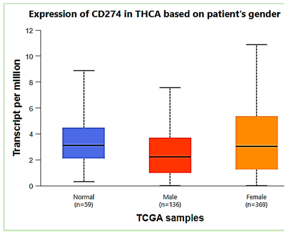
Figure 2.
ACTA2 (α-smooth muscle actin) transcript-per-million expression in the TCGA THCA cohort, stratified by normal thyroid tissue (n=59), male PTC (n=136), and female PTC (n=369). Box shows interquartile range with median line; whiskers show minimum/maximum. Male PTC expression was significantly higher than female PTC (p<0.05), consistent with the immunohistochemical cancer-associated-fibroblast literature linking stromal α-SMA to extrathyroidal extension, BRAF mutation, and lymph node metastasis in PTC [37,38], clinicopathologic features independently over-represented in male PTC (Section 3.11). Source: UALCAN TCGA THCA gene expression module [34].
Figure 2.
ACTA2 (α-smooth muscle actin) transcript-per-million expression in the TCGA THCA cohort, stratified by normal thyroid tissue (n=59), male PTC (n=136), and female PTC (n=369). Box shows interquartile range with median line; whiskers show minimum/maximum. Male PTC expression was significantly higher than female PTC (p<0.05), consistent with the immunohistochemical cancer-associated-fibroblast literature linking stromal α-SMA to extrathyroidal extension, BRAF mutation, and lymph node metastasis in PTC [37,38], clinicopathologic features independently over-represented in male PTC (Section 3.11). Source: UALCAN TCGA THCA gene expression module [34].
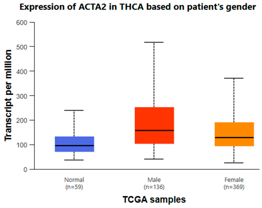
Table 3.
Age-stratified hazard ratios for female (vs. male) disease-specific survival in PTC, illustrating the disappearance of the female survival advantage after approximately age 55.
Table 3.
Age-stratified hazard ratios for female (vs. male) disease-specific survival in PTC, illustrating the disappearance of the female survival advantage after approximately age 55.
| Age at diagnosis | Female vs. male HR for disease-specific survival |
| <55 years | HR 0.33, strong female advantage |
| 55–69 years | HR 1.01, advantage disappears |
| HR 1.17, reverses slightly |
Table 4.
BRAF- and TERT-stratified male-sex hazard ratios for PTC mortality, illustrating that male sex is prognostically neutral in BRAF-wild-type disease but confers substantial, compounding risk in BRAF- and BRAF/TERT-altered disease.
Table 4.
BRAF- and TERT-stratified male-sex hazard ratios for PTC mortality, illustrating that male sex is prognostically neutral in BRAF-wild-type disease but confers substantial, compounding risk in BRAF- and BRAF/TERT-altered disease.
| BRAF / histology context | Male sex hazard ratio for mortality |
| Wild-type BRAF PTC | HR 0.70 (male sex confers no excess risk) |
| BRAF V600E-positive PTC | HR 2.74 (independent risk factor) |
| BRAF V600E, conventional-variant PTC | HR 3.51 |
| BRAF V600E + TERT promoter co-alteration (“genetic duet”) | HR 37.77 vs. wild-type for both genes |
Table 5.
Representative studies reporting null or contradictory findings regarding sex-based prognostic effects and hormonal causality in PTC, included here to avoid a one-sided synthesis.
Table 5.
Representative studies reporting null or contradictory findings regarding sex-based prognostic effects and hormonal causality in PTC, included here to avoid a one-sided synthesis.
| Study | Finding |
| Nilubol et al., 2013 (Thyroid) | Male sex not independently associated with disease-specific survival after adjusting for tumor aggressiveness features |
| Oyer et al., 2013 (Laryngoscope) | Sex not an independent risk factor for survival in differentiated thyroid cancer after adjustment |
| Kim et al., 2022 (Cancers) | Single-institution Korean cohort (n=1,252): comparable recurrence-free survival between sexes after adjusting for confounders |
| Park et al., 2025 (Thyroid) | Mendelian randomization found no strong causal evidence for reproductive/hormonal factors driving differentiated thyroid cancer risk |
Table 2.
High-confidence BRAF and TERT alteration prevalence by sex in the institutional DNA cohort (n=18).
Table 2.
High-confidence BRAF and TERT alteration prevalence by sex in the institutional DNA cohort (n=18).
| Sex | n | BRAF+, n (%) | TERT+, n (%) | BRAF+/TERT+ co-alteration |
| Female | 13 | 9 (69.2%) | 0 (0%) | 0 |
| Male | 5 | 4 (80.0%) | 1 (20.0%) | 1 (the same patient) |
Disclaimer/Publisher’s Note: The statements, opinions and data contained in all publications are solely those of the individual author(s) and contributor(s) and not of MDPI and/or the editor(s). MDPI and/or the editor(s) disclaim responsibility for any injury to people or property resulting from any ideas, methods, instructions or products referred to in the content. |
© 2026 by the authors. Licensee MDPI, Basel, Switzerland. This article is an open access article distributed under the terms and conditions of the Creative Commons Attribution (CC BY) license (http://creativecommons.org/licenses/by/4.0/).
Copyright: This open access article is published under a Creative Commons CC BY 4.0 license, which permit the free download, distribution, and reuse, provided that the author and preprint are cited in any reuse.