

Submitted:
23 September 2026
Posted:
24 September 2026
You are already at the latest version
Abstract
Norovirus infection has been increasingly recognized as a potential trigger of necrotizing enterocolitis (NEC) in preterm neonates. Although previous reports have described severe cases associated with intestinal pneumatosis, bowel ischemia, and the need for surgical intervention, the clinical spectrum of Norovirus-related intestinal disease remains poorly defined. We report the case of a 29-week gestational age preterm infant who developed abdominal distension on the 35th day of life. Stool reverse transcriptase polymerase chain reaction confirmed Norovirus infection. Abdominal radiographs showed progressive development of intestinal pneumatosis, leading to the confirmed diagnosis of a stage IIA NEC. Abdominal pneumatosis resolved completely within five days, without evidence of perforation or clinical deterioration. The infant received only supportive therapy and intravenous immunoglobulin (IVIG). This case highlighted the heterogeneous clinical spectrum of Norovirus-associated intestinal involvement in preterm infants and the possible protective and immunomodulatory role of the IVIG.
Keywords:
intestinal pneumatosis
; intravenous immunoglobulin
; necrotizing enterocolitis
; norovirus infection
; preterm infant
1. Introduction
Necrotizing enterocolitis (NEC) is a devastating gastrointestinal emergency affecting approximately 5% of very preterm or very low birth weight neonates [1,2].
Despite decades of research and advances in neonatal care, NEC remains a leading cause of mortality and morbidity in neonatal intensive care units worldwide, with mortality rates approaching 20% overall and 30-50% in advanced cases [3].
The risk factors for NEC include extreme prematurity, extremely low birth weight, intrauterine growth restriction with absent or reversed end-diastolic flow on umbilical artery Doppler studies, formula feeding, sepsis, congenital heart disease and blood transfusions. The disease spectrum ranges from gastrointestinal symptoms to catastrophic illness characterized by circulatory compromise, respiratory and metabolic acidosis, disseminated intravascular coagulopathy and multiorgan system failure [4].
The Bell staging system [5], first proposed in 1978 and subsequently modified by Walsh and Kliegman [6], remains the most commonly used method to assess NEC severity.
Emerging evidence demonstrated a significant association between viral infections and NEC development in preterm neonates. Among specific viruses, Norovirus showed the strongest association with NEC [7]. The reported cases concerned premature infants who showed intestinal pneumatosis requiring laparotomy with colectomy and stoma creation. In many of the infants described, laparotomy showed intestinal stenotic areas.
We report the favourable clinical and radiological evolution of a NEC in a preterm neonate with Norovirus infection.
2. Case Presentation
The baby was a 29 weeks’ gestation male infant who experienced sudden onset of abdominal distension on the 35th day of life with worsening of general condition and sepsis-like clinical appearance. Laboratory investigations revealed leukopenia with severe neutropenia together with elevated inflammatory markers and anaemia which requiring red blood cell transfusion. Blood, urine, and cerebrospinal fluid cultures were negative for bacteria. The presence of Norovirus in the stool was confirmed by reverse transcriptase polymerase chain reaction (RT-PCR). The main laboratory findings and vital signs during the acute phase of illness are summarized in Table 1.
The neonate was maintained for 7 days under mechanical ventilation to minimize gastrointestinal air insufflation in the setting of marked abdominal distension. Empirical antibiotic therapy with intravenous vancomycin and meropenem was initiated from the first day of symptom onset and currently. A central venous catheter was placed for the administration of total parenteral nutrition. Serial abdominal plain radiographs were performed: at the beginning, we observed only distension of bowel loops (Figure 1a). Intestinal “soap bubbles” pneumatosis appeared on the second day of illness (Figure 1b) and persisted for three days (Figure 1c-e), leading to a diagnostic definition of stage IIA NEC.
On the second day of illness, the infant developed generalized pitting edema, associated with a significant increase in body weight and preserved renal function. Due to the clinical diagnosis of capillary leak syndrome, treatment with intravenous immunoglobulins (IVIG) was started at a dose of 500 mg/day for 4 days.
On the fifth day, pneumatosis was no longer visible (Figure 1f). No free air has ever been detected.
After seven days, the patient was transitioned to non-invasive ventilation, which was discontinued after 7 days, on the 49th fay of life, because of satisfactory respiratory function. Antibiotic therapy was discontinued after 7 days following normalization of inflammatory markers and clinical improvement.
After eight days, the infant began to have spontaneous bowel movements and to feed. He was discharged at 71 days of life without abdominal complications due to NEC.
The baby is currently one year old and is in good clinical condition, shows adequate growth, and has not experienced any gastrointestinal symptoms.
3. Discussion
We reported the favourable evolution of NEC in a preterm infant, which did not reach the advanced stages characterized by intestinal perforation and which resolved without long-term gastrointestinal sequelae. This clinical course was different from that described so far in the literature in neonates with Norovirus infections [8,9].
Multiple outbreaks of NEC associated with Norovirus have been documented in neonatal intensive care units. A 1998 outbreak in New York involved eight cases of NEC with two deaths. In this cohort of neonates, six stool samples were available for analysis and Norovirus particles were identified in 4 of 6 specimens. Of the eight reported cases, seven demonstrated intestinal pneumatosis on abdominal radiography. Persistent dilated bowel loops and portal venous gas were reported in two cases, while abdominal distension was present in four cases. No intestinal perforation was reported, but, except for the two fatal cases, the clinical outcomes and long-term evolution of the remaining patients were not described. The severity of NEC according to Bell’s staging criteria was not specified, nor were the causes of death, including whether they were directly related to intestinal complications. Furthermore, no detailed information was provided regarding the therapeutic management and clinical interventions [10].
Armbrust et al. described another outbreak of eleven cases of low birth weight-premature infants with suggestive symptoms of viral infection. Norovirus were detected in stools in eight cases. Seven infants developed abdominal distension, and two developed NEC. In the first case with confirmed Norovirus infection, NEC stage IIB according to Bell’s criteria occurred and required surgical intervention with hemicolectomy and ileostomy. The second infant presented colitis requiring stoma formation, capillary leak syndrome, and multiorgan failure, resulting in death [8]. Interesting, among infants with Norovirus infections described in the literature, only this infant presented capillary leak syndrome as a complication of the infection, as in our patient. No details of the therapeutic management of capillary lek syndrome were reported for this neonate in the Armbrust et al. case series. In our infant, IVIG were administered as capillary leak syndrome therapy [11,12]. Given the absence of specific antiviral therapies for Norovirus infections and for other invasive Enterovirus infections, the use of IVIG were commonly considered, based on their reported immunomodulatory and neutralizing effects [13,14]. Available evidence on the use of IVIG in neonatal invasive Enterovirus infections is mainly derived from case series and observational studies, and no definitive conclusions regarding its efficacy can be drawn in the absence of randomized clinical trials [15,16,17,18]. However, animal studies have shown that systemic IgG infused via IVIG are actively retro-transported from the serum into the intestinal lumen, binding to the crystallizable fragment (Fc) receptor expressed on erythrocytes [19]. In our neonate, systemic IVIG could have created a protective endoluminal antibody shield against Norovirus.
In a cohort of 34 infants with viral enteric infections admitted to a neonatal intensive care unit in Germany, Norovirus was detected in eight patients. The predominant clinical signs were abdominal distension, diarrhoea, and apnoea. Four infants presented with bloody stools, and only one showed an acute inflammatory response with elevated C-reactive protein and lactate, suggesting stage IB NEC. In this infant, enteral nutrition was discontinued and empiric antibiotic therapy was administered. In these eight reported cases, abdominal symptoms did not progress beyond Bell’s stage 1 and, therefore, did not overcome the suspicion of NEC [20].
Finally, Pelizzo et al. described a case series of three preterm infants with Norovirus-associated isolated colon ischemia from a NICU in Italy. The first case presented abdominal distension and radiological evidence of intestinal pneumatosis. Four days after symptom onset, the development of free intraperitoneal air led to surgical intervention, consisting of colectomy with ileostomy formation. Intraoperative findings revealed areas of intestinal stenosis associated with necrosis and intestinal perforation. The second and third cases presented with haematochezia and abdominal distension; intestinal pneumatosis was observed in the third case. These two patients underwent exploratory laparotomy, which revealed stenotic intestinal segments, requiring subsequent ileostomy formation. In all cases, enteral feeding was discontinued, and antibiotic therapy was administered. Therefore, Pellizzo et al. suggested early surgical management with ileostomy once colonic pneumatosis is identified on abdominal radiography [9]. These findings contrast with our reported infant, in which the clinical course was favourable with conservative management, despite the presence of intestinal pneumatosis which persisted for three days and without the need for surgical intervention. Our infant developed abdominal distension and radiographic evidence of pneumatosis intestinalis in the setting of RT-PCR-confirmed Norovirus infection, leading to the diagnosis of confirmed NEC. However, the abdominal clinical course remained stable, without progression to intestinal perforation. Serial abdominal imaging demonstrated gradual resolution of pneumatosis under conservative management, including bowel rest and supportive care. It is difficult to define exactly what influenced the favourable course of NEC in our newborn. Since NEC is a condition that recognizes an inflammatory pathogenesis, we can hypothesize that IVIG may provide the neutralization of enteral and circulating viral particles, modulating the systemic and intestinal inflammatory response.
4. Conclusions
Our report further supports the concept that Norovirus-related intestinal pathology in neonates encompasses a broad clinical spectrum, ranging from self-limiting forms of pneumatosis intestinalis to fulminant, life-threatening disease. Our case suggested that intestinal pneumatosis associated with Norovirus infection could resolve completely and without sequelae with conservative management in the absence of clinical deterioration or abdominal perforation. The role of immunoglobulins in invasive enterovirus infections deserves to be tested in randomized clinical trials.
Author Contributions
Conceptualization, L.S. and S.C.; clinical investigation, S.C., G.V., F.P., N.M., A.F. and M.S.; data curation, L.M., L.S., A.F., M.S. and M.T.; writing—original draft preparation, L.S., S.C. and M.T.; writing—review and editing, L.S., S.C. and G.V.; supervision, S.C. and G.V.; literature review, L.S., S.C., N.M., F.P. and G.V. All authors have read and agreed to the published version of the manuscript.
Funding
This research received no external funding.
Institutional Review Board Statement
Ethical review and approval were waived for this study because it is a single case report and, according to institutional policies, did not require approval by an Institutional Review Board or Ethics Committee.
Informed Consent Statement
Written informed consent was obtained from the patient’s parents/legal guardians for the publication of this case report and any accompanying images.
Data Availability Statement
No new datasets were generated or analyzed in this study. All relevant data are included in the article.
Conflicts of Interest
The authors declare no conflicts of interest.
References
- Sharif, S.; Oddie, S.J.; Heath, P.T.; McGuire, W. Prebiotics to prevent necrotising enterocolitis in very preterm or very low birth weight infants. Cochrane Database Syst. Rev. 2023, 6, CD015133. [Google Scholar] [CrossRef] [PubMed]
- Sharif, S.; Meader, N.; Oddie, S.J.; Rojas-Reyes, M.X.; McGuire, W. Probiotics to prevent necrotising enterocolitis in very preterm or very low birth weight infants Update in. Cochrane Database Syst. Rev. 2020, 10, CD005496. [Google Scholar] [CrossRef]
- Li, B.; Yeganeh, M.; Lee, D.; Chusilp, S.; Balsamo, F.; Ganji, N.; Wang, C.Y.; Zito, A.; Biouss, G.; Pierro, A. Exploring the Complex Pathophysiology of Necrotizing Enterocolitis in Preterm Neonates. Annu Rev. Pathol. 2026, 21, 37–58. [Google Scholar] [CrossRef] [PubMed]
- Lin, P.W.; Stoll, B.J. Necrotising enterocolitis. Lancet 2006, 368, 1271–1283. [Google Scholar] [CrossRef] [PubMed]
- Bell, M.J.; Ternberg, J.L.; Feigin, R.D.; Keating, J.P.; Marshall, R.; Barton, L.; Brotherton, T. Neonatal necrotizing enterocolitis. Therapeutic decisions based upon clinical staging. Ann. Surg. 1978, 187, 1–7. [Google Scholar] [CrossRef] [PubMed]
- Kliegman, R.M.; Walsh, M.C. Neonatal necrotizing enterocolitis: Pathogenesis, classification, and spectrum of illness. Curr. Probl. Pediatr. 1987, 17, 213–288. [Google Scholar] [CrossRef] [PubMed]
- Mani, S.; Hazra, S.; Hagan, J.; Sisson, A.; Nair, J.; Pammi, M. Viral Infections and Neonatal Necrotizing Enterocolitis: A Meta-analysis. Pediatrics 2023, 152, e2022060876. [Google Scholar] [CrossRef] [PubMed]
- Armbrust, S.; Kramer, A.; Olbertz, D.; Zimmermann, K.; Fusch, C. Norovirus infections in preterm infants: Wide variety of clinical courses. BMC Res. Notes 2009, 2, 96. [Google Scholar] [CrossRef] [PubMed]
- Pelizzo, G.; Nakib, G.; Goruppi, I.; Fusillo, M.; Scorletti, F.; Mencherini, S.; Parigi, G.B.; Stronati, M.; Calcaterra, V. Isolated colon ischemia with norovirus infection in preterm babies: A case series. J. Med. Case Rep. 2013, 7, 108. [Google Scholar] [CrossRef] [PubMed]
- Turcios-Ruiz, R.M.; Axelrod, P.; St John, K.; Bullitt, E.; Donahue, J.; Robinson, N.; Friss, H.E. Outbreak of necrotizing enterocolitis caused by norovirus in a neonatal intensive care unit. J. Pediatr. 2008, 153, 339–344. [Google Scholar] [CrossRef] [PubMed]
- Druey, K.M.; Arnaud, L.; Parikh, S.M. Systemic capillary leak syndrome. Nat. Rev. Dis. Primers 2024, 10, 86. [Google Scholar] [CrossRef] [PubMed]
- Abgueguen, P.; Chennebault, J.M.; Pichard, E. Immunoglobulins for treatment of systemic capillary leak syndrome. Am. J. Med. 2010, 123, e3-4. [Google Scholar] [CrossRef] [PubMed]
- Abzug, M.J. Presentation, diagnosis, and management of enterovirus infections in neonates. Paediatr. Drugs 2004, 6, 1–10. [Google Scholar] [CrossRef] [PubMed]
- Chuang, Y.Y.; Huang, Y.C. Enteroviral infection in neonates. J. Microbiol. Immunol. Infect. 2019, 52, 851–857. [Google Scholar] [CrossRef] [PubMed]
- Lin, T.Y.; Kao, H.T.; Hsieh, S.H.; Huang, Y.C.; Chiu, C.H.; Chou, Y.H.; Yang, P.H.; Lin, R.I.; Tsao, K.C.; Hsu, K.H.; et al. Neonatal enterovirus infections: Emphasis on risk factors of severe and fatal infections. Pediatr. Infect. Dis. J. 2003, 22, 889–894. [Google Scholar] [CrossRef] [PubMed]
- Harik, N.; DeBiasi, R.L. Neonatal nonpolio enterovirus and parechovirus infections. Semin Perinatol. 2018, 42, 191–197. [Google Scholar] [CrossRef] [PubMed]
- Zhang, M.; Wang, H.; Tang, J.; He, Y.; Xiong, T.; Li, W.; Qu, Y.; Mu, D. Clinical characteristics of severe neonatal enterovirus infection: A systematic review. BMC Pediatr. 2021, 21, 127. [Google Scholar] [CrossRef] [PubMed]
- Menzella, N.; Costa, S.; Fattore, S.; De Tomaso, D.; Fusco, F.P.; Sessa, L.; Tirone, C.; Priolo, F.; Paladini, A.; Serrao, S.; et al. Invasive neonatal Enterovirus infection: A case series and review of the literature. Children 2026. [Google Scholar] [CrossRef]
- Harris, N.L.; Spoerri, I.; Schopfer, J.F.; Nembrini, C.; Merky, P.; Massacand, J.; Urban, J.F., Jr.; Lamarre, A.; Burki, K.; Odermatt, B.; et al. Mechanisms of neonatal mucosal antibody protection. J. Immunol. 2006, 177, 6256–6262. [Google Scholar] [CrossRef] [PubMed]
- Bagci, S.; Eis-Hübinger, A.M.; Yassin, A.F.; Simon, A.; Bartmann, P.; Franz, A.R.; Mueller, A. Clinical characteristics of viral intestinal infection in preterm and term neonates. Eur. J. Clin. Microbiol. Infect. Dis. 2010, 29, 1079–1084. [Google Scholar] [CrossRef] [PubMed]
Figure 1.
The radiological evolution of intestinal pneumatosis. Distension of intestinal loops, with a diameter greater than the width of the L5 vertebra, appeared at the onset of gastrointestinal symptoms (a). “Soap bubbles” appearance (arrows) emerged after two days from the onset of symptoms (b) and persisted for three days into treatment for necrotising enterocolitis (c-e). Pneumatosis was no longer visible after five days from the onset of symptoms (f).
Figure 1.
The radiological evolution of intestinal pneumatosis. Distension of intestinal loops, with a diameter greater than the width of the L5 vertebra, appeared at the onset of gastrointestinal symptoms (a). “Soap bubbles” appearance (arrows) emerged after two days from the onset of symptoms (b) and persisted for three days into treatment for necrotising enterocolitis (c-e). Pneumatosis was no longer visible after five days from the onset of symptoms (f).
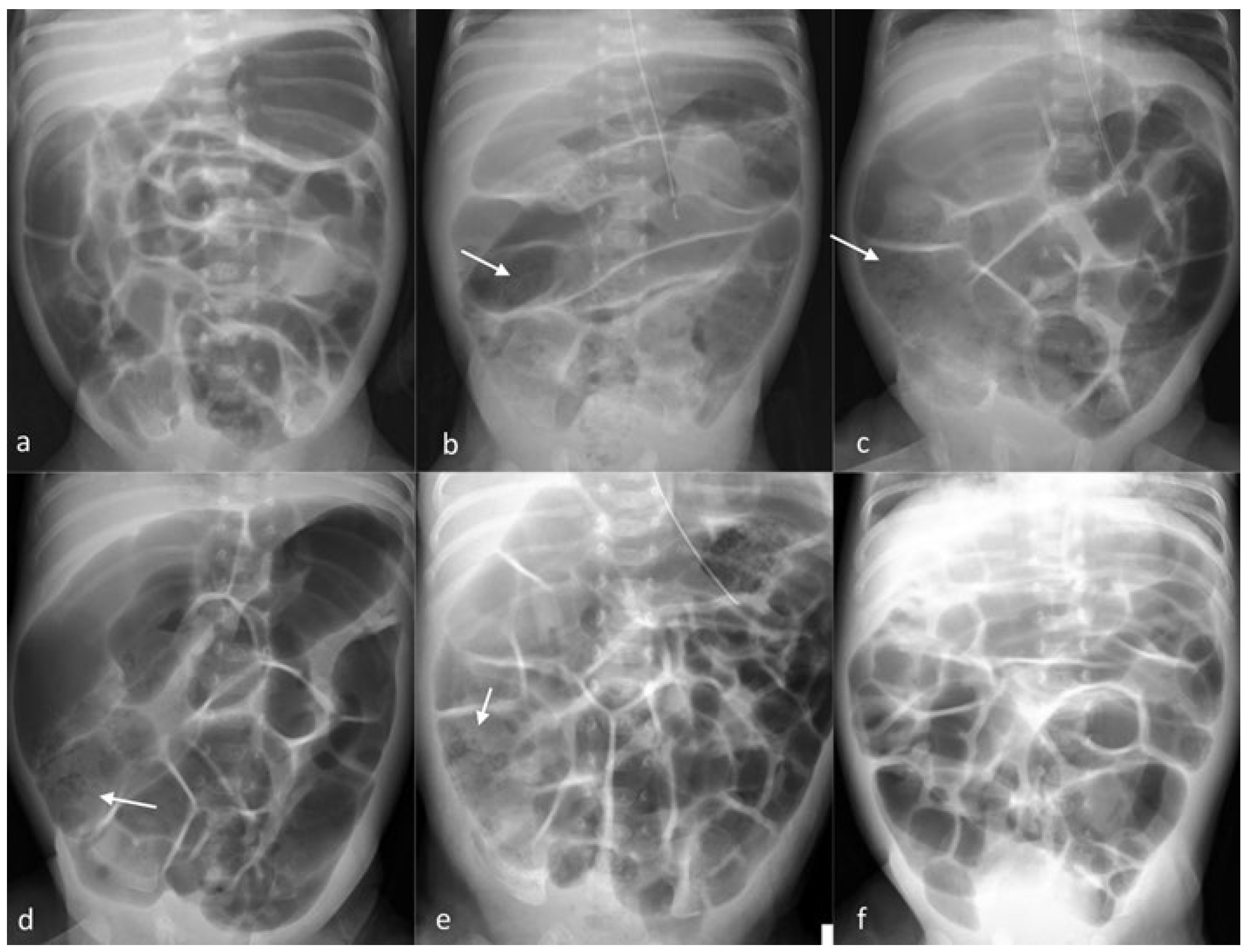

Table 1.
Laboratory findings and vital signs during the acute phase of illness.
| Parameter | Measured value | Reference range |
|
Vital sign Heart rate |
155 beats/min |
100–180 beats/min |
| Peripheral oxygen saturation | 97% | ≥90% |
|
Hematological parameters White blood cell count |
3.37 × 10³/μL |
5.00–17.00 × 10³/μL |
| Absolute neutrophil count | 0.61 × 10³/μL | 1.00–12.00 × 10³/μL |
| Haemoglobin | 11.6 g/dL | 9.0–15.0 g/dL |
| Haematocrit | 33.2% | 27.0–44.0% |
| Platelet count | 387 × 103/μL | 220–500 × 103/μL |
| Inflammatory markers | ||
| C-reactive protein | 77.6 mg/L | 0–5 mg/L |
| Serum electrolytes | ||
| Sodium | 135 mEq/L | 136–145 mEq/L |
| Potassium | 4.71 mEq/L | 3.50–5.10 mEq/L |
| Chloride | 103 mEq/L | 98–107 mEq/L |
| Total calcium | 9.3 mg/dL | 8.4–10.6 mg/dL |
| Microbiological investigations | ||
| Blood culture | Negative | Negative |
| Urine culture | Negative | Negative |
| Cerebrospinal fluid culture | Negative | Negative |
| Stool RT-PCR for Norovirus | Positive | Negative |
| Cerebrospinal fluid RT-PCR for Norovirus |
Negative |
Negative |
Legend: RT-PCR reverse transcriptase polymerase chain reaction.
Disclaimer/Publisher’s Note: The statements, opinions and data contained in all publications are solely those of the individual author(s) and contributor(s) and not of MDPI and/or the editor(s). MDPI and/or the editor(s) disclaim responsibility for any injury to people or property resulting from any ideas, methods, instructions or products referred to in the content. |
© 2026 by the authors. Licensee MDPI, Basel, Switzerland. This article is an open access article distributed under the terms and conditions of the Creative Commons Attribution (CC BY) license (http://creativecommons.org/licenses/by/4.0/).
Copyright: This open access article is published under a Creative Commons CC BY 4.0 license, which permit the free download, distribution, and reuse, provided that the author and preprint are cited in any reuse.