
Submitted:
23 September 2026
Posted:
24 September 2026
You are already at the latest version
Abstract
A novel lytic bacteriophage, designated vB_ValS-L9, infecting the potential pathogenic bacterium Vibrio alginolyticus L4, was isolated from a coastal aquaculture area in Beihai, China. Transmission electron microscopy revealed that vB_ValS-L9 possesses an icosa-hedral head (56.50 ± 4.19 nm in diameter) and a long tail (143.03 ± 5.32 nm in length), ex-hibiting a typical siphovirus-like morphology within the class Caudoviricetes. The phage demonstrated wide stability across various temperatures, pH levels, and salinities. Genomic sequencing showed that vB_ValS-L9 has a circular double-stranded DNA genome of 43,884 bp with a G+C content of 43.55% and 61 predicted ORFs. No tRNA, virulence, or antibiotic resistance genes were identified. Genomic and phylogenetic analyses, including ANI, VICTOR, VIRIDIC, and conserved protein phylogenies, identified vB_ValS-L9 as a novel species that, together with Vibrio phages Artemius and VapS30, forms a new genus in the class Caudoviricetes, for which the name Artemiovirus is proposed. Furthermore, in vivo trials using juvenile pearl oysters (Pinctada martensii) revealed that vB_ValS-L9 pro-vides substantial prophylactic and therapeutic protection against V. alginolyticus L4 infec-tion, especially when the phage was administered before bacterial challenge. Collectively, these findings suggest that vB_ValS-L9 shows potential as a safe, environmentally robust, and promising biocontrol candidate for managing vibriosis in aquaculture.
Keywords:
Vibrio alginolyticus
; Vibrio phage
; morphology and genomic characterization
; taxonomic assignment
; aquaculture biocontrol
1. Introduction
The genus Vibrio encompasses a diverse group of Gram-negative, halophilic bacteria that are ubiquitous in marine and estuarine ecosystems [1,2]. Several Vibrio species are major pathogens in mariculture, such as Vibrio parahaemolyticus, Vibrio alginolyticus, Vibrio anguillarum, Vibrio harveyi, responsible for severe disease outbreaks that cause substantial economic losses globally [3,4,5,6]. Infections such as vibriosis can result in mass mortality of farmed fish, crustaceans, and mollusks, threatening the sustainability and profitability of the aquaculture industry [7,8]. Furthermore, certain pathogentic Vibrio species are also zoonotic human pathogens capable of causing foodborne or direct-contact infections [9,10,11]. Among them, V. alginolyticus is one of the most frequently encountered species in warm coastal waters [12]. Initially isolated from seawater and characterized for its pronounced alginate-degrading ability [13], V. alginolyticus was later recognized as an important opportunistic pathogen for both marine animals and humans [14]. In aquaculture settings, it has been implicated in acute vibriosis outbreaks affecting a wide range of economically important species, including the oyster, crab, abalone, fish, and shrimp [15,16,17,18,19,20].
Compounding this threat, the overuse and misuse of conventional antibiotics have accelerated the emergence of multidrug-resistant strains, complicating infection management and increasing mortaltiy rates in both aquaculture animals and humans [21]. Multidrug-resistant “superbugs” are currently recognized among the top threats to global health [22]. Therefore, it is imperative to explore alternative, eco-friendly biocontrol strategies, such as phage therapy.
Bacteriophages (phages for short), the natural viral predators of bacteria, have gained widespread attention as sustainable alternatives to antibiotics for controlling pathogenic Vibrio in aquaculture [6,23,24,25]. To date, numerous Vibrio phages have been isolated from diverse marine environments, revealing remarkable morphological and genomic diversity. The vast majority of these phages are tailed viruses classified within the class Caudoviricetes (NCBI GenBank, accessed 15 May 2026). Despite this recognized diversity, the number of isolated and sequenced Vibrio phages remains limited relative to the immense viral richness predicted by metagenomic surveys. Moreover, phylogenetic analyses based on whole-genome nucleotide sequences (e.g., ViPTree, VIRIDIC) frequently reveal that many newly isolated Vibrio phages cannot be reliably assigned to any recognized family or genus, highlighting significant taxonomic gaps [26,27,28,29]. Under the latest ICTV taxonomy releases (ratified in 2025), a considerable proportion of phage genome sequences deposited in public databases remain unassigned at multiple taxonomic ranks [30], impeding our comprehensive understanding of phage evolution and diversification. Consequently, the isolation and in-depth characterization of novel lytic Vibrio phages are not only to bridge these taxonomic and ecological knowledge gaps but also to expand the candidate phage pool for targeted biocontrol applications.
In the present study, a novel lytic V. alginolyticus phage, designated vB_ValS-L9, was isolated from coastal aquaculture seawater. Its morphological features, physico-chemical stability, and genomic properties were systematically characterized, and its taxonomic assignment was delineated through comprehensive comparative genomic and phylogenetic analyses. Furthermore, its in vivo prophylactic and therapeutic efficacy was evaluated using a juvenile pearl oyster (Pinctada martensii) challenge model, and the effect of varying the timing of phage administration on therapeutic efficacy was investigated. This study broadens the current understanding of V. alginolyticus phage biology and introduces a safe, highly effective candidate for phage-based biocontrol in aquaculture.
2. Results
2.1. Phage Isolation, Morphology, Host Range, and Infection Kinetics
2.1.1. Phage Isolation and Morphological Characterization
Phage vB_ValS-L9 was isolated from surface seawater collected from an aquaculture area of Beihai, China, using Vibrio alginolyticus L4 as the indicator host. On double-layer agar plates, vB_ValS-L9 produced small, clear, and round plaques after 24 h of incubation at 28 °C, demonstrating robust lytic activity against the host strain. Chloroform sensitivity assays showed that viral infectivity remained essentially unchanged across all tested chloroform volume fractions (1%, 10%, and 50%, v/v), confirming that the vB_ValS-L9 virion is non-enveloped and lacks essential structural lipids.
Transmission electron microscopy (TEM) examination revealed that vB_ValS-L9 possesses an icosahedral head with a diameter of 56.50 ± 4.19 nm and a long, flexible, non-contractile tail measuring 143.03 ± 5.32 nm in length (Figure 1A). Based on these morphological characteristics, vB_ValS-L9 exhibits a typical siphovirus-like morphotype, classifying it within the class Caudoviricetes (traditionally classified under the former family Siphoviridae).
2.1.2. Host Specificity and Infection Kinetics
The host range of phage vB_ValS-L9 was evaluated against 53 marine bacterial isolates, comprising 43 Vibrio strains, seven Shewanella strains, two Pseudoalteromonas strains, and one Sulfitobacter strain (Table S1). Spot assays revealed that vB_ValS-L9 formed distinct, clear plaques exclusively on its original isolation host, V. alginolyticus L4, whereas no lytic activity or plaque formation was observed across the remaining 52 tested strains.
To characterize the replication dynamics of phage vB_ValS-L9, a one-step growth curve assay was performed at a multiplicity of infection (MOI) of 0.01 using exponentially growing V. alginolyticus L4. The growth curve exhibited a latent period of approximately 40 min, followed by a rapid burst phase that yielded an estimated burst size of 231 PFU per infected cell (Figure 1B). To further assess its bactericidal potency under varying infection pressures, liquid lysis kinetics were monitored across MOIs spanning 0.001 to 10 (Figure 1C). Notably, even at the lowest tested MOI (0.001), phage vB_ValS-L9 exerted robust bactericidal activity against strain L4, as evidenced by a profound reduction in culture turbidity (OD600) compared to the uninfected control after both 4 h and 8 h of co-incubation (Figure 1D).
2.2. Environmental Stability of Phage vB_ValS-L9
To evaluate the environmental resilience of phage vB_ValS-L9, its stability under various temperature, pH, and salinity conditions was systematically determined following a 24-h exposure period (Figure 2). Thermal stability assays indicated an inverse relationship between temperature and viral viability; nevertheless, phage vB_ValS-L9 exhibited moderate thermostability, retaining a 46.39% survival rate even after incubation at 40 °C (Figure 2A). In pH stability tests, vB_ValS-L9 demonstrated a notable tolerance across a broad pH spectrum ranging from 5.0 to 10.0, with optimal infectivity observed at physiological marine pH (pH 8.0) (Figure 2B). Notably, the phage retained approximately 81.76% and 62.94% of its initial titer under acidic (pH 5.0) and alkaline (pH 10.0) conditions, respectively. Regarding salinity tolerance, vB_ValS-L9 maintained substantial lytic capability across diverse salinity gradients (Figure 2C). Although viral titers displayed a slight, progressive decline when seawater concentration exceeded 60% (~20.40‰ salinity), the phage preserved more than 44.44% of its maximum viability even under pronounced hypotonic stress (10% seawater, ~3.40‰ salinity).
2.3. Genomic Features and Functional Annotation
2.3.1. General Genomic Features and Functional Modularity
High-throughput sequencing and de novo assembly yielded a complete, circular, double-stranded DNA genome for phage vB_ValS-L9, comprising 43,884 bp with an overall G+C content of 43.55%. Quality evaluation via CheckV corroborated that the assembled genome was 100% complete and circular, with no detectable host contamination. A total of 61 ORFs were predicted across the genome, yielding a high coding density of 95.28% (Figure 3; Table S2). Among these, 31 ORFs (50.82%) were assigned putative biological functions based on homology searches and conserved domain analysis, whereas the remaining 30 ORFs (49.18%) were designated as hypothetical proteins with uncharacterized functions. Comprehensive screening confirmed the absence of tRNA genes, antimicrobial resistance genes, virulence factor determinants, and lysogeny-associated marker enzymes (e.g., integrases, recombinases, and excisionases), reinforcing the strictly lytic lifestyle and biosafety of vB_ValS-L9.
The functionally annotated ORFs were organized into five distinct modules according to their predicted roles in the phage life cycle: DNA replication and repair (6 ORFs: ORF10, ORF14, ORF31, ORF36, ORF41, ORF43), viral packaging and morphogenesis (5 ORFs: ORF2, ORF3, ORF58, ORF59, ORF60), structural components (7 ORFs: ORF44, ORF49, ORF50, ORF52, ORF54, ORF55, ORF57), host lysis (1 ORF: ORF28), and transcriptional regulation (2 ORF: ORF9, ORF22). The remaining 10 ORFs lacked clear functional assignments but were predicted to encode proteins containing conserved structural features, including transmembrane helices (ORF25, ORF26, ORF27, ORF29, ORF45), coiled-coil domains (ORF33, ORF35), an ATPase (ORF34), a single-stranded DNA-binding protein (ORF38), and a DUF4326 domain-containing protein (ORF30).
2.3.2. Annotation of Predicted ORFs
Host cell lysis is mediated by a single lytic gene, ORF28, which encodes an endolysin with a conserved peptidoglycan hydrolase domain predicted to degrade the bacterial cell wall during the late stage of infection to facilitate progeny release [56]. Virion packaging and capsid maturation are orchestrated by four coordinated proteins: the small (TerS, ORF3) and large (TerL, ORF60) subunits of the terminase complex mediate viral DNA recognition, cleavage, and translocation into the procapsid via the portal protein (ORF59) [57,58], while a prohead serine protease (ORF58) drives scaffolding protein cleavage and capsid shell maturation [59].
The core DNA replication and repair machinery comprises six ORFs, including a DNA polymerase (ORF43), a DNA primase (ORF41), and a DNA helicase (ORF31), supplemented by auxiliary nucleases and processing enzymes including an HNH endonuclease (ORF10), a putative polynucleotide kinase (ORF14), and a PD-(D/E)XK family nuclease (ORF36).
Virion architecture is encoded by seven structural genes, including the major capsid protein (ORF57), neck protein (ORF50), stopper protein (ORF55), tail tubular protein (ORF44), tail tube protein (ORF52), tail length ape measure protein (TMP) (ORF49) and major tail protein (ORF54). Notably, TMP (ORF49) comprising 945 amino acids, corresponding to a predicted tail length of approximately 141.75 nm based on the 0.15 nm/residue ratio [60,61], consistent with the tail length measured by TEM (143.03 ±5.32 nm)(Figure 1; Figure 4).
To overcome host barriers and regulate gene expression, vB_ValS-L9 encodes two regulatory and counter-defense effectors: ORF9 (TreK) is predicted to act during early infection to shut down host macromolecular synthesis and redirect cellular metabolism toward viral replication (NCBI Protein Family Model NF049213, https://www.ncbi.nlm.nih.gov/genome/annotation_prok/evidence/NF049213/#1), whereas ORF22 encodes a restriction alleviation protein (Lar) that assists in overcoming host restriction-modification (R-M) barriers (Encyclopedia of Viral Anti-DefencE Systems, http://www.ebi.ac.uk/finn-srv/evades/details/lar/).
2.4. Comparative Genomics, Phylogenetic Classification, and Taxonomic Clustering
2.4.1. Phylogenetic Classification, Comparative genomics, and Evolutionary Divergence
Initial BLASTn searches against the NCBI nucleotide database revealed that phage vB_ValS-L9 shares the highestsequence similarity with two Vibrio phages, including Vibrio phage VapS30 (95.11% identity with 71% query coverage; accession no. PV528705) and Vibrio phage Artemius (95.81% identity with 73% query coverage) [62]. This indicated an exceptionally close evolutionary relationship among these three phages, prompting their triadic comparative analysis throughout subsequent genomic investigations.
To delineate their global evolutionary placement, a whole-genome proteomic tree was constructed via the ViPTree server (Figure 4A, B). Phages vB_ValS-L9, VapS30, and Artemius formed a tightly clustered, well-supported monophyletic clade that was distinctly separated from all other recognized Vibrio-infecting phages, confirming their unique taxonomic position. Based on the ViPTree topology, a set of phylogenetically neighboring phage genomes was selected for subsequent comparative analyses.
Subsequently, intergenomic pairwise comparisons using OrthoANI demonstrated that vB_ValS-L9 shares 90.79% average nucleotide identity (ANI) with VapS30 and 91.42% with Artemius (Figure 4C). Because these values are above the 70% genus-level demarcation threshold but below the 95% species-level boundary [63], vB_ValS-L9 represents a novel viral species within the same genus alongside VapS30 and Artemius. Intergenomic similarity profiling via VIRIDIC yielded completely concordant clustering and boundaries (Figure 4D).
Furthermore, genome-BLAST distance phylogeny generated using the VICTOR pipeline robustly placed vB_ValS-L9, VapS30, and Artemius into an independent monophyletic lineage representing a distinct genus within the family (Figure 4E). Single-gene maximum-likelihood (ML) phylogenetic trees reconstructed from the amino acid sequences of the terminase (ORF60) and portal protein (ORF59) further validated this topology (Figure 5), demonstrating that vB_ValS-L9 invariably coalesces into the same tight clade with VapS30 and Artemius with maximal bootstrap support.
2.4.2. Comparative Genomics and ORF-Level Variations Underlying Phenotypic Divergence
Pairwise genomic comparisons across vB_ValS-L9, VapS30, Artemius, and Escherichia phage PTXU04 [64] using tBLASTx revealed high overall genomic synteny and modular conservation among the three vibriophages (Figure 6). A total of 47 ORFs (77.05% of the 61 predicted ORFs) in vB_ValS-L9 exhibited significant homology to genes in Artemius (amino acid identity: 30.56%–100%), while 46 ORFs (75.41%) were homologous to those in VapS30 (amino acid identity: 35.23%–100%).
Beyond the conserved core, distinct patterns of gene acquisition and loss were identified among the three phages. Notably, ORF9 (the early host-takeover effector TreK) and ORF28 (endolysin) were unique to vB_ValS-L9, with no detectable homologs in either VapS30 or Artemius. Furthermore, ORF33 (a coiled-coil domain-containing protein) was exclusively present in vB_ValS-L9. Gene distribution differences were also observed across pairwise comparisons: ORF22 (the restriction alleviation protein Lar) and ORF34 (ATPase) were shared between vB_ValS-L9 and VapS30 (sharing 60.00% and 91.87% amino acid identity, respectively) but were completely absent in Artemius. Conversely, ORF30 (DUF4326 domain-containing protein) was conserved between vB_ValS-L9 and Artemius (100.00% identity) but missing in VapS30. In contrast, enzymatic genes such as ORF6 (cellulase) and ORF11 (hydrolase) were present in VapS30 but absent in both vB_ValS-L9 and Artemius.
In addition to gene presence/absence polymorphisms, structural sequence divergence was evident. Most notably, ORF44 (tail tubular protein), a structural component typically implicated in host cell attachment and penetration, displayed marked sequence divergence, exhibiting only 75.76% amino acid identity to its VapS30 counterpart and 75.37% to that of Artemius.
2.4.3. Bipartite Network Analysis and Taxonomic Clustering
To further validate the taxonomic framework of vB_ValS-L9 at the viral community scale, a bipartite gene-sharing network analysis was performed using datasets from GenBank (Coheviridae, order Caudoviricetes) and IMG/VR (v3). Network topology and density-based clustering using ClusterONE demonstrated that phage vB_ValS-L9, together with VapS30 and Artemius, grouped cohesively into a single distinct virus cluster (VC) module (VC_19), clearly separated from other established phage families (Figure 7).
2.5. Protective and Therapeutic Efficacy of Phage vB_ValS-L9 in P. martensii Challenge Trials
The in vivo biocontrol capacity of phage vB_ValS-L9 in mitigating V. alginolyticus L4-induced mortality in juvenile pearl oysters was assessed over a 14-day trial (Figure 8). In the untreated negative control group, oysters maintained high baseline viability throughout the experiment, exhibiting a 14-day cumulative survival rate of 96.67% (cumulative mortality: 3.33%). Conversely, oysters in the positive control group (challenged with V. alginolyticus L4 without intervention) experienced severe mortality beginning on day 2, with the survival rate declining steeply to 56.67% at day 7 and reaching a final survival of only 38.33% by day 14 (final mortality: 61.67%) (P < 0.001). Prophylactic phage administration conferred pronounced protective effects, with survival directly correlating with the proximity of phage delivery to the bacterial challenge. Among the prophylaxis groups, the Prophylaxis-1d group yielded the highest protection, achieving a final 14-day survival rate of 96.67% (comparable to the unchallenged negative control, P > 0.05). Prophylaxis-2d and Prophylaxis-4d groups also conferred substantial protection, yielding final survival rates of 90.00% and 78.33%, respectively, both significantly higher than the positive control (P < 0.01). In therapeutic regimens, the Phage-1h-pi group effectively rescued infected oysters, resulting in an 85.00% survival rate at day 14, which surpassed the survival rate of the Antibiotic-1h-pi group (+1 h antibiotic, 78.33% survival). Although delayed phage intervention at 24 h post-infection (+24 h) showed reduced efficacy due to advanced bacterial colonization and tissue injury, it still significantly enhanced juvenile survival (58.33%) relative to the positive control (38.33%, P < 0.05).
The effects of phage treatment on the physiological status of juvenile pearl oysters were also assessed by monitoring their ingestion rates (Figure 8B). Overall, the positive control group exhibited a pronounced reduction in ingestion activity following V. alginolyticus L4 challenge, with the lowest ingestion rate observed on day 3, followed by a partial recovery thereafter. In contrast, oysters receiving phage prophylaxis or early therapeutic treatment generally maintained higher and more stable ingestion rates throughout the 14-day experiment. The prophylactic groups showed relatively stable ingestion activity across the observation period, whereas the Phage-24h-pi group exhibited a more pronounced reduction in ingestion rate, particularly during the early and middle stages of infection. Notably, the ingestion rates of phage-treated oysters were generally comparable to those of the negative control and were higher than those of the positive control at several time points. These results indicate that phage treatment not only improved survival following V. alginolyticus challenge but also helped preserve the feeding activity of juvenile pearl oysters.
3. Discussion
3.1. Taxonomic Novelty and Genomic Characteristics of Phage vB_ValS-L9 and Its Relatives
The combined evidence from whole-genome proteomic trees, ANI, VIRIDIC, VICTOR, and single-gene phylogenies places phages vB_ValS-L9, VapS30, and Artemius in a well-supported monophyletic lineage that is clearly distinct from other known Vibrio phages [62]. The ANI values among these phages fall above the genus-level threshold but below the species-level boundary [63], justifying the designation of vB_ValS-L9 as a novel species within a new genus. We tentatively propose the name “Artemiovirus” for this genus. Bipartite gene-sharing network analysis further supports this grouping, as the three phages formed a cohesive virus cluster separated from established phage families (Figure 7). This taxonomic placement highlights the still-underexplored diversity of marine Vibrio phages and the need for continued isolation and genomic characterization.
3.2. Conserved and Lineage-Specific Genomic Features
Comparative genomics revealed a conserved core of genes involved in DNA replication, genome processing, and virion assembly, consistent with the close evolutionary relationship among the three phages. This core likely represents the functional backbone required for lytic replication in this lineage. In contrast, several genes showed lineage-specific distributions, including ORF9 (TreK), ORF28 (endolysin), and ORF33 in vB_ValS-L9; ORF22 (Lar) shared with VapS30; and ORF30 shared with Artemius. These patterns suggest that gene gain, loss, and retention have contributed to divergence after speciation.
Phage vB_ValS-L9 encodes five TM helix-containing proteins, and similar TM-rich proteins are present in VapS30 and Artemius, with amino acid sequence identities ranging from 52.63% to 99.57%. Multiple TM helices may enable functional division and coordination among holin/antiholin proteins with distinct topologies, allowing precise timing of lysis initiation and optimization of lysis efficiency [65,66]. In F1 cluster phages, two holin-like genes are required for efficient lysis and temporal control; deletion of either gene alone impairs lysis, and co-production causes growth arrest [66]. Phage SPP1 likewise requires two holin-like proteins for coordinated lysis [67]. The Mycobacterium phage D29 holin uses a C-terminal coiled-coil domain and basic residues to regulate oligomerization and pore formation (Bavda et al., 2021). Thus, the TM-rich lysis module of these three phages may confer fine-tuned regulation of host lysis and enhance their ability to counter host defenses.
3.3. Host Range, Tail-Associated Divergence, and Early Infection
Among the 53 bacterial strains tested (including 43 Vibrio strains), phage vB_ValS-L9 exhibited lytic activity only against its original host, indicating a relatively narrow host range, whereas phage Artemius infected 13 of the 20 tested Vibrio strains [62]. Tail-associated structural proteins are key determinants of host recognition. Notably, the tail tubular protein ORF44 of vB_ValS-L9 shares only 75.76% and 75.37% amino acid sequence identity with its counterparts in VapS30 and Artemius, respectively. The portal protein ORF59 also shows relatively low conservation, with 68.46% and 68.70% identity to Artemius and VapS30, respectively. In addition, the tail tube protein ORF52 is highly conserved with Artemius (96.55% identity) and VapS30 (98.71% identity), whereas the major capsid protein ORF57 exhibits only 52.41% and 52.63% identity with the corresponding proteins of VapS30 and Artemius, respectively. Collectively, despite a conserved overall genome organization, these three phages show substantial divergence in virion structural proteins, especially tail-associated components involved in host recognition and genome delivery, which may underlie their different host-interaction properties[68,69,70]. However, causality between these sequence differences and host specificity remains to be experimentally established. Expanding host-range testing to include more V. alginolyticus isolates from diverse sources will help determine whether the narrow host range of vB_ValS-L9 is an intrinsic property or an artifact of limited sampling.
Early infection requires phages to overcome host metabolic competition and defense systems (Fitzpatrick et al., 2025; Hinton, 2025; Jacobson et al., 2021). Phage vB_ValS-L9 encodes TreK (ORF9), a putative early host-takeover effector that may suppress host macromolecular synthesis and redirect resources toward viral replication (Silva et al., 2024). However, this gene is absent in the closely related phages VapS30 and Artemius. TreK homologs have been identified in several staphylococcal Twort-like phages, and their conservation suggests possible roles in host-range determination or genome packaging[71]. In phage SPO1, the early gene cluster within the terminal redundancy constitutes a typical “host-takeover module” comprising 24 genes organized into 12 operons, whose products inhibit host macromolecular synthesis to create favorable conditions for viral replication (Gutiérrez et al., 2015; Sampath and Stewart, 2004; Stewart et al., 1998). The presence of TreK only in vB_ValS-L9 among the three closely related phages suggests that it may contribute to its specific host-adaptation strategy. In addition, ORF22 encodes Lar, a restriction alleviation protein that likely helps the phage bypass host R-M barriers (Zabeau et al., 1980; Loenen and Murray, 1986; Gomez and Waters, 2024; Vizzarro et al., 2024).
3.4. Prophylactic and Therapeutic Efficacy in Pearl Oysters
Vibriosis caused by V. alginolyticus remains a major constraint on the sustainable development of marine aquaculture [72]. In the juvenile P. martensii challenge model, vB_ValS-L9 conferred substantial prophylactic and therapeutic protection. The three prophylaxis groups and the Phage-1h-pi group achieved 14-day survival rates significantly higher than that of the Positive control, indicating that early administration provides superior protection. This finding is consistent with previous observations that prophylactic phage application generally outperforms delayed treatment in aquaculture systems [73,74]. Filter-feeding bivalves can bioaccumulate virus particles in digestive tissues, gills, and mantle [75].
The efficacy of prophylaxis declined when phage was administered 4 days before challenge compared with 2 days and 1 day before challenge, likely due to phage decay in seawater and clearance by the host. Nakai et al. [76](1999) found that phages survived for only 3 days in natural seawater; Suttle and Chen (1992) reported decay rates of 0.4-0.8 h-1 under full sunlight[77]; and Suttle (1994) estimated an average daily decay of approximately 0.48, corresponding to about 38% inactivation per day[78]. On this basis, phage administered 4 days earlier would retain only a fraction of the titer of that administered 1 day earlier, making it difficult to maintain an effective concentration at the time of infection. In addition, bivalve hemolymph and phagocytes can clear phages [79,80]. Therefore, the prophylactic effect follows an inverted U-shaped relationship with administration time, peaking at approximately 1 day, suggesting that shorter intervals or repeated dosing may be preferable in practice.
The Phage-24h-pi group showed lower efficacy than the early-intervention groups, likely related to deep bacterial colonization and tissue damage at later stages. Lomelí-Ortega and Martínez-Díaz (2014) showed that phage therapy within 6 h post-infection effectively prevented mortality of Litopenaeus vannamei larvae, whereas administration after 6 h still reduced mortality but was less effective than early intervention. The reduced efficacy of delayed administration may be related to the difficulty of phages reaching infected tissues such as the hepatopancreas [81]. Alternatively, bacteria may have already begun to colonize or invade pearl oyster cells [82], and phages may need to compete with established bacterial communities for adsorption sites [83]. Therefore, the optimal window for phage therapy should be early infection or prophylactic use. Notably, the Phage-1h-pi and Antibiotic-1h-pi groups produced similar protective effects, indicating that phage therapy is comparable or superior to antibiotic treatment.
Furthermore, the vB_ValS-L9 genome contains no tRNA genes, virulence factors, antibiotic resistance genes, or lysogeny-associated markers (integrases, recombinases, and excisionases), consistent with its strictly lytic lifestyle and supporting its safety as a biocontrol agent [84]. Its narrow host range may minimize disturbance to non-target microbial communities and protect beneficial bacteria in aquaculture environments [23,72], but it also limits its applicability as a broad-spectrum antimicrobial. Phage cocktail strategies can increase bacterial coverage and reduce the probability of resistance emergence [72,85], therefore, vB_ValS-L9 may need to be combined with phages targeting other V. alginolyticus strains.
4. Materials and Methods
4.1. Bacterial Strains and Culture Conditions
Vibrio alginolyticus L4, originally isolated from pearl oyster (Pinctada martensii) tissues sampled from an aquaculture site on Weizhou Island (Beihai, China), was employed as the host bacterium for bacteriophage isolation and propagation. In addition, 53 bacterial strains were utilized to evaluate the phage host range (Table S1). All bacterial strains were routinely grown in 2216E marine broth with shaking at 180 rpm or maintained on 2216E marine agar plates (Hope Bio-Technology Co., Ltd., Qingdao, China) at 28 °C.
4.2. Phage Isolation, Purification, and Biological Characterization
4.2.1. Phage Isolation and Purification
Water samples were collected from the seawater in the coastal aquaculture area in Beihai, China (21.46 °N, 109.35 °E). Samples were clarified by low-speed centrifugation and passed through 0.22-μm membrane filters (Millipore, Bedford, MA, USA) to remove cellular debris and bacteria. To enrich for bacteriophages, the resulting filtrate was mixed with an equal volume of early logarithmic-phase Vibrio alginolyticus L4 culture in 2216E broth and incubated at 28 °C. After 24 and 48 h of incubation, the enrichment mixtures were centrifuged at 6,000 × g for 10 min at 4 °C. The supernatants were filtered through 0.22-μm membrane filters and screened for lytic phages using the double-layer agar method [31]. Single plaques were collected in sodium chloride-magnesium sulfate (SM) buffer (100 mM NaCl, 50 mM Tris, 10 mM MgSO4, and 0.01% [w/v] gelatin, at pH 7.5) and subjected to five consecutive rounds of plaque purification to obtain a clonal isolate. The purified phage was suspended in SM buffer and stored at 4 °C for routine experiments, with long-term stocks supplemented with chloroform at -80 °C.
4.2.2. Chloroform Sensitivity Test
To determine whether the phage possesses a lipid-containing envelope or membrane components, its sensitivity to chloroform was evaluated following the protocol described by Lu et al. (2017). Briefly, 500-µL aliquots of purified phage suspension (~10⁷ PFU/mL) were mixed with 5, 50, or 500 µL of chloroform (corresponding to final volume fractions of 1%, 10%, and 50%, respectively). A phage suspension without chloroform served as the negative control. The mixture was vortexed vigorously for 2 min and incubated at 28 °C for 30 min. After incubation, the samples were centrifuged at 4,000 × g for 5 min at 4 °C to separate the organic and aqueous phases. The upper aqueous phase containing active phages was carefully collected, serially 10-fold diluted, and titrated using the double-layer agar method. All assays were performed in triplicate.
4.2.3. Transmission Electron Microscopy (TEM) and Morphological Analysis
To observe virion morphology, 1 L of phage lysate was clarified through a 0.22-μm membrane and precipitated by adding 10% (w/v) polyethylene glycol (PEG) 8000 supplemented with 1 M NaCl. Following overnight incubation at 4 °C, phage particles were pelleted by centrifugation at 10,000 × g for 50 min at 4 °C and gently resuspended in 10 mL of SM buffer. Phage particles were further purified via CsCl isopycnic gradient ultracentrifugation (ρ =1.5 g/mL) at 200,000 × g for 24 h at 4 °C using an Optima L-100 XP Ultracentrifuge (Beckman Coulter, Brea, CA, USA). The purified phage band was harvested using a sterile syringe and dialyzed twice against SM buffer (4 °C, 12 h per exchange) to completely remove residual CsCl.
For grid preparation, 10 µL of the purified phage suspension was deposited onto a carbon-coated copper grid (200-mesh) and allowed to adsorb for 10 min. Excess liquid was blotted with filter paper, and the sample was negatively stained with 2% (w/v) aqueous uranyl acetate (pH 4.5) for 1 min, followed by air-drying. Specimen grids were examined using a JEOL JEM-1200EX transmission electron microscope operating at 100 kV. Micrographs were recorded using a GATAN CCD camera system. Phage dimensions (head diameter and tail length) were determined by measuring at least 15 randomly selected, intact virions across five independent fields of view.
4.2.4. Host Range Determination
To determine the infective host range of the isolated phage, spot assays were conducted on 53 distinct bacterial strains, including 43 Vibrio species, seven Shewanella species, two Pseudoalteromonas species, and one Sulfitobacter species (Table S1). Briefly, 5-μL aliquots of serially diluted phage suspensions (106, 107, and 108 PFU/mL) were carefully spotted onto the surface of freshly prepared double-layer agar plates pre-inoculated with the respective test bacterium in the top agar. Following absorption of the droplets into the agar matrix, the plates were incubated at 28 °C for up to 7 days. Plates were monitored periodically for the formation of distinct lysis zones or individual plaques, which indicated susceptibility to phage infection. All spot assays were performed in triplicate.
4.2.5. One-Step Growth Curve Assay
A one-step growth assay was performed to determine the latent period, eclipse period, and burst size of the phage, following established protocols [31,32] with minor modifications. Briefly, an exponentially growing culture of V. alginolyticus L4 (OD600 = 0.2, ~1 × 10⁸ CFU/mL) was infected with the phage suspension at a multiplicity of infection (MOI) of 0.01. The phage–host mixture was incubated at 28 °C for 10 min to facilitate viral adsorption, followed by centrifugation at 6000 × g for 5 min at 4 °C to remove unadsorbed phages. The pellet of infected bacterial cells was gently resuspended in 2216E liquid medium and continuously incubated at 28 °C.
Samples were collected in triplicate at 10-min intervals over a 60-min period. To monitor intracellular phage progeny (determining the eclipse phase), a parallel set of aliquots was treated with chloroform (1% final volume fraction) to artificially lyse the cells. Both untreated and chloroform-treated samples were immediately diluted serially and titrated using the double-layer agar plaque assay. Bacterial growth was simultaneously monitored by recording the OD600. The burst size was calculated as the ratio of the final titer of liberated progeny virions to the initial quantity of infected bacterial cells during the latent period. All assays were conducted in three independent biological replicates.
4.3. Thermal, pH, and Salinity Stability Assays
To assess the environmental resilience of phage vB_ValS-L9, its tolerance to variations in temperature, pH, and salinity was evaluated in parallel using phage suspensions at ~108 PFU/mL in SM buffer. Thermal stability was determined by incubating phage suspensions at temperatures ranging from 4 to 40 °C for 24 h, followed by immediate cooling to 4 °C and titration via the double-layer agar method. For the pH stability assay, the pH of 0.2 M Na2HPO4/NaH2PO4 buffered SM solutions was adjusted to values ranging from 5.0 to 10.0 using 5 M HCl or NaOH, followed by filtration through a 0.22-μm membranes (Millipore, Bedford, MA, USA). Aliquots of phage suspension were mixed with 950 μL of the respective pH-adjusted buffer and incubated at 28 °C for 24 h prior to plaque enumeration. Salinity tolerance was similarly examined by inoculating 50 µL of phage suspension into 950 µL of sterile seawater matrices with salinities ranging from 0‰ to 34‰ (prepared by serially diluting 34‰ sterile seawater with sterile distilled water), followed by 24 h incubation at 28 °C and subsequent plaque assay.
4.4. Genomic Sequencing, Annotation, and Phylogenetic Analysis
4.4.1. Phage DNA Extraction and Genome Sequencing
Phage genomic DNA was extracted using the standard phenol–chloroform protocol [33] with minor modifications. Briefly, CsCl-purified phage particles were pretreated with DNase I (1 μg/mL) and RNase A (1 μg/mL) (Takara Bio Inc., Shiga, Japan) at 37 °C for 1 h to digest residual host genomic DNA and RNA. The mixture was subsequently incubated with proteinase K (100 μg/mL) in the presence of sodium dodecyl sulfate (SDS, final concentration: 0.5%, w/v) at 56 °C for 2 h to disrupt the viral capsid. Genomic DNA was isolated by sequential extractions with equal volumes of phenol:chloroform:isoamyl alcohol (25:24:1, v/v/v) and chloroform:isoamyl alcohol (24:1, v/v), precipitated with ice-cold isopropanol, washed twice with 70% ethanol, and finally dissolved in sterile TE buffer (pH 8.0). The purity and concentration of the extracted DNA were determined using a NanoDrop ND-1000 spectrophotometer (Thermo Fisher Scientific, Waltham, MA, USA) and verified by 1% (w/v) agarose gel electrophoresis.
High-throughput sequencing was conducted on an Illumina NovaSeq 6000 platform (paired-end, 2 × 150 bp) by the Shanghai Biozeron Biotechnology Co., Ltd. (Shanghai, China). Raw reads were quality-filtered and adapter-trimmed using Trimmomatic (v0.36) with parameters SLIDINGWINDOW:4:15 and MINLEN:75 [34]. Genome assembly of the clean reads was performed using SPAdes (v3.15.5) under default multi-k-mer modes [35]. Genome completeness, potential host contamination, and circularity were evaluated using CheckV (v1.0.1) [36]. The complete genome sequence of phage vB_ValS-L9 has been deposited in the GenBank database under the accession number PZ784640.
4.4.2. Genome Annotation and Bioinformatic Analysis
Putative open reading frames (ORFs) were predicted by integrating GeneMarkS [37] and PRODIGAL v2.6.3 [38]. Functional annotation was performed via GenomeNet nr-aa database (a non-redundant protein sequence database merging sequences from RefSeq, SwissProt, TrEMBL, and GenPept), with a cut-off E-value of 10-5 [39]. Remote homology and structural domain were refined using HHpred against PDB, Pfam, and SwissProt database [40,41]. Putative tRNA genes were screened using tRNAscan-SE v1.23 [42,43]. To evaluate biosafety and lifestyle, predicted ORFs were queried against the Virulence Factor Database (VFDB) [44] and the Comprehensive Antibiotic Resistance Database (CARD) [45] using local BLASTp (E-value ≤ 10-5). Hallmark lysogenic markers (e.g., transposases, integrases, and recombinases) were systematically checked to confirm a lytic lifestyle [46,47]. A circular genome map was generated using the Proksee server [48].
4.4.3. Comparative Genomics and Phylogenetic Classification
A whole-genome proteomic tree was constructed using the Viral Proteomic Tree server (ViPTree, https://www.genome.jp/viptree/, accessed on 20 February 2026) based on normalized tBLASTx scores [39]. Intergenomic pairwise similarities and nucleotide identities were computed using OrthoANI (OAT v0.93) [49] and the VIRIDIC web server (https://rhea.icbm.uni-oldenburg.de/VIRIDIC/) [50]. Genome-BLAST distance phylogeny was inferred via the Virus Classification and Tree Building Online Resource (VICTOR; https://ggdc.dsmz.de/victor.php) pipeline [51]. Single-gene maximum-likelihood (ML) phylogenetic trees were constructed using IQ-TREE v3.0.1 [52] with 1,000 ultrafast bootstrap replicates. Phylogenetic trees were annotated and rendered using iTOL v7.6. Pairwise ORF-level amino acid synteny and similarities were analyzed using local tBLASTx with a cut-off E-value of 10-5 [53].
To analyze modular taxonomic frameworks and protein cluster conservation of phage vB_ValS-L9 within a broader viral landscape, a bipartite network-based clustering approach was conducted using reference datasets from the NCBI RefSeq database (n = 18,969). Pairwise protein sequence similarities were evaluated, and highly cohesive subgraphs and putative protein modules were identified using the ClusterONE density-based graph clustering algorithm [54]. The resulting network topology and cluster memberships were visualized and structured using Gephi v0.9.7 [55].
4.5. In Vivo Potential Biocontrol Efficacy in Pearl Oysters
Juvenile pearl oysters (Pinctada martensii, shell length: 4.5 ± 0.3 cm) were acclimatized in recirculating seawater systems (temperature: 20 ± 1 °C, salinity: 30 ± 1, pH: 8.0 ± 0.2) for 7 days prior to experimentation. Oysters were fed daily with Chlorella vulgaris, and half of the rearing seawater was renewed daily at 09:00 AM. To evaluate the prophylactic and therapeutic potential of phage vB_ValS-L9 against V. alginolyticus L4 infection, eight experimental groups were established in triplicate (n = 20 individuals per replicate, total 60 oysters per group) (Table 1). These included three phage prophylaxis groups (at Immunization Stage), in which a single phage dose was administered at 4, 2, or 1 day before bacterial challenge (Prophylaxis-4d, Prophylaxis-2d, and Prophylaxis-1d, respectively), with no further phage added during the 14-day Vibrio challenge period; two phage post-challenge groups, in which phage was added at 1 h and 24 h after Vibrio addition (Phage-1h-pi and Phage-24h-pi, respectively); one antibiotic group, in which florfenicol was added at 1 h after Vibrio addition (Antibiotic-1h-pi); one negative control (no phage and no Vibrio); and one positive control (Vibrio only).
For groups receiving bacterial challenge, immersion challenge was performed by inoculating mid-log-phase V. alginolyticus L4 into the culture water to achieve a final density of ~1.0 × 10⁶ CFU/mL. In phage-treated groups, phage was administered to a final concentration of ~1.0 × 10⁶ PFU/mL according to the designated schedule. Cumulative mortality was monitored, and samples were collected daily over a 14-day observation period. Ingestion rates of juvenile pearl oysters were measured on days 1, 3, 7, 10, and 14 after bacterial challenge for each corresponding treatment regimen. Dead individuals were promptly recorded and removed.
5. Conclusions
In summary, this study reports the isolation and comprehensive characterization of a novel lytic phage, vB_ValS-L9, infecting V. alginolyticus. The phage exhibits a siphovirus-like morphology, a circular double-stranded DNA genome of 43,884 bp with 61 ORFs, and remarkable stability across diverse environmental conditions. Comparative genomic and phylogenetic analyses place vB_ValS-L9 in a distinct genus-level lineage together with VapS30 and Artemius, representing a novel species and a previously under-recognized taxonomic group, namely Artemiovirus. Its narrow host range, environmental robustness, absence of virulence, antibiotic resistance, or lysogeny-associated genes, and significant in vivo prophylactic and therapeutic efficacy collectively indicate that vB_ValS-L9 is a safe and effective candidate for phage-based biocontrol of V. alginolyticus in aquaculture. This study expands the understanding of Vibrio phage diversity and provides a promising resource for sustainable aquaculture disease management.
Supplementary Materials
The following supporting information can be downloaded at the website of this paper posted on Preprints.org., Table S1: Host range of phage vB_VasP-L10; Table S2: Genome annotation of phage vB_VasP-L10; Table S3: Comparison of phage vB_ValS-L9 ORFs with homologous ORFs from phage VapS30 and Artemius.
Author Contributions
Conceptualization, X.C., X.L., Y.Z., Y.L., and L.L.; methodology, X.C., X.L., Y.Z., X.R., and S.Z.; data curation, X.C., X.L., Y.Z., J.T., and K.Z.; visualization, Y.Z.; writing-original draft preparation, X.C., X.L., and Y.Z.; supervision, L.L.; project administration, L.L.; funding acquisition, L.L and X.L. All authors have read and agreed to the published version of the manuscript.
Funding
This research was funded by the Guangxi Science and Technology Program, grant number 025GXNSFAA069859; the Science and technology plan project of Beihai City, grant number 202082031; the Scientific Research Foundation of the Fourth Institute of Oceanography, grant number JKF202406 and JKF-20260; the National Natural Science Foundation of China, grant number 42606270; the Guangxi Economic Talent Development Support Special Project, Guangxi Zhuang Autonomous Region Marine Bureau, grant number 2026XHRC07 and 2026XHRC11.
Institutional Review Board Statement
Not applicable.
Informed Consent Statement
Not applicable.
Data Availability Statement
The complete genomic sequence of phage vB_VasP-L10 has been deposited into GenBank under accession number PZ784640.
Acknowledgments
We are grateful to aquaculture staff for the help with pearl oysters culture. We would like to thank the reviewers. Their indispensable suggestions improved the quality of our study.
Conflicts of Interest
The authors declare no conflicts of interest.
References
- Thompson, F.L.; Iida, T.; Swings, J. Biodiversity of vibrios. Microbiol. Mol. Biol. Rev. 2004, 68, 403–431. [Google Scholar] [CrossRef] [PubMed]
- Sampaio, A.; Silva, V.; Poeta, P.; Aonofriesei, F. Vibrio spp.: life strategies, ecology, and risks in a changing environment. Diversity 2022, 14, 97. [Google Scholar] [CrossRef]
- Kumar, V.; Roy, S.; Behera, B.K.; Bossier, P.; Das, B.K. Acute hepatopancreatic necrosis disease (AHPND): virulence, pathogenesis and mitigation strategies in shrimp aquaculture. Toxins 2021, 13, 524. [Google Scholar] [CrossRef] [PubMed]
- Mohd Yazid, S.H.; Mohd Daud, H.; Azmai, M.N.A.; Mohamad, N.; Mohd Nor, N. Estimating the economic loss due to vibriosis in net-cage cultured Asian seabass (Lates calcarifer): Evidence from the East coast of peninsular Malaysia. Front. Vet. Sci. 2021, 8, 644009. [Google Scholar] [CrossRef] [PubMed]
- Shafiee, N.S.; Mazlan, N.; Abd Rahman, N.N.; Shapawi, R.; Shah, M.D. Vibriosis in aquaculture: pathogenic Vibrio species, affected animals, diagnosis and adverse effects. Essent. Aquac. Pract. 2024, 113–138. [Google Scholar] [CrossRef]
- Sen, B.K.; Ghosh, R.R.; Rahman, M.; Ahmed, I.; Hossain, M.M.M. Vaccines as a strategic approach to combat Vibrio parahaemolyticus and control of early mortality syndrome sin shrimp aquaculture: A comprehensive review. Vaccine 2026, 71, 128049. [Google Scholar] [CrossRef] [PubMed]
- Vandeputte, M.; Kashem, M.A.; Bossier, P.; Vanrompay, D. Vibrio pathogens and their toxins in aquaculture: A comprehensive review. Rev. Aquac. 2024, 16, 1858–1878. [Google Scholar] [CrossRef]
- Yang, F.; Xu, L.; Huang, W.; Li, F. Highly lethal Vibrio parahaemolyticus strains cause acute mortality in Penaeus vannamei post-larvae. Aquaculture 2022, 548, 737605. [Google Scholar] [CrossRef]
- Baker-Austin, C.; Oliver, J.D.; Alam, M.; Ali, A.; Waldor, M.K.; Qadri, F.; Martinez-Urtaza, J. Vibrio spp. infections. Nat. Rev. Dis. Primers 2018, 4, 1–19. [Google Scholar] [CrossRef] [PubMed]
- Baker-Austin, C.; Oliver, J.D. Vibrio vulnificus. Trends Microbiol. 2020, 28, 81–82. [Google Scholar] [CrossRef] [PubMed]
- Mustapha, S.; Mustapha, E.M.; Nozha, C. Vibrio alginolyticus: an emerging pathogen of foodborne diseases. Int. J. Sci. Technol. 2013, 2, 302–309. [Google Scholar]
- Di, D.Y.; Lee, A.; Jang, J.; Han, D.; Hur, H.-G. Season-specific occurrence of potentially pathogenic Vibrio spp. on the southern coast of South Korea. Appl. Environ. Microbiol. 2017, 83, e02680-02616. [Google Scholar] [CrossRef] [PubMed]
- SAKAZAKI, R. Proposal of Vibrio alginolyticus for the biotype 2 of Vibrio parahaemolyticus. Jpn. J. Med. Sci. Biol. 1968, 21, 359–362. [Google Scholar] [CrossRef] [PubMed]
- Sheikh, H.; Alhamadin, N.; Liew, H.; Fadhlina, A.; Wahid, M.; Musa, N.; Jalal, K. Virulence factors of the zoonotic pathogen Vibrio alginolyticus: a review and bibliometric analysis. Appl. Biochem. Microbiol. 2024, 60, 514–531. [Google Scholar] [CrossRef]
- Yang, B.; Zhai, S.; Li, X.; Tian, J.; Li, Q.; Shan, H.; Liu, S. Identification of Vibrio alginolyticus as a causative pathogen associated with mass summer mortality of the Pacific Oyster (Crassostrea gigas) in China. Aquaculture 2021, 535, 736363. [Google Scholar] [CrossRef]
- Kim, H.J.; Park, K.-I.; Kim, S.G. Genomic and Pathogenic Characterization of Vibrio alginolyticus CG-1 Isolated from Crassostrea gigas. Curr. Microbiol. 2025, 82, 1–12. [Google Scholar] [CrossRef] [PubMed]
- Kwok, C.T.-K.; Yu, R.C.-W.; Hau, P.-T.; Cheung, K.Y.-C.; Ng, I.C.-F.; Fung, J.; Wong, I.T.-F.; Yau, M.C.-Y.; Liu, W.-M.; Kong, H.-K. Characteristics and pathogenicity of Vibrio alginolyticus SWS causing high mortality in mud crab (Scylla serrata) aquaculture in Hong Kong. Front. Cell. Infect. Microbiol. 2024, 14, 1425104. [Google Scholar] [CrossRef] [PubMed]
- Chang, L.; Wei, Z.; Lu, L.; Liu, L.; Yao, Y.; Ding, W.; Ni, T.; Chen, X.; Wang, H.; Bao, Z. Vibrio alginolyticus AbY-1904: A neotype pathogen and its pathogenic characterization in Haliotis discus hannai. Aquaculture 2025, 602, 742322. [Google Scholar] [CrossRef]
- Hao, Y.; Zhao, Y.; Zhang, Y.; Liu, Y.; Wang, G.; He, Z.; Cao, W.; Han, T.; Zhang, X.; Zhang, Z. Population response of intestinal microbiota to acute Vibrio alginolyticus infection in half-smooth tongue sole (Cynoglossus semilaevis). Front. Microbiol. 2023, 14, 1178575. [Google Scholar] [CrossRef] [PubMed]
- Hannan, M.A.; Rahman, M.M.; Mondal, M.N.; Chandra, D.S.; Chowdhury, G.; Islam, M.T. Molecular identification of Vibrio alginolyticus causing vibriosis in shrimp and its herbal remedy. Pol. J. Microbiol. 2019, 68, 429. [Google Scholar] [CrossRef] [PubMed]
- Preena, P.G.; Swaminathan, T.R.; Kumar, V.J.R.; Singh, I.S.B. Antimicrobial resistance in aquaculture: A crisis for concern. Biologia 2020, 75, 1497–1517. [Google Scholar] [CrossRef]
- Algammal, A.; Hetta, H.F.; Mabrok, M.; Behzadi, P. Emerging multidrug-resistant bacterial pathogens “superbugs”: A rising public health threat. Front. Microbiol. 2023, 14, 1135614. [Google Scholar] [CrossRef] [PubMed]
- Rai, S.; Kaur, B.; Singh, P.; Singh, A.; Benjakul, S.; Vijay Kumar Reddy, S.; Nagar, V.; Tyagi, A. Perspectives on phage therapy for health management in aquaculture. Aquac. Int. 2024, 32, 1349–1393. [Google Scholar] [CrossRef]
- Kalatzis, P.G.; Castillo, D.; Katharios, P.; Middelboe, M. Bacteriophage interactions with marine pathogenic vibrios: implications for phage therapy. Antibiotics 2018, 7, 15. [Google Scholar] [CrossRef] [PubMed]
- Plaza, N.; Castillo, D.; Pérez-Reytor, D.; Higuera, G.; García, K.; Bastías, R. Bacteriophages in the control of pathogenic vibrios. Electron. J. Biotechnol. 2018, 31, 24–33. [Google Scholar] [CrossRef]
- Cai, R.; Li, D.; Qin, W.; Lin, W.; Pan, L.; Qian, M.; Wang, F.; Wang, C.; Mu, C.; Tong, Y. A novel Vibrio alginolyticus phage and its therapy application in Portunus trituberculatus larvae. Aquaculture 2024, 579, 740165. [Google Scholar] [CrossRef]
- Chen, L.; Liu, Q.; Fan, J.; Yan, T.; Zhang, H.; Yang, J.; Deng, D.; Liu, C.; Wei, T.; Ma, Y. Characterization and genomic analysis of ValSw3-3, a new Siphoviridae bacteriophage infecting Vibrio alginolyticus. J. Virol. 2020, 94, e00066-00020. [Google Scholar] [CrossRef] [PubMed]
- PK, S.; Ammini, P.; Sriram, P.R.A. Characterization and genomic analysis of vB_CoeS_P1, a novel marine bacteriophage, and definition of the new family Estoviridae. Arch. Virol. 2026, 171, 140. [Google Scholar] [CrossRef] [PubMed]
- Tu, A.V.T.; Pham-Khanh, N.H.; Nguyen, S.H.; Sunahara, H.; Xuan, T.D.T.; Kamei, K. Isolation, characterization, and complete genome sequence of vibrio phage KIT04, a novel lytic phage of the subfamily ermolyevavirinae. Virology 2023, 579, 148–155. [Google Scholar] [CrossRef] [PubMed]
- Turner, D.; Adriaenssens, E.M.; Amann, R.I.; Bardy, P.; Bartlau, N.; Barylski, J.; Błażejak, S.; Bouzari, M.; Briegel, A.; Briers, Y. Summary of taxonomy changes ratified by the International Committee on Taxonomy of Viruses (ICTV) from the Bacterial Viruses Subcommittee, 2025. J. General. Virol. 2025, 106, 002111. [Google Scholar] [CrossRef] [PubMed]
- Pajunen, M.; Kiljunen, S.; Skurnik, M. Bacteriophage phiYeO3-12, specific for Yersinia enterocolitica serotype O:3, is related to coliphages T3 and T7. J. Bacteriol. 2000, 182, 5114–5120. [Google Scholar] [CrossRef] [PubMed]
- Wu, L.T.; Chang, S.Y.; Yen, M.R.; Yang, T.C.; Tseng, Y.H. Characterization of extended-host-range pseudo-T-even bacteriophage Kpp95 isolated on Klebsiella pneumoniae. Appl. Environ. Microbiol. 2007, 73, 2532–2540. [Google Scholar] [CrossRef] [PubMed]
- Sambrook, J.; Fritsch, E.F.; Maniatis, T. Molecular cloning: a laboratory manual; Cold spring harbor laboratory press, 2001. [Google Scholar]
- Bolger, A.M.; Lohse, M.; Usadel, B. Trimmomatic: a flexible trimmer for Illumina sequence data. Bioinformatics 2014, 30, 2114–2120. [Google Scholar] [CrossRef] [PubMed]
- Bankevich, A.; Nurk, S.; Antipov, D.; Gurevich, A.A.; Dvorkin, M.; Kulikov, A.S.; Lesin, V.M.; Nikolenko, S.I.; Pham, S.; Prjibelski, A.D. SPAdes: a new genome assembly algorithm and its applications to single-cell sequencing. J. Comput. Biol. 2012, 19, 455–477. [Google Scholar] [CrossRef] [PubMed]
- Nayfach, S.; Camargo, A.P.; Schulz, F.; Eloe-Fadrosh, E.; Roux, S.; Kyrpides, N.C. CheckV assesses the quality and completeness of metagenome-assembled viral genomes. Nat. Biotechnol. 2021, 39, 578–585. [Google Scholar] [CrossRef] [PubMed]
- Besemer, J.; Lomsadze, A.; Borodovsky, M. GeneMarkS: a self-training method for prediction of gene starts in microbial genomes. Implications for finding sequence motifs in regulatory regions. Nucleic Acids Res. 2001, 29, 2607–2618. [Google Scholar] [CrossRef] [PubMed]
- Hyatt, D.; Chen, G.-L.; LoCascio, P.F.; Land, M.L.; Larimer, F.W.; Hauser, L.J. Prodigal: prokaryotic gene recognition and translation initiation site identification. BMC Bioinform. 2010, 11, 119. [Google Scholar] [CrossRef] [PubMed]
- Nishimura, Y.; Yoshida, T.; Kuronishi, M.; Uehara, H.; Ogata, H.; Goto, S. ViPTree: the viral proteomic tree server. Bioinformatics 2017, 33, 2379–2380. [Google Scholar] [CrossRef] [PubMed]
- Zimmermann, L.; Stephens, A.; Nam, S.-Z.; Rau, D.; Kübler, J.; Lozajic, M.; Gabler, F.; Söding, J.; Lupas, A.N.; Alva, V. A completely reimplemented MPI bioinformatics toolkit with a new HHpred server at its core. J. Mol. Biol. 2018, 430, 2237–2243. [Google Scholar] [CrossRef] [PubMed]
- Mistry, J.; Chuguransky, S.; Williams, L.; Qureshi, M.; Salazar, G.A.; Sonnhammer, E.L.; Tosatto, S.C.; Paladin, L.; Raj, S.; Richardson, L.J. Pfam: The protein families database in 2021. Nucleic Acids Res. 2021, 49, D412–D419. [Google Scholar] [CrossRef] [PubMed]
- Chan, P.P.; Lowe, T.M. tRNAscan-SE: searching for tRNA genes in genomic sequences. In Gene prediction; Springer, 2019; pp. 1–14. [Google Scholar] [CrossRef] [PubMed]
- Lowe, T.M.; Chan, P.P. tRNAscan-SE On-line: integrating search and context for analysis of transfer RNA genes. Nucleic Acids Res. 2016, 44, W54–W57. [Google Scholar] [CrossRef] [PubMed]
- Chen, L.; Yang, J.; Yu, J.; Yao, Z.; Sun, L.; Shen, Y.; Jin, Q. VFDB: a reference database for bacterial virulence factors. Nucleic Acids Res. 2005, 33, D325–D328. [Google Scholar] [CrossRef] [PubMed]
- Liu, B.; Pop, M. ARDB—antibiotic resistance genes database. Nucleic Acids Res. 2009, 37, D443–D447. [Google Scholar] [CrossRef] [PubMed]
- Xia, R.; Sun, M.; Balcázar, J.L.; Yu, P.; Hu, F.; Alvarez, P.J. Benzo [a] pyrene stress impacts adaptive strategies and ecological functions of earthworm intestinal viromes. ISME J. 2023, 17, 1004–1014. [Google Scholar] [CrossRef] [PubMed]
- Liu, B.; Zheng, D.; Zhou, S.; Chen, L.; Yang, J. VFDB 2022: a general classification scheme for bacterial virulence factors. Nucleic Acids Res. 2022, 50, D912–D917. [Google Scholar] [CrossRef] [PubMed]
- Grant, J.R.; Enns, E.; Marinier, E.; Mandal, A.; Herman, E.K.; Chen, C.-y.; Graham, M.; Van Domselaar, G.; Stothard, P. Proksee: in-depth characterization and visualization of bacterial genomes. Nucleic Acids Res. 2023, 51, W484–W492. [Google Scholar] [CrossRef] [PubMed]
- Lee, I.; Ouk Kim, Y.; Park, S.-C.; Chun, J. OrthoANI: an improved algorithm and software for calculating average nucleotide identity. Int. J. Syst. Evol. Microbiol. 2016, 66, 1100–1103. [Google Scholar] [CrossRef] [PubMed]
- Moraru, C.; Varsani, A.; Kropinski, A.M. VIRIDIC—A novel tool to calculate the intergenomic similarities of prokaryote-infecting viruses. Viruses 2020, 12, 1268. [Google Scholar] [CrossRef] [PubMed]
- Meier-Kolthoff, J.P.; Göker, M. VICTOR: genome-based phylogeny and classification of prokaryotic viruses. Bioinformatics 2017, 33, 3396–3404. [Google Scholar] [CrossRef] [PubMed]
- Wong, T.K.; Ly-Trong, N.; Ren, H.; Demotte, P.; Baños, H.; Roger, A.J.; Susko, E.; Bielow, C.; De Maio, N.; Goldman, N. IQ-TREE 3: phylogenomic inference software using complex evolutionary models. Mol. Biol. Evol. 2026, msag117. [Google Scholar] [CrossRef] [PubMed]
- Altschul, S.F.; Madden, T.L.; Schäffer, A.A.; Zhang, J.; Zhang, Z.; Miller, W.; Lipman, D.J. Gapped BLAST and PSI-BLAST: a new generation of protein database search programs. Nucleic Acids Res. 1997, 25, 3389–3402. [Google Scholar] [CrossRef] [PubMed]
- Nepusz, T.; Yu, H.; Paccanaro, A. Detecting overlapping protein complexes in protein-protein interaction networks. Nat. Methods 2012, 9, 471–472. [Google Scholar] [CrossRef] [PubMed]
- Bastian, M.; Heymann, S.; Jacomy, M. Gephi: an open source software for exploring and manipulating networks. In Proceedings of the Proceedings of the international AAAI conference on web and social media, 2009; pp. 361–362. [Google Scholar] [CrossRef]
- Samir, S. Molecular machinery of the triad Holin, Endolysin, and Spanin: key players orchestrating bacteriophage-induced cell lysis and their therapeutic applications. Protein Pept. Lett. 2024, 31, 85–96. [Google Scholar] [CrossRef] [PubMed]
- Hawkins, D.E.; Godwin, O.C.; Antson, A.A. Viral genomic DNA packaging machinery. Macromol. Protein Complexes V. Struct. Funct. 2024, 181–205. [Google Scholar] [CrossRef] [PubMed]
- Feiss, M.; Rao, V.B. The bacteriophage DNA packaging machine. Viral Mol. Mach. 2012, 489–509. [Google Scholar] [CrossRef] [PubMed]
- Liu, J.; Mushegian, A. Displacements of prohead protease genes in the late operons of double-stranded-DNA bacteriophages. J. Bacteriol. 2004, 186, 4369–4375. [Google Scholar] [CrossRef] [PubMed]
- Katsura, I. Determination of bacteriophage λ tail length by a protein ruler. Nature 1987, 327, 73–75. [Google Scholar] [CrossRef] [PubMed]
- Katsura, I.; Hendrix, R.W. Length determination in bacteriophage lambda tails. Cell 1984, 39, 691–698. [Google Scholar] [CrossRef] [PubMed]
- Droubogiannis, S.; Pavlidi, L.; Tsertou, M.I.; Kokkari, C.; Skliros, D.; Flemetakis, E.; Katharios, P. Vibrio phage Artemius, a novel phage infecting Vibrio alginolyticus. Pathogens 2022, 11, 848. [Google Scholar] [CrossRef] [PubMed]
- Turner, D.; Kropinski, A.M.; Adriaenssens, E.M. A roadmap for genome-based phage taxonomy. Viruses 2021, 13, 506. [Google Scholar] [CrossRef] [PubMed]
- Korf; Meier-Kolthoff; Adriaenssens; Kropinski; Wittmann. Still Something to Discover: Novel Insights into Escherichia coli Phage Diversity and Taxonomy. Viruses 2019, 11, 454. [Google Scholar] [CrossRef] [PubMed]
- Pollenz, R.S.; Bland, J.; Pope, W.H. Bioinformatic characterization of endolysins and holin-like membrane proteins in the lysis cassette of phages that infect Gordonia rubripertincta. PLoS ONE 2022, 17, e0276603. [Google Scholar] [CrossRef] [PubMed]
- Pollenz, R.S.; Ruiz-Houston, K.; Dean, W.; Nguyen, L. Genetic analysis of F1 cluster phages that infect Mycobacterium smegmatis identifies two distinct holin-like proteins that regulate the host lysis event. PLoS ONE 2026, 21, e0339202. [Google Scholar] [CrossRef] [PubMed]
- Fernandes, S.; São-José, C. Probing the function of the two holin-like proteins of bacteriophage SPP1. Virology 2017, 500, 184–189. [Google Scholar] [CrossRef] [PubMed]
- de Jonge, P.A.; Nobrega, F.L.; Brouns, S.J.; Dutilh, B.E. Molecular and evolutionary determinants of bacteriophage host range. Trends Microbiol. 2019, 27, 51–63. [Google Scholar] [CrossRef] [PubMed]
- Yehl, K.; Lemire, S.; Yang, A.C.; Ando, H.; Mimee, M.; Torres, M.D.T.; de la Fuente-Nunez, C.; Lu, T.K. Engineering phage host-range and suppressing bacterial resistance through phage tail fiber mutagenesis. Cell 2019, 179, 459–469. e459. [Google Scholar] [CrossRef] [PubMed]
- Nobrega, F.L.; Vlot, M.; De Jonge, P.A.; Dreesens, L.L.; Beaumont, H.J.; Lavigne, R.; Dutilh, B.E.; Brouns, S.J. Targeting mechanisms of tailed bacteriophages. Nat. Rev. Microbiol. 2018, 16, 760–773. [Google Scholar] [CrossRef] [PubMed]
- Łobocka, M.; Hejnowicz, M.S.; Dąbrowski, K.; Gozdek, A.; Kosakowski, J.; Witkowska, M.; Ulatowska, M.I.; Weber-Dąbrowska, B.; Kwiatek, M.; Parasion, S. Genomics of staphylococcal Twort-like phages-potential therapeutics of the post-antibiotic era. Adv. Virus Res. 2012, 83, 143–216. [Google Scholar] [CrossRef] [PubMed]
- Albarella, D.; Dall’Ara, P.; Rossi, L.; Turin, L. Bacteriophage therapy in freshwater and saltwater aquaculture species. Microorganisms 2025, 13, 831. [Google Scholar] [CrossRef] [PubMed]
- Jun, J.W. A concise overview of studies on successful real-world applications of bacteriophages in aquaculture. Viruses 2024, 16, 1843. [Google Scholar] [CrossRef] [PubMed]
- Kumari, R.; Yadav, R.; Kumar, D.; Chaube, R.; Nath, G. Evaluation of bacteriophage therapy of Aeromonas hydrophila infection in a freshwater fish, Pangasius buchanani. Front. Aquac. 2023, 2, 1201466. [Google Scholar] [CrossRef]
- Silva, V.C.d.; Elois, M.; Savi, B.P.; Miotto, M.; Lindner, J.D.D.; Fongaro, G.; Souza, D.S.M. Bioaccumulation Dynamic byCrassostrea gigasOysters of Viruses That Are Proposed as Surrogates for Enteric Virus Contamination in Environmental Samples. Food Environ. Virol. 2022, 15, 1–7. [Google Scholar] [CrossRef] [PubMed]
- Nakai, T.; Sugimoto, R.; Park, K.; Matsuoka, S.; Mori, K.; Nishioka, T.; Maruyama, K. Protective effects of bacteriophage on experimental Lactococcus garvieae infection in yellowtail. Dis. Aquat. Org. 1999, 37, 33–41. [Google Scholar] [CrossRef] [PubMed]
- Suttle, C.A.; Chen, F. Mechanisms and rates of decay of marine viruses in seawater. Appl. Environ. Microbiol. 1992, 58, 3721–3729. [Google Scholar] [CrossRef] [PubMed]
- Suttle, C.A. The significance of viruses to mortality in aquatic microbial communities. Microb. Ecol. 1994, 28, 237–243. [Google Scholar] [CrossRef] [PubMed]
- Acton, R.T.; Evans, E.E. Bacteriophage clearance in the oyster (Crassostrea virginica). J. Bacteriol. 1968, 95, 1260–1266. [Google Scholar] [CrossRef] [PubMed]
- Canesi, L.; Gallo, G.; Gavioli, M.; Pruzzo, C. Bacteria–hemocyte interactions and phagocytosis in marine bivalves. Microsc. Res. Tech. 2002, 57, 469–476. [Google Scholar] [CrossRef] [PubMed]
- Lomelí-Ortega, C.O.; Martínez-Díaz, S.F. Phage therapy against Vibrio parahaemolyticus infection in the whiteleg shrimp (Litopenaeus vannamei) larvae. Aquaculture 2014, 434, 208–211. [Google Scholar] [CrossRef]
- Wang, Z.; Wang, B.; Chen, G.; Jian, J.; Lu, Y.; Xu, Y.; Wu, Z. Transcriptome analysis of the pearl oyster (Pinctada fucata) hemocytes in response to Vibrio alginolyticus infection. Gene 2016, 575, 421–428. [Google Scholar] [CrossRef] [PubMed]
- Leader, A.; Molad, O.; Dombrovsky, A.; Reches, M.; Mandler, D. Interactions of microorganisms with lipid langmuir layers. Langmuir 2021, 37, 10340–10347. [Google Scholar] [CrossRef] [PubMed]
- Xu, J.; Tang, L.; Hong, J.; Zhu, X.; Jia, Q.; Li, Y.; Zhang, W. Reduction of Vibrio alginolyticus Contamination in Raw Seafood by a Novel Phage vB_Va_ZWPVA056. J. Food Prot. 2025, 100672. [Google Scholar] [CrossRef] [PubMed]
- Çağatay, İ.T. Bacteriophages Against Aeromonas Species in Aquaculture: An Integrative Review of Therapeutic Applications, Genomic Advances and Future Directions. Viruses 2026, 18, 868. [Google Scholar] [CrossRef] [PubMed]
Figure 1.
Morphological and infection dynamics of phage vB_ValS-L9. (A) Transmission electron microscopy image of Vibrio phage vB_ValS-L9. (B) One-step curve of phage vB_ValS-L9. (C) Killing curves of Vibrio alginolyticus L4 by phage vB_ValS-L9 infection at different MOIs. Error bars represent the standard deviations of triplicate sampls. (D) Bacterial abundance at 4h and 8 h at differet MOIs. Results are presented as mean values ± SD and shown as log10.2.2. Figures, Tables and Schemes.
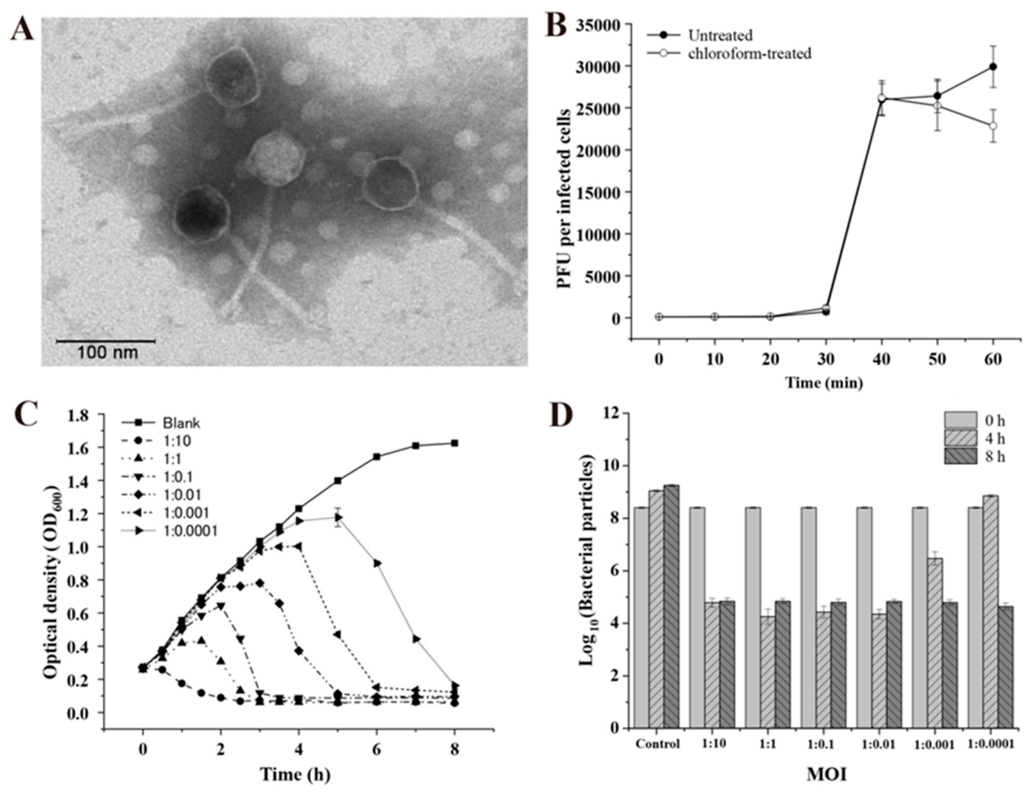
Figure 2.
The stability of phage vB_ValS-L9 under different environmental conditions. (A) Stability of phage vB_ValS-L9 in different temperatures. (B) Stability of phage vB_ValS-L9 under different pH conditions. (C) Stability of phage vB_ValS-L9 under different salinity conditions. Error bars represent the standard deviations of triplicate samples.
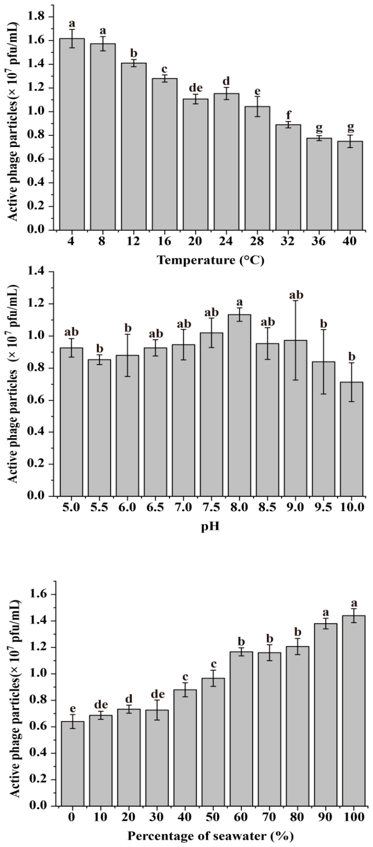
Figure 3.
Circular genomic map of phage vB_ValS-L9. Predicted ORFs are color-coded according to their functional annotation categories, as indicated in the legend, where the coding regions are indicated by the arrows illustrating the direction of transcription. From outside to inside: ORFs, GC content, GC skew, the scale.
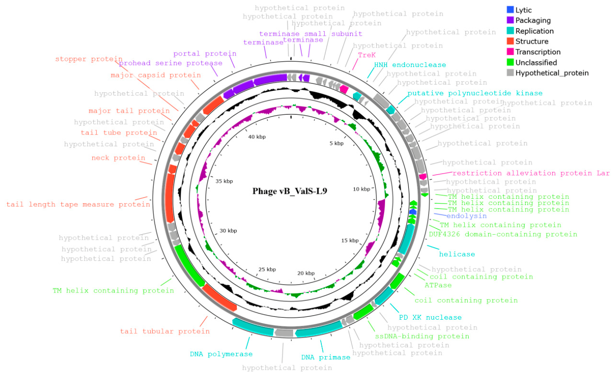
Figure 4.
Phylogenomic and comparative genomic analysis of phage vB_ValS-L9 and its closely related Vibrio phages. (A) Proteome-based phylogenetic tree generated by VIPTree displaying the global genomic relationships between phage vB_ValS-L9 and related double-stranded DNA viruses in the NCBI database. The colored rings represent the virus family (inner ring) and host group (outer ring). The red star marks the position of phage vB_ValS-L9. (B) Expanded sub-tree from VIPTree detailing the clade containing phages vB_ValS-L9, VapS30, and Artemius and their closely related genomic neighbor. The left and right color bars indicate the taxonomic virus family and host group, respectively. (C) Heatmap of Average Nucleotide Identity (ANI) values showing pairwise sequence comparison between phage vB_ValS-L9 and representative bacteriophages. (D) Heatmap of intergenomic similarities between phage vB_ValS-L9 and its closest relatives, calculated using the VIRIDIC pipeline. The upper-right half displays pairwise intergenomic identities (%), while the lower-left half represents aligned genome fractions. (E) Genome-based phylogenetic tree constructed using the VICTOR pipeline at the nucleotide level (D0), depicting the taxonomical classification and clade support values calculated via GBDP (Genome BLAST Distance Phylogeny).
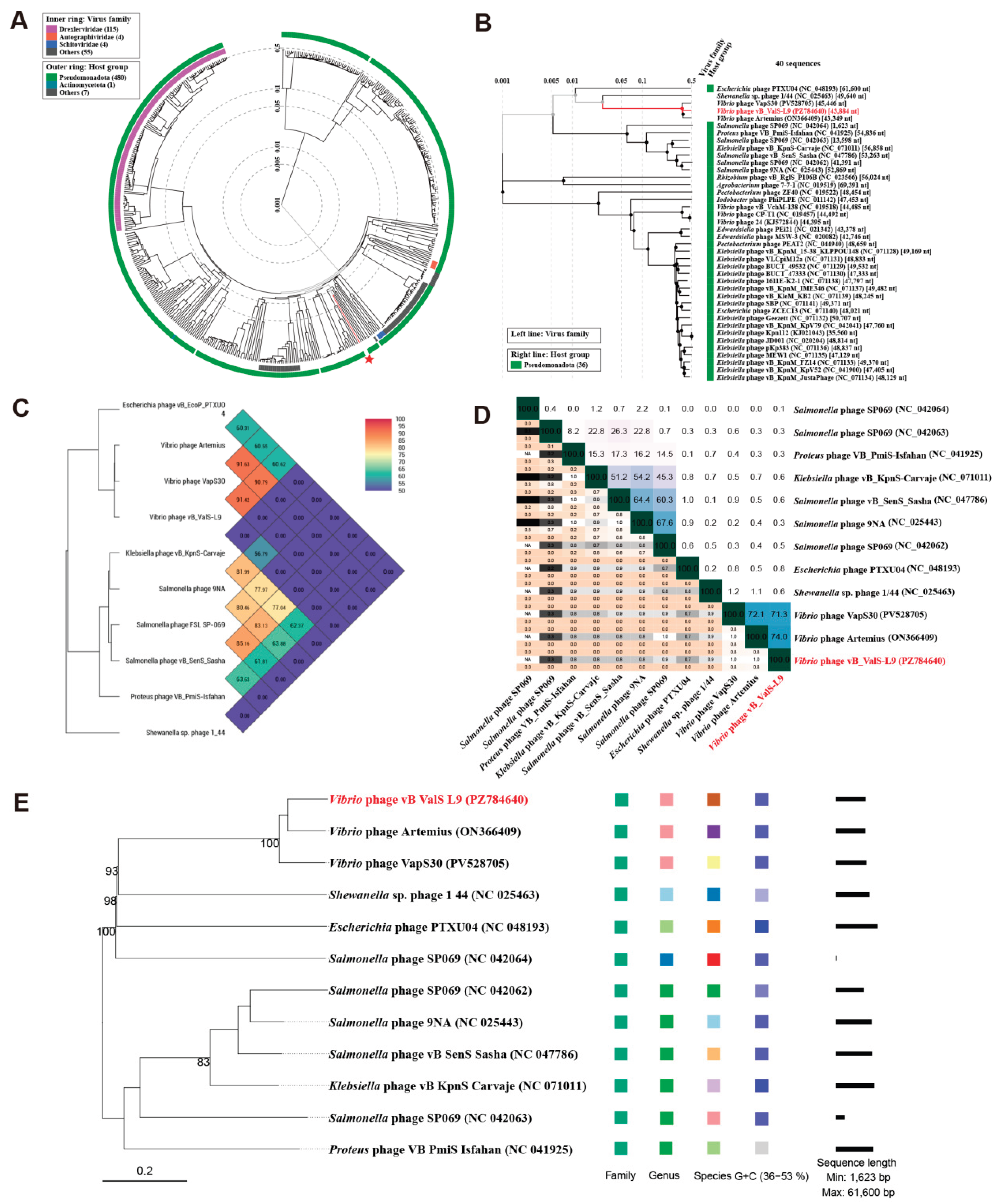
Figure 5.
Maximum-likelihood (ML) phylogenetic analysis of phage vB_ValS-L9 based on conservative packaging proteins. (A) ML phylogenetic tree inferred from the amino acid sequences of the terminase. (B) ML phylogenetic tree constructed from the amino acid sequences of the portal protein. Branch support values were evaluated using 1,000 ultrafast bootstrap replicates, and node values greater than 70% are indicated at the corresponding branches. Phage vB_ValS-L9 is highlighted in red, and phage Artemius and phage VapS30 are highlighted in blue.
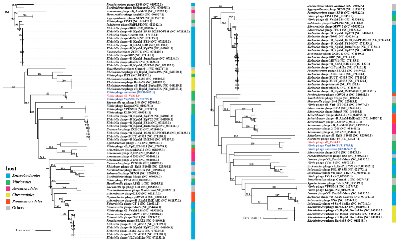
Figure 6.
Genomic comparison between Vibrio phage vB_ValS-L9, VapS30, Artemius, and Escherichia phage PTXU04. Arrows present the ORFs. The direction of each arrow represents the direction of transcription. Genome regions showing similarity were searched using tBLASTX and matches satisfying length and E-value (< 10−5) cutoffs were indicated by the rectangle according to the color scale on the top.
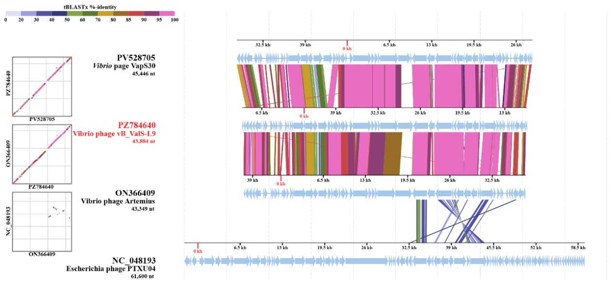
Figure 7.
Gene content-based viral network created for phage vB_ValS-L9, VapS30, and Artemius. Viruses at different family levels are shown in different colors. Dotted lines enclose different virus group.
Figure 7.
Gene content-based viral network created for phage vB_ValS-L9, VapS30, and Artemius. Viruses at different family levels are shown in different colors. Dotted lines enclose different virus group.
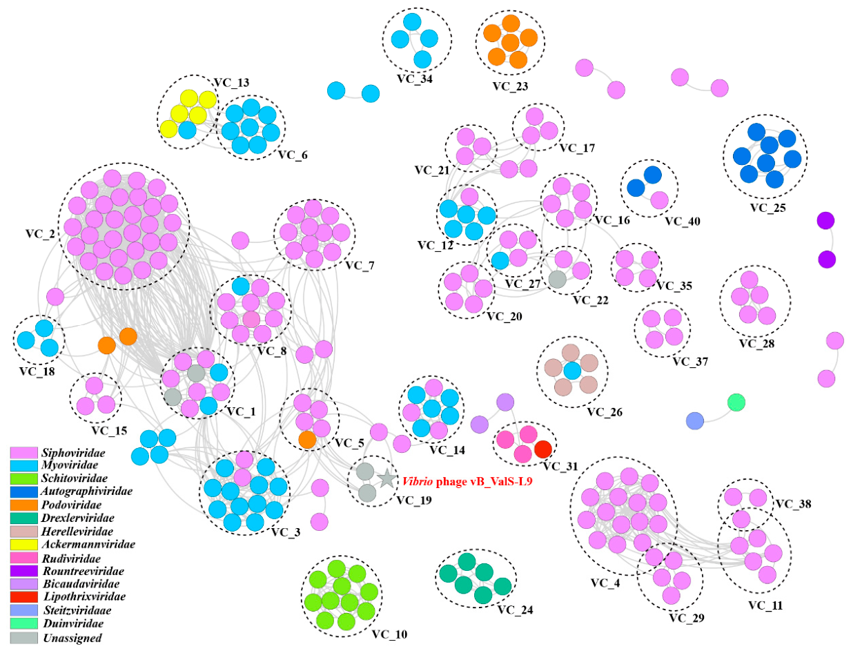
Figure 8.
Effects of phage vB_ValS-L9 treatment on survival and ingestion rate of juvenile pearl oysters challenged with Vibrio alginolyticus L4. (A) In vivo prophylactic and therapeutic efficacy of phage vB_ValS-L9 in juvenile pearl oysters (Pinctada martensii) challenged with Vibrio alginolyticus L4. (B) Ingestion rates of juvenile pearl oysters measured on days 1, 3, 7, 10, and 14 after bacterial challenge under the corresponding treatment regimens.
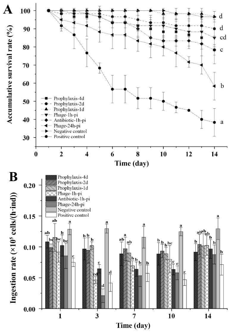

Table 1.
Experiment design for phage therapy on pearl oyster.
| Groups | Acclimation Stage | Immunization Stage | Inoculation Stage |
|---|---|---|---|
| Day 1 to Day 6 | 3 days | 14 days | |
| Prophylaxis-4d | Add phage 4 days in advance | Phage was added via immersion at a final concentration of 1.0 × 10⁶ PFU/mL at 4 days prior to the bacterial challenge | Vibrio was added daily via immersion to a final concentration of 1.0 × 10⁶ CFU/mL without phage supplementation. |
| Prophylaxis-2d | Add phage 2 days in advance | Phage was added via immersion at a final concentration of 1.0 × 10⁶ PFU/mL at 2 days prior to the bacterial challenge | Vibrio was added daily via immersion to a final concentration of 1.0 × 10⁶ CFU/mL without phage supplementation. |
| Prophylaxis-1d | Add phage 1 days in advance | Phage was added via immersion at a final concentration of 1.0 × 10⁶ PFU/mL at 1 days prior to the bacterial challenge | Vibrio was added daily via immersion to a final concentration of 1.0 × 10⁶ CFU/mL without phage supplementation. |
| Phage-1h-pi | Add the phage one hour after adding the vibrio. | - | Vibrio was added daily via immersion to a final concentration of 1.0 × 10⁶ CFU/mL; 1 h later, phages were added to a final concentration of 1.0 × 10⁶ PFU/mL. |
| Antibiotic-1h-pi | Add antibiotics one hour after adding the vibrio | - | Vibrio was added daily via immersion to a final concentration of 1.0 × 10⁶ CFU/mL; 1 h later, antibiotics (florfenicol) were added. |
| Phage-24h-pi | Add the phage 24 hour after adding the vibrio. | - | Vibrio was added daily via immersion to a final concentration of 1.0 × 10⁶ CFU/mL; 24 h later, phages were added to a final concentration of 1.0 × 10⁶ PFU/mL. |
| Negative control | Negative control | − | - |
| Positive control | Positive control | − | Vibrio was added daily via immersion to a final concentration of 1.0 × 10⁶ CFU/mL. |
Disclaimer/Publisher’s Note: The statements, opinions and data contained in all publications are solely those of the individual author(s) and contributor(s) and not of MDPI and/or the editor(s). MDPI and/or the editor(s) disclaim responsibility for any injury to people or property resulting from any ideas, methods, instructions or products referred to in the content. |
© 2026 by the authors. Licensee MDPI, Basel, Switzerland. This article is an open access article distributed under the terms and conditions of the Creative Commons Attribution (CC BY) license (http://creativecommons.org/licenses/by/4.0/).
Copyright: This open access article is published under a Creative Commons CC BY 4.0 license, which permit the free download, distribution, and reuse, provided that the author and preprint are cited in any reuse.