

Submitted:
23 September 2026
Posted:
23 September 2026
You are already at the latest version
Abstract
Porcine deltacoronavirus (PDCoV) is an emerging enteric coronavirus that causes severe mortality in neonatal piglets that results in significant economic burden to the global swine industry. The spike protein, particularly the C-terminal domain of S1 subunit (S1-CTD), contains immunodominant epitopes and represents an attractive target for antibody development. In this study, we expressed and purified recombinant PDCoV S1-CTD proteins in Escherichia coli and used it to immunize BALB/c mice for generating S1-CTD-specific antibodies. Following hybridoma production, we successfully established four monoclonal antibodies (2B7-G10, 2C7-F6, 7G3-G8, and 4H4 mAbs) that specifically recognized recombinant S1-CTD as well as native PDCoV spike protein. We further engineered a single-chain variable fragment derived from clone 4H4 (4H4-scFv) that retained antigen-binding ability to against S1-CTD and native PDCoV spike. Collectively, these findings demonstrate a practical framework for generating targeted mAbs and scFvs while expanding the repertoire of available reagents for future PDCoV S-targeted studies.
Keywords:
porcine deltacoronavirus (PDCoV)
; spike
; S1-CTD domain
; monoclonal antibody (mAb)
; single-chain variable fragment (scFv)
1. Introduction
Porcine deltacoronavirus (PDCoV) is an enveloped, single-stranded positive-sense RNA virus belonging to the genus Deltacoronavirus within the family Coronaviridae[1]. PDCoV gained global attention following a large-scale diarrheal outbreak in the United States in 2014, which was first identified in 2012 during a molecular epidemiological surveillance study in Hong Kong (cite)[2,3,4,5]. It has since been reported in major swine-producing regions worldwide and now considered an endemic disease[6,7,8,9,10,11]. PDCoV primarily infects mature enterocytes in the small intestine, leading to villous atrophy and enteric dysfunction that manifests as acute watery diarrhea, vomiting, and dehydration, with mortality rates reaching 30-50% in nursing piglets[12,13,14].
The genome of PDCoV is approximately 25 kb in length and encodes four structural proteins. Among these, the spike (S) glycoprotein mediates viral attachment and entry through binding to porcine aminopeptidase N (pAPN)[15,16], and also serves as the main immunogenic target for the induction of neutralizing antibodies in the host[1,5]. Structurally, the S protein is a trimeric class I fusion glycoprotein that undergoes cleavage into two functional subunits: the S1 receptor binding subunit and the membrane-anchored S2 membrane fusion subunit[15,17,18]. The S1 subunit can be further divided into two domains: the N-terminal domain (S1-NTD) and the C-terminal domain (S1-CTD). Cryo-electron microscopy has identified S1-CTD as the major receptor-binding domain (RBD)[17], and functional mapping further demonstrated that antisera raised against S1-CTD exhibit significantly higher neutralizing activity than those against S1-NTD or S2[18]. These findings establish S1-CTD as the primary immunodominant region of PDCoV S involved in both receptor recognition and protective humoral immunity, highlighting its potential as a vaccine candidate.
Despite the central role of S1-CTD in mediating PDCoV host-virus interactions, there remains a relative paucity of reagents specific for S1-CTD that is currently available for research purposes. For example, several monoclonal antibodies (mAbs) targeting the nucleocapsid (N) protein have been developed and widely used in assays including ELISA, immunohistochemistry, and immunofluorescence[19,20,21]. These efforts have also been extended to single-chain variable fragments (scFvs)[22], which offer several advantages over mAbs including lower production costs, ease of expression, and greater flexibility for molecular engineering[23,24]. In contrast, only a limited number of S-specific mAbs have been reported[25,26,27], with no scFvs currently available[22]. This represents a barrier for antigenic characterization and development of next-generation diagnostics and antivirals for PDCoV.
In this study, we generated and characterized four mAbs and one scFv targeting the PDCoV S1-CTD domain. All of them exhibited binding specificity against S1-CTD and the native S protein, while being compatible with routine experimental methods including ELISA and immunofluorescence assay (IFA). These reagents expand the limited repertoire of PDCoV S-specific antibodies and may provide a framework for future recombinant antibody engineering efforts.
2. Materials and Methods
2.1. Cells and Virus
LLC-PK1 and HEK-293T cells were cultured in Dulbecco’s Modified Eagle Medium (DMEM, Gibco) and SP2/0 myeloma cells myeloma cells in RPMI 1640 Medium (Gibco) at 37 °C in a 5% CO₂ humidified incubator. All culture media were supplemented with 10% fetal bovine serum (FBS, Gibco) and 1% penicillin-streptomycin. PDCoV strain used in the initial experiments was house-kept and propagated in LLC-PK1 cells as described previously [28]. For immunofluorescence assay (IFA) detection, a novel PDCoV isolate with higher infection efficiency was obtained in 2021 from clinical samples collected in Jinshi, China. This isolate was designated PDCoV/China/Jinshi/2021 and was propagated in LLC-PK1 cells under the same conditions. Because this isolate has not yet been publicly reported, its full characterization is ongoing and will be reported elsewhere.
2.2. Expression and Purification of Recombinant S1-Ctd Protein
The PDCoV S1-CTD gene fragment (1036 bp) was amplified by PCR from PDCoV cDNA using primers S1-CTD-F (5′-CCAAGCTTTGAGGTGTATTGTGCCAGT-3′, containing a HindIII site) and S1-CTD-R (5′-CGGAATTCATGGATGGGTTCTACTCCGAC-3′, containing an EcoRI site). The amplicon was cloned into the pET-28a vector (Novagen) between the EcoRI and HindIII sites. The recombinant plasmid pET-28a-PDCoV-S1-CTD was confirmed by colony PCR, double enzyme digestion, and DNA sequencing.
For protein expression, the plasmid was transformed into E. coli Transetta (DE3) competent cells (TransGen Biotech). A single colony was inoculated into LB medium containing 50 μg/mL kanamycin and grown at 37 °C with shaking (220 rpm) to an OD₆₀₀ of 0.6–0.8. Protein expression was induced by adding 1 mM isopropyl β-D-1-thiogalactopyranoside (IPTG) at 37 °C for 12 h. Cells were harvested by centrifugation (4000 × g, 10 min, 4 °C), resuspended in lysis buffer (50 mM Tris-HCl, 300 mM NaCl, pH 8.0), and disrupted by sonication on ice (300 W, 3 s on/5 s off, 20 min). The lysate was centrifuged (12,000 × g, 20 min, 4 °C), and the pellet (containing inclusion bodies) was collected. The inclusion bodies were washed three times with wash buffer (50 mM Tris-HCl, 300 mM NaCl, 2 M urea, 1% Triton X-100, pH 8.0) and then solubilized in denaturing buffer (50 mM Tris-HCl, 300 mM NaCl, 8 M urea, 10 mM imidazole, pH 8.0) overnight at 4 °C with gentle stirring. After centrifugation (12,000 × g, 15 min, 4 °C), the supernatant was subjected to Ni-NTA affinity chromatography (GE Healthcare) under denaturing conditions. The bound protein was eluted with elution buffer (50 mM Tris-HCl, 300 mM NaCl, 8 M urea, 250 mM imidazole, pH 8.0). Refolding was achieved by stepwise dialysis against decreasing concentrations of urea (6 M, 4 M, 2 M, 1 M, 0 M) in dialysis buffer (50 mM Tris-HCl, 300 mM NaCl, 10% glycerol, pH 8.0) at 4 °C. The refolded protein was concentrated using Amicon Ultra centrifugal filters (Millipore) and stored at –80 °C. Protein concentration was determined by the BCA method (Thermo Scientific). The purified protein was verified by SDS-PAGE and western blot with anti-His antibody (Proteintech).
2.3. Mouse Immunization
Six-week-old female BALB/c mice were immunized subcutaneously with 100 μg of purified S1-CTD protein emulsified 1:1(v/v) with Freund’s complete adjuvant (Sigma) for the primary immunization. Two booster immunizations with 50 μg of protein emulsified with Freund’s incomplete adjuvant (Sigma) were administered at 2-week intervals. A fourth immunization without adjuvant was given intraperitoneally three days before cell fusion. Blood samples were collected from the tail vein before each immunization, and serum antibody titers were determined by indirect ELISA.
2.4. Indirect Elisa for Antibody Detection
Microtiter plates (Corning) were coated with 100 μL/well of purified S1-CTD protein (2 μg/mL in 0.05 M carbonate-bicarbonate buffer, pH 9.6) overnight at 4 °C. After washing three times with PBST (PBS containing 0.05% Tween-20), the plates were blocked with 5% skim milk in PBST for 2 hours at 37 °C. Serial dilutions of mouse serum or hybridoma supernatants were added (100 μL/well) and incubated for 1 hour at 37 °C. After washing, HRP-conjugated goat anti-mouse IgG (1:5000 dilution, Abcam) was added and incubated for 1 hour at 37 °C. The plates were washed, and 100 μL/well of TMB substrate (Solarbio) was added. The reaction was stopped with 50 μL/well of 2 M H₂SO₄ after 15 minutes, and the absorbance was read at 450 nm using a microplate reader (Bio-Rad). The cutoff value for positivity was set as the mean OD of negative controls plus three standard deviations.
2.5. Hybridoma Generation and Screening
Splenocytes from the immunized mouse #1 were fused with SP2/0 myeloma cells at a ratio of 5:1 using polyethylene glycol 1500 (Roche) following standard protocols. Fused cells were seeded in 96-well plates and selected in HAT medium (DMEM containing 20% FBS, 100 μM hypoxanthine, 0.4 μM aminopterin, and 16 μM thymidine). Hybridoma supernatants were screened by indirect ELISA using S1-CTD protein as the coating antigen. Positive clones were subcloned at least three times by limiting dilution to establish stable monoclonal hybridoma cell lines. The selected hybridoma clones (2B7-G10, 2C7-F6, 7G3-G8, and 4H4) were expanded and cryopreserved in liquid nitrogen.
2.6. Monoclonal Antibody Production and Purification
For ascites production, 8- to 10-week-old BALB/c mice were injected intraperitoneally with 0.5 mL of pristine (Sigma). After 7–10 days, 5 × 10⁶ of 7G3-G8 hybridoma cells were injected intraperitoneally. Ascites fluid was collected 10–14 days later, clarified by centrifugation (10,000 × g, 10 min, 4 °C), and stored at –80 °C. mAbs were purified from ascites using Protein G Sepharose (GE Healthcare) according to the manufacturer’s instructions.
2.7. Construction of Single-Chain Variable Fragment (Scfv)
Total RNA was extracted from 4H4 hybridoma cells using TRIzol reagent (Invitrogen). cDNA was synthesized by reverse transcription. The variable regions of the VH and VL were amplified by RT-PCR using degenerate primer sets: VH forward 5′-CGCGGATCCSAGGTSMAGCTGCAGSAGTCWGG-3′, VH reverse 5′-CGAGCCGCCACCTCCAGAGCCACCTCCGCCTGAGGAGACTATGAGAGTGG-3′; VL forward 5′-GGCGGAGGTGGCTCTGGAGGTGGCGGCTCGGAYATTGTGMTSACMCARWCTMCA-3′, VL reverse 5′-CCGCTCGAGTTATTTGATTTCCAGCTTGG-3′. The VH and VL fragments were purified and assembled into a single-chain variable fragment (scFv) by overlap extension PCR containing a flexible (Gly₄Ser)₃ linker. The assembled VH-linker-VL product (~750 bp) was cloned into the pET-28a vector using standard restriction enzyme ligation (primers incorporated appropriate restriction sites). The recombinant plasmid pET-28a-4H4 was confirmed by colony PCR and DNA sequencing.
2.8. Expression and Purification of Scfv
The pET-28a-4H4 plasmid was transformed into E. coli Transetta (DE3) cells. A single colony was inoculated into LB medium containing 50 μg/mL kanamycin and grown at 37 °C to an OD₆₀₀ of 0.6–0.8. Expression was induced with 1 mM IPTG at 16 °C for 12 h. Cells were harvested by centrifugation (4000 × g, 10 min, 4 °C), resuspended in lysis buffer (50 mM Tris-HCl, 300 mM NaCl, pH 8.0) containing 1 mM PMSF, and disrupted by sonication on ice. The lysate was centrifuged (12,000 × g, 20 min, 4 °C), and the pellet (containing inclusion bodies) was washed three times with wash buffer (50 mM Tris-HCl, 300 mM NaCl, 2 M urea, 1% Triton X-100, pH 8.0). The inclusion bodies were solubilized in denaturing buffer (50 mM Tris-HCl, 300 mM NaCl, 8 M urea, 10 mM imidazole, pH 8.0) overnight at 4 °C. After centrifugation (12,000 × g, 15 min, 4 °C), the supernatant was loaded onto a Ni-NTA column (GE Healthcare). The column was washed with 5 column volumes of wash buffer (50 mM Tris-HCl, 300 mM NaCl, 8 M urea, 20 mM imidazole, pH 8.0), and the bound scFv was eluted with elution buffer (50 mM Tris-HCl, 300 mM NaCl, 8 M urea, 250 mM imidazole, pH 8.0). Refolding was performed by stepwise dialysis against decreasing urea concentrations (6 M, 4 M, 2 M, 1 M, 0 M) in dialysis buffer (50 mM Tris-HCl, 300 mM NaCl, 10% glycerol, pH 8.0) at 4 °C. The refolded scFv was concentrated using Amicon Ultra centrifugal filters (Millipore) and stored at –80 °C. Purity was assessed by SDS-PAGE, and the protein was verified by western blot using anti-His antibody (1:2000, Proteintech).
2.9. Western Blot
Protein samples were separated by 12% SDS-PAGE and transferred onto PVDF membranes (Millipore). The membranes were blocked with 5% skim milk in TBST (TBS containing 0.1% Tween-20) for 2 hours at room temperature, then incubated with primary antibodies (mouse serum, hybridoma supernatant, purified mAb, or scFv) overnight at 4 °C. After washing three times with TBST, the membranes were incubated with HRP-conjugated secondary antibodies (1:5000 dilution) for 1 h at room temperature. Protein bands were visualized using enhanced chemiluminescence (ECL) substrate (Thermo Scientific) and imaged with a ChemiDoc system (Bio-Rad).
2.10. Immunofluorescence Assay (Ifa)
LLC-PK1 cells grown on coverslips were infected with PDCoV(the Jinshi/2021 isolate) at an MOI of 0.1 for 24 h. For transfected cells, 293T cells were transfected with GFP-PDCoV-S1-CTD or empty GFP-C2 vector using Lipofectamine 3000 (Invitrogen). Cells were fixed with 4% paraformaldehyde for 15 min at room temperature, permeabilized with 0.1% Triton X-100 in PBS for 10 min, and blocked with 5% BSA in PBS for 1 h. The cells were then incubated with primary antibodies (mouse serum, hybridoma supernatant, or scFv followed by anti-His antibody) for 1 h at 37 °C. After washing, cells were incubated with FITC-conjugated goat anti-mouse IgG (1:200, Beyotime) for 1 h at 37 °C in the dark. Nuclei were counterstained with DAPI (Beyotime). Fluorescence was observed using a fluorescence microscope (Nikon).
2.11. Double-Antibody Sandwich Elisa
Microtiter plates were coated with various concentrations of purified 4H4-scFv (0.5, 1.0, and 2.0 μg/mL) in carbonate-bicarbonate buffer overnight at 4 °C. After blocking with 5% skim milk in PBST, serial dilutions of S1-CTD antigen (10, 5, 2, 0.5, 0.05, and 0.005 μg/mL) were added and incubated for 1 h at 37 °C. After washing, mouse anti-PDCoV S1-CTD mAb (ascites, 1:1000 dilution) was added and incubated for 1 h at 37 °C, followed by HRP-conjugated goat anti-mouse IgG (1:5000). The plates were developed with TMB substrate, and the reaction was stopped with 2 M H₂SO₄. Absorbance was measured at 450 nm. A sample was considered positive if the P/N ratio (OD of positive well / OD of negative well) exceeded 2.1.
3. Results
3.1. Recombinant Pdcov S1-Ctd Is Efficiently Expressed in Prokaryotic Cells
To express recombinant PDCoV S1-CTD, we first used PCR to amplify the corresponding gene fragment from PDCoV cDNA (Figure 1A) and cloned it into the pET-28a vector between the EcoRI and HindIII restriction sites, downstream of the vector-encoded His tag. We then transformed the resulting recombinant plasmid, pET-28a-PDCoV-S1-CTD, into Escherichia coli Transetta (DE3) cells, and induced protein expression with 1 mM IPTG at either 16 °C or 37 °C for 12 hours.
To confirm protein expression, we performed SDS-PAGE analysis and detected a prominent band between 37-43 kDa, with stronger expression following induction at 37 °C (Figure 1B). Further analysis revealed that the expected 42 kDa band was primarily found in the insoluble fraction (inclusion bodies) rather than the soluble supernatant (Figure 1C). We subsequently purified the recombinant PDCoV S1-CTD protein using Ni-NTA affinity chromatography under denaturing conditions, followed by stepwise dialysis to enable protein refolding. The purified protein appeared as a single 42 kDa band on Coomassie-stained SDS-PAGE (Figure 1D), and its identity was further confirmed by western blot analysis using an anti-His antibody (Figure 1E).
3.2. Immunization with Purified Pdcov S1-Ctd Elicits Specific Antibody Responses in Mice
Having successfully expressed and purified recombinant PDCoV S1-CTD, we next immunized two BALB/c mice subcutaneously using the protein emulsified in Freund’s adjuvant to induce antigen-specific antibody production. After three immunizations, we evaluated serum antibody titers by indirect ELISA using S1-CTD-coated plates and found that mouse #1 exhibited substantially higher titers and OD values compared to mouse #2 (Figure 2A). To maximize antigen-specific immune response prior to hybridoma generation, we administered a fourth booster immunization which resulted in mouse #1 reaching a titer of 1:2¹⁰ in contrast to a lack of appreciable increases for mouse #2 (Figure 2B).
Based on the superior antibody response in mouse #1, we tested the specificity of its serum by western blot and IFA using 293T cells transfected with either GFP-PDCoV-S1-CTD or empty GFP-C2 vector. By western blot, we found that mouse #1 antisera specifically recognized a ~70 kDa band corresponding to the GFP-S1-CTD fusion protein, while no reactivity was detected in the empty vector control (Figure 2C). Consistently, IFA demonstrated specific staining in 293T cells expressing GFP-PDCoV-S1-CTD, while no fluorescence signal was observed in cells transfected with the empty vector (Figure 2D). Based on these results, mouse #1 was selected for subsequent cell fusion.
3.3. Pdcov S1-Ctd-Specific Monoclonal Antibodies Recognize Native Pdcov S
Following fusion of splenocytes from immunized mouse #1 with SP2/0 myeloma cells, we selected hybridoma clones in HAT medium and screened culture supernatants by indirect ELISA using purified S1-CTD protein as the coating antigen. We identified four positive clones, designated 2B7-G10, 2C7-F6, 7G3-G8, and 4H4, and subjected them to further subcloning by limiting dilution. All four clones remained stable and continuously secreted antibodies after multiple rounds of subcloning.
We next tested the specificity of these four mAbs against PDCoV S under denaturing or native conditions, by western blot and IFA staining respectively, in PDCoV-infected LLC-PK1 cells. For western blot, we found that all four mAbs specifically recognized a ~150 kDa band corresponding to the S protein in infected cell lysates, while no bands were detected in mock-infected controls (Figure 3A). Consistently, IFA revealed specific membranous staining in PDCoV-infected LLC-PK1, while no fluorescence signal was observed in uninfected cells (Figure 3B). These results indicate that all four mAbs recognized PDCoV S protein in both denatured and native conformations.
3.4. Recombinant 4h4-Scfv Is Also Efficiently Expressed in Prokaryotic Cells
To extend the utility of the generated mAbs, we sought to construct an scFv using clone 4H4. We first used RT-PCR to amplify the variable heavy (VH) and variable light (VL) chain sequences from RNA isolated from the 4H4 hybridoma cells (Figure 4A), and then assembled them into an scFv using a flexible (Gly₄Ser)₃ linker (Figure 4B). The resulting construct, designated as 4H4-scFv, was similarly His-tagged when cloned into the pET-28a vector, and further verified by DNA sequencing (SEQ ID No. 1, Supplementary Materials).
After transformation into Escherichia coli DE3 cells for protein expression under IPTG induction, we were able to identify a clear band between 24-31 kDa using SDS-PAGE, which was similarly found only in the inclusion body fraction (Figure 4C). We then similarly performed Ni-NTA affinity purification and stepwise dialysis, which revealed a single 30 kDa band on Coomassie-stained SDS-PAGE (Figure 4D) corresponding to 4H4-scFv as shown by western blot analysis using an anti-His antibody (Figure 4E).
3.54. h4-Scfv Can Also Recognize Native Pdcov S
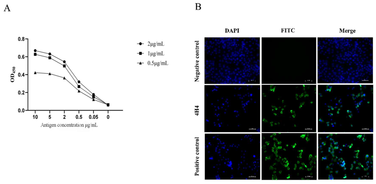
We evaluated the binding activity of 4H4-scFv against S1-CTD using a double-antibody sandwich ELISA. Plates were coated with scFv (0.5–2.0 μg/mL) and incubated with serial dilutions of S1-CTD (10–0.005 μg/mL), and bound S1-CTD was then detected using anti-S1-CTD mAb ascites (1:1000) derived from the 7G3-G8 hybridoma clone. We found that 4H4-scFv specifically bound to the S1-CTD antigen in a dose-dependent manner, with positive reactivity detected at scFv concentrations as low as 0.5 μg/mL with an antigen concentration of 0.05 μg/mL (Figure 5A). This also indicates that 4H4 and 7G3-G8 recognize non-overlapping epitopes on S1-CTD.
Next, we similarly tested the specificity of 4H4-scFv against PDCoV S under denaturing or native conditions, by western blot and IFA staining respectively. For western blot, we were unable to detect any bands corresponding to PDCoV S in infected and uninfected controls (data not shown). In contrast, IFA revealed specific membranous staining in uninfected controls (Figure 5B). These results suggest that 4H4-scFv is specific to the native conformation of PDCoV S protein in infected cells while confirming its utility for immunofluorescence applications.
4. Discussion
In this study, we successfully leveraged the immunogenic potential of PDCoV S1-CTD[29] and generated four monoclonal antibodies (2B7-G10, 2C7-F6, 7G3-G8, and 4H4) and one single-chain variable fragment (4H4-scFV) that recognized both recombinant S1-CTD and native S protein. These reagents expand the limited toolbox of PDCoV S-specific detection reagents and may provide a framework for enabling future recombinant antibody development.
Although our recombinant PDCoV S1-CTD was expressed as insoluble inclusion bodies in E. coli, we were able to recover high-quality proteins through denaturing purification followed by gradual refolding. This was demonstrated by the ability of the recombinant S1-CTD to elicit robust antibody responses in mice alongside the recognition of native S protein by the resulting antisera. These findings indicate that our protein production process could preserve antigenically relevant epitopes and further support the strong antigenicity of the S1-CTD domain. Collectively, these results suggest that S1-CTD produced in a prokaryotic expression system can serve as an effective immunogen despite the absence of native glycosylation and other post-translational modifications present in mammalian expression systems.
Our new mAbs and scFv, rationally designed to target the S1-CTD domain and validated across routine immunological assays including ELISA and IFA, may facilitate further investigation into PDCoV biology and pathogenesis. Currently, the N protein is most commonly used for PDCoV detection due to its high abundance during infection[20,30,31], but the S protein offers distinct advantages for mechanistic studies[15,25]. For instance, as the S protein is exposed on the surface of virions and infected cells, antibodies raised against S would enable direct visualization of S-mediated functional processes such as viral entry and cell-to-cell spread[32,33]. Furthermore, antibodies raised against extracellular domains such as S1-CTD can be applied to intact virions and unpermeabilized cells, which may enable opportunities for applications such as live-cell imaging.
Limitations of this study include the lack of assessment of neutralization potential for our mAbs and scFv, as our primary objective was to develop sensitive detection reagents. In addition, the precise epitope recognized by these antibodies remain unknown, and their potential protective efficacy was also not tested. Future studies on appropriate infection models, coupled to neutralization assays and epitope mapping, would provide additional insights into the functional utility of these reagents beyond diagnostic applications. Furthermore, 4H4-scFv failed to detect denatured PDCoV S by western blot but could recognize native S by IFA. While this may suggest that 4H4-scFv preferentially recognizes epitopes that are disrupted under reducing conditions, a reduced binding affinity relative to the parental mAb may also contribute to the lack of detectable reactivity. Additional studies will be required to determine the molecular basis underlying this observation.
In conclusion, the mAbs and scFv generated in this study provide new reagents for the field and support ongoing efforts toward the development of targeted diagnostics. The successful production of immunologically relevant S1-CTD antigen in a prokaryotic expression system, which was extended to development of a novel S-targeting recombinant scFv, also forms a proof-of-concept framework for future antibody engineering efforts for PDCoV proteins. Collectively, these findings highlight the utility of rational antigen design and recombinant protein engineering for advancing next-generation antibody-based technologies.
5. Conclusions
In this study, four monoclonal antibodies and one single-chain variable fragment targeting the PDCoV S1-CTD domain were successfully generated. All antibodies recognized both recombinant S1-CTD and native spike protein. These reagents expand the available toolkit for PDCoV spike protein research and may facilitate future diagnostic and functional studies.
Supplementary Materials
The following supporting information can be downloaded at the website of this paper posted on Preprints.org.
Author Contributions
Conceptualization: W.Y. and Z.D.; Methodology: W.Y., L.X., Z.L., L.Y., Y.H., H.M.; Validation: W.Y., L.X., and Z.L.; Formal Analysis: W.Y., L.X., Z.L., L.Y., Y.H., H.M., G.W., Y.Y., and D.H.; Investigation: L.X., Z.L., L.Y., Y.H., H.M.; Resources: W.Y. and Z.D., G.W., Y.Y., and D.H., D.S, S.D; Data Curation: W.Y., L.X., Z.L., L.Y., Y.H., H.M., G.W., Y.Y., and D.H.; Writing – Original Draft: W.Y. and Z.D.; Writing – Review & Editing: W.Y., Y.Y.Y., and Z.D.; Visualization: W.Y., L.X., Z.L., L.Y., Y.H., H.M.; Supervision: S.D. and Z.D.; Project Administration: Z.D.; Funding Acquisition: W.Y. and Z.D.
Funding
This work was supported by the National Natural Science Foundation of China (No. 31860704); the Jiangxi Provincial Natural Science Foundation (CJJ200450); the Natural Science Foundation for Youth of Jiangxi Province (20242BAB20315); the Science and Technology Program of Jiangxi Provincial Department of Education (GJJ2200410).
Institutional Review Board Statement
The animal study protocol was approved by the Experimental Animal Ethics (Management) Committee of Jiangxi Agricultural University (Approval number: JXAULL-2022-06-15, approved on 15 June 2022). All animal experiments were conducted in accordance with the guidelines and regulations of the committee, and BALB/c mice were housed under specific pathogen-free conditions with free access to food and water.
Acknowledgments
We appreciated all the funds supported this work.
Conflicts of Interest
The authors declare no conflict of interest.
References
- Jung, K.; Hu, H.; Saif, L.J. Porcine deltacoronavirus infection: Etiology, cell culture for virus isolation and propagation, molecular epidemiology and pathogenesis. Virus Res. 2016, 226, 50–59. [Google Scholar] [CrossRef]
- Woo, P.C.; Lau, S.K.; Lam, C.S.; Tsang, A.K.; Hui, S.W.; Fan, R.Y.; Martelli, P.; Yuen, K.Y. Discovery of a novel bottlenose dolphin coronavirus reveals a distinct species of marine mammal coronavirus in Gammacoronavirus. J. Virol. 2014, 88, 1318–1331. [Google Scholar] [CrossRef]
- Wang, L.; Byrum, B.; Zhang, Y. Detection and genetic characterization of deltacoronavirus in pigs, Ohio, USA, 2014. Emerg. Infect. Dis. 2014, 20, 1227–1230. [Google Scholar] [CrossRef]
- Li, G.; Chen, Q.; Harmon, K.M.; Yoon, K.J.; Schwartz, K.J.; Hoogland, M.J.; Gauger, P.C.; Main, R.G.; Zhang, J. Full-Length Genome Sequence of Porcine Deltacoronavirus Strain USA/IA/2014/8734. Genome Announc 2014, 2. [Google Scholar] [CrossRef]
- Woo, P.C.; Lau, S.K.; Lam, C.S.; Lau, C.C.; Tsang, A.K.; Lau, J.H.; Bai, R.; Teng, J.L.; Tsang, C.C.; Wang, M.; et al. Discovery of seven novel Mammalian and avian coronaviruses in the genus deltacoronavirus supports bat coronaviruses as the gene source of alphacoronavirus and betacoronavirus and avian coronaviruses as the gene source of gammacoronavirus and deltacoronavirus. J. Virol. 2012, 86, 3995–4008. [Google Scholar] [CrossRef]
- Tang, P.; Cui, E.; Song, Y.; Yan, R.; Wang, J. Porcine deltacoronavirus and its prevalence in China: a review of epidemiology, evolution, and vaccine development. Arch. Virol. 2021, 166, 2975–2988. [Google Scholar] [CrossRef]
- Lednicky, J.A.; Tagliamonte, M.S.; White, S.K.; Elbadry, M.A.; Alam, M.M.; Stephenson, C.J.; Bonny, T.S.; Loeb, J.C.; Telisma, T.; Chavannes, S.; et al. Independent infections of porcine deltacoronavirus among Haitian children. Nature 2021, 600, 133–137. [Google Scholar] [CrossRef]
- Liu, C.; Zhang, X.; Zhang, Z.; Chen, R.; Zhang, Z.; Xue, Q. Complete Genome Characterization of Novel Chinese Porcine Deltacoronavirus Strain SD. Genome Announc 2017, 5. [Google Scholar] [CrossRef]
- Suzuki, T.; Hayakawa, J.; Ohashi, S. Complete Genome Characterization of the Porcine Deltacoronavirus HKD/JPN/2016, Isolated in Japan, 2016. Genome Announc 2017, 5. [Google Scholar] [CrossRef]
- Lee, S.; Lee, C. Complete Genome Characterization of Korean Porcine Deltacoronavirus Strain KOR/KNU14-04/2014. Genome Announc 2014, 2. [Google Scholar] [CrossRef]
- More-Bayona, J.A.; Ramirez-Velasquez, M.; Hause, B.; Nelson, E.; Rivera-Geronimo, H. First isolation and whole genome characterization of porcine deltacoronavirus from pigs in Peru. Transbound. Emerg. Dis. 2022, 69, e1561–e1573. [Google Scholar] [CrossRef]
- Shan, X.; Li, R.; Ma, X.; Qiu, G.; Xiang, Y.; Zhang, X.; Wu, D.; Wang, L.; Zhang, J.; Wang, T.; et al. Epidemiology, pathogenesis, immune evasion mechanism and vaccine development of porcine Deltacoronavirus. Funct. Integr. Genom. 2024, 24, 79. [Google Scholar] [CrossRef]
- Duan, C. An Updated Review of Porcine Deltacoronavirus in Terms of Prevalence, Pathogenicity, Pathogenesis and Antiviral Strategy. Front Vet. Sci. 2021, 8, 811187. [Google Scholar] [CrossRef]
- Wang, L.; Hayes, J.; Sarver, C.; Byrum, B.; Zhang, Y. Porcine deltacoronavirus: histological lesions and genetic characterization. Arch. Virol. 2016, 161, 171–175. [Google Scholar] [CrossRef]
- Du, W.; Debski-Antoniak, O.; Drabek, D.; van Haperen, R.; van Dortmondt, M.; van der Lee, J.; Drulyte, I.; van Kuppeveld, F.J.M.; Grosveld, F.; Hurdiss, D.L.; et al. Neutralizing antibodies reveal cryptic vulnerabilities and interdomain crosstalk in the porcine deltacoronavirus spike protein. Nat. Commun. 2024, 15, 5330. [Google Scholar] [CrossRef]
- Wang, B.; Liu, Y.; Ji, C.M.; Yang, Y.L.; Liang, Q.Z.; Zhao, P.; Xu, L.D.; Lei, X.M.; Luo, W.T.; Qin, P.; et al. Porcine Deltacoronavirus Engages the Transmissible Gastroenteritis Virus Functional Receptor Porcine Aminopeptidase N for Infectious Cellular Entry. J. Virol. 2018, 92. [Google Scholar] [CrossRef]
- Shang, J.; Zheng, Y.; Yang, Y.; Liu, C.; Geng, Q.; Tai, W.; Du, L.; Zhou, Y.; Zhang, W.; Li, F. Cryo-Electron Microscopy Structure of Porcine Deltacoronavirus Spike Protein in the Prefusion State. J. Virol. 2018, 92. [Google Scholar] [CrossRef]
- Chen, R.; Fu, J.; Hu, J.; Li, C.; Zhao, Y.; Qu, H.; Wen, X.; Cao, S.; Wen, Y.; Wu, R.; et al. Identification of the immunodominant neutralizing regions in the spike glycoprotein of porcine deltacoronavirus. Virus Res. 2020, 276, 197834. [Google Scholar] [CrossRef]
- Okda, F.; Lawson, S.; Liu, X.; Singrey, A.; Clement, T.; Hain, K.; Nelson, J.; Christopher-Hennings, J.; Nelson, E.A. Development of monoclonal antibodies and serological assays including indirect ELISA and fluorescent microsphere immunoassays for diagnosis of porcine deltacoronavirus. BMC Vet. Res. 2016, 12, 95. [Google Scholar] [CrossRef]
- Han, F.; Shan, F.; Hou, J.; Guo, D.; Xiang, Y.; Yuan, J.; Wei, Z. Establishment and application of PDCoV antigen-specific DAS-ELISA detection method. BMC Vet. Res. 2024, 20, 342. [Google Scholar] [CrossRef]
- Wang, W.; Fan, B.; Zhang, X.; Yang, S.; Zhou, J.; Guo, R.; Zhao, Y.; Zhou, J.; Li, J.; Li, B. Development and evaluation of a monoclonal antibody-based competitive ELISA for detecting porcine deltacoronavirus antibodies. Anim. Dis. 2024, 4. [Google Scholar] [CrossRef]
- Zhang, Y.; Song, Y.; Ren, H.; Zeng, Q.; Yuan, Y.; Xia, L.; Wei, Z. Preparation of a Single-Chain Antibody against Nucleocapsid Protein of Porcine Deltacoronavirus by Phage Display Technology. Viruses 2022, 14. [Google Scholar] [CrossRef]
- Ahmad, Z.A.; Yeap, S.K.; Ali, A.M.; Ho, W.Y.; Alitheen, N.B.; Hamid, M. scFv antibody: principles and clinical application. Clin. Dev. Immunol. 2012, 2012, 980250. [Google Scholar] [CrossRef]
- Holliger, P.; Hudson, P.J. Engineered antibody fragments and the rise of single domains. Nat. Biotechnol. 2005, 23, 1126–1136. [Google Scholar] [CrossRef]
- Chen, R.; Wen, Y.; Yu, E.; Yang, J.; Liang, Y.; Song, D.; Wen, Y.; Wu, R.; Zhao, Q.; Du, S.; et al. Identification of an immunodominant neutralizing epitope of porcine Deltacoronavirus spike protein. Int. J. Biol. Macromol. 2023, 242, 125190. [Google Scholar] [CrossRef]
- Li, X.; Ye, H.; Ge, X.; Zhou, L.; Guo, X.; Han, J.; Zhang, Y.; Yang, H. Identification of two immunodominant and neutralizing linear B-cell epitopes exposed on the surface of the porcine deltacoronavirus spike protein. Vet. Res. 2026, 57, 33. [Google Scholar] [CrossRef]
- Lu, W.; Cao, H.; Yang, Y.; Sun, Y.; Yang, D.; Gerber, P.F.; Li, X.; Huang, Y.; Wang, B. Characterization of two neutralizing monoclonal antibodies with conformational epitopes against porcine deltacoronavirus. Anim. Dis. 2025, 5. [Google Scholar] [CrossRef]
- Zhang, M.; Peng, Q.; Ye, Y.; Gong, W.; Guo, N.; Zhang, F.; Li, A.; Zhou, X.; Huang, D.; Song, D.; et al. Correction for Zhang et al., “Complete Genome Sequence of Porcine Deltacoronavirus Strain CH/JXJGS01/2016, Isolated in Jiangxi Province, China, 2016”. Genome Announc 2017, 5. [Google Scholar] [CrossRef]
- Chen, Y.; Liu, Q.; Guo, D. Emerging coronaviruses: Genome structure, replication, and pathogenesis. J. Med. Virol. 2020, 92, 2249. [Google Scholar] [CrossRef]
- Wang, W.; Li, J.; Fan, B.; Zhang, X.; Guo, R.; Zhao, Y.; Zhou, J.; Zhou, J.; Sun, D.; Li, B. Development of a Novel Double Antibody Sandwich ELISA for Quantitative Detection of Porcine Deltacoronavirus Antigen. Viruses 2021, 13. [Google Scholar] [CrossRef]
- Wang, W.; Fan, B.; Zhang, X.; Guo, R.; Zhao, Y.; Zhou, J.; Zhou, J.; Peng, Q.; Zhu, M.; Li, J.; et al. Development of a colloidal gold immunochromatographic assay strip using monoclonal antibody for rapid detection of porcine deltacoronavirus. Front Microbiol. 2022, 13, 1074513. [Google Scholar] [CrossRef]
- Ke, Z.; Oton, J.; Qu, K.; Cortese, M.; Zila, V.; McKeane, L.; Nakane, T.; Zivanov, J.; Neufeldt, C.J.; Cerikan, B.; et al. Structures and distributions of SARS-CoV-2 spike proteins on intact virions. Nature 2020, 588, 498–502. [Google Scholar] [CrossRef]
- Yu, S.; Zheng, X.; Zhou, Y.; Gao, Y.; Zhou, B.; Zhao, Y.; Li, T.; Li, Y.; Mou, J.; Cui, X.; et al. Antibody-mediated spike activation promotes cell-cell transmission of SARS-CoV-2. PLoS Pathog. 2023, 19, e1011789. [Google Scholar] [CrossRef]
Figure 1.
Prokaryotic expression and purification of recombinant PDCoV S1-CTD. (A) Schematic of PDCoV S protein. RBD: receptor binding domain, TMD: transmembrane domain. NTD and CTD refers to the N-terminus and C-terminus of the S1 subunit. (B) SDS-PAGE of total protein expression at different induction temperatures. M: protein ladder; Lane 1: uninduced; Lane 2: 1 mM IPTG induction at 37 °C; Lane 3: 1 mM IPTG induction 16 °C; Lane 4: pET-28a empty vector. (C) SDS-PAGE of proteins found in the soluble supernatant and insoluble pellet fractions. M: protein ladder; Lanes 1 & 3: supernatant (16 °C & 37 °C respectively); Lanes 2 & 4: pellet (16 °C & 37 °C respectively). (D) SDS-PAGE of elution fractions after Ni-NTA purification. M: protein ladder; Lanes 1–5: elution fractions. (E) Detection of dialyzed protein by SDS-PAGE (Lane 1) and Western blot using anti-His antibody (Lane 2); M: protein ladder.
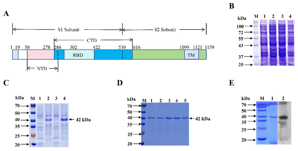
Figure 2.
Generation of S1-CTD antisera in mice. (A, B) Indirect ELISA of mouse serum antibody titers after three rounds of immunization (A) and a fourth booster (B). (C) Western blot using mouse #1 serum (left) or anti-GFP antibody (right) to detect GFP-S1-CTD fusion protein. Lanes 1,3: 293T cells expressing GFP-C2 empty vector; Lanes 2,4: 293T cells expressing GFP-PDCoV-S1-CTD. (D) IFA using mouse #1 serum to stain 293T cells transfected with GFP-PDCoV-S1-CTD (top row) or empty vector (bottom row). Scale bar: 50 μm.
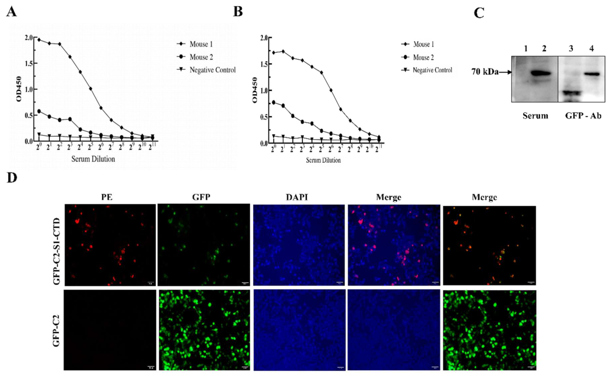
Figure 3.
Assessment of PDCoV S binding specificity of monoclonal antibodies. (A) Western blot using hybridoma (2B7-G10, 7C7-F6, 7G3-G8, 4H4) supernatants to detect denatured PDCoV S protein in infected or mock-infected LLC-PK1 cell lysates. (B) IFA using hybridoma (2B7-G10, 7C7-F6, 7G3-G8, 4H4) or negative control supernatants to detect native PDCoV S protein in infected or negative LLC-PK1 cells. Scale bar: 50 μm.
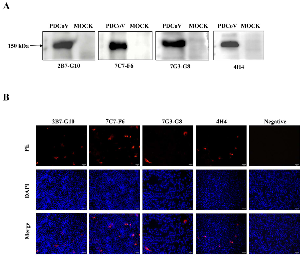
Figure 4.
Amplification of variable regions and prokaryotic expression and purification of 4-H4-scFv. (A) Agarose gel electrophoresis of DNA from PCR-amplified VH and VL genes from 4-H4 hybridomas. M: DNA ladder; Lanes 1–2: 4H4-VH; Lanes 3–4: 4H4-VL. (B) Agarose gel electrophoresis of DNA from overlap extension PCR to assemble VH-linker-VL fragment. M: DNA ladder; Lane 1: assembled product. (C) SDS-PAGE of 4H4-scFv expression and purification. M: protein ladder; Lane 1: uninduced whole cells; Lane 2: induced whole cells; Lane 3: supernatant fraction from induced whole cells; Lane 4: pellet fraction from induced whole cells; Lane 5: Ni-NTA flow-through; Lane 6: wash; Lane 7: purified protein fraction. (D) SDS-PAGE to show dialyzed 4H4-scFv. M: protein ladder; Lane 1: dialyzed protein. (E) Western blot using anti-His antibody to detect 4H4-scFv. M: protein marker; Lane 1: purified scFv.
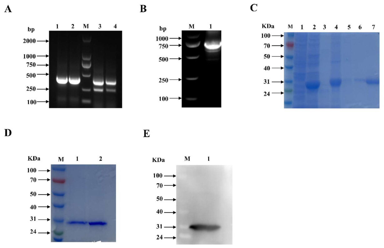
Figure 5.
Assessment of PDCoV S binding specificity of 4H4-scFv. (A) Double-antibody sandwich ELISA with 4H4-scFv as the capture antibody and S1-CTD as the target antigen, with bound S1-CTD detected using anti-S1-CTD ascites from clone 7G3-G8. (B) IFA using 4H4-scFv to detect native PDCoV S protein in negative control or PDCoV-infected LLC-PK1 cells. Scale bar: 50 μm.
Disclaimer/Publisher’s Note: The statements, opinions and data contained in all publications are solely those of the individual author(s) and contributor(s) and not of MDPI and/or the editor(s). MDPI and/or the editor(s) disclaim responsibility for any injury to people or property resulting from any ideas, methods, instructions or products referred to in the content. |
© 2026 by the authors. Licensee MDPI, Basel, Switzerland. This article is an open access article distributed under the terms and conditions of the Creative Commons Attribution (CC BY) license.
Copyright: This open access article is published under a Creative Commons CC BY 4.0 license, which permit the free download, distribution, and reuse, provided that the author and preprint are cited in any reuse.