

Submitted:
22 September 2026
Posted:
23 September 2026
You are already at the latest version
Abstract
The 2021 WHO Classification of Tumours of the Central Nervous System introduced three molecularly defined mesenchymal entities: intracranial mesenchymal tumour, FET::CREB fusion-positive; CIC-rearranged sarcoma; and primary intracranial sarcoma, DICER1-mutant. Each is genetically defined yet morphologically non-specific, and each mimics a commoner neoplasm: meningioma, Ewing or embryonal tumour, and high-grade glioma respectively. Existing accounts describe these tumours descriptively and in parallel. This review instead reads all three through a single mechanistic lens, tracing each defining lesion to its diagnostic phenotype. We compare the available diagnostic modalities and propose a practical, tiered testing algorithm. Two findings stand out. DNA methylation profiling is the diagnostic backbone for CIC-rearranged and DICER1-mutant sarcoma but has no dedicated class for FET::CREB fusion-positive intracranial mesenchymal tumour, which it actively misassigns to meningioma, making RNA-based fusion testing indispensable. Alone among the three, DICER1-mutant sarcoma may arise on a germline background, so its diagnosis triggers counselling and surveillance for the patient's family. Across all three, molecular characterisation has largely resolved diagnostic ambiguity without yet delivering therapy: mechanistically rational targets remain preclinical, and management rests on resection and borrowed adjuvant regimens. Closing that diagnostic-to-therapeutic gap will require registries, standardised outcome reporting and molecularly stratified trials.
Keywords:
CNS sarcoma
; intracranial mesenchymal tumour
; CIC-rearranged sarcoma
; DICER1
; FET::CREB fusion
; DNA methylation profiling
; molecular diagnostics
; precision oncology
; WHO CNS5
1. Introduction
Perhaps better known as soft-tissue-type or sarcomatous tumours (sarcomas) of the Central Nervous System (CNS), mesenchymal, non-meningothelial neoplasms arising in the CNS are distinct from meningiomas (which are meningothelial in origin) and from the far commoner gliomas and neuronal tumours. Historically they were diagnosed by morphology and lineage-marker immunohistochemistry, but they have long posed a diagnostic problem: their morphology is often non-specific, and they overlap both with commoner CNS tumours and with their soft-tissue counterparts. Few have possessed defining markers, leaving classification unstable and reproducibility poor. The advance of molecular tools, fusion detection, targeted sequencing and, in particular, genome-wide DNA methylation profiling, has reshaped this landscape, changing the histological diagnosis in a substantial minority of CNS tumours and enabling defined entities to be carved out of previously ambiguous groupings [1].
Building on this shift, the 2021 WHO classification of CNS tumours (CNS5) formalised a category of "mesenchymal, non-meningothelial tumours of uncertain differentiation" [2]. Within it sit three molecularly defined entities: intracranial mesenchymal tumour, FET::CREB fusion-positive (ICMT) [3,4]; CIC-rearranged sarcoma [5,6]; and primary intracranial sarcoma, DICER1-mutant [7]. Linking the three is the clinical problem they share: each is genetically defined yet morphologically non-specific, mimicking a commoner tumour, meningioma, Ewing/embryonal tumour, and high-grade glioma, respectively. This review is confined to these three molecularly defined entities; other mesenchymal or dural-based CNS sarcomas of distinct lineage, and pathologically related tumours such as Ewing sarcoma, are considered only where they arise as differential diagnoses.
The existing literature has described these entities, individually or together [8,9,10] but tends to treat them descriptively; cataloguing clinical, histological and molecular features in parallel. To our knowledge, no review has compared the three through a unified mechanistic lens, tracing how each defining lesion produces its diagnostic phenotype and drawing out the logic they share. All three are rare, and the published evidence largely consists of small retrospective series and individual case reports. No standardised treatment exists for any of them, and much of the underlying biology is extrapolated from their soft-tissue counterparts.
As such, the aim of this review is to address that gap; adopting a mechanism-led framework, it traces each entity from its defining molecular lesion to its diagnostic phenotype. It then sets out a comparative analysis of the available diagnostic modalities and proposes a practical testing algorithm, before drawing out the themes that unite the three entities. It closes by weighing what molecular definition currently delivers in practice – accurate classification, prognostic interpretation and identification of hereditary risk – against what it does not yet deliver.
2. Materials and Methods
This paper is a critical narrative review rather than a systematic review, and no formal protocol was employed. Literature was identified through a structured search of PubMed covering the period January 2000 to July 2026, using combinations of the terms "intracranial mesenchymal tumour," "FET::CREB," "EWSR1," "CIC-rearranged sarcoma," "CIC::DUX4", "CIC::NUTM1", "ATXN1", "primary intracranial sarcoma", "DICER1", "DNA methylation profiling", and "RNA sequencing". This was supplemented by hand-searching the reference lists of included articles and by citation tracking of the key primary series. Given the rarity of these entities, inclusion was deliberately broad: original molecular characterisation studies, cohort studies, clinically informative case reports and series, and major reviews were all considered. Reports lacking molecular confirmation were excluded unless of historical significance to the classification of these tumours.
3. Classification & Nomenclature
Prior to molecular characterisation, these tumours occupied some of the least stable categories in neuropathology, appearing variously as "primary intracranial sarcoma," "intracranial myxoid mesenchymal tumour," "intracranial angiomatoid fibrous histiocytoma," and CNS "PNET." Diagnosis relied on morphology together with lineage-marker immunohistochemistry; reproducibility was limited, and the same tumour could be classified under different names at different institutions [2,8,9]. The consequence was not merely semantic: cases scattered across incompatible labels could not be pooled, so the natural history of each entity remained obscure.
The 2021 WHO classification used molecular signatures to carve defined entities out of this space, grouping them under "mesenchymal, non-meningothelial tumours of uncertain differentiation" (Figure 1). Three sit within it: intracranial mesenchymal tumour, FET::CREB fusion-positive; CIC-rearranged sarcoma; and primary intracranial sarcoma, DICER1-mutant. Notably, ICMT entered the classification as a provisional type rather than a fully established entity, reflecting the fact that its defining criteria remain unsettled [2,3,7]. The wider mesenchymal, non-meningothelial family also houses more established entities such as solitary fibrous tumour and chondrosarcoma; the uncertain-differentiation subgroup that this review concerns contains, alongside these three, Ewing sarcoma, considered here only as a differential diagnosis. What unites the three is that each was carved out of previously ambiguous territory by a single recurrent molecular lesion rather than by appearance alone, which is what made stable classification possible for the first time.
Both ICMT and CIC-rearranged sarcoma already carry proposed renames, and for the same underlying reason: each name was fixed before the biology behind it was fully understood. For ICMT, the proposal is "CNS mesenchymal tumour, FET::CREB fusion-positive," since spinal cases have since been described and "intracranial" is unduly restrictive [4]. For CIC-rearranged sarcoma, the proposal is "CNS sarcoma, CIC/ATXN1 complex-fused," since the non-CIC ATXN1 fusions share the same biology and methylation class yet are excluded by the current name [6,11]. This instability is not a cosmetic problem, because a tumour’s name directs the workup it receives. A label that says “intracranial” invites a spinal example of the same entity to be overlooked, and a label that says “CIC-rearranged” invites a laboratory to test for a CIC fusion and stop there, missing the ATXN1-fused tumours that are biologically identical. In each case the name can narrow the differential prematurely and send testing down the wrong path.
Despite the advances that molecular characterisation has brought, then, the nomenclature of these entities still lags the mechanisms that define them; and that lag is itself an argument for the molecularly integrated diagnosis that CNS5 champions.
4. Molecular Diagnostic Framework
4.1. Histology and Immunohistochemistry: Role and Limitations
Immunohistochemistry is the first molecular-adjacent step in evaluating these tumours, and for each entity a small panel of markers either support the diagnosis, or aids in excluding its principal mimic. No single marker, however, is specific enough to stand alone: the same panels that point toward a diagnosis may also generate characteristic misreads. Table 1 summarizes the supportive and discriminatory markers for each entity alongside the reasons immunohistochemistry alone cannot be solely relied upon.
Morphology remains central to tumour diagnosis, but across all three of these entities it fails on its own. Their appearances, undifferentiated round cell, spindle-to-pleomorphic, and variably mesenchymal, are non-specific, and each overlaps with a commoner neoplasm. Immunohistochemistry (IHC) is therefore the natural next step, and it does useful work: it narrows the differential and points toward a lineage. Its markers fall into three groups. Supportive markers, which establish the mesenchymal phenotype; desmin, EMA, CD99, CD68 and MUC4 in ICMT; desmin and myogenin in DICER1-mutant sarcoma. Discriminatory markers are the genuinely informative ones: nuclear ETV4 in CIC-rearranged sarcoma, the combination of H3K27me3 loss with nuclear TLE1 in DICER1-mutant sarcoma, and a patchy CD99 pattern together with NKX2.2 negativity to separate CIC-rearranged sarcoma from Ewing sarcoma. Finally, partner-specific markers can resolve the underlying fusion: NUT in CIC::NUTM1 cases, and nuclear DUX4 in DUX4-fused cases.
Yet immunohistochemistry has three shortcomings that prevent it from closing the diagnosis. First, in ICMT it is actively misleading: the synaptophysin, SSTR2A and GFAP profile pushes the observer toward meningioma, the very tumour ICMT is most often mistaken for. Second, it is partner-dependent — WT1 is positive in only two of six CIC::NUTM1 cases [13], and NUTM1 fusions are precisely those enriched intracranially, so the marker fails where it is most needed. Third, and most tellingly, the series do not agree with one another: Tauziède-Espariat et al. (2022) [12] report synaptophysin and GFAP as constantly negative across eleven ICMT cases, whereas Rajan et al. (2026) [4] find synaptophysin positive in over half. Immunohistochemistry can therefore triage; it cannot decide.
4.2. Molecular Methods: Comparative Strengths and Limitations
Immunohistochemistry can only narrow the differential; molecular testing, however, is what establishes the diagnosis. That being said, it should be noted that no single assay resolves all three entities, and each modality carries a characteristic blind spot. Table 2 compares the principal molecular methods, from targeted fusion and mutation testing through to genome-wide DNA methylation profiling, setting each entity’s diagnostic contribution against its documented limitations in these tumours.
If immunohistochemistry can only triage, molecular testing must decide; and among the available modalities, genome-wide DNA methylation profiling has the strongest claim to being the backbone. It defines CIC-rearranged sarcoma, splitting SARC-CIC from HGNET-CIC, and DICER1-mutant sarcoma as a distinct class, and Capper et al. [1] showed that it altered the histological diagnosis in approximately 12% of prospectively examined CNS tumours.
Its value is most visible where conventional testing fails. Satomi et al. [19] reported an ATXN1::DUX4 tumour that was negative on both break-apart FISH and targeted panel sequencing, yet matched the CIC-rearranged sarcoma methylation class. Jiang et al. (2025) [20] described a CIC::DUX4 tumour missed by targeted NGS and resolved the same way. The pattern extends beyond CIC: a 52-year-old man whose tumour was diagnosed histologically as high-grade glioma, not otherwise specified, was reclassified as DICER1-mutant sarcoma by methylome profiling, with sequencing subsequently confirming a DICER1 mutation [16]. In each case, methylation profiling rescued a diagnosis that other modalities had missed.
ICMT, however, breaks this pattern — and does so in a way that matters. The classifier does not merely fail to recognize ICMT; it actively misassigns these tumours to meningioma, the very entity they are most often confused with [3,12], and in one pooled series UMAP analysis scattered ICMT cases directly into meningioma methylation classes [18]. A methylation result is therefore not simply unhelpful in ICMT; it is actively misleading, and RNA-based fusion testing is indispensable.
No single modality is therefore sufficient. FISH is rapid and widely available but carries a notable false-negative rate and cannot identify the fusion partner — a real limitation here, since the partner determines both the applicable immunostain (NUT or nuclear DUX4) and the SARC-CIC/HGNET-CIC distinction. Targeted DNA sequencing reliably detects DICER1 hotspot mutations and co-alterations but can miss fusions entirely. RNA-based fusion detection identifies the partner and is the only modality that resolves ICMT. Copy-number profiling is supportive rather than diagnostic. Germline testing, finally, is the one investigation whose result changes management beyond the tumour itself.
4.3. Practical Testing Algorithm
Drawing on the strengths and limitations described above, the following pragmatic diagnostic sequence, summarized in Figure 2, is proposed. It is offered as a synthesis of the available evidence rather than a validated pathway; no prospectively tested algorithm currently exists for these entities. The principle is that testing should be tiered rather than exhaustive, sequenced according to what each modality can and cannot resolve.
Clinical and radiological features narrow the field first. A dural-based, meningioma-like extra-axial mass should raise ICMT [3,4], while a cortical or subcortical lesion containing intratumoral blood products on susceptibility-weighted and T1-weighted imaging should raise DICER1-mutant sarcoma — blood products were present in all eight patients in the multicentre imaging series of Eldaya et al. (2024) [23], and a solid-cystic supratentorial mass with haemorrhage is the characteristic appearance [24] .
Histology and a targeted immunohistochemical panel narrow it further, guided by the morphological pattern. An undifferentiated round-cell tumour prompts ETV4, CD99 and NKX2.2 [6,13]; a spindle-to-pleomorphic tumour with eosinophilic globules prompts H3K27me3, TLE1 and myogenic markers [14,15] and a meningioma-like mesenchymal tumour prompts desmin, EMA, CD99, CD68 and, where available, CREM [3,4,12,25].
RNA-based fusion testing follows, and it is indispensable rather than optional — most of all in ICMT, where for the reasons set out in Section 4.2 it is the only modality capable of establishing the diagnosis [3,4,12,18]. Fusion testing also identifies the partner in CIC/ATXN1-fused tumours, which determines both the applicable immunostain and the SARC-CIC/HGNET-CIC distinction [6,11,17]. Targeted DNA sequencing with copy-number profiling is performed in parallel, capturing DICER1 hotspot mutations and their frequent TP53, ATRX and MAPK-pathway co-alterations [7], together with the recurrent 10p, 19 and 22q losses described in ICMT [4].
DNA methylation profiling is then reserved for cases that remain unresolved or discordant, where it functions as the rescue step [1] — as demonstrated in tumours negative on FISH and panel sequencing [19], on targeted NGS [20], and in a sarcoma initially diagnosed as high-grade glioma [16]. Where a DICER1 alteration is identified, germline testing and genetic counselling must follow [21,22].
5. Intracranial Mesenchymal Tumour, FET::CREB Fusion-Positive (ICMT)
5.1. Molecular Lesion and Mechanism
ICMT is defined by the fusion of a FET-family gene; usually EWSR1, rarely FUS, to a CREB-family transcription factor: ATF1, CREB1 or CREM [3], with rare cases involving CREB3L3 [18]. Mechanistically, the FET partner contributes a strong N-terminal transactivation domain (low-complexity, prion-like), while the CREB-family partners are bZIP transcription factors normally gated by cAMP/PKA phosphorylation that bind cAMP-response elements (CRE). The fusion couples the constitutively active FET domain to CREB DNA-binding, driving ligand- and cAMP-independent transactivation of CRE-regulated targets, a mechanism so far demonstrated in the related angiomatoid fibrous histiocytoma and clear-cell sarcoma models rather than intracranially [26,27]. In the largest series (81 cases), 65 presented with a documented fusion, where EWSR1::ATF1 (n = 25) and EWSR1::CREM (n = 21) were the commonest partners. EWSR1::CREB1 (n = 12) was roughly half as frequent, and FUS::CREM (n = 3) and SMARCA2::CREM (n = 4) rare [4]. CREM is overexpressed regardless of partner, marking it as the convergent node of the entity and the basis for a proposed CREM immunohistochemical surrogate [4]. Notably, the same FET::CREB fusions recur in extracranial angiomatoid fibrous histiocytoma, clear-cell sarcoma, hyalinizing clear-cell carcinoma and primary pulmonary myxoid sarcoma, so the fusion is not entity-specific; DNA methylation is what distinguishes them [12]. The molecular spectrum also extends beyond the FET genes: SMARCA2::CREM and SMARCA4::CREM fusions implicate the CREM partner together with SWI/SNF chromatin remodelling rather than the FET moiety alone [28,29].
5.2. Epigenetic Subgroups and Methylation Profiling
The suggestion that ICMT comprises more than one epigenetic subgroup was first raised in a small integrative series, in which FET::CREB tumours emerged as a family rather than a single entity, with a subset forming a distinct methylation cluster, a finding independently supported in a parallel cohort [3,12,25]. The largest study to date formally resolved this into two subclasses: subclass A (n = 69, 85%; median age 26) and subclass B (n = 12, 15%; median age 15), the latter female-predominant, enriched for CREM-partner fusions, containing all GFAP-positive cases, and, importantly, associated with significantly shorter progression-free survival (P < 0.01) [4]. However, no consistent histological difference separates the subclasses, so the distinction is presently epigenetic and prognostic rather than morphological. Whether subclass assignment carries independent prognostic value beyond resection status remains to be established in prospective cohorts.
5.3. Histologic Spectrum and Diagnostic Mimicry
ICMT arises predominantly in an extra-axial, meningeal or intraventricular location, frequently with dural attachment or a dural tail [3,4] (see Section 4). It is this dural-based growth, combined with a variable and often bland morphology, ranging from syncytial sheets to reticular, cord-like arrangements in a myxoid or collagenous stroma, which sometimes presents with angiomatoid fibrous histiocytoma-like pseudo angiomatoid spaces and lymphoid cuffing [3,30], which produces its principal diagnostic trap: mistaking ICMT for meningioma. Rather than resolving this overlap, the immunophenotype compounds it: the EMA, synaptophysin and SSTR2A positivity found in ICMT is shared with meningioma (full marker profile in Table 1). The proliferation index is typically low [30]. Thus, definitive distinction rests on molecular testing, as detailed in Section 4.
5.4. Clinical Behaviour, Prognosis and Therapy
ICMT affects children and young adults; across 74 pooled cases the median age was 23 years (range 4–79), with a slight female predominance (F:M ≈ 1.55) and supratentorial location in roughly 80%. However, it should be noted that around 18.5% of patients are over 40 [4,18] . The tumours are generally indolent but recurrence-prone, with recurrence occurring in approximately 40% of cases at a median of about ten months [18]. Reported prognostic determinants remain few and derive from small retrospective series, so they should be read with caution. The clearest is extent of resection — subtotal resection carries significantly shorter progression-free and overall survival than gross-total resection (P = 0.0003) — making maximal safe resection the first-line treatment [3,18]. The role and benefit of adjuvant radiotherapy or chemotherapy have not yet been established; however, emerging systemic options are considered in Section 9.
5.5. Key unresolved Questions
Despite formal recognition in WHO CNS5, and for all that molecular profiling has clarified, ICMT remains a provisionally defined entity with more unanswered questions than settled ones. Its relationship with extracranial AFH, a variant of the same tumour or distinct entity, remains unresolved, and the cell of origin is undetermined, though the arachnoidal features of the dominant methylation cluster offer a hint. The presumption that these are indolent, surgically curable lesions is also being tested: extracranial metastases have now been documented [31], and a metastatic, PD-L1-high tumour achieved durable remission on immune-checkpoint inhibition [32], hinting at behaviour and treatment options beyond local control. Prognostic stratification, too, rests on small numbers: the subclass A/B split, though promising, derives from a single 81-case cohort, so behaviour and optimal management remain open [4]. Finally, whether the spinal cases that motivate the proposed rename behave differently from intracranial ones is unknown.
6. CIC-Rearranged Sarcoma of the CNS
6.1. Molecular Lesion and Mechanism
CIC (Capicua), the human homolog of Drosophila capicua, is an HMG-box transcriptional repressor and a downstream effector of the RTK-RAS-MAPK pathway that normally represses the PEA3 subfamily of ETS transcription factors: ETV1, ETV4 and ETV5[5] . The prototypic rearrangement, CIC::DUX4, arising from t(4;19)(q35;q13), retains the CIC HMG DNA-binding domain but replaces its C-terminus with a fragment of DUX4 that confers neomorphic transcriptional activity, sufficient to transform NIH 3T3 fibroblasts and effectively converting CIC from a repressor into an aberrant activator [5]. In the defining study, CIC::DUX4 bound the ERM (ETV5) promoter and upregulated ETV5 and ETV1; ETV4 induction was not itself demonstrated, although nuclear ETV4 is now the diagnostic readout of the pathway (see Section 4). Subsequent work has extended the mechanism: the DUX4 C-terminus, like the alternative partners NUTM1 and LEUTX, recruits the p300/CBP co-activator with H3K27ac deposition, and the downstream programme includes CCNE1 and the anti-apoptotic MCL1, the latter now identified as a therapeutic dependency in patient-derived models [33,34]. These tumours belong to the family of undifferentiated round-cell, Ewing-like sarcomas first characterised in the soft tissues, but the intracranial cases are not simply its copies. The CNS fusion spectrum is broader than in soft tissue, where CIC::DUX4 accounts for roughly 95% of cases: intracranial tumours are enriched for CIC::NUTM1 and CIC::LEUTX, and for non-CIC fusions of ATXN1 and ATXN1L, including ATXN1::DUX4 and ATXN1/ATXN1L::NUTM2A [6,11,19,35]. Since ATXN1 and ATXN1L normally form a repressor complex with CIC, the disruption of that complex produces the same PEA3/ETV de-repression, which is the biological argument for the proposed “CNS sarcoma, CIC/ATXN1 complex-fused” rename discussed in Section 3 [6,11].
6.2. CNS-Specific Molecular Features and Methylation Profiling
Beyond the shift in fusion partners, little is firmly established that separates CNS CIC/ATXN1-rearranged sarcoma biologically from its soft-tissue counterpart, and whether the intracranial location reflects a distinct cell of origin or simply anatomical context is unknown. DNA methylation profiling separates CNS CIC/ATXN1-rearranged tumours into two classes named in Table 2: an aggressive SARC-CIC group, shared with soft-tissue CIC-rearranged sarcoma, and an intermediate-grade HGNET-CIC group [6,17]. Sievers et al. [17] defined HGNET-CIC as a distinct cluster with no methylation similarity to soft-tissue SARC-CIC, driven predominantly by a recurrent CIC::LEUTX fusion and showing neuroepithelial or glial features in children [17]. Whether these represent one tumour spanning a morphological spectrum or two separate entities is unsettled, since the same CIC::LEUTX fusion appears in both groups and blurs the boundary [6]. At present the distinction is more taxonomic than therapeutic, because neither class yet carries a validated, class-specific management implication.
6.3. Histologic Spectrum, Diagnostic Mimicry and Biomarkers
Histologically these are undifferentiated round-cell, “Ewing-like” neoplasms: sheets of round-to-ovoid cells with focal spindling or an epithelioid appearance in a myxoid matrix, typically high-grade, with frequent necrosis and a high proliferation index (Ki-67 median about 30%) [6]. The immunophenotype both supports the diagnosis and excludes the principal mimic but does neither on its own (full profile in Table 1). Nuclear ETV4, the readout of PEA3/ETV de-repression, is constant and is the most accessible diagnostic surrogate, although it marks the pathway rather than the fusion and cannot identify the partner. WT1 is unreliable in the CNS, positive in only two of six CIC::NUTM1 cases, and NUTM1 fusions are exactly those enriched intracranially, so the marker is weakest where it is most needed [6,13]. Separation from Ewing sarcoma rests on two markers read together: CD99 is only patchy or focally membranous rather than diffusely membranous, and NKX2.2 is negative [6]. Partner-specific stains then resolve the fusion, NUT in NUTM1-fused and nuclear DUX4 in DUX4-fused cases, with INI1/BRG1 retained; the diagnosis is ultimately confirmed molecularly (Section 4) [13,19].
6.4. Clinical Behaviour, Prognosis and Therapy
Within a CNS-specific cohort, CIC/ATXN1-rearranged sarcoma presents at a median age of about 9.5 years (range 0 to 40), roughly two-thirds in children, with a female predominance and a supratentorial location in most cases [6]. The evidence supports an aggressive course, though it rests on small numbers: the reported median overall survival in the CNS cohort is approximately 17.5 months, with a high rate of local recurrence [6], and recurrent intracranial disease is documented in individual reports [20]. The same 17.5-month median overall survival was reported independently in Le Loarer’s mixed-site CIC::NUTM1 series (range 7-37 months), in which five of six patients died of disease [13]. Documented CNS behaviour is dominated by local recurrence; the frequent metastatic spread of the extracranial prototype is not well characterised intracranially. No standard regimen exists; management combines maximal safe resection with chemoradiation, and these tumours are relatively chemo-resistant compared with Ewing sarcoma[36]. The candidate targets that follow from the fusion, principally MCL1 and p300/CBP, are considered alongside the other entities in Section 9 [33,34].
6.5. Key Unresolved Questions
Despite a firm genetic definition, the entity’s boundaries remain disputed. The SARC-CIC versus HGNET-CIC split raises the unresolved question of whether these are one tumour across a morphological spectrum or two distinct entities, a question the shared CIC::LEUTX fusion keeps open [6,17]. The nomenclature also lags the biology: “CIC-rearranged” excludes the non-CIC ATXN1 fusions that share its mechanism and methylation class, which is the basis for the proposed rename [6,11]. Above all, the intracranial evidence base is thin, a single 15-case series and scattered reports, so outcome data, optimal treatment and even the fusion-partner distribution still lean on soft-tissue literature now known to diverge from the CNS [6,13,35].
7. Primary Intracranial Sarcoma, DICER1-Mutant
7.1. Molecular Lesion and Mechanism
DICER1, on chromosome 14q32, encodes an RNase III endoribonuclease that processes precursor microRNA into its mature form; more specifically, the RNase IIIa domain cleaves the 3p arm of the hairpin and the RNase IIIb domain the 5p arm [37]. These tumours arise through a two-hit, biallelic mechanism, which is the crux of why germline testing matters here. Simplistically put, a loss-of-function (truncating) allele is combined in trans with a missense hotspot mutation in the RNase IIIb domain (E1705K, D1709, D1810 or E1813), or with loss of heterozygosity [7]. In the canonical model, the RNase IIIb hotspot selectively cripples the maturation of the 5p miRNA arm whilst sparing the 3p arm, depleting tumour-suppressor 5p miRNAs such as let-7 and miR-30 and thus de-repressing oncogenic and de-differentiation programmes. This account should be hedged, however: Vedanayagam et al. [38] found that RNase IIIa and IIIb mutations exert broadly similar effects on miRNA biogenesis, so a strict 5p-only model likely oversimplifies the biology.
7.2. Co-Alterations and DICER1 Tumour Predisposition Syndrome
Several recurrent co-alterations amplify this phenotype. Koelsche et al. [7] found TP53 mutations in 55% of cases (12 of 22) and MAPK-pathway alterations in 77% (17 of 22); most often involving KRAS, NF1, FGFR4, NRAS or EGFR amplification, alongside frequent ATRX loss. Whether any of these carries independent prognostic weight has not yet been established. The tumour occurs both sporadically and in the germline setting of DICER1 syndrome, and the balance varies by cohort: germline variants were found in a minority of young adults (two of eight in one series), whereas a large Peruvian paediatric cohort was entirely somatic [21,24]. A reported adult case makes the sporadic pattern more concrete. In a 52-year-old male, a DICER1 exon 25 variant (Glu1813Ala) was confirmed in tumour tissue but absent from blood, excluding inherited DICER1 syndrome [16]. A CNS sarcoma diagnosis nonetheless warrants a syndrome-level workup, because DICER1 syndrome predisposes to a family of embryonal, mesenchymal neoplasms, among them pleuropulmonary blastoma, cystic nephroma, ovarian Sertoli-Leydig cell tumour and thyroid disease [22], and the rhabdomyoblastic differentiation these sarcomas frequently display reflects that shared lineage [14].
7.3. Histologic Heterogeneity, Diagnostic Mimicry and Methylation
Morphologically these are high-grade spindle-to-pleomorphic sarcomas, characteristically containing eosinophilic cytoplasmic globules, with frequent rhabdomyoblastic or myogenic differentiation and occasional immature cartilage, which gives a pleuropulmonary-blastoma-like organoid appearance [14,39]. This heterogeneity is what drives misdiagnosis: the spindled, undifferentiated areas mimic high-grade glioma or gliosarcoma, while the myogenic foci suggest rhabdomyosarcoma. Immunohistochemistry resolves this less cleanly than might be expected. The tumour cells are GFAP-negative, but negativity does not exclude a glioma, since GFAP-negative glioblastomas occur and gliosarcoma with diffuse mesenchymal metaplasia may retain no residual glial component [16]. The immunophenotype narrows but does not settle the differential (full profile in Table 1): patchy desmin and myogenin mark the myogenic foci, and the two most useful surrogates are H3K27me3 loss, though its extent varies - diffuse in only four of six cases in one series, and mosaic or minimal in the remainder [15] - and nuclear TLE1. Because morphology and immunohistochemistry are individually insufficient, the diagnosis is confirmed molecularly, by DNA methylation profiling together with DICER1 sequencing (Section 4.2); however, unlike ICMT, this entity has a distinct methylation class that reliably defines it [15,39].
7.4. Clinical Behaviour and Prognosis
This is primarily a paediatric tumour (median age ~ 6 years), although adult cases have been reported (median ~20), and it is characteristically supratentorial [7,21,24]. Similar to CIC-rearranged sarcoma, this tumour behaves aggressively; although outcomes can vary. In the Peruvian paediatric cohort two-year overall survival was 66.3% and two-year progression-free survival 51%, with the best outcomes reported in the subgroup who was treated with postoperative ICE chemotherapy, followed by radiotherapy and then further ICE. This group reached a two-year event-free survival of 79% [24]. Young adults had a median overall survival of 30.8 months and a time to progression of 14.5 months [21]. The entity also has a striking epidemiology: its incidence in Peru (0.19 per 100,000 children), where it is the second commonest paediatric high-grade CNS malignancy, far exceeds that in Germany (0.007 per 100,000; P < 0.001) [24].
7.5. Germline Testing, Counselling and Surveillance
Uniquely among the three entities, a molecular finding here carries management consequences beyond the tumour itself, which makes this the point of most direct clinical action. Identification of a germline DICER1 variant should trigger genetic counselling for both the patient and family, and enrollment in surveillance for the other DICER1-syndrome neoplasms [21]. This procedure should follow guidelines set out by the literature; namely that by Schultz et al. [22] who describe which individual counts as at-risk, and the recommended screening: chest imaging in early childhood for pleuropulmonary blastoma, abdominal and pelvic ultrasound for renal and ovarian tumours, and thyroid assessment. Since the germline result changes management for relatives as much as for the patient, germline testing is not optional once a DICER1 alteration is identified.
7.6. Current Therapy and Emerging Approaches
Standard treatment focuses on maximal safe resection, ICE chemotherapy and focal radiotherapy, with one caveat to carry into any outcome comparison: ICE denotes ifosfamide, carboplatin and etoposide in the paediatric series but ifosfamide, cisplatin and etoposide in the adult series, so the platinum agent should be specified [21,24]. A biologically rational lead has emerged but remains preliminary: in the first patient-derived xenograft model of the entity, high-throughput drug screening found that activation of retinoic acid receptor (RAR) signalling reduced tumour-cell viability, identifying RAR as a candidate target rather than an established treatment [40]. This lead is discussed further in Section 9.
7.7. Key Unresolved Questions
Much about this entity remains open. It is unclear whether the recurrent co-alterations carry independent prognostic value, what drives the marked geographic difference in incidence between Peru and Germany, and how treatment should be standardised across the paediatric-adult divide.
8. Comparative Molecular Pathogenesis and Cross-Entity Analysis
The three entities are driven by different molecular lesions, yet converge on a single clinical problem: each is a genetically defined tumour whose morphology is non-specific and mimics a commoner CNS neoplasm; meningioma in the case of ICMT, Ewing or embryonal tumour for CIC-rearranged sarcoma, and high-grade glioma or gliosarcoma for DICER1-mutant sarcoma. Comparing them along four axes, namely: mechanism, diagnosis, clinical behaviour and heredity, shows both why molecular testing has become indispensable to their diagnosis, and where it still falls short.
8.1. Convergent Mechanisms and Diagnostic Implications
Their driver lesions fall into two mechanistic classes. ICMT and CIC-rearranged sarcoma are fusion-driven, each producing a chimeric transcription factor: a FET::CREB activator in ICMT, and in CIC-rearranged sarcoma a fusion that converts CIC from a repressor into a neomorphic activator of the PEA3/ETS programme. DICER1-mutant sarcoma is different, defined not by a fusion but by a biallelic loss of function combined with an RNase IIIb hotspot mutation that cripples microRNA processing. The routes nonetheless converge on one theme: each is a failure of transcriptional or epigenetic regulation, whether a gain of aberrant activation, a loss of repression, or a loss of post-transcriptional control, and all three end at a dysregulated programme that drives an undifferentiated mesenchymal phenotype. It is this shared endpoint that produces the morphological non-specificity underlying the diagnostic mimicry, so the convergence that unites the three biologically is also what makes them so indistinguishable.
Molecular testing is decisive for all three, with DNA methylation profiling as the common backbone; in prospective use it has changed the histological diagnosis in roughly one in eight CNS tumours [1], a measure of how far morphology alone can mislead. The modality that actually secures the diagnosis, however, differs, and one difference is clinically critical. Because methylation has no category for ICMT, the decisive assay differs by entity: RNA fusion detection for ICMT and for partner identification in CIC-rearranged sarcoma, targeted DNA sequencing with methylation for DICER1-mutant sarcoma (Section 4). The diagnostic algorithm is thus not uniform across all three entities. Where methylation is unavailable, even the accessible immunohistochemical surrogates differ: CREM for ICMT, nuclear ETV4 for CIC-rearranged sarcoma, and combined H3K27me3 loss with nuclear TLE1 for DICER1-mutant sarcoma.
8.2. Clinical Divergence and the Diagnostic-to-Therapeutic Gap
Clinically the three span a range. ICMT is generally indolent but recurrence-prone, recurring in roughly 40% of cases, with extent of resection the main determinant of outcome [18], whereas CIC-rearranged and DICER1-mutant sarcomas are both aggressive, with short survival in the small series so far reported (Section 5, Section 6 and Section 7). Heredity is the sharpest point of divergence: alone among the three, DICER1-mutant sarcoma can arise on a germline background, so its diagnosis by itself triggers genetic counselling and syndrome surveillance for the patient and family, while ICMT and CIC-rearranged sarcoma are somatic events confined to the tumour. These comparisons are summarised in Table 3.
Across all four axes one conclusion recurs. Each lesion points to a mechanistically rational targets: immune checkpoint inhibition in ICMT, MCL1 and p300/CBP in CIC-rearranged sarcoma, and retinoic acid receptor or RAS-pathway signalling in DICER1-mutant sarcoma. All three remain preclinical, and treatment remains surgery-centred with conventional adjuvant therapy. Encouragingly, the first functional models of these tumours, a patient-derived xenograft for DICER1-mutant sarcoma [40] and tumoroids for CIC-rearranged sarcoma [34], are beginning to appear. Closing that diagnostic-to-therapeutic gap is the task the following sections take up.
9. Precision Oncology and Therapeutic Opportunities
9.1. Established Clinical Practice
Treatment currently rests on maximal safe surgical resection, the one intervention with a consistent effect on outcome; in ICMT, gross-total resection confers significantly longer progression-free and overall survival than subtotal resection (Section 5), and extent of resection is treated as the principal modifiable prognostic factor for the others as well. Radiotherapy is delivered pragmatically and individualised to tumour site and patient age rather than by any entity-specific protocol, and systemic therapy is borrowed rather than validated: ICE-type regimens in DICER1-mutant sarcoma and Ewing-type regimens in CIC-rearranged sarcoma, the latter with generally inferior responses (Section 6 and Section 7). Two further steps are now integral to good practice. Molecular confirmation, by DNA methylation profiling together with fusion or targeted sequencing, is required to reach the diagnosis at all (Section 4); and in DICER1-mutant sarcoma, germline assessment with genetic counselling and syndrome surveillance follows any DICER1 alteration[22]. Since the molecular result increasingly shapes management and not only the label, most clearly the germline finding in DICER1-mutant sarcoma, these tumours are best handled in specialist neuro-oncology centres with integrated molecular diagnostics and multidisciplinary, molecular tumour-board review. It should be stated, nonetheless, that no prospective, entity-specific standard-of-care guideline exists for any of the three: management is extrapolated from general neuro-oncological and soft-tissue sarcoma practice.
9.2. Emerging Clinical and Biological Opportunities
What has changed recently is the ability to nominate and prioritise targets experimentally. The first functional models of these tumours, a patient-derived xenograft for DICER1-mutant sarcoma and tumoroids for CIC-rearranged sarcoma, now support high-throughput drug screening, and it is precisely such screens that produced the leads set out in the entity sections: retinoic acid receptor signalling in DICER1-mutant sarcoma[40] (Section 7) and MCL1 dependency in CIC-rearranged sarcoma [34] (Section 6). The DICER1 lead is the more immediately translatable of the two, because retinoic acid receptor agonists such as all-trans retinoic acid are already licensed oncology drugs [41], which would shorten the path from screen to trial should the finding replicate. These same models also allow drug combinations and mechanisms of resistance to be tested before any patient is exposed, which matters where single-agent activity is likely to be modest. Complementing functional screening, transcriptomic and phosphoproteomic mapping offers a route to the dependencies created by fusion-driven transcriptional programmes; because the ICMT and CIC fusions act as aberrant transcription factors, their downstream output and the transcriptional co-activators they depend on, such as p300/CBP, become candidate points of attack, and indirect targeting of the fusion through this machinery, rather than the undruggable fusion protein itself, is an active strategy across the fusion-driven sarcomas that provides the conceptual template here. Immune-checkpoint inhibition, meanwhile, has produced anecdotal benefits in a PD-L1-high ICMT [32] (Section 5).
9.3. Trial Strategy and Barriers to Translation
As each of these tumours is individually rare, conventional entity-specific randomised trials are impractical, and the rational alternative is molecularly stratified or basket designs that enrol patients by shared driver or pathway rather than by histological label, an approach already established for other fusion-driven and pathway-defined sarcomas. None of the leads described above has yet advanced beyond preclinical or single-case evidence, so each is best read as a biologically rational hypothesis rather than a treatment. Two barriers temper the optimism: the blood-brain barrier constrains which systemic agents reach an intracranial tumour, and the absence of validated predictive biomarkers means candidate targets cannot yet be matched to patients prospectively. Turning these leads into therapy will therefore require cooperative, multi-institutional trial structures able to test shared-pathway strategies across entities that are, individually, too rare to study alone.
10. Future Directions
10.1. Research Priorities and Infrastructure
The most pressing needs are structural rather than technological. As each entity is defined by a handful of cases, progress depends first on international registries and co-ordinated tissue banking, able to assemble cohorts large enough to answer questions no single centre can, and on standardised outcome reporting so that survival and recurrence data from different series can actually be compared. The reliance on small, retrospective series that recurs throughout this review is a direct consequence of their absence. A second priority is conceptual. Most of the molecular markers now in use are descriptive, establishing what a tumour is, whereas the field has almost no predictive biomarkers to indicate what will work against it. Drawing that distinction and prospectively collecting the paired tissue and outcome data needed to validate predictive markers, is what will move molecular classification from a diagnostic instrument toward a therapeutic one. Systematic multi-omic comparison of primary and recurrent tumours is one concrete starting point, since it would show how these tumours evolve under treatment.
10.2. Liquid Biopsy and
Liquid biopsy is an attractive prospect for tumours that are difficult to sample repeatedly. For an intracranial tumour, cerebrospinal fluid is a more logical source of circulating tumour DNA than plasma, because the blood-brain barrier limits how much tumour-derived material reaches the peripheral blood. Two analytes suit these entities particularly well: the tumour-specific fusion transcripts of ICMT and CIC-rearranged sarcoma, which could serve as highly specific markers of residual or recurrent disease, and methylation-based cell-free DNA signatures, which would extend the same profiling that already underpins diagnosis (Section 4) to a minimally invasive setting. Both remain contingent on assay development and validation in tumours this rare, and neither is yet clinical. In parallel, single-cell RNA sequencing, spatial transcriptomics and proteomic or phosphoproteomic profiling offer a route to the questions bulk analysis cannot answer, above all the cell of origin, undetermined for all three entities, and the intratumoral heterogeneity that may underlie treatment resistance. Together these approaches would convert the static, single-timepoint snapshot on which classification currently rests into a dynamic picture of how these tumours arise, evolve and escape therapy.
11. Discussion
Compared to where these tumours stood a decade ago, molecular characterisation has delivered a great deal: each of the three now has a defining lesion, and diagnosis no longer rests on morphology alone. Reading the three side by side, as done in this review, also exposes a shared mechanistic logic that a single-entity account may miss: three different routes to a dysregulated transcriptional programme. What molecular pathology now offers, is not just a label but rather an account of why each tumour behaves and misleads the way it does.
What a molecular definition has not yet been able to achieve is a treatment plan. No targeted therapy is validated for any of the three, and prospective outcome data do not exist. For the clinician the practical consequence is stark: a diagnosis can now be made with confidence that was impossible a decade ago, yet it rarely changes what can be offered beyond maximal resection. The central imbalance of the field is therefore that diagnostic maturity has outpaced therapeutic maturity, and closing that distance is the work that remains.
Several limitations qualify this synthesis. The evidence base for all three entities is small and almost entirely retrospective, drawn from case reports and modest series, so the frequencies, survival figures and marker profiles cited here should be read as provisional and subject to revision as larger cohorts accrue. A caveat intrinsic to any rare-tumour literature built on reports is that unusual or aggressive cases are more likely to be published than indolent ones, so the aggregate picture may overstate how these tumours typically behave. Much of the biology, particularly for CIC-rearranged sarcoma, is extrapolated from extracranial counterparts (Section 6.5). The nomenclature is still in motion, with two of the three entities carrying proposed renames, so terminology used here may date quickly. And as a narrative rather than a systematic review, drawing on a single-database (PubMed) search, this account reflects a structured but non-exhaustive reading of a fast-moving literature, so relevant work indexed only elsewhere may have been missed.
12. Conclusions
The three entities discussed in this review show how a single molecular lesion can ‘fix’ the identity of an otherwise non-specific CNS tumour, and how far that principle has transformed a once-unstable corner of neuropathology. Molecular definition has made these tumours diagnosable; the task now is to make them treatable. Three steps would do most to close that gap. First, international registries and coordinated tissue banking, to assemble cohorts large enough to yield reliable outcome data and to validate prognostic and predictive markers. Second, molecularly stratified and basket trial designs that enrol by shared driver or pathway, the only realistic route to testing targeted therapy in tumours this rare. Third, standardised germline management pathways for DICER1-mutant sarcoma, so that the hereditary risk a molecular diagnosis reveals is acted on consistently. Each of these depends more on coordination than on new discovery. Biology is now defined; the next decade belongs to translation.
Abbreviations
The following abbreviations are used in this manuscript:
| AFH | angiomatoid fibrous histiocytoma |
| ATF1 | activating transcription factor 1 |
| ATRX | ATRX chromatin remodeler |
| ATXN1/ATXN1L | ataxin 1 / ataxin 1-like |
| BRG1 | Brahma-related gene 1 (SMARCA4) |
| bZIP | basic leucine zipper |
| cAMP | cyclic adenosine monophosphate |
| CBP | CREB-binding protein |
| CCNE1 | cyclin E1 |
| CD34 / CD68 / CD99 | cluster of differentiation 34 / 68 / 99 |
| CIC | capicua transcriptional repressor |
| CNS | central nervous system |
| CNS5 | WHO Classification of Tumours of the Central Nervous System, fifth edition (2021) |
| CNV | copy-number variation |
| CRE | cAMP-response element |
| CREB | cAMP-response element-binding protein |
| CREB1 / CREB3L3 | cAMP-response element-binding protein 1 / 3-like 3 |
| CREM | cAMP-response element modulator |
| DKFZ | Deutsches Krebsforschungszentrum (German Cancer Research Centre) |
| DNA | deoxyribonucleic acid |
| DUX4 | double homeobox 4 |
| EMA | epithelial membrane antigen |
| ETS | E26 transformation-specific |
| ETV1 / ETV4 / ETV5 | ETS variant transcription factor 1 / 4 / 5 |
| EWSR1 | EWS RNA-binding protein 1 |
| FET | FUS/EWSR1/TAF15 RNA-binding protein family |
| FISH | fluorescence in situ hybridisation |
| FUS | FUS RNA-binding protein |
| GFAP | glial fibrillary acidic protein |
| H3K27ac | histone H3 lysine 27 acetylation |
| H3K27me3 | histone H3 lysine 27 trimethylation |
| HGNET-CIC | high-grade neuroepithelial tumour with CIC alteration (methylation class) |
| HMB45 | human melanoma black 45 |
| HMG | high-mobility group |
| ICE | ifosfamide, carboplatin or cisplatin, and etoposide |
| ICMT | intracranial mesenchymal tumour, FET::CREB fusion-positive |
| IHC | immunohistochemistry |
| INI1 | integrase interactor 1 (SMARCB1) |
| Ki-67 | proliferation marker Ki-67 (MKI67) |
| LEUTX | leucine twenty homeobox |
| LOF | loss of function |
| MAPK | mitogen-activated protein kinase |
| MCL1 | myeloid cell leukaemia 1 |
| miRNA | microRNA |
| MUC4 | mucin 4 |
| NGS | next-generation sequencing |
| NKX2.2 | NK2 homeobox 2 |
| NUT | nuclear protein in testis |
| NUTM1 / NUTM2A | NUT family member 1 / 2A |
| OS | overall survival |
| p300 | E1A-binding protein p300 (EP300) |
| PD-L1 | programmed death-ligand 1 |
| PEA3 | polyomavirus enhancer activator 3 |
| PKA | protein kinase A |
| PNET | primitive neuroectodermal tumour |
| RAR | retinoic acid receptor |
| RNA | ribonucleic acid |
| RNA-seq | RNA sequencing |
| RNase III | ribonuclease III |
| RTK | receptor tyrosine kinase |
| SARC-CIC | CIC-rearranged sarcoma (methylation class) |
| SMA | smooth muscle actin |
| SMARCA2/ SMARCA4 | SWI/SNF-related matrix-associated actin-dependent regulator of chromatin, subfamily A, member 2 / 4 |
| SOX10 | SRY-box transcription factor 10 |
| SSTR2A | somatostatin receptor 2A |
| SWI/SNF | SWItch/Sucrose Non-Fermentable |
| TLE1 | transducin-like enhancer of split 1 |
| TP53 | tumour protein p53 |
| UMAP | uniform manifold approximation and projection |
References
- Capper, D.; Jones, D.T.W.; Sill, M.; et al. DNA methylation-based classification of central nervous system tumours. Nature 2018, 555, 469–474. [Google Scholar] [CrossRef]
- Louis, D.N.; Perry, A.; Wesseling, P.; et al. The 2021 WHO Classification of Tumors of the Central Nervous System: a summary. Neuro Oncol. 2021, 23, 1231–1251. [Google Scholar] [CrossRef]
- Sloan, E.A.; Chiang, J.; Villanueva-Meyer, J.E.; et al. Intracranial mesenchymal tumour with FET-CREB fusion—A unifying diagnosis for the spectrum of intracranial myxoid mesenchymal tumours and angiomatoid fibrous histiocytoma-like neoplasms. Brain Pathol. 2021, 31, e12918. [Google Scholar] [CrossRef]
- Rajan, S.; Chung, H.-J.; Wu, Z.; et al. Intracranial mesenchymal tumour, FET::CREB fusion-positive: An integrative analysis of 81 cases. Neuro Oncol. 2026, 28, 939–951. [Google Scholar] [CrossRef]
- Kawamura-Saito, M.; Yamazaki, Y.; Kaneko, K.; et al. Fusion between CIC and DUX4 up-regulates PEA3 family genes in Ewing-like sarcomas with t(4;19)(q35;q13) translocation. Hum. Mol. Genet 2006, 15, 2125–2137. [Google Scholar] [CrossRef]
- Tauziède-Espariat, A.; Ebrahimi, A.; Boddaert, N.; et al. CIC/ATXN1-rearranged tumours in the central nervous system are mainly represented by sarcomas: A comprehensive clinicopathological and epigenetic series. Brain Pathol. 2025, 35, e13303. [Google Scholar] [CrossRef]
- Koelsche, C.; Mynarek, M.; Schrimpf, D.; et al. Primary intracranial spindle cell sarcoma with rhabdomyosarcoma-like features share a highly distinct methylation profile and DICER1 mutations. Acta Neuropathol. 2018, 136, 327–337. [Google Scholar] [CrossRef]
- Pizzimenti, C.; Gianno, F.; Gessi, M. Expanding the spectrum of “mesenchymal” tumours of the central nervous system. Pathol.-J. Ital. Soc. Anat. Pathol. Diagn. Cytopathol. 2022, 114, 455–464. [Google Scholar] [CrossRef]
- Jacobo, J. Central nervous system tumours of uncertain differentiation. World Neurosurg. X 2024, 22, 100349. [Google Scholar] [CrossRef]
- Tauziède-Espariat, A.; Hasty, L.; Métais, A.; et al. Mesenchymal non-meningothelial tumours of the central nervous system: a literature review and diagnostic update of novelties and emerging entities. Acta Neuropathol. Commun. 2023, 11, 22. [Google Scholar] [CrossRef]
- Pratt, D.; Kumar-Sinha, C.; Cieślik, M.; et al. A novel ATXN1-DUX4 fusion expands the spectrum of ‘CIC-rearranged sarcoma’ of the CNS to include non-CIC alterations. Acta Neuropathol. 2021, 141, 619–622. [Google Scholar] [CrossRef]
- Tauziède-Espariat, A.; Sievers, P.; Larousserie, F.; et al. An integrative histopathological and epigenetic characterization of primary intracranial mesenchymal tumours, FET:CREB-fused broadening the spectrum of tumour entities in comparison with their soft tissue counterparts. Brain Pathol. 2022, 32, e13010. [Google Scholar] [CrossRef]
- Le Loarer, F.; Pissaloux, D.; Watson, S.; et al. Clinicopathologic Features of CIC-NUTM1 Sarcomas, a New Molecular Variant of the Family of CIC-Fused Sarcomas. Am. J. Surg. Pathol. 2019, 43, 268–276. [Google Scholar] [CrossRef]
- Kamihara, J.; Paulson, V.; Breen, M.A.; et al. DICER1-associated central nervous system sarcoma in children: comprehensive clinicopathologic and genetic analysis of a newly described rare tumour. Mod. Pathol. 2020, 33, 1910–1921. [Google Scholar] [CrossRef]
- Alexandrescu, S.; Meredith, D.M.; Lidov, H.G.; et al. Loss of histone H3 trimethylation on lysine 27 and nuclear expression of transducin-like enhancer 1 in primary intracranial sarcoma, DICER1-mutant. Histopathology 2021, 78, 265–275. [Google Scholar] [CrossRef]
- Marinelli, A.; Cuomo, M.; Franca, R.A.; et al. A Rare Adult Primary Intracranial Sarcoma, DICER1-Mutant Identified by Epigenomic Profiling: A Case Report. Brain Sci. 2023, 13, 235. [Google Scholar] [CrossRef]
- Sievers, P.; Sill, M.; Schrimpf, D.; et al. Pediatric-type high-grade neuroepithelial tumours with CIC gene fusion share a common DNA methylation signature. npj Precis Onc 2023, 7, 30. [Google Scholar] [CrossRef]
- Mezzacappa, F.M.; Smith, F.K.; Zhang, W.; et al. Potential prognostic determinants for FET::CREB fusion-positive intracranial mesenchymal tumour. Acta Neuropathol. Commun. 2024, 12, 17. [Google Scholar] [CrossRef]
- Satomi, K.; Ohno, M.; Kubo, T.; et al. Central nervous system sarcoma with ATXN1::DUX4 fusion expands the concept of CIC-rearranged sarcoma. Genes Chromosom. Cancer 2022, 61, 683–688. [Google Scholar] [CrossRef]
- Jiang, S.; Li, K.K.-W.; Hu, J.; et al. Recurrent CIC-rearranged sarcoma of central nervous system: a clinicopathological case report. Front Oncol. Epub ahead of print. 2026, 15. [Google Scholar] [CrossRef]
- Cardona, A.F.; Chamorro Ortiz, D.F.; Ruíz-Patiño, A.; et al. DICER1-associated central nervous system sarcoma: A comprehensive clinical and genomic characterization of case series of young adult patients. Neurooncol Pract. 2023, 10, 381–390. [Google Scholar] [CrossRef]
- Schultz, K.A.P.; Nelson, A.T.; Mallinger, P.H.R.; et al. DICER1-Related Tumour Predisposition: Identification of At-risk Individuals and Recommended Surveillance Strategies. Clin. Cancer Res. 2024, 30, 5681–5692. [Google Scholar] [CrossRef]
- Eldaya, R.W.; Fagan, R.J.; Dagher, S.A.; et al. Imaging Features of Primary Intracranial Sarcoma with DICER1 Mutation: A Multicenter Case Series. Am. J. Neuroradiol. 2024, 45, 626–631. [Google Scholar] [CrossRef]
- Diaz Coronado, R.Y.; Mynarek, M.; Koelsche, C.; et al. Primary central nervous system sarcoma with DICER1 mutation—treatment results of a novel molecular entity in pediatric Peruvian patients. Cancer 2022, 128, 697–707. [Google Scholar] [CrossRef]
- Sloan, E.A.; Gupta, R.; Koelsche, C.; et al. Intracranial mesenchymal tumours with FET-CREB fusion are composed of at least two epigenetic subgroups distinct from meningioma and extracranial sarcomas. Brain Pathol. 2022, 32, e13037. [Google Scholar] [CrossRef]
- Brown, A.D.; Lopez-Terrada, D.; Denny, C.; et al. Promoters containing ATF-binding sites are de-regulated in cells that express the EWS/ATF1 oncogene. Oncogene 1995, 10, 1749–1756. [Google Scholar]
- Yamada, K.; Ohno, T.; Aoki, H.; et al. EWS/ATF1 expression induces sarcomas from neural crest–derived cells in mice. J. Clin. Invest 2013, 123, 600–610. [Google Scholar] [CrossRef]
- Tauziède-Espariat, A.; Pierron, G.; Guillemot, D.; et al. A novel SMARCA2-CREM fusion: expanding the molecular spectrum of intracranial mesenchymal tumours beyond the FET genes. Acta Neuropathol. Commun. 2021, 9, 174. [Google Scholar] [CrossRef]
- Cyrta, J.; Dermawan, J.K.; Tauziède-Espariat, A.; et al. Expanding the clinicopathologic spectrum and genomic landscape of tumours with SMARCA2/4::CREM fusions. J. Pathol. 2024, 264, 305–317. [Google Scholar] [CrossRef]
- Bale, T.A.; Oviedo, A.; Kozakewich, H.; et al. Intracranial myxoid mesenchymal tumours with EWSR1–CREB family gene fusions: myxoid variant of angiomatoid fibrous histiocytoma or novel entity? Brain Pathol. 2018, 28, 183–191. [Google Scholar] [CrossRef]
- Wang, J.; He, S.; Ran, B.; et al. Extracranial Metastases From FET-CREB Fusion-positive Intracranial Mesenchymal Tumour on FDG PET/CT. Clin. Nucl. Med. 2026, 51, 735–737. [Google Scholar] [CrossRef]
- D’Antonio, F.; Rossi, S.; Giovannoni, I.; et al. Case Report: Remarkable breakthrough: successful treatment of a rare intracranial mesenchymal, FET::CREB fusion-positive tumour treated with patient-tailored multimodal therapy. Front Oncol. Epub ahead of print. 2023, 13. [Google Scholar] [CrossRef]
- Ponce, R.K.M.; Luck, C.; Okimoto, R.A. Molecular and therapeutic advancements in Capicua (CIC)-rearranged sarcoma. Front Cell Dev. Biol. Epub ahead of print 31 May 2024. 12. [CrossRef]
- Breunis, W.; Brack, E.; Ehlers, A.C.; et al. Patient-derived tumoroids from CIC::DUX4 rearranged sarcoma identify MCL1 as a therapeutic target. Nat. Commun. 2025, 16, 7688. [Google Scholar] [CrossRef]
- Xu, F.; Viaene, A.N.; Ruiz, J.; et al. Novel ATXN1/ATXN1L::NUTM2A fusions identified in aggressive infant sarcomas with gene expression and methylation patterns similar to CIC-rearranged sarcoma. Acta Neuropathol. Commun. 2022, 10, 102. [Google Scholar] [CrossRef]
- Antonescu, C.R.; Owosho, A.A.; Zhang, L.; et al. Sarcomas With CIC-rearrangements Are a Distinct Pathologic Entity With Aggressive Outcome: A Clinicopathologic and Molecular Study of 115 Cases. Am. J. Surg. Pathol. 2017, 41, 941–949. [Google Scholar]
- Foulkes, W.D.; Priest, J.R.; Duchaine, T.F. DICER1: mutations, microRNAs and mechanisms. Nat. Rev. Cancer 2014, 14, 662–672. [Google Scholar] [CrossRef]
- Vedanayagam, J.; Chatila, W.K.; Aksoy, B.A.; et al. Cancer-associated mutations in DICER1 RNase IIIa and IIIb domains exert similar effects on miRNA biogenesis. Nat. Commun. 2019, 10, 3682. [Google Scholar] [CrossRef]
- Lee, J.C.; Villanueva-Meyer, J.E.; Ferris, S.P.; et al. Primary intracranial sarcomas with DICER1 mutation often contain prominent eosinophilic cytoplasmic globules and can occur in the setting of neurofibromatosis type 1. Acta Neuropathol. 2019, 137, 521–525. [Google Scholar] [CrossRef]
- Honma, H.; Tateishi, K.; Iwashita, H.; et al. Primary intracranial sarcoma associated with DICER1 mutant: a case report and preclinical investigation. Brain Tumour Pathol. 2025, 42, 12–20. [Google Scholar] [CrossRef]
- di Martino, O.; Welch, J.S. Retinoic Acid Receptors in Acute Myeloid Leukemia Therapy. Cancers 2019, 11, 1915. [Google Scholar] [CrossRef]
Figure 1.
Position of the three molecularly defined entities within the WHO CNS5 (2021) classification of mesenchymal, non-meningothelial tumours of the central nervous system [2]. The family divides into soft tissue and chondro-osseous tumours; within the soft tissue group, the subgroup of tumours of uncertain differentiation (highlighted) contains the three entities reviewed here together with Ewing sarcoma, which is considered only as a differential diagnosis. Intracranial mesenchymal tumour, FET::CREB fusion-positive entered CNS5 as a provisional type.
Figure 1.
Position of the three molecularly defined entities within the WHO CNS5 (2021) classification of mesenchymal, non-meningothelial tumours of the central nervous system [2]. The family divides into soft tissue and chondro-osseous tumours; within the soft tissue group, the subgroup of tumours of uncertain differentiation (highlighted) contains the three entities reviewed here together with Ewing sarcoma, which is considered only as a differential diagnosis. Intracranial mesenchymal tumour, FET::CREB fusion-positive entered CNS5 as a provisional type.
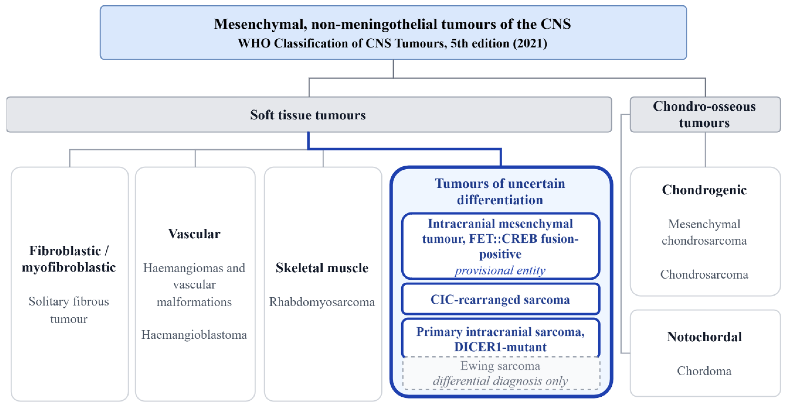
Figure 2.
Proposed tiered diagnostic algorithm for the three molecularly defined CNS sarcomas. Clinical and radiological features narrow the differential, and morphology guides a targeted immunohistochemical panel that triages but cannot establish the diagnosis. RNA-based fusion testing and targeted DNA sequencing with copy-number profiling are performed in parallel on the same specimen; DNA methylation profiling is reserved as a rescue step for cases that remain unresolved or discordant, and germline testing follows any DICER1 alteration. Two checkpoints are highlighted: a methylation call of "meningioma" in a dural-based mesenchymal tumour does not exclude intracranial mesenchymal tumour and requires fusion testing, and a DICER1 alteration mandates germline testing because the result changes management for the patient's family as well as the patient. The algorithm is a synthesis of the available evidence, not a prospectively validated pathway.
Figure 2.
Proposed tiered diagnostic algorithm for the three molecularly defined CNS sarcomas. Clinical and radiological features narrow the differential, and morphology guides a targeted immunohistochemical panel that triages but cannot establish the diagnosis. RNA-based fusion testing and targeted DNA sequencing with copy-number profiling are performed in parallel on the same specimen; DNA methylation profiling is reserved as a rescue step for cases that remain unresolved or discordant, and germline testing follows any DICER1 alteration. Two checkpoints are highlighted: a methylation call of "meningioma" in a dural-based mesenchymal tumour does not exclude intracranial mesenchymal tumour and requires fusion testing, and a DICER1 alteration mandates germline testing because the result changes management for the patient's family as well as the patient. The algorithm is a synthesis of the available evidence, not a prospectively validated pathway.
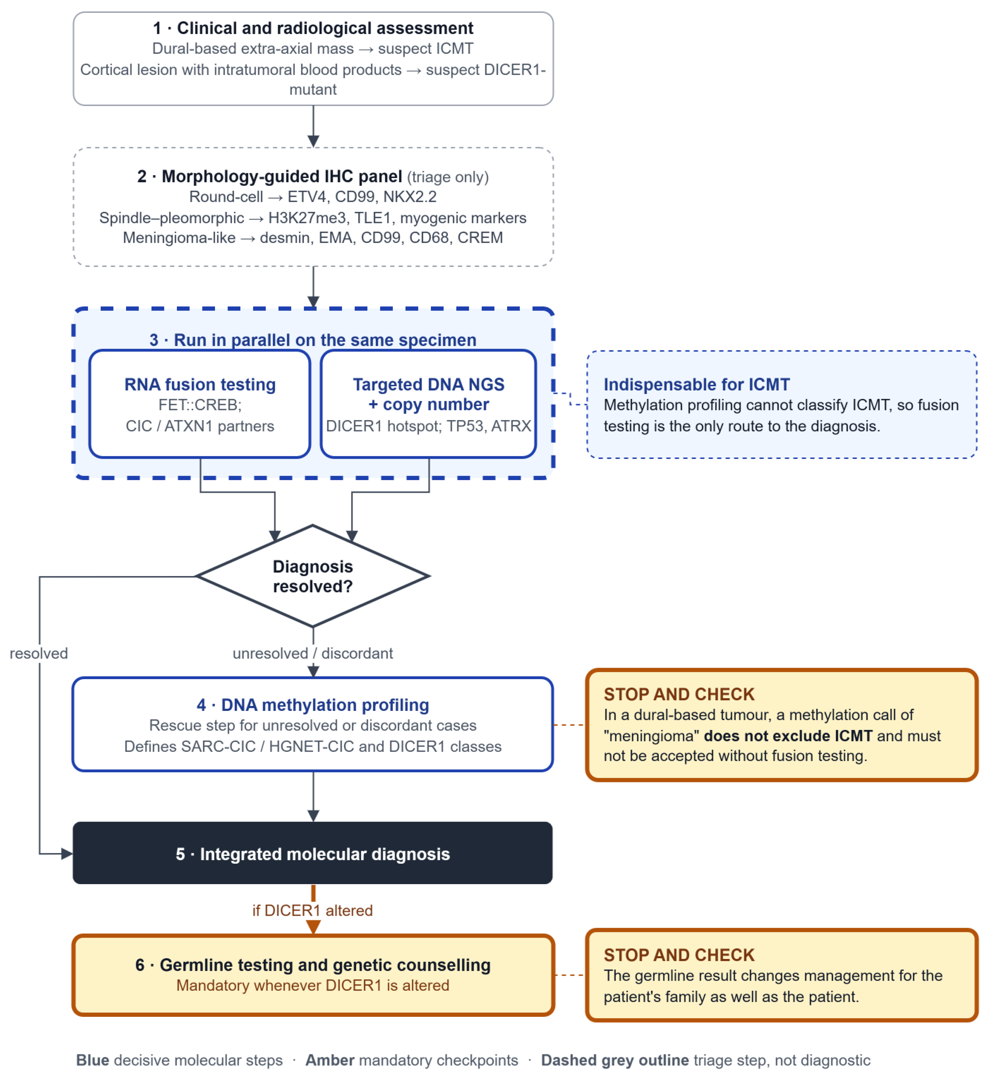

Table 1.
Diagnostic immunohistochemical markers and their limitations.
| Entity | Supportive/ confirmatory markers | Discriminatory value | Why IHC alone is insufficient |
| ICMT, FET::CREB | desmin, EMA, CD99, CD68 - near-ubiquitous [3,4,12]; in the largest series, desmin 84%, EMA 73%, CD99 88% [4], and all 11 cases in one series expressed CD68 and EMA [12]. MUC4 in a subset (5/7 [3]; 3/11 [12]. CREM proposed surrogate [4]. Negative: CD34, SMA, SOX10, HMB45 [12] |
No single specific marker | Synaptophysin >50%, SSTR2A ~⅓, GFAP in subclass B [4] → actively drives the meningioma / neuroepithelial misread. SERIES DISAGREE: Tauziède-Espariat (2022) [12] reports synaptophysin and GFAP constantly negative across 11 cases, whereas Sloan (2021) [3] finds synaptophysin frequent and Rajan (2026) [4] positive in 60% |
| CIC-rearranged sarcoma | Nuclear ETV4 - constant; readout of PEA3 de-repression [6,13] | CD99 patchy / focal (vs diffuse membranous in Ewing) + NKX2.2 negative - read together, separate from Ewing [6] | WT1 unreliable in the CNS: positive in only 2/6 CIC::NUTM1 cases [13], and NUTM1 fusions are enriched intracranially, so the marker fails where it is most needed. NUT positive only in NUTM1-fused cases; nuclear DUX4 only in DUX4-fused cases → partner-dependent [13]. |
| DICER1-mutant | desmin, myogenin (patchy, myogenic foci) [14] | H3K27me3 loss + nuclear TLE1 - combined, the accessible surrogate; nuclear TLE1 in 6/6 cases [15] | The extent of H3K27me3 loss varies rather than being all-or-none: diffuse in 4 of 6 cases, mosaic in one and minimal (≤ 5%) in one [15]. GFAP is negative in tumour cells, but negativity does not exclude high-grade glioma, since GFAP-negative glioblastoma and gliosarcoma without a residual glial component both occur [16]. |
Table 2.
Molecular diagnostic modalities.
| Modality | What it detects | Strength in these entities | Documented limitation |
| DNA methylation array profiling | Genome-wide methylation class | The diagnostic backbone: defines CIC (SARC-CIC/HGNET-CIC) and DICER1 (distinct class), and rescues cases missed by other assays [7,17]; changed the histological diagnosis in ~12% of prospective CNS tumours [1] | Blind spot for ICMT — no dedicated class; actively misassigns these tumours to meningioma [3,12,18] |
| RNA-based fusion detection (RNA-seq / fusion panel) | Fusion transcripts + partner identity | Required for ICMT (methylation cannot classify it). Identifies the CNS-enriched CIC partners (NUTM1, LEUTX) and non-CIC ATXN1 fusions [3,4,6,11] | Needs adequate RNA quality; novel partners may be missed by fixed panels |
| Targeted DNA NGS panel | Point mutations, indels, some fusions | Detects DICER1 hotspots + co-alterations (TP53, ATRX, MAPK) [7,19] | Can miss the fusion entirely; documented false negatives in CIC [20] |
| FISH (break-apart) | Rearrangement of a locus | Rapid, widely available | Notable false-negative rate; cannot identify the partner (critical, since partner drives CIC subtype and NUT/DUX4 IHC) [19] |
| Copy-number profiling | CNVs | Supportive; ICMT shows recurrent 10p, 19, 22q loss; HGNET-CIC shows 1p/13q/14q/22q loss, gain 8 [4,17] | Not diagnostic alone |
| Germline testing | Constitutional DICER1 variant | Mandatory when a DICER1 alteration is found — changes management for patient and family [21,22] | Requires counselling infrastructure |
Table 3.
Comparison of the three molecularly defined CNS sarcomas.
| Feature | ICMT, FET::CREB | CIC-rearranged sarcoma (CNS) | Primary intracranial sarcoma, DICER1-mutant |
| Class of molecular lesion | Chimeric transcription factor (gain of an aberrant activator) | Converted transcriptional repressor (repressor -> neomorphic activator) | Crippled miRNA-processing enzyme (loss of regulation) |
| Defining alteration | FET::CREB fusion (EWSR1/FUS :: ATF1/CREB1/CREM; rarely CREB3L3) | CIC::DUX4 + CNS-enriched variants (NUTM1, LEUTX); non-CIC ATXN1 fusions | Biallelic DICER1 (LOF allele + RNase IIIb hotspot) |
| Downstream consequence | Constitutive CRE-driven transactivation; CREM overexpression | PEA3/ETV (ETV1/4/5) de-repression | 5p-miRNA depletion (let-7, miR-30) |
| Morphologically / mimics | Variable mesenchymal; meningioma | Undifferentiated round cell; Ewing / CNS embryonal | Spindle-pleomorphic; high-grade glioma / gliosarcoma |
| DNA methylation profiling | Blind spot -- no DKFZ class; mislabels as meningioma | Defines (SARC-CIC / HGNET-CIC) | Defines (distinct class) |
| Confirmatory test | Fusion testing (NGS/RNA-seq) -- required | Methylation +/- fusion (FISH false-negatives) | Methylation + DICER1 sequencing |
| Accessible IHC surrogate | CREM | ETV4 (nuclear) | H3K27me3 loss + nuclear TLE1 |
| Epidemiology/ location | Children-young adults; extra-axial/meningeal | Children-young adults; supratentorial | Paediatric (adults reported); supratentorial |
| Behaviour | Indolent but recurrence-prone | Aggressive (median OS ~17.5 mo) | Aggressive (2-yr OS ~66%) |
| Emerging molecular target | Immune-checkpoint inhibition (PD-L1) | MCL1; p300/CBP | RAR / RAS-pathway; germline surveillance |
Disclaimer/Publisher’s Note: The statements, opinions and data contained in all publications are solely those of the individual author(s) and contributor(s) and not of MDPI and/or the editor(s). MDPI and/or the editor(s) disclaim responsibility for any injury to people or property resulting from any ideas, methods, instructions or products referred to in the content. |
© 2026 by the authors. Licensee MDPI, Basel, Switzerland. This article is an open access article distributed under the terms and conditions of the Creative Commons Attribution (CC BY) license (http://creativecommons.org/licenses/by/4.0/).
Copyright: This open access article is published under a Creative Commons CC BY 4.0 license, which permit the free download, distribution, and reuse, provided that the author and preprint are cited in any reuse.