
Submitted:
21 September 2026
Posted:
22 September 2026
You are already at the latest version
Abstract
Periodontal disease (PD) is a multi-factorial condition that may lead to tissue destruction and tooth loss. Multirooted teeth are at high risk due to the furcation space where roots diverge, and new regenerative therapies are needed to support complete repair of furcation defects. This study assessed safety and preliminary efficacy of an injectable piezoelectric-based hydrogel (Ambrilux Dental Gel) in a canine model of ligature-induced PD. Animals were randomized to study treatment (n=3) (two applications 28 days apart) or control (n=3). Laboratory results were normal throughout the study. Radiographs demonstrated notable increase in furcation bone density in all animals in the study treatment group by 28 days after the first treatment, with continued increase following the second treatment. The control group showed continued loss of bone or minimal regeneration. Quantitative CT showed treated animals had greater furcation bone mineral density, bone volume and bone volume to tissue volume ratio compared to control. This study provides the first direct evidence that a degradable, piezo-based hydrogel is efficacious for treatment of PD in large animals, serving as a foundation for future studies. The ability of Ambrilux to promote bone growth in tooth furcations suggests it represents a safe and novel approach to PD.
Keywords:
periodontal disease
; piezomaterials
; furcation defect
; bone regeneration
; hydrogel
1. Introduction
Periodontal disease (PD) is a multi-factorial condition that impacts two in five adults in the United States, with prevalence increasing with age [1]. It is characterized by increased bacterial colonization of the impacted teeth, followed by the generation of a substantial inflammatory response in the surrounding tissues. If left untreated, PD may lead to the destruction of the bone and ligaments surrounding the tooth and subsequent loss of the tooth. PD requires ongoing supportive treatment, but tooth loss is common even with treatment; it was reported that over half of patients lost at least one molar over ten years of periodontal treatment [2].
The goal of periodontal treatment is to improve the tissue environment by reducing or eliminating dental plaque biofilm and calculus from the surface of the tooth, thereby stopping progression of tissue damage and ideally restoring healthy tissue. This can be accomplished through a broad range of non-surgical and surgical options including scaling to remove supragingival bacterial plaque and calculus; comprehensive root planing to treat root surface irregularities or alterations; chemotherapeutic agents to address microbial pathogens or alter host response; resective procedures; and regenerative procedures [3]. Periodontal regenerative procedures include soft tissue grafts, bone replacement grafts, root biomodification, tissue regeneration, and combinations of these procedures. Severe cases of PD may require reconstructive procedures involving bone regeneration, ridge augmentation and preservation, implant site development, and sinus augmentation.
Multirooted teeth, such as molars, are anatomically complex due to the presence of the furcation space where the roots of the tooth diverge. The furcation of a tooth, which plays an important role in stabilizing the tooth, is difficult to reach and therefore more prone to plaque deposition and subsequent tissue destruction. The degree of furcation involvement (FI) closely correlates with the long-term prognosis of the tooth.
Furcation defects are particularly difficult to treat, and they may require extensive resective therapy such as tunneling, root amputation or hemisection [4]. However, resective surgical approaches have a considerable failure rate, high morbidity, and high cost [5]. A consensus panel convened by the American Academy of Periodontology (AAP) Regeneration Workshop found that periodontal regeneration has been established as a viable treatment option for various furcation defects, and regenerative therapy should be considered before resective therapy or extraction for Class II defects, where there is loss of inter-radicular bone but a portion of bone and periodontal ligament remain intact [6]. The panel also supported research to investigate novel regenerative approaches, as new regenerative therapies are needed to improve the complete resolution of furcation defects [7].
One new approach harnesses the piezoelectric effect to induce tissue regeneration. Bone, and cartilage as well as the collagen fibers, are natural piezoelectric materials and therefore can generate an electric charge when subject to mechanical forces. These piezoelectric properties of bone are thought to play an important role in mediating natural remodeling of the tissue as well as assist in the repair of bone following injury [8]. A new regenerative therapy for PD is under development (Ambrilux™ Dental Gel, formerly known as PiezoGEL, Oral Biolife, Bethlehem, PA), which utilizes these piezoelectric properties of bone to regenerate tissue. Ambrilux is an innovative hydrogel placed in the periodontal pocket and is embedded with piezoelectric fillers designed for tissue regeneration through electrical stimulation. This hydrogel is administered directly into the periodontal pocket via a minimally invasive probe and subsequently light-cured to form an in-situ graft. The primary functional mechanism of Ambrilux is mechanical [9]; the biocompatible piezoelectric ceramic fillers embedded in the gel are activated by natural biomechanical vibrations, such as those from chewing, jaw movements, and vocalizations, to produce the electrical charges necessary for stimulating osteogenic cells and promoting bone tissue regeneration. In addition, the hydrogel component (GelMA) provides an important physical scaffold for the recruitment and growth of bone cells.
The current exploratory study was designed to examine the safety profile and efficacy signals of an injectable piezoelectric-based hydrogel in canines following the development of ligature-induced PD. The study objective was to generate key preliminary data and establish effect sizes for future, adequately powered confirmatory studies. In adherence with the ethical principles of animal research, the group sizes were restricted to the minimum required for establishing the initial dataset for evaluating early-stage efficacy and safety trends.
2. Materials and Methods
2.1. Study Design
This preclinical study was conducted to examine the impact of a piezoelectric-based hydrogel (Ambrilux™, Oral Biolife, Bethlehem, PA) on periodontal healing of teeth in canines following the development of ligature-induced PD. Periodontal disease was induced in six female beagle dogs over a period of 55 days. Animals were then randomized into two groups with treated animals (n=3) receiving two applications of the study treatment 28 days apart. Non-treated animals (n=3) served as the control group. Periodontal repair was assessed in all animals by radiographic analysis at Day -1 (prior to study treatment), Day 29 (just prior to dose 2), and Day 57 (28 days after dose 2). Urine and blood samples were obtained at the end of the study for evaluation of safety parameters. All animals were sacrificed 70 days after the start of the study treatment phase, when tissue samples were harvested and subjected to quantitative computed tomography (CT) and histopathologic analysis. See Figure 1 for details on study design.
2.2. Animals and Housing
All handling and treatment procedures involved in conducting this study were approved by the Institutional Animal Care and Use Committee (IACUC) (AC24017C; KAC24017.01C; KAC24017.02C) and in accordance with the Animal Welfare Act and the Guide for the Care and Use of Laboratory Animals [10].
Animals (non-pregnant female beagles) were allowed to acclimate to the housing, food and water for a minimum of 7 days. General health observations and physical examination were performed twice a day during the acclimation phase. Animals were housed in an Animal Biosafety Level (ABSL)-2 room where air is HEPA filtered both on intake and exhaust. The room was 40 × 32 feet and equipped with continuous environmental monitoring and automatic light timing systems. The dogs were housed in kennels providing a minimum of 12 square feet for each dog.
Feed was adjusted based on the stage of the protocol. Animals were fed Purina Pro Plan Veterinary Diets EN Gastroenteric bagged, hard dog food (Nestle Purina PetCare Company, St. Louis, MO) throughout the acclimation period and up to placement of ligatures. Following placement of ligatures, a canned wet food formulation of Purina Pro Plan Veterinary Diets EN Gastroenteric was provided throughout the remainder of the study until ligature removal. Feed volume and changes were determined by the Attending Veterinarian.
2.3. Periodontal Preparation
Two weeks prior to induction of PD, the dogs were premedicated with acepromazine (0.05 mg/kg, subcutaneous (SQ) or intravenous (IV)) and hydromorphone (0.10 mg/kg, SQ or IV), catheterized, induced to general anesthesia with propofol (to effect, IV), intubated and maintained under general anesthesia with oxygen and isoflurane. Periodontal instrumentation was then performed with ultrasonic and hand instruments, and the teeth were carefully polished. During this preparation phase, animals had their teeth brushed once daily for 1 minute using a commercially available canine toothpaste (Virbac C.E.T. Enzymatic, Westlake, TX). Daily brushing was continued until the beginning of the PD induction phase.
2.4. Periodontal Disease Induction
Ligatures were placed on teeth #407, #408, and #409 of the mandible using the methods described by Kim et al. [11]. These teeth, which form the bottom half of the carnassial apparatus, were chosen based on their role as the primary area of mastication as well as their similarity to human dental spaces [11]. While under general anesthesia, the gingival attachments of the teeth were incised using a number 11 scalpel blade and undermined until the pocket depth measured 3 mm with a straight elevator. Following undermining of the gingival attachment, a shallow notch was created on the mesial and distal aspects of each tooth using a burr to prevent slippage and facilitate retention of the ligatures. Lastly, a dental ligature wire twisted with 2-0 braided silk suture was placed on each tooth. The knot of the ligature faced distally and was covered with a dental composite (3M™ Filtek™ Supreme Ultra Universal Restorative, Maplewood, MN) to prevent iatrogenic damage to the oral cavity. Buprenorphine (0.015 mg/kg, SQ) was administered during anesthetic recovery following the placement of the ligatures. Then, the oral cavity was examined daily to confirm retention of each of the ligatures, which remained in place for 55 days. The ligatures were removed before the periodontal evaluation on Day -1 and were not replaced.
2.5. Periodontal Treatment
2.5.1. Fabrication of Piezoelectric Hydrogel
The piezoelectric hydrogel was fabricated as previously described [12] by mixing 200 mg/mL gelatin methacryloyl hydrogel (GelMA;Advanced Biomaterials, 5208) with 9 mg/mL silanized particles of barium titanate BTO (US Nanomaterials, US3830, 200 nm) and sterile phosphate-buffered saline (PBS). The photoinitiator lithium phenyl-2,4,6-trimethylbenzoyl phosphonate (LAP) (Advanced Biomaterials, 5269) was added at a proportion of 0.35% wt of the solubilized GelMA. Lyophilized GelMA and PBS (70 °∆C) were mixed using a vortex mixer (Daigger Vortex-Genie) until GelMA was fully solubilized. BTO particles were added during the continuous shaking of the mixture. Subsequently, LAP was dissolved in 100 μL of warm PBS (70 °∆C) and mixed with the dissolved GelMA/BTO mixture.
2.5.2. Randomization
Animals (n=6) were randomized to either active treatment group (scaling, root planing, and application of the piezolectric gel) or control group (scaling and root planning only), at the end of the PD induction phase on Day -1. Randomization was performed by ranking the PD severity of all subjects based on the periodontal evaluation combined score of plaque index (PI), gingival index (GI), bleeding on probing (BoP), and the difference between the periodontal probing pocket depth (PPD) and the clinical attachment level (CAL). Dogs were then randomly assigned to a study group within independent permutations of the two treatments. Of the two dogs with the highest periodontal scores, one was randomly allocated to the study treatment group and the other to the control group, and the same for the remaining two pairs of subjects. Randomization was performed within a commercially available data software (Microsoft Excel).
2.5.3. Application of Study Treatment
Application of the piezolectric gel was performed on Day 1 for animals in the active treatment group. Each dog received dexmedetomidine and butorphanol injectable anesthesia intravenously for the application of the study treatment and was monitored throughout sedation. The piezolectric gel was applied into the periodontal pocket using prefilled syringes and a 22-gauge dental application tip (Figure 2a). A new application device was used for each dog. Approximately 0.5 mL in total of the piezolectric gel was applied to the three teeth being studied until visible above the gingival margin. The gel was then cured with a LED light for 60 seconds (Figure 2b). Following curing, the periodontal pocket was covered with tissue adhesive (Vetbond, 3M, Maplewood MN) and allowed to dry. The second study treatment application occurred on Day 29 following the same procedure and was performed while the dogs were under general anesthesia for periodontal evaluations. The buccal surface of teeth 407, 408, 409 were observed every 7 days after application by unmasked personnel to determine the retention of the study treatment. In addition, the retention of the study treatment was documented during necropsy on Day 71.
2.5.4. Masking
Masking was in effect during the study treatment phase of the study and was accomplished by separation of functions. The Study Investigator and limited other personnel designated by the Investigator were unmasked and responsible for study treatment application and weekly post-application retention observations, while the Attending Veterinarian remained masked and was responsible for performing periodontal measurements subsequent to the study treatment application. During the treatment phase, routine general health observations were performed by masked personnel except on the morning of Day 60 when an unmasked individual performed general health observations. On Day 1 following morning general health observations, one member of the study staff was directed to become unmasked by the Study Investigator to assist with recovery from the injectable anesthesia used to facilitate treatment administration.
2.6. Study Assessments
2.6.1. Laboratory Analysis
Comprehensive chemistry, complete blood count, and urinalysis were conducted at IDEXX BioAnalytics (West Sacramento, CA) to assess animal health, specific organ system health as well as assist in identifying any potential toxicities. Venous blood samples were collected for hematology (CBC and serum chemistry) using a 20 ga × ¾ inch needle on Days -1, 29, and 57. Following collection, the blood was transferred into a 5 mL serum separator tube and a 3 mL K2EDTA tube. The K2EDTA tubes were placed on ice immediately after collection. The serum samples were allowed to clot for a minimum of 30 minutes before ambient centrifugation at 2,000 × g for 10 minutes. Following centrifugation, the serum was decanted into 2 aliquots (one primary and a backup). The K2EDTA tube and a serum aliquot were shipped on the day of collection.
2.6.2. Safety Assessment
General health observations were conducted twice daily throughout the study. As periodontal disease was induced in this study, observations localized to the oral cavity were not considered adverse events. Systemic sequalae secondary to periodontal disease with severity such that emergency intervention or observation beyond protocol specifications were recorded as adverse events and classified as mild, moderate, or severe.
2.6.3. Periodontal Evaluation
Because of the inherent variability in clinical scoring, as well as the low number of animals per group, periodontal evaluations were conducted primarily to ensure onset and satisfactory progression of disease in each animal and to assure proper randomization of subjects prior to treatment. Periodontal evaluations were performed on Day -56 before placement of ligatures, Day -1 after ligature removal, Day 29 and Day 57 while the subjects were under general anesthesia. The evaluation was performed using a scoring system that accounts for PI, GI, PPD, CAL, and BoP [13,14]. Scores ranged from 0 to 3 for PI with 0=none and 3=abundant; from 0 to 3 for GI with 0=no inflammation and 3=severe inflammation. Measurement of PPD was defined as the distance between the gingival margin and the bottom of the probable pocket, and CAL was the distance between the cementum-enamel junction and the bottom of the probable pocket. A score of 0 indicated absence of bleeding within 10 seconds following probing and a score of 1 indicated presence of bleeding.
Each of the parameters were evaluated at the disto-buccal site. Periodontal evaluations were performed by the same individual throughout the course of the study who was masked to treatment assignments.
2.6.4. Intraoral Radiographs
Digital intraoral radiographs were obtained to evaluate the temporal changes of alveolar bone loss after ligature application and subsequent administration of study treatment. Intraoral radiographs were captured using a handheld radiology unit (DEXIS NOMAD™ Pro 2, Quakertown, PA) concurrent with the periodontal evaluations at the start and end of PD induction, 28 days after dose 1 (just prior to dose 2), and 28 days after dose 2. To ensure precise alignment of the digital sensor throughout the course of the study, dental molds were constructed for each individual animal using Braval VPS Putty (Patterson Dental, St. Paul, MN). Briefly, putty was mixed according to instruction and placed into an adhesive-coated tray. The tray was inserted into the mouth and held in position until set. For each radiograph, the mold was inserted into the mouth and the digital sensor placed in the same location on the mold.
Qualitative analysis of radiographs of the furcations of teeth #409 and #408 was conducted in a blinded manner and scored for changes in furcation bone radiopatency over time. Issues with superimposition of adjacent teeth in some animals precluded use of tooth #407.
2.6.5. Micro-CT Imaging
In order limit variability, the CT imaging, VOI and quantitative analysis were conducted by a single individual in a blinded fashion. For high-resolution micro-CT imaging, harvested jaw specimens were scanned with a SKYSCAN 1273 micro-CT system (Micro Photonics Inc, Allentown, PA) at a resolution of 13.85 μm (achieved using 130 kV and 60 μA). Individual mandible samples were wrapped in gauze saturated with physiological saline and placed within a sealed plastic container to maintain hydration during imaging. Each sample was imaged with an isotropic voxel size of 25 μm using 120 kV and 300 μA X-ray settings with exposure time of 501 ms per frame, with 4 frames averaged at each projection angle. Volumetric reconstructions were performed using NRecon 2 software (Bruker Corp, Billerica, Massachusetts) before visualization of samples within Bruker’s DataViewer and CTVox programs. Quantitative assessment of bone in the furcation of tooth 409 was performed using a morphological escalator styled selection method to isolate the thinner trabecular bone within each furcation volume of interest. The morphological escalator is a fully automated, image-processing technique that is commonly utilized in CT bone morphometry and is packaged into the analysis software. We used the technique to help segment and delineate trabecular bone from surrounding dense cortical bone, thereby more accurately defining our region of interest.
After the tissue mineral density was determined, volumetric analysis of the volume of interest was determined. The mineralized tissue within the VOI was first segmented using an adaptive mean threshold applied in 2D with a round kernel, radius of 3, and constant of 1. A pre-thresholding was also employed to minimize analysis processing time defining all pixels with intensity less than 65 as non-mineralized and all pixels with intensity 75 or greater as mineralized. Thus, the adaptive threshold was used to decide on the classification of each pixel falling between 66 and 74.
After segmentation, a despeckle function was applied in 2D to remove all black speckles less than 7 pixels in area followed by a repeat of the function again at the same selection criteria but now focused on white pixels in the dataset less than 7 pixels in area. After these cleanup steps, a volumetric analysis was completed on the dataset in 3D which captured the VOI volume, mineralized tissue volume, surface area, mineralized tissue thickness, and open space thickness among other reported values.
2.6.6. Qualitative Histology
Qualitative histology, as opposed to quantitative histology, was utilized due to the small sample size and associated statistical limitations. Histopathologic analysis focused on both the proximal regions of the teeth as well as the furcation of tooth #409.
Following CT analysis, samples were decalcified in 22% formic acid until suitable for sectioning, processed using routine histologic processing techniques, embedded in paraffin, and cut at 4-μm thickness. Sections were stained with Hematoxylin and Eosin (H & E) and examined using Aperio ImageScope digital pathology system (Leica Biosystems, Deer Park, IL).
3. Results
3.1. Safety Data
Health observations (general health, physical examination and final necropsy) were within normal results for all animals (control and treated) in the study. No adverse events were reported.
Comprehensive chemistry, hematology, and urinalysis were used to assess animal health, specific organ system health as well as assist in identifying any potential toxicities. The laboratory data for Days -1, 29 and 57 (Supplemental Tables S1–S3) showed normal results throughout the study indicating that the subjects remained in good health throughout the study, and the study treatment did not elicit any untoward changes in the measured parameters.
3.2. Clinical Evaluations
Periodontal evaluations were performed on Day -56 before placement of ligatures, Day -1 after ligature removal, Day 29 and Day 57. The dataset derived from the in-life periodontal examinations is provided in Supplemental Tables S4 and S5. A review of the PI, GI, PPD, CAL, and BoP data did not show any appreciable trends between groups.
3.3. Radiographic Analysis
Radiographic analysis showed that at the end of the PD induction phase, all animals in both the control and treatment groups demonstrated multiple molars that showed moderate interproximal bone loss and furcation defects. This indicates that the ligature protocol utilized was successful in initiating a moderate degree of PD.
All the animals in the treatment group showed increasing radiopacity of the interproximal and furcation bone over time, with a corresponding decrease in the distance between the furcation fornix and base of the defect. Temporal analysis of radiographs showed that within 30 days post-application of the piezolectric gel, a notable bone-growth response could be observed in the furcation region (Figure 3). Bone growth in the furcations of treated animals continued to be observed following the second application period. This degree of response was noted for all treated animals and was not observed in non-treated animals, in which either continuing bone loss or minimal bone regrowth in the furcations were observed by the end of the study.
3.4. Qualitative Histological Analysis
3.4.1. Proximal Regions
Qualitative histologic examination was utilized to determine the degree of inflammation and other morphologic changes to the periodontal tissues between those teeth where PD was induced (Figure 4). Tissues from control animals exhibited a thickened gingival epithelium (hyperplasia) with a moderate degree of inflammation, composed primarily of lymphocytes and neutrophils, in the surrounding regions. The connective tissue beneath the gingival layer was composed of disorganized collagen fibers containing fibroblastic cells with a high density of blood vessels. Numerous osteoclasts, indicative of active bone resorption, were noted in the alveolar bone of control animals. In contrast, treated animals exhibited greater organization in the collagen matrix with a notable decrease in the number of blood vessels as compared to control animals. Some areas of low-grade inflammation were present in the junctional epithelium. Few osteoclasts were noted in the alveolar bone of treated animals.
3.4.2. Tooth Furcations
Histologic analysis of the furcation from tooth #409 was conducted to both determine the morphologic alterations to the periodontal ligament (PDL) as well as correlate the changes noted in CT to alterations in histopathology (Figure 5). The PDL in control animals exhibited loss of ligament structure as characterized by the presence of disordered, ligament like tissue composed primarily of loosely organized collagen fibers and fibroblasts with increased extracellular spacing. The internal zone of the PDL was found to contain numerous blood vessels of variable sizes and diminished vascular integrity resulting in notable micro-hemorrhaging. Bone within the furcation of control animals may be characterized by the presence of multi-nucleated osteoclasts with an associated increase in bone resorption. Two out of three control animals exhibited replacement of bone in the fornix region with increased number of fibroblasts, granulation tissue and a limited number of inflammatory cells (Figure 5a).
In contrast to control subjects, Ambrilux-treated animals exhibited well-organized periodontal ligaments (PDL) characterized by fibrous connective tissue and densely packed fibroblasts. Moderate-sized blood vessels were present within the ground substance, and PDL fiber bundles extended directly into the alveolar bone. Qualitative evaluation of the furcation areas revealed newly formed alveolar bone lined with osteoblasts, alongside mature lamellar bone. Inflammatory cell infiltration was minimal.
3.5. Micro CT Analysis
To further evaluate the ability of the study treatment to promote bone regeneration, micro CT imaging was utilized to generate sagittal and 3D reconstructed (frontal and sagittal) images in animals in both the control and treatment groups (Figure 6). Analysis of frontal 3D reconstructions in animals of the treatment group had a smooth, less mottled appearance to the bone surface. The 2D and 3D sagittal views of the samples from the treated animal showed more profound bone density within the tooth furcation with thicker bone walls. In contrast, the control sites revealed an uneven bone surface topography in the furcation and proximal areas, indicative of active PD and subsequent bone loss. Sagittal section views (2D and 3D reconstruction) and of the furcation showed a loss of bone density with wide alveolar spacing.
Quantitative analysis was also completed on the harvested jaw specimens, with the highlighted volume of interest (VOI) (Figure 7a) quantified for thickness, volume, and local bone mineral density (BMD). Representative heatmap images representing the registered VOI for a control animal and a treated animal are presented in Figure 7b and Figure 7c, respectively. Mean values for quantitative CT at furcation are shown in Table 1. Mean bone volume (BV) was markedly higher in the treatment group as compared to control with a similar difference observed for the BV/TV and bone mineral density (BMD). Trabecular measurements were consistent with a greater degree of repair in treated animals as opposed to control animals. While trabecular thickness was similar between the groups, the treated animals had a notable increase in the number of trabeculae as well as a corresponding decrease in trabecular separation. Increased bone density was visually noted in treated animals at proximal areas as well, although quantitative analysis was not feasible due to inherent structural differences across animals. Because the small sample size compromises hypothesis testing reliability, p-values were excluded to avoid potential data misinterpretation.
4. Discussion
The results of the current proof of concept study provide evidence that the tested piezoelectric-based hydrogel (Ambrilux™, Oral Biolife, Bethlehem, PA) is safe and can positively modulate periodontal wound healing and promote bone regrowth in a canine model of ligature-induced PD. These conclusions are based primarily on quantitative and observational results. Quantitative CT was utilized to gauge bone regeneration and was conducted on the furcation areas of tooth #409. This tooth was chosen as it is the largest molar and therefore the most prominent tooth in the lower jaw, while teeth #407 and 408 are premolars. In the dog, tooth #409 is responsible for shredding food and is therefore subjected to the highest bite pressures and food entrapment. This tooth, as compared to the premolars, is subject to more severe wear and is a frequent site of naturally occurring periodontal disease [15,16]. Analysis using quantitative CT showed that furcation bone volume was 37% higher in treated animals than in control animals, and bone mineral density was over 18% higher in treated animals. These data suggest that treatment with Ambrilux has the potential to regenerate bone in the furcation area. Temporal radiographic evidence showed that all treated animals demonstrated increasing radiopacity at the furcation area. In contrast, control animals demonstrated stable or worsening levels of disease. It was noted that 3/3 treated animals demonstrated bone regeneration over time, while in the control group 2/3 animals showed continued bone loss with one animal exhibiting no change. However, because of the low number of subjects, as well as the moderate degree of induced disease, we cannot definitively state that the injury was of critical size and therefore cannot exclude the possibility of spontaneous/natural healing in either group. Histological findings support a lower degree of inflammation in treated animals, and potential modulation of the periodontal ligament tissues. It should be noted that while the histological data offers valuable insight into Ambrilux-induced cellular changes, the limited sample size precludes any definitive conclusions. Periodontal evaluations were performed throughout the study, but review of the data did not highlight appreciable differences, likely due to the small sample size and/or moderate severity of the disease. Additional ad hoc analyses are needed to investigate trends in these evaluations and identify potential areas of improvement for future studies.
Furcation involvement in periodontal disease is a key pathologic and diagnostic feature, since bone loss in the furcation is an important predictive indicator of disease severity and clinical outcome (i.e., tooth loss) [17]. Bone loss at the furcation is apparent on radiographs as indicated by a decrease in radiopacity, which signifies that severe periodontal disease is present that will require significant and complex treatment [18,19,20] to save the affected tooth. Bone regeneration in the furcation area is a clinical challenge, thus, bone loss at the furcation was an important efficacy endpoint in this study. Various regenerative therapies have been investigated to address furcation defects, including bone grafts, guided tissue regeneration (GTR), application of growth factors and enamel matrix derivatives, and bone morphogenetic proteins, with variable clinical outcomes reported [7,18,19,20]. A consensus report from the AAP Regeneration Workshop reported that histological evidence of periodontal regeneration has been demonstrated in several studies for maxillary facial, mesial, distal, and mandibular facial or lingual Class II furcation defects [6]. However, evidence for periodontal regeneration for Class III defects (with through and through lesion) is limited to only case reports. The panel recommended that future research should focus emerging regenerative therapies that have the potential to enhance outcomes [6], including approaches like the current study treatment with piezoelectric properties that can promote bone regeneration and serve as a physical scaffold. The structural and physical properties of Ambrilux are conducive to meeting these goals. The piezoelectric component, as described in more detail below, serves to provide the stimulus to initiate stem cell differentiation as well as promote cell recruitment, while the GelMA hydrogel serves to provide a suitable scaffold for cell migration and maturation. Key to Ambrilux’s capacity to promote bone regeneration is its ability to gain full access to small anatomical regions such as difficult to access areas of the periodontal pocket as well as the tooth furcation. Comprehensive rheological and injectability analysis have been utilized to determine the viscosity of Ambrilux. It was determined that, in the uncured form and at body temperature, the viscosity of Ambrilux is low, allowing it to access tight areas [12]. The ability to act as viscoelastic material means that Ambrilux behaves like an elastic solid (stretching and returning to shape) as well as a liquid [21]. These physical properties are complemented by the uniform dispersion of BTO nanoparticles within the fluid matrix, thereby avoiding the formation of aggregates.
Electric fields play a critical role in mediating various physiological processes, impacting tissue development and regeneration [22]. Reports of the impact of electric fields on bone regeneration date back to 1812, and the concept of piezoelectric properties of bone were described in 1953 [23]. Bone, cartilage as well as the collagen fibers within bone, are piezoelectric materials and therefore retain the ability to generate an electric charge when subject to mechanical forces. The piezoelectric properties of bone are thought to play an important role in mediating natural remodeling of the tissue as well as assist in the repair of bone injury. The mechanism(s) by which the piezoelectric properties of bone facilitate bone repair have focused on how mechanical stimuli is converted to bioelectrical signals that impact target cells, primarily osteoprogenitors cells and osteocytes. In this regard, the release of negative charges by the piezomaterials in bone, and their subsequent interaction with voltage-gated calcium channels, specifically Piezo1 channels, appear to be the primary mechanism for initiation of bone formation/repair [9]. Piezo1 channels, which are found on several cell types including stem cells and osteocytes, function as important mechanosensors in bone and serve to regulate bone formation, remodeling, and repair by modulating gene expression. Roldan et al. (2023) have further elucidated the mechanism of the piezoelectric hydrogel Ambrilux analyzing changes in the expression of key genes involved in osteogenic differentiation [12]. These studies demonstrated that mechanostimulation of Ambrilux upregulates Runt-related transcription factor 2 (RUNX2), a protein that is crucial for bone formation/maintenance as well as other important osteogenic genes including Collagen Type I Alpha 1 Chain (COL1A1) and alkaline phosphatase (ALP) [12,24]. Activation of these genes suggests that the mechanostimulation, and the subsequent release of physiologic microcurrents by this piezoelectric hydrogel, may trigger the differentiation of target stems cells to an osteogenic phenotype. It should be noted that the aforementioned studies were conducted both in vitro and in a murine model of PD and therefore do not present definitive proof that these molecular interactions play a role in the present study.
Bone possesses inherent regenerative capabilities; however, instability, poor blood supply, and infection-driven inflammation can severely impair this process. In PD, persistent bacterial inflammation triggers cascading tissue damage, leading to alveolar bone and tooth loss [25,26]. The impact of Ambrilux in altering the microbiome and local inflammatory response in the present study requires further exploration. Nevertheless, previous in vitro results reveal that Ambrilux significantly diminishes viable bacteria and biofilm biomass, operating in a reactive oxygen species (ROS)-independent manner [12,27]. To build upon these encouraging markers of localized healing, future research will comprehensively evaluate the gel’s ability to reduce planktonic bacteria and prevent biofilm formation.
From a pathophysiologic perspective, the canine ligature model effectively mimics the human condition [28], thereby providing a degree of confidence that these data will translate to the human setting. While this exploratory study was not powered for definitive statistical analysis—thereby limiting absolute conclusions—it provides a valuable, semi-quantitative overview of bone regeneration in the challenging furcation environment. These data establish foundational effect sizes to guide future, adequately powered confirmatory studies while adhering to ethical principles of animal reduction. To expand on these findings, future research will utilize fully powered, adequately controlled study designs to rigorously define the regenerative potential of Ambrilux within both bone and the periodontal ligament.
Despite sample size constraints, the robust bone growth observed in the treated furcation areas marks a significant advancement in treating these complex anatomical defects. Ultimately, these results provide critical baseline insights into both the safety and overall efficacy of piezoelectric hydrogels. This technology shows strong promise as a safe, innovative regenerative therapy to reverse bone loss in periodontal disease, particularly within challenging furcation defects.
Supplementary Materials
The following supporting information can be downloaded at website of this paper posted on Preprints.org, Supplemental Table S1. Clinical chemistry results; Supplemental Table S2. Hematology results – blood count; Supplemental Table S3. Urinalysis results; Supplemental Table S4. Periodontal evaluations for treated animals (disto-buccal); Supplemental Table S5. Periodontal evaluations for control animals (disto-buccal).
Author Contributions
KK: study conceptualization, project administration, supervision, data interpretation, writing – first draft, and writing – review & editing. RS: data interpretation, writing – review & editing.
Funding
This preclinical study was funded by Oral Biolife.
Institutional Review Board Statement
The animal study protocol was approved by the Midwest Veterinary Services (Manhattan, Kansas) Institutional Animal Care and Use Committee (IACUC) on March 1, 2024
(AC24017C; KAC24017.01C; KAC24017.02C).
Data Availability Statement
Some datasets presented in this article are not readily available because of technical limitations. The available datasets are included as Supplementary tables. Further inquiries can be directed to the corresponding author.
Acknowledgments
The authors wish to thank the following for their support: Pascal Kunz, MD, DMD for creating individualized molds for the study animals to ensure consistent radiographs over time, and for support with data interpretation; Midwest Veterinary Services for conducting the preclinical study, funded by Oral Biolife; and Maria Paluselli for medical writing support, funded by Oral Biolife.
Conflicts of Interest
Dr. Kilgore serves as a consultant to Oral Biolife as Chief Scientific Officer. Dr. Santana serves as a consultant on the Oral Biolife Scientific Advisory Board.
Abbreviations
The following abbreviations are used in this manuscript:
| AAP | American Academy of Periodontology |
| ABSL | Animal Biosafety Level |
| ALP | alkaline phosphatase |
| BoP | Bleeding on probing |
| BMD | bone mineral density |
| BV | bone volume |
| CAL | clinical attachment level |
| CBC | complete blood count |
| COL1A1 | Collagen Type I Alpha 1 Chain |
| CT | computed tomography |
| FDA | Food and Drug Administration |
| FI | furcation involvement |
| GI | gingival index |
| GTR | guided tissue regeneration |
| H&E | Hematoxylin and Eosin |
| HEPA | High-Efficiency Particulate Air |
| IACUC | Institutional Animal Care and Use Committee |
| IV | intravenous |
| K2EDTA | dipotassium ethylenediaminetetraacetic acid |
| PD | periodontal disease |
| PDL | periodontal ligament |
| PI | plaque index |
| PPD | periodontal probing pocket depth |
| ROS | reactive oxygen species |
| RUNX2 | Runt-related transcription factor 2 |
| SQ | subcutaneous |
| TV | total volume |
| VOI | volume of interest |
References
- National Institute of Health, Dental and Craniofacial Research. Periodontal Disease in Adults (Age 30 or Older). Available online at: https://www.nidcr.nih.gov/research/data-statistics/periodontal-disease/adults (accessed 30April2026).
- Dannewitz B, Zeidler A, Hüsing J, Saure D, Pfefferle T, Eickholz P, et al. Loss of molars in periodontally treated patients: results 10 years and more after active periodontal therapy. J Clin Periodontol. 2016, 43(1), 53-62. [CrossRef]
- American Academy of Periodontology. Guidelines for periodontal therapy. J Periodontol. 2001, 72, 1624-1628. https://www.aapd.org/globalassets/media/policies_guidelines/e_periotherapy.pdf.
- Carnevale G, Pontoriero R, Hurzeler MB. Management of furcation involvement. Periodontol 2000. 1995, 9, 69–89. [CrossRef]
- Georgiou GO, Tarallo F, Marchetti E, Bizzarro S. Overview of the Effect of Different Regenerative Materials in Class II Furcation Defects in Periodontal Patients. Materials (Basel). 2022, 15(9), 3194. [CrossRef]
- Reddy MS, Aichelmann-Reidy ME, Avila-Ortiz G, Klokkevold PR, Murphy KG, Rosen PR, et al. Periodontal regeneration - furcation defects: a consensus report from the AAP Regeneration Workshop. J Periodontol. 2015, 86(2 Suppl), S131-S133. [CrossRef]
- Sanz M, Jepsen K, Eickholz P, Jepsen S. Clinical concepts for regenerative therapy in furcations. Periodontol 2000. 2015, 68, 308–332. [CrossRef]
- Heng BC, Bai Y, Li X, Meng Y, Lu Y, Zhang X, Deng X. The bioelectrical properties of bone tissue. Animal Model Exp Med 2023, 6(2), 120-130. [CrossRef]
- Kao FC, Chiu PY, Tsai TT, Lin ZH. The application of nanogenerators and piezoelectricity in osteogenesis. Sci Technol Adv Mater. 2019, 20(1), 1103-1117. [CrossRef]
- National Research Council (US) Committee for the Update of the Guide for the Care and Use of Laboratory Animals. Guide for the Care and Use of Laboratory Animals, 8th ed.; The National Academies Press.: Washington, DC, USA, 2011. [CrossRef]
- Kim SE, Lee ER, Lee Y, Jeong M, Park YW, Ahn JS, et al. A modified method for inducing periodontitis in dogs using a silk-wire twisted ligature. J Vet Sci. 2012, 13(2), 193-7. [CrossRef]
- Roldan L, Montoya C, Solanki V, Cai KQ, Yang M, Correa S, et al. A Novel Injectable Piezoelectric Hydrogel for Periodontal Disease Treatment. ACS Appl Mater Interfaces. 2023, 15(37), 43441-43454. [CrossRef]
- Loe H, Silness J. Periodontal disease in pregnancy. I. Prevalence and severity. Acta Odontol Scand. 1963, 21, 533–551. [CrossRef]
- Wennstrom JL, Newman HN, MacNeill SR, Killoy WJ, Griffiths GS, Gillam DG, et al. Utilization of locally delivered doxycycline in non-surgical treatment of chronic periodontitis. A comparative multi-centre trial of 2 treatment approaches. J Clin Periodontol. 2001, 28, 753–761. [CrossRef]
- Cunha E, Tavares L, Oliveira M. Revisiting Periodontal Disease in Dogs: How to Manage This New Old Problem? Antibiotics (Basel). 2022, 1(12), 1729. [CrossRef]
- O’Morrow C. Extraction of a mandibular first molar tooth (409) in a dog. Can Vet J. 2010, 51(4), 416-20.
- Santana RB, Uzel MI, Gusman H, Gunaydin Y, Jones JA, Leone CW. Morphometric analysis of the furcation anatomy of mandibular molars. J Periodontol. 2004, 75(6), 824-9. [CrossRef]
- de Santana RB, Gusman HC, Van Dyke TE. The response of human buccal maxillary furcation defects to combined regenerative techniques--two controlled clinical studies. J Int Acad Periodontol. 1999, 1(3), 69-77.
- Santana RB, de Mattos CM, Van Dyke T. Efficacy of combined regenerative treatments in human mandibular class II furcation defects. J Periodontol. 2009, 80(11), 1756-64. [CrossRef]
- de Miranda JL, Santana CM, Santana RB. Influence of endodontic treatment in the post-surgical healing of human Class II furcation defects. J Periodontol. 2013, 84(1), 51-7. [CrossRef]
- Vijayakanth T, Shankar S, Finkelstein-Zuta G, Rencus-Lazar S, Gilead S, Gazit E. Perspectives on recent advancements in energy harvesting, sensing and bio-medical applications of piezoelectric gels. Chem Soc Rev, 2023, 52(17):6191-6220. [CrossRef]
- Deng X, Zhuang Y, Cui J, Wang L, Zhan H, Wang X, et al. Open Challenges and Opportunities in Piezoelectricity for Tissue Regeneration. Adv Sci (Weinh), 2025, 12(38), e10349. [CrossRef]
- Evans RD, Foltz D, Foltz K. Electrical stimulation with bone and wound healing. Clin Podiatr Med Surg. 2001, 18(1), 79-95.
- Komori T. Roles of Runx2 in Skeletal Development. Adv Exp Med Biol. 2017, 962, 83-93. [CrossRef]
- Sheen JR, Mabrouk A, Garla VV. Fracture Healing Overview. In:StatPearls [Internet]. StatPearls Publishing: Treasure Island, FL, USA, 2025. https://www.ncbi.nlm.nih.gov/books/NBK551678/.
- Cho YD, Kim KH, Lee YM, Ku Y, Seol YJ. Periodontal Wound Healing and Tissue Regeneration: A Narrative Review. Pharmaceuticals (Basel). 2021, 14(5), 456. [CrossRef]
- Solanki, V.; Montoya, C.; Neelakantan, P.; Yang, M.; Orrego, S. Injectable Piezoelectric Hydrogel for Vital Pulp Therapy. J. Funct. Biomater. 2025, 16, 452. [CrossRef]
- U.S. Department of Health and Human Services Food and Drug Administration, Center for Devices and Radiological Health. Animal Studies for Dental Bone Grafting Material Devices – Premarket Notification (510(k)) Submissions, Guidance for Industry. August 22, 2025. https://www.fda.gov/media/177340/download.
Figure 1.
Overview of study design.
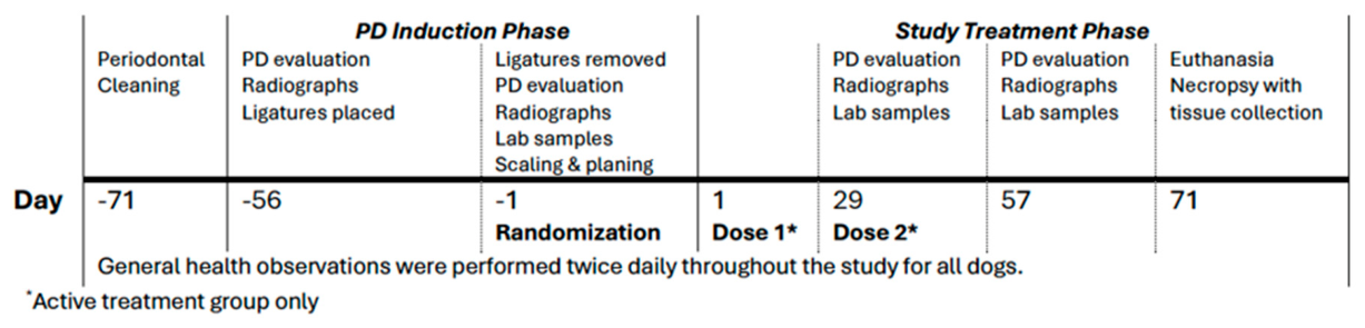
Figure 2.
(a) Application of Ambrilux to the periodontal pocket of teeth 407-409 following induction of ligature-induced PD. Excess Ambrilux (arrow) was removed prior to curing. The bracket represents area of PD that has yet to be treated. (b) Curing of Ambrilux within the periodontal pocket using a standard UV light (395-405 nm) for approximately 60 seconds.
Figure 2.
(a) Application of Ambrilux to the periodontal pocket of teeth 407-409 following induction of ligature-induced PD. Excess Ambrilux (arrow) was removed prior to curing. The bracket represents area of PD that has yet to be treated. (b) Curing of Ambrilux within the periodontal pocket using a standard UV light (395-405 nm) for approximately 60 seconds.
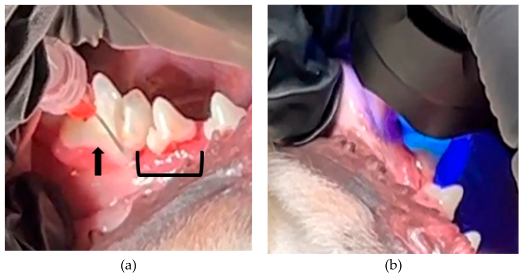
Figure 3.
Temporal radiographs of Control (#588) (a, b, c) and Ambrilux-treated animal (#913) (d, e, f). Images were obtained on Day -1 prior to Dose 1, Day 29 prior to Dose 2 and Day 57. The treated animal demonstrated increased bone formation, specifically in the tooth furcation.
Figure 3.
Temporal radiographs of Control (#588) (a, b, c) and Ambrilux-treated animal (#913) (d, e, f). Images were obtained on Day -1 prior to Dose 1, Day 29 prior to Dose 2 and Day 57. The treated animal demonstrated increased bone formation, specifically in the tooth furcation.
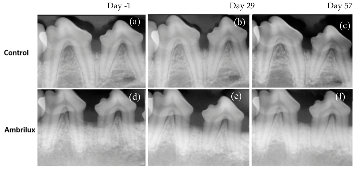
Figure 4.
Histopathology of H&E-stained canine periodontal tissue located between teeth 408 and 409 for control animal #834 (a, b, c) and Ambrilux-treated animal #892 (d, e, f). Outlined insets of (a, d) are shown at higher magnification view in (b, c) and (e, f) respectively. (a) Representative low magnification view of periodontium from control animal showing inflammation (I) in the gingiva and surrounding connective tissue (CT). Numerous osteoclasts may be noted in the alveolar bone (arrows). Higher magnification view (b) of the gingiva also shows an increase in tissue vascularity (arrowheads). (c) Osteoclast nests (arrow) are indicative of active bone resorption. (d) Ambrilux-treated animal exhibited a greater degree of organization within the connective tissue with decreased blood vessels and reduced inflammation, (e) as highlighted in higher magnification view. (f) Osteoclast activity was also reduced. I, Inflammation; CT, Connective Tissue; R, Tooth Root; AB, Alvolar Bone; BC, Bone Crest; PDL, Periodontal Ligament. Micrographs were obtained at magnifications of x6 (a, d) and x20 (b, c, e and f).
Figure 4.
Histopathology of H&E-stained canine periodontal tissue located between teeth 408 and 409 for control animal #834 (a, b, c) and Ambrilux-treated animal #892 (d, e, f). Outlined insets of (a, d) are shown at higher magnification view in (b, c) and (e, f) respectively. (a) Representative low magnification view of periodontium from control animal showing inflammation (I) in the gingiva and surrounding connective tissue (CT). Numerous osteoclasts may be noted in the alveolar bone (arrows). Higher magnification view (b) of the gingiva also shows an increase in tissue vascularity (arrowheads). (c) Osteoclast nests (arrow) are indicative of active bone resorption. (d) Ambrilux-treated animal exhibited a greater degree of organization within the connective tissue with decreased blood vessels and reduced inflammation, (e) as highlighted in higher magnification view. (f) Osteoclast activity was also reduced. I, Inflammation; CT, Connective Tissue; R, Tooth Root; AB, Alvolar Bone; BC, Bone Crest; PDL, Periodontal Ligament. Micrographs were obtained at magnifications of x6 (a, d) and x20 (b, c, e and f).
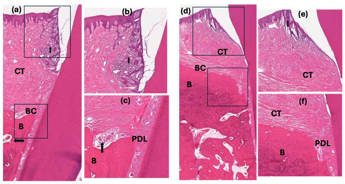
Figure 5.
H&E-stained histology images highlighting the furcation periodontal ligament from (a) a control animal #323 and (c)Ambrilux-treated animal #892 (both tooth 409). Outlined insets of (a), (c) are shown at higher magnification view in (b) and (d) respectively. Severe degradation of the PDL was noted in control animals as compared to treated animals. Micrographs were obtained at magnifications of x6 (a, c) and x20 (c, d).
Figure 5.
H&E-stained histology images highlighting the furcation periodontal ligament from (a) a control animal #323 and (c)Ambrilux-treated animal #892 (both tooth 409). Outlined insets of (a), (c) are shown at higher magnification view in (b) and (d) respectively. Severe degradation of the PDL was noted in control animals as compared to treated animals. Micrographs were obtained at magnifications of x6 (a, c) and x20 (c, d).
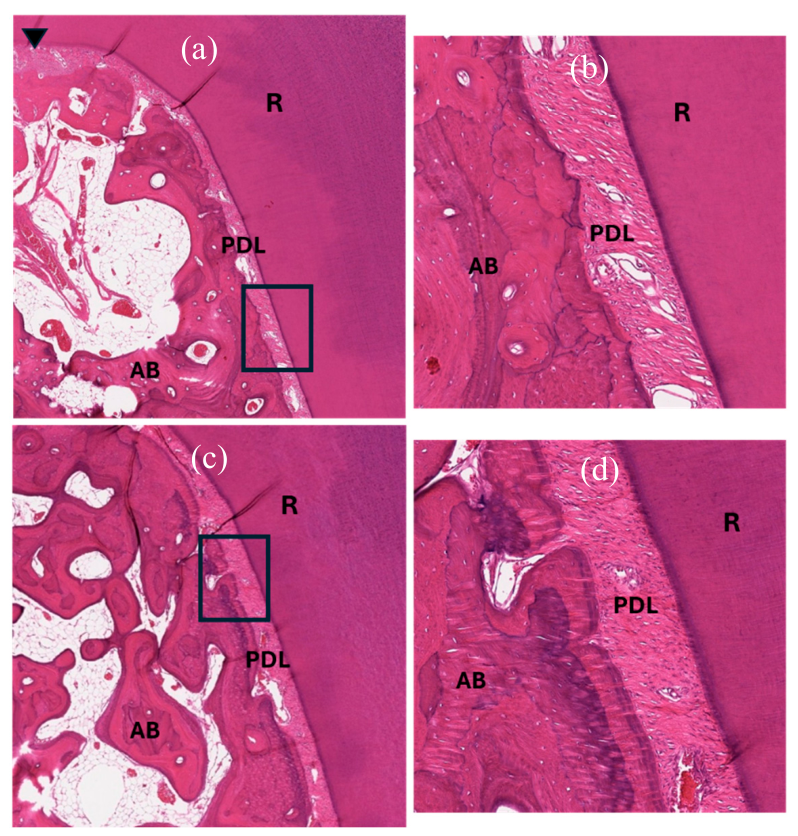
Figure 6.
Representative 3D and sagittal images of control (a, c, d) and Ambrilux-treated (b, e, f) animals (Animal #323 and #913 respectively). The degree of bone loss (black arrows) in the external 3D view (a, b) is more pronounced in control samples as compared to treated samples. Internal 3D (c, e) and sagittal (d, f) images exemplify the increased bone formation in the furcation areas of treated animals.
Figure 6.
Representative 3D and sagittal images of control (a, c, d) and Ambrilux-treated (b, e, f) animals (Animal #323 and #913 respectively). The degree of bone loss (black arrows) in the external 3D view (a, b) is more pronounced in control samples as compared to treated samples. Internal 3D (c, e) and sagittal (d, f) images exemplify the increased bone formation in the furcation areas of treated animals.
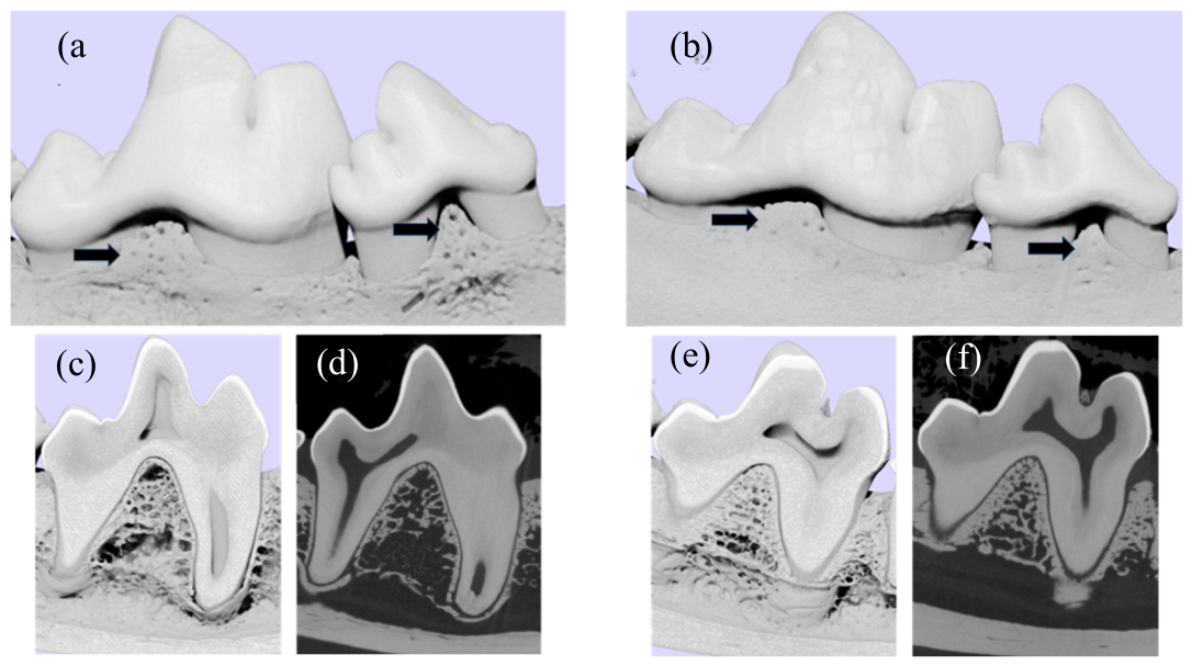
Figure 7.
Volumetric analysis of the furcation of tooth 409. (a) Furcation area of tooth 409 representing the registered VOI for analysis. Bone within red highlighted volume of interest was quantified for thickness, volume, and local bone mineral density. (b) Representative image of furcation from a control animal (#588). (c) Representative image of furcation from a and a treated animal (#542).
Figure 7.
Volumetric analysis of the furcation of tooth 409. (a) Furcation area of tooth 409 representing the registered VOI for analysis. Bone within red highlighted volume of interest was quantified for thickness, volume, and local bone mineral density. (b) Representative image of furcation from a control animal (#588). (c) Representative image of furcation from a and a treated animal (#542).
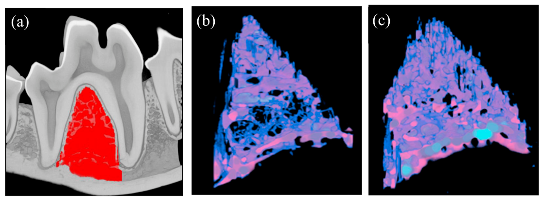

Table 1.
Quantitative CT assessment of bone density, volume and trabecular parameters at furcation.
| BMD (g CaHA/cm3) |
Bone Volume (mm3) | BV/TV (%) |
Trabecular Number (1/mm) | Trabecular Thickness (mm) | Trabecular Separation (mm) | |
|---|---|---|---|---|---|---|
| Control | 0.205 ± 0.017 | 44.35 ± 8.44 | 16.42 ± 1.38 | 0.481 ± 0.016 | 0.139 + 0.007 | 2.03 ± 0.26 |
| Treated | 0.243 ± 0.004 | 60.7 ± 2.92 | 19.9 ± 0.71 | 0.561 ± 0.009 | 0.142 + 0.004 | 2.18 ± 0.11 |
| BMD=bone mineral density; BV=bone volume; TV=total volume | ||||||
Disclaimer/Publisher’s Note: The statements, opinions and data contained in all publications are solely those of the individual author(s) and contributor(s) and not of MDPI and/or the editor(s). MDPI and/or the editor(s) disclaim responsibility for any injury to people or property resulting from any ideas, methods, instructions or products referred to in the content. |
© 2026 by the authors. Licensee MDPI, Basel, Switzerland. This article is an open access article distributed under the terms and conditions of the Creative Commons Attribution (CC BY) license (http://creativecommons.org/licenses/by/4.0/).
Copyright: This open access article is published under a Creative Commons CC BY 4.0 license, which permit the free download, distribution, and reuse, provided that the author and preprint are cited in any reuse.