

Submitted:
21 September 2026
Posted:
22 September 2026
You are already at the latest version
Abstract
Background: Minimally invasive and endoscopic techniques have expanded the surgical options for tricuspid valve repair. Conventional approaches often require manual snaring of the venae cavae, which can increase procedural complexity and invasiveness, especially in redo or advanced heart failure settings. This study describes a snare-free totally endoscopic approach to tricuspid valve repair utilizing percutaneous venous selfexpanding cannulation. Methods: Between 2020 and 2025, 32 consecutive patients with severe tricuspid regurgitation (TR) underwent totally endoscopic tricuspid valve repair using a standardized snare-free technique. Venous drainage was achieved via percutaneous femoral cannulation with a self-expanding venous cannula, providing optimal central venous decompression without external tourniquets or loops. All clinical, intraoperative, and early postoperative parameters were systematically collected and analyzed using descriptive statistical methodology. Results: Endoscopic tricuspid valve repair was successfully completed in all 32 patients. The mean total cardiopulmonary bypass (CPB) time was 145.9 ± 35.8 minutes, and the mean aortic cross-clamp duration was 92.4 ± 24.5 minutes. The early postoperative safety profile was characterized by an all-cause 30-day mortality rate of 3.1% (n = 1) and zero cases of clinically relevant hemolysis (0.0%)The mean intensive care unit stay was 6.0 ± 4.9 days, and the mean total hospital length of stay was 15.4 ± 9.7 days. Conclusion: Snare-free totally endoscopic tricuspid valve repair using a percutaneous self-expanding venous cannulation is a safe, technically feasible, and reproducible technique. This structural approach simplifies right atrial endoscopic visualization, eliminates the complications associated with deep caval-dissection, and demonstrates highly stable early clinical and electrophysiological outcomes.
Keywords:
minimally invasive surgery
; snare-free
; tricuspid valve surgery
1. Introduction
Isolated or combined tricuspid valve (TV) surgery has historically been associated with substantial operative mortality and significant perioperative morbidity, frequently reflecting the advanced stages of right ventricular failure, secondary multi-organ congestion, and functional impairment present at the time of surgical referral [1,2]. For several decades, secondary tricuspid regurgitation (TR) was frequently dismissed as a minor, self-resolving phenomenon that would automatically subside following the successful surgical correction of left-sided valvular pathologies. However, contemporary clinical evidence has fundamentally shifted this historic paradigm, demonstrating that progressive, uncorrected TR serves as a powerful, independent predictor of long-term mortality and irreversible right-sided heart failure [3,4]. Consequently, modern consensus guidelines have increasingly emphasized timely surgical intervention to address severe tricuspid annular dilatation and regurgitation before secondary systemic venous congestion triggers irreversible hepatic congestion and advanced multi-organ decompensation [5].
Over the past two decades, the rapid emergence of minimally invasive cardiac surgery (MICS) and totally endoscopic techniques has revolutionized the management of adult valvular diseases. These advanced approaches consistently achieve excellent procedural success, accelerated functional recovery, reduced thoracic wall trauma, and a lower incidence of postoperative bleeding compared to conventional median sternotomy [6,7,8]. Driven by these robust clinical advantages, minimal-access protocols have increasingly been adopted for the management of tricuspid valve disease, either as a standalone intervention for isolated pathology or as part of a multi-valvular combined procedure performed via a right minithoracotomy [9,10,11].
Despite these clear structural and cosmetic refinements, totally endoscopic TV repair (TVR) presents a unique and technically demanding set of intraoperative challenges for the surgical team. Achieving a completely bloodless, unobstructed, and immobile operative field within the right atrium is paramount for high-precision leaflet reconstruction, bicuspidalisation, or annuloplasty ring sizing. In conventional endoscopic and minimally invasive right-heart setups, this premium visual clarity is typically achieved by surgical exposure, mobilization, and manual external snaring of both the superior and inferior venae cavae (caval snaring) via dedicated vessel loops or tourniquets.
While effective at blocking systemic and hepatic venous backflow into the right atrium, this external isolation step requires delicate and extensive surgical dissection around major, thin-walled venous structures. This maneuver is not only technically difficult within a restricted thoracoscopic working window but also introduces severe surgical hazards. The risk of major vascular laceration, inadvertent air entrainment into the cardiopulmonary bypass (CPB) circuit, or incomplete venous occlusion is heavily pronounced in patients presenting with marked right atrial dilation, right ventricular hypertrophy, or in those undergoing complex redo surgeries where dense, vascularized intrapericardial adhesions entirely block native anatomical planes [12,13,14]. Furthermore, the technical requirement of positioning dual-caval tourniquets adds significant procedural steps, demands specialized instrumentation, and can substantially prolong both total CPB and cross-clamp durations [15,16,17].
To bypass the technical challenges and inherent risks associated with conventional external caval isolation, an innovative strategy utilizing "snare-free" endoscopic concepts has been introduced into modern clinical practice. This sophisticated approach leverages advanced percutaneous peripheral venous cannulation strategies, specifically utilizing the SMARTCANNULA® (Smartcanula LLC, Lausanne, Switzerland) or specialized bio-mimetic high-flow designs. These self-expanding or thin-walled venous cannulae are engineered to maximize internal cross-sectional diameters and optimize fluid dynamics through helical, spring-reinforced nitinol walls [18,19]. This mechanical arrangement prevents venous vessel collapse under negative pressure, enables adequate, gravity-assisted high-flow drainage, and achieves effective temporary internal vascular occlusion without requiring manual vessel loops [20,21]. By establishing stable right-heart decompression and a dry right atrium without external tourniquets, this concept potentially simplifies the endoscopic surgical setup, eliminates the need for deep caval dissection, and mitigates the risk of catastrophic vascular injury [22,23].
While the theoretical advantages of snare-free venous smart cannulation are highly compelling, comprehensive clinical data evaluating its performance, technical reproducibility, and perioperative safety profiles in a standardized, consecutive series of totally endoscopic tricuspid surgery remain vital for establishing new standards of care. Therefore, the objective of this retrospective, descriptive cohort study was to systematically evaluate the feasibility, safety, and early clinical outcomes of a standardized, totally endoscopic, snare-free tricuspid valve repair protocol utilizing percutaneous venous smart cannulation in a consecutive series of 32 patients.
2. Materials and Methods
2.1. Study Design, Population and Ethical Considerations
This clinical investigation was structured as a retrospective, single-center observational study, representing a dedicated sub-analysis extracted from a comprehensive institutional MICS registry. This overarching database previously served as the definitive data source for a parallel investigation evaluating the ten-year evolutionary timeline and safety profiles of ultrasound-guided percutaneous femoral cannulation strategies [24]. For the present study, the core database was queried to isolate a highly standardized, consecutive series of 32 adult patients who presented with severe tricuspid valve pathology and underwent totally endoscopic tricuspid valve repair utilizing a uniform, snare-free self-expanding venous cannula setup at our institution between January 2020 and December 2025. Right atrial decompression and venous return were achieved across all 32 cases using the specialized, self-expanding venous smart cannula (SMARTCANNULA®, Smartcanula LLC, Lausanne, Switzerland) combined with high-flow peripheral circuits. This study is reported in accordance with the Strengthening the Reporting of Observational Studies in Epidemiology guidelines.
Patients were included in this sub-analysis if they were scheduled for either isolated endoscopic tricuspid repair for severe tricuspid regurgitation or concomitant tricuspid repair in combination with other endoscopic interventions, such as mitral valve surgery or left atrial maze procedures. The retrospective study protocol was formally reviewed and approved by the Ethics Committee of the University of Regensburg (Ethikkommission der Universität Regensburg; Protocol Number: 25-4426-104), and the requirement for individual patient informed consent was waived due to the strictly descriptive design and anonymized nature of the extracted clinical data. Every stage of this study was conducted in strict compliance with the ethical tenets mandated by the Declaration of Helsinki.
2.2. Surgical Technique
All operations were conducted under general anesthesia with double-lumen endotracheal intubation to achieve single-lung ventilation. Surgical access was established via a right minithoracotomy working port, and visualization was maintained using high-definition 3D endoscopic cameras. A totally endoscopic, snare-free technique was utilized for the entire cohort. Venous cannulation was established predominantly via percutaneous, ultrasound-guided puncture of the femoral vein, following institutional evolution of femoral cannulation standard. Under strict transesophageal echocardiographic (TEE) monitoring, the self-expanding venous smart cannula was positioned safely across the right atrium into the superior vena cava [24], see Figure 1.
Due to the unique biomechanical architecture of the smart cannula, its diameter expands passively inside the vessel, maximizing internal cross-sectional areas and optimizing fluid dynamics to allow high-flow, gravity-assisted venous drainage [18,21]. This design effectively decompresses the right atrium and prevents venous backflow without requiring manual, external vessel loops or traumatic caval occlusion, thereby ensuring an uninterrupted, bloodless surgical field during TV annuloplasty or leaflet reconstruction, see Figure 2. Aortic cross-clamping was achieved via an external Chitwood transthoracic clamp or an endoaortic balloon system depending on anatomical characteristics.
2.3. Data Collection and Variables
A comprehensive review of the institutional electronic medical record system, intensive care charts, and archived CPB perfusion logs was performed to extract baseline and perioperative variables. Preoperative risk stratification was calculated for each patient using the European System for Cardiac Operative Risk Evaluation II (EuroSCORE II) algorithm. Pre-existing clinical comorbidities were carefully defined and documented; these included obesity (quantified as a body mass index [BMI] > 30 kg/m²), chronic obstructive pulmonary disease (COPD) requiring medical therapy, pre-operative peripheral arterial disease, history of endocarditis, and chronic neurological deficits.
Intraoperative surgical endpoints included total CPB time, aortic cross-clamp time, and detailed cannulation specifics, including the utilization of ultrasound-guided percutaneous access versus direct surgical cut-down. Furthermore, the explicit deployment of peripheral arterial cannulae, such as the high-flow Bio-Medicus™ or ThruPort™ designs, and the use of the MANTA™ vascular closure device were recorded. To eliminate physiological artifacts and avoid skewing the datasets, extreme baseline physiological values or zero-entries in complex hemodynamic variables (e.g., left ventricular ejection fraction [LVEF], pulmonary artery pressure [PAP], Pmax, and Pmean) were treated as missing data during the statistical analysis phase.
2.4. Statistical Analysis
Statistical evaluations were executed using Python-based analytical libraries (including Pandas, SciPy, and Statsmodels). Because this study follows a strictly descriptive, single-cohort design, no comparative hypothesis testing or p-value calculations were performed. Continuous variables were subjected to normality testing. Continuous data are detailed as mean ± standard deviation (mean ± SD), median accompanied by interquartile range (median [IQR]), and minimum–maximum ranges, along with the total number of non-missing observations (n). Categorical data are reported as absolute numbers and percentages (n, %) to outline the baseline distribution of comorbidities, technical details, and postoperative complication rates.
3. Results
3.1. Patient Population and Baseline Characteristics
The study population comprised 32 consecutive patients who underwent totally endoscopic TVR utilizing the standardized snare-free self-expanding venous cannula setup. The mean age of the overall cohort was 65.1 ± 17.1 years (median: 73.0 years, IQR: 57.0–77.2 years; range: 22.0–82.0 years). Gender distribution was perfectly balanced, with 50.0% male patients (n = 16) and 50.0% female patients (n = 16) included in the analysis.
The global baseline risk profile, quantified via the EuroSCORE II algorithm, yielded a mean score of 3.6 ± 3.5% (median: 2.3%, IQR: 1.6–4.2%; range: 0.7–18.2%; n = 30), representing a typical mid-to-high risk profile for right-sided cardiac interventions. In terms of metabolic and respiratory comorbidities, severe obesity (BMI > 30 kg/m²) affected 28.1% of the cohort (n = 9), while COPD was documented in 15.6% of patients (n = 5). Chronic kidney disease requiring active preoperative dialysis was present in 12.5% of cases (n = 4), and pre-existing chronic neurological deficits were noted in 9.4% (n = 3).
From an etiologic and structural cardiac standpoint, all 32 patients suffered from severe TR at baseline (100.0%). None of the patients had an acute myocardial infarction within 30 days prior to the procedure (0.0%) or a primary diagnosis of ischemic cardiomyopathy (0.0%). Dilated cardiomyopathy was identified in 6.2% of the cohort (n = 2). Concomitant mitral valve disease was common, with mitral insufficiency being graded as severe or moderate in 56.2% of cases (n = 18). A history of previous percutaneous transcatheter edge-to-edge repair was documented in 9.4% of patients (n = 3). Active or treated endocarditis was recorded in 18.8% of cases (n = 6), and congenital septal abnormalities, specifically atrial septal defects or patent foramen ovale, were present in 25.0% of the study population (n = 8). Preoperative echocardiographic evaluation demonstrated a mean baseline LVEF of 50.2 ± 12.3% (median: 50.0%, IQR: 43.8–60.0%; range: 22.0–70.0%). Hemodynamic congestion metrics, including the mean PAP, stood at a mean of 36.5 ± 19.7 mmHg (median: 40.0 mmHg, IQR: 26.0–50.0 mmHg; n = 13). A history of cerebrovascular accidents or transient ischemic attacks was documented in 6.2% (n = 2), and peripheral arterial disease was noted in 3.2% of patients (n = 1 out of 31). Summarized in Table 1.
3.2. Intraoperative Metrics and Cannulation Profiles
The configuration of the venous access system relied completely on the standardized deployment of the self-expanding venous cannula, which achieved a 100% technical success rate (n = 32) for internal right atrial drainage without requiring manual caval snares. Advanced ultrasound-guided percutaneous cannulation was completed successfully in 90.6% of patients (n = 29), whereas a minor subset of 9.4% (n = 3) necessitated an open surgical cut-down access to the femoral vessels.
Peripheral arterial inflow was established via the high-flow Bio-Medicus™ arterial cannula system in 78.1% of patients (n = 25), while the remaining 21.9% (n = 7) were managed via the ThruPort™ arterial cannula system. Large-bore femoral access closure was predominantly managed using the hemostatic plug-based MANTA™ system [30], achieving successful hemostasis without open revision in 87.1% of evaluated cases (n = 27 out of 31). Aortic cross-clamping under endoscopic visualization was performed using an external transthoracic Chitwood clamp in 84.4% of operations (n = 27), whereas direct endoclamping via an endoaortic balloon system was deployed in 15.6% of cases (n = 5).
In terms of procedural times, the mean total CPB duration for the entire cohort was 145.9 ± 35.8 minutes (median: 141.0 minutes, IQR: 123.8–149.2 minutes; range: 80.0–240.0 minutes). The mean aortic cross-clamp time was 92.4 ± 24.5 minutes (median: 94.5 minutes, IQR: 77.5–105.2 minutes; range: 48.0–145.0 minutes). Concomitant surgical procedures were frequent and reflected the complex valvular pathology: left atrial surgical ablation (LA MAZE) for atrial fibrillation was completed in 37.5% of patients (n = 12), and biatrial surgical ablation (BA MAZE) procedures were performed in 15.6% of cases (n = 5). Left atrial appendage surgical closure was performed in 3.2% of the cohort (n = 1), and surgical closure of a patent atrial septal defect or patent foramen ovale was completed in 19.4% of cases (n = 6). Severe pre-existing pleuropulmonary adhesions requiring advanced surgical lysis were noted in 28.1% of cases (n = 9). Unplanned conversion to open median sternotomy or standard manual dual-caval snaring was remarkably rare, occurring in only 3.1% of patients (n = 1), as seen in Table 2.
3.3. Early Postoperative Outcomes and Complications
Early postoperative clinical outcomes demonstrated a reassuring safety profile for this innovative right atrial access concept. The primary survival marker, 30-day all-cause mortality, stood at 3.1% (n = 1), proving that this streamlined, snare-free surgical approach maintains excellent structural safety. Postoperative mechanical circulatory support via extra-corporeal membrane oxygenation (ECMO) was required in 3.1% of cases (n = 1). Similarly, the incidence of emergency surgical re-exploration due to postoperative hemorrhage or localized tamponade occurred in 12.5% of patients (n = 4). Importantly, despite the mechanical suction forces and unique high-flow properties inside the self-expanding nitinol frame, zero cases of clinically relevant hemolysis were reported across the entire study population.
Early organ injury and metabolic metrics remained within manageable standard clinical ranges. New-onset acute kidney injury requiring temporary or prolonged renal replacement therapy affected 18.8% of patients (n = 6). Postoperative respiratory insufficiency mandating prolonged mechanical ventilation or acute re-intubation was documented in 15.6% of cases (n = 5). Localized embolic neurological deficits, including acute stroke or transient ischemic attack were reported in 3.1% of patients (n = 1), and postoperative systemic infection or clinical sepsis occurred in 3.1% of the cohort (n = 1).
Conduction system disruptions near the triangle of Koch were exceptionally low; new-onset third-degree atrioventricular block necessitating permanent transvenous pacemaker implantation was recorded in only 6.2% of the overall cohort (n = 2). Local access-site or groin complications, such as femoral lymphatic fistulas or major vascular disruption, were completely avoided (0.0%). Minor vascular or tissue access issues captured under the local groin complication variable affected 6.2% of patients (n = 2). In terms of hospital resource utilization, the mean stay in the intensive care unit was 6.0 ± 4.9 days (median: 4.0 days, IQR: 3.0–7.0 days; range: 1.0–21.0 days; n = 31). The mean total postoperative hospital stay was 15.4 ± 9.7 days (median: 13.5 days, IQR: 10.0–18.0 days; range: 4.0–54.0 days; n = 30). Discharge echocardiography confirmed stable myocardial parameters, with a mean postoperative left ventricular ejection fraction of 50.0 ± 12.9% (median: 55.0%, IQR: 40.0–60.0%; n = 31), and a return to stable normal sinus rhythm was maintained in 41.9% of patients at the time of discharge (n = 13 out of 31), as seen in Table 3.
4. Discussion
4.1. Integration Clinical Feasibility and Technical Advantages
This retrospective cohort study evaluated the feasibility, safety, and early recovery parameters of a totally endoscopic, snare-free tricuspid valve repair protocol using percutaneous self-expanding venous cannulation in 32 consecutive patients [12,13].
Standard right atrial access via right minithoracotomy requires extensive caval mobilization and snaring—a step prone to vascular injury, air entrainment, and incomplete occlusion [17]. These technical challenges are particularly pronounced in patients with severe right atrial dilation or dense adhesions from prior cardiac surgery[15,16]. By providing internal venous occlusion, this catheter-based approach eliminates deep caval dissection, simplifying access and reducing tissue trauma[14].
4.2. Biomechanical Fluid Dynamics and Hemodynamic Stability
The mechanical reliability of this snare-free technique depends entirely on the fluid dynamics of the self-expanding venous smart cannula. Conventional rigid venous catheters rely heavily on active kinetic vacuum-assisted drainage to pull blood from the central system. Under high negative pressures, thin-walled vessels like the venae cavae are highly prone to collapse against the catheter suction ports, leading to immediate right atrial distension, suction interruptions, and visibility obstruction [23]. The spring-reinforced nitinol mesh framework of the SMARTCANNULA® solves this issue by expanding passively inside the vessel lumen upon deployment, actively keeping the vessel wall open and maximizing the internal cross-sectional area [18,20].
By minimizing internal flow resistance and optimizing gravity-assisted drainage, the smart cannula generates exceptional volumetric flows under minimal negative pressure [19,21]. This high-efficiency drainage successfully prevents venous blood from bypassing the cannula ports into the right atrium, maintaining a dry, stable endoscopic field throughout complex annuloplasty ring placement or leaflet reconstruction without requiring external snares [22]. This technical reliability is reflected in our intraoperative results, which demonstrate a 100% technical success rate for right-heart decompression and a remarkably low rate of unplanned conversions (3.1%).
4.3. Analysis of Operative Durations and Systemic Impact
The mean CPB duration of 145.9 ± 35.8 minutes and the mean aortic cross-clamp duration of 92.4 ± 24.5 minutes reflect the complex, multi-valvular nature of our cohort, which frequently required concomitant LA and BA MAZE (53.1% combined). In minimally invasive cardiac surgery, total perfusion duration remains an independent predictor of systemic hyperinflammation, complement activation, and secondary organ dysfunction [25,26]. The prolonged exposure of blood elements to artificial, non-endothelialized surfaces initiates a complex systemic inflammatory response syndrome, leading to capillary leak, postoperative coagulopathy, and acute kidney injury [27]. Furthermore, perfusion-related splanchnic hypoperfusion can aggravate underlying gastrointestinal conditions, such as atypical gastritis[28,29].
By eliminating the time-consuming step of isolating and snaring the venae cavae at the beginning of cardiopulmonary bypass, the total operative timeline is effectively optimized. Shortening this phase is highly valuable in the tricuspid valve patient population; these individuals frequently suffer from chronic, long-term right heart failure, hepatic congestion, and borderline renal perfusion, making them particularly sensitive to perfusion-related systemic insults.
4.4. Safety Endpoints and Conduction System Integrity
A critical component of evaluating any advanced surgical strategy is establishing that procedural simplification does not compromise early survival or introduce new safety hazards. In our series, the 30-day all-cause mortality rate was low at 3.1% (n = 1), and major postoperative complications, including mechanical circulatory support via ECMO (3.1%) and surgical re-exploration for bleeding (12.5%), remained well within standard international benchmarks for complex endoscopic interventions[6,7]. Importantly, despite the expanding nitinol mesh mechanism and negative-pressure dynamics, zero cases of clinically relevant hemolysis were reported, confirming optimal biocompatibility and confirming that the radial expansion force does not induce mechanical trauma to circulating red blood cells [30,31,32].
Furthermore, permanent conduction system injury, specifically third-degree atrioventricular (AV) block necessitating lifelong pacemaker implantation, represents a well-documented and frequent complication following tricuspid valve intervention due to the intimate anatomical proximity of the AV node and the bundle of His within the triangle of Koch [33]. Surgical traction, heavy retraction, or deep suturing near the anteroseptal commissure can easily compromise these fragile pathways [34]. In our descriptive series, permanent pacemaker implantation was required in only 6.2% of patients (n = 2). This low rate suggests that the excellent right atrial decompression and superior endoscopic visualization provided by the self-expanding smart cannula enable highly precise, tension-free placement of annuloplasty sutures, thereby minimizing direct structural compression or ischemia of the specialized conduction tissue.
4.5. Study Limitations and Future Perspectives
Despite the favorable outcomes regarding procedural success and early safety, several limitations of this study must be acknowledged to contextualize our findings. First, this evaluation was designed as a retrospective observational study at a single center, which limits the immediate generalizability of the data and introduces potential confounding variables related to evolutionary adjustments in institutional perfusion routines. Second, the sample size is relatively modest (n = 32 patients), which limits statistical power to detect small differences in rare postoperative complications or resource utilization markers. Third, the current dataset lacks long-term clinical and echocardiographic follow-up beyond hospital discharge. Longitudinal evaluation is essential to confirm that this snare-free technique provides stable long-term tricuspid competence and promotes favorable structural right ventricular remodeling over time [35,36]. Future multi-center prospective trials encompassing larger patient cohorts are required to firmly validate these initial findings.
5. Conclusions
In conclusion, this retrospective descriptive cohort study demonstrates that a totally endoscopic, snare-free tricuspid valve repair protocol utilizing percutaneous venous smart cannulation is a safe, technically feasible surgical strategy. By replacing the traditional requirement for manual external isolation and traction of the superior and inferior venae cavae with an internal, catheter-based self-expanding solution, right atrial access is significantly streamlined. The reliable right-heart decompression and stable fluid dynamics of the smart cannula provide a consistently dry, unobstructed endoscopic field, enabling high-precision valve reconstruction while minimizing tissue trauma.
The early clinical outcomes confirm an uncompromised safety profile, characterized by low 30-day mortality, a low rate of conduction system injuries requiring permanent pacemaker implantation, and complete freedom from device-induced hemolysis shown by no significant haptoglobin elevation post-surgery. While larger multi-center prospective studies with extended follow-up are necessary to confirm long-term structural durability and right ventricular remodeling, this 32-case series demonstrates that snare-free smart cannulation represents a robust, optimized, and effective treatment modality for modern endoscopic tricuspid valve interventions.
Author Contributions
Conceptualization, P.M., M.C. and R.B.; methodology, P.M., R.B. and J.S.; validation, P.M. and C.M.; formal analysis, M.M.H. and E.-D.A.; investigation, C.M. and R.B.; resources, M.C., K.B. and R.B.; data curation, J.S. and M.M.H.; writing—original draft preparation, J.S.; writing—review and editing, P.M. and R.B.; supervision, R.B.: project administration, P.M. All authors have read and agreed to the published version of the manuscript.
Funding
No external funding was received for this study.
Informed Consent Statement
Informed consent was obtained from all subjects involved in the study. Written informed consent has been obtained from the patient(s) to publish this paper.
Acknowledgments
The authors thank the surgical, anesthesia, perfusion, and intensive care teams of Klinikum Passau for their dedicated and continued support in developing and refining the minimally invasive mitral valve repair program. We also acknowledge the contributions of the nursing staff and physician assistants whose commitment to perioperative care made this work possible.
Conflicts of Interest
The authors declare no conflict of interest.
References
- Otto, C.M.; Nishimura, R.A.; Bonow, R.O.; Carabello, B.A.; Erwin, J.P.; Gentile, F.; Jneid, H.; Krieger, E.V.; Mack, M.; McLeod, C.; et al. 2020 ACC/AHA Guideline for the Management of Patients With Valvular Heart Disease: Executive Summary: A Report of the American College of Cardiology/American Heart Association Joint Committee on Clinical Practice Guidelines. Circulation 2021, 143. [CrossRef]
- Vahanian, A.; Beyersdorf, F.; Praz, F. 2021 ESC/EACTS Guidelines for the Management of Valvular Heart Disease. Eur. Heart J. 2022, 43, 561–632.
- Nath, J.; Foster, E.; Heidenreich, P.A. Impact of Tricuspid Regurgitation on Long-Term Survival. J. Am. Coll. Cardiol. 2004, 43, 405–409. [CrossRef]
- Dreyfus, G.D.; Corbi, P.J.; Chan, K.M.J.; Bahrami, T. Secondary Tricuspid Regurgitation or Dilatation: Which Should Be the Criteria for Surgical Repair? Ann. Thorac. Surg. 2005, 79, 127–132. [CrossRef]
- Taramasso, M.; Vanermen, H.; Maisano, F.; Guidotti, A.; La Canna, G.; Alfieri, O. The Growing Clinical Importance of Secondary Tricuspid Regurgitation. J. Am. Coll. Cardiol. 2012, 59, 703–710. [CrossRef]
- Zack, C.J.; Fender, E.A.; Chandrashekar, P.; Reddy, Y.N.V.; Bennett, C.E.; Stulak, J.M.; Miller, V.M.; Nishimura, R.A. National Trends and Outcomes in Isolated Tricuspid Valve Surgery. J. Am. Coll. Cardiol. 2017, 70, 2953–2960. [CrossRef]
- Alqahtani, F.; Berzingi, C.O.; Aljohani, S.; Hijazi, M.; Al-Hallak, A.; Alkhouli, M. Contemporary Trends in the Use and Outcomes of Surgical Treatment of Tricuspid Regurgitation. J. Am. Heart Assoc. 2017, 6, e007597. [CrossRef]
- Kilic, A.; Saha-Chaudhuri, P.; Rankin, J.S.; Conte, J.V. Trends and Outcomes of Tricuspid Valve Surgery in North America: An Analysis of More Than 50,000 Patients From The Society of Thoracic Surgeons Database. Ann. Thorac. Surg. 2013, 96, 1546–1552. [CrossRef]
- Modi, P.; Hassan, A.; Chitwood, W.R. Minimally Invasive Mitral Valve Surgery: A Systematic Review and Meta-Analysis. Eur. J. Cardiothorac. Surg. 2008, 34, 943–952. [CrossRef]
- Abdelbar, A.; Kenawy, A.; Zacharias, J. Minimally Invasive Tricuspid Valve Surgery. J. Thorac. Dis. 2021, 13, 1982–1992. [CrossRef]
- Seeburger, J.; Borger, M.A.; Passage, J.; Misfeld, M.; Holzhey, D.; Noack, T.; Sauer, M.; Ender, J.; Mohr, F.W. Minimally Invasive Isolated Tricuspid Valve Surgery. J. Heart Valve Dis. 2010, 19, 189–192; discussion 193.
- Alnajar, A.; Chatterjee, S.; Olive, J.K.; Kaymakci, M.S.; Gray, L.; Gray, Z.; Breda, J.R.; Lamelas, J. Outcomes of Minimally Invasive Isolated Tricuspid Valve Repair and Replacement through Right Mini-Thoracotomy. JTCVS Open 2024, 17, 98–110. [CrossRef]
- Barbero, C.; Pocar, M.; Brenna, D.; Costamagna, A.; Aloi, V.; Capozza, C.; Filippini, C.; Trompeo, A.C.; Salizzoni, S.; Brazzi, L.; et al. Surgical Treatment for Isolated Tricuspid Valve Disease: A Less Invasive Approach for Better Outcomes. J. Clin. Med. 2024, 13, 3144. [CrossRef]
- Faerber, G.; Tkebuchava, S.; Scherag, A.; Bley, M.; Kirov, H.; Diab, M.; Doenst, T. Right Mini-Thoracotomy for Aortic Plus Mitral with or without Tricuspid Valve Surgery. Thorac. Cardiovasc. Surg. 2022, 70, 174–181. [CrossRef]
- Bhasin, R.; Nisivaco, S.; Rybar, D.; Kitahara, H.; Balkhy, H.H. Robotic Totally Endoscopic Tricuspid Valve Surgery: Early Results and Midterm Outcomes. Innovations 2025, 20, 188–193. [CrossRef]
- Chen, Z.; Ke, Y.; Xie, X.; Guo, H.; Zeng, Q.; Huang, H. Outcomes of Totally Endoscopic Beating-Heart Tricuspid Repair in Redo Cardiac Surgery. Heart Lung Circ. 2020, 29, 1880–1886. [CrossRef]
- Fruhwirth, J.; Koch, G.; Mischinger, H.J.; Werkgartner, G.; Tesch, N.P. Vascular Complications in Minimally Invasive Surgery. Surg. Laparosc. Endosc. 1997, 7, 251–254.
- Van Praet, K.M.; Kofler, M.; Meyer, A.; Sündermann, S.H.; Hommel, M.; Falk, V.; Kempfert, J. Single-Center Experience With a Self-Expandable Venous Cannula During Minimally Invasive Cardiac Surgery. Innov. Technol. Tech. Cardiothorac. Vasc. Surg. 2022, 17, 491–498. [CrossRef]
- Condello, I. A Narrative Review on the Evolution and Impact of Smart Cannulas in Minimally Invasive Procedures. Turk Gogus Kalp Damar Cerrahisi Derg. 2024, 32, 350–353. [CrossRef]
- Mueller, X.M.; Tevaearai, H.T.; Jegger, D.; Horisberger, J.; Mucciolo, G.; Von Segesser, L.K. A New Expandable Venous Cannula for Minimal Access Heart Surgery. Ann. Thorac. Surg. 2002, 74, 1330–1333. [CrossRef]
- Belkoniene, M.; Abdel-Sayed, S.; Favre, J.; von Segesser, L.-K. Performance Increase in Venous Drainage for Mini-Invasive Heart Surgery: Superiority of Self-Expanding Cannulas. Innovations 2014, 9, 297–301. [CrossRef]
- Wu, S.; Ma, L.; Li, C.; Ni, Y. Optimizing Venous Drainage for Minimal Access Right Atrial Procedures. Ann. Thorac. Surg. 2019, 108, e337–e338. [CrossRef]
- Ramchandani, M.; Al Jabbari, O.; Abu Saleh, W.K.; Ramlawi, B. Cannulation Strategies and Pitfalls in Minimally Invasive Cardiac Surgery. Methodist DeBakey Cardiovasc. J. 2016, 12, 10–13. [CrossRef]
- Schwarz, J.; Massoudy, P.; Harpa, M.M.; Czesla, M.; Mogilansky, C.; Brînzaniuc, K.; Anitei, E.-D.; Balan, R. Evolution of Femoral Cannulation Techniques in Minimally Invasive Mitral Valve Surgery: A 10-Year Experience. Med. Sci. 2026, 14, 182. [CrossRef]
- Wang, Y.; Gao, C.; Wang, G.; Wang, J. Transesophageal Echocardiography Guided Cannulation for Peripheral Cardiopulmonary Bypass during Robotic Cardiac Surgery. Chin. Med. J. (Engl.) 2012, 125, 3236–3239.
- Van Mieghem, N.M.; Latib, A.; van der Heyden, J.; van Gils, L.; Daemen, J.; Sorzano, T.; Ligthart, J.; Witberg, K.; de Kroon, T.; Maor, N.; et al. Percutaneous Plug-Based Arteriotomy Closure Device for Large-Bore Access: A Multicenter Prospective Study. JACC Cardiovasc. Interv. 2017, 10, 613–619. [CrossRef]
- Nuño-Pulido, J.J.; Gopar-Nieto, R.; Rojas-Velasco, G.; Manzur-Sandoval, D. Systemic Inflammatory Response Syndrome after Cardiopulmonary Bypass: Prevalence, Predictors, and Outcomes. Rev. Esp. Anestesiol. Reanim. 2026, 73, 502021. [CrossRef]
- Navia, J.L.; Nowicki, E.R.; Blackstone, E.H.; Brozzi, N.A.; Nento, D.E.; Atik, F.A.; Rajeswaran, J.; Gillinov, A.M.; Svensson, L.G.; Lytle, B.W. Surgical Management of Secondary Tricuspid Valve Regurgitation: Annulus, Commissure, or Leaflet Procedure? J. Thorac. Cardiovasc. Surg. 2010, 139, 1473-1482.e5. [CrossRef]
- Velho, T.R.; Pereira, R.M.; Guerra, N.C.; Ferreira, R.; Pedroso, D.; Neves-Costa, A.; Nobre, Â.; Moita, L.F. The Impact of Cardiopulmonary Bypass Time on the Sequential Organ Failure Assessment Score after Cardiac Surgery. Interdiscip. Cardiovasc. Thorac. Surg. 2024, 38, ivae082. [CrossRef]
- Ferrari, E.; Von Segesser, L.K.; Berdajs, D.; Müller, L.; Halbe, M.; Maisano, F. Clinical Experience in Minimally Invasive Cardiac Surgery with Virtually Wall-Less Venous Cannulas. Innov. Technol. Tech. Cardiothorac. Vasc. Surg. 2018, 13, 104–107. [CrossRef]
- Von Segesser, L.K.; Ferrari, E.; Delay, D.; Maunz, O.; Horisberger, J.; Tozzi, P. Routine Use of Self-Expanding Venous Cannulas for Cardiopulmonary Bypass: Benefits and Pitfalls in 100 Consecutive Cases☆. Eur. J. Cardiothorac. Surg. 2008, 34, 635–640. [CrossRef]
- Mueller, X. Optimized Venous Return with a Self-Expanding Cannula: From Computational Fluid Dynamics to Clinical Application. Interact. Cardiovasc. Thorac. Surg. 2002, 1, 23–27. [CrossRef]
- Kassab, J.; Harb, S.C.; Desai, M.Y.; Gillinov, A.M.; Layoun, H.; El Dahdah, J.; Chedid El Helou, M.; Nakhla, S.; Elgharably, H.; Kapadia, S.R.; et al. Incidence, Risk Factors, and Outcomes Associated With Permanent Pacemaker Implantation Following Tricuspid Valve Surgery. J. Am. Heart Assoc. 2024, 13, e032760. [CrossRef]
- Bazire, B.; Algalarrondo, V.; Dreyfus, J. Permanent Pacemaker Implantation After Tricuspid Valve Surgery. JACC Case Rep. 2023, 13, 101805. [CrossRef]
- Sobieraj, J.; Rdzanek, A.; Kapłon-Cieślicka, A.; Huczek, Z.; Tomaniak, M.; Ostrowska, E.; Piasecki, A.; Pędzich, E.; Scisło, P. Heart 3D: Echocardiographic and Anatomical Features of the Tricuspid Valve in a Heterogeneous Population with Severe Regurgitation-Implications for Edge-to-Edge Procedure Suitability. Front. Cardiovasc. Med. 2025, 12, 1637158. [CrossRef]
- Badano, L.P.; Penso, M.; Tomaselli, M.; Kim, K.; Clement, A.; Radu, N.; Hong, G.-R.; Hădăreanu, D.R.; Buta, A.; Delcea, C.; et al. Advanced Echocardiography and Cluster Analysis to Identify Secondary Tricuspid Regurgitation Phenogroups at Different Risk. Rev. Esp. Cardiol. Engl. Ed. 2025, 78, 838–847. [CrossRef]
Figure 1.
Percutaneus femoral cannulation with the Smartcanula (Smartcanula LLC, Lausanne, Switzerland) and the EndoReturn (Edwards, Irvine, USA) arterial canula.
Figure 1.
Percutaneus femoral cannulation with the Smartcanula (Smartcanula LLC, Lausanne, Switzerland) and the EndoReturn (Edwards, Irvine, USA) arterial canula.
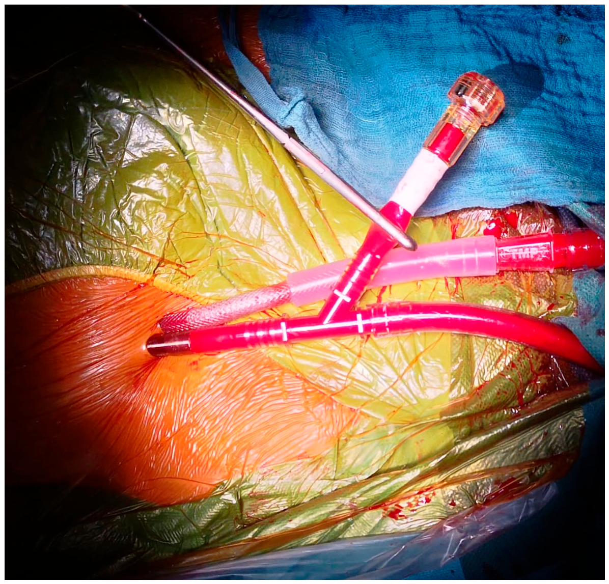
Figure 2.
View on the right atrium and the selfexpanding cannula.
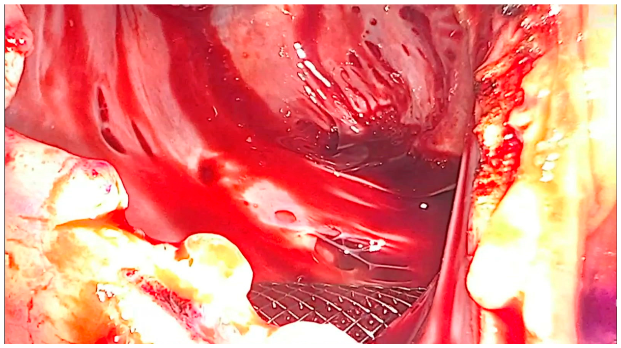

Table 1.
Baseline characteristics of the patient population.
| Variable | Descriptive Cohort Statistics (n = 32) |
| Age (years, mean ± SD) | 65.1 ± 17.1 (Median: 73.0, Range: 22.0–82.0) |
| Male / Female Gender (n, %) | 16 (50.0%) / 16 (50.0%) |
| EuroSCORE II (%, mean ± SD) | 3.6 ± 3.5 (Median: 2.3, n = 30) |
| Obesity (BMI >30 kg/m², n, %) | 9 (28.1%) |
| COPD (n, %) | 5 (15.6%) |
| Preoperative Dialysis (n, %) | 4 (12.5%) |
| Active/Treated Endocarditis (n, %) | 6 (18.8%) |
| Baseline Left Ventricular EF (%) | 50.2 ± 12.3 (Median: 50.0, Range: 22.0–70.0) |
| Pulmonary Artery Pressure (mmHg) | 36.5 ± 19.7 (Median: 40.0, Range: 1.0–67.0, n = 13) |
Table 2.
Intraoperative Data and Technical Profiles.
| Variable | Descriptive Cohort Statistics (n = 32) |
| Cardiopulmonary Bypass Time (min, mean ± SD) | 145.9 ± 35.8 (Median: 141.0, Range: 80.0–240.0) |
| Aortic Cross-Clamp Time (min, mean ± SD) | 92.4 ± 24.5 (Median: 94.5, Range: 48.0–145.0) |
| Percutaneous Venous Cannulation (n, %) | 29 (90.6%) |
| SMARTCANNULA Deployment (n, %) | 32 (100.0%) |
| MANTA Vascular Closure Device Utilization (n, %) | 27 (87.1%, n = 31 evaluated) |
| Concomitant LA MAZE Ablation (n, %) | 12 (37.5%) |
| Surgical ASD/PFO Defect Closure (n, %) | 6 (19.4%) |
Table 3.
Early Postoperative Endpoints and Complications.
| Postoperative Variable | Descriptive Cohort Statistics (n = 32) |
| 30-Day All-Cause Mortality (n, %) | 1 (3.1%) |
| Postoperative ECMO Support (n, %) | 1 (3.1%) |
| Surgical Re-thoracotomy Rate (n, %) | 4 (12.5%) |
| Clinical Hemolysis Incidence (n, %) | 0 (0.0%) |
| Acute Kidney Injury (ANV, n, %) | 6 (18.8%) |
| Postoperative Respiratory Insufficiency (n, %) | 5 (15.6%) |
| Neurological Deficit / Stroke (n, %) | 1 (3.1%) |
| Permanent Pacemaker Implantation (n, %) | 2 (6.2%) |
| Intensive Care Unit Length of Stay (days, mean ± SD) | 6.0 ± 4.9 (Median: 4.0, n = 31) |
| Total Postoperative Hospital Stay (days, mean ± SD) | 15.4 ± 9.7 (Median: 13.5, n = 30) |
Disclaimer/Publisher’s Note: The statements, opinions and data contained in all publications are solely those of the individual author(s) and contributor(s) and not of MDPI and/or the editor(s). MDPI and/or the editor(s) disclaim responsibility for any injury to people or property resulting from any ideas, methods, instructions or products referred to in the content. |
© 2026 by the authors. Licensee MDPI, Basel, Switzerland. This article is an open access article distributed under the terms and conditions of the Creative Commons Attribution (CC BY) license (http://creativecommons.org/licenses/by/4.0/).
Copyright: This open access article is published under a Creative Commons CC BY 4.0 license, which permit the free download, distribution, and reuse, provided that the author and preprint are cited in any reuse.