
Submitted:
03 September 2026
Posted:
20 September 2026
You are already at the latest version
Abstract
Fulminant Varicella Zoster Virus (VZV) associated with myocarditis is rare but may result in severe myocardial dysfunction requiring veno-arterial extracorporeal mem-brane oxygenation (VA-ECMO), which itself increases the risk of left ventricular (LV) distension and thrombus formation. We report the case of a 50-year-old woman who developed refractory cardiogenic shock following out-of-hospital ventricular fibrilla-tion cardiac arrest and required peripheral VA-ECMO. VZV DNA was detected in the blood, while coronary angiography demonstrated normal coronary arteries, support-ing a diagnosis of fulminant VZV-associated myocarditis. Despite anticoagulation and multimodal LV unloading with an intra-aortic balloon pump and Rashkind atrial sep-tostomy, persistent LV distension and absence of aortic valve opening were followed by formation of a large and organized LV thrombus. After neurological assessment confirmed preserved responsiveness, surgical thrombectomy was undertaken. Thrombus removal was performed through the aortic valve and mitral valve, followed by pulsed irrigation of the LV cavity and surgical apical venting connected to the ECMO venous circuit. Histopathology demonstrated lymphocytic myocarditis of probable viral origin. The patient subsequently developed multiorgan failure and died on postoperative day six. This case highlights the challenges of thrombus formation during VA-ECMO and the importance of early, individualized ventricular unloading. We additionally review available left ventricular unloading strategies and right ven-tricular unloading techniques in patients requiring VA-ECMO, emphasizing their re-spective indications and technical considerations.
Keywords:
Varicella Zoster Virus
; fulminant myocarditis
; VA-ECMO
; left ventricular thrombus
; ventricular unloading
; surgical thrombectomy
; cardiogenic shock
1. Introduction
Myocarditis represents a challenging diagnosis, particularly in critically ill patients [1,2]. Varicella Zoster Virus (VZV)–induced myocarditis is an uncommon but potentially severe cause of myocardial inflammation [3,4,5]. The situation becomes more complex when myocardial dysfunction progresses to cardiogenic shock requiring mechanical circulatory support.
Veno-arterial extracorporeal membrane oxygenation (VA-ECMO) is often used in the setting of witnessed cardiac arrest to provide systemic perfusion and oxygenation. However, the increased afterload from ECMO flow and the absence of native left ventricular ejection can lead to stasis within the left ventricle and subsequent thrombus formation. This case illustrates the complex management of viral myocarditis, mechanical circulatory support, and surgical intervention.
2. Introduction
Myocarditis represents a challenging diagnosis, particularly in critically ill patients [1,2]. Varicella Zoster Virus (VZV)–induced myocarditis is an uncommon but potentially severe cause of myocardial inflammation[3,4,5]. The situation becomes more complex when myocardial dysfunction progresses to cardiogenic shock requiring mechanical circulatory support.
Veno-arterial extracorporeal membrane oxygenation (VA-ECMO) is often used in the setting of witnessed cardiac arrest to provide systemic perfusion and oxygenation. However, the increased afterload from ECMO flow and the absence of native left ventricular ejection can lead to stasis within the left ventricle and subsequent thrombus formation. This case illustrates the complex management of viral myocarditis, mechanical circulatory support, and surgical intervention.
3. Case Presentation
A 50 years old female patient was admitted after an out-of-hospital witnessed cardiac arrest due to ventricular fibrillation, a shockable rhythm. Following successful resuscitation, she remained in refractory cardiogenic shock despite optimal medical support. Peripheral VA-ECMO was promptly initiated[6]. On admission, polymerase chain reaction (PCR) testing revealed the presence of Varicella Zoster Virus (VZV) DNA in her blood, suggesting active viremia that was treated with acyclovir[4,7]. Coronary angiography was performed and revealed normal coronary arteries (Figure 1), thereby ruling out an ischemic cause of her cardiac arrest.
At this stage, a provisional diagnosis of fulminant Varicella Zoster Virus (VZV)–associated myocarditis was made, based on clinical presentation and laboratory findings.
As the next step following VA-ECMO initiation, protecting the heart from excessive distension becomes paramount[8,9]. Strategies for left ventricular (LV) unloading[10], which have been associated with improved survival, must be considered early. Despite maximal support, including intra-aortic balloon pump (IABP)[11] placement and creation of a Rashkind atrial septostomy[12], the patient exhibited complete absence of aortic valve opening and persistent left ventricular distension. (Figure 2)
Despite an adequately maintained anticoagulation profile, monitored through serial anti-Xa assays a transthoracic echocardiogram revealed a large, organized thrombus occupying the apex and mid-left ventricular (LV) cavity. (Figure 3)
While on mechanical ventilation, a structured sedation vacation permitted a reassuring neurological assessment. Upon sedation withdrawal, the patient demonstrated spontaneous eye opening, visual tracking, and appropriate response to simple commands. These findings confirmed intact brainstem function and supported the decision to continue full intensive care measures[13].
A multidisciplinary team, including specialists in cardiac surgery, infectious diseases, intensive care, and cardiology, opted for a surgical left ventricular thrombectomy[14]. The goals were to prevent systemic thromboembolism and enable potential recovery of left ventricular function. Should myocardial recovery prove unattainable, expedited evaluation for heart transplantation was planned as a secondary intervention, necessitating transfer to a specialized center by aerial transportation while on ECMO support.
It is noteworthy that the island of Martinique, where the patient initially presented, situated in the Lesser Antilles of the West Indies, functions as a regional hub for cardiac surgery. However, it currently lacks facilities for heart transplantation.
4. Surgical Management
The patient was taken to the operating room. A median sternotomy was performed, and upon opening the pericardium, a pressurized, hemorrhagic effusion consistent with tamponade was observed. After systemic heparinization, cardiopulmonary bypass (CPB) was established via bicaval venous cannulation and femoral arterial cannulation.
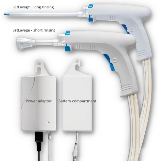
Moderate hypothermia was initiated, and the aorta was clamped. Myocardial protection was achieved using retrograde hyperkalemic blood cardioplegia delivered through the coronary sinus, minimizing the risk of dislodging thrombus that could occur with antegrade delivery. Upon initial aortotomy, no visible thrombus was detected in the aortic root, allowing for the safe addition of ostial antegrade cardioplegia. The left ventricle was thoroughly irrigated and suctioned by a pulse irrigator (Figure 4) after manual debulking of the thrombus, which was done via the aortic valve to remove residual debris and optimize visualization.
Subsequently, a transseptal left atriotomy was performed, allowing direct access to the LV via the mitral valve. Copious amounts of thrombi were removed, and the cavity was thoroughly irrigated with the pulse irrigator. The Rashkind septostomy previously done was closed.
An LV apical muscular biopsy was taken and sent for pathology for analysis. A 22 Fr cannula was placed through the apex of the LV and connected to the venous line of the ECMO circuit to provide continuous decompression [15]. The LV vent cannula was partially clamped by a Hoffman tubing clamp to control flow and avoid recirculation.
The aortic clamping time was 49 minutes, time that was necessary to clean all the residual thrombi. The heart resumed spontaneous sinus rhythm with successful defibrillation. CPB was discontinued after 98 minutes, and ECMO flow resumed.
5. Discussion
5.1. VZV Myocarditis
VZV is a DNA herpesvirus classically associated with varicella (chickenpox) and herpes zoster (shingles). Although it more commonly affects the skin and nervous system, VZV can cause myocarditis via direct viral invasion or immune-mediated mechanisms. Few cases have been documented, mostly in immunocompromised or elderly patients. Given the increased risk of severe complications in this population, current guidelines recommend administration of a booster dose of the varicella zoster vaccine starting at age 65 to enhance immunity and reduce the likelihood of viral reactivation.[16]
Our patient had no rash or neurological symptoms, but VZV DNA positivity in blood strongly suggests systemic reactivation. Cardiac catheterization was normal, ruling out ischemic causes and supporting a diagnosis of viral myocarditis.
5.2. Thrombus Formation Under ECMO
Thrombus formation in the LV under ECMO occurs due to:[17]
- Increased afterload from retrograde aortic flow
- Absence of native LV ejection
- Poor myocardial contractility (as in myocarditis)
- Inadequate anticoagulation
- Tamponade or external compression
Blood stasis in a non-contractile LV leads to a prothrombotic environment. In the presence of viable myocardium and preserved end-organ function, surgical decompression and thrombectomy may facilitate recovery.
6. Left Ventricular Unloading Techniques Under VA-ECMO
Effective LV unloading prevents thrombus formation, improves coronary perfusion, and reduces pulmonary edema. Various techniques exist; we present in this table four common techniques:

Table 1.
A comparative table of LV unloading methods.
| Technique | Type | Access | Effectiveness | Notes |
| Intra-Aortic counter pulsation Balloon (IACPB) | Percutaneous | Femoral artery | Low to moderate | Reduces afterload, promotes aortic valve opening |
| LV Apical Vent | Surgical | Sternotomy/ thoracotomy |
Very high | Direct decompression, requires surgical access |
| Atrial Transseptal septostomy (Rashkind) | Percutaneous | Atrial septum | Low to moderate | Simple, often inadequate alone |
| ECMELLA (ECMO + Impella) | Hybrid | Dual access | Very high | Expensive, effective unloading and perfusion |
7. Right Ventricular Failure and Unloading Techniques Under VA-ECMO
RV failure can coexist with LV dysfunction or develop as a complication of ECMO[18,19,20]. It contributes to elevated right atrial pressure, hepatic congestion, and reduced ECMO drainage[21]. Right ventricular (RV) unloading is increasingly recognized as a critical adjunct in biventricular failure or when RV dysfunction occurs during VA-ECMO
Management Strategies:
Management of right ventricular (RV) failure during VA-ECMO requires a multimodal approach tailored to the hemodynamic profile and underlying pathology. Initial strategies include volume optimization to reduce preload and alleviate RV strain, followed using pulmonary vasodilators such as inhaled nitric oxide or sildenafil to decrease pulmonary vascular resistance. In cases of persistent RV dysfunction, inotropic support with agents like milrinone or dobutamine can enhance contractility. For patients with severe RV overload or persistent congestion, surgical pulmonary artery venting may provide effective decompression. In more advanced scenarios, right ventricular assist devices (RVADs) offer direct mechanical support to the failing RV. Additionally, conversion to a veno-veno-arterial (VVA) ECMO configuration may be considered with venous cannulas in the superior and inferior vena cava to augment both RV and LV unloading while maintaining systemic perfusion.
Table 2.
A comparative table of RV unloading methods.
| Technique | Type | Access/Method | Effectiveness | Notes |
| Volume Optimization | Medical | Diuretics, fluid restriction | Moderate | First-line approach; risk of hypoperfusion |
| Pulmonary Vasodilators | Pharmacologic | Inhaled NO, Sildenafil | Variable | Reduce pulmonary pressure; monitor for hypotension |
| Inotropes | Pharmacologic | Milrinone, Dobutamine | Moderate | Improve RV contractility; watch for arrhythmias |
| Pulmonary Artery Venting | Surgical | Via PA cannula | High | Direct RV unloading; requires surgical access |
| RVAD (Right Ventricular Assist Device) | Mechanical | Percutaneous or surgical | Very High | Direct support of RV output; technically complex |
| VVA-ECMO Configuration | ECMO Adjustment | Dual ECMO cannulas | High | Supports both ventricles; may need additional flow regulation |
8. ECMO Setup and Technical Parameters
The patient was placed on VA-ECMO using a Cardiohelp HLS Advanced 7.0 system, with cannulas sizing was as follows:
- Arterial Cannula: 17 Fr
- Venous Cannula: 25 Fr
- Reperfusion Cannula (Femoral): 6 Fr
- Heparin Dose on Insertion: 5,000 IU
Body Surface Area (DuBois Formula): BSA (m²) =1.85
9. Myocarditis of Probable Viral Origin: Pathology Review
Histological analysis of LV apex biopsy revealed:
- Plurifocal myocyte damage
- Infiltrate rich in T-lymphocytes (CD3+), few B cells (CD20+), rare eosinophils and macrophages (CD68+)
- No granulomas, giant cells, or fungal elements
- Pericardial fluid: fibrinous pericarditis without specific features
These findings are compatible with a lymphocytic myocarditis of probable viral origin. Eosinophils raise the possibility of a drug reaction or parasitic etiology, but the clinical context and positive VZV PCR favor viral myocarditis. Tissue quality limited advanced PCR/metagenomic testing.
10. Conclusion
This case illustrates the rare occurrence of VZV-induced myocarditis presenting with cardiac arrest and evolving LV thrombus formation under VA-ECMO. Early recognition and aggressive management—including surgical thrombectomy and apical venting—are crucial to prevent irreversible myocardial damage or embolic events. Comprehensive LV and RV unloading strategies must be individualized and initiated promptly to optimize outcomes in patients with severe myocardial dysfunction.
Unfortunately, despite these efforts, the patient passed away due to multi-organ failure on postoperative day six.
Conflicts of Interest
“The authors declare no conflicts of interest.”.
References
- Franken, R.A.; Franken, M. Pseudo-myocardial infarction during an episode of herpes zoster. Arq. Bras. Cardiol. 2000, 75. [Google Scholar] [CrossRef] [PubMed]
- I Donoiu 1, O.I. 1: Varicella-zoster myocarditis mimicking acute myocardial infarction. Curr. Health Sci. J. 2014. [Google Scholar] [CrossRef] [PubMed]
- Cherukuri, A.S.S.; Belay, N.F.; Nasereldin, D.S.; Mohammed, D.O.; Mohamed, S.; Elkhazeen, A.; Ghobriel, N.G.; Alatta, L.; Alsafi, W.; Abdalla, Y.; Brry, G.; Abdelrahman, N. Varicella-Zoster Virus Myocarditis: Early Clinical Diagnosis and Outcome; Cureus, 2023. [Google Scholar] [CrossRef] [PubMed]
- Kociol, R.D.; Cooper, L.T.; Fang, J.C.; Moslehi, J.J.; Pang, P.S.; Sabe, M.A.; Shah, R. V.; Sims, D.B.; Thiene, G.; Vardeny, O. Recognition and Initial Management of Fulminant Myocarditis. Circulation 2020, 141. [Google Scholar] [CrossRef] [PubMed]
- Maggi, U.; Russo, R.; Conte, G.; Chiumello, D.; Lunghi, G.; Maggioni, M.; Caspani, M.L.; Arnoldi, R.; Dondossola, D.; Rossi, G. Fulminant Multiorgan Failure Due to Varicella Zoster Virus and HHV6 in an Immunocompetent Adult Patient, and Anhepatia. Transpl. Proc. 2011, 43, 1184–1186. [Google Scholar] [CrossRef] [PubMed]
- Keebler, M.E.; Haddad, E. V.; Choi, C.W.; McGrane, S.; Zalawadiya, S.; Schlendorf, K.H.; Brinkley, D.M.; Danter, M.R.; Wigger, M.; Menachem, J.N.; Shah, A.; Lindenfeld, J.A. Venoarterial Extracorporeal Membrane Oxygenation in Cardiogenic Shock. JACC Heart Fail. 2018, 6, 503–516. [Google Scholar] [CrossRef] [PubMed]
- Contamine, M.; Ader, F.; Lepiller, Q.; Martha, B.; Cagnon-Chapalain, J.; Leturnier, P.; Frober, E.; Bouiller, K.; Binquet, C.; Auvray, C.; Piroth, L.; Blot, M. Acyclovir treatment of varicella-zoster virus meningeal infections and acute kidney injury: a multicentre case series study. Infect. Dis. 2024, 56, 842–850. [Google Scholar] [CrossRef] [PubMed]
- Bernhardt, A.M.; Schrage, B.; Westermann, D.; Reichenspurner, H. Extracorporeal membrane oxygenation evolution: Left ventricular unloading strategies. JTCVS Open 2021, 8, 85–89. [Google Scholar] [CrossRef] [PubMed]
- Cevasco, M.; Takayama, H.; Ando, M.; Garan, A.R.; Naka, Y.; Takeda, K. Left ventricular distension and venting strategies for patients on venoarterial extracorporeal membrane oxygenation. J. Thorac. Dis. 2019, 11, 1676–1683. [Google Scholar] [CrossRef] [PubMed]
- Zhang, H.; Wang, T.; Wang, J.; Liu, G.; Yan, S.; Teng, Y.; Wang, J.; Ji, B. Different strategies in left ventricle unloading during venoarterial extracorporeal membrane oxygenation: A network meta-analysis. IJC Heart Vasc. 2024, 54, 101506. [Google Scholar] [CrossRef] [PubMed]
- Wang, K.; Wang, L.; Ma, J.; Xie, H.; Li, C.; Hao, X.; Du, Z.; Wang, H.; Hou, X. Intra-aortic balloon pump after VA-ECMO reduces mortality in patients with cardiogenic shock: an analysis of the Chinese extracorporeal life support registry. Crit. Care 2024, 28, 394. [Google Scholar] [CrossRef] [PubMed]
- AlGhamdi, M.; Saiydoun, G.; Lebreton, G.; Mazzucotelli, J.-P. Percutaneous atrial septostomy for left ventricular unloading in patients on peripheral venoarterial extracorporeal membrane oxygenation: A systematic review and meta-analysis. Am. Heart J. Plus Cardiol. Res. Pract. 2025, 54, 100542. [Google Scholar] [CrossRef] [PubMed]
- Aboul-Nour, H.; Jumah, A.; Abdulla, H.; Sharma, A.; Howell, B.; Jayaprakash, N.; Gardner-Gray, J. Neurological monitoring in ECMO patients: current state of practice, challenges and lessons. Acta Neurol. Belg. 2023, 123, 341–350. [Google Scholar] [CrossRef] [PubMed]
- Ortuno, S.; Delmas, C.; Diehl, J.-L.; Bailleul, C.; Lancelot, A.; Naili, M.; Cholley, B.; Pirracchio, R.; Aissaoui, N. Weaning from veno-arterial extra-corporeal membrane oxygenation: which strategy to use? Ann. Cardiothorac. Surg. 2019, 8, E1–E8. [Google Scholar] [CrossRef] [PubMed]
- Rescigno, G.; Aratari, C.; Matteucci, M.L.S.; Massi, F.; Capestro, F.; D’Alfonso, A.; Torracca, L. Management of transapical left venting during adult peripheral extracorporeal membrane oxygenation. Mech. Circ. Support 2011, 2, 11826576. [Google Scholar] [CrossRef]
- Bresse, X.; Annemans, L.; Préaud, E.; Bloch, K.; Duru, G.; Gauthier, A. Vaccination against herpes zoster and postherpetic neuralgia in France: a cost–effectiveness analysis. Expert Rev. Pharmacoecon Outcomes Res. 2013, 13, 393–406. [Google Scholar] [CrossRef] [PubMed]
- Weber, C.; Deppe, A.-C.; Sabashnikov, A.; Slottosch, I.; Kuhn, E.; Eghbalzadeh, K.; Scherner, M.; Choi, Y.-H.; Madershahian, N.; Wahlers, T. Left ventricular thrombus formation in patients undergoing femoral veno-arterial extracorporeal membrane oxygenation. Perfusion 2018, 33, 283–288. [Google Scholar] [CrossRef] [PubMed]
- DeFilippis, E.M.; Topkara, V.K.; Kirtane, A.J.; Takeda, K.; Naka, Y.; Garan, A.R. Mechanical Circulatory Support for Right Ventricular Failure. Card. Fail. Rev. 2022, 8, e14. [Google Scholar] [CrossRef] [PubMed]
- Grant, C.; Richards, J.B.; Frakes, M.; Cohen, J.; Wilcox, S.R. ECMO and Right Ventricular Failure: Review of the Literature. J. Intensive Care Med. 2021, 36, 352–360. [Google Scholar] [CrossRef] [PubMed]
- Puerto, E.; Tavazzi, G.; Gambaro, A.; Cirillo, C.; Pecoraro, A.; Martín-Asenjo, R.; Delgado, J.; Bueno, H.; Price, S. Interaction between VA-ECMO and the right ventricle. Hell. J. Cardiol. 2022, 68, 17–24. [Google Scholar] [CrossRef] [PubMed]
- Kim, D.; Park, Y.; Choi, K.H.; Park, T.K.; Lee, J.M.; Cho, Y.H.; Choi, J.O.; Jeon, E.S.; Yang, J.H. Prognostic Implication of RV Coupling to Pulmonary Circulation for Successful Weaning From Extracorporeal Membrane Oxygenation. JACC Cardiovasc Imaging 2021, 14, 1523–1531. [Google Scholar] [CrossRef] [PubMed]
- Møller, J.E.; Sionis, A.; Aissaoui, N.; Ariza, A.; Bělohlávek, J.; De Backer, D.; Färber, G.; Gollmann-Tepeköylu, C.; Mebazaa, A.; Price, S.; Swol, J.; Thiele, H.; Hassager, C. Step by step daily management of short-term mechanical circulatory support for cardiogenic shock in adults in the intensive cardiac care unit: a clinical consensus statement of the Association for Acute CardioVascular Care of the European Society of Cardiology SC, the European Society of Intensive Care Medicine, the European branch of the Extracorporeal Life Support Organization, and the European Association for Cardio-Thoracic Surgery. Eur. Heart J. Acute Cardiovasc Care 2023, 12, 475–485. [Google Scholar] [CrossRef] [PubMed]
- Burkhoff, D.; Sayer, G.; Doshi, D.; Uriel, N. Hemodynamics of Mechanical Circulatory Support. J. Am. Coll. Cardiol. 2015, 66, 2663–2674. [Google Scholar] [CrossRef] [PubMed]
Figure 1.
Normal coronary angiogram.
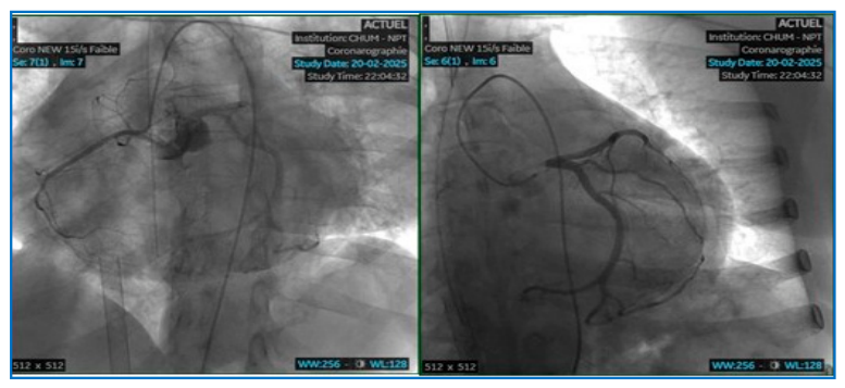
Figure 2.
Showing the complete setting of the patient through different stages of the management. 1- The VA- ECMO. 2- Intra-aortic counter pulsation balloon. 3- Percutaneous atrial septostomy (closed intra operatively). 4- LV apical vent connected to the venous line (added surgically post thrombectomy). 5- Hoffman tubing clamp to control LV vent flow and adequate LV unloading.
Figure 2.
Showing the complete setting of the patient through different stages of the management. 1- The VA- ECMO. 2- Intra-aortic counter pulsation balloon. 3- Percutaneous atrial septostomy (closed intra operatively). 4- LV apical vent connected to the venous line (added surgically post thrombectomy). 5- Hoffman tubing clamp to control LV vent flow and adequate LV unloading.
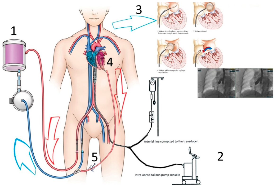
Figure 3.
Showing a TEE image of the large thrombus occupying the left ventricular cavity.
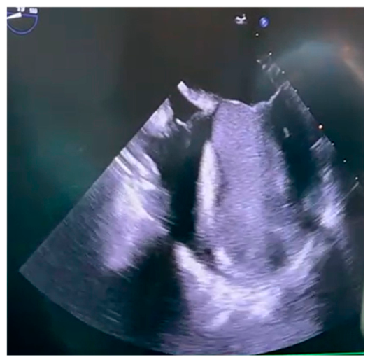
Figure 4.
Showing the pulsed irrigation system (JetLavage ™ ,endocon GmbH) used for thrombectomy and lavage of the left ventricle.
Figure 4.
Showing the pulsed irrigation system (JetLavage ™ ,endocon GmbH) used for thrombectomy and lavage of the left ventricle.
Disclaimer/Publisher’s Note: The statements, opinions and data contained in all publications are solely those of the individual author(s) and contributor(s) and not of MDPI and/or the editor(s). MDPI and/or the editor(s) disclaim responsibility for any injury to people or property resulting from any ideas, methods, instructions or products referred to in the content. |
© 2026 by the authors. Licensee MDPI, Basel, Switzerland. This article is an open access article distributed under the terms and conditions of the Creative Commons Attribution (CC BY) license (http://creativecommons.org/licenses/by/4.0/).
Copyright: This open access article is published under a Creative Commons CC BY 4.0 license, which permit the free download, distribution, and reuse, provided that the author and preprint are cited in any reuse.