

Submitted:
18 September 2026
Posted:
20 September 2026
You are already at the latest version
Abstract
Background/Objectives: Non-clear cell renal cell carcinoma (nccRCC) comprises biologically heterogeneous renal malignancies with distinct molecular drivers, clinical behavior, and therapeutic responses. This review summarizes prognosis and systemic therapy and evaluates emerging histology-specific and biomarker-directed approaches. Methods: We analyzed overall survival by histology in the SEER 17 Registries database (2000–2022) and conducted a narrative review of trials, guidelines, and translational studies. The ICD-O-3-defined population included 43,966 adults; 43,789 with known survival time were included in survival analyses. Results: Median follow-up was 96 months. Compared with papillary RCC, adjusted HRs were 0.67 (95% CI 0.64–0.70) for chromophobe RCC, 3.73 (3.37–4.11) for collecting duct carcinoma, 0.97 (0.80–1.17) for oncocytic tumors, and 33.23 (27.47–40.20) for medullary carcinoma. VEGFR- and MET-directed therapies and immune checkpoint inhibitor combinations show activity that varies by histology. Conclusions: nccRCC should be studied and managed as a collection of distinct entities. Registry associations require cautious interpretation because of residual confounding, evolving classification, and non-proportional hazards; subtype-specific trials and molecular stratification remain priorities.
Keywords:
non-clear cell renal cell carcinoma
; papillary renal cell carcinoma
; chromophobe renal cell carcinoma
; immune checkpoint inhibitors
; molecular biomarkers
; prognosis
; systemic therapy
1. Introduction
Clear cell RCC (ccRCC), driven predominantly by alterations in the VHL pathway, constitutes most renal cell carcinomas and has well-established therapeutic paradigms. Non-clear cell renal cell carcinomas (nccRCCs) account for approximately 30% of renal malignancies.[1] Papillary RCC (pRCC), chromophobe RCC (chRCC), and tumors with mixed histology are among the more common forms, whereas collecting duct carcinoma, renal medullary carcinoma, oncocytic tumors, and other or unclassified tumors are rare and have distinct molecular drivers, cellular contexts, and clinical behavior.[2]
Historically, patients with nccRCC have been underrepresented in prospective clinical trials, and management strategies have largely been extrapolated from studies conducted in ccRCC.[3] As a result, outcomes for patients with advanced nccRCC have generally been inferior, with lower response rates to conventional VEGF- and mTOR-targeted therapies and limited high-quality evidence to guide treatment selection. Moreover, the rarity of individual subtypes has posed challenges in conducting adequately powered, histology-specific studies, further contributing to gaps in evidence-based care.
Recent advances in molecular profiling and tumor biology have reshaped nccRCC as a collection of distinct diseases rather than a single clinical category.[4] These insights have identified actionable pathways, including MET alterations in papillary RCC, and have supported biomarker-driven and histology-enriched trials. Immune checkpoint inhibitors and combinations with tyrosine kinase inhibitors (TKIs) have shown promising activity in selected subtypes, although responses remain heterogeneous.
In this review, we summarize the prognostic landscape and systemic treatment options for nccRCC, emphasizing subtype-specific outcomes, emerging targeted and immunotherapeutic strategies, and the evolving role of molecular characterization. We also discuss ongoing trials and future directions for personalized management informed by tumor origin and biology.
2. Materials and Methods
We used a hybrid approach that combined a population-based SEER analysis with a narrative literature review to summarize demographic characteristics, survival outcomes, tumor biology, and therapeutic evidence in nccRCC.
We extracted de-identified data from the SEER Research Data, 17 Registries, November 2024 submission (diagnosis years 2000–2022).[2] Eligible records were malignant kidney-parenchyma tumors (SEER site recode: Kidney Parenchyma) diagnosed at age ≥20 years and assigned to one of five prespecified ICD-O-3 histology groups: papillary (8260 and 8050), chromophobe (8317 and 8270), collecting duct (8319), oncocytic (8290 and 8342), or medullary (8510). Other renal parenchymal malignancies outside of the WHO 2022 classification were excluded. Patients younger than 20 years (n = 91) were excluded, yielding an adult cohort of 43,966 tumors.
Overall survival (OS) was defined as the interval from diagnosis to death from any cause or last follow-up. Patients with unknown survival time (n = 177) were excluded from survival analyses, leaving 43,789 evaluable patients. For 778 patients reported as surviving zero months, survival time was set to 0.5 months to retain early events.
Kaplan–Meier methods were used to estimate OS by histologic group, and differences were assessed using the log-rank test. Median follow-up was estimated with the reverse Kaplan–Meier method. Numbers at risk and deaths are reported with survival results.
Cox proportional hazards regression was used to estimate hazard ratios (HRs) and 95% confidence intervals (CIs), with papillary RCC as the reference group. Univariable models evaluated histology alone. Multivariable models adjusted for age, sex, race, and year of diagnosis. Stage-specific analyses were restricted to patients diagnosed during 2018–2022 with known Derived EOD 2018 stage because comparable stage data were unavailable for earlier years. The proportional-hazards assumption was assessed using Schoenfeld residuals. Because histology did not satisfy the assumption, Cox estimates are reported as average associations over follow-up and are interpreted together with Kaplan–Meier estimates at clinically relevant time points rather than as constant effects.
Analyses were performed using Stata and independently reproduced for data verification. All tests were two-sided, and p < 0.05 was considered statistically significant.
The narrative review contextualized the population-based findings and summarized evidence on tumor biology, molecular drivers, biomarkers, and systemic therapy. PubMed/MEDLINE, major oncology journals, and ClinicalTrials.gov were searched through July 23, 2026, using combinations of “non-clear cell renal cell carcinoma,” “papillary,” “chromophobe,” “collecting duct,” “renal medullary,” “oncocytic,” “systemic therapy,” “clinical trial,” “molecular,” “transcriptomic,” and “epigenomics.” We prioritized peer-reviewed prospective phase II/III studies, contemporary clinical guidelines, subtype-specific translational studies, and registered ongoing interventional trials. Reference lists of included reports were also screened. Evidence was synthesized narratively because of substantial heterogeneity in histology, treatment, study design, and endpoints; cross-trial comparisons were considered indirect.
3. Results
Among 43,966 adults in the ICD-O-3-defined cohort, papillary RCC was the predominant histology (n = 29,824; 67.83%), followed by chromophobe RCC (n = 13,297; 30.24%), collecting duct carcinoma (n = 511; 1.16%), oncocytic tumors (n = 208; 0.47%), and medullary carcinoma (n = 126; 0.29%) (Table 1).
Sex- and race-based distributions differed across histologic groups (Table 1). Papillary RCC comprised 74.02% of tumors among men and 53.09% among women and was particularly common among African American patients (81.23%). Chromophobe RCC comprised 44.82% of tumors among women compared with 24.13% among men. Medullary carcinoma represented 0.99% of tumors among African American patients but only 0.08% among White patients.
Among 43,789 patients with known survival time, 13,762 deaths occurred and 30,027 patients were censored alive. Median follow-up was 96 months. OS differed significantly by histology (global log-rank χ² = 2914.51; df = 4; p < 0.001). Median OS was 229 months for chromophobe RCC, 161 months for papillary RCC, 131 months for oncocytic tumors, 16 months for collecting duct carcinoma, and 9 months for medullary carcinoma. With papillary RCC as the reference, univariable HRs were 0.57 (95% CI 0.55–0.60) for chromophobe RCC, 3.70 (95% CI 3.35–4.08) for collecting duct carcinoma, 1.29 (95% CI 1.07–1.56) for oncocytic tumors, and 11.42 (95% CI 9.46–13.78) for medullary carcinoma (Table 2).
After adjusting for age, sex, race, and diagnosis year, chromophobe RCC remained associated with better OS than papillary RCC (adjusted HR 0.67; 95% CI 0.64–0.70), whereas collecting duct carcinoma (adjusted HR 3.73; 95% CI 3.37–4.11) and medullary carcinoma (adjusted HR 33.23; 95% CI 27.47–40.20) remained associated with worse OS. Oncocytic histology was not independently associated with OS (adjusted HR 0.97; 95% CI 0.80–1.17; p = 0.743). Kaplan–Meier curves showed clear separation across histologies (Figure 1). Stage-specific analyses in the 2018–2022 subset also demonstrated significant histology-related survival differences within stages I–IV (all p < 0.001); median OS was 13 months for stage IV and was not reached for stages I–III. Schoenfeld-residual testing indicated non-proportional hazards for histology (global p < 0.001); consequently, the HRs should be interpreted as average associations over follow-up. Comparable stage, grade, metastatic-site, and treatment variables were not uniformly available across 2000–2022, precluding valid full-period adjustment for these factors.
3.1. Clinical trials and Evolving Treatment Strategies
Non–clear cell renal cell carcinoma (nccRCC) represents a biologically heterogeneous group of malignancies comprising several histologic subtypes, each with distinct molecular and clinical characteristics.[5,6] Historically, therapeutic advances in this population have lagged behind those seen in clear cell RCC, largely due to the lower incidence of these tumors and their frequent exclusion from large, randomized trials. Consequently, treatment recommendations for nccRCC have often been extrapolated from studies conducted in clear cell RCC despite significant biological differences between these entities. Recently, however, there has been increasing recognition of the need for histology-specific investigation, leading to the development of prospective clinical trials dedicated to nccRCC.
Several prospective studies have helped define the current therapeutic landscape. The randomized phase II ASPEN and ESPN trials compared VEGF-targeted therapy with mTOR inhibition and generally favored sunitinib over everolimus across most non-clear cell histologies, supporting VEGF-directed therapy as an initial approach. [7,8] Subsequent subtype-focused studies further refined treatment selection.
3.2. Papillary RCC
Papillary RCC has the strongest subtype-specific prospective evidence among nccRCCs. In PAPMET (SWOG-1500), cabozantinib improved progression-free survival and response rate compared with sunitinib.[9] SAVOIR suggested greater activity with savolitinib than with sunitinib in MET-driven papillary RCC, although the trial closed early.[10] KEYNOTE-427 Cohort B demonstrated activity with pembrolizumab monotherapy, particularly in papillary tumors.[11] Other studies reported encouraging results with cabozantinib plus nivolumab and with bevacizumab plus erlotinib in papillary and FH-deficient or HLRCC-associated disease; the latter regimen achieved an ORR of 72%, median PFS of 21.1 months, and median OS of 44.6 months in HLRCC-associated disease.[12,13] CALYPSO evaluated savolitinib combined with durvalumab showing meaningful activity in MET-driver subset. [14]
ICI–TKI combinations have produced some of the strongest prospective activity reported in advanced nccRCC. In KEYNOTE-B61, pembrolizumab plus lenvatinib achieved an ORR of 50.6% with longer follow-up, while cabozantinib plus nivolumab achieved an ORR of 47.5% in the papillary, unclassified, and translocation-associated cohort. [13,15] Pembrolizumab monotherapy achieved an ORR of 26.7% in KEYNOTE-427 Cohort B. [11] In PAPMET, ORRs were 23% with cabozantinib and 4% with sunitinib in papillary RCC.[9]
3.3. Chromophobe RCC
While papillary RCC appears to derive the greatest benefit from modern ICI-TKI combinations, chromophobe RCC has shown consistently lower activity with immunotherapy-based approaches. In KEYNOTE-427 Cohort B, ORRs were 28.8% for papillary RCC and 9.5% for chromophobe RCC; no responses were observed in the chromophobe cohort treated with cabozantinib plus nivolumab. [11,13] In ASPEN, chromophobe RCC was among the few settings in which outcomes numerically favored everolimus over sunitinib, supporting distinct biology and treatment sensitivity.[8]
3.4. Collecting Duct Carcinoma
The phase II BONSAI study evaluated cabozantinib in 23 patients with metastatic collecting duct carcinoma and reported an ORR of 35%, median PFS of 4 months, and median OS of 7 months. These results indicate measurable activity but do not establish comparative superiority because the study was small and single arm.[16]
An emerging strategy is antibody-drug conjugate and immune checkpoint inhibitor combination therapy. The phase II REPRINT trial is evaluating pembrolizumab plus enfortumab vedotin in collecting duct carcinoma.[17] This histology-specific study is particularly important because the rarity of CDC has historically resulted in treatment recommendations derived from small prospective studies, retrospective series, and extrapolation from urothelial and other renal malignancies. For metastatic CDC, platinum-based chemotherapy remains an important treatment option, while cabozantinib represents a targeted therapeutic consideration and clinical trial enrollment should be prioritized whenever feasible. Current trials are evaluating combination regimens rather than targeting a single dominant molecular alteration.
Ongoing prospective studies reflect the shift toward histology-specific and immunotherapy–TKI strategies. Broad trials include the phase III STELLAR-304 study of zanzalintinib (XL092) plus nivolumab versus sunitinib and phase II studies of tivozanib plus nivolumab, cabozantinib plus nivolumab plus ipilimumab, and fruquintinib plus serplulimab.[18,19,20,21] Histology-enriched studies include PAPMET2 and the phase III SAMETA trial in MET-driven papillary RCC. REPRINT in collecting duct carcinoma further illustrates the move toward rare-subtype-specific trial design. [17,22,23,25,26]

Table 3.
Completed prospective trials in advanced nccRCC.
| Completed trials | |||||
| Name of trial | Phase | Population | Intervention | Comparator | Key points |
| KEYNOTE-B61 (NCT04704219) [15] | II | Advanced nccRCC (n = 158) | Pembrolizumab + Lenvatinib | Single arm | ORR 50.6%; median PFS 17.9 months; responses varied by histology, with lower activity in chromophobe RCC |
| NCT01130519[12] | II | HLRCC-associated or sporadic papillary RCC (n = 83) | Bevacizumab + Erlotinib | Single arm |
HLRCC: ORR 72%, median PFS 21.1 months, median OS 44.6 months; sporadic pRCC: ORR 35%, median PFS 8.9 months |
| ASPEN (NCT01108445)[8] | II | Metastatic nccRCC (n = 108) | Sunitinib | Everolimus | Median PFS 8.3 months with sunitinib versus 5.6 months with everolimus; interpretation by histology was exploratory |
| PAPMET/SWOG-1500 (NCT02761057)[9] | II | Metastatic papillary RCC (n = 147) | Cabozantinib | Sunitinib | Cabozantinib improved median PFS (9.0 vs. 5.6 months) and ORR (23% vs. 4%) versus sunitinib |
| SAVOIR (NCT03091192)[10] | III | MET-driven papillary RCC (n = 60) | Savolitinib | Sunitinib | ORR 27% with savolitinib versus 7% with sunitinib; trial closed early and was underpowered for definitive comparison |
| KEYNOTE-427 (NCT02853344)[11] | II | Advanced nccRCC (n = 165) | Pembrolizumab | Single arm | ORR ~26%, highest in papillary RCC |
| NCT03635892[13] | II | Advanced nccRCC (n = 47) | Cabozantinib + Nivolumab | Single arm | ORR 47.5% in the papillary, unclassified, and translocation-associated cohort; no responses in the chromophobe cohort |
| CALYPSO (NCT02819596) [14] | II | Advanced papillary RCC (n = 41) | Savolitinib + Durvalumab | Single arm | ORR 29%; median PFS 4.9 months with savolitinib plus durvalumab |
| NCT01399918[24] | II | Advanced pRCC or nccRCC with papillary features (n = 35) | Everolimus + bevacizumab | Single arm | ORR 29%; median PFS 11.0 months with everolimus plus bevacizumab |
| BONSAI NCT03354884[16] | II | Metastatic collecting duct carcinoma (n = 23) | Cabozantinib | Single arm |
ORR 35%; median PFS 4 months and median OS 7 months with cabozantinib |
pRCC: Papillary renal cell carcinoma; uRCC: unclassified renal cell carcinoma.
Table 4.
Selected ongoing or recently active clinical trials in nccRCC.
| Ongoing trials | |||||
| Name of trial | Phase | Population | Intervention | Comparator arm | Status |
| STELLAR-304 (NCT05678673)[21] | III | Advanced/metastatic non-clear cell RCC | Zanzalintinib + Nivolumab | Sunitinib | Recruiting |
| NCT06053658[20] | II | Advanced nccRCC | Tivozanib + Nivolumab | Single arm | Recruiting |
| NCT04413123[19] | II | Advanced nccRCC | Cabozantinib + Nivolumab + Ipilimumab | Single arm | Not currently recruiting |
| NCT05831891[18] | II | Advanced nccRCC | Fruquintinib + Serplulimab | Single arm | Recruiting |
| PAPMET2/S2200 (NCT05411081)[25] | II | Metastatic papillary RCC | Cabozantinib + Atezolizumab | Cabozantinib | Recruiting |
| SAMETA (NCT05043090)[26] | III | MET-driven papillary RCC | Savolitinib + Durvalumab | Sunitinib or Durvalumab | Not currently recruiting |
| NCT05096390[23] | II | Papillary RCC | Axitinib + Pembrolizumab | Axitinib | Recruiting |
| NCT04981509[27] | II | Papillary RCC | Bevacizumab + Erlotinib + Atezolizumab | Single arm | Not currently recruiting |
| REPRINT (NCT06302569) [17] | II | Collecting duct carcinoma | Pembrolizumab + Enfortumab vedotin | Single arm | Recruiting |
| NCT05808608 [22] | I | Sarcomatoid or rare RCC subtypes (including nccRCC) | Cadonilimab* + Axitinib |
Single arm | Recruiting |
*AK104 is a PD-1/CTLA-4 bispecific antibody.
4. Discussion
4.1. Tumor Biology
nccRCC subtypes demonstrate distinct molecular, histological, and clinical features compared to clear cell RCC, with limited effective therapeutic options and poor representation in clinical trials. Distinct RCC subtypes likely arise from progenitor cells located in different segments of the nephron, such as the proximal versus distal tubules, each retaining unique transcriptional and epigenetic programs established during kidney development. In this context, identical or overlapping genetic alterations may produce divergent phenotypes depending on the cellular context in which they occur. This paradigm aligns with a stem cell–based model of oncogenesis, wherein tumor heterogeneity reflects the differentiation capacity and plasticity of the cell-of-origin. Consequently, biomarkers based solely on genomic alterations may be insufficient, as they fail to capture the hierarchical organization and stem-like properties of tumors. Instead, integrating developmental lineage, cellular differentiation state, and microenvironmental context may improve diagnostic precision, prognostication, and therapeutic targeting. Such a framework has important implications for translational research, emphasizing the need to move beyond reductionist genomic approaches toward integrated, context-dependent models of cancer biology.
4.1.1. Developmental and Embryonic Origins
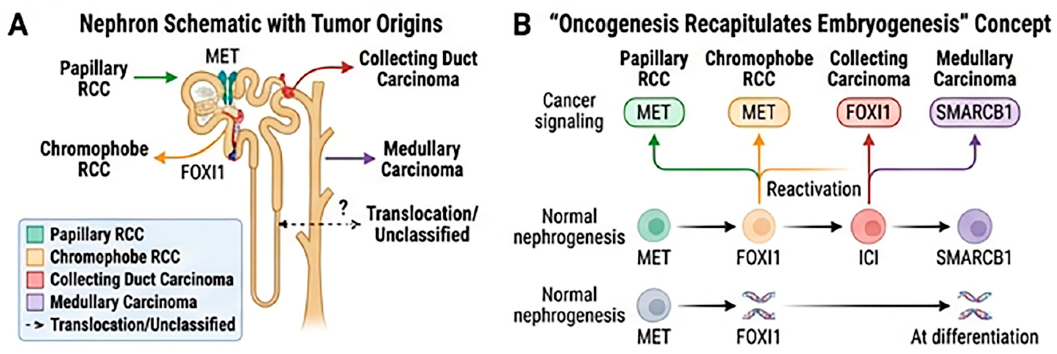
The kidney develops through reciprocal interactions between the metanephric mesenchyme and ureteric bud, which generate nephron epithelia and the collecting system, respectively.[28] Single-cell transcriptomic and chromatin-accessibility (scATAC-seq) data support a model in which major nccRCC subtypes arise within distinct nephron-segment or progenitor contexts, with tumor heterogeneity reflecting both developmental lineage and subsequent oncogenic and metabolic reprogramming. Most papillary RCCs show proximal-tubule programs, although subsets may reflect collecting-duct principal cells or renal tubular progenitors that expand in chronic renal insufficiency or in association with papillary adenomas.[29,30,31,32] Chromophobe RCC is linked to distal nephron and collecting-duct intercalated-cell programs, supported by gene-expression, methylation, and immunohistochemical markers including KIT (CD117), CK7, and FOXI1. Collecting duct carcinoma also shows distal nephron or collecting-duct features.[33,34] Renal medullary carcinoma is thought to arise in the medullary epithelium under chronic hypoxic stress.
Several molecular programs that characterize nccRCC—including MET signaling in pRCC, FOXI1-driven transcription in chRCC, and Wnt/β-catenin activity across subtypes—may represent reactivation of developmental pathways rather than entirely de novo programs acquired during malignant transformation. This concept, in which oncogenesis partially recapitulates embryogenesis in an inappropriate cellular context, may explain lineage-associated phenotypes and therapeutic vulnerabilities.[35]
During kidney development, nephron progenitors acquire spatially and temporally regulated identities rather than following a simple fixed hierarchy. A stem-cell framework therefore suggests that tumor phenotype may reflect both the developmental context of the initiating cell and subsequent differentiation or dedifferentiation. This hypothesis may help explain why some distal-nephron-associated tumors are aggressive, whereas a highly differentiated tumor such as chromophobe RCC may remain relatively indolent. These relationships remain conceptual and should not be interpreted as proof that nephron position alone determines clinical behavior.
Figure 2.
Proposed developmental context of major nccRCC subtypes. (A) Nephron segments associated with papillary, chromophobe, collecting duct, medullary, translocation-associated, and unclassified tumors. (B) Conceptual model in which oncogenic signaling reactivates lineage-associated developmental programs.
Figure 2.
Proposed developmental context of major nccRCC subtypes. (A) Nephron segments associated with papillary, chromophobe, collecting duct, medullary, translocation-associated, and unclassified tumors. (B) Conceptual model in which oncogenic signaling reactivates lineage-associated developmental programs.
4.1.2. Heterogeneity Across nccRCC Subtypes
Heterogeneity in nccRCC occurs at several levels. Intertumoral variation reflects distinct developmental and molecular contexts and may be further shaped by stem-like plasticity, such that similar genetic or epigenetic alterations produce different phenotypes depending on the cellular state in which they occur.[32] Spatial and temporal diversity within tumors also permits resistant subclones to emerge during treatment.
Transcriptomic studies in ccRCC provide proof of principle for classifying renal tumors by functional state rather than morphology alone. Verbiest et al. described favorable angiogenic or normal-like groups (ccrcc2/classical, approximately 45%; ccrcc3/normal-like, approximately 5%) and less favorable, less differentiated groups (ccrcc1/c-MYC-up, approximately 30%; ccrcc4/c-MYC-up and immune-up, approximately 20%). The ccrcc4 phenotype was enriched for inflammatory and sarcomatoid features and expressed an immunosuppressive program. Although these ccRCC classifiers cannot be applied directly to nccRCC, the same principles—transcriptomic state, immune contexture, methylation pattern, and differentiation status—may help explain divergent behavior and treatment response among tumors with similar morphology or overlapping mutations.[36]
In the TRACERx Renal model, multiregion sequencing of ccRCC identified seven recurrent evolutionary subtypes and linked metastatic behavior to intratumoral heterogeneity (ITH) and somatic copy-number alteration (SCNA) burden. [37,38] Tumors with low ITH but high SCNA burden were associated with rapid multisite progression and included multiple-clonal-driver, VHL-wild-type, and BAP1-driven trajectories. Highly branched, high-ITH tumors, often involving PBRM1-led trajectories, tended to progress more slowly, whereas low-ITH, low-SCNA tumors had relatively low metastatic potential. Metastases were more clonally homogeneous than matched primary tumors, consistent with a metastatic bottleneck. These evolutionary classes are biological and prognostic frameworks, not validated treatment-selection rules, and their application to nccRCC requires histology-specific multiregion cohorts and prospective validation.
Sarcomatoid dedifferentiation can arise in any RCC histology and exemplifies an extreme epithelial-to-mesenchymal transition, with loss of epithelial identity, acquisition of invasive and stem-like properties, treatment resistance, and paradoxically greater sensitivity to immune checkpoint inhibition. [35,39] Sarcomatoid change is therefore best interpreted as a dedifferentiation pattern rather than a distinct histology.
The tumor microenvironment further contributes to heterogeneity by shaping immune infiltration, angiogenesis, and stromal interactions. Notably, single-cell analyses suggest that tumor epithelial cells actively recruit immune cells, which may partly explain the sensitivity of some RCCs to immune checkpoint inhibitors despite relatively low neo-antigen burden.[30]
Papillary RCC is associated with MET alterations, particularly in tumors previously classified as type 1, whereas CDKN2A loss, SETD2 mutations, and NRF2–ARE pathway activation are associated with less favorable biology. [40,41] TP53 and PTEN mutations occur in chromophobe RCC, which otherwise has a low somatic mutation burden and a distinct oncogenic process. Collecting duct and renal medullary carcinomas are rare, aggressive tumors; renal medullary carcinoma is characterized by SMARCB1 (INI-1) loss and is strongly associated with sickle cell trait.[1]
4.1.3. Translational and Therapeutic Implications
Cancer treatment has increasingly shifted from grouping tumors solely by histomorphology toward integrating developmental lineage and molecular context, although their clinical utility in nccRCC remains unproven. [43] Treatment selection should therefore remain grounded in histology-specific clinical evidence and validated molecular findings.
In papillary RCC, germline or somatic MET pathway activation represents one of the most actionable molecular alterations. This rationale supports MET-directed agents, including cabozantinib and savolitinib, particularly in molecularly selected patients. Transcriptome-derived stemness indices in pRCC have been associated with stage and histologic phenotype; tumors previously classified as type 2 showed higher stemness scores and worse outcomes than type 1 tumors, suggesting that dedifferentiation may track with clinical aggressiveness.[30]
In chromophobe RCC, intercalated-cell lineage programs and FOXI1-associated epigenomic features distinguish the tumor from pRCC. Low mutation burden, limited neoantigen load, and T-cell exclusion may contribute to reduced responsiveness to immune checkpoint inhibition.[42] In ccRCC models, SNX5-mediated CD44 internalization reduced stemness-associated and EMT phenotypes; whether this mechanism influences immune exclusion or ICI response in nccRCC is unknown.[44] PTEN loss, chromosomal alterations, and mTOR-pathway activity may help explain the modest activity observed with everolimus in the chromophobe subgroup of ASPEN.
Collecting duct and renal medullary carcinomas are aggressive distal-nephron-associated tumors in which tumor-suppressor loss and epigenomic dysregulation may be more important than a single dominant oncogenic driver. Their plasticity and molecular complexity suggest that combinations targeting multiple compartments may be required. In collecting duct carcinoma, co-occurring CDKN2A loss and SLC7A11 overexpression have been associated with cell-cycle dysregulation and potential cisplatin resistance.[45]
This transition from histomorphology-based classification to biology-informed treatment selection supports a broader focus on therapy development: determining how, when, and in whom to deploy existing agents, in addition to developing new drugs.[43]
4.1.4. Biomarkers
Genomic and epigenomic biomarkers should be distinguished when interpreting nccRCC biology. TP53 alterations can occur in both chromophobe RCC, which had favorable survival in our cohort (univariable HR 0.57), and collecting duct carcinoma, which had poor survival (univariable HR 3.70), demonstrating that mutation status alone does not determine phenotype. Lineage-associated transcriptional programs, including FOXI1 in chRCC and HNF1B in pRCC, and subtype-specific methylation patterns may add prognostic information beyond mutation burden. Cellular context and epigenomic state should therefore be integrated with genomic alterations.[39]
Table 5.
Developmental context, molecular alterations, transcriptomic/epigenomic biomarkers, and biological features of selected nccRCC subtypes and related patterns.
Table 5.
Developmental context, molecular alterations, transcriptomic/epigenomic biomarkers, and biological features of selected nccRCC subtypes and related patterns.
| Subtype | Cell/segment of origin | Key molecular alterations | Transcriptomic / epigenomic markers and pathways | Biological features | |
| Papillary | PCT with subset from CD | MET (~15%), CDKN2A loss, SETD2, FH | HNF1B lineage program; MET signaling; C1 epigenomic cluster | Heterogeneous | |
| Chromophobe | DCT/CD | TP53, PTEN | FOXI1 intercalated-cell program; KIT/CK7; C2 epigenomic cluster | Generally indolent | |
| Collecting duct | DCT | NF2, SMARCB1 | Distal-nephron program; NF2/SMARCB1-associated and methylation abnormalities | Aggressive | |
| Renal Medullary | Medullary CD | SMARCB1 (INI-1) loss | SMARCB1-loss program; C2 epigenomic cluster | Highly aggressive (classically associated with sickle cell trait) | |
| Sarcomatoid | Dedifferentiation from any RCC subtype | TP53, CDKN2A, others (heterogeneous) | Variable | Highly aggressive, poor prognosis | |
| Translocation RCC | Variable | TFE3/TFEB gene fusions | C1 epi-cluster, ACTG1-MITF fusion | Younger patients | |
| Unclassified or emerging | Variable | TSC2/MTOR, ALK, FH | Variable | Heterogeneous |
4.2. Future Directions
Future progress in nccRCC will require moving beyond treatment as a single clinical entity toward histology-specific and biologically informed strategies. Papillary, chromophobe, collecting duct, medullary, oncocytic, translocation-associated, and other or unclassified tumors harbor distinct molecular features that influence behavior and treatment response.[46] Subtype-enriched and biomarker-informed trials are needed to evaluate these biologically distinct tumors. A refined understanding of tumor pathophysiology may improve the use of existing drugs as much as the identification of new targets. For example, preclinical and early clinical observations with panitumumab in SMARCB1-deficient renal medullary carcinoma support EGFR dependence as a potential vulnerability and justify further evaluation of EGFR-directed antibodies.[47]
At the same time, accumulating experimental evidence suggests that intratumoral heterogeneity and therapeutic resistance in renal cancers may be partly sustained by tumor cell populations with stem-like properties.[48] Several putative stem cell-associated markers, including CD133, CD44, and CXCR4, have been described in RCC and have been associated with stem-like behavior, tumorigenicity, and adverse clinical features in preclinical and translational studies.[49] However, their precise biologic and clinical significance, particularly across individual nccRCC subtypes, remains incompletely defined.
An important area for future study involves the development of adaptive and biologically stratified clinical trials. Given the rarity of many nccRCC subtypes, basket or platform trial designs that incorporate molecular stratification may accelerate the evaluation of targeted therapies and combination regimens in defined patient subsets. Finally, integrating longitudinal sampling strategies, such as circulating tumor DNA or serial tumor biopsies, may provide insight into clonal evolution and mechanisms of therapeutic resistance over time.[50] Epigenomic characterization of ctDNA, including methylation-based assays, warrants prospective evaluation for longitudinal monitoring in nccRCC. Whether these assays identify stem-like residual cell states more effectively than mutation-based assays has not been established.
ctDNA may complement lineage- and tissue-based biomarkers as a dynamic marker of molecular residual disease and treatment response. In colorectal cancer, longitudinal methylation-based ctDNA testing has demonstrated strong associations between postoperative or post-adjuvant positivity and recurrence risk.[51] Although ctDNA is not specific to cancer stem cells, methylation and other epigenomic features may detect clinically relevant residual cell states that mutation-only assays do not fully capture.
In RCC, tumor-informed ctDNA positivity has been associated with recurrence in localized disease and with inferior progression-free survival in advanced disease. Pretreatment positivity was associated with shorter PFS (HR 5.51; p = 0.008), and positivity at 3 months was associated with worse PFS (HR 18.42; p = 0.007); favorable ctDNA dynamics corresponded to a longer median time to progression (10.3 vs. 1.7 months).[52] In a separate post-nephrectomy cohort of 77 patients, including 13 with nccRCC, molecular residual disease positivity was associated with inferior recurrence-free survival (HR 18; p < 0.0001).[53] Small studies also suggest that ctDNA can precede radiographic progression, clarify equivocal imaging, and track response or clearance after resection of recurrent RCC.[54,55]
However, RCC is often a low-shedding tumor, and the available evidence is dominated by clear cell disease, retrospective cohorts, conference reports, and small case series. A positive ctDNA result may complement imaging, but a negative result cannot exclude residual disease. Cost, assay sensitivity, and limited negative predictive value also constrain routine use. Prospective, histology-specific studies are required before methylation- or other epigenomic-based ctDNA assays can guide therapy or serve as validated biomarker of stemness.
4.3 Limitations
This study has several limitations. First, the SEER analysis was retrospective and relied on registry-recorded demographic, pathological, and survival data. Consequently, the observed associations cannot establish causality and may be affected by residual confounding, incomplete data, coding errors, and unmeasured differences in comorbidities, performance status, treatment selection, and access to care. SEER records tumors rather than unique individuals; therefore, an eligible tumor was retained when a patient had multiple primary malignancies. In addition, overall survival includes deaths from both cancer-related and noncancer causes and may be influenced by differences in age and comorbidity across histologic groups.
Second, histologic groups were defined using prespecified ICD-O-3 codes. Because the 2022 WHO classification include molecularly defined entities and SEER does not provide comprehensive molecular characterization, the registry-based categories do not fully correspond to contemporary WHO entities.
Third, comparable stage, pathological grade, metastatic-status, and treatment variables were not uniformly available throughout the study period. Stage-, grade-, and M-stage analyses were consequently restricted primarily to patients diagnosed from 2018 onward. Information regarding systemic therapy, treatment sequence, drug exposure, response, recurrence, comorbidity, and performance status was unavailable. Although the multivariable models adjusted for age, sex, race, and year of diagnosis, full-period adjustment for stage, grade, metastatic burden, surgery, and systemic treatment was not possible. Surgery-stratified findings may also be influenced by selection bias and should not be interpreted as evidence of a treatment effect.
Finally, collecting duct, oncocytic, and medullary tumors were uncommon, producing sparse data and wide confidence intervals in several stage- and grade-specific analyses. Some subgroup estimates could not be calculated reliably and should be considered exploratory.
5. Conclusions
nccRCC comprises biologically heterogeneous malignancies with marked prognostic diversity. In our ICD-O-3-defined SEER cohort, chromophobe RCC had the most favorable survival, whereas collecting duct and medullary carcinomas had the poorest outcomes. These differences are consistent with distinct developmental contexts, epigenomic programs, and degrees of cellular plasticity across subtypes. Similar or overlapping somatic alterations may therefore produce different clinical phenotypes depending on lineage and epigenomic state.[35,39]
Prospective nccRCC trials likewise demonstrate heterogeneous treatment activity and support therapy development that identifies the appropriate agents, combinations, sequences, and patient populations.[43] Integrating molecular profiling, histology-enriched trial design, and biomarker-informed patient selection offers the most promising path toward improved outcomes in these rare malignancies.
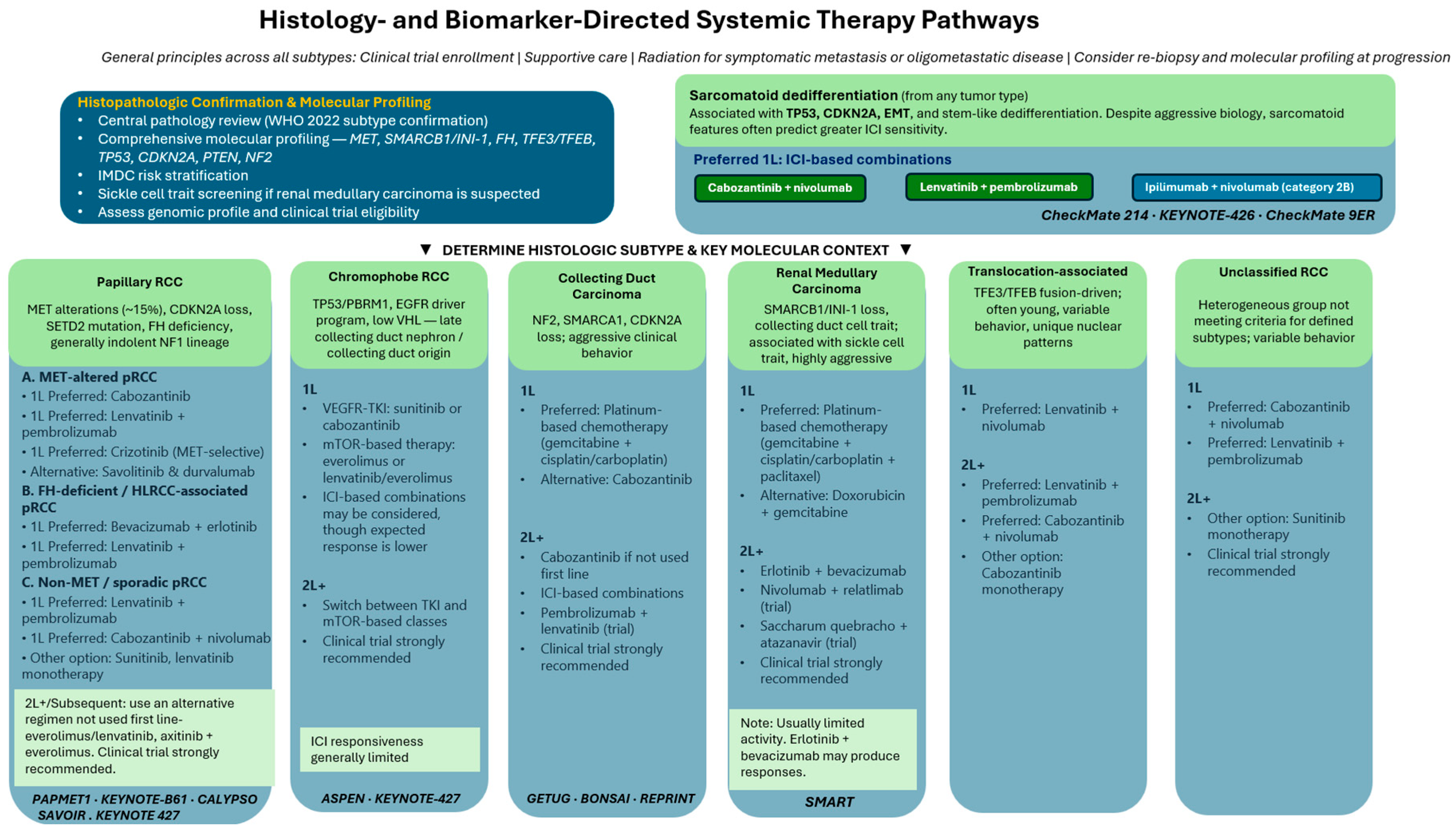
Figure 3.
Histology- and biomarker-directed systemic therapy pathways for advanced nccRCC. Recommendations should be individualized according to histology, molecular context, prior therapy, comorbidity, guideline availability, and clinical-trial eligibility.
Figure 3.
Histology- and biomarker-directed systemic therapy pathways for advanced nccRCC. Recommendations should be individualized according to histology, molecular context, prior therapy, comorbidity, guideline availability, and clinical-trial eligibility.
Supplementary Materials
The following supporting information can be downloaded at the website of this paper posted on Preprints.org.
Author Contributions
T.S., conceptualization, literature review, writing, and data analysis; Y.M., literature review and writing; N.S.K., literature review and writing; S.M.T., conceptualization, literature review, writing, and supervision; A.K., conceptualization, literature review, writing, and supervision. All authors have read and agreed to the published version of the manuscript.
Funding
This research received no external funding.
Institutional Review Board Statement
This study used de-identified, publicly available SEER research data under the SEER Research Data Use Agreement. The analysis did not involve interaction with human participants or access to identifiable private information and did not require institutional review board approval.
Informed Consent Statement
Not applicable.
Data Availability Statement
The data analyzed in this study are available from the National Cancer Institute SEER Program after execution of the SEER Research Data Use Agreement (SEER 17 Registries Research Data, November 2024 submission, 2000–2022). The ICD-O-3 selection criteria and analytic methods are reported in the manuscript.
Acknowledgments
During manuscript preparation, the authors used Mendeley for reference management and Stata for statistical analysis. Open Evidence and Ask Doximity were used to support literature searching. The authors reviewed and edited all content and take full responsibility for the final manuscript.
Conflicts of Interest
The authors declare no conflicts of interest.
Abbreviations
The following abbreviations are used in this manuscript:
| ALK | Anaplastic lymphoma kinase |
| ccRCC | Clear cell renal cell carcinoma |
| CD | Collecting duct |
| CDC | Collecting duct carcinoma |
| CDKN2A | Cyclin-dependent kinase inhibitor 2A |
| chRCC | Chromophobe renal cell carcinoma |
| CI | Confidence interval |
| CK7 | Cytokeratin 7 |
| CSC | Cancer stem cell |
| CTLA-4 | Cytotoxic T-lymphocyte-associated protein 4 |
| ctDNA | Circulating tumor DNA |
| CXCR4 | C-X-C chemokine receptor type 4 |
| DCT | Distal convoluted tubule |
| EMT | Epithelial-to-mesenchymal transition |
| FH | Fumarate hydratase |
| FOXI1 | Forkhead box I1 |
| HLRCC | Hereditary leiomyomatosis and renal cell carcinoma |
| HNF1B | Hepatocyte nuclear factor 1 beta |
| ICI | Immune checkpoint inhibitor |
| INI-1 | Integrase interactor 1 |
| KIT/CD117 | KIT proto-oncogene receptor tyrosine kinase/cluster of differentiation 117 |
| MITF | Microphthalmia-associated transcription factor |
| mTOR | Mechanistic target of rapamycin |
| MTF | Master transcription factor |
| nccRCC | Non-clear cell renal cell carcinoma |
| NF2 | Merlin |
| NRF2-ARE | Nuclear factor erythroid 2-related factor 2–antioxidant response element |
| PCT | Proximal convoluted tubule |
| PD-1 | Programmed death-1 |
| PFS | Progression-free survival |
| pRCC | Papillary renal cell carcinoma |
| scATAC-seq | Single-cell assay for transposase-accessible chromatin sequencing |
| SETD2 | SET domain-containing 2 |
| SLC7A11 | Solute carrier family 7 member 11 |
| SMARCB1 | SWI/SNF-related matrix-associated actin-dependent regulator of chromatin subfamily B member 1 |
| TFE3 | Transcription factor E3 |
| TFEB | Transcription factor EB |
| TGF-beta | Transforming growth factor beta |
| TKI | Tyrosine kinase inhibitor |
| TSC2 | Tuberous sclerosis complex 2 |
| uRCC | Unclassified renal cell carcinoma |
| VEGF | Vascular endothelial growth factor |
| VEGFR | Vascular endothelial growth factor receptor |
| VHL | Von Hippel-Lindau |
References
- Nepali, P.R.; Eraky, A.; Okhawere, K.E.; Dogra, N.; Mehrazin, R.; Badani, K.; Kyprianou, N. Molecular and therapeutic landscape of non-clear cell renal carcinoma. Nat. Rev. Urol. 2025, 22(11), 735–755. [Google Scholar] [CrossRef] [PubMed]
- National Cancer Institute. Surveillance, Epidemiology, and End Results Program. SEER*Stat Database: Incidence - SEER Research Data, 17 Registries, November 2024 Submission (2000-2022). April 2025. Available online: https://seer.cancer.gov/data-software/documentation/seerstat/.
- Giles, R.H.; Choueiri, T.K.; Heng, D.Y.; Albiges, L.; Hsieh, J.J.; Linehan, W.M.; Pal, S.; Maskens, D.; Paseman, B.; Jonasch, E.; et al. Recommendations for the Management of Rare Kidney Cancers. Eur. Urol. 2017, 72(6), 974–983. [Google Scholar] [CrossRef] [PubMed]
- Signoretti, S.; Flaifel, A.; Chen, Y.B.; Reuter, V.E. Renal Cell Carcinoma in the Era of Precision Medicine: From Molecular Pathology to Tissue-Based Biomarkers. JCO 2018, 36(36), 3553–3559. [Google Scholar] [CrossRef] [PubMed]
- Albiges, L.; Flippot, R.; Rioux-Leclercq, N.; Choueiri, T.K. Non–Clear Cell Renal Cell Carcinomas: From Shadow to Light. JCO 2018, 36(36), 3624–3631. [Google Scholar] [CrossRef] [PubMed]
- Young, M.; Jackson-Spence, F.; Beltran, L.; Day, E.; Suarez, C.; Bex, A.; Powles, T.; Szabados, B. Renal cell carcinoma. The Lancet 2024, 404(10451), 476–491. [Google Scholar] [CrossRef] [PubMed]
- Tannir, N.M.; Jonasch, E.; Albiges, L.; Altinmakas, E.; Ng, C.S.; Matin, S.F.; Wang, X.; Qiao, W.; Dubauskas Lim, Z.; Tamboli, P.; et al. Everolimus Versus Sunitinib Prospective Evaluation in Metastatic Non–Clear Cell Renal Cell Carcinoma (ESPN): A Randomized Multicenter Phase 2 Trial. Eur. Urol. 2016, 69(5), 866–874. [Google Scholar] [CrossRef] [PubMed]
- Armstrong, A.J.; Halabi, S.; Eisen, T.; Broderick, S.; Stadler, W.M.; Jones, R.J.; Garcia, J.A.; Vaishampayan, U.N.; Picus, J.; Hawkins, R.E.; et al. Everolimus versus sunitinib for patients with metastatic non-clear cell renal cell carcinoma (ASPEN): a multicentre, open-label, randomised phase 2 trial. Lancet Oncol. 2016, 17(3), 378–388. [Google Scholar] [CrossRef] [PubMed]
- Pal, S.K.; Tangen, C.; Thompson, I.M.; Balzer-Haas, N.; George, D.J.; Heng, D.Y.C.; Shuch, B.; Stein, M.; Tretiakova, M.; Humphrey, P.; et al. A comparison of sunitinib with cabozantinib, crizotinib, and savolitinib for treatment of advanced papillary renal cell carcinoma: a randomised, open-label, phase 2 trial. The Lancet 2021, 397(10275), 695–703. [Google Scholar] [CrossRef] [PubMed]
- Choueiri, T.K.; Heng, D.Y.C.; Lee, J.L.; Cancel, M.; Verheijen, R.B.; Mellemgaard, A.; Ottesen, L.H.; Frigault, M.M.; L’Hernault, A.; Szijgyarto, Z.; et al. Efficacy of Savolitinib vs Sunitinib in Patients WithMET-Driven Papillary Renal Cell Carcinoma. JAMA Oncol. 2020, 6(8), 1247. [Google Scholar] [CrossRef] [PubMed]
- McDermott, D.F.; Lee, J.L.; Ziobro, M.; Suarez, C.; Langiewicz, P.; Matveev, V.B.; Wiechno, P.; Gafanov, R.A.; Tomczak, P.; Pouliot, F.; et al. Open-Label, Single-Arm, Phase II Study of Pembrolizumab Monotherapy as First-Line Therapy in Patients With Advanced Non–Clear Cell Renal Cell Carcinoma. JCO 2021, 39(9), 1029–1039. [Google Scholar] [CrossRef] [PubMed]
- Srinivasan, R.; Gurram, S.; Singer, E.A.; Sidana, A.; Al Harthy, M.; Ball, M.W.; Friend, J.C.; Mac, L.; Purcell, E.; Vocke, C.D.; et al. Bevacizumab and Erlotinib in Hereditary and Sporadic Papillary Kidney Cancer. N Engl. J. Med. 2025, 392(23), 2346–2356. [Google Scholar] [CrossRef] [PubMed]
- Lee, C.H.; Voss, M.H.; Carlo, M.I.; Chen, Y.B.; Zucker, M.; Knezevic, A.; Lefkowitz, R.A.; Shapnik, N.; Dadoun, C.; Reznik, E.; et al. Phase II Trial of Cabozantinib Plus Nivolumab in Patients With Non–Clear-Cell Renal Cell Carcinoma and Genomic Correlates. JCO 2022, 40(21), 2333–2341. [Google Scholar] [CrossRef] [PubMed]
- Suárez, C.; Larkin, J.M.G.; Patel, P.; Valderrama, B.P.; Rodriguez-Vida, A.; Glen, H.; Thistlethwaite, F.; Ralph, C.; Srinivasan, G.; Mendez-Vidal, M.J.; et al. Phase II Study Investigating the Safety and Efficacy of Savolitinib and Durvalumab in Metastatic Papillary Renal Cancer (CALYPSO). JCO 2023, 41(14), 2493–2502. [Google Scholar] [CrossRef] [PubMed]
- Albiges, L.; Gurney, H.; Atduev, V.; Suarez, C.; Climent, M.A.; Pook, D.; Tomczak, P.; Barthelemy, P.; Lee, J.L.; Stus, V.; et al. Pembrolizumab plus lenvatinib as first-line therapy for advanced non-clear-cell renal cell carcinoma (KEYNOTE-B61): a single-arm, multicentre, phase 2 trial. Lancet Oncol. 2023, 24(8), 881–891. [Google Scholar] [CrossRef] [PubMed]
- Procopio, G.; Sepe, P.; Claps, M.; Buti, S.; Colecchia, M.; Giannatempo, P.; Guadalupi, V.; Mariani, L.; Lalli, L.; Fucà, G.; et al. Cabozantinib as First-line Treatment in Patients With Metastatic Collecting Duct Renal Cell Carcinoma. JAMA Oncol. 2022, 8(6), 910. [Google Scholar] [CrossRef] [PubMed]
- ClinicalTrials.gov. Pembrolizumab Plus Enfortumab Vedotin in Collecting Duct and Renal Medullary Carcinoma (REPRINT). Identifier NCT06302569. Available online: https://clinicaltrials.gov/study/NCT06302569.
- Huang, J.; Hu, X.; Wang, H.; Guo, J.; Zhang, H.; Dong, P.; Xue, W. Fruquintinib plus serplulimab as first-line therapy in metastatic or unresectable non-clear cell renal cell carcinoma (nccRCC): Updated efficacy and safety data from a multicenter, single-arm, phase II trial. J. Clin. Oncol. 2026, 44((7_) suppl, 422–422. [Google Scholar] [CrossRef]
- McGregor, B.A.; Huang, J.; Xie, W.; Xu, W.; Bilen, M.A.; Braun, D.A.; Zhang, T.; McKay, R.R.; McDermott, D.F.; Hammers, H.J.; et al. Phase II study of cabozantinib (Cabo) with nivolumab (Nivo) and ipilimumab (Ipi) in advanced renal cell carcinoma with variant histologies (RCCvh). JCO 2023, 41((16_) suppl, 4520–4520. [Google Scholar] [CrossRef]
- Nguyen, C.B.; Vaishampayan, U.N.; Campbell, M.T.; Hammers, H.J.; Brooks, R.; Solley, T.; Wood, L.M.; Jonasch, E.; Alva, A.S. Phase 2 study of combination tivozanib and nivolumab in advanced non-clear cell renal cell carcinoma (RCC): FORTUNE trial. JCO 2024, 42((16_) suppl, TPS4612–TPS4612. [Google Scholar] [CrossRef]
- Pal, S.K.; Powles, T.; Kanesvaran, R.; Molina-Cerrillo, J.; Feldman, D.R.; Barata, P.; Liu, M.; Bhatt, A.; Wang, Z.; Nandoskar, P.; et al. STELLAR-304: a phase III study of zanzalintinib (XL092) plus nivolumab in advanced non-clear cell renal cell carcinoma. Future Oncol. 2025, 21(7), 787–794. [Google Scholar] [CrossRef] [PubMed]
- ClinicalTrials.gov. A Study of AK104 Plus Axitinib in Advanced/Metastatic Special Pathological Subtypes of Renal Cell Carcinoma. Identifier NCT05808608. Available online: https://clinicaltrials.gov/study/NCT05808608.
- ClinicalTrials.gov. Axitinib +/- Pembrolizumab in First Line Treatment of mPRCC (PAXIPEM). Identifier NCT05096390. Available online: https://clinicaltrials.gov/study/NCT05096390.
- Feldman, D.R.; Ged, Y.; Lee, C.H.; Knezevic, A.; Molina, A.M.; Chen, Y.B.; Chaim, J.; Coskey, D.T.; Murray, S.; Tickoo, S.K.; et al. Everolimus plus bevacizumab is an effective first-line treatment for patients with advanced papillary variant renal cell carcinoma: Final results from a phase II trial. Cancer 2020, 126(24), 5247–5255. [Google Scholar] [CrossRef] [PubMed]
- Maughan, B.L.; Plets, M.; Pal, S.K.; Ged, Y.; Tangen, C.; Vaishampayan, U.N.; Lerner, S.P.; Thompson, I.M. SWOG S2200 (PAPMET2): A phase II randomized trial of cabozantinib with or without atezolizumab in patients with advanced papillary renal cell carcinoma (PRCC). JCO 2024, 42((4_) suppl, TPS493–TPS493. [Google Scholar] [CrossRef]
- Choueiri, T.K.; Xu, W.; Poole, L.; Telaranta-Keerie, A.; Hartmaier, R.; Powles, T. SAMETA: An open-label, three-arm, multicenter, phase III study of savolitinib + durvalumab versus sunitinib and durvalumab monotherapy in patients with MET-driven, unresectable, locally advanced/metastatic papillary renal cell carcinoma (PRCC). JCO 2022, 40((16_) suppl, TPS4601–TPS4601. [Google Scholar] [CrossRef]
- Bravo Montenegro, G.L.; Sharon, E.; Pickett-Gies, C.A.; Mac, L.; Friend, J.C.; Purcell, E.; Akshintala, S.; Glod, J.; McKay, R.R.; Bilen, M.A.; et al. A phase 2 study of bevacizumab, erlotinib, and atezolizumab in subjects with advanced hereditary leiomyomatosis and renal cell cancer (HLRCC) associated or sporadic papillary renal cell cancer (pRCC). JCO 2022, 40((16_) suppl, TPS4604–TPS4604. [Google Scholar] [CrossRef]
- Upadhyay, K.; Silverstein, D. Renal Development: A Complex Process Dependent on Inductive Interaction. CPR 2014, 10(2), 107–114. [Google Scholar] [CrossRef] [PubMed]
- Wang, Q.; Zhang, Y.; Zhang, B.; Fu, Y.; Zhao, X.; Zhang, J.; Zuo, K.; Xing, Y.; Jiang, S.; Qin, Z.; et al. Single-cell chromatin accessibility landscape in kidney identifies additional cell-of-origin in heterogenous papillary renal cell carcinoma. Nat. Commun. 2022, 13(1), 31. [Google Scholar] [CrossRef] [PubMed]
- Zhang, Y.; Narayanan, S.P.; Mannan, R.; Raskind, G.; Wang, X.; Vats, P.; Su, F.; Hosseini, N.; Cao, X.; Kumar-Sinha, C.; et al. Single-cell analyses of renal cell cancers reveal insights into tumor microenvironment, cell of origin, and therapy response. Proc. Natl. Acad. Sci. U.S.A. 2021, 118(24), e2103240118. [Google Scholar] [CrossRef] [PubMed]
- Saleeb, R.M.; Farag, M.; Ding, Q.; Downes, M.; Bjarnason, G.; Brimo, F.; Plant, P.; Rotondo, F.; Lichner, Z.; Finelli, A.; et al. Integrated Molecular Analysis of Papillary Renal Cell Carcinoma and Precursor Lesions Unfolds Evolutionary Process from Kidney Progenitor-Like Cells. Am. J. Pathol. 2019, 189(10), 2046–2060. [Google Scholar] [CrossRef] [PubMed]
- Tu, S.M.; Zhang, M.; Wood, C.G.; Pisters, L.L. Stem Cell Theory of Cancer: Origin of Tumor Heterogeneity and Plasticity. Cancers 2021, 13(16), 4006. [Google Scholar] [CrossRef] [PubMed]
- Trpkov, K.; Hes, O.; Williamson, S.R.; Adeniran, A.J.; Agaimy, A.; Alaghehbandan, R.; Amin, M.B.; Argani, P.; Chen, Y.B.; Cheng, L.; et al. New developments in existing WHO entities and evolving molecular concepts: The Genitourinary Pathology Society (GUPS) update on renal neoplasia. Mod. Pathol. 2021, 34(7), 1392–1424. [Google Scholar] [CrossRef] [PubMed]
- Nassar, A.H.; Abou Alaiwi, S.; Baca, S.C.; Adib, E.; Corona, R.I.; Seo, J.H.; Fonseca, M.A.S.; Spisak, S.; El Zarif, T.; Tisza, V.; et al. Epigenomic charting and functional annotation of risk loci in renal cell carcinoma. Nat. Commun. 2023, 14(1), 346. [Google Scholar] [CrossRef] [PubMed]
- Tu, S.M.; Aydin, A.M.; Maraboyina, S.; Chen, Z.; Singh, S.; Gokden, N.; Langford, T. Stem Cell Origin of Cancer: Implications of Oncogenesis Recapitulating Embryogenesis in Cancer Care. Cancers 2023, 15(9), 2516. [Google Scholar] [CrossRef] [PubMed]
- Verbiest, A.; Van Hoef, V.; Rodriguez-Antona, C.; García-Donas, J.; Graña-Castro, O.; Albersen, M.; Baldewijns, M.; Laenen, A.; Roussel, E.; Schöffski, P.; et al. MicroRNA expression profiles in molecular subtypes of clear-cell renal cell carcinoma are associated with clinical outcome and repression of specific mRNA targets. PLoS ONE 2020, 15(9), e0238809. [Google Scholar] [CrossRef] [PubMed]
- Turajlic, S.; Xu, H.; Litchfield, K.; Rowan, A.; Chambers, T.; Lopez, J.I.; Nicol, D.; O’Brien, T.; Larkin, J.; Horswell, S.; et al. Tracking Cancer Evolution Reveals Constrained Routes to Metastases: TRACERx Renal. Cell 2018, 173(3), 581–594.e12. [Google Scholar] [CrossRef] [PubMed]
- Turajlic, S.; Xu, H.; Litchfield, K.; Rowan, A.; Horswell, S.; Chambers, T.; O’Brien, T.; Lopez, J.I.; Watkins, T.B.K.; Nicol, D.; et al. Deterministic Evolutionary Trajectories Influence Primary Tumor Growth: TRACERx Renal. Cell 2018, 173(3), 595–610.e11. [Google Scholar] [CrossRef] [PubMed]
- Tu, S.M.; Chen, J.Z.; Singh, S.R.; Aydin, A.M.; Gokden, N.; Tam, N.N.C.; Leung, Y.K.; Langford, T.; Ho, S.M. Stem Cell Theory of Cancer: Clinical Implications of Epigenomic versus Genomic Biomarkers in Cancer Care. Cancers 2023, 15(23), 5533. [Google Scholar] [CrossRef] [PubMed]
- Lobo, J.; Ohashi, R.; Amin, M.B.; Berney, D.M.; Compérat, E.M.; Cree, I.A.; Gill, A.J.; Hartmann, A.; Menon, S.; Netto, G.J.; et al. WHO 2022 landscape of papillary and chromophobe renal cell carcinoma. Histopathology 2022, 81(4), 426–438. [Google Scholar] [CrossRef] [PubMed]
- Cancer Genome Atlas Research Network. Comprehensive Molecular Characterization of Papillary Renal-Cell Carcinoma. N Engl. J. Med. 2016, 374(2), 135–145. [Google Scholar] [CrossRef] [PubMed]
- Durinck, S.; Stawiski, E.W.; Pavía-Jiménez, A.; Modrusan, Z.; Kapur, P.; Jaiswal, B.S.; Zhang, N.; Toffessi-Tcheuyap, V.; Nguyen, T.T.; Pahuja, K.B.; et al. Spectrum of diverse genomic alterations define non–clear cell renal carcinoma subtypes. Nat. Genet 2015, 47(1), 13–21. [Google Scholar] [CrossRef] [PubMed]
- Tu, S.M.; Singh, S.R.; Arnaoutakis, K.; Malapati, S.; Bhatti, S.A.; Joon, A.Y.; Atiq, O.T.; Pisters, L.L. Stem Cell Theory of Cancer: Implications for Translational Research from Bedside to Bench. Cancers 2022, 14(14), 3345. [Google Scholar] [CrossRef] [PubMed]
- Zhou, Q.; Li, J.; Ge, C.; Chen, J.; Tian, W.; Tian, H. SNX5 suppresses clear cell renal cell carcinoma progression by inducing CD44 internalization and epithelial-to-mesenchymal transition. Mol. Ther.-Oncolytics 2022, 24, 87–100. [Google Scholar] [CrossRef] [PubMed]
- Wang, J.; Papanicolau-Sengos, A.; Chintala, S.; Wei, L.; Liu, B.; Hu, Q.; Miles, K.M.; Conroy, J.M.; Glenn, S.T.; Costantini, M.; et al. Collecting duct carcinoma of the kidney is associated withCDKN2Adeletion andSLCfamily gene up-regulation. Oncotarget 2016, 7(21), 29901–29915. [Google Scholar] [CrossRef] [PubMed]
- Xie, D.; Li, G.; Zheng, Z.; Zhang, X.; Wang, S.; Jiang, B.; Li, X.; Wang, X.; Wu, G. The molecular code of kidney cancer: A path of discovery for gene mutation and precision therapy. Mol. Asp. Med. 2025, 101, 101335. [Google Scholar] [CrossRef] [PubMed]
- Zacharias, N.M.; Rupe, E.S.; Chen, X.; Kotecha, R.R.; Rieke, D.T.; McGregor, B.A.; Pesquera, P.; Yu, Z.; Grover, A.; Daw, N.C.; et al. Panitumumab-Based EGFR Blockade in SMARCB1-Deficient Renal Medullary Carcinoma: Preclinical Basis and Prospective Clinical Activity. Clin. Cancer Res. 2026, OF1–OF19. [Google Scholar] [CrossRef] [PubMed]
- Peired, A.J.; Sisti, A.; Romagnani, P. Renal Cancer Stem Cells: Characterization and Targeted Therapies. Stem Cells Int. 2016, 2016(1), 8342625. [Google Scholar] [CrossRef] [PubMed]
- Cheng, B.; Yang, G.; Jiang, R.; Cheng, Y.; Yang, H.; Pei, L.; Qiu, X. Cancer stem cell markers predict a poor prognosis in renal cell carcinoma: a meta-analysis. Oncotarget 2016, 7(40), 65862–65875. [Google Scholar] [CrossRef] [PubMed]
- Basu, A.; Au, C.; Kommalapati, A.; Kandala, H.; Sudhaman, S.; Mahmood, T.; Carson, C.; Pajak, N.; Dutta, P.; Calhoun, M.; et al. Longitudinal Testing of Circulating Tumor DNA in Patients With Metastatic Renal Cell Carcinoma. JCO Precis Oncol. 2024, (8), e2400667. [Google Scholar] [CrossRef] [PubMed]
- Mo, S.; Ye, L.; Wang, D.; Han, L.; Zhou, S.; Wang, H.; Dai, W.; Wang, Y.; Luo, W.; Wang, R.; et al. Early Detection of Molecular Residual Disease and Risk Stratification for Stage I to III Colorectal Cancer via Circulating Tumor DNA Methylation. JAMA Oncol. 2023, 9(6), 770. [Google Scholar] [CrossRef] [PubMed]
- Suri, Y.; Sudhaman, S.; Dwivedi, B.; Kollipara, R.; Singh, A.; Velichko, S.; Carson, C.; Wray, A.; Dutta, P.; Malhotra, M.; et al. 2609P Clinical performance of a tumor-informed whole genome-based (WGS) ctDNA assay for recurrence detection and treatment response monitoring in localized and advanced/metastatic renal cell carcinoma (RCC). Ann. Oncol. 2025, 36, S1309–S1310. [Google Scholar] [CrossRef]
- Bhanvadia, R.; Smigelski, M.; Sudhaman, S.; Nagpal, S.; Brooks, B.; Gerald, T.; Sanchez-Mendez, R.; Battista, C.; Kazarian, A.; Choi, S.H.; et al. Utility of Circulating Tumor DNA Monitoring in Postnephrectomy Renal Cancer. JCO Oncol. Adv. 2026, 3(1), e2500066. [Google Scholar] [CrossRef]
- Ghavshough, M.H.; Fu, M.Z.; Patel, D.; Packiam, V.T.; Ghodoussipour, S. Tumor-informed ctDNA surveillance after radical nephrectomy and level IV IVC thrombectomy for clear cell renal cell carcinoma: Early detection of molecular residual disease. Urol. Case Rep. 2026, 66, 103460. [Google Scholar] [CrossRef] [PubMed]
- Alam, R.; Fuchs, J.W.; Shenoy, N.K. Utility of circulating tumor DNA in management of diverse renal malignancies: Insights from a case series in adjuvant and recurrent/metastatic settings. Med 2025, 6(10), 100806. [Google Scholar] [CrossRef] [PubMed]
Figure 1.
Kaplan–Meier estimates of overall survival for various nccRCC histologies. The risk table shows the number of patients remaining under observation.
Figure 1.
Kaplan–Meier estimates of overall survival for various nccRCC histologies. The risk table shows the number of patients remaining under observation.
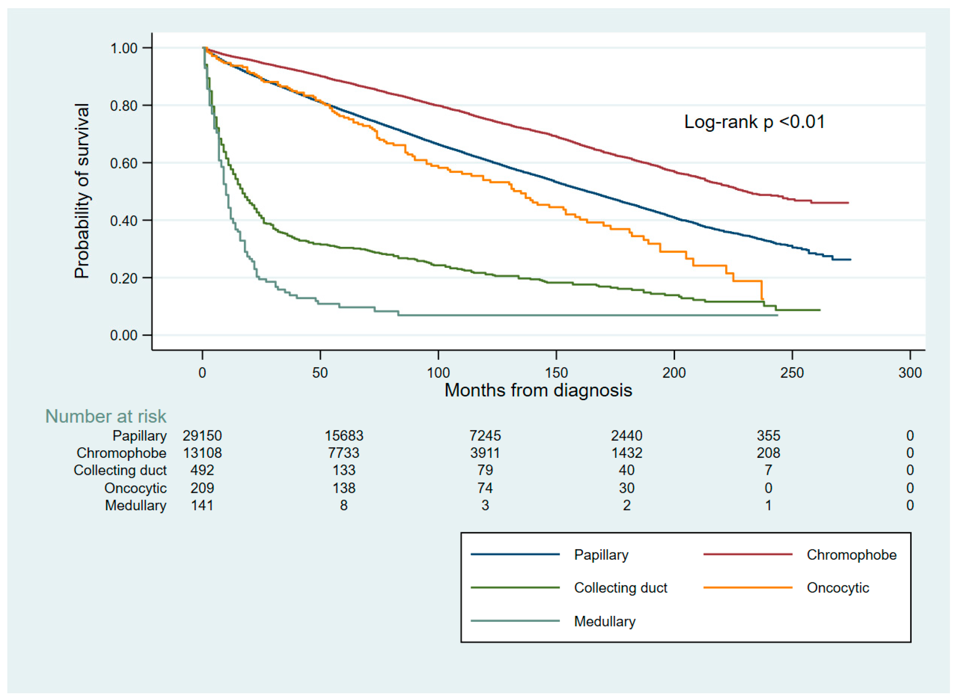
Table 1.
Distribution of various nccRCC histologies overall and by sex, age, race and stage IV.
| Histological type |
Total, n (%) |
Sex |
Median age, years |
Race | Stage IV, n/N known (%) | |||
| Male, n (%) | Female, n (%) | White, n (%) | African American, n (%) | Other, n (%) | ||||
| Papillary | 29,824 (67.83) | 22,924 (74.02) | 6,900 (53.09) | 62 | 20,425 (64.44) | 8,102 (81.23) | 1,297 (56.46) | 388/8,467 (4.6) |
| Chromophobe | 13,297 (30.24) | 7,472 (24.13) | 5,825 (44.82) | 62 | 10,721 (33.83) | 1,628 (16.32) | 948 (41.27) | 82/3,993 (2.1) |
| Collecting duct | 511 (1.16) | 354 (1.14) | 157 (1.21) | 62 | 361 (1.14) | 111 (1.11) | 39 (1.70) | 30/66 (45.5) |
| Oncocytic | 208 (0.47) | 130 (0.42) | 78 (0.60) | 72 | 164 (0.52) | 34 (0.34) | 10 (0.44) | NE (0 known) |
| Medullary | 126 (0.29) | 90 (0.29) | 36 (0.28) | 30 | 24 (0.08) | 99 (0.99) | 3 (0.13) | 33/40 (82.5) |
Table 2.
Univariable Cox proportional hazards estimate for survival by histology (papillary RCC as the reference group).
Table 2.
Univariable Cox proportional hazards estimate for survival by histology (papillary RCC as the reference group).
| Histological type | HR | 95% CI | p-value |
| Chromophobe | 0.57 | 0.55–0.60 | <0.001 |
| Collecting duct | 3.70 | 3.35–4.08 | <0.001 |
| Oncocytic | 1.29 | 1.07–1.56 | 0.007 |
| Medullary | 11.42 | 9.46–13.78 | <0.001 |
Disclaimer/Publisher’s Note: The statements, opinions and data contained in all publications are solely those of the individual author(s) and contributor(s) and not of MDPI and/or the editor(s). MDPI and/or the editor(s) disclaim responsibility for any injury to people or property resulting from any ideas, methods, instructions or products referred to in the content. |
© 2026 by the authors. Licensee MDPI, Basel, Switzerland. This article is an open access article distributed under the terms and conditions of the Creative Commons Attribution (CC BY) license (http://creativecommons.org/licenses/by/4.0/).
Copyright: This open access article is published under a Creative Commons CC BY 4.0 license, which permit the free download, distribution, and reuse, provided that the author and preprint are cited in any reuse.