
Submitted:
18 September 2026
Posted:
20 September 2026
You are already at the latest version
Abstract
The mainstream approach to splicing therapy today involves synthetic antisense oligonucleotides. ASOs interfere with spliceosomal ribozyme assembly by blocking splice sites or influence protein regulators by blocking or adding their binding sites. ASOs are not compatible with human biology, which limits their efficacy and they come with a burden of chemical toxicity. Spliceosomal snRNAs adapted for a specific target can either enhance or suppress exon inclusion. Recombinant snRNAs with their pre-mRNA binding sites changed still assemble into functional snRNPs and integrate into active spliceosomes. Native to human cells, they are versatile for in vivo delivery and can be either encoded and expressed from rAAVs or delivered as RNA molecules by exosomes, benefitting from excellent biodistribution. Imported snRNAs are taken up by cytoplasmic maturation factors and transferred into the nucleus. Adapted snRNAs can be used to target different stage spliceosomes: U1 and U2 for early complexes, U2/U6 for precatalytic or U5 for catalytic complexes. U1 is the only spliceosomal snRNA thoroughly examined in pre-clinical studies (~100 mutations in 24 genes) and it is necessary to review the past 20 years of experience with U1 before moving on to other snRNAs. U1 is involved in the initial splice site selection and usually binds the 5’ss, but it also promotes 5’ss usage ‘at a distance’ if bound in the vicinity. Adapted U1 suppresses 3’ss usage, as likely does WT U1 if 3’ss CAG|GU protosplice site repeat is followed by a sequence resembling the start of the intron. However, historically the non-spliceosomal U7-OPT is often used as a scaffold molecule for exon-skipping ASOs. This chimeric U7 snRNA cannot join histone bodies, its WT destination. While both U1 and U7 show promising safety profiles in mice, WT U7 expression is 1000 times lower, than U1. U7 snRNA modulates master transcription regulators by binding histone-fold domain protein NF-Y; the effects of U7-OPT overexpression remain underexplored. U1, a splicing molecule, naturally works better for promoting exon inclusion, than U7, but years of experiments also revealed U1 and U7 limitations. New adaptations of the core spliceosomal snRNAs are necessary to overcome these limitations.
Keywords:
splice sites
; U1 snRNA
; protosplice site repeats
; U7 snRNA
; ASO
; Duchenne muscular dystrophy
; spinal muscular atrophy
; cell penetrating peptides
; AAV
; exosomes
1. Introduction
The focus of translation research and industrial development is largely shifted towards effective delivery of drugs, often overlooking the limitations of the drugs themselves. Yet, the performance of drugs depends on their compatibility with human biology. Here we will discuss why snRNAs are better than existing splicing and gene therapies and how they are well suited for in vivo delivery.
Parts 1 and 2 (Artemyeva-Isman, 2026a,b) reviewed spliceosomal ribozyme evolution and structure, highlighting catalytic capabilities and the mechanism of splice sites recognition, which ensures splicing precision (Artemyeva-Isman and Porter, 2021) The process of modular assembly of the ribozyme allows to target early, pre-catalytic or catalytic spliceosome complexes for splicing and gene therapy. This paper will focus on the early splice site choice for exon inclusion or skipping, reviewing the performance of U1 snRNA in preclinical studies and the technologies it is competing against.
In Zhuang and Weiner, 1986 discovered that U1 snRNA adapted to match a 5’ss mutation can correct splicing. The same authors demonstrated (Zhuang and Weiner, 1989) that a branchpoint site mutation that led to the use of a cryptic 3’ss can be suppressed by U2 snRNA made complementary to the mutant BP site. These important early works show that snRNAs with altered pre-mRNA binding sites, which still have their protein-binding sites unchanged, are recognized by spliceosomal protein components and undergo normal assembly process to form functional ribonucleoproteins - spliceosomal snRNPs. Despite this, synthetic antisense oligonucleotides ASOs (or SSO - splice-switching oligos) with modified sugar-phosphate chemistry to enhance their stability are used for therapeutic splicing modulation today. Here it is necessary to introduce ASO technology and its shortcomings.
2. Antisense Oligonucleotides
2.1. Therapy for Spinal Muscular Atrophy
The first and notable ASO success is Nusinersen (Spinraza® Biogen) for Spinal Muscular Atrophy. SMA is an exceptional case, because Survival Motor Neuron gene (SMN1) mutated in SMA, has a gene double, SMN2. It is identical in coding sequence except for a CU change in exon 7 position +6 (cDNA 840CT; exon 7 is now exon 8 in ensembl and LOVD databases, traditional exon numbering is continued below). This change is synonymous, Phe280Phe, but apparently affects exon 7 inclusion. About 90% of SMN2 transcripts lack exon 7 and lead to production of an unstable protein. Both orthologous genes are located on the short arm of chromosome 5 q13.2 and while the functional SMN1 is a unique copy, 0 to 4 copies of SMN2 gene can be present (Prior et al., 2009). Notably, 10-15% of healthy individuals lack SMN2 altogether, but SMN1-/- is incompatible with foetus survival in the absence of SMN2, which supplies some of the functional protein. Accordingly, SMA severity is inversely corelated with SMN2 copy number: severe SMA, a leading genetic cause of infant mortality, is linked to 1 or 2 SMN2 copies. However, if SMN2 splicing is corrected, it can compensate for any SMN1 mutations providing treatment for all SMA patients. Earlier studies showed that SMN2 exon 7 inclusion is ruled by RBPs, competitive binding of hnRNP A1/A2 and SR protein SF2/ASF, and the mutation destroys an exonic splicing enhancer ESE (Singh et al., 2004; Cartegni et al., 2006). Coincidentally, it also adds a Watson-Crick pair for the ectopic U1 binding at the SMN2 exon 7 3’ss – here we refer to protosplice site repeats CAG|GU at both 5’ and 3’ss (see Part 2, Artemyeva-Isman, 2026b). The nusinersen oligonucleotide blocks an intronic splicing silencer ISS in intron 7, which is effective to counterbalance the ESE disruption and promotes exon 7 inclusion (Singh et al. 2007; US patent 8,110,560 - Singh et al. 2012). Another advantage for SMA treatment is that efficient delivery of ASO is achieved intrathecally. To prolong stability, Nusirensen ASO has a modified sugar-phosphate backbone: a 2’O-methoxyethyl ribose (MOE) and 5’-3’ phosphorothioate linkage (PS). Since Nusinersen was approved for clinic by the FDA in December 2016, then worldwide between 2017 and 2019, it had improved the condition and life expectancy of SMA patients. However, it appeared that ASO also promotes Histone 3 methylation adding the H3K9me2 epigenetic silencing mark to the target SMN2 locus (Marasco et al., 2022). To prevent the unwanted chromatin remodelling Nusinersen is used in combination with valproic acid (VPA), a histone deacetylase (HDAC) inhibitor. More concerningly, high doses of Nusirensen appear to deregulate off-target splicing events (Ottesen et al., 2021). Crucially, in the long-term repeated intrathecal administrations are traumatic to the patients, so while Nusirensen helps to improve the condition it is far from providing cure. Here I am obliged to deviate from ASOs and include the performance of other SMA therapies. Risdiplam (FDA approved in August 2020), a small molecule administered orally, can enhance SMN2 exon 7 inclusion. The proposed mechanism of action is stabilisation of U1 snRNA binding with the end of exon 7 that has an unusual A-1 and an interaction with purine-rich ESE2 of exon 7 (SMN-C5, same chemistry as Risdiplam – Sivaramakrishnan et al., 2017; Campagne et al., 2019). Combined therapy of Risdiplam and VPA is preferred by some patients (Koterazawa et al., 2023). However, this is again supportive treatment only, which cannot reverse the disease, and off-target effects are a risk. In a dose dependent manner, Risdiplam deregulates expression and splicing of thousands to hundreds of genes involved in replication, cell cycle, signalling and metabolism (Ottesen et al., 2023). Finally, since May 2019 severe SMA is treated in infants with single intravenous injection of AAV9-SMN, which is lifesaving and produces beneficial long-term effects. However, the FDA halted a clinical trial of this drug in older patients with milder SMA, as SMN overexpression appeared toxic in the long-term in animal models. Excessive SMN was shown to sequester Sm proteins in the cytoplasm of motor neurons and proprioceptor neurons in the dorsal root ganglia, leading to neurodegeneration, synapse disruption and inflammatory complications – a disease resembling SMN deficiency, as in both cases nuclear snRNPs are depleted, leading to widespread aberrant splicing (Van Alstyne et al., 2021). This brief account of splicing and gene therapies for SMA shows that an approach more coherent with natural gene regulation can help to avoid side effects. Preclinical studies on U1 snRNA achieved boosting SMN2 exon 7 inclusion (Singh et al., 2007; Fernandez Alanis et al., 2012; Donadon et al., 2019; Dal Mas et al., 2015a; Rogalska et al., 2016), as discussed below (5.1 Boosting exon inclusion by U1 snRNA). Further possibilities include adapting U6, U2 and U5 snRNA of the pre-catalytic spliceosome to enhance splicing of introns 6 and/or 7, and correcting SMN2 transcripts or gene by reverse splicing (see Part 4, Artemyeva-Isman, 2026d).
2.2. Therapy for Duchenne Muscular Dystrophy
Encouraged by the initial success of SMA treatment by Nusirensen, ASOs were tried out in the clinic for exon skipping to correct the reading frame of the dystrophin gene (Xp21), which is a hotspot for deletion of 1 or multiple exons. The general rule is that exon deletions that cause a frame shift lead to a severe phenotype - Duchenne muscular dystrophy DMD. Conversely, deletions that happen to preserve the reading frame and affect only the rod-domain produce a shortened dystrophin and can lead to a much milder disease – Becker dystrophy BMD (Monaco et al., 1988). As 3’ deletion breakpoints often occur in introns 50, 52 and 44, it is possible to correct the reading frame for the majority of patients by skipping exons 51, 53 and 45. Accordingly, the FDA has conditionally approved four ASOs for clinical use. These ASOs have phosphorodiamidate morpholino (PMO) modification of the sugar-phosphate backbone designed to block inclusion of one of these exons: Eteplirsen Ex51 (Sarepta, 2016), Golodirsen Ex53 (Sarepta, 2019), Viltolarsen Ex53 (NS Pharma, 2020), Casimersen Ex45 (Sarepta, 2021). The current problem is the delivery of PMOs to skeletal muscle throughout the body, the diaphragm and the heart. PMOs are infused into the bloodstream once weekly, but the treatment typically achieves low levels of dystrophin production in skeletal muscle, <1% to 9% (Torres-Masjoan et al., 2025; Haque et al., 2024) with limited benefits for the patients. The same PMOs used in patients with single exon 45 and 53 duplications produce 1-6% of normal dystrophin levels (Nicolau et al., 2024), in addition to almost 1% that is naturally produced in these patients by compensatory duplication skipping, altogether leading to somewhat better therapeutic effect. As for adverse effects of PMOs, renal toxicity is flagged by the FDA for all but Eteplirsen, which has a warning of hypersensitive reactions (Haque et al., 2024). Another drug, Ataluren (PTC124), a small molecule that promotes stop-codon read-through was the first drug approved in Europe in 2014 for DMD patients with nonsense mutations (~15% of all cases). Despite some positive reports (McDonald et al., 2022), the evidence was inconclusive, the drug was ruled ineffective, and in March 2025 the EMA withdrew the authorisation. In parallel, AAV micro-dystrophin therapies are also in clinical trials. To fit into an AAV, micro-dystrophins are designed with abridged rod domains modelled on shortened dystrophins found in some BMD patients with mild disease (Elangkovan and Dickson, 2021). ELEVIDYS got FDA approval in June 2023, but since three boys died after treatment, the drug has been suspended. Apart from adverse reactions of the liver, Hart et al., 2024 reported a potential problem: micro-dystrophin used in ELEVIDYS appeared to accelerate cardiomyopathy in DBA/2-mdx mice, a model of severe DMD. In this study overexpression in the heart was 10 times more efficient and micro-dystrophin appeared to displace utrophin, a shorter dystrophin orthologue that has a distinct, yet not understood function at the sarcolemma of cardiomyocytes. In skeletal muscle the expression was an order less, utrophin was not displaced from neuromuscular junctions, where its presence is essential. Another suggested cause of cardiac failure is overloading of protein degradation ubiquitin proteosome system. Postmitotic cells are particularly susceptible to proteotoxicity by accumulation of misfolded proteins. Finally, AAV-CRISPR-hfCas12Max, an optimised Cas12i derivative (Zhang et al., 2023) is tried in the clinic by HuidaGene to destroy DMD exon 51 5’ss by a DBS. This strategy aims to achieve permanent exon 51 skipping in patients with deletions of exon(s) 52, 52-61 and 52-63. Apart from off-target effects, which the designers endeavour to avoid with hfCas12Max, a recent preprint warns of on-target whole AAV integration at DSBs (Jia et al., 2025). This is a particular concern in case of small Cas12i, as the whole functional endonuclease is encoded by a single AAV, integration can lead to permanent Cas12 expression and gradual accumulation of genotoxic events. The risks for trial participants highlight the need for DMD therapy effort to try methods compatible with endogenous splicing and gene expression regulation to avoid toxicity and dangerous complications. DMD splicing modulation was tried out with U1 snRNA (Denti et al., 2006, 2008; Incitti et al., 2010 – see comment on designs below in 5.2 Inducing exon skipping by U1 and U7 snRNAs). Modified U7 snRNA, which is not a component of the spliceosome and has a stand-alone role in histone pre-mRNA 3’end processing, was used as a carrier of antisense sequences blocking DMD splice sites, ESE or adding ESS, hnRNP A binding site to promote exon skipping (Goyenvalle et al., 2004, 2009; Vulin et al., 2012 – see U7 design and limitations explained below). The advantage over ASOs is that snRNAs can be expressed from an AAV vector. Presently, U7 carrying sequences to block exon 2 splice sites progressed to clinical trials (scAAV9.U7.ACCA) for patients with the most common duplications of exon 2. A previous study recorded beneficial effects of a single intravenous AAV injection persisting for 18 months in Dup2 mice (Gushchina et al., 2023; Roberts, 2023). This is a very promising approach, based on an outstanding investigation of the exceptions from the frame-shift rule in BMD patients with nonsense mutations in dystrophin exons 1 and 2 and frame-shift deletion of exon 2. These mutations activate an internal ribosome entry site IRES in exon 5, leading to translation initiation in exon 6, which produces a functional N-truncated dystrophin (Flanigan et al., 2009; Gurvich et al., 2009; Wein et al., 2014). However, exon 5 IRES is inactivated by exon 2 duplications, leading to a severe phenotype. U7.ACCA aims to skip one or both exon 2 copies to produce either normal dystrophin or the functional N-truncated dystrophin isoform. However, this strategy can only help a minority of patients. For most patients, improvement of exon skipping can be also tried with snRNAs, which can be expressed from AAV (see below: the design principles for U1 are explained, and U1 is compared to U7). However, rather than exon skipping, or mini-dystrophin, both of which only aim to convert DMD to BMD, including deleted exons from an exogenous template is a better solution. This implies naturally controlled normal dystrophin expression. Trans-splicing was tried out, but admittedly only two works are published on this approach for DMD (Lorain et al., 2010, 2013) compared to hundreds of studies on improvements of ASOs for DMD exon skipping. The efficiency of trans-splicing remained low, and there is a way to ameliorate it, if all snRNA interactions are improved and some extended for the trans-exon(s) to win the splicing competition. Moreover, defining U5 snRNA binding register by statistical dependencies at human splice sites (Artemyeva-Isman and Porter, 2021) and the U5 Loop1 structure presented in Part 2 (Artemyeva-Isman, 2026b, Figure 7) are aimed to open a new way for development of reverse splicing to insert deleted exons into the DMD gene (Part 4, Artemyeva-Isman, 2026d).
2.3. Important Developments: Targeting Non-Productive Splicing and Hotspot Exons
Whether effective or not, ASOs are approved drugs, and that encourages their application to other disease targets, which I will not review here, but for two features of special interest. ‘Aberrant’ or non-productive splicing is one of the mechanisms to control gene expression. ASO splicing modulation to reduce aberrant isoforms destined for NMD can benefit all patients with haploinsufficient genetic diseases regardless of their individual mutations (Lim et al., 2020). Stoke therapeutics now partnered with Biogene (August 2025) to produce zorevunersen (STK-001) ASO targeting SCN1A (sodium channel Nav1.1) transcripts in Dravet syndrome, severe progressive epilepsy. This is a rare autosomal dominant condition caused by haploinsufficiency of SCN1A gene, the expression of which is charismatically downregulated by splicing, normally producing 89% of non-productive mRNAs. The second feature is a recent paper promoting an idea to develop ‘single’ ASOs for the correction of any disease-causing splice site mutations (Duan et al., 2025). A previous study (Glidden et al., 2021) shows that disease-causing ss mutations appear to be concentrated around exons with low splice site usage – termed ‘hotspot exons’, 1400 identified in the human genome. Typically, such exons depend on multiple RBPs for their inclusion. This means that exons which already have divergent splice sites (i.e. not following the consensus C - Part 1, Artemyeva-Isman, 2026a) cannot tolerate any further mutations, as suboptimal base-pairing with snRNAs will be made even worse or RBP binding will be blocked. Targeting individual disease-causing mutations is commercially impractical and the principle of ‘single’ ASOs is not to target hotspot exons themselves, but to slow down splicing of the flanking introns by targeting the neighbouring exons. Reduced splicing rate promotes the inclusion of the hotspot exon and can suppress various mutations using the same ASO design.
2.4. ASO Toxicity and Off-Target Effects
The shortcoming of ASOs, apart from the unexpected problem of the target gene silencing, as with Nusinersen, is that different formulations of the oligonucleotide backbone are toxic to various degrees - kidneys and liver being the ‘high exposure’ organs (Goyenvalle et al., 2023; Haque et al., 2024). Natural RNA or DNA oligos are rapidly degraded and as the therapeutic oligos cannot be expressed by the target cell, efficient treatment requires a large dose and chemical modification for sustained activity. Moreover, diffusing oligonucleotides into the bloodstream is prone to immunostimulatory effects and adverse effects on haemostasis. These include thrombocytopenia, inhibition of coagulation and complement activation (Goyenvalle et al., 2023). Intrathecal delivery has a risk of neurotoxic effects, which includes immune reaction, tremors and seizures (Goyenvalle et al., 2023; Miller et al., 2024; Li et al., 2024). Furthermore, the very specificity of ASO effect on splicing is dose-dependent, as can be expected for hybridization of nucleic acids: at high dose Nusinersen massively perturbs the entire transcriptome (Ottesen et al., 2021). The sum of these adverse effects is aggravating, because patients are exposed to repeated ASO treatment, every several months for intrathecal and weekly for intravenous administration.
3. Splicing Switches as Therapeutic Targets in Common Diseases
Splicing therapy started with SMA and DMD because of unmet need for treatment, but modulation of splicing is by no means limited to neuromuscular field or correction of inherited ss mutations, as indeed aberrant splicing is evident as soon as it is studied in disease states generally. ASO-associated toxicity, delivery limitations and the need for repeated administration impede new splicing therapies to compete with existing treatments. SnRNAs are more versatile than ASOs: snRNAs are themselves components of the spliceosome that base-pair with the splice sites, they do not need chemical modifications as they join the natural maturation pathway in the cytoplasm that protects them from degradation and shuttles them to the nucleus, and they can be expressed by the target cell if snRNA genes are delivered by AAV. In this light, we can consider a wide field of future disease targets for snRNAs and show a few examples.
3.1. Immunology and Haematology
Switching between splicing isoforms with antagonistic functions is a common regulatory mechanism of physiological states, such as immune response and haemostasis. Intronic and exonic splice site mutations can jam or skew these switches leading to autoimmune or coagulation diseases. A famous example, human FAS/CD95 (10q23) exon 6 inclusion controls T-cell proliferation and clearance by apoptosis. Full length FAS is a transmembrane death receptor, while FasD6 is a soluble FAS that stimulates T-cell growth. The dysregulation of this switch by intron 5 -3CG mutation leads to autoimmune lymphoproliferative syndrome ALPS (Corrionero et al., 2011). -3C is one of the most conserved 3’ss positions, which we have suggested pairs with U2 G31 (C in Part 1, Artemyeva-Isman, 2026a; see section 2.3 Branchpoint pairing with U2 snRNA in Part 2, Artemyeva-Isman, 2026b). Apart from ALPS, FAS mutations and decreased FAS-mediated T-cell clearance were found in patients with primary immune thrombocytopenic purpura ITP (Vandrovcova et al., 2019; Boggio et al., 2017). FAS dysregulation plays a role in T-cell accumulation in the brain in multiple sclerosis (Volpe et al., 2016), and elevated serum soluble FAS is a common marker in autoimmune conditions (Vincent et al., 2020; Guillot et al, 2001), making it a therapeutic target. It can be addressed by snRNA-mediated regulation of the FAS splicing switch, provided specific delivery to T-cells is feasible (Hamilton et al., 2024).
Another example, coagulation Factor V F5 (1q24) alternative intron inside exon 13 controls haemostasis. Full length F5 is a co-factor of F10 in the prothrombinase complex, while F5-short - with most of exon 13 excised - is a co-factor of the Tissue Factor Pathway Inhibitor (TFPI), a potent coagulation suppressor. The full-length F5 isoform is by far predominant, normally only sub-nanomolar amounts of F5-short are in circulation and it was discovered by chance due to a mutation that causes a sharp upregulation of F5-short splicing leading to a bleeding disorder (Vincent et al, 2013; Broze & Girard, 2013). This mutation, F5 c2350 AG in exon 13 (F5-Texas), introduces the conserved exon-end G-1 preceding the alternative intron and establishes the most important G=C pair with U5 C39 (C). SnRNAs can be adapted to regulate F5 splicing switch: a very slight upregulation of F5-short can be beneficial for patients with chronic thrombosis; delivery to the liver is realistic (see 4 Delivery limitations).
3.2. Cancer
Cell transformation, tumour growth and immunosuppression are also controlled by splicing switches. Let us consider a few usual cancer targets. HER2 /ERBB2 (17q12) Human Epidermal Growth Factor Receptor 2 overexpression is associated with bad prognosis in breast, non-small cell lung and other cancers (Smith et al., 2020). HER2 is an exceptional Epidermal Growth Factor Receptor (EGFR) tyrosine kinase, which unlike other members of this family does not require a ligand for dimerization. Moreover, HER2 is a preferred partner for heterodimerisation by other HER/EGFR, which leads to kinase activation of both partners and drives cell transformation. Remarkably, HER2 splicing isoforms differ in their effects. The exclusion of exon 16 makes it worse, as HER2D16 is particularly prone to form constitutively active homodimers, promoting aggressive tumours. On the contrary, retention of HER2 intron 8 produces a short isoform with a distinct C-teminus Herstatin, which is beneficial: it disrupts HER2 dimers and inhibits proliferation of transformed cells, acting as a potent tumour suppressor (Doherty et al., 1999; Silipo et al., 2017). Splicing modulation is a way to reverse the oncogenic activity of HER2.
The vascular endothelial growth factor VEGFA (6p21) exists as two antagonistic forms: pro- or anti-angiogenic - depending on the usage of the alternative 3’ss of the last exon 8 and producing distinct C-termini of six amino acids (Di Matteo et al., 2020). VEGFA165-a isoform is the main factor of tumour vascularisation – its suppression by snRNA-mediated switching to the distal 3’ss producing anti-angiogenic VEGFA165-b will prevent tumour growth.
The androgen receptor AR (Xq12), the main culprit in prostate cancer, becomes constitutively active at a dangerous castrate-resistant later stage – CRPC. AR variant 7 splicing isoform includes a cryptic exon 3b and produces a functional receptor that lacks ligand-binding domain (Rana et al., 2021; Zhu and Luo, 2020) - ARv7 is a specific biomarker in circulating cancer cells for bad prognosis in CRPC (Sartor and Dong, 2015). Directing splicing to androgen-controlled isoforms can be beneficial for CRPC management.
Finally, immune evasion of tumours is mediated by Programmed Death PD1 receptor of T-cells, that is triggered by its ligand PDL1 (9p24) overexpressed by cancer cells. Again, PDL1 splicing plays a key role in the regulation of this axis. Affinity to PD1 is increased if exon 3 is excluded - PDL1 176aa compared to the full-length PDL1 290aa (colorectal cancer study Wang et al., 2021). Worse, while PD1-PDL1 interaction requires surface contact between T-cells and tumour cells, another isoform PDL1 245aa lacking trans-membrane domain has an alternative C-terminus, which serves to form stable dimers that are secreted by tumour cells and drive systemic immunosuppression and promotes metastases and leads to poorer survival (Wang et al., 2021; Mahoney et al., 2019). In addition, a long non-coding RNA splicing variant is produced by excising an alternative intron from exon 4 and using a distal 3’ss to splice out intron 5 - PDL1lnc enhances c-Myc oncogenic activity (Qu et al., 2021). Clearly, it is important to distinguish between these four different PDL1s and consider switching off potent immunosuppressive shorter protein isoforms and PDL1lnc when devising cancer immunotherapy.
4. Delivery Limitations
The chief problem with delivery is that most of systemically injected drugs accumulate in the liver. Exceptionally, liver therapeutic targets can readily benefit by efficient drug delivery, which can be further enhanced by targeting asialoglycoprotein receptors ASGPR on hepatocytes (Springer & Dowdy, 2018) with synthetic N-acetylgalactosamine GalNac (Prieve et al., 2018; Kasiewicz et al., 2023) or antibodies for ASGPR (Malecova et al., 2023). For all other organs and tissues systemic delivery is assessed by specific intake and liver toxicity.
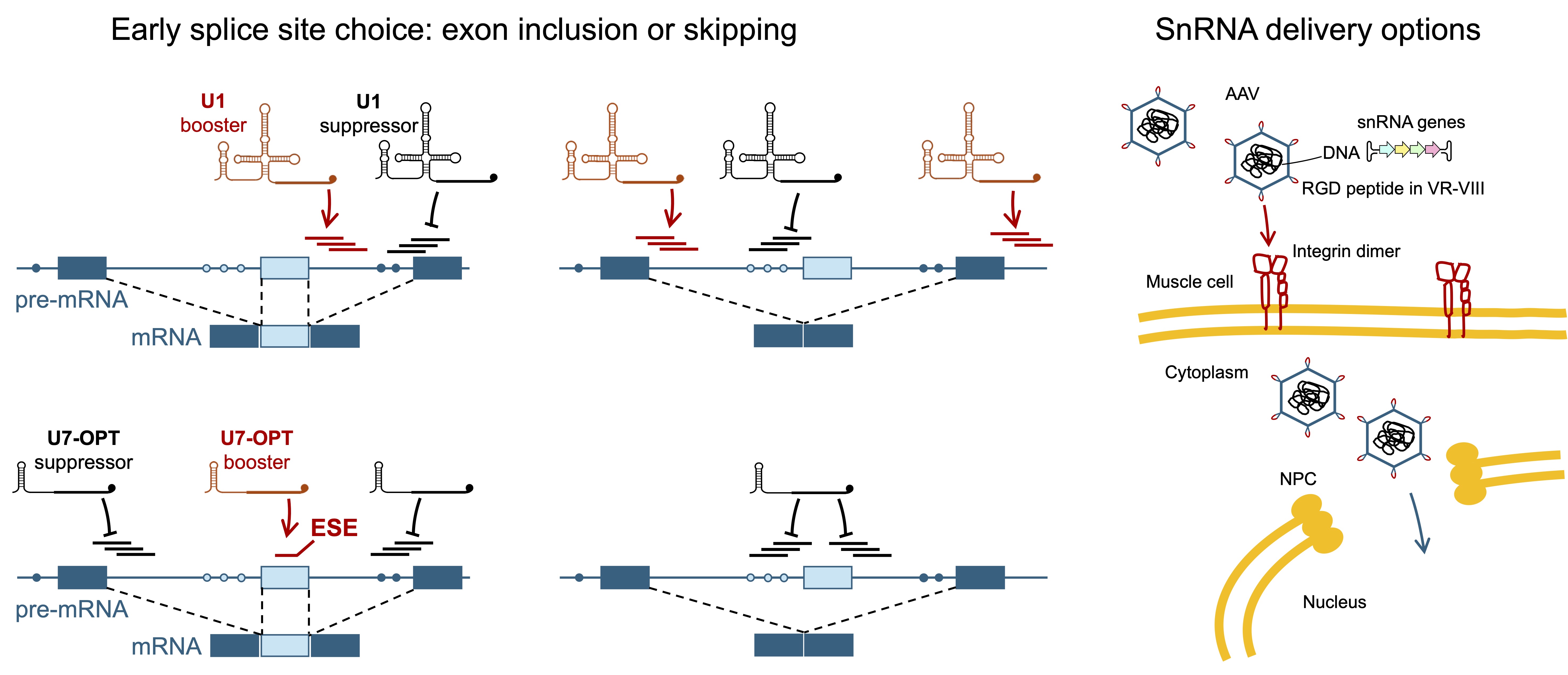
Another important point is that delivery is defined by the nature of the drug itself. ASOs are synthetic molecules: their in vivo delivery is limited to synthetic and otherwise ex vivo engineered means. In contrast, spliceosomal snRNAs are biological molecules, their genes or snRNAs themselves can be delivered by biological vehicles (such as AAV, exosomes – see below, or enveloped VLP – see Part 4, Artemyeva-Isman, 2026d) and if delivered as RNA to the cytoplasm, they are assured by an existing mechanism to be imported to the nucleus to their pre-mRNA target.
4.1. ASO-Peptide Conjugates
Low efficacy of DMD-targeting PMO ASOs (antisense oligoes with PMO backbone, hereafter PMOs) injected naked into the bloodstream is attributed to poor delivery. Cellular uptake, albeit not specific to muscle, can be increased by conjugating oligonucleotides with cell-penetrating peptides CPP. These are short peptides (<30aa) that traverse cell membrane by diverse translocation or endocytosis mechanisms; often CPPs are rich in Arg+ (R) and Lys+ (K) that can disorder negatively charged membranes and/or hydrophobic amino acids contributing to amphipathic a-helical structures that can insert into the lipid bilayer (Ruseska and Zimmer, 2020; Hadjicharalambous et al., 2022). Natural CPPs derived from transcription factors and signalling proteins include TAT peptide RKKRRQRRR from HIV1 Trans-Activator of Transcription, Penetratin RQIKIWFQNRRMKWKK of the Drosophila melanogaster Antennapedia Antp homeodomain transcription factor, pVEC LLIILRRRIRKQAHAHSK from murine Vascular Endothelial Cell adhesion E-Cadherin Cdh1, and YARVRRRGPRR protein transduction domain PTD of the human HPH1 (Polyhomeotic 1 homolog) transcription factor (Park et al., 2013). Synthetic CPPs are designed to mimic the properties of natural peptides, or chimeric CPPs combine natural peptides with enhancing additions. Peptides designed for conjugation with PMOs targeting DMD splicing include Penetratin-derived Pip series of peptides (Gait et al., 2019) and HPH1 PTD-derived DG9 (Haque and Yokota, 2023; Shah et al., 2025). The first problem with CPP drugs is their enzymatic degradation in serum. This problem is usually addressed by using amino acid analogues, that are not found in biological proteins and thus make the peptides resistant to proteolysis. Commonly included into CPPs are 6-aminohexanoic acid (Ahx or X, Lys analogue lacking a-amino group), b-Ala (B, aliphatic chain is longer by one C than in Ala, a-Ala) and D-Arg (dR, the D enantiomer of Arg, L-Arg). All three of these are included in Pip2 to 6 versions, but DG9 peptide made of 2x HPH1 PTD has exclusively 5x dR modification. Despite improved biodistribution in animal models, clinical trials of the Sarepta cationic CPP (SRP-5051) and the PepGen Pip CPP (PGN-EDO51) showed dose-dependent escalation of renal toxicity with hypomagnesemia and hypokalaemia, causing the cancellation of the Sarepta drug and limiting the dosage for PepGen product (Torres-Masjoan et al., 2025; Haque et al., 2024). The adverse effects are thought to be linked to positively charged peptides damaging cellular membranes at high doses. This is hardly surprising, as closely related to CPPs are natural antimicrobial peptides AMPs, which also carry an overall positive charge and ‘their initial interaction with the anionic phospholipids of bacterial membranes is an important early step in their direct killing mechanism’ (Hadjicharalambous et al., 2022). AMP and CPP compositions, structures and activities largely overlap. Many CPPs have antibacterial activity, and most AMPs penetrate cells, with some showing no selectivity between eukaryotic and bacterial membranes (remember that eukaryotes inherited bacterial membranes – see Part 1, Artemyeva-Isman, 2026a). DG9 is not yet in clinical trials, it remains to be seen if its positive charge can be better tolerated. Possible adverse effects of non-canonical amino acids included in CPPs also need to be considered. 6-aminohexanoic acid (X), mimics Lys and binds the Lys-binding sites of plasmin and its precursor plasminogen, which blocks proteolysis of fibrin and fibrinogen, and inhibits dissolution of blood clots (Markowska et al., 2021). Ahx is an antifibrinolytic drug likely to cause thrombosis if misused. The dangers of therapeutic use of peptides made of D-Arg are difficult to predict. Homochirality (‘same handedness’ from the Greek ceir, hand) or the same 3D arrangement of chemical groups around the Ca atom in amino acids is required for protein stability and was a precondition for the origin of Life on Earth. As a start, a tiny excess of L-enantiomers possibly occurred due to preferential photolysis of D-amino acids by stellar right circularly polarised light R-CPL (Fukue et al., 2010; Breslow and Cheng, 2010; Bocková et al., 2023 - experimental L-CPL irradiation produces the excess of D-isomer). Further amplification of L-isomers occurred by asymmetric autocatalysis (Soai et al., 1995; Soai, 2019) and/or enrichment in evaporating solutions under plausible prebiotic conditions (Breslow and Levine, 2006; Levine et al, 2008). Predominant L-amino acids in turn led to preferential synthesis of D-sugars (Breslow and Cheng, 2010). Curiously, the same is true the other way round: D-sugars catalyse amino acid synthesis with the excess of L-products (Wagner et al., 2017). Different prebiotic chemical scenarios are suggested, but undoubtedly the synergy between homochirality of amino acids and sugars for the RNA backbone produced the basis for genetic code of the primordial RNA -peptide world: stereoselective aminoacylation catalysis by D-RNA making proteins exclusively of L-amino acids (Müller et al., 2022; Tamura, 2011), the property inherited by the ribosome (Melnikov et al., 2019 – ribosome structure with D-Phe)1. However, D-amino acids are present in the biosphere: many organisms, from bacteria to humans, possess racemases to convert L-forms to D-forms that play striking roles. D amino acids are included into peptidoglycans of bacterial cell wall, making them resistant to enzymes that process L forms (Cava et al., 2011). D-amino acids are also found in bacterial and eukaryotic AMPs and antibiotics, which like peptidoglicans are synthesised by specialised enzymes, not by ribosomes (Cava et al., 2011). For example, biosynthesis of penicillin requires d-(L-a-Aminoadipyl)-L-cysteinyl-D-valine synthetase initially making a tripeptide, then D-Val is locked with L-Cys into the b-lactam ring (van der Lende et al., 2002). In animals, peptides containing D-amino acids are synthesised by the ribosome with L counterparts, that are isomerised post-translationally. Such peptides are rare and serve for defence, attack or signalling, although it is certain that more will be discovered. Examples include neurotransmitters and toxins of molluscs, neurohormones in Decapod crustaceans, spider venom, skin secretion of frogs (Bai et al., 2009; Cava et al., 2011) and most extraordinarily in the venom of male platypuses (Torres et al., 2006). Free D-amino acids are also biologically active. Bacteria releases D-amino acids to the environment that appear to control microbial communities. Specifically, D-Arg is lethal to many bacteria, but the Vibrio species appear exceptionally tolerant. The cholera pathogen V. cholerae synthesises and secretes D-Arg to inhibit nearby competitors. D-Arg is also a negative chemotactic agent that promotes spread of V. cholerae in response to stress, for example to move away from spent environments (Alvarez et al., 2018; Irazoki et al., 2023). Signalling roles also belong to D amino acids in higher organisms: a classic example is D-Ser, a neurotransmitter in the mammalian cortex involved in learning and memory. In addition, D-Asp is involved in hormone secretion in mammalian brain and endocrine tissues (Fuchs et al., 2011; Kiriyama and Nochi, 2016). D-Arg also has CNS-specific activity: it was reported as a beneficial agent protecting against high levels of glucocorticoids (Canteros, 2014). This is attributed to a role in the nitric oxide NO production, which usually starts from L-Arg, but as nitric oxide synthase NOS is stereospecific, the alternative remains that D-Arg reacts directly with reactive oxygen species ROS to yield NO. This idea was put forward previously to explain the increase of NO for both L- and D-Arg, when nebulised into the airways of patients with asthma (Chambers and Ayres, 2001). Yet, this hypothesis did not appear correct when tested in a rat hypertension model (Wölkart et al., 2004). While the mechanism of action is unclear, the fact remains that D-Arg interferes with NO signalling. Current interest in the therapeutic use of D-amino acids, and peptides that include them for extra stability, raises the question of mammalian tolerance. High levels of D-amino acids can lead to chiral mistakes by aa-tRNA synthases and slow down effective translation, which leads to growth arrest both in E. coli and S. cerevisiae. The first response in all organisms, including mammals, to control D-amino acids level is their catabolic degradation by D-amino acid oxidases. These are highly expressed in the human brain where D-Ser is produced, the gut where microbiome produces D-amino acids and the kidneys for systemic chiral detoxification. In addition, kidneys excrete the excess of D-forms: more than 97% of amino acids in the blood are L-forms, while urine amino acids are 50% D-forms (mainly D-Ser). Alarmingly, renal dysfunction, which is also flagged in clinical trials of CPPs, leads to a decrease of urinal D/L amino acid ratio and the accumulation of D-forms in the blood (Taniguchi et. al., 2025). Translation of D-Arg or D-Arg-rich peptides like DG9 for repeated administrations in the clinic requires a careful safety assessment of bioactivities with regard to new bacterial pathogens, and physiological signalling roles (Navarro et al., 2005 - the first relevant toxicology study).
Apart from cellular uptake, PMO delivery to the cytoplasm is prevented by their retention in the endosomes, as they lack specific signals for a destination compartment and are subject to degradation when the late endosome fuses with a lysosome. Endosomal degradation of PMOs can be prevented by peptide-based endosomal escape vehicles EEV. Entrada ENTR-601-44 targeting exon 44 is in trials with no toxic effects for these PMO-peptide conjugates yet reported, but exon skipping is below 0.5% so far (Torres-Masjoan et al., 2025).
Finally, the best idea is to target PMOs to muscle cells. Enhanced delivery to muscle and heart was achieved by PMOs conjugated with antibodies for transferrin receptor 1 TfR1 (TFRC), although liver uptake is still 5 times greater (Malecova et al., 2023) and only 15% of dystrophin exon skipping was achieved in mice (Cochran et al., 2024). Avidity Biosciences’ antibody-oligonucleotide conjugates AOC-1044 is in clinical trials with no data available so far, but competitors DYNE-251 PMO conjugates with TfR1 antibody fragments (otherwise known as single chain variable fragment scFv or single chain antibody) flagged dose-dependent acute kidney injury, thrombocytopenia and pancytopenia (Torres-Masjoan et al., 2025).
It appears that PMOs and PMO-peptide conjugates are locked in a vicious circle of toxicity: naked PMOs come with adverse effects, which limits the dose, and most of the drug does not get to the target, while improved delivery of PMO-peptide conjugates is marred by additional toxicity keeping the tolerable dose much below that of the naked PMOs. Crucially, the very mechanism of ASO activity is not supported biologically. ASOs are degraded in the serum and a small proportion of them that gets in the muscle cells is not marked for escape from the endosomes before lysosomal fusion, so even smaller fraction enters the cytoplasm, where ASOs are awkward non-biological molecules not supported by any clear mechanism of nuclear import to reach their pre-mRNA target. ASO backbone modifications and peptide-conjugation are forcing ASO activity against the odds of physiology and molecular cell biology.
4.2. Recombinant Adeno-Associated Virus Vectors
snRNAs are biological molecules that can be encoded and delivered by viral vectors. AAVs are clinically approved and were injected into the eye, CNS or delivered systemically through the bloodstream in over 200 clinical trials (reviewed in Wang et al., 2024). Different natural AAV serotypes allow some degree of preference to specific organs or tissues, however, high doses of AAV delivered intravenously can cause hepatotoxicity, and intrathecally – neurotoxicity (see Wang et al., 2024 for all adverse effects). Another drawback of AAV is that it can only be used once, as immune response will not allow for an efficient second administration. Despite these complications, AAV is instrumental to true therapeutic achievement, notably AAV9-SMN for infants with severe SMA (see above 2.1 Therapy for Spinal Muscular Atrophy). Another example, a recent clinical trial success for early onset Huntington’s disease used rAAV5-miHTT to destroy HTT mRNA by RNA interference and lower the level of mutant HTT proteins (uniQure AMT130, trials NCT0543017 and NCT04120493). The efforts to engineer enhanced tropism to specific organs and reduce uptake by the liver can boast gradual attainment. Such efforts are focused on modification of the exposed variable regions VR-I to VR-IX of the capsid viral protein 1 VP1, that mediate AAV affinity to cell surface receptors and antibodies. (VP2 and VP3 are shorter versions produced by the same CAP gene; a.a. numbering below is as in VP1). Strikingly, a single amino acid change in VR-I AAV8 Asn271Asp or AAV9 Asn270Asp can be sufficient to reduce hepatic uptake (Xing et al., 2025), and, on the other hand, an insertion of a single Thr at VR-I position 265 from AAV1 to AAV2 changed humoral immune profile, but not the virus tropism (Li et al., 2012) – potentially evading the immunity for repeated doses of AAV-carried medication. Apart from rational designs like these, directed evolution in vivo is used to select desired tropism from a library of random peptides inserted into VR-VIII surface loop of AAV9: Deverman et al., 2016 selected for increased delivery to the brain, which was later improved by Nonnenmacher et al., 2020 using brain-specific promoters for viral RNA transcription, and Tabebordbar et al., 2021 selected MyoAAV for delivery to skeletal muscle using muscle-specific promoter. AAVMYO (Weinmann et al., 2020; El Andari et al., 2022) was developed by AAV capsid shuffling, as opposed to starting with AAV9 alone, in addition to peptide insertion at VR-VIII and muscle-specific promoters for viral RNA. Muscle-targeting peptides of MyoAAV and AAVMYO have a motif in common: ArgGlyAsp (RGD), which binds cell-surface integrin heterodimers, highly expressed in skeletal muscle (Bönnemann, 2021, Figure 8). These muscle-tropic AAVs also significantly diminish uptake by the liver. Using a muscle-specific promoter for the therapeutic transgene can further ensure efficient expression in muscle and abolish ectopic expression (Darbey et al., 2024). AAV small genome size is usually a problem for packaging large protein genes, but for snRNAs, there is room enough to include multiple gene copies in tandem. AAV was used to deliver U1 snRNA in pre-clinical studies (see below).
Encouragingly, past trials show that transgene expression can persist for more than 10 years after a single AAV administration (reviewed in Muhuri et al., 2022).
4.3. Exosomes
Exosomes are a class of small (30-200nm) extracellular vesicles (EV) that originate from multivesicular bodies by exocytosis, as opposed to macrovesicles (100-1000nm) that originate by budding of the cell membrane (Gurung et al., 2021). Compared to synthetic lipid nanoparticles LNPs, exosomes are not toxic and have superior biodistribution by systemic delivery. They are produced by different cell types to carry molecular messages, including snRNAs, between neighbouring cells and body-wide, penetrating through tissues, carried in the bloodstream and capable to cross the blood-brain barrier. Moreover, mammals deliver exosomes with milk to their offspring. Orally administered exosomes also have impressive biodistribution capacity, they get into the bloodstream and cross the blood-brain barrier, and they are less accumulated in the liver, compared to exosomes injected intravenously (reviewed in Gurung et al., 2021). They are on the way to the clinic, as at least 20 trials of exosomes are ongoing in USA and worldwide (reviewed in Herrmann et al., 2021). Efficient systemic exosome delivery to the brain and skeletal muscle was achieved in pre-clinical studies. Alvarez-Erviti et al., 2011 (Alvarez-Erviti et al., 2011) developed dendritic cells expressing exosomal membrane protein Lamp2b fused with a peptide derived from Rabies virus glycoprotein that binds a7 nicotinic acetylcholine receptor CHRNA7 specific to neurons, oligodendrocytes and microglia in the brain (RVG peptide). Exosomes harvested from these cells were electroporated with siRNA against amyloid b producing BACE1, a therapeutic target in Alzheimer’s disease. Single intravenous delivery achieved 60% knock-down of mRNA and protein in mice. Repeated delivery was not hampered by immune reactions. Kojima et al., 2018 demonstrated that exosome-producer cells can be implanted subcutaneously into mice to deliver catalase CAT mRNA to the brain to reduce neuroinflammation for the treatment of Parkinson’s disease. Engineering such cells involved overexpressing genes for exosome biogenesis and expression of Connexin 43 (gap junction a-1 GJA) S368A mutant, that enhances exosomal cargo delivery to the cytoplasm of the recipient cells (Soares et al., 2015). CAT mRNA packaging was ensured by fusing exosomal membrane protein CD63 with the archaeal L7Ae (SNU13 homologue), that interacts with a kink turn of the C/D box sRNA in Archaea (snoRNAs and U4 snRNA in eukaryotes, see C in Part 1, Artemyeva-Isman, 2026a; Moore et al., 2004 – crystal structure). C/D box was inserted into the 3′-UTR of the CAT mRNA. Implanting exosome producer cells to disseminate snRNAs boosting SMN2 exon 7 inclusion can be beneficial for late onset SMA. In these cases, with 3+ SMN2 copies, AAV9-SMN risks harmful overexpression (see above 2.1 Therapy for Spinal Muscular Atrophy). While snRNAs can be delivered by AAV9, the experience with mRNA shows that intrathecal route is less protective for neuromuscular junctions, but intravenous injection works less well for motor neuron preservation (Reilly et al., 2024). Exosomes have an advantage of body-wide distribution, ideal for ubiquitously expressed SMN protein. Finally, specific targeting of EVs to activated muscle precursor cells was ingeniously achieved by engineering producer cells to express myomarker Mymk and myomerger Mymg, the membrane proteins specifically expressed only during muscle regeneration to promote fusion with injured fibres or formation of new fibres (Hindi et al., 2024, Figure 9). Potentially, autologous fibroblasts can be engineered to express Mymk, Mymg and snRNAs targeting dystrophin splicing. It will be interesting to see if re-implantation of such producer cells improves muscle repair.
5. U1 (and U7) in Pre-Clinical Studies
Following the discovery by Zhuang and Weiner, 1986, 17 years have elapsed without follow-up studies, until Baralle et al., 2003 again demonstrated that a pathogenic splicing variant associated with neurofibromatosis type 1 NF1 can be corrected by U1 matching the mutation. In this case, adapted U1 restores a G=C pair, albeit swapped between the interacting RNAs (A: NF1). Since then, U1snRNAs suppressing splicing mutations have been designed and tested in at least a dozen independent laboratories worldwide (including: Carmel et al., 2004; Singh et al., 2007; Hartmann et al., 2010; Matos et al., 2014; van der Woerd et al., 2015; Lee et al., 2016; Martínez-Pizarro et al., 2018; Hatch et al., 2022 and numerous studies from groups of Prof Franco Pagani, Dr Dario Balestra and Prof Mirco Pinotti, and Prof John Neidhardt), and no doubt U1 will soon find the way to the clinic (Donadon et al., 2019; Romano et al., 2022; Swirski et al., 2023). Altogether U1 was adapted to correct splicing disrupted by ~100 different mutations in 24 genes linked to 22 diseases (38 studies reviewed in Gonçalves et al., 2023; and more recent studies include Zhang et., al., 2024; Peretto et al., 2025).
5.1. Boosting Exon Inclusion by U1 snRNA
5.1.1. U1 snRNA Design and Mechanism of Action
Firstly, U1 can be adapted to suppress substitutions in the 5’ss: a single base in the U1 5’ end is altered to match a specific mutation in the 5’ss, as done by Zhuang & Weiner, 1986 and Baralle et al., 2003 (A: NF1). Alternatively, U1 5’ end is made entirely complementary to the last 3 bases of the exon and the first 8nt of the intron (Singh et al., 2007; van der Woerd et al., 2015; Swirski et al., 2023). These adapted U1 molecules are also called compensatory U1 (Balestra et al., 2019), and they are most effective for divergent 5’ss, especially with the unusual exon 3’end, which renders them more specific to the target 5’ss (A: SMN2, OPA1, CDKL5). Another approach is based on U1’s ability to activate 5’ss ‘at a distance’ - discovered by Cohen et al., 1994 (see section 2.1 5’ss binding by U1 snRNA in Part 2, Artemyeva-Isman, 2026b) and widely used since Fernandez Alanis et al., 2012 adopted such U1 designs and termed them exon-specific U1 ExSpeU1. Rather than targeting the exact 5’ss exon/intron boundary, an overlapping array is designed for sites from 5’ss into the start of the intron and screened for the best splicing booster. Such U1s were adapted to bind targets of different lengths, from 9nt (Balestra et al., 2019, here the first two bases of U1 were disregarded as less important for recognition) to longer sites: 16nt (Donadon et al., 2018; Romano et al., 2022), 18nt (Donadon et al., 2019), 20 and 25nt (Hatch et al., 2022). How do ExSpeU1s work? Fernandez Alanis et al., 2012 and Donadon et al., 2018 show that if the effective 5’ sequence is transferred from U1 to U7 snRNA, exon skipping is favoured instead of inclusion. U7, which is not involved in splicing and has a separate role in histone mRNA 3’end processing, is usually used to carry antisense sequences that act as ASOs (Gorman et al., 1998, 2000; De Angelis et al., 2002; Goyenvalle et al., 2004, 2009). U7-induced exon skipping works by sterically preventing spliceosomal snRNPs from binding at splice sites, or by blocking protein regulators from binding splicing enhancer sites (Donadon et al., 2018; see more on U7 design below). On the contrary, it appears that U1 positioned in the vicinity of the 5’ss still functions as an Early spliceosome complex (Figure 5 in Part 2, Artemyeva-Isman, 2026b): it interacts with U2 snRNP in complex A and transitions to pre-catalytic spliceosome, which in turn defines the precise 5’ss by combined action of U6 and U5 snRNAs and 3D RNA folding, involving intron ends pair demarcated by a gap from helices formed by exons with U5 (see Part 2, C). Subsequently, Martínez-Pizarro et al., 2018 explored the effect of intronic mutations found in patients with phenylketonuria PKU and observed an additional 5’ss motif 18nt downstream of PAH (12q23) exon 11. Wild-type U1 snRNP binding at this site appeared to support both correct and cryptic splicing. Intronic mutations +17GA and +20GC led to exon skipping and proved to be pathogenic variants. This splicing defect was partially corrected by adapted U1 with a binding site shifted by 2nt into the exon to increase specificity (A, PAH).
As a matter stands now, researchers have different opinions on U1 design, adopting mostly U1 targeting 5’ss in the canonical binding register (Neidhardt group), preferentially intronic ExSpeU1 (Pagani group) or both canonical compensatory U1 and ExSpeU1 (Balestra and Pinotti group and others). The opinion that ExSpeU1 are better splicing boosters, than U1 complementary to 5’ss, is backed by ExSpeU1 binding a longer unique intronic sequence, rather than a short splice site, that is conserved, albeit imperfectly, and this ensures specificity and eliminates competition for the same site with abundant endogenous U1. Moreover, ExSpeU1 binding downstream from 5’ss do not target specific substitutions and can be effective for different mutations, inclusive of changes in the polypyrimidine tract PPT upstream of the target exon (Fernandez Alanis et al., 2012; Donegà et al., 2020).To these arguments we can add that excessive affinity of the canonical compensatory U1 to 5’ss can be detrimental to the task, making subsequent U1 disassociation less efficient and slowing down the transition from complex pre-B to B (Lund and Kjems, 2002, discussed in section 2.1 5’ss binding by U1 snRNA, Part 2, Artemyeva-Isman, 2026b). On the contrary, ExSpeU1 bound downstream, leaves 5’ss clear for U6 and U5 binding. However, the main concern is that ExSpeU1s are prone to activate cryptic splice sites (van der Woerd et al., 2015; Balestra et al., 2020a - FAH; Peretto et al., 2023) and their mechanism of action implies the need of adjustment when 5’ss is defined in the subsequent pre-catalytic spliceosome complex. In support of compensatory U1 at 5’ss, we already remarked their efficacy for exons with unusual 3’ends and the impact of additional G=C pairs for stabilisation of the U1/5’ss helix –these are factors that increase specificity and efficacy. Moreover, like ExSpeU1, compensatory U1 targeting a divergent 5’ss can be effective without being adapted to a specific substitution (Carmel et al., 2004; Balestra et al., 2019; A - SMN2, CDKL5). Equally, a U1 that adds just one extra G=C pair to the U1/5’ss helix can partially compensate for a frequent 3’ss mutation linked to intrahepatic cholestasis in the Netherlands (A - ATP8B1; van der Woerd et al., 2015). Several studies compared effectiveness of compensatory U1 alongside ExSpeU1s (Martínez-Pizarro et al., 2018; Balestra et al., 2019; Balestra et al., 2020a,b; Peretto et al., 2023; Sacchetto et al., 2021; Peretto et al., 2025), showing that the choice of the best splicing booster depends both on the target exon sequence environment and a specific mutation.
In the past 20 years disease targets for U1 therapy fall mainly into four categories: diseases caused by a common splicing mutation, as the same U1 can treat all the patients, and diseases with target organs that are easy to reach by gene delivery: liver, CNS or eye.
5.1.2. Targeting Common Splicing Mutations
A common splicing mutation can be a frequent recurring mutation of the same base. For example, Shwachman–Diamond syndrome (SDS), a rare autosomal recessive exocrine pancreatic insufficiency, bone malformation, and haematopoiesis defect often linked to leukaemia, is usually (90%) caused by SBDS2 intron 2 +2TC change. This mutation frequently arises by conversion with the locally duplicated pseudogene copy SBDSP, that shares 97% sequence identity with SBDS (Boocock et al., 2003). Compensatory U1 at 5’ss restores trace amounts of the correct transcript, while ExSpeU1s promote usage of cryptic sites, which increases with the distance of ExSpeU1 binding site from the 5’ss (Peretto et al., 2023).
A common splicing mutation can be a founder mutation that persists in the population due to some selective advantages in heterozygous state, or a mutation that goes on in an isolated population. In the latter category, familial dysautonomia (FD), an autosomal recessive neuropathy affecting sympathetic and sensory neurons, is caused by the deficiency of ELP1 (IKBKAP)3. FD is very rare in populations in general, but in Ashkenazi Jews the frequency is 1:3,600, due to ELP1 intron 20 +6TC mutation, that leads to skipping of exon 20 and nonsense-mediated decay, reducing mRNA to ~20% of normal level. This might seem surprising, as more than half of human pre-mRNA splice sites do not conserve +6U (C in Part 1, Artemyeva-Isman, 2026a). The reason for the importance of +6U here is likely to be that exon 20 ends with an A (A, ELP1), rather than a G, that is conserved at 82% (C). Experiments with ELP1 minigene show that changing the exon-end AG leads to 100% exon 20 inclusion, despite +6TC mutation. Co-transfection of ELP1 +6TC minigene with U1 3AG matching +6TC mutation partially restores exon 20 inclusion. Equally, U1 9CU matching the unusual exon-end A-1 compensates for +6TC mutation (Carmel et al., 2004). While fully complementary U1 to ELP1 exon 20 5’ss was never tried out, developed ExSpeU1 achieves ELP1 exon 20 inclusion at wt level (Donadon et al., 2018; US patent 9,669,109 – Pagani and Pinotti, 2017) and delivery by AAV9 extends survival and improves cardiac, renal and motor functions in an FD mouse model (Romano et al., 2022). This successful pre-clinical study is remarkable, as no gene or splicing therapy is currently available for FD patients.
Another example of a founder mutation in people of Southern Chinese descent (Taiwan, South China and neighbouring South-East Asia) is dopa decarboxylase DDC (AADC)4 intron 6 +4AT that accounts for higher frequency of this severe autosomal recessive neurotransmitter deficiency (Hwu et al., 2023). U1 snRNA made complementary to 9nt of the mutant 5’ss partially restores correct splicing in dose-dependent manner. This compensatory U1 packaged into AAV9 and injected into bilateral cerebral ventricles in a mouse model improves survival, but efficacy remained below that achieved by overexpression of DDC gene (Lee et al., 2016) and the latter is for now the therapeutic option for AADC deficiency in Taiwan.
Netherton syndrome, a severe autosomal recessive skin disease caused by SPINK55 deficiency, in Greece is frequently due to a founder synonymous mutation c.891CT (p.Cys297Cys) that disrupts a splicing enhancer in SPINK5 exon 11 (Lacroix et al., 2012). Lentiviral-mediated transduction of patient-derived skin keratinocytes with ExSpeU1 restores correct SPINK5 splicing in dose-dependent manner (Dal Mas et al., 2015b).
ATP8B16 deficiency is an autosomal recessive liver disease (inrahepatic cholestasis – impairment of bile flow), which in the Netherlands is frequently caused by ATP8B1 intron 23 -3CA mutation, that results in exon 24 skipping and in-frame deletion (A). Remarkably, compensatory U1 to exon 24 5’ss significantly rescues exon inclusion (van der Woerd et al., 2015).
Hereditary tyrosinemia type 1 HT1 is an autosomal recessive metabolic disorder caused by FAH7 deficiency. In Quebec HT1 patients usually carry FAH intron 12 +5GA mutation (Grompe et al., 1994), that induces exon 12 (and 13) skipping and usage of a cryptic 5’ss with partial intron 12 inclusion, resulting in complete absence of the enzyme in the liver. An attempt to correct this defect with U1 fully complementary to exon 12 5’ss failed (Scalet et al., 2018), which is frustrating, because in a mouse model with FAH exon 8 -1GA mutation (missense Ala236Thr) a trace amount of correctly spliced FAH induced by compensatory U1 AAV8 transfer was sufficient to detect FAH enzyme by immunohistochemical analysis of mouse liver sections (Balestra et al., 2020a).
Perhaps the most important global target that falls into the ‘common mutation’ disease category remains spinal muscular atrophy SMA (see above 2.1 Therapy for Spinal Muscular Atrophy on the unsatisfactory performance of existing therapies). SMA is an exceptional case: rather than targeting SMN1 with different mutations, the therapeutic target is the suboptimal splicing of its near identical orthologue SMN2, present in 1-4 copies. In comparison, our previous examples of founder mutations in neurological conditions, FD and AADC deficiency, are relevant to much smaller cohorts of patients living with these rare diseases in the world: estimated respectively as 350 people (all of Ashkenazi descent, Familial Dysautonomia Foundation, Inc., 2026 - website) and 130 people (of which ~70 are Taiwanese, Dai et al., 2020). The incidence of SMA is on average 1:10,000 in populations worldwide, ~70 babies with SMA are born every year in the UK alone. SMN2 carries a mutation in exon 7, that converts an exonic splicing enhancer into a silencer (ESEESS, A). SMN1/2 exon 7 has an unusual 3’end A-1, and a change for the conserved G-1, which improves the interactions with endogenous U1 and U5 snRNAs effectively rescues exon inclusion, suppressing the effect of the ESEESS mutation in SMN2 (Singh et al., 2004). Equally, snRNAs can be adapted for the divergent exon 3’end: an excellent follow up study (Singh et al., 2007) demonstrated that a compensatory U1 fully complementary to 5’ss achieves preferential inclusion of exon 7 (SMN2, A). Unfortunately, the authors focused on ASOs blocking the intronic splicing silencer, leading to Nusinersen drug development (see above 2 Antisense oligonucleotides), disregarding U1 therapeutic potential, until Pagani group considered SMA as a target (Fernandez Alanis et al., 2012) and patented several ExSpeU1s capable to boost SMN2 exon 7 inclusion (US patent 9,669,109, Pagani and Pinotti, 2017). One of these ExSpeU1, that binds SMN2 intron 7 at position +25 (A), rescues the phenotype as a transgene in SMA mice (Rogalska et al., 2016) and when systemically delivered by AAV9 (Donadon et al., 2019). These studies are very promising for clinical development and need to be followed by parallel assessment of compensatory U1 targeting SMN2 exon 7 5’ss (Singh et al., 2007) in animal models (see below safety considerations).
5.1.3. Liver Targets
Apart from the previously reviewed FAH and ATP8B1 with population-specific common mutations, liver targets investigated for U1 include ornithine transcarbamylase deficiency (OTCD8), the most common X-linked urea cycle defect causing life-threatening toxic ammonia accumulation and encephalopathy in newborns. Both compensatory U1 for 5’ss and ExSpeU1 were effective to correct OTCexon 4 -1GA mutation of the mouse OTCD model and ExSpeU1 tested in vivo by systemic AAV8 delivery achieved 50% of normal expression of OTC enzyme in mouse liver (Balestra et al., 2020b -OTC). Both in mice and in humans OTC intron 4 is unusual, as instead of the conserved adenines there are U+3U+4 (A and see section 3.1 5’ss helix in Part 2, Artemyeva-Isman, 2026b), which still produces 91% of exon 4 inclusion normally in humans (WT exon 4 is subject to 5% skipping and 4% cryptic 5’ss usage). Remarkably, only two peripheral base differences at 5’ss, exon -3C and intron +10U in humans and -3U, +10A in mice lead to very different activity of compensatory U1 at 5’ss and intronic ExSpeU1 in these species, highlighting the importance of 5’ss sequence environment when designing splicing therapeutic agents and testing them in mice (Sacchetto et al., 2021).
Splicing defects of coagulation factors F7, 8 and 9, produced in the liver, were also targeted by adapted U1s (Balestra et al., 2014, 2015; Scalet et al., 2019; Zhang et al., 2024; Peretto et al., 2025). The most frequent, affecting 1:5000, is X-linked Haemophilia A HA - F8 deficiency, and as full-length 7kb F8 cDNA exceeds AAV packaging capacity an abridged version F8-SQ with regulatory domain B (Pipe, 2009) deleted is used for gene therapy. Therefore, correction of F8 splicing mutations by snRNAs has the advantage of restoring the endogenous F8 activity. Peretto et al., 2025 achieved splicing correction of nine intronic F8 mutations using compensatory 5’ss-targeting U1s and ExSpeU1s for F8 exons 8, 11 and 22.
5.1.4. CNS Targets
Seckel syndrome, a rare autosomal recessive disease involving dwarfism, microcephaly and mental retardation, is caused by ATR9 deficiency; the cDNA is too long for AAV delivery. ATR exon 9 lacks both conserved guanines at the ends (half of human exons start with G+1 and 82% end with G-1, C in Part 1 (Artemyeva-Isman, 2026a), featuring C+1 and U-1 instead and the inclusion of this exon depends on the exonic splicing enhancer, which is mutated in an examined case of Seckel syndrome (Scalet et al., 2017): ATRss c.2101 AG (synonymous p.Gly674Gly). ATR exon 9 changes +1CG or -1UG fix exon 9 inclusion suppressing the ATRss ESE mutation, remarkably like SMN2 exon 7 -1UG change suppresses the ESEESS mutation (A; Singh et al., 2004, see SMN2 explanation above). U1 made complementary to the ATR exon 9 5’ss induced preferential exon inclusion suppressing the effect of the ATRss mutation (Scalet et al., 2017).
Another important study explores the correction of CDKL510 splicing mutations (Balestra et al., 2019). CDKL5 deficiency is characterised by drug-resistant epileptic seizures in infancy and cognitive and motor disability (Fehr et al., 2013). It is X-linked dominant, in fact the ratio is 4:1 female-to-male, indicating decreased male foetal survival. Phenotypic severity in heterozygous girls and hemizygous boys can be equal and instead depends on the type of CDKL5 mutation, the pattern of X-inactivation in females and postzygotic genetic mosaicism (Benke et al., 2025). Both CDKL5 deficiency and overexpression are pathogenic, as duplications cause mental retardation, autism and macrocephaly, but no epilepsy, in both sexes (Szafranski et al., 2015). The dominant and dosage-dependent effects of the mutations are bad premises for CDKL5 cDNA therapy and highlights the importance of correction of endogenous gene expression. Balestra et al., 2019 designed compensatory U1s for 5’ss of exons 3, 9 and 16 and ExSpeU1s binding at the start of their following introns and were able to rescue exon 3 and 16 inclusion for +5GA mutations, but not for +1GU/C mutations for all these exons. According to the observation that compensatory U1 adapted to a divergent 5’ss is particularly effective, CDKL5 exon 16 inclusion was rescued equally well by ExSpeU1+12 and compensatory U1, even though it was designed complementary to the wt 5’ss with +5G for splicing correction of mt +5A (A, CDKL5). Phenotypic correction was demonstrated in mouse Cdlk5null neurons by lentiviral co-transfections of adapted U1 and CDKL5 expression construct containing intron 3 with +5GA mutation.
5.1.5. Eye Targets
Moving on, studies of U1 therapy for vision loss is a major contribution of Neidhardt group. Tanner et al., 2009 targeted retinitis pigmentosa (RP), a degenerative retinopathy that affects ~1.5 million people in the world. Most common cause of autosomal dominant RP are rhodopsin RHO11 mutations. They explored an individual case of RHO exon 4 -1GA (c.936GA) mutation, that led to both exon skipping and activation of a cryptic 5’ss in minigenes transfected into COS-712 cells and mouse retinal explants. Co-transfection with compensatory mutation-adapted U1 snRNA rescued correct exon 4 inclusion, although some usage of the cryptic 5’ss was still observed. Glaus et al., 2011 followed by targeting X-linked RPGR13 intron 10 +3AU (c.1245+3AT) mutation, that causes exon 10 skipping and showed that compensatory U1 fully complementary to the 5’ss partially restores exon inclusion.
Their next target was Bardet-Biedl Syndrome BBS, an autosomal recessive primary ciliopathy, characterised by progressive retinal dystrophy and vision loss in children, learning difficulties, renal and genital abnormalities, obesity and polydactyly. Some twenty genes are linked to BBS, but almost a quarter of all patients of European descent have BBS114 deficiency. A very mild form of BBS diagnosed in an extended family with the only manifestation of RP-like retinopathy is caused by BBS1 exon 5 -1GA (c.479GA). Lentiviral transduction of patient-derived fibroblasts with compensatory U1 adapted to the mutation produced dose-dependent partial correction of aberrant splicing (Schmid et al., 2011). The authors systematically proceeded to mutate 5’ss positions of their BBS1 exon 5 minigene and employed U1 made complementary to these mutations to correct splicing when co-transfected with the BBS1 minigene into COS-7 cells. Adapted U1s effectively suppressed mutations in all positions, except they failed for +1GU and +2UG, and only partially worked for +5GU mutation (Schmid et al., 2013).
Their recent target is autosomal dominant optic atrophy ADOA, a progressive degeneration of retinal ganglion cells and their axons, which form the optic nerve. The genetic defect was unknown until in 2000, it was linked to OPA1 (3q29), which is responsible for 75% of cases. OPA1 is a ubiquitously expressed GTPase of the inner mitochondrial membrane and homozygous mutations are embryonic lethal, while heterozygous mutations in ADOA exhibit haploinsufficiency or dominant effects, which can cause a more severe ADOA+ with additional manifestations: deafness, ataxia, peripheral neuropathy, impaired eye movement, and myopathy. ADOA affects 1:30,000 people worldwide (Ding et al., 2025), but in Denmark and southern-eastern Sicily the frequency is higher due to founder OPA1 mutations (1:10,000 Danish and estimated even higher in the Italian province of Siracusa, Gallus et al., 2012). The true incidence of OPA1 mutations might be underestimated – a recent case study (Tachibana et al., 2025) reports intron 23 +2UG (c2331+2TG) mutation discovered in a 55-year-old Japanese patient with a very slow onset of the vision deterioration. Curiously, a knock down of another mitochondrial protein SARM115 prevents optic nerve degeneration in ADOA model mice (Ding et al., 2025), showing that the development of visual impairment depends on other factors in addition to OPA1 defect and makes SARM1 a promising alternative therapeutic target. Adapted U1 snRNA partially suppressed OPA1 intron 10 +5GA (c.1065+5GA) mutation, producing 15% increase of exon 10 inclusion in patient-derived primary skin fibroblasts (Jüschke et al., 2021). In a mouse model with the same OPA1 mutation, delivery of adapted snRNA genes by AAV2/8 subretinal injections equally showed 15% increase of OPA1 total protein expression and the ratio of OPA1 long and short proteins produced by alternative splicing appeared preserved (Swirski et al., 2023). Remarkably, compensatory U1 adapted to match the mutation at the 5’ss was effective in a mouse (A, Opa1), but not in human cells – showing the variation in activity of adapted U1 between mouse and human, independently observed by Sacchetto et al., 2021 for OTC exon 4 -1GA mutation (see above). In ADOA human cells, the Neidhardt group, exceptionally to their previous practice, had to test a series of intronic-binding U1s, selected by MaxEnt top scores for 5’ss downstream from the actual exon/intron boundary. Their approach did not aim for a unique sequence like ExSpeU1: instead, they chose the target for U1 closest to the 5’ss motif and preserved the same length of the binding sequence – 11bp with only 9bp made complementary to the target, keeping the first two U1 bases unaltered. U1+18 was the most effective and did not give aberrant products with a cryptic 5’ss+18 (Jüschke et al., 2021). Despite the differences in the premise for U1 design, these intronic-binding U1s and ExSpeU1s are likely to share their mechanism of action with the WT U1 binding cryptic 5’ss motifs downstream from an exon and activating both correct and aberrant splicing (Martínez-Pizarro et al., 2018; A - PAH, see explanation above). It is important to remember that intronic-binding U1s designed for other targets showed cryptic 5’ss activation (van der Woerd et al., 2015; Balestra et al., 2020a - FAH; Peretto et al., 2023). In every individual case an experiment alone can show if the correct 5’ss is preferred at the pre-catalytic stage.
5.2. Inducing Exon Skipping by U1 and U7 snRNAs
While U1 is primarily used to boost exon inclusion, it can also be employed to promote exon skipping. In connection to this, let’s remember that both 5’ and 3’ss have a common core sequence CAG|GU, protosplice site repeat (see Parts 1 and 2, Artemyeva-Isman, 2026a,b). While U1 snRNA selects 5’ss by 6-7 Watson-Crick pairs, it is probable that some 3’ss also can bind U1, if more matches to U1 appear in exon positions +3 to +8. In such cases U1 snRNP is competing with U2AF for 3’ss binding – suggesting that U1 affinity to 3’ss is a natural mechanism that promotes exon skipping. However, the first snRNA used for exon-skipping was not U1, but U7 – an snRNA unconnected with splicing. Like spliceosomal snRNAs, U7 is processed in the cytoplasm, where SMN complex decorates it with LSm10 and LSm1116, which upon the re-entry into the nucleus target U7 to histone locus bodies. U7 snRNA binds the 3’end of histone pre-mRNA and recruits HCC, histone cleavage complex (reviewed in Geisler et al., 2023). In order to use U7 snRNA as a scaffold for splicing-modulating ASOs, Gorman et al., 1998 replaced the Lsm binding site for Sm of spliceosomal snRNAs creating a chimeric “U7-OPT”. The anti-histone sequence at the 5’end is then replaced by ASO targeting 5’ss or 3’ss or two tandem ASOs targeting both 5’ss+3’ss (Suter et al., 1999). U1 snRNA was tried next to suppress cryptic exon inclusion in intron 2 of the b-globin gene and naturally could not suppress 5’ss, as it promotes 5’ss usage, but was effective blocking 3’ss (Gorman et al., 2000). Unfortunately, subsequent works that tested U1 for DMD exon skipping never utilised U1 targeting exclusively 3’ss. De Angelis et al., 2002 initially targeted exon 51 5’ss by U1, BP by U2, 3’ss by U7 and 5’ss+3’ss by U7 in patient-derived myoblasts with DMD deletion of exons 48-50. Again, U1 and U2 were ineffective for exon skipping, as they naturally induce splicing when bound to 5’ss and BP respectively. U7 was effective when carrying antisense sequences to 3’ss or both splice sites. These reports provoked the use of U7 as a carrier of ASOs (Brun et al., 2003; Goyenvalle et al., 2004, 2009; and recently for gRNA as well – Byrne et al., 2025). The idea of targeting both 5’ss and 3’ss by the same snRNA somehow gets transferred from U7 to U1: subsequent U1 studies (Denti et al., 2006, 2008) used U1 carrying antisense sequences to both splice sites of exon 23 to skip a point mutation in the mdx mouse model of DMD. The last work that considered U1 for DMD exon 51 skipping combined three antisense sequences targeting 3’ss, ESE and 5’ss in one U1 molecule (Incitti et al., 2010). The authors achieved 50% exon 51 skipping in patients’ skin fibroblasts converted to myoblasts. A more recent study, Hatch et al., 2022, explored using U1 to regulate a classic example of a splicing switch between protein isoforms with antagonistic functions in HEK293T cells. FAS exon 6 inclusion produces a membrane death receptor, that triggers T-cell clearance upon the completion of an immune response. On the contrary, FASD6 is a soluble protein, that promotes T-cell proliferation (see above 3 Splicing switches as therapeutic targets in common diseases). U1 designed to be complementary to 20 or 25nt across 3’ss efficiently promotes FAS exon 6 skipping. The following study by Covello et al., 2022 targeted RPGR alternative exon 9a, which is normally spliced in only at a low level, but a deep intronic variant detected in a patient with mild retinopathy increased exon 9a inclusion, as detected in peripheral blood cells (Neidhardt et al., 2007) and when expressed from an RPGR minigene in HEK293T, PC-12 and 661W cell lines17 (Covello et al., 2022). U1-3’ss designed to bind 21nt at the 3’ss was an effective suppressor of exon 9a inclusion, which fully agrees with experimental results in Hatch et al., 2022 and with what we might expect happens naturally if U1 affinity to 3’ss can compete with U2AF for binding. The authors equally used U1-5’ss complementary to 24nt across 5’ss, which naturally favoured exon 9a inclusion in two cell lines, except PC-19, where it resulted in increased exon skipping. U1-5’ss+3’ss, a heritage design carried over from DMD studies reviewed above (Denti et al., 2006, 2008; Incitti et al., 2010), induced exon skipping with the same efficiency as U1-3’ss in two cell lines, and lower efficiency in PC-19. Finally, co-transfection of U1-5’ss and U1-3’ss resulted in a moderate increase of exon 9a inclusion, indicating that U1-5’ss stimulation is stronger than U1-3’ss inhibition. In view of U1 role as a splicing factor, not just a mere scaffold for antisense sequences, C proposes U1 designs for exon skipping based either on blocking U2AF binding at the 3’ss or by increasing the rate of splicing of the neighbouring exons. The latter possibility is based on the fact, that deceleration of splicing of the neighbouring exons by ASOs results in the increase of the alternative or mutated exon inclusion (see above 2.3 Important developments: targeting non-productive splicing and hotspot exons, Duan et al., 2024).
5.3. U1 and U7 snRNA Safety
Safety profile of U1 in pre-clinical experiments is very positive so far. Altogether 10 studies applied AAV delivery of therapeutic U1 in mouse disease models (DMD Denti et al., 2006, 2008; F7 Balestra et al., 2014; SMN2 Dal Mas et al., 2015a; Donadon et al., 2019; DDC Lee et al., 2016; FAH Balestra et al., 2020a; OTC Sacchetto et al., 2021; ELP1 Romano et al., 2022; OPA1 Swirski et al., 2023), with only one study showing adverse effects due to liver toxicity triggered by the high dose of AAV8 (Balestra et al., 2014). Importantly, Denti et al., 2008 continued monitoring mice for 18 months after the AAV1-U1 systemic injection, confirming long-term safety and durable therapeutic effect. Rogalska et al., 2016 created germline transgenic mice carrying 1 copy of ExSpeU1SMN2 cassette integrated in Chr1 (no further details provided). U1SMN2 was expressed in all the tissues examined at a level below that of the endogenous U6 snRNA. These transgenic mice appeared healthy, indicating that overexpression of designer U1 is not toxic. ExSpeU1SMN2 was effective at concentrations measured as ~1/500th of the endogenous U1 in mice 1 month after a single systemic AAV delivery (Donadon et al., 2019). Off-target transcription and splicing alterations were further analysed by RNAseq in mouse models (Rogalska et al., 2016; Donadon et al., 2019; Balestra et al., 2020a – FAH; Romano et al., 2022). The outcomes of these studies show no global perturbation of either gene expression or splicing. Very few altered splicing events are dose-dependent and principally, do not appear to cause adverse effects. Swirski et al., 2023 assessed off-target events by a more focused approach. As they performed subretinal injections of AAV2/8 that carried snRNA genes, the effects on splicing of eye-specific transcripts were examined by first searching for potential off-target binding sites for the adapted U1 and then RT-PCR and Sanger sequencing for 11 splicing events that could be affected by off-target binding. No changes were detected in the splicing patterns against the contralateral control eye injected with U1 WT. The reasons for such an encouraging safety profile might be nonsense-mediated decay NMD and for compensatory U1 that targets 5’ss – competition with by far more abundant endogenous U1 snRNA. The claim, that ‘binding of the ExSpeU1s to intronic sequences, which are not conserved, will significantly reduce the possibility of off-target events’ (Fernandez Alanis et al., 2012) does not have any real supporting evidence. It is simply inferred that U1 designed to match a specific 5’ss will also affect some other off-target 5’ss, regardless of the competing abundant cellular WT U1. Although ExSpeU1s are designed to bind unique sequences, dose-dependent off-target binding cannot be excluded, and random off-target effects will be relevant, as with ASOs (Ottesen et al., 2021 – Nusirensen dose-dependent off-target effects as an example). So far, both designs, U1s targeting splice sites (DMD Denti et al., 2006, 2008; DDC Lee et al., 2016; FAH Balestra et al., 2020a; OPA1 Swirski et al., 2023) and intronic-binding ExSpeU1 (SMN2 Dal Mas et al., 2015a; Rogalska et al., 2016; Donadon et al., 2019; OTC Sacchetto et al., 2021; ELP1 Romano et al., 2022) appear safe in preclinical studies.
U7 safety data is less extensive but also appears positive. Germline transgenic mice carrying U7-bifunctional-SMN2 (see the next section) also appear healthy (Meyer et al., 2009). RNAseq analysis for off-target splicing effects also did not show any major perturbations of splicing (Domenger et al., 2018; Wein et al., 2021). However, it is not clear if the chimeric U7-OPT (see above) used for splicing modulation can interfere with other roles of U7 snRNA not tied to histone-bodies. Apart from histone pre-mRNA, U7 snRNA is known to associate with and inhibit histone fold domain HFD transcription factor NF-Y (Higuchi et al., 2008; Plewka et al., 2024). Ubiquitously expressed NF-Y regulates expression of housekeeping genes, and promotes chromatin accessibility for other factors, as for example, master regulators of cell differentiation (Oldfield et al., 2014). NF-Y that can bind ‘closed’ chromatin, drives expression of human endogenous retroviruses especially of LTR12 type. Interestingly, these retroelements also carry a perfectly conserved histone pre-mRNA motif for U7 snRNA that in turn suppresses NF-Y binding (Plewka et al., 2024). Furthermore, NF-Y binding motifs often colocalise with FOS protooncogene binding sites (Fleming et al., 2013) suggesting some functional interplay. These studies indicate regulatory roles of U7, that are yet little explored. U7 overexpression therapy will significantly increase the U7 level (normally U7 is ~103 times less abundant than U1, Gorman et al., 2000), which might be a safety issue in the long-term.
5.4. Limitations of U7 and U1 Technologies
It is important to distinguish between these two snRNAs: U1 primary function is splice site selection, but U7 is not a splicing factor; chimeric “U7-OPT” made deficient for histone pre-mRNA processing is simply used as a scaffold for antisense oligos. Effectively, U7 is an extension of ASO technology that allows AAV delivery avoiding the problem of ASO chemical toxicity. Compared with ASOs or their peptide conjugates it is an improvement. However, U1 clearly outperforms U7 for boosting exon inclusion. The only study that used U7 for this purpose involves the classic example of SMN2 target: “U7-OPT” with a 5’ end substituted for a bifunctional ASO with an ESE of 3x SRSF1 binding sites was tried out for boosting SMN2 exon 7 inclusion (Odermatt et al., 2016). It was necessary to use 4 tandem copies of this chimeric U7 gene in self-complementary scAAV9 (see the legend to Figure 8 explaining scAAV) injected into the mouse cerebral ventricle to ensure maximum possible effect. U1, the natural booster of 5’ss, does not require all these enhancing devices. All U1 studies use only one U1 gene copy per AAV genome, which can additionally carry a reporter gene to visualize vector expression (Donadon et al., 2018; Balestra et al., 2020b). ScAAV was used to deliver U1 in two instances (Dal Mas et al., 2015a, Swirski et al., 2023), the rest of the works reviewed here use standard AAV. In fact, the expression levels of 0.1-0.3% of the endogenous U1 is sufficient to maintain the therapeutic effect in different mouse tissues after intraperitoneal administration of AAV9-ExSpeU1-SMN2 (Donadon et al., 2019).
There is no data that allows adequate comparison of U1 and U7 potential for exon skipping. Initially, U1 designs were flawed by targeting 5’ss (De Angelis et al., 2002), which naturally has the opposite effect. Subsequent works used U1, targeting a combination of 5’ss and other sites. Among these studies, the work by Denti et al., 2006 was the best U1 and U7 side by side comparison, as the same antisense sequence that combines 5’ss and 3’ss was substituted for 5’ ends of both snRNAs. These recombinant snRNAs performed equally well for dystrophin exon 23 skipping in mdx mice when delivered by AAV2/1 intramuscular injections. In their subsequent work the authors used U1 for systemic injections and long-term follow-up (Denti et al., 2008). Regrettably, U1, like U7, was used as a mere scaffold, which defies the potential of U1 as a splicing factor. The affinity of the WT U1 to 3’ss CAG|GU suggests a natural suppressor role (see explained above 5.2 Inducing exon skipping by U1 and U7 snRNAs). Adapted U1s targeting 3’ss were effective for exon skipping (Hatch et al., 2022; Covello et al., 2022), but their potential remains underexplored and was never compared to U7 side by side.
Now let us look at the data on U1 activity boosting exon inclusion in detail. Accumulated experience of almost 100 attempted targets revealed the limitations of this technology. Firstly, mutations of the first base of the intron, +1G are generally untreatable by U1 (Fernandez Alanis et al., 2012; Schmid et al., 2013; Balestra et al., 2019), because after U1 disassociation intron end guanines +1G and -1G form a pair in the tWW configuration, with the only other nearly isosteric pair produced by +1A and -1C combination (see Part 2, Artemyeva-Isman, 2026b, Figure 7). U1s engineered to correct +1G mutations instead activate nearby cryptic 5’ss, but with two reported exceptions. Hartmann et al., 2010 report FANCC18 intron 2 +1GU ‘leaky’ mutation; ~30% of correct transcript is still produced in patient-derived skin fibroblasts. Compensatory U1 fully complementary to the mutant 5’ss increases the proportion of the correct transcript to almost 60%. Matos et al., 2014 attempted to correct two +1GA mutations of HGSNAT19 introns 2 and 6 and got partial correction of intron 2, but not intron 6 splicing, explained by a cryptic 5’ss site with a high score in the latter case. Curiously, both studies showed that correct splicing was better achieved from the endogenous genes in patient-derived cells, rather than from minigene constructs in cell lines. FANCC intron 2 +1GU does not ‘leak’ if expressed from a minigene in HeLa cells and correction by U1 is less efficient. HGSNAT intron 2 +1GA cannot be corrected by U1 if expressed from a minigene in COS-7 cells but was partially corrected in cells derived from two unrelated patients. The support of endogenous splicing factors is evident, and surely, lineage-specific factors will also play a role in disease manifestation and U1 therapeutic efficacy. Moreover, the fact that some correct splicing can occur for FANCC intron 2 with +1U shows that a combination of splicing factors can sometimes compensate for a steric deviation from the highly conserved intron ends pair, which explains how extremely rare human introns that do not have the G...G or A...C ends are still spliced out. The A••C pair is nearly isosteric to G••G in tWW configuration (see Part 2, Artemyeva-Isman, 2026b), yet it requires support of specialised protein factors (Niwa et al., 2026 and refs therein).
Secondly, excluding +1G mutations, U1 efficacy greatly varies for different targets and is difficult to predict, as splicing is achieved by competition of a multitude of factors. ‘Partial’ correction means that only some level of normal splicing is rescued by engineered U1, keeping in mind that WT splicing also can be only partially correct to control gene expression. Poor efficacy was a problem for compensatory 5’ss-binding U1s targeting Taiwanese DDC mutation (Lee et al., 2016), Quebec FAH mutation (Scalet et al., 2018), common SBDS mutation (Peretto et al., 2023), and unique mutations in RPGR (Glaus et al., 2011) and BBS1 (Schmid et al., 2011), while ExSpeU1 were prone to activate cryptic splicing in transcripts of ATP8B1 (van der Woerd et al., 2015), FAH (Balestra et al., 2020a) and SBDS (Peretto et al., 2023). To resolve these problems some studies combined the activation of the correct 5’ss by U1 with ASOs blocking cryptic sites (F9 Balestra et al., 2015; BBS1 Breuel et al., 2019; SLC26A4 Lee et al., 2019), but this rather defies the point of snRNAs. Finally, two studies used U6 snRNA in combination with U1 snRNA (BBS1 Schmid et al., 2013; Opa1 Swirski et al., 2023) and reported an improvement in splicing correction. These are important observations that open a new avenue for adapting pre-catalytic spliceosome complex – a stage of precise definition of splice sites that might offer to overcome the U1 limitations of efficacy and cryptic splicing activation (see Part 4, Artemyeva-Isman, 2026d).
6. Discussion
Currently approved splicing and gene therapy drugs are divided between antisense oligonucleotides ASOs, small molecules, coding DNA expression and RNA interference. RNAi targets the activity of the human RISC complex destroying mRNA for a dominant disease trait, which is safe and beneficial, but it cannot repair mRNA or correct the mutation to restore the normal function. ASOs and small molecules are toxic chemicals used to correct or modulate mRNA splicing with very limited efficiency and a wide range of side effects. Adding cDNA is effective in some cases, but overexpression can be harmful or even fatal. Moreover, this approach is unsuitable for dominant mutations, as it does not correct the endogenous gene expression. Besides, cDNA can be too long and problematic for packaging into recombinant viruses used for in vivo delivery.
Human spliceosomal snRNAs have many advantages over current splicing and gene therapies. They do not come with the chemical toxicity of ASOs or small molecules. As opposed to cDNA therapy, snRNAs target transcripts correcting the endogenous gene expression, avoiding the danger of overexpression, and capable of correcting dominant mutations. Besides, snRNAs can preserve or regulate the relative abundance of alternative splicing isoforms. Versatile for in vivo delivery, snRNAs can be expressed from rAAV. Importantly, snRNAs have a cytoplasmic maturation stage and their import to the nucleus is ensured if delivered by exosomes, to benefit from excellent biodistribution. SnRNAs can be expressed by producer cells and biologically packaged into exosomes, and producer cells (that can be autologous) were already tried as subcutaneous implants, producing exosomes with an RNA drug in vivo. A future attraction is the development of transgenic livestock producing snRNA drugs packaged in milk exosomes. If targeting ubiquitously expressed gene, like SMN2 in spinal muscular atrophy (SMA), bodywide distribution is the goal, but if tissue-specific transcript is the aim, both viruses and exosomes can be decorated with ligands or fusogens to target specific organs or tissues. U1 snRNA delivered by AAV in SMA mice was effective and safe; moreover, germline transgenic mice expressing U1 targeting SMN2 were healthy. U1 was used for dystrophin exon-skipping to correct the reading frame in a mouse model of Duchenne muscular dystrophy (DMD) and appeared to be still efficacious 18 months (the typical lifespan for a mouse) after a single intravenous AAV injection. In fact, the effect of AAV-administered cDNA drugs appears to persist for at least 10 years in humans with expression in postmitotic tissues.
Splicing therapies are by no means limited to rare genetic diseases like SMA or DMD. Splicing switches often regulate protein isoforms with antagonistic functions, controlling physiological states. Examples include haemostasis, T-cell proliferation control, angiogenesis, tumour growth and metastases, indicating that snRNAs can be developed to treat common diseases.
U1 is the only spliceosomal snRNA tested in pre-clinical studies so far, with a total of almost a 100 mutations targeted in more than 20 human disease genes, and 10 studies of in vivo AAV-U1 delivery in mouse disease models. Most of these employ U1 to promote 5’ss usage, which is well understood since the late 1980s. Non-spliceosomal snRNA U7 was tried for exon-skipping first and became popular as a scaffold RNA molecule for ASOs and recently for gRNA. U7 scaffold for splicing-modifying ASOs allows rAAV delivery for long-term expression and avoids chemical toxicity. However, native U7 function is not limited to histone pre-mRNA 3’end processing. Strikingly, U7, which is normally expressed 1000 times less than U1 in human cells, has other regulatory roles through inhibiting NF-Y, a histone fold domain protein, essential for chromatin accessibility in transcription. It is not clear if transcription control effects are potential risks for U7 overexpression therapy. Moreover, U7 is naturally less effective, when used instead of U1 to promote 5’ss usage. On the contrary, U1 can be as effective for exon skipping as U7, if targeting 3’ss. I argue, that U1 naturally binds a proportion of human 3’ss, competing with U2AF35 and controls the usage of some 3’ss, because of complementarity to CAG|GU conserved at 3’ss like at 5’ss.
Limitations of U1 technology became apparent from the accumulated experience from more than 10 different laboratories: the efficacy of adapted U1s greatly varies, depending on the sequence environment of the target. This prompted attempts to combine it with U6, albeit with limited improvement, because of the insufficient consideration for U6 binding register (see Part 4, Artemyeva-Isman, 2026d).
In this paper we reviewed pre-clinical studies of spliceosomal U1 snRNA and the chimeric U7 and explained their mechanism of action and advantages over ASOs, small molecules and cDNA overexpression. Parts 1 and 2 (Artemyeva-Isman, 2026a,b) provided an overview of spliceosomal ribozyme evolution and structure, showing the catalytic capabilities and the mechanism of precise recognition. U1, although an attribute of the spliceosome, is only involved in the initial splice site selection, but does not form part of the core splicing ribozyme. The final Part 4, Artemyeva-Isman, 2026d provides a perspective on U2, U6 and U5 snRNAs and outlines spliceosomal ribozyme adaptation for splicing and gene therapy.
Acknowledgments
I thank Professor Terrence A. Partridge (UCL, London), Dr Alberto Malerba (RVC, London) and Professor Richard Piercy (RVC, London) for helpful discussions of the current trends in the DMD gene therapy field. I thank Dr Suzie Lefebvre (Université Paris Cité) for a discussion of the current trends in SMA therapy.
Conflicts of Interest
The author declares no conflicts of interest.
| 1 | “Mirrow life” is theoretically possible (Lander et al., 2023), PCR works with chemically synthesised Pfu polymerase of D-amino acids and L-dNTPs producing L-DNA (Fan et al., 2021). |
| 2 |
SBDS (7q11) - Shwachman-Bodian-Diamond Syndrome gene is conserved in Eukaryotes and Archaea, encodes RNA processing protein, that is involved in ribosome biogenesis, iron homeostasis and supports the activity of Fe/S cluster enzymes, inclusive of those with the role in DNA repair (Jain et al., 2020). |
| 3 |
ELP1 (9q31) - elongator complex protein 1 (formerly IKBKAP - I k B kinase complex-associated protein) is the largest subunit of the Elongator complex, which plays critical role in translation by modifying uridine in the wobble position (U34) in tRNAs (Kojic et al., 2023). |
| 4 |
DDC (7p12, synonym AADC - aromatic L-amino acid decarboxylase) synthesises dopamine and serotonin |
| 5 |
SPINK5 (5q32) - serine protease inhibitor Kazal-type 5 gene, encodes LEKTI - lympho-epithelial Kazal-type-related inhibitor protein which is secreted by the epidermis and hair follicles to inhibit dermal proteases. SPINK5 is essential for normal skin barrier function (Lacroix et al., 2012). |
| 6 |
ATP8B1 (18q21) encodes phosphorylation (P) -type ATPase that uses ATP hydrolysis to translocate aminophospholipids from the outer to the inner layer of the hepatocellular membrane, supporting the function of the bile salt export pump (van der Woerd et al., 2015). |
| 7 |
FAH (15q25) – fumarylacetoacetase hydrolase, the final enzyme in the pathway of tyrosine catabolism |
| 8 |
OTC (Xp11) – mitochondrial urea cycle enzyme in hepatocytes that synthesises citrulline of ornithine and carbamoyl phosphate (a product of toxic ammonia). OTCD is X-linked recessive, severe disease usually occurs in newborn boys, while males with partial deficiency, or heterozygous females, depending on X-inactivation pattern, can experience post-neonatal, or late disease onset (Ibrahim et al., 2023). |
| 9 |
ATR (3q23) – Ataxia Telangiectasia and Rad3 related, Ser/Thr kinase involved in DNA damage response at replication forks. ATR activates Checkpoint kinase 1 CHK1 that slows down or arrests cell cycle. ATR is related to two other protein kinases ATM Ataxia Telangiectasia Mutated, and DNA-PK DNA-dependent. Protein Kinase (Blackford and Jackson, 2017), all of which are large proteins with cDNAs much exceeding AAV packaging capacity. ATR is also associated with Cutaneous Telangiectasia And Cancer Syndrome. |
| 10 |
CDKL5 (Xp22) – cyclin dependent kinase like 5, forms a complex with and phosphorylates MeCP2, methyl-CpG binding protein 2, which is a transcription silencer, promoter activator and alternative splicing regulator (Rusconi et al., 2008). Mutations in CDKL5 (Xp21) and MECP2 (Xq28) both cause X-linked dominant neurological conditions. MECP2 is linked to Rett syndrome RTT (progressive loss of acquired speech and motor skills), X-linked mental retardation, psychiatric conditions and autism. RTT affects almost exclusively girls, due to embryonic lethality of hemizygous males, except for XXY aneuploidy cases and mosaics (Young et al., 2005). |
| 11 |
RHO (3q22), the light-sensitive pigment, embedded in the membrane disks, that are packed in the outer segment of the rod cells in the retina. |
| 12 | COS-7 are fibroblasts derived from CV-1 African Green Monkey and transformed with SV-40 T-antigen. |
| 13 |
RPGR (Xp11) - retinitis pigmentosa GTPase regulator, essential for the function of connecting cilium – a narrow bottleneck channel for transport and signalling between the mitochondria-containing inner segment and the outer segment of the rod and cone photoreceptor cells (Gilliam et al., 2012 – CryoEM tomography images). |
| 14 |
BBS1 (11q13) – a component of the octameric BBSome complex, that facilitates ciliary transport, cilia development and maintenance. BBS1 controls lipid composition and prevents protein accumulation in the outer segment – see 13 (Masek et al., 2022). |
| 15 |
SARM1 (17q11) - Sterile α and Toll/IL-1 receptor motif-containing 1 is a mitochondrial enzyme that cleaves NAD+ into nicotinamide and cyclic ADP-ribose. SARM1 main role is to execute axonal degeneration, and its knockdown was identified as a therapeutic strategy for Charcot-Marie-Tooth disease type 2A CMT2A (Sato-Yamada et al., 2022) and other mitochondrial neurodegenerative diseases. |
| 16 |
LSm10 and LSm11 (like Sm) are distinct from LSm proteins that bind U6 snRNA and Sm proteins common for U1, U2 and U5. U7 snRNA is the only non-spliceosomal snRNA (the missing U3 is a C/D box snoRNA processing pre-rRNA). U7 is transcribed by Pol II as U1, U2, and U5 (U6 is transcribed by Pol III). |
| 17 |
RPGR is widely expressed in different organs and cell types, including kidneys. RPGR exon 9a and, presumably, its splicing regulation is conserved between humans and chimpanzees, but exon 9a does not exist in rodents. Cell lines used by Covello et al., 2022 are HEK293, human embryonic kidney cells, 661W, mouse retinal ganglion precursor-like cell line, and PC-12 rat pheochromocytoma (adrenal tumour) cell line and can be differentiated to resemble chromaffin cells of the adrenal medulla – specialized neurons with endocrine function, that synthesise, store and release adrenaline, noradrenaline and dopamine. |
| 18 |
FANCC (9q22) - is a second most frequently mutated among 22 genes linked to Fanconi anaemia FA (Fang et al., 2025), progressive haematopoietic failure linked to myelodysplasia, other cancers and variable congenital malformations. FA genes encode proteins involved in the repair of DNA interstrand crosslinks, and patient-derived cells are hypersensitive to DNA crosslinking agents displaying increased chromosome breakage on metaphase spreads and G2 cell cycle arrest in flow cytometry analysis (Hartmann et al., 2010). |
| 19 |
HGSNAT (8p11) - heparan sulphate acetyl-CoA: a-glucosaminide N-acetyltransferase, an enzyme of lysosomal membrane that transfers acetyl from acetyl-CoA to the terminal glucosamine residue of heparan sulphate, making it a substrate for hydrolysis by a-N-acetylglucosaminidase. HGSNAT deficiency leads to lysosomal storage of heparan sulphate, causing severe neurodegeneration - a rare autosomal recessive disease, mucopolysaccharidosis IIIC (Sanfilippo syndrome). |
References
- Alvarez-Erviti, L.; Seow, Y.; Yin, H.; Betts, C.; Lakhal, S.; Wood, M. J. Delivery of siRNA to the mouse brain by systemic injection of targeted exosomes. Nature biotechnology 2011, 29(4), 341–345. [Google Scholar] [CrossRef]
- Alvarez, L.; Aliashkevich, A.; de Pedro, M. A.; Cava, F. Bacterial secretion of D-arginine controls environmental microbial biodiversity. The ISME journal 2018, 12(2), 438–450. [Google Scholar] [CrossRef]
- Artemyeva-Isman, O. V. The origins of spliceosomal ribozymes; Preprints.org, 2026a. [Google Scholar]
- Artemyeva-Isman, O. V. Spliceosomal ribozyme assembly; Preprints.org, 2026b. [Google Scholar]
- Artemyeva-Isman, O. V. U2, U6 and U5 snRNAs for splicing and gene therapy; Preprints.org, 2026d. [Google Scholar]
- Artemyeva-Isman, O. V.; Porter, A. C. G. U5 snRNA Interactions With Exons Ensure Splicing Precision. Frontiers in genetics 2021, 12, 676971. [Google Scholar] [CrossRef]
- Bai, L.; Sheeley, S.; Sweedler, J. V. Analysis of Endogenous D-Amino Acid-Containing Peptides in Metazoa. Bioanalytical reviews 2009, 1(1), 7–24. [Google Scholar] [CrossRef]
- Balestra, D.; Barbon, E.; Scalet, D.; Cavallari, N.; Perrone, D.; Zanibellato, S.; Bernardi, F.; Pinotti, M. Regulation of a strong F9 cryptic 5'ss by intrinsic elements and by combination of tailored U1snRNAs with antisense oligonucleotides. Human molecular genetics 2015, 24(17), 4809–4816. [Google Scholar] [CrossRef]
- Balestra, D.; Faella, A.; Margaritis, P.; Cavallari, N.; Pagani, F.; Bernardi, F.; Arruda, V. R.; Pinotti, M. An engineered U1 small nuclear RNA rescues splicing defective coagulation F7 gene expression in mice. Journal of thrombosis and haemostasis: JTH 2014, 12(2), 177–185. [Google Scholar] [CrossRef]
- Balestra, D.; Ferrarese, M.; Lombardi, S.; Ziliotto, N.; Branchini, A.; Petersen, N.; Bosma, P.; Pinotti, M.; van de Graaf, S. F. J. An Exon-Specific Small Nuclear U1 RNA (ExSpeU1) Improves Hepatic OTC Expression in a Splicing-Defective spf/ash Mouse Model of Ornithine Transcarbamylase Deficiency. International journal of molecular sciences 2020b, 21(22), 8735. [Google Scholar] [CrossRef]
- Balestra, D.; Giorgio, D.; Bizzotto, M.; Fazzari, M.; Ben Zeev, B.; Pinotti, M.; Landsberger, N.; Frasca, A. Splicing Mutations Impairing CDKL5 Expression and Activity Can be Efficiently Rescued by U1snRNA-Based Therapy. International journal of molecular sciences 2019, 20(17), 4130. [Google Scholar] [CrossRef]
- Balestra, D.; Scalet, D.; Ferrarese, M.; Lombardi, S.; Ziliotto, N.; C Croes, C.; Petersen, N.; Bosma, P.; Riccardi, F.; Pagani, F.; Pinotti, M.; van de Graaf, S. F. J. A Compensatory U1snRNA Partially Rescues FAH Splicing and Protein Expression in a Splicing-Defective Mouse Model of Tyrosinemia Type I. International journal of molecular sciences 2020a, 21(6), 2136. [Google Scholar] [CrossRef]
- Baralle, M.; Baralle, D.; De Conti, L.; Mattocks, C.; Whittaker, J.; Knezevich, A.; Ffrench-Constant, C.; Baralle, F. E. Identification of a mutation that perturbs NF1 agene splicing using genomic DNA samples and a minigene assay. Journal of medical genetics 2003, 40(3), 220–222. [Google Scholar] [CrossRef]
- Benke, T.A.; Demarest, S.; Angione, K. CDKL5 Deficiency Disorder. In GeneReviews® [Internet]; Adam, M.P., Feldman, J., Mirzaa, G.M., Eds.; University of Washington, 2024 Apr 11 [Updated 2025 May 1; pp. 1993–2025. Available online: https://www.ncbi.nlm.nih.gov/books/NBK602610/.
- Blackford, A. N.; Jackson, S. P. ATM, ATR, and DNA-PK: The Trinity at the Heart of the DNA Damage Response. Molecular cell 2017, 66(6), 801–817. [Google Scholar] [CrossRef]
- Bocková, J.; Jones, N. C.; Topin, J.; Hoffmann, S. V.; Meinert, C. Uncovering the chiral bias of meteoritic isovaline through asymmetric photochemistry. Nature communications 2023, 14(1), 3381. [Google Scholar] [CrossRef]
- Boggio, E.; Gigliotti, C. L.; Rossi, D.; Toffoletti, E.; Cappellano, G.; Clemente, N.; Puglisi, S.; Lunghi, M.; Cerri, M.; Vianelli, N.; Cantoni, S.; Tieghi, A.; Beggiato, E.; Gaidano, G.; Comi, C.; Chiocchetti, A.; Fanin, R.; Dianzani, U.; Zaja, F. Decreased function of Fas and variations of the perforin gene in adult patients with primary immune thrombocytopenia. British journal of haematology 2017, 176(2), 258–267. [Google Scholar] [CrossRef]
- Bönnemann, C. G. Designer AAV muscle up. Cell 2021, 184(19), 4845–4847. [Google Scholar] [CrossRef]
- Boocock, G. R.; Morrison, J. A.; Popovic, M.; Richards, N.; Ellis, L.; Durie, P. R.; Rommens, J. M. Mutations in SBDS are associated with Shwachman-Diamond syndrome. Nature genetics 2003, 33(1), 97–101. [Google Scholar] [CrossRef]
- Breslow, R.; Cheng, Z. L. L-amino acids catalyze the formation of an excess of D-glyceraldehyde, and thus of other D sugars, under credible prebiotic conditions. Proceedings of the National Academy of Sciences of the United States of America 2010, 107(13), 5723–5725. [Google Scholar] [CrossRef]
- Breslow, R.; Levine, M. S. Amplification of enantiomeric concentrations under credible prebiotic conditions. Proceedings of the National Academy of Sciences of the United States of America 2006, 103(35), 12979–12980. [Google Scholar] [CrossRef]
- Breuel, S.; Vorm, M.; Bräuer, A. U.; Owczarek-Lipska, M.; Neidhardt, J. Combining Engineered U1 snRNA and Antisense Oligonucleotides to Improve the Treatment of a BBS1 Splice Site Mutation. Molecular therapy. Nucleic acids 2019, 18, 123–130. [Google Scholar] [CrossRef]
- Broze, G. J., Jr.; Girard, T. J. Factor V, tissue factor pathway inhibitor, and east Texas bleeding disorder. The Journal of clinical investigation 2013, 123(9), 3710–3712. [Google Scholar] [CrossRef]
- Brun, C.; Suter, D.; Pauli, C.; Dunant, P.; Lochmüller, H.; Burgunder, J. M.; Schümperli, D.; Weis, J. U7 snRNAs induce correction of mutated dystrophin pre-mRNA by exon skipping. Cellular and molecular life sciences: CMLS 2003, 60(3), 557–566. [Google Scholar] [CrossRef]
- Byrne, S. M.; Burleigh, S. M.; Fragoza, R.; Jiang, Y.; Savva, Y.; Pabon, R.; Kania, E.; Rainaldi, J.; Portell, A.; Mali, P.; Briggs, A. W. An engineered U7 small nuclear RNA scaffold greatly increases ADAR-mediated programmable RNA base editing. Nature communications 2025, 16(1), 4860. [Google Scholar] [CrossRef]
- Campagne, S.; Boigner, S.; Rüdisser, S.; Moursy, A.; Gillioz, L.; Knörlein, A.; Hall, J.; Ratni, H.; Cléry, A.; Allain, F. H. Structural basis of a small molecule targeting RNA for a specific splicing correction. Nature chemical biology 2019, 15(12), 1191–1198. [Google Scholar] [CrossRef]
- Canteros, M. G. D-Arginine as a neuroprotective amino acid: promising outcomes for neurological diseases. Drug discovery today 2014, 19(5), 627–636. [Google Scholar] [CrossRef]
- Carmel, I.; Tal, S.; Vig, I.; Ast, G. Comparative analysis detects dependencies among the 5' splice-site positions. RNA (New York, N.Y.) 2004, 10(5), 828–840. [Google Scholar] [CrossRef]
- Cartegni, L.; Hastings, M. L.; Calarco, J. A.; de Stanchina, E.; Krainer, A. R. Determinants of exon 7 splicing in the spinal muscular atrophy genes, SMN1 and SMN2. American journal of human genetics 2006, 78(1), 63–77. [Google Scholar] [CrossRef]
- Cava, F.; Lam, H.; de Pedro, M. A.; Waldor, M. K. Emerging knowledge of regulatory roles of D-amino acids in bacteria. Cellular and molecular life sciences: CMLS 2011, 68(5), 817–831. [Google Scholar] [CrossRef]
- Chambers, D. C.; Ayres, J. G. Effect of nebulised L- and D-arginine on exhaled nitric oxide in steroid naive asthma. Thorax 2001, 56(8), 602–606. [Google Scholar] [CrossRef]
- Cochran, M.; Marks, I.; Albin, T.; Arias, D.; Kovach, P.; Darimont, B.; Huang, H.; Etxaniz, U.; Kwon, H. W.; Shi, Y.; Diaz, M.; Tyaglo, O.; Levin, A.; Doppalapudi, V. R. Structure-Activity Relationship of Antibody-Oligonucleotide Conjugates: Evaluating Bioconjugation Strategies for Antibody-Phosphorodiamidate Morpholino Oligomer Conjugates for Drug Development. Journal of medicinal chemistry 2024, 67(17), 14868–14884. [Google Scholar] [CrossRef]
- Cohen, J. B.; Snow, J. E.; Spencer, S. D.; Levinson, A. D. Suppression of mammalian 5' splice-site defects by U1 small nuclear RNAs from a distance. Proceedings of the National Academy of Sciences of the United States of America 1994, 91(22), 10470–10474. [Google Scholar] [CrossRef]
- Corrionero, A.; Raker, V. A.; Izquierdo, J. M.; Valcárcel, J. Strict 3' splice site sequence requirements for U2 snRNP recruitment after U2AF binding underlie a genetic defect leading to autoimmune disease. RNA (New York, N.Y.) 2011, 17(3), 401–411. [Google Scholar] [CrossRef]
- Dai, W.; Lu, D.; Gu, X.; Yu, Y. & Mainland Chinese League of AADC Rare Disease. Aromatic L-amino acid decarboxylase deficiency in 17 Mainland China patients: Clinical phenotype, molecular spectrum, and therapy overview. Molecular genetics & genomic medicine 2020, 8(3), e1143. [Google Scholar] [CrossRef]
- Dal Mas, A.; Fortugno, P.; Donadon, I.; Levati, L.; Castiglia, D.; Pagani, F. Exon-Specific U1s Correct SPINK5 Exon 11 Skipping Caused by a Synonymous Substitution that Affects a Bifunctional Splicing Regulatory Element. Human mutation 2015b, 36(5), 504–512. [Google Scholar] [CrossRef]
- Dal Mas, A.; Rogalska, M. E.; Bussani, E.; Pagani, F. Improvement of SMN2 pre-mRNA processing mediated by exon-specific U1 small nuclear RNA. American journal of human genetics 2015a, 96(1), 93–103. [Google Scholar] [CrossRef]
- Darbey, A.; Jin, W.; Greensmith, L.; Sleigh, J. N.; Counsell, J.; Fratta, P. Refining gene delivery to skeletal muscle with a dual-strategy approach of muscle-tropic AAV capsids and muscle-specific promoters. bioRxiv 2024, 2024.08.02.605568. [Google Scholar] [CrossRef]
- De Angelis, F. G.; Sthandier, O.; Berarducci, B.; Toso, S.; Galluzzi, G.; Ricci, E.; Cossu, G.; Bozzoni, I. Chimeric snRNA molecules carrying antisense sequences against the splice junctions of exon 51 of the dystrophin pre-mRNA induce exon skipping and restoration of a dystrophin synthesis in Delta 48-50 DMD cells. Proceedings of the National Academy of Sciences of the United States of America 2002, 99(14), 9456–9461. [Google Scholar] [CrossRef]
- Denti, M. A.; Incitti, T.; Sthandier, O.; Nicoletti, C.; De Angelis, F. G.; Rizzuto, E.; Auricchio, A.; Musarò, A.; Bozzoni, I. Long-term benefit of adeno-associated virus/antisense-mediated exon skipping in dystrophic mice. Human gene therapy 2008, 19(6), 601–608. [Google Scholar] [CrossRef]
- Denti, M. A.; Rosa, A.; D'Antona, G.; Sthandier, O.; De Angelis, F. G.; Nicoletti, C.; Allocca, M.; Pansarasa, O.; Parente, V.; Musarò, A.; Auricchio, A.; Bottinelli, R.; Bozzoni, I. Chimeric adeno-associated virus/antisense U1 small nuclear RNA effectively rescues dystrophin synthesis and muscle function by local treatment of mdx mice. Human gene therapy 2006, 17(5), 565–574. [Google Scholar] [CrossRef]
- Deverman, B. E.; Pravdo, P. L.; Simpson, B. P.; Kumar, S. R.; Chan, K. Y.; Banerjee, A.; Wu, W. L.; Yang, B.; Huber, N.; Pasca, S. P.; Gradinaru, V. Cre-dependent selection yields AAV variants for widespread gene transfer to the adult brain. Nature biotechnology 2016, 34(2), 204–209. [Google Scholar] [CrossRef]
- Di Matteo, A.; Belloni, E.; Pradella, D.; Cappelletto, A.; Volf, N.; Zacchigna, S.; Ghigna, C. Alternative splicing in endothelial cells: novel therapeutic opportunities in cancer angiogenesis. Journal of experimental & clinical cancer research: CR 2020, 39(1), 275. [Google Scholar] [CrossRef]
- Ding, C.; Ndiaye, P. S.; Campbell, S. R.; Fry, M. Y.; Gong, J.; Wienbar, S. R.; Gibbs, W.; Morquette, P.; Chao, L. H.; Do, M. T. H.; Schwarz, T. L. SARM1 loss protects retinal ganglion cells in a mouse model of autosomal dominant optic atrophy. The Journal of clinical investigation 2025, 135(12), e191315. [Google Scholar] [CrossRef]
- Doherty, J. K.; Bond, C.; Jardim, A.; Adelman, J. P.; Clinton, G. M. The HER-2/neu receptor tyrosine kinase gene encodes a secreted autoinhibitor. Proceedings of the National Academy of Sciences of the United States of America 1999, 96(19), 10869–10874. [Google Scholar] [CrossRef]
- Domenger, C.; Allais, M.; François, V.; Léger, A.; Lecomte, E.; Montus, M.; Servais, L.; Voit, T.; Moullier, P.; Audic, Y.; Le Guiner, C. RNA-Seq Analysis of an Antisense Sequence Optimized for Exon Skipping in Duchenne Patients Reveals No Off-Target Effect. Molecular therapy Nucleic acids 2018, 10, 277–291. [Google Scholar] [CrossRef]
- Donadon, I.; Bussani, E.; Riccardi, F.; Licastro, D.; Romano, G.; Pianigiani, G.; Pinotti, M.; Konstantinova, P.; Evers, M.; Lin, S.; Rüegg, M. A.; Pagani, F. Rescue of spinal muscular atrophy mouse models with AAV9-Exon-specific U1 snRNA. Nucleic acids research 2019, 47(14), 7618–7632. [Google Scholar] [CrossRef]
- Donadon, I.; Pinotti, M.; Rajkowska, K.; Pianigiani, G.; Barbon, E.; Morini, E.; Motaln, H.; Rogelj, B.; Mingozzi, F.; Slaugenhaupt, S. A.; Pagani, F. Exon-specific U1 snRNAs improve ELP1 exon 20 definition and rescue ELP1 protein expression in a familial dysautonomia mouse model. Human molecular genetics 2018, 27(14), 2466–2476. [Google Scholar] [CrossRef]
- Donegà, S.; Rogalska, M. E.; Pianigiani, G.; Igreja, S.; Amaral, M. D.; Pagani, F. Rescue of common exon-skipping mutations in cystic fibrosis with modified U1 snRNAs. Human mutation 2020, 41(12), 2143–2154. [Google Scholar] [CrossRef]
- Duan, C.; Rong, S.; Buerer, L.; Neil, C. R.; Zhong, Y.; Lyu, Z.; Savatt, J. M.; Strande, N. T.; Fairbrother, W. G. Single Antisense Oligonucleotides Correct Diverse Splicing Mutations in Hotspot Exons. Proceedings of the National Academy of Sciences of the United States of America 2025, 122(25), e2425659122. [Google Scholar] [CrossRef]
- El Andari, J.; Renaud-Gabardos, E.; Tulalamba, W.; Weinmann, J.; Mangin, L.; Pham, Q. H.; Hille, S.; Bennett, A.; Attebi, E.; Bourges, E.; Leborgne, C.; Guerchet, N.; Fakhiri, J.; Krämer, C.; Wiedtke, E.; McKenna, R.; Guianvarc'h, L.; Toueille, M.; Ronzitti, G.; Hebben, M.; …; Grimm, D. Semirational bioengineering of AAV vectors with increased potency and specificity for systemic gene therapy of muscle disorders. Science advances 2022, 8(38), eabn4704. [Google Scholar] [CrossRef]
- Elangkovan, N.; Dickson, G. Gene Therapy for Duchenne Muscular Dystrophy. Journal of neuromuscular diseases 2021, 8(s2), S303–S316. [Google Scholar] [CrossRef]
- Familial Dysautonomia Foundation; Inc. About Familial Dysautonomia (FD). 2026. Available online: https://familialdysautonomia.org/about/facts (accessed on 23 July 2026).
- Fan, C.; Deng, Q.; Zhu, T. F. Bioorthogonal information storage in L-DNA with a high-fidelity mirror-image Pfu DNA polymerase. Nature biotechnology 2021, 39(12), 1548–1555. [Google Scholar] [CrossRef]
- Fang, C.; Zhu, Z.; Cao, J.; Huang, J.; Xu, Y. Comprehensive review on Fanconi anemia: insights into DNA interstrand cross-links, repair pathways, and associated tumors. Orphanet journal of rare diseases 2025, 20(1), 389. [Google Scholar] [CrossRef]
- Fehr, S.; Wilson, M.; Downs, J.; Williams, S.; Murgia, A.; Sartori, S.; Vecchi, M.; Ho, G.; Polli, R.; Psoni, S.; Bao, X.; de Klerk, N.; Leonard, H.; Christodoulou, J. The CDKL5 disorder is an independent clinical entity associated with early-onset encephalopathy. European journal of human genetics: EJHG 2013, 21(3), 266–273. [Google Scholar] [CrossRef]
- Fernandez Alanis, E.; Pinotti, M.; Dal Mas, A.; Balestra, D.; Cavallari, N.; Rogalska, M. E.; Bernardi, F.; Pagani, F. An exon-specific U1 small nuclear RNA (snRNA) strategy to correct splicing defects. Human molecular genetics 2012, 21(11), 2389–2398. [Google Scholar] [CrossRef]
- Flanigan, K. M.; Dunn, D. M.; von Niederhausern, A.; Howard, M. T.; Mendell, J.; Connolly, A.; Saunders, C.; Modrcin, A.; Dasouki, M.; Comi, G. P.; Del Bo, R.; Pickart, A.; Jacobson, R.; Finkel, R.; Medne, L.; Weiss, R. B. DMD Trp3X nonsense mutation associated with a founder effect in North American families with mild Becker muscular dystrophy. Neuromuscular disorders: NMD 2009, 19(11), 743–748. [Google Scholar] [CrossRef]
- Fleming, J. D.; Pavesi, G.; Benatti, P.; Imbriano, C.; Mantovani, R.; Struhl, K. NF-Y coassociates with FOS at promoters, enhancers, repetitive elements, and inactive chromatin regions, and is stereo-positioned with growth-controlling transcription factors. Genome research 2013, 23(8), 1195–1209. [Google Scholar] [CrossRef]
- Fuchs, S. A.; Berger, R.; de Koning, T. J. D-serine: the right or wrong isoform? Brain research 2011, 1401, 104–117. [Google Scholar] [CrossRef]
- Fukue, T.; Tamura, M.; Kandori, R.; Kusakabe, N.; Hough, J. H.; Bailey, J.; Whittet, D. C.; Lucas, P. W.; Nakajima, Y.; Hashimoto, J. Extended high circular polarization in the Orion massive star forming region: implications for the origin of homochirality in the solar system. Origins of life and evolution of the biosphere: the journal of the International Society for the Study of the Origin of Life 2010, 40(3), 335–346. [Google Scholar] [CrossRef]
- Gait, M. J.; Arzumanov, A. A.; McClorey, G.; Godfrey, C.; Betts, C.; Hammond, S.; Wood, M. J. A. Cell-Penetrating Peptide Conjugates of Steric Blocking Oligonucleotides as Therapeutics for Neuromuscular Diseases from a Historical Perspective to Current Prospects of Treatment. Nucleic acid therapeutics 2019, 29(1), 1–12. [Google Scholar] [CrossRef]
- Gallus, G. N.; Cardaioli, E.; Rufa, A.; Collura, M.; Da Pozzo, P.; Pretegiani, E.; Tumino, M.; Pavone, L.; Federico, A. High frequency of OPA1 mutations causing high ADOA prevalence in south-eastern Sicily, Italy. Clinical genetics 2012, 82(3), 277–282. [Google Scholar] [CrossRef]
- Geisler, M. S.; Kemp, J. P., Jr.; Duronio, R. J. Histone locus bodies: a paradigm for how nuclear biomolecular condensates control cell cycle regulated gene expression. Nucleus (Austin, Tex.) 2023, 14(1), 2293604. [Google Scholar] [CrossRef]
- Gilliam, J. C.; Chang, J. T.; Sandoval, I. M.; Zhang, Y.; Li, T.; Pittler, S. J.; Chiu, W.; Wensel, T. G. Three-dimensional architecture of the rod sensory cilium and its disruption in retinal neurodegeneration. Cell 2012, 151(5), 1029–1041. [Google Scholar] [CrossRef]
- Glaus, E.; Schmid, F.; Da Costa, R.; Berger, W.; Neidhardt, J. Gene therapeutic approach using mutation-adapted U1 snRNA to correct a RPGR splice defect in patient-derived cells. Molecular therapy: the journal of the American Society of Gene Therapy 2011, 19(5), 936–941. [Google Scholar] [CrossRef]
- Glidden, D. T.; Buerer, J. L.; Saueressig, C. F.; Fairbrother, W. G. Hotspot exons are common targets of splicing perturbations. Nature communications 2021, 12(1), 2756. [Google Scholar] [CrossRef]
- Gonçalves, M.; Santos, J. I.; Coutinho, M. F.; Matos, L.; Alves, S. Development of Engineered-U1 snRNA Therapies: Current Status. International journal of molecular sciences 2023, 24(19), 14617. [Google Scholar] [CrossRef]
- Gorman, L.; Mercatante, D. R.; Kole, R. Restoration of correct splicing of thalassemic beta-globin pre-mRNA by modified U1 snRNAs. The Journal of biological chemistry 2000, 275(46), 35914–35919. [Google Scholar] [CrossRef]
- Gorman, L.; Suter, D.; Emerick, V.; Schümperli, D.; Kole, R. Stable alteration of pre-mRNA splicing patterns by modified U7 small nuclear RNAs. Proceedings of the National Academy of Sciences of the United States of America 1998, 95(9), 4929–4934. [Google Scholar] [CrossRef]
- Goyenvalle, A.; Babbs, A.; van Ommen, G. J.; Garcia, L.; Davies, K. E. Enhanced exon-skipping induced by U7 snRNA carrying a splicing silencer sequence: Promising tool for DMD therapy. Molecular therapy: the journal of the American Society of Gene Therapy 2009, 17(7), 1234–1240. [Google Scholar] [CrossRef]
- Goyenvalle, A.; Jimenez-Mallebrera, C.; van Roon, W.; Sewing, S.; Krieg, A. M.; Arechavala-Gomeza, V.; Andersson, P. Considerations in the Preclinical Assessment of the Safety of Antisense Oligonucleotides. Nucleic acid therapeutics 2023, 33(1), 1–16. [Google Scholar] [CrossRef]
- Goyenvalle, A.; Vulin, A.; Fougerousse, F.; Leturcq, F.; Kaplan, J. C.; Garcia, L.; Danos, O. Rescue of dystrophic muscle through U7 snRNA-mediated exon skipping. Science (New York, N.Y.) 2004, 306(5702), 1796–1799. [Google Scholar] [CrossRef]
- Grompe, M.; St-Louis, M.; Demers, S. I.; al-Dhalimy, M.; Leclerc, B.; Tanguay, R. M. A single mutation of the fumarylacetoacetate hydrolase gene in French Canadians with hereditary tyrosinemia type I. The New England journal of medicine 1994, 331(6), 353–357. [Google Scholar] [CrossRef]
- Guillot, R.; Bringuier, A. F.; Porokhov, B.; Guillausseau, P. J.; Feldmann, G. Increased levels of soluble Fas in serum from diabetic patients with neuropathy. Diabetes & metabolism 2001, 27(3), 315–321. [Google Scholar]
- Gurung, S.; Perocheau, D.; Touramanidou, L.; Baruteau, J. The exosome journey: from biogenesis to uptake and intracellular signalling. Cell communication and signaling: CCS 2021, 19(1), 47. [Google Scholar] [CrossRef]
- Gurvich, O. L.; Maiti, B.; Weiss, R. B.; Aggarwal, G.; Howard, M. T.; Flanigan, K. M. DMD exon 1 truncating point mutations: amelioration of phenotype by alternative translation initiation in exon 6. Human mutation 2009, 30(4), 633–640. [Google Scholar] [CrossRef]
- Gushchina, L. V.; Bradley, A. J.; Vetter, T. A.; Lay, J. W.; Rohan, N. L.; Frair, E. C.; Wein, N.; Flanigan, K. M. Persistence of exon 2 skipping and dystrophin expression at 18 months after U7snRNA-mediated therapy in the Dup2 mouse model. Molecular therapy. Methods & clinical development 2023, 31, 101144. [Google Scholar] [CrossRef]
- Hadjicharalambous, A.; Bournakas, N.; Newman, H.; Skynner, M. J.; Beswick, P. Antimicrobial and Cell-Penetrating Peptides: Understanding Penetration for the Design of Novel Conjugate Antibiotics. Antibiotics (Basel, Switzerland) 2022, 11(11), 1636. [Google Scholar] [CrossRef]
- Hamilton, J. R.; Chen, E.; Perez, B. S.; Sandoval Espinoza, C. R.; Kang, M. H.; Trinidad, M.; Ngo, W.; Doudna, J. A. In vivo human T cell engineering with enveloped delivery vehicles. Nature biotechnology 2024, 42(11), 1684–1692. [Google Scholar] [CrossRef]
- Haque, U. S.; Yokota, T. Enhancing Antisense Oligonucleotide-Based Therapeutic Delivery with DG9, a Versatile Cell-Penetrating Peptide. Cells 2023, 12(19), 2395. [Google Scholar] [CrossRef]
- Haque, U.S.; Kohut, M.; Yokota, T. Comprehensive review of adverse reactions and toxicology in ASO-based therapies for Duchenne Muscular Dystrophy: From FDA-approved drugs to peptide-conjugated ASO. Current research in toxicology 2024, 7, 100182. [Google Scholar] [CrossRef]
- Hart, C. C.; Lee, Y. I.; Xie, J.; Gao, G.; Lin, B. L.; Hammers, D. W.; Sweeney, H. L. Potential limitations of microdystrophin gene therapy for Duchenne muscular dystrophy. JCI insight 2024, 9(11), e165869. [Google Scholar] [CrossRef]
- Hartmann, L.; Neveling, K.; Borkens, S.; Schneider, H.; Freund, M.; Grassman, E.; Theiss, S.; Wawer, A.; Burdach, S.; Auerbach, A. D.; Schindler, D.; Hanenberg, H.; Schaal, H. Correct mRNA processing at a mutant TT splice donor in FANCC ameliorates the clinical phenotype in patients and is enhanced by delivery of suppressor U1 snRNAs. American journal of human genetics 2010, 87(4), 480–493. [Google Scholar] [CrossRef]
- Hatch, S. T.; Smargon, A. A.; Yeo, G. W. Engineered U1 snRNAs to modulate alternatively spliced exons; Methods, 2022; Volume 205, pp. 140–148. [Google Scholar] [CrossRef]
- Herrmann, I. K.; Wood, M. J. A.; Fuhrmann, G. Extracellular vesicles as a next-generation drug delivery platform. Nature nanotechnology 2021, 16(7), 748–759. [Google Scholar] [CrossRef]
- Higuchi, T.; Anzai, K.; Kobayashi, S. U7 snRNA acts as a transcriptional regulator interacting with an inverted CCAAT sequence-binding transcription factor NF-Y. Biochimica et biophysica acta 2008, 1780(2), 274–281. [Google Scholar] [CrossRef]
- Hindi, S. M.; Petrany, M. J.; Greenfeld, E.; Focke, L. C.; Cramer, A. A. W.; Whitt, M. A.; Khairallah, R. J.; Ward, C. W.; Chamberlain, J. S.; Prasad, V.; Podbilewicz, B.; Millay, D. P. Enveloped viruses pseudotyped with mammalian myogenic cell fusogens target skeletal muscle for gene delivery. Cell 2023, 186(16), 3520. [Google Scholar] [CrossRef]
- Hwu, W. L.; Hsu, R. H.; Li, M. H.; Lee, H. M.; Chen, H. A.; Lee, N. C.; Chien, Y. H. Aromatic l-amino acid decarboxylase deficiency in Taiwan. JIMD reports 2023, 64(5), 387–392. [Google Scholar] [CrossRef]
- Ibrahim, M. S.; Gold, J. I.; Woodall, A.; Yilmaz, B. S.; Gissen, P.; Stepien, K. M. Diagnostic and Management Issues in Patients with Late-Onset Ornithine Transcarbamylase Deficiency. Children (Basel, Switzerland) 2023, 10(8), 1368. [Google Scholar] [CrossRef]
- Incitti, T.; De Angelis, F. G.; Cazzella, V.; Sthandier, O.; Pinnarò, C.; Legnini, I.; Bozzoni, I. Exon skipping and duchenne muscular dystrophy therapy: selection of the most active U1 snRNA antisense able to induce dystrophin exon 51 skipping. Molecular therapy: the journal of the American Society of Gene Therapy 2010, 18(9), 1675–1682. [Google Scholar] [CrossRef]
- Irazoki, O.; Ter Beek, J.; Alvarez, L.; Mateus, A.; Colin, R.; Typas, A.; Savitski, M. M.; Sourjik, V.; Berntsson, R. P.; Cava, F. D-amino acids signal a stress-dependent run-away response in Vibrio cholerae. Nature microbiology 2023, 8(8), 1549–1560. [Google Scholar] [CrossRef]
- Jain, A.; Nilatawong, P.; Mamak, N.; Jensen, L. T.; Jensen, A. N. Disruption in iron homeostasis and impaired activity of iron-sulfur cluster containing proteins in the yeast model of Shwachman-Diamond syndrome. Cell & bioscience 2020, 10, 105. [Google Scholar] [CrossRef]
- Jia, M. S.; Padmaswari, M. H.; Burcham, L. A.; Agrawal, S.; Bulliard, G. N.; Stokes, A. L.; Nelson, C. E. Beyond the Cut: Long-read sequencing reveals complex genomic and transcriptomic changes in AAV-CRISPR therapy for Duchenne Muscular Dystrophy. bioRxiv: the preprint server for biology 2025, 2025.08.01.668007. [Google Scholar] [CrossRef]
- Kasiewicz, L. N.; Biswas, S.; Beach, A.; Ren, H.; Dutta, C.; Mazzola, A. M.; Rohde, E.; Chadwick, A.; Cheng, C.; Garcia, S. P.; Iyer, S.; Matsumoto, Y.; Khera, A. V.; Musunuru, K.; Kathiresan, S.; Malyala, P.; Rajeev, K. G.; Bellinger, A. M. GalNAc-Lipid nanoparticles enable non-LDLR dependent hepatic delivery of a CRISPR base editing therapy. Nature communications 2023, 14(1), 2776. [Google Scholar] [CrossRef]
- Kiriyama, Y.; Nochi, H. D-Amino Acids in the Nervous and Endocrine Systems. Scientifica 2016, 2016, 6494621. [Google Scholar] [CrossRef]
- Kojic, M.; Abbassi, N. E. H.; Lin, T. Y.; Jones, A.; Wakeling, E. L.; Clement, E.; Nakou, V.; Singleton, M.; Dobosz, D.; Kaliakatsos, M.; Glatt, S.; Wainwright, B. J. A novel ELP1 mutation impairs the function of the Elongator complex and causes a severe neurodevelopmental phenotype. Journal of human genetics 2023, 68(7), 445–453. [Google Scholar] [CrossRef]
- Kojima, R.; Bojar, D.; Rizzi, G.; Hamri, G. C.; El-Baba, M. D.; Saxena, P.; Ausländer, S.; Tan, K. R.; Fussenegger, M. Designer exosomes produced by implanted cells intracerebrally deliver therapeutic cargo for Parkinson's disease treatment. Nature communications 2018, 9(1), 1305. [Google Scholar] [CrossRef]
- Koterazawa, K.; Kitayama, S.; Nishio, H. Combined therapy of valproic acid and an SMN2 splicing modifier for an adult patient with SMA type II. Brain and Development Case Reports 2023, 1(1), 100002. [Google Scholar] [CrossRef]
- Lacroix, M.; Lacaze-Buzy, L.; Furio, L.; Tron, E.; Valari, M.; Van der Wier, G.; Bodemer, C.; Bygum, A.; Bursztejn, A. C.; Gaitanis, G.; Paradisi, M.; Stratigos, A.; Weibel, L.; Deraison, C.; Hovnanian, A. Clinical expression and new SPINK5 splicing defects in Netherton syndrome: unmasking a frequent founder synonymous mutation and unconventional intronic mutations. The Journal of investigative dermatology 2012, 132 3 Pt 1, 575–582. [Google Scholar] [CrossRef]
- Lander, A. J.; Jin, Y.; Luk, L. Y. P. D-Peptide and D-Protein Technology: Recent Advances, Challenges, and Opportunities. Chembiochem: a European journal of chemical biology 2023, 24(4), e202200537. [Google Scholar] [CrossRef]
- Lee, B.; Kim, Y. R.; Kim, S. J.; Goh, S. H.; Kim, J. H.; Oh, S. K.; Baek, J. I.; Kim, U. K.; Lee, K. Y. Modified U1 snRNA and antisense oligonucleotides rescue splice mutations in SLC26A4 that cause hereditary hearing loss. Human mutation 2019, 40(8), 1172–1180. [Google Scholar] [CrossRef]
- Lee, N. C.; Lee, Y. M.; Chen, P. W.; Byrne, B. J.; Hwu, W. L. Mutation-adapted U1 snRNA corrects a splicing error of the dopa decarboxylase gene. Human molecular genetics 2016, 25(23), 5142–5147. [Google Scholar] [CrossRef]
- Levine, M.; Kenesky, C. S.; Mazori, D.; Breslow, R. Enantioselective synthesis and enantiomeric amplification of amino acids under prebiotic conditions. Organic letters 2008, 10(12), 2433–2436. [Google Scholar] [CrossRef]
- Li, C.; Diprimio, N.; Bowles, D. E.; Hirsch, M. L.; Monahan, P. E.; Asokan, A.; Rabinowitz, J.; Agbandje-McKenna, M.; Samulski, R. J. Single amino acid modification of adeno-associated virus capsid changes transduction and humoral immune profiles. Journal of virology 2012, 86(15), 7752–7759. [Google Scholar] [CrossRef]
- Li, Y.; Zhang, N.; Jiang, T.; Gan, L.; Su, H.; Wu, Y.; Yang, X.; Xiang, G.; Ni, R.; Xu, J.; Li, C.; Liu, Y. Disproportionality Analysis of Nusinersen in the Food and Drug Administration Adverse Event Reporting System: A Real-World Postmarketing Pharmacovigilance Assessment. Pediatric neurology 2024, 158, 71–78. [Google Scholar] [CrossRef]
- Lim, K. H.; Han, Z.; Jeon, H. Y.; Kach, J.; Jing, E.; Weyn-Vanhentenryck, S.; Downs, M.; Corrionero, A.; Oh, R.; Scharner, J.; Venkatesh, A.; Ji, S.; Liau, G.; Ticho, B.; Nash, H.; Aznarez, I. Antisense oligonucleotide modulation of non-productive alternative splicing upregulates gene expression. Nature communications 2020, 11(1), 3501. [Google Scholar] [CrossRef]
- Lorain, S.; Peccate, C.; Le Hir, M.; Garcia, L. Exon exchange approach to repair Duchenne dystrophin transcripts. PloS one 2010, 5(5), e10894. [Google Scholar] [CrossRef]
- Lorain, S.; Peccate, C.; Le Hir, M.; Griffith, G.; Philippi, S.; Précigout, G.; Mamchaoui, K.; Jollet, A.; Voit, T.; Garcia, L. Dystrophin rescue by trans-splicing: a strategy for DMD genotypes not eligible for exon skipping approaches. Nucleic acids research 2013, 41(17), 8391–8402. [Google Scholar] [CrossRef]
- Mahoney, K. M.; Shukla, S. A.; Patsoukis, N.; Chaudhri, A.; Browne, E. P.; Arazi, A.; Eisenhaure, T. M.; Pendergraft, W. F., 3rd; Hua, P.; Pham, H. C.; Bu, X.; Zhu, B.; Hacohen, N.; Fritsch, E. F.; Boussiotis, V. A.; Wu, C. J.; Freeman, G. J. A secreted PD-L1 splice variant that covalently dimerizes and mediates immunosuppression. Cancer immunology, immunotherapy: CII 2019, 68(3), 421–432. [Google Scholar] [CrossRef]
- Malecova, B.; Burke, R. S.; Cochran, M.; Hood, M. D.; Johns, R.; Kovach, P. R.; Doppalapudi, V. R.; Erdogan, G.; Arias, J. D.; Darimont, B.; Miller, C. D.; Huang, H.; Geall, A.; Younis, H. S.; Levin, A. A. Targeted tissue delivery of RNA therapeutics using antibody-oligonucleotide conjugates (AOCs). Nucleic acids research 2023, 51(12), 5901–5910. [Google Scholar] [CrossRef]
- Marasco, L. E.; Dujardin, G.; Sousa-Luís, R.; Liu, Y. H.; Stigliano, J. N.; Nomakuchi, T.; Proudfoot, N. J.; Krainer, A. R.; Kornblihtt, A. R. Counteracting chromatin effects of a splicing-correcting antisense oligonucleotide improves its therapeutic efficacy in spinal muscular atrophy. Cell 2022, 185(12), 2057–2070.e15. [Google Scholar] [CrossRef]
- Markowska, A.; Markowski, A. R.; Jarocka-Karpowicz, I. The Importance of 6-Aminohexanoic Acid as a Hydrophobic, Flexible Structural Element. International journal of molecular sciences 2021, 22(22), 12122. [Google Scholar] [CrossRef]
- Martínez-Pizarro, A.; Dembic, M.; Pérez, B.; Andresen, B. S.; Desviat, L. R. Intronic PAH gene mutations cause a splicing defect by a novel mechanism involving U1snRNP binding downstream of the 5' splice site. PLoS genetics 2018, 14(4), e1007360. [Google Scholar] [CrossRef]
- Masek, M.; Etard, C.; Hofmann, C.; Hülsmeier, A. J.; Zang, J.; Takamiya, M.; Gesemann, M.; Neuhauss, S. C. F.; Hornemann, T.; Strähle, U.; Bachmann-Gagescu, R. Loss of the Bardet-Biedl protein Bbs1 alters photoreceptor outer segment protein and lipid composition. Nature communications 2022, 13(1), 1282. [Google Scholar] [CrossRef]
- Matos, L.; Canals, I.; Dridi, L.; Choi, Y.; Prata, M. J.; Jordan, P.; Desviat, L. R.; Pérez, B.; Pshezhetsky, A. V.; Grinberg, D.; Alves, S.; Vilageliu, L. Therapeutic strategies based on modified U1 snRNAs and chaperones for Sanfilippo C splicing mutations. Orphanet journal of rare diseases 2014, 9, 180. [Google Scholar] [CrossRef]
- McDonald, C. M.; Muntoni, F.; Penematsa, V.; Jiang, J.; Kristensen, A.; Bibbiani, F.; Goodwin, E.; Gordish-Dressman, H.; Morgenroth, L.; Werner, C.; Li, J.; Able, R.; Trifillis, P.; Tulinius, M. & Study 019 investigators Ataluren delays loss of ambulation and respiratory decline in nonsense mutation Duchenne muscular dystrophy patients. Journal of comparative effectiveness research 2022, 11(3), 139–155. [Google Scholar] [CrossRef]
- Melnikov, S. V.; Khabibullina, N. F.; Mairhofer, E.; Vargas-Rodriguez, O.; Reynolds, N. M.; Micura, R.; Söll, D.; Polikanov, Y. S. Mechanistic insights into the slow peptide bond formation with D-amino acids in the ribosomal active site. Nucleic acids research 2019, 47(4), 2089–2100. [Google Scholar] [CrossRef]
- Meyer, K.; Marquis, J.; Trüb, J.; Nlend Nlend, R.; Verp, S.; Ruepp, M. D.; Imboden, H.; Barde, I.; Trono, D.; Schümperli, D. Rescue of a severe mouse model for spinal muscular atrophy by U7 snRNA-mediated splicing modulation. Human molecular genetics 2009, 18(3), 546–555. [Google Scholar] [CrossRef]
- Miller, R.; Paquette, J.; Barker, A.; Sapp, E.; McHugh, N.; Bramato, B.; Yamada, N.; Alterman, J.; Echeveria, D.; Yamada, K.; Watts, J.; Anaclet, C.; DiFiglia, M.; Khvorova, A.; Aronin, N. Preventing acute neurotoxicity of CNS therapeutic oligonucleotides with the addition of Ca2+ and Mg2+ in the formulation. Molecular therapy Nucleic acids 2024, 35(4), 102359. [Google Scholar] [CrossRef]
- Monaco, A. P.; Bertelson, C. J.; Liechti-Gallati, S.; Moser, H.; Kunkel, L. M. An explanation for the phenotypic differences between patients bearing partial deletions of the DMD locus. Genomics 1988, 2(1), 90–95. [Google Scholar] [CrossRef]
- Moore, T.; Zhang, Y.; Fenley, M. O.; Li, H. Molecular basis of box C/D RNA-protein interactions; cocrystal structure of archaeal L7Ae and a box C/D RNA; Structure, 2004; Volume 12, 5, pp. 807–818. [Google Scholar] [CrossRef]
- Muhuri, M.; Levy, D. I.; Schulz, M.; McCarty, D.; Gao, G. Durability of transgene expression after rAAV gene therapy. Molecular therapy: the journal of the American Society of Gene Therapy 2022, 30(4), 1364–1380. [Google Scholar] [CrossRef]
- Müller, F.; Escobar, L.; Xu, F.; Węgrzyn, E.; Nainytė, M.; Amatov, T.; Chan, C. Y.; Pichler, A.; Carell, T. A prebiotically plausible scenario of an RNA-peptide world. Nature 2022, 605(7909), 279–284. [Google Scholar] [CrossRef]
- Navarro, E.; Alonso, S. J.; Martín, F. A.; Castellano, M. A. Toxicological and pharmacological effects of D-arginine. Basic & clinical pharmacology & toxicology 2005, 97(3), 149–154. [Google Scholar] [CrossRef]
- Neidhardt, J.; Glaus, E.; Barthelmes, D.; Zeitz, C.; Fleischhauer, J.; Berger, W. Identification and characterization of a novel RPGR isoform in human retina. Human mutation 2007, 28(8), 797–807. [Google Scholar] [CrossRef]
- Nicolau, S.; Malhotra, J.; Kaler, M.; Magistrado-Coxen, P.; Iammarino, M. A.; Reash, N. F.; Frair, E. C.; Wijeratne, S.; Kelly, B. J.; White, P.; Lowes, L. P.; Waldrop, M. A.; Flanigan, K. M. Increase in Full-Length Dystrophin by Exon Skipping in Duchenne Muscular Dystrophy Patients with Single Exon Duplications: An Open-label Study. Journal of neuromuscular diseases 2024, 11(3), 679–685. [Google Scholar] [CrossRef]
- Niwa, T.; Miyamoto, J.; Iwase, N.; Iwai, H.; Kojima, T.; Suzuki, T. Arabidopsis TITAN LIKE is required for U12-type intron splicing, especially of AT-AC subtypes. Plant & cell physiology Advance online publication. 2026, pcag057. [Google Scholar] [CrossRef]
- Nonnenmacher, M.; Wang, W.; Child, M. A.; Ren, X. Q.; Huang, C.; Ren, A. Z.; Tocci, J.; Chen, Q.; Bittner, K.; Tyson, K.; Pande, N.; Chung, C. H.; Paul, S. M.; Hou, J. Rapid evolution of blood-brain-barrier-penetrating AAV capsids by RNA-driven biopanning. Molecular therapy. Methods & clinical development 2020, 20, 366–378. [Google Scholar] [CrossRef]
- Odermatt, P.; Trüb, J.; Furrer, L.; Fricker, R.; Marti, A.; Schümperli, D. Somatic Therapy of a Mouse SMA Model with a U7 snRNA Gene Correcting SMN2 Splicing. Molecular therapy: the journal of the American Society of Gene Therapy 2016, 24(10), 1797–1805. [Google Scholar] [CrossRef]
- Oldfield, A. J.; Yang, P.; Conway, A. E.; Cinghu, S.; Freudenberg, J. M.; Yellaboina, S.; Jothi, R. Histone-fold domain protein NF-Y promotes chromatin accessibility for cell type-specific master transcription factors. Molecular cell 2014, 55(5), 708–722. [Google Scholar] [CrossRef]
- Ottesen, E. W.; Luo, D.; Singh, N. N.; Singh, R. N. High Concentration of an ISS-N1-Targeting Antisense Oligonucleotide Causes Massive Perturbation of the Transcriptome. International journal of molecular sciences 2021, 22(16), 8378. [Google Scholar] [CrossRef]
- Ottesen, E. W.; Singh, N. N.; Luo, D.; Kaas, B.; Gillette, B. J.; Seo, J.; Jorgensen, H. J.; Singh, R. N. Diverse targets of SMN2-directed splicing-modulating small molecule therapeutics for spinal muscular atrophy. Nucleic acids research 2023, 51(12), 5948–5980. [Google Scholar] [CrossRef]
- Pagani, F.; Pinotti, M. Modified human U1 snRNA molecule, a gene encoding for the modified human U1 snRNA molecule, an expression vector including the gene, and the use thereof in gene therapy of Familial Dysautonomia and Spinal Muscular Atrophy. U.S. Patent; U.S. Patent and Trademark Office, 2017. Available online: https://patentimages.storage.googleapis.com/0c/83/ef/4b6a6ebdc47ee9/US9669109.pdf.
- Park, T. Y.; Kim, S. H.; Shin, Y. C.; Lee, N. H.; Lee, R. K.; Shim, J. H.; Glimcher, L. H.; Mook-Jung, I.; Cheong, E.; Kim, W. K.; Honda, F.; Morio, T.; Lim, J. S.; Lee, S. K. Amelioration of neurodegenerative diseases by cell death-induced cytoplasmic delivery of humanin. Journal of controlled release: official journal of the Controlled Release Society 2013, 166(3), 307–315. [Google Scholar] [CrossRef]
- Peretto, L.; D'angiolillo, C.; Ferraresi, P.; Balestra, D.; Pinotti, M. Rescue of a panel of Hemophilia A-causing 5'ss splicing mutations by unique Exon-specific U1snRNA variants; Molecular medicine, 2025; Volume 31, 1, p. 121. [Google Scholar] [CrossRef]
- Peretto, L.; Tonetto, E.; Maestri, I.; Bezzerri, V.; Valli, R.; Cipolli, M.; Pinotti, M.; Balestra, D. Counteracting the Common Shwachman-Diamond Syndrome-Causing SBDS c.258+2T. International journal of molecular sciences 2023, 24(4), 4024. [Google Scholar] [CrossRef]
- Pipe, S. W. Functional roles of the factor VIII B domain. Haemophilia: the official journal of the World Federation of Hemophilia 2009, 15(6), 1187–1196. [Google Scholar] [CrossRef]
- Plewka, P.; Szczesniak, M. W.; Stepien, A.; Pasieka, R.; Wanowska, E.; Makalowska, I.; Raczynska, K. D. Novel function of U7 snRNA in the repression of HERV1/LTR12s and lincRNAs in human cells. Nucleic acids research 2024, 52(17), 10504–10519. [Google Scholar] [CrossRef]
- Prieve, M. G.; Harvie, P.; Monahan, S. D.; Roy, D.; Li, A. G.; Blevins, T. L.; Paschal, A. E.; Waldheim, M.; Bell, E. C.; Galperin, A.; Ella-Menye, J. R.; Houston, M. E. Targeted mRNA Therapy for Ornithine Transcarbamylase Deficiency. Molecular therapy: the journal of the American Society of Gene Therapy 2018, 26(3), 801–813. [Google Scholar] [CrossRef]
- Prior, T. W.; Krainer, A. R.; Hua, Y.; Swoboda, K. J.; Snyder, P. C.; Bridgeman, S. J.; Burghes, A. H.; Kissel, J. T. A positive modifier of spinal muscular atrophy in the SMN2 gene. American journal of human genetics 2009, 85(3), 408–413. [Google Scholar] [CrossRef]
- Qu, S.; Jiao, Z.; Lu, G.; Yao, B.; Wang, T.; Rong, W.; Xu, J.; Fan, T.; Sun, X.; Yang, R.; Wang, J.; Yao, Y.; Xu, G.; Yan, X.; Wang, T.; Liang, H.; Zen, K. PD-L1 lncRNA splice isoform promotes lung adenocarcinoma progression via enhancing c-Myc activity. Genome biology 2021, 22(1), 104. [Google Scholar] [CrossRef]
- Rana, M.; Dong, J.; Robertson, M. J.; Basil, P.; Coarfa, C.; Weigel, N. L. Androgen receptor and its splice variant, AR-V7, differentially induce mRNA splicing in prostate cancer cells. Scientific reports 2021, 11(1), 1393. [Google Scholar] [CrossRef]
- Reilly, A.; Yaworski, R.; Beauvais, A.; Schneider, B. L.; Kothary, R. Long term peripheral AAV9-SMN gene therapy promotes survival in a mouse model of spinal muscular atrophy. Human molecular genetics 2024, 33(6), 510–519. [Google Scholar] [CrossRef]
- Roberts, T. C. Long-term dystrophin restoration supports development of splice correction therapy for DMD patients with exon 2 duplications. Molecular therapy. Methods & clinical development 2023, 31, 101160. [Google Scholar] [CrossRef]
- Rogalska, M. E.; Tajnik, M.; Licastro, D.; Bussani, E.; Camparini, L.; Mattioli, C.; Pagani, F. Therapeutic activity of modified U1 core spliceosomal particles. Nature communications 2016, 7, 11168. [Google Scholar] [CrossRef]
- Romano, G.; Riccardi, F.; Bussani, E.; Vodret, S.; Licastro, D.; Ragone, I.; Ronzitti, G.; Morini, E.; Slaugenhaupt, S. A.; Pagani, F. Rescue of a familial dysautonomia mouse model by AAV9-Exon-specific U1 snRNA. American journal of human genetics 2022, 109(8), 1534–1548. [Google Scholar] [CrossRef]
- Rusconi, L.; Salvatoni, L.; Giudici, L.; Bertani, I.; Kilstrup-Nielsen, C.; Broccoli, V.; Landsberger, N. CDKL5 expression is modulated during neuronal development and its subcellular distribution is tightly regulated by the C-terminal tail. The Journal of biological chemistry 2008, 283(44), 30101–30111. [Google Scholar] [CrossRef]
- Ruseska, I.; Zimmer, A. Internalization mechanisms of cell-penetrating peptides. Beilstein journal of nanotechnology 2020, 11, 101–123. [Google Scholar] [CrossRef]
- Sacchetto, C.; Peretto, L.; Baralle, F.; Maestri, I.; Tassi, F.; Bernardi, F.; van de Graaf, S. F. J.; Pagani, F.; Pinotti, M.; Balestra, D. OTC intron 4 variations mediate pathogenic splicing patterns caused by the c. Molecular medicine (Cambridge, Mass.) 2021, 27(1), 157. [Google Scholar] [CrossRef]
- Sartor, O.; Dong, Y. Androgen receptor variant-7: an important predictive biomarker in castrate resistant prostate cancer. Asian journal of andrology 2015, 17(3), 439–440. [Google Scholar] [CrossRef]
- Sato-Yamada, Y.; Strickland, A.; Sasaki, Y.; Bloom, J.; DiAntonio, A.; Milbrandt, J. A SARM1-mitochondrial feedback loop drives neuropathogenesis in a Charcot-Marie-Tooth disease type 2A rat model. The Journal of clinical investigation 2022, 132(23), e161566. [Google Scholar] [CrossRef]
- Scalet, D.; Balestra, D.; Rohban, S.; Bovolenta, M.; Perrone, D.; Bernardi, F.; Campaner, S.; Pinotti, M. Exploring Splicing-Switching Molecules For Seckel Syndrome Therapy. Biochimica et biophysica acta. Molecular basis of disease 2017, 1863(1), 15–20. [Google Scholar] [CrossRef]
- Scalet, D.; Maestri, I.; Branchini, A.; Bernardi, F.; Pinotti, M.; Balestra, D. Disease-causing variants of the conserved +2T of 5' splice sites can be rescued by engineered U1snRNAs. Human mutation 2019, 40(1), 48–52. [Google Scholar] [CrossRef]
- Scalet, D.; Sacchetto, C.; Bernardi, F.; Pinotti, M.; van de Graaf, S. F. J.; Balestra, D. The somatic FAH C.1061C. Journal of human genetics 2018, 63(5), 683–686. [Google Scholar] [CrossRef]
- Schmid, F.; Glaus, E.; Barthelmes, D.; Fliegauf, M.; Gaspar, H.; Nürnberg, G.; Nürnberg, P.; Omran, H.; Berger, W.; Neidhardt, J. U1 snRNA-mediated gene therapeutic correction of splice defects caused by an exceptionally mild BBS mutation. Human mutation 2011, 32(7), 815–824. [Google Scholar] [CrossRef]
- Schmid, F.; Hiller, T.; Korner, G.; Glaus, E.; Berger, W.; Neidhardt, J. A gene therapeutic approach to correct splice defects with modified U1 and U6 snRNPs. Human gene therapy 2013, 24(1), 97–104. [Google Scholar] [CrossRef]
- Shah, M. N. A.; Wilton-Clark, H.; Haque, F.; Powell, B.; Sutanto, L. E.; Maradiya, R.; Zhabyeyev, P.; Roshmi, R. R.; Anwar, S.; Aslesh, T.; Lim, K. R. Q.; Maruyama, R.; Bigot, A.; Young, C. S.; Bittner, S.; Spencer, M. J.; Moulton, H. M.; Oudit, G. Y.; Yokota, T. DG9 boosts PMO nuclear uptake and exon skipping to restore dystrophic muscle and cardiac function. Nature communications 2025, 16(1), 4477. [Google Scholar] [CrossRef]
- Silipo, M.; Gautrey, H.; Satam, S.; Lennard, T.; Tyson-Capper, A. How is Herstatin, a tumor suppressor splice variant of the oncogene HER2, regulated? RNA biology 2017, 14(5), 536–543. [Google Scholar] [CrossRef]
- Singh, N. N.; Androphy, E. J.; Singh, R. N. In vivo selection reveals combinatorial controls that define a critical exon in the spinal muscular atrophy genes. RNA (New York, N.Y.) 2004, 10(8), 1291–1305. [Google Scholar] [CrossRef]
- Singh, N. N.; Singh, R. N.; Androphy, E. J. Modulating role of RNA structure in alternative splicing of a critical exon in the spinal muscular atrophy genes. Nucleic acids research 2007, 35(2), 371–389. [Google Scholar] [CrossRef]
- Singh, R. N.; Singh, N. N.; Singh, N. K.; Androphy, E. J. Spinal Muscular Atrophy (SMA) treatment via targeting of SMN2 splice site inhibitory sequences. U.S. Patent; U.S. Patent and Trademark Office, 2012. Available online: https://patentimages.storage.googleapis.com/16/e4/02/0b3be408bb90d1/US8110560.pdf.
- Sivaramakrishnan, M.; McCarthy, K. D.; Campagne, S.; Huber, S.; Meier, S.; Augustin, A.; Heckel, T.; Meistermann, H.; Hug, M. N.; Birrer, P.; Moursy, A.; Khawaja, S.; Schmucki, R.; Berntenis, N.; Giroud, N.; Golling, S.; Tzouros, M.; Banfai, B.; Duran-Pacheco, G.; Lamerz, J.; …; Metzger, F. Binding to SMN2 pre-mRNA-protein complex elicits specificity for small molecule splicing modifiers. Nature communications 2017, 8(1), 1476. [Google Scholar] [CrossRef]
- Smith, H. W.; Yang, L.; Ling, C.; Walsh, A.; Martinez, V. D.; Boucher, J.; Zuo, D.; Sokol, E. S.; Pavlick, D. C.; Frampton, G. M.; Chmielecki, J.; Jones, L. M.; Roux, P. P.; Lockwood, W. W.; Muller, W. J. An ErbB2 splice variant lacking exon 16 drives lung carcinoma. Proceedings of the National Academy of Sciences of the United States of America 2020, 117(33), 20139–20148. [Google Scholar] [CrossRef]
- Soai, K. Asymmetric autocatalysis. Chiral symmetry breaking and the origins of homochirality of organic molecules. Proceedings of the Japan Academy. Series B, Physical and biological sciences 2019, 95(3), 89–110. [Google Scholar] [CrossRef]
- Soai, K.; Shibata, T.; Morioka; H. Choji, K. Asymmetric autocatalysis and amplification of enantiomeric excess of a chiral molecule. Nature 1995, 378, 767–768. [Google Scholar] [CrossRef]
- Soares, A. R.; Martins-Marques, T.; Ribeiro-Rodrigues, T.; Ferreira, J. V.; Catarino, S.; Pinho, M. J.; Zuzarte, M.; Isabel Anjo, S.; Manadas, B.; P G Sluijter, J.; Pereira, P.; Girao, H. Gap junctional protein Cx43 is involved in the communication between extracellular vesicles and mammalian cells. Scientific reports 2015, 5, 13243. [Google Scholar] [CrossRef]
- Springer, A. D.; Dowdy, S. F. GalNAc-siRNA Conjugates: Leading the Way for Delivery of RNAi Therapeutics. Nucleic acid therapeutics 2018, 28(3), 109–118. [Google Scholar] [CrossRef]
- Suter, D.; Tomasini, R.; Reber, U.; Gorman, L.; Kole, R.; Schümperli, D. Double-target antisense U7 snRNAs promote efficient skipping of an aberrant exon in three human beta-thalassemic mutations. Human molecular genetics 1999, 8(13), 2415–2423. [Google Scholar] [CrossRef]
- Swirski, S.; May, O.; Ahlers, M.; Wissinger, B.; Greschner, M.; Jüschke, C.; Neidhardt, J. In Vivo Efficacy and Safety Evaluations of Therapeutic Splicing Correction Using U1 snRNA in the Mouse Retina. Cells 2023, 12(6), 955. [Google Scholar] [CrossRef]
- Szafranski, P.; Golla, S.; Jin, W.; Fang, P.; Hixson, P.; Matalon, R.; Kinney, D.; Bock, H. G.; Craigen, W.; Smith, J. L.; Bi, W.; Patel, A.; Wai Cheung, S.; Bacino, C. A.; Stankiewicz, P. Neurodevelopmental and neurobehavioral characteristics in males and females with CDKL5 duplications. European journal of human genetics: EJHG 2015, 23(7), 915–921. [Google Scholar] [CrossRef]
- Tabebordbar, M.; Lagerborg, K. A.; Stanton, A.; King, E. M.; Ye, S.; Tellez, L.; Krunnfusz, A.; Tavakoli, S.; Widrick, J. J.; Messemer, K. A.; Troiano, E. C.; Moghadaszadeh, B.; Peacker, B. L.; Leacock, K. A.; Horwitz, N.; Beggs, A. H.; Wagers, A. J.; Sabeti, P. C. Directed evolution of a family of AAV capsid variants enabling potent muscle-directed gene delivery across species. Cell 2021, 184(19), 4919–4938.e22. [Google Scholar] [CrossRef]
- Tachibana, M.; Hayashi, T.; Igawa, Y.; Mizobuchi, K.; Miyasaka, Y.; Tsuchihashi, T.; Shinoda, K. Case of autosomal dominant optic atrophy with relatively good visual function. BMC ophthalmology 2025, 25(1), 443. [Google Scholar] [CrossRef]
- Tamura, K. Molecular basis for chiral selection in RNA aminoacylation. International journal of molecular sciences 2011, 12(7), 4745–4757. [Google Scholar] [CrossRef]
- Taniguchi, S.; Adachi, K.; Tran, X.; Suzuki, M.; Sasabe, J. Mammalian Tolerance to Amino Acid Heterochirality. Chembiochem: a European journal of chemical biology 2025, 26(13), e202500273. [Google Scholar] [CrossRef]
- Tanner, G.; Glaus, E.; Barthelmes, D.; Ader, M.; Fleischhauer, J.; Pagani, F.; Berger, W.; Neidhardt, J. Therapeutic strategy to rescue mutation-induced exon skipping in rhodopsin by adaptation of U1 snRNA. Human mutation 2009, 30(2), 255–263. [Google Scholar] [CrossRef]
- Torres-Masjoan, L.; Aguti, S.; Zhou, H.; Muntoni, F. Clinical applications of exon-skipping antisense oligonucleotides in neuromuscular diseases. Molecular therapy: the journal of the American Society of Gene Therapy 2025, 33(6), 2689–2704. [Google Scholar] [CrossRef]
- Torres, A. M.; Tsampazi, M.; Tsampazi, C.; Kennett, E. C.; Belov, K.; Geraghty, D. P.; Bansal, P. S.; Alewood, P. F.; Kuchel, P. W. Mammalian l-to-d-amino-acid-residue isomerase from platypus venom. FEBS letters 2006, 580(6), 1587–1591. [Google Scholar] [CrossRef]
- Van Alstyne, M.; Tattoli, I.; Delestrée, N.; Recinos, Y.; Workman, E.; Shihabuddin, L. S.; Zhang, C.; Mentis, G. Z.; Pellizzoni, L. Gain of toxic function by long-term AAV9-mediated SMN overexpression in the sensorimotor circuit. Nature neuroscience 2021, 24(7), 930–940. [Google Scholar] [CrossRef]
- van der Lende, T. R.; van de Kamp, M.; Berg, M.; Sjollema, K.; Bovenberg, R. A.; Veenhuis, M.; Konings, W. N.; Driessen, A. J. delta-(L-alpha-Aminoadipyl)-L-cysteinyl-D-valine synthetase, that mediates the first committed step in penicillin biosynthesis, is a cytosolic enzyme. Fungal genetics and biology: FG & B 2002, 37(1), 49–55. [Google Scholar] [CrossRef]
- van der Woerd, W. L.; Mulder, J.; Pagani, F.; Beuers, U.; Houwen, R. H.; van de Graaf, S. F. Analysis of aberrant pre-messenger RNA splicing resulting from mutations in ATP8B1 and efficient in vitro rescue by adapted U1 small nuclear RNA. Hepatology (Baltimore, Md.) 2015, 61(4), 1382–1391. [Google Scholar] [CrossRef]
- Vandrovcova, J.; Salzer, U.; Grimbacher, B.; Wanders, J.; Rao, K.; Thrasher, A.; Burns, S.; Gilmore, K.; Bussel, J.; Cooper, N. FAS mutations are an uncommon cause of immune thrombocytopenia in children and adults without additional features of immunodeficiency. British journal of haematology 2019, 186(6), e163–e165. [Google Scholar] [CrossRef]
- Vincent, F. B.; Kandane-Rathnayake, R.; Koelmeyer, R.; Harris, J.; Hoi, A. Y.; Mackay, F.; Morand, E. F. Associations of serum soluble Fas and Fas ligand (FasL) with outcomes in systemic lupus erythematosus. Lupus science & medicine 2020, 7(1), e000375. [Google Scholar] [CrossRef]
- Vincent, L. M.; Tran, S.; Livaja, R.; Bensend, T. A.; Milewicz, D. M.; Dahlbäck, B. Coagulation factor V(A2440G) causes east Texas bleeding disorder via TFPIα. The Journal of clinical investigation 2013, 123(9), 3777–3787. [Google Scholar] [CrossRef]
- Volpe, E.; Sambucci, M.; Battistini, L.; Borsellino, G. Fas-Fas Ligand: Checkpoint of T Cell Functions in Multiple Sclerosis. Frontiers in immunology 2016, 7, 382. [Google Scholar] [CrossRef]
- Vulin, A.; Barthélémy, I.; Goyenvalle, A.; Thibaud, J. L.; Beley, C.; Griffith, G.; Benchaouir, R.; le Hir, M.; Unterfinger, Y.; Lorain, S.; Dreyfus, P.; Voit, T.; Carlier, P.; Blot, S.; Garcia, L. Muscle function recovery in golden retriever muscular dystrophy after AAV1-U7 exon skipping. Molecular therapy: the journal of the American Society of Gene Therapy 2012, 20(11), 2120–2133. [Google Scholar] [CrossRef]
- Wagner, A. J.; Zubarev, D. Y.; Aspuru-Guzik, A.; Blackmond, D. G. Chiral Sugars Drive Enantioenrichment in Prebiotic Amino Acid Synthesis. ACS central science 2017, 3(4), 322–328. [Google Scholar] [CrossRef]
- Wang, C.; Weng, M.; Xia, S.; Zhang, M.; Chen, C.; Tang, J.; Huang, D.; Yu, H.; Sun, W.; Zhang, H.; Lai, M. Distinct roles of programmed death ligand 1 alternative splicing isoforms in colorectal cancer. Cancer science 2021, 112(1), 178–193. [Google Scholar] [CrossRef]
- Wang, J. H.; Gessler, D. J.; Zhan, W.; Gallagher, T. L.; Gao, G. Adeno-associated virus as a delivery vector for gene therapy of human diseases. Signal transduction and targeted therapy 2024, 9(1), 78. [Google Scholar] [CrossRef]
- Wein, N.; Dunn, D. M.; Waldrop, M. A.; Gushchina, L. V.; Frair, E. C.; Weiss, R. B.; Flanigan, K. M. Absence of Significant Off-Target Splicing Variation with a U7snRNA Vector Targeting DMD Exon 2 Duplications. Human gene therapy 2021, 32(21-22), 1346–1359. [Google Scholar] [CrossRef]
- Wein, N.; Vulin, A.; Falzarano, M. S.; Szigyarto, C. A.; Maiti, B.; Findlay, A.; Heller, K. N.; Uhlén, M.; Bakthavachalu, B.; Messina, S.; Vita, G.; Passarelli, C.; Brioschi, S.; Bovolenta, M.; Neri, M.; Gualandi, F.; Wilton, S. D.; Rodino-Klapac, L. R.; Yang, L.; Dunn, D. M.; …; Flanigan, K. M. Translation from a DMD exon 5 IRES results in a functional dystrophin isoform that attenuates dystrophinopathy in humans and mice. Nature medicine 2014, 20(9), 992–1000. [Google Scholar] [CrossRef]
- Weinmann, J.; Weis, S.; Sippel, J.; Tulalamba, W.; Remes, A.; El Andari, J.; Herrmann, A. K.; Pham, Q. H.; Borowski, C.; Hille, S.; Schönberger, T.; Frey, N.; Lenter, M.; VandenDriessche, T.; Müller, O. J.; Chuah, M. K.; Lamla, T.; Grimm, D. Identification of a myotropic AAV by massively parallel in vivo evaluation of barcoded capsid variants. Nature communications 2020, 11(1), 5432. [Google Scholar] [CrossRef]
- Wölkart, G.; Stessel, H.; Brunner, F. In vivo administration of D-arginine: effects on blood pressure and vascular function in angiotensin II-induced hypertensive rats. Atherosclerosis 2004, 176(2), 219–225. [Google Scholar] [CrossRef]
- Xing, R.; Xu, M.; Reil, D.; Destefano, A.; Cui, M.; Liu, N.; Liang, J.; Xu, G.; Luo, L.; Xu, M.; Zhang, F.; Tai, P. W. L.; Wei, Y.; Gruntman, A. M.; Flotte, T. R.; Gao, G.; Wang, D. A single amino acid variant in the variable region I of AAV capsid confers liver detargeting. PLoS pathogens 2025, 21(9), e1013533. [Google Scholar] [CrossRef]
- Young, J. I.; Hong, E. P.; Castle, J. C.; Crespo-Barreto, J.; Bowman, A. B.; Rose, M. F.; Kang, D.; Richman, R.; Johnson, J. M.; Berget, S.; Zoghbi, H. Y. Regulation of RNA splicing by the methylation-dependent transcriptional repressor methyl-CpG binding protein 2. Proceedings of the National Academy of Sciences of the United States of America 2005, 102(49), 17551–17558. [Google Scholar] [CrossRef]
- Zhang, H.; Kong, X.; Xue, M.; Hu, J.; Wang, Z.; Wei, Y.; Wang, H.; Zhou, J.; Zhang, W.; Xu, M.; Shen, X.; Yin, F.; Ai, Z.; Huang, G.; Xia, J.; Song, X.; Li, H.; Yuan, Y.; Li, J.; Zhong, N.; …; Yang, H. An engineered xCas12i with high activity, high specificity, and broad PAM range. Protein & cell 2023, 14(7), 538–543. [Google Scholar] [CrossRef]
- Zhang, H.; Xin, M.; Lin, L.; Chen, C.; Balestra, D.; Ding, Q. Pleiotropic effects of different exonic nucleotide changes at the same position contribute to hemophilia B phenotypic variation. Journal of thrombosis and haemostasis: JTH 2024, 22(4), 975–989. [Google Scholar] [CrossRef]
- Zhu, Y.; Luo, J. Regulation of androgen receptor variants in prostate cancer. Asian journal of urology 2020, 7(3), 251–257. [Google Scholar] [CrossRef]
- Zhuang, Y.; Weiner, A. M. A compensatory base change in U1 snRNA suppresses a 5' splice site mutation. Cell 1986, 46(6), 827–835. [Google Scholar] [CrossRef]
- Zhuang, Y.; Weiner, A. M. A compensatory base change in human U2 snRNA can suppress a branch site mutation. Genes & development 1989, 3(10), 1545–1552. [Google Scholar] [CrossRef]
Figure 8.
AAV delivery of spliceosomal snRNA genes to regenerating muscle. AAV are small unenveloped viruses with icosahedral capsid Ø ~25nm that penetrate nuclear pores (Ø ~64nm). AAV has a small 4.7kb genome, recombinant AAV (rAAV) have all viral genes replaced by a therapeutic gene of up to 4.4kb flanked by AAV terminal repeats. Once in the nucleus AAV ssDNA genome is converted to dsDNA and transcription begins. It appears that skipping second strand DNA synthesis for rAAV by delivering self-complementary DNA (scAAV) significantly increases efficacy of transduction and expression (Wang et al., 2024). This reduces the packaging capacity by half, but snRNA genes are short and multiple genes can be easily accommodated in tandem within 2.2kb. WT AAV can integrate into a specific site on human chromosome 19, but rAAV devoid of viral genes are non-integrating, they undergo circularisation or form circular concatemers and persist as episomes in the nuclei of human postmitotic cells with therapeutic expression lasting for a decade (AAV clinical experience reviewed in Muhuri et al., 2022). AAV capsids are engineered for tissue specificity to enhance targeted delivery and to reduce hepatotoxicity. RGD peptides inserted in the exposed variable region VIII (VR-VIII) of the capsid protein CAP incurs specificity to Integrin receptors abundantly expressed on muscle cells (Bönnemann, 2021). NPC – nuclear pore complex.
Figure 8.
AAV delivery of spliceosomal snRNA genes to regenerating muscle. AAV are small unenveloped viruses with icosahedral capsid Ø ~25nm that penetrate nuclear pores (Ø ~64nm). AAV has a small 4.7kb genome, recombinant AAV (rAAV) have all viral genes replaced by a therapeutic gene of up to 4.4kb flanked by AAV terminal repeats. Once in the nucleus AAV ssDNA genome is converted to dsDNA and transcription begins. It appears that skipping second strand DNA synthesis for rAAV by delivering self-complementary DNA (scAAV) significantly increases efficacy of transduction and expression (Wang et al., 2024). This reduces the packaging capacity by half, but snRNA genes are short and multiple genes can be easily accommodated in tandem within 2.2kb. WT AAV can integrate into a specific site on human chromosome 19, but rAAV devoid of viral genes are non-integrating, they undergo circularisation or form circular concatemers and persist as episomes in the nuclei of human postmitotic cells with therapeutic expression lasting for a decade (AAV clinical experience reviewed in Muhuri et al., 2022). AAV capsids are engineered for tissue specificity to enhance targeted delivery and to reduce hepatotoxicity. RGD peptides inserted in the exposed variable region VIII (VR-VIII) of the capsid protein CAP incurs specificity to Integrin receptors abundantly expressed on muscle cells (Bönnemann, 2021). NPC – nuclear pore complex.
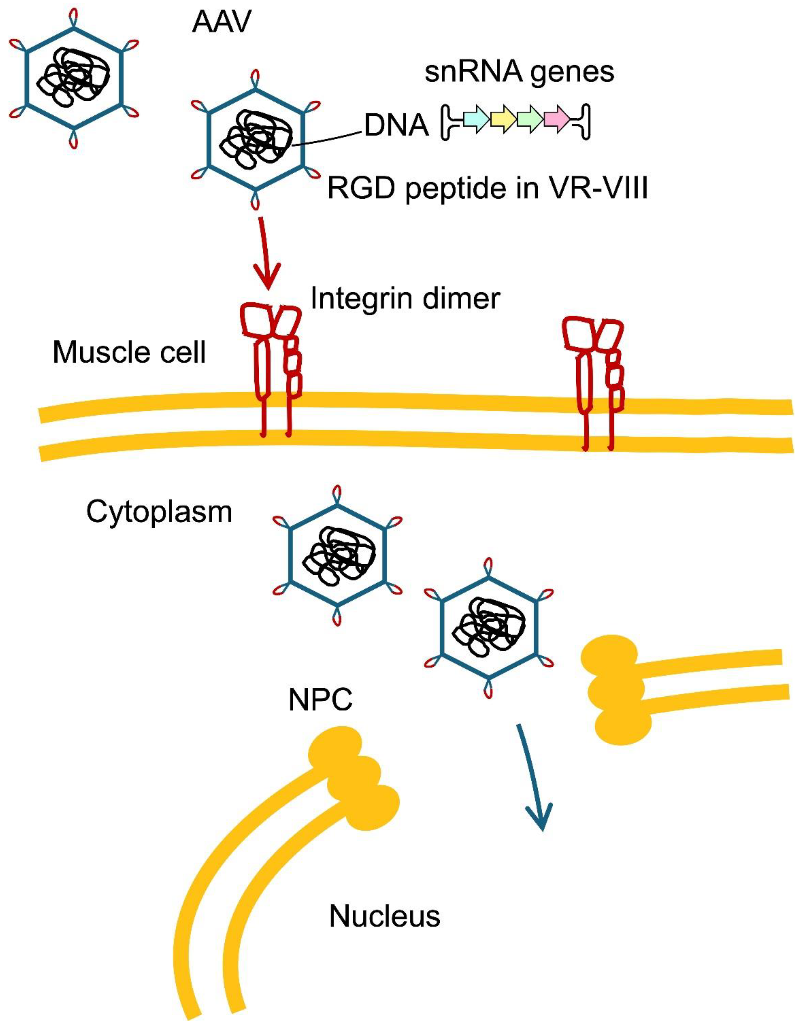
Figure 9.
Extravesicular vehicles (EV) are promising for snRNA delivery to muscle. EVs (Ø 30-1000nm) including exosomes (Ø 30-200nm) are produced by different cell types as vehicles for molecular signals and include many RNA species (Gurung et al., 2021). They benefit from excellent body-wide distribution and can be targeted to specific tissues by inclusion of specialised fusogens or ligands. Activated muscle precursor cells and damaged fibres switch on their fusogens Myomaker Mymk and Myomerger Mymg, that mediate muscle regeneration by cell fusion. EVs produced by cells that express Mymk and Mymg specifically deliver their cargo to the cytoplasm of regenerating muscle (Hindi et al., 2024). Ex vivo loading of in vitro transcribed RNA is usually done by electroporation and damages exosome membranes, but overexpressed snRNAs can be packaged into EVs by producer cells themselves. Production of exosomes in cell cultures for systemic injections is still inefficient, but subcutaneous implantation of producer cells, which can be autologous, provides a source of EVs with therapeutic effect in mice (Kojima et al., 2018). In future, transgenic livestock can produce RNA drug packaged into milk exosomes, that efficiently deliver biomolecules body-wide. EVs and exosomes are particularly suitable for snRNAs, which do not require nuclear delivery, as they join the cytoplasmic maturation pathway and get imported into the nucleus. SMN– survival motor neuron complex adorns snRNAs with Sm proteins; TGS1 – Trimethylguanosine Synthase responsible for converting the m7G cap (m7G-5’-ppp-5’) acquired in the nucleus when transcribed by Pol II into 2,2,7m3G or TMG cap (2,2,7m3G-5’-ppp-5’); SPN-Snurportin, a protein adaptor that binds the TMG cap and interacts with importin β; NPC - nuclear pore complex.
Figure 9.
Extravesicular vehicles (EV) are promising for snRNA delivery to muscle. EVs (Ø 30-1000nm) including exosomes (Ø 30-200nm) are produced by different cell types as vehicles for molecular signals and include many RNA species (Gurung et al., 2021). They benefit from excellent body-wide distribution and can be targeted to specific tissues by inclusion of specialised fusogens or ligands. Activated muscle precursor cells and damaged fibres switch on their fusogens Myomaker Mymk and Myomerger Mymg, that mediate muscle regeneration by cell fusion. EVs produced by cells that express Mymk and Mymg specifically deliver their cargo to the cytoplasm of regenerating muscle (Hindi et al., 2024). Ex vivo loading of in vitro transcribed RNA is usually done by electroporation and damages exosome membranes, but overexpressed snRNAs can be packaged into EVs by producer cells themselves. Production of exosomes in cell cultures for systemic injections is still inefficient, but subcutaneous implantation of producer cells, which can be autologous, provides a source of EVs with therapeutic effect in mice (Kojima et al., 2018). In future, transgenic livestock can produce RNA drug packaged into milk exosomes, that efficiently deliver biomolecules body-wide. EVs and exosomes are particularly suitable for snRNAs, which do not require nuclear delivery, as they join the cytoplasmic maturation pathway and get imported into the nucleus. SMN– survival motor neuron complex adorns snRNAs with Sm proteins; TGS1 – Trimethylguanosine Synthase responsible for converting the m7G cap (m7G-5’-ppp-5’) acquired in the nucleus when transcribed by Pol II into 2,2,7m3G or TMG cap (2,2,7m3G-5’-ppp-5’); SPN-Snurportin, a protein adaptor that binds the TMG cap and interacts with importin β; NPC - nuclear pore complex.
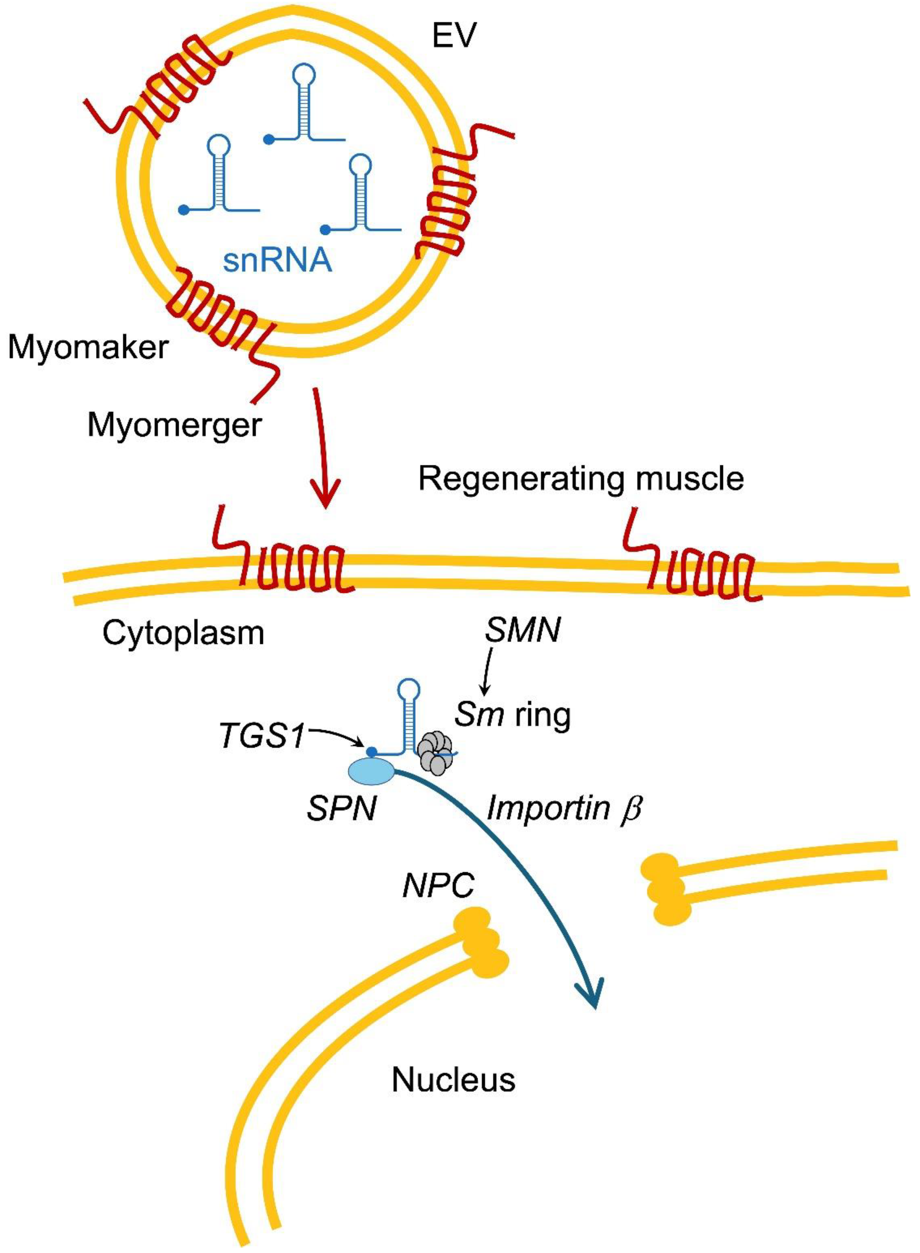
Figure 10.
U1 and U7 snRNAs targeting splice site choices in the early spliceosome (for successive spliceosomal complexes see Figure 5 in Part 2, Artemyeva-Isman, 2026b). Exons: rectangles; in panels B and C, alternative or mutant exon: in light blue; branchpoints: dots; snRNA binding sites: lines above the transcript; arrays to choose the optimal activity: multiple lines. A Examples of tested U1 designs for the suppression of splicing mutations. Compensatory U1 binds the 5’ss - it is usually effective if the target exon has a divergent 3’end or if at least one new G=C pair is introduced. ‘Exon-specific’ ExSpeU1s bind intronic sequences downstream from the exon but still stimulate the use of the correct 5’ss. U1 selection of 5’ss from a distance can occur naturally: wild-type U1 bound to the cryptic 5’ss motif downstream from PAH exon 11 stimulates the use of both correct and cryptic 5’ss and mutations in the cryptic motif cause exon 11 skipping. ExSpeU1s and the ectopic-bound WT U1 are encircled in blue ovals with arrows indicating upregulated 5’ss. B Inclusion of an alternative or mutated exon can be boosted by adapted U1: array in red - compensatory or ExSpeU1s (as in A). U1 in black: reducing the rate of splicing for a flanking exon with U1 blocking the 3’ss is likely to stimulate alternative exon inclusion (same principle as for ‘single’ ASOs around hotspot exons - Duan et al., 2025). I argue that U1 naturally acts as a suppressor for some 3’ss (see text). U7 is not a splicing factor, chimeric U7-OPT (with Lsm binding site substituted for Sm binding site, Gorman et al., 1998) is used as a mere scaffold for ASOs. U7-OPT booster (in red) adds an exonic splicing enhancer (ESE), but it is less effective than U1, a native 5’ss booster (see 5.4 Limitations of U7 and U1 technologies). U7-OPT suppressor (in black) to slow down splicing of flanking exons can be tried to promote the alternative or mutant exon inclusion (same principle as described for ‘single’ ASOs - Duan et al., 2025). C Skipping of an alternative or mutant exon can be induced by blocking 3’ss by U1 (see text, Hatch et al., 2022; Covello et al., 2022). Enhancing splicing rate of the flanking exons with U1 adapted for 5’ss can be tried to promote alternative exon skipping. U7-OPT (in black) blocking 5’ss, 3’ss or ESE or carrying an ASO complementary to two or three of these sites are used as splicing suppressors.
Figure 10.
U1 and U7 snRNAs targeting splice site choices in the early spliceosome (for successive spliceosomal complexes see Figure 5 in Part 2, Artemyeva-Isman, 2026b). Exons: rectangles; in panels B and C, alternative or mutant exon: in light blue; branchpoints: dots; snRNA binding sites: lines above the transcript; arrays to choose the optimal activity: multiple lines. A Examples of tested U1 designs for the suppression of splicing mutations. Compensatory U1 binds the 5’ss - it is usually effective if the target exon has a divergent 3’end or if at least one new G=C pair is introduced. ‘Exon-specific’ ExSpeU1s bind intronic sequences downstream from the exon but still stimulate the use of the correct 5’ss. U1 selection of 5’ss from a distance can occur naturally: wild-type U1 bound to the cryptic 5’ss motif downstream from PAH exon 11 stimulates the use of both correct and cryptic 5’ss and mutations in the cryptic motif cause exon 11 skipping. ExSpeU1s and the ectopic-bound WT U1 are encircled in blue ovals with arrows indicating upregulated 5’ss. B Inclusion of an alternative or mutated exon can be boosted by adapted U1: array in red - compensatory or ExSpeU1s (as in A). U1 in black: reducing the rate of splicing for a flanking exon with U1 blocking the 3’ss is likely to stimulate alternative exon inclusion (same principle as for ‘single’ ASOs around hotspot exons - Duan et al., 2025). I argue that U1 naturally acts as a suppressor for some 3’ss (see text). U7 is not a splicing factor, chimeric U7-OPT (with Lsm binding site substituted for Sm binding site, Gorman et al., 1998) is used as a mere scaffold for ASOs. U7-OPT booster (in red) adds an exonic splicing enhancer (ESE), but it is less effective than U1, a native 5’ss booster (see 5.4 Limitations of U7 and U1 technologies). U7-OPT suppressor (in black) to slow down splicing of flanking exons can be tried to promote the alternative or mutant exon inclusion (same principle as described for ‘single’ ASOs - Duan et al., 2025). C Skipping of an alternative or mutant exon can be induced by blocking 3’ss by U1 (see text, Hatch et al., 2022; Covello et al., 2022). Enhancing splicing rate of the flanking exons with U1 adapted for 5’ss can be tried to promote alternative exon skipping. U7-OPT (in black) blocking 5’ss, 3’ss or ESE or carrying an ASO complementary to two or three of these sites are used as splicing suppressors.
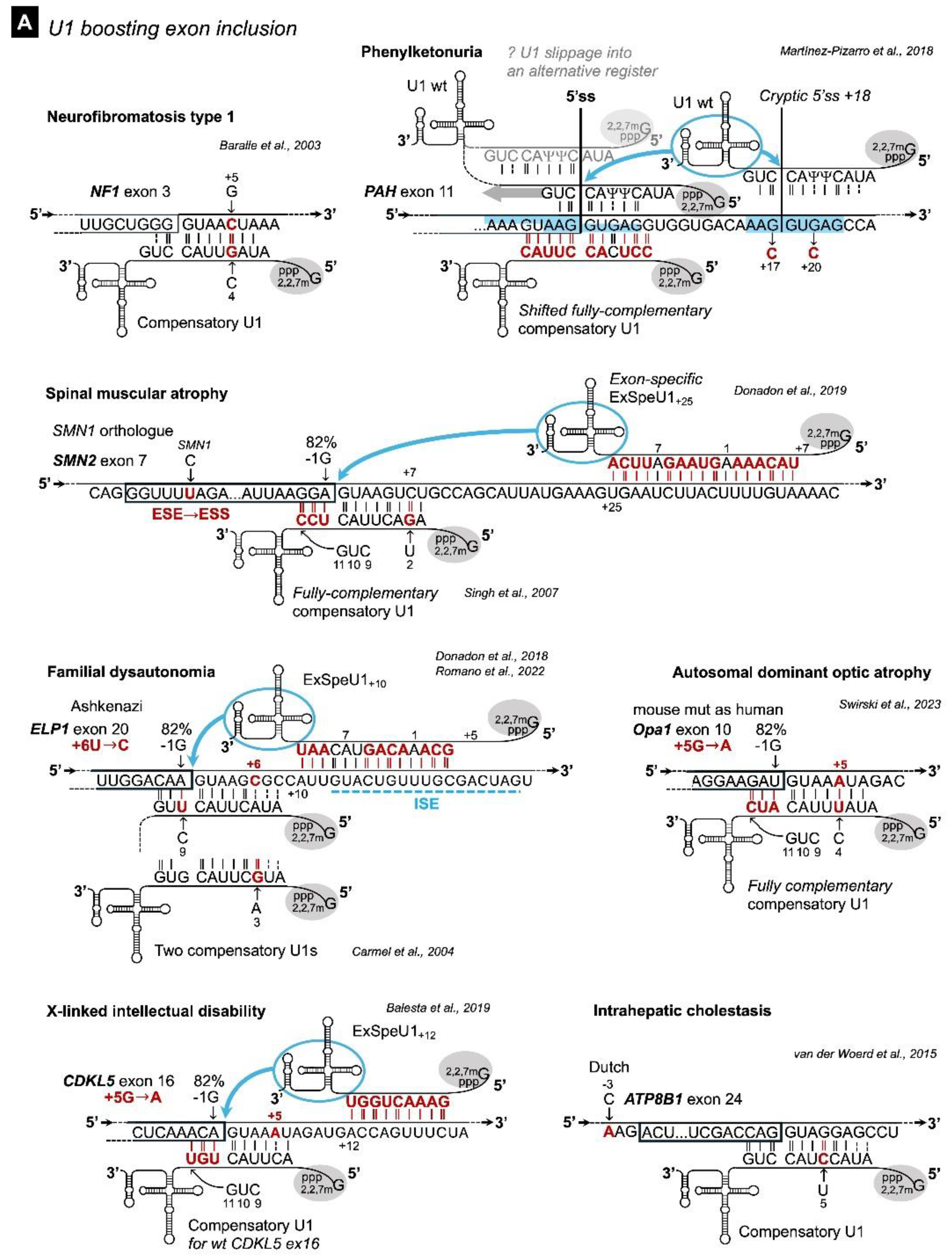
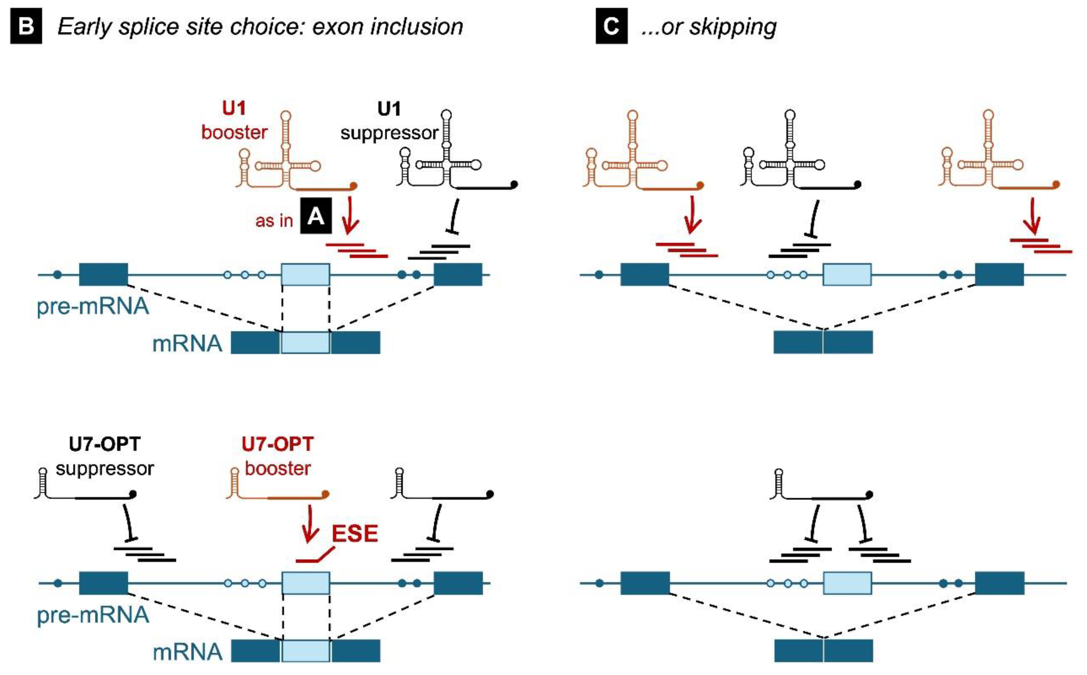
Disclaimer/Publisher’s Note: The statements, opinions and data contained in all publications are solely those of the individual author(s) and contributor(s) and not of MDPI and/or the editor(s). MDPI and/or the editor(s) disclaim responsibility for any injury to people or property resulting from any ideas, methods, instructions or products referred to in the content. |
© 2026 by the authors. Licensee MDPI, Basel, Switzerland. This article is an open access article distributed under the terms and conditions of the Creative Commons Attribution (CC BY) license (http://creativecommons.org/licenses/by/4.0/).
Copyright: This open access article is published under a Creative Commons CC BY 4.0 license, which permit the free download, distribution, and reuse, provided that the author and preprint are cited in any reuse.