
Submitted:
18 September 2026
Posted:
20 September 2026
You are already at the latest version
Abstract
Whole-exome sequencing (WES) is widely used to diagnose pediatric-onset inherited metabolic disorders (IEM), yet how often such patients carry more than one independent molecular diagnosis is rarely examined, particularly where testing is outsourced to commercial laboratories. We reviewed eight years (2018–2025) of outsourced WES at a Taiwanese pediatric genetics center. Of 227 referrals, 91 were solved, including 13 patients with pediatric-onset IEM or lysosomal storage disease; all variants were reannotated against ACMG/AMP criteria. Eighteen molecular diagnoses supported by 27 variants were established. Five of 13 patients (38.5%; exact 95% CI 13.9–68.4%) carried two independent molecular diagnoses, nearly eight times the 4.9% multi-locus rate of large unselected exome series and 3.9 times the 9.9% in our overall cohort; the proportion stayed above both benchmarks under progressively stricter counting rules, falling to 7.7% when pathogenic or likely pathogenic variants were required at both loci. Multi-locus cases typically paired a primary IEM with an unrelated Mendelian disorder. Disease-specific therapy was initiated in 8 of 13 patients (61.5%). In selected pediatric-onset metabolic disorders a single molecular diagnosis may not fully explain the presentation, underscoring the need for comprehensive variant interpretation and periodic reanalysis.
Keywords:
whole-exome sequencing
; inborn errors of metabolism
; lysosomal storage disease
; multi-locus diagnosis
; blended phenotype
; diagnostic odyssey
; Taiwan
1. Introduction
Whole-exome sequencing (WES) has evolved from a research tool into a first-tier diagnostic test for children with suspected Mendelian disorders. In large, unselected referral series from clinical laboratories, a molecular diagnosis has been achieved in approximately one-quarter to one-third of cases: 25% in the first 250 probands reported by Yang and colleagues [1], 25.2% in a subsequent cohort of 2,000 patients [2], and 28.8% in 3,040 consecutive cases at another laboratory, where the inclusion of parental samples further increased the diagnostic yield [3]. These figures, derived largely from in-house sequencing programs in North America and Europe, have become benchmarks against which newer testing settings are measured.
Inherited metabolic disorders (also termed inborn errors of metabolism [IEM]) occupy a unique place within this landscape. The most recent international nosology includes more than 1,400 distinct conditions, most of which affect multiple organ systems [4]. Two features make an accurate molecular diagnosis especially important. First, IEMs often present with overlapping, nonspecific features, such as developmental delay, hypotonia, organomegaly, and dysmorphism, making phenotype-driven targeted testing challenging. Second, unlike many other pediatric rare diseases, a substantial proportion of IEMs are treatable, allowing genomic confirmation to directly guide enzyme replacement therapy, dietary modification, cofactor supplementation, or transplantation [5,6]. WES, particularly alongside detailed biochemical phenotyping, has shortened the diagnostic odyssey for many of these children [5].
A less widely appreciated consequence of genome-wide testing is that a single patient may harbor two or more independent molecular diagnoses. Early clinical WES series noted it only occasionally—four of the first 250 probands received two unrelated diagnoses [1]—but the phenomenon proved consistent, with blended phenotypes resulting from two single-gene disorders accounting for 4.6% of solved cases in a cohort of 2,000 patients [2]. Posey and colleagues subsequently evaluated this systematically and reported that 101 of 2,076 molecularly diagnosed patients (4.9%) carried variants in two or more disease-associated loci, with the distinct diagnoses typically affecting different organ systems [7]. The prevalence is not fixed: it was substantially higher in a consanguineous neurodevelopmental cohort [8], reflecting the influence of ascertainment and population structure. Clinically, this distinction is important because a single “unifying” diagnosis that fails to explain all aspects of the phenotype may simply be incomplete.
However, the frequency of multiple molecular diagnoses specifically among pediatric-onset IEMs remains poorly defined. Available estimates come from large Western centers with in-house sequencing and mixed-indication referral populations; in many Asian countries, including Taiwan, clinical WES is commercially outsourced, and metabolic disorders are rarely examined separately. Because IEMs are inherently multisystem disorders and are often investigated by WES only after conventional metabolic and targeted testing has been exhausted, they may represent a subgroup enriched for multi-locus diagnoses. We therefore reviewed eight consecutive years of outsourced WES performed at a single Taiwanese pediatric genetics center and identified 13 patients with pediatric-onset IEM or lysosomal storage disease. We characterized the molecular and inheritance spectrum, estimated the prevalence of multi-locus diagnoses against published benchmarks, and documented the pre-WES workup and the clinical impact of diagnosis.
2. Results
2.1. Cohort Characteristics
Of the 227 patients referred for outsourced WES between January 2018 and December 2025, 91 had a documented molecular diagnosis, and 13 of these met the definition of inherited metabolic disorders described above (Figure 1A; per-patient clinical summaries are provided in Table S1). Ten patients were male, and three were female. The median age at WES referral was 2.7 yr (interquartile range [IQR], 0.7–9.6 yr; range, 0.0–23.1 yr), and referrals were weighted toward infancy and early childhood: four patients (30.8%) were referred before their first birthday and a further five (38.5%) between 1 and 5 yr of age (Figure 1B and Table 1). Referrals were distributed across the study period except for 2021, when the COVID-19 pandemic most disrupted our outpatient genetics service (Figure 1C). Testing reflected funding rather than study design: five patients (38.5%) were sequenced through the research-funded program in 2018–2019 and eight (61.5%) by self-pay commercial testing in 2020–2025 across three providers (Figure 1D). Four research-funded patients underwent trio WES; the remaining nine exomes (69.2%), including one research-funded neonate, were singletons. Twelve families were of Han Chinese Taiwanese ancestry, and one was of mixed Taiwanese and European ancestry.
Mucopolysaccharidoses (MPS) formed the largest phenotypic group, accounting for four of the 13 patients (30.8%), followed by mitochondrial disorders in two (15.4%). The remaining seven categories—organic acidemia, glycogen storage disease, amino acid metabolism, creatine metabolism, lipid storage disease, congenital disorder of glycosylation, and other inborn errors of metabolism—each contributed one patient (Figure S1). This distribution reflects our center’s referral base rather than the underlying epidemiology of inherited metabolic diseases. The mucopolysaccharidosis cases were referred through newborn screening follow-up and established enzyme replacement therapy clinics, whereas patients with less recognizable phenotypes were generally referred only after conventional testing had failed.
2.2. Molecular Diagnoses and Variant Spectrum
The 13 patients accounted for 18 molecular diagnoses supported by 27 variants (Table 2 and Table S2). Autosomal recessive inheritance predominated, accounting for 9 of the 18 diagnoses (50.0%), consistent with the genetic architecture of classical inborn errors of metabolism. Four diagnoses (22.2%) followed autosomal dominant inheritance, two of them attributable to de novo variants—the PTPN11 hotspot variant in P2-03 and the recurrent ZSWIM6 frameshift variant in P2-10. The remaining five diagnoses were X-linked (three X-linked recessive and two X-linked), a distribution that reflects the male predominance of the cohort (Figure 2B).
Among the 27 variants, six (22.2%) were classified as pathogenic and 11 (40.7%) as likely pathogenic, such that 63.0% received a definitive classification. The remaining 10 variants (37.0%) were variants of uncertain significance (Figure 2A). These variants were not distributed evenly. They clustered among the secondary findings in patients with multi-locus diagnoses—COG4, FLNA, and PIEZO2—and among deep intronic variants, of which the GALNS c.423-862C>T allele is the clearest example. It was missed by WES and identified only by whole-genome sequencing, commissioned when WES returned a single GALNS allele in a child with absent leukocyte enzyme activity. In four patients, the diagnosis relied on a variant of uncertain significance in a setting where the biochemical findings made the diagnosis unambiguous, and enzymatic evidence, rather than in silico prediction, supported pathogenicity. Compound heterozygous variants accounted for 16 of the 27 variants (59.3%), heterozygous variants for six (22.2%), and hemizygous variants for five (18.5%). No homozygous variants were identified, which is unsurprising in a non-consanguineous population (Figure 2C).
One reported finding was ultimately excluded from the diagnosis count. P2-09 carried two ZNF469 variants, p.Arg3090* and p.Pro1363Arg, which the reporting laboratory classified as pathogenic and likely benign, respectively. Because brittle cornea syndrome 1 is inherited in an autosomal recessive manner and requires biallelic pathogenic variants, one pathogenic allele together with one likely benign allele indicates carrier status rather than a molecular diagnosis. In addition, the characteristic ocular features of the disorder—corneal thinning, blue sclerae, and keratoconus—were absent. We therefore report the finding but excluded it from the diagnosis count, and P2-09 was classified as having a single-locus diagnosis.
The second locus in P2-11 was assigned after the initial analysis had closed: a heterozygous PIEZO2 p.Val1660Ala variant of uncertain significance in a gene whose gain-of-function missense variants cause the autosomal dominant Marden–Walker syndrome and distal arthrogryposis types 3 and 5, which McMillin and colleagues have proposed may represent variable expressivity of a single disorder. On review, the phenotype was concordant, with mild joint contractures together with moderate intellectual disability and surgically corrected esotropia. Nevertheless, the variant remained classified as uncertain because both parents died during the patient’s childhood, precluding segregation analysis, and no functional studies were performed.
2.3. Multi-Locus Diagnoses
Five of the 13 patients (38.5%; exact 95% confidence interval [95% CI], 13.9–68.4) carried two independent molecular diagnoses involving distinct, functionally unrelated loci (Table 3 and Figure 3). This was 7.8 times the 4.9% multi-locus diagnosis rate reported among 2,076 molecularly diagnosed patients in an unselected clinical exome series [7] and 3.9 times the 9.9% observed in our own full cohort of 227 referrals (submitted). The confidence interval is wide, as expected for this sample size, but its lower bound of 13.9% still exceeds both comparator values.
The pairings were clinically heterogeneous and, in four of the five patients, involved organ systems that could not be explained by a single diagnosis. P2-03 presented on day 2 of life with severe metabolic acidosis, hyperammonemia, and methylmalonic aciduria attributable to compound heterozygous MMUT variants. The same report identified a PTPN11 p.Arg265Gln hotspot variant, indicating that the atrial septal defect noted on echocardiography, initially considered incidental during the acute crisis, was an early manifestation of Noonan syndrome. In P2-08, xanthomas and a total cholesterol level of 482 mg/dL were fully explained by biallelic ABCG5 variants, whereas a hemizygous FLNA variant plausibly explained the pulmonary atresia with intact ventricular septum, which sitosterolemia does not. In P2-10, the metabolic diagnosis was secondary, with COG4-related Saul–Wilson syndrome accompanying a de novo ZSWIM6 neurodevelopmental disorder. P2-12 was distinct in carrying variants in two mitochondrial solute carrier genes, SLC25A41 and SLC25A5.
Three of the 13 patients had cytogenetic findings identified before WES that, in retrospect, suggested genomic complexity without fully resolving it, although only one was ultimately classified as multi-locus. P2-11 carried a complex balanced rearrangement involving chromosomes 2, 3, 6, 7, and 9 with normal parental karyotypes and a negative chromosomal microarray, whereas P2-09 carried a maternally inherited 7q duplication. The third patient, P2-13, carried a 5.1-Mb 4q23–q24 deletion spanning 25 OMIM genes together with biallelic NDUFA10 variants. Under our predefined criteria this patient was classified as single-locus because the deletion was not WES-derived, although the phenotype clearly reflected both abnormalities.
2.4. Pre-WES Investigation and Age at Referral
Patients underwent substantial evaluation before WES, and the pattern of investigations was informative. Biochemical testing was nearly universal, whereas specific enzyme assays and urinary glycosaminoglycan analysis were requested mainly when a lysosomal disorder was clinically suspected. When performed, these investigations directly contributed to the diagnosis of the mucopolysaccharidosis cases. Karyotyping and chromosomal microarray were performed in the patients with the most complex phenotypes and more often raised questions than answered them. Four patients underwent targeted single-gene or panel testing before WES, none of which established the diagnosis. In P2-07, a standard IDS panel detected no pathogenic variant, and the causative deep intronic variant was subsequently identified by WES. Because documentation was uneven across the study period, particularly for patients referred from other institutions, we describe these patterns qualitatively.
Age at referral spanned infancy through adulthood (Figure S2). P2-03 illustrates what is achievable when clinical suspicion is high: WES was initiated on day 2 of life concurrently with newborn screening by tandem mass spectrometry. At the other extreme, P2-12 underwent testing at 23.1 yr of age and P2-11 at 14.8 yr. Both had multi-locus diagnoses and complex phenotypes despite inconclusive karyotyping, microarray, and targeted testing, and both were ultimately diagnosed through the research-funded trio program. Turnaround time from sample dispatch to report release could be determined for four self-pay cases with complete records and had a median of 35.5 days (IQR, 27.5–80.0 days; range, 27–117 days; Table 4A). The single outlier (117 days) occurred in the patient who required complementary whole-genome sequencing after WES identified only one GALNS allele. Reporting dates were unavailable for research-funded cases and were not routinely recorded before 2023, so this estimate is illustrative rather than representative.
2.5. Impact of the Molecular Diagnosis on Management
A molecular diagnosis changed clinical management in the majority of patients (Table 4B). Disease-specific therapy was initiated in 8 of 13 patients (61.5%), including enzyme replacement therapy in three patients with mucopolysaccharidoses (P2-02, P2-05, and P2-07), disease-specific dietary management for methylmalonic aciduria, glycogen storage disease type Ib, and sitosterolemia, and cofactor or vitamin supplementation for the mitochondrial and creatine deficiency disorders. One patient (P2-07) subsequently underwent hematopoietic stem cell transplantation. All 13 families received genetic counseling and multidisciplinary specialist care, and prenatal diagnosis was offered in two subsequent pregnancies.
Two patients illustrate the range of therapeutic impact following molecular diagnosis. In P2-05, 4 yr of elosulfase alfa for MPS IVA showed urinary keratan sulfate fluctuating around each infusion cycle but declining overall, while GALNS activity rose progressively yet remained below the reference range. The peak observed in the second quarter of 2023 coincided with reduced treatment adherence during the COVID-19 pandemic, and neutralizing anti-drug antibodies developed during follow-up (Figure 4A). In P2-07, who was identified through newborn screening at 0.4 yr of age with severe MPS II, urinary heparan sulfate decreased from 992.71 to 7.74 μg/mL after idursulfase treatment—a 99% reduction—and remained low after transplantation, with donor chimerism confirmed by short tandem repeat analysis (Figure 4B). By contrast, P2-06 illustrates the limits of diagnosis without disease-modifying therapy: MPS IIIC was diagnosed at 8.9 yr, and its principal value was to reinterpret a decade of attention-deficit and developmental difficulties rather than to change treatment.
3. Discussion
Five of the 13 children in this metabolic subset carried two independent molecular diagnoses. At 38.5%, this is nearly eight times the 4.9% multi-locus rate reported among 2,076 molecularly diagnosed patients in the largest unselected clinical exome series published to date [7]. The direction of that difference is not in doubt; its magnitude is. With 13 patients, the exact confidence interval ranges from 13.9% to 68.4%, and the point estimate should be interpreted as reflecting a highly filtered referral population at a single center rather than the prevalence of pediatric metabolic disease.
Because the estimate depends entirely on what is allowed to count as a diagnosis, we deliberately varied that definition, each step a subset of the one before. All five pairs qualify under the criteria specified in the Methods section. Excluding P2-12, on the grounds that neither SLC25A41 nor SLC25A5 has an established gene–disease association—SLC25A41 has a gene entry in OMIM but no associated phenotype, and SLC25A5 has been proposed as a candidate gene for non-syndromic intellectual disability rather than being confirmed [19,20]—leaves four cases (30.8%; 95% CI, 9.1–61.4). Requiring, in addition, a pathogenic or likely pathogenic variant at both loci, thereby excluding the FLNA, COG4, and PIEZO2 variants of uncertain significance, leaves P2-03 alone: 7.7% (95% CI, 0.2–36.0). A sixth patient, P2-09, would have entered the numerator (46.2%) had the second ZNF469 allele been pathogenic rather than likely benign; we did not count it. The point estimate thus exceeds the unselected exome rate under every definition tested, but the margin narrows from nearly eightfold to under twofold as criteria tighten, and no definition establishes by how much.
Several features of how these patients reached us plausibly account for the enrichment. Outsourced exome sequencing was not a first-line test at our center for most of the study period; it followed failure of biochemistry, enzyme assays, and targeted panels, selecting for phenotypes that one gene was unlikely to explain. The denominator compounds this effect, because a second diagnosis is far more likely to be sought and identified when the first leaves part of the phenotype unexplained. Inherited metabolic disorders are also intrinsically multisystem, overlapping with neurodevelopmental, connective tissue, and RASopathy phenotypes, so clinicians are already primed to consider coexisting diagnoses. Three of the 13 patients had partial cytogenetic findings before WES—a complex balanced rearrangement, a maternally inherited 7q duplication, and a 4q23–q24 deletion—of whom one proved to have a multi-locus diagnosis. None of these findings explained the phenotype, but each suggested that more than one locus might be involved. This differs from the mechanism proposed for consanguineous Turkish neurodevelopmental cohorts [8], where an elevated burden of homozygous variation is thought responsible; our families were non-consanguineous, making ascertainment rather than population structure the likelier explanation.
The clinically useful message is not the percentage but the habit it argues for. When a molecularly confirmed diagnosis accounts for only part of what is wrong with a child, the reasonable inference is that the analysis is incomplete rather than that the remaining features are incidental. Two cases illustrate this point. In P2-03, an atrial septal defect had been recorded as an incidental echocardiographic finding during a neonatal metabolic crisis dominated by methylmalonic aciduria; the PTPN11 hotspot variant on the same report reclassified it as the first sign of Noonan syndrome, with different implications for cardiac and growth surveillance. In P2-10, the metabolic diagnosis is the secondary one, with COG4 underlying a de novo ZSWIM6 neurodevelopmental disorder, so a review confined to metabolic genes would have missed the finding that places this patient in our cohort. P2-11 makes the same point from the opposite direction: referred for intellectual disability and behavioral difficulties, this patient also had mild joint contractures and corrected esotropia that no one had connected to a diagnosis, yet both fall within the PIEZO2 spectrum. The designation of primary and secondary reflects which diagnosis matched the referral rather than which is better supported.
The outsourced model imposed its own limitations, worth stating because they apply to many pediatric genomic services across Asia. No provider released primary sequencing data with its reports, so we could not realign, re-filter, or systematically reanalyze. A pooled analysis of 29 reanalysis studies reported an additional diagnostic yield of approximately 10% from revisiting previously unsolved cases [21], but that opportunity is structurally unavailable to centers that never receive the primary data. Transcript selection and nomenclature also differed between providers, which is why every variant was reannotated rather than accepted as reported. The experience of P2-05 is illustrative: WES identified only a single GALNS allele in a child with absent leukocyte enzyme activity, and the second, deep intronic allele was detected only after whole-genome sequencing. When the biochemical phenotype is unequivocal but the exome findings are incomplete, reflex whole-genome sequencing is a more appropriate next step than repeating WES. Access to primary data and a defined reanalysis interval belong in the service contract rather than in case-by-case negotiation.
Diagnosis translated into treatment more often than the multisystem complexity of this cohort might suggest, with disease-specific therapy initiated in eight of the 13 patients. The clearest example is P2-07, identified through newborn screening and treated with enzyme replacement therapy followed by transplantation, in whom urinary heparan sulfate fell by 99% and remained low thereafter. P2-06 provides the necessary counterpoint: MPS IIIC was diagnosed at 8.9 yr of age, no disease-modifying therapy was available, and the value of the diagnosis lay in reinterpreting a decade of attention-deficit and developmental difficulties, securing rare disease registration, and providing the family with an explanation. Both outcomes justify testing, but only one directly changes treatment.
Limitations
This is a retrospective series of 13 patients from a single center, and the confidence intervals reported above honestly reflect that limitation. The denominator deserves particular caution: it comprises patients who already had a molecular diagnosis, so the figure we report is the probability of a second diagnosis conditional on having a first, not the probability of multi-locus disease in a referred child. The 136 patients in the parent cohort without a diagnosis were not reanalyzed and could not be, and some of them may harbor exactly the kind of complex genotypes described here. Multi-locus cases are also more memorable, and their records are correspondingly more complete, which may have influenced the findings.
Gene–disease validity is the weakest aspect of this dataset, and we have tried not to disguise it. Beyond SLC25A41 and SLC25A5, the FLNA variant in P2-08, the COG4 variant in P2-10, and the PIEZO2 variant in P2-11 all involve variants of uncertain significance without functional assays or completed segregation studies. The mild joint contracture in P2-11 is consistent with the PIEZO2 phenotype, but both parents had died before testing, so segregation could not be assessed. Similarly, in P2-06 only one HGSNAT allele was identified, with the second inferred from the enzyme result and clinical phenotype. The converse decision was made for P2-09, where a pathogenic ZNF469 allele was paired with a likely benign allele. Because brittle cornea syndrome 1 is recessive and the characteristic ocular features were absent, we considered this to represent carrier status rather than a molecular diagnosis, reducing the headline estimate from 46.2% to 38.5%. Phasing of the two SLC25A5 variants was not established; their assignment as hemizygous assumes both lie on the single maternal X. We performed no functional characterization, which is the obvious next step, particularly for the dual solute-carrier finding. Documentation also limited the analysis: onset and treatment-start dates were too inconsistent to support analysis of the diagnostic interval, the pre-exome workup could not be reliably tabulated, and turnaround time was available for only four patients. Finally, comparisons with published series are descriptive by design, since those cohorts differ in ascertainment, sequencing era, and reporting practice. Had we counted the 4q23–q24 deletion in P2-13 as a second independent diagnosis rather than a parallel cytogenetic finding, the multi-locus proportion would have been 6 of 13.
4. Materials and Methods
4.1. Study Design and Setting
We performed a retrospective observational study at the Division of Pediatric Genetics and the International Rare Disease Centre of MacKay Memorial Hospital, Taipei, a tertiary referral center for inherited metabolic diseases in northern Taiwan. The hospital had no in-house exome facility during the study period, so all diagnostic WES was outsourced. Between 2018 and 2019, sequencing was performed through a government-funded research program, which provided trio sequencing for most, but not all, patients. Thereafter (2020–2025), testing was performed through self-pay commercial clinical laboratories. Reporting followed the STROBE recommendations for observational research [9]. The study protocol was approved by the Institutional Review Board of MacKay Memorial Hospital (21MMHIS109e) and was conducted in accordance with the Declaration of Helsinki.
4.2. Case Ascertainment and Eligibility
Cases were identified from the divisional WES registry, which records all exome referrals since January 2018. Of 227 consecutive patients referred between January 2018 and December 2025 with complete registry records, 91 received a documented molecular diagnosis. Among these 91 patients, we included those whose primary or secondary diagnosis belonged to the inherited metabolic disorder, lysosomal storage disease, mitochondrial disorder, or congenital disorder of glycosylation categories of the International Classification of Inherited Metabolic Disorders [4]. The remaining 78 patients, whose diagnoses were exclusively non-metabolic, were excluded. Thirteen patients met the eligibility criteria and comprised the study cohort (Figure 1A). Patients with uninformative WES were outside the scope of this analysis, which addresses the architecture of confirmed diagnoses rather than diagnostic yield; the latter is reported separately for the parent cohort.
Pediatric onset was defined as documented onset of disease-related clinical or biochemical manifestations before 18 yr of age, regardless of age at WES. Accordingly, one patient who underwent WES at 23.1 yr of age was included because generalized seizures and psychomotor regression had begun in early childhood.
4.3. WES and Complementary Genomic Testing
Four patients underwent trio WES through the research-funded program, and one additional patient referred from the neonatal intensive care unit underwent proband-only WES through the same program. The remaining eight patients underwent singleton WES at one of three commercial Laboratories, designated Laboratories A, B, and C to preserve vendor anonymity.
Because sequencing was outsourced, capture chemistry, sequencing platforms, mean read depth, coverage thresholds, and variant-calling pipelines were determined by the individual laboratories and were not reported uniformly. In addition, raw sequencing data were not routinely released with the clinical reports. Consequently, our analyses were based on the variants reported by each laboratory rather than independently reprocessed primary data, and systematic reanalysis of unsolved regions was not possible. This limitation of the outsourced testing model is discussed further in the Discussion section.
When only a single pathogenic allele was identified in an autosomal recessive gene despite a concordant enzymatic phenotype, complementary whole-genome sequencing was requested. In one patient (P2-05), this identified a deep intronic GALNS variant that had not been captured by WES. Parental Sanger sequencing was performed whenever samples were available to determine inheritance and variant phase. In several families, this was not feasible because of parental death or loss to follow-up; in these cases, inheritance was recorded as presumed.
4.4. Clinical Data Extraction
Two investigators independently extracted data from electronic and paper records using a structured form. Variables included demographics and ancestry; age at first documented symptom and at WES referral; presenting phenotype by organ system; pre-WES investigations, recorded narratively because documentation was too heterogeneous to tabulate (basic biochemistry, tandem mass spectrometry newborn screening, enzyme assays, urinary glycosaminoglycan analysis, brain imaging, echocardiography, karyotyping, chromosomal microarray, targeted single-gene or panel testing, and electroencephalography); biochemical and enzymatic findings with laboratory-specific reference intervals; treatment and its timing; and clinical status at the most recent follow-up.
An investigation was considered performed only when a dated result was available in the medical record and was further categorized as contributing to the final diagnosis, abnormal but nonspecific, or normal. Discrepancies between investigators were resolved by joint review of the source records with a senior clinical geneticist. The complete abstracted clinical information for each patient, including presentation, pre-exome investigations, molecular findings, treatment, and outcome, is provided in the Table S1.
4.5. Variant Re-Annotation and Classification
All variants were re-extracted from the original laboratory reports and reannotated for this study rather than accepted as originally reported. Variant descriptions were standardized according to HGVS nomenclature using the specified RefSeq transcript [11], and each variant was reclassified according to the 2015 American College of Medical Genetics and Genomics/Association for Molecular Pathology (ACMG/AMP) guidelines [10]. Supporting evidence included population allele frequencies from gnomAD v4 [12], ClinVar submissions and assertion conflicts [13], and in silico predictions from SIFT [14], PROVEAN [15], and CADD [16]. For intronic and synonymous variants, predicted splice effects were evaluated using SpliceAI delta scores [17].
Two authors independently classified all variants, with disagreements resolved by consensus and, when necessary, adjudication by a senior author. When our classification differed from that of the reporting laboratory, the discrepancy was documented, and the more conservative classification was retained. Variants of uncertain significance were considered supportive of a diagnosis only when they were present in trans with a pathogenic or likely pathogenic variant in a gene consistent with the biochemical phenotype or when independent enzymatic and published functional evidence supported pathogenicity. The rationale for each case is provided in Table S2.
4.6. Definitions: Molecular, Single-Locus, and Multi-Locus Diagnosis
Because the primary outcome depended on how diagnoses were counted, definitions were established before analysis. A molecular diagnosis was defined as a gene–disease pair that explained all or part of the patient’s phenotype and was supported either by pathogenic or likely pathogenic variants in the expected zygosity or by variants of uncertain significance with the corroborating evidence described above.
Diagnoses were counted at the gene–disease pair level; therefore, a patient with two qualifying gene–disease pairs contributed two diagnoses. Variants were counted individually, such that a compound heterozygous diagnosis contributed two variants. Patients were classified as having a multi-locus diagnosis when two or more qualifying diagnoses involved distinct, unlinked loci, consistent with the definition used by Posey and colleagues [7].
Cytogenetic and copy number abnormalities identified before WES—including a maternally inherited 7q duplication, a 4q23–q24 interstitial deletion, and a complex balanced rearrangement—were tabulated separately and were not counted among WES-derived diagnoses, although they are described where relevant to the phenotype. Two findings were incompletely characterized when analysis began—a secondary variant awaiting gene assignment and a ZNF469 allele pair whose classification was not yet specified—and were resolved from the original reports before the analysis closed; both were then judged by the criteria above.
4.7. Turnaround Time and Management Impact
Turnaround time was defined as the number of calendar days from sample submission to release of the final report and was calculated only for self-pay commercial cases because the research-funded program followed a research rather than a clinical reporting schedule. The interval between symptom onset and WES referral was not analyzed because dates of symptom onset were unavailable or imprecise in most records. Instead, age at referral is reported.
A change in clinical management was attributed to the molecular diagnosis only when the medical record explicitly linked the intervention to the genetic findings. Prespecified categories included initiation of disease-specific therapy, genetic counseling, parental confirmatory testing, prenatal diagnosis in a subsequent pregnancy, rare disease registration or disability certification, and referral for multidisciplinary care.
4.8. Statistical Analysis
Statistical analysis was descriptive. Continuous variables are presented as median, interquartile range, and range, whereas categorical variables are presented as counts and percentages, with the denominator (patients, diagnoses, or variants) specified for each proportion. The prevalence of multi-locus diagnosis is presented with exact (Clopper–Pearson) 95% confidence intervals. Comparisons with published benchmarks are presented descriptively as ratios of proportions because the sample size (n = 13) was too small to support formal hypothesis testing.
Missing data were not imputed. Variables with incomplete documentation are reported using the available denominator and identified accordingly. Statistical analyses were performed using MedCalc Statistical Software version 23.6.3 (MedCalc Software Ltd., Ostend, Belgium; https://www.medcalc.org; 2026). Percentiles were calculated using the program’s default rank-based method, in which the pth percentile corresponds to the observation of rank p × n + 0.5, with linear interpolation between adjacent observations [18]. Because different percentile definitions can produce appreciably different quartiles in small samples, the reported interquartile ranges should be interpreted using this definition.
4.9. De-Identification
Patients are identified throughout by study codes (P2-01 to P2-13), which cannot be linked to individual identities without access to the master registry maintained at our institution. Sequencing providers are identified only as the research-funded program or Laboratories A, B, and C. Referring and treating clinicians are not named, external hospitals are described generically, and dates are reported only to the quarter-year level. No images or other potentially identifiable materials are included.
5. Conclusions
In this pediatric metabolic subset drawn from eight years of outsourced exome sequencing, two independent molecular diagnoses were identified in almost two-fifths of the children and remained more common than in unselected exome series under every counting convention tested. The finding argues less for a revised prevalence estimate than for a change in clinical interpretation: in a child whose phenotype is only partly explained by a confirmed diagnosis, the remaining features are evidence rather than noise, and the appropriate response is to keep interrogating the data. For centers that depend on commercial sequencing, this is possible only if primary-data access and a reanalysis schedule are secured at contracting, and genome sequencing is available when biochemistry and exome results disagree.
Supplementary Materials
The following supporting information can be downloaded at the website of this paper posted on Preprints.org, Table S1: Detailed per-patient clinical summaries; Table S2: Complete variant-level annotation of the 27 variants; Figure S1: Distribution of primary phenotypic diagnoses; Figure S2: Age at whole-exome sequencing referral by patient.
Author Contributions
Conceptualization, C.-L.L., H.-Y.L. and S.-P.L.; methodology, C.-L.L., C.-K.C. and Y.-R.T.; validation, C.-L.L., C.-K.C., H.-C.C. and Y.-R.T.; formal analysis, C.-L.L. and Y.-T.L.; investigation, C.-L.L., Y.-H.C., H.-C.C., H.-Y.H. and J.-Y.W.; resources, H.-Y.L. and S.-P.L.; data curation, C.-L.L., Y.-T.L. and J.-Y.W.; writing—original draft preparation, C.-L.L.; writing—review and editing, all authors; visualization, C.-L.L.; supervision, H.-Y.L. and S.-P.L.; funding acquisition, H.-Y.L. and S.-P.L. All authors have read and agreed to the published version of the manuscript.
Funding
This work was supported by MacKay Memorial Hospital (MMH-E-112-13, MMH-MM-112-14, MMH-E-113-13, MMH-MM-113-13, MMH-E-114-13, and MMH-E-115-13) and the National Science and Technology Council, Taiwan (NSTC-113-2314-B-195-003, NSTC-113-2314-B-195-004, NSTC-113-2314-B-195-021, NSTC-113-2314-B-715-002, NSTC-113-2811-B-195-001, NSTC-114-2314-B-195-001, NSTC-114-2314-B-195-002, NSTC-114-2314-B-715-001, NSTC-114-2811-B-195-002, NSTC-115-2124-M-001-016, NSTC-115-2314-B-195-001, NSTC-115-2314-B-195-002, NSTC-115-2314-B-195-012-MY3, NSTC-115-2314-B-195-013, NSTC-115-2314-B-715-001, and NSTC-115-2811-B-195-001). The funders had no role in the study design; data collection, analysis, or interpretation; manuscript preparation; or the decision to publish.
Institutional Review Board Statement
The study was conducted in accordance with the Declaration of Helsinki and approved by the Institutional Review Board of MacKay Memorial Hospital, Taipei, Taiwan (protocol code 21MMHIS109e; approved 1 October 2021, with annual renewals).
Informed Consent Statement
Written informed consent for genetic testing and for research use of de-identified data was obtained from all patients or their legal guardians at the time of clinical referral. All identifying information has been removed; patients are referred to only by study identifier.
Data Availability Statement
The de-identified case-level dataset generated and analyzed in this study is provided in the supplementary material accompanying this article (Table S1 and Table S2). Raw sequencing data are not available because sequencing was performed by commercial laboratories that did not release FASTQ, BAM, or VCF files with their clinical reports. Further information may be requested from the corresponding authors, subject to institutional review board approval and applicable data protection regulations in Taiwan.
Acknowledgments
We thank the patients and families who participated in this study as well as the clinical and laboratory staff of the Division of Pediatric Genetics, MacKay Memorial Hospital, for their long-term contribution to the divisional case registry. We also thank the staff of the International Rare Disease Centre at MacKay Memorial Hospital for their support with case enrollment and data management.
Conflicts of Interest
The authors declare no conflicts of interest. The funders had no role in the design of the study; in the collection, analyses, or interpretation of data; in the writing of the manuscript; or in the decision to publish the results.
Use of Generative AI: During the preparation of this work, the authors used a large language model (Anthropic Claude) to assist with language editing and the drafting of descriptive text. The authors reviewed and edited all generated content, independently verified every reference and numerical value against the source records, and take full responsibility for the content of the published article.
References
- Yang, Y.; Muzny, D.M.; Reid, J.G.; et al. Clinical whole-exome sequencing for the diagnosis of mendelian disorders. N Engl. J. Med. 2013, 369, 1502–1511. [Google Scholar] [CrossRef] [PubMed]
- Yang, Y.; Muzny, D.M.; Xia, F.; et al. Molecular findings among patients referred for clinical whole-exome sequencing. JAMA 2014, 312, 1870–1879. [Google Scholar] [CrossRef] [PubMed]
- Retterer, K.; Juusola, J.; Cho, M.T.; et al. Clinical application of whole-exome sequencing across clinical indications. Genet Med. 2016, 18, 696–704. [Google Scholar] [CrossRef] [PubMed]
- Ferreira, C.R.; Rahman, S.; Keller, M.; Zschocke, J.; ICIMD Advisory Group. An international classification of inherited metabolic disorders (ICIMD). J. Inherit. Metab. Dis. 2021, 44, 164–177. [Google Scholar] [CrossRef] [PubMed]
- Tarailo-Graovac, M.; Shyr, C.; Ross, C.J.; et al. Exome sequencing and the management of neurometabolic disorders. N Engl. J. Med. 2016, 374, 2246–2255. [Google Scholar] [CrossRef] [PubMed]
- Hoytema van Konijnenburg, E.M.M.; Wortmann, S.B.; Koelewijn, M.J.; et al. Treatable inherited metabolic disorders causing intellectual disability: 2021 review and digital app. Orphanet J. Rare Dis. 2021, 16, 170. [Google Scholar] [CrossRef] [PubMed]
- Posey, J.E.; Harel, T.; Liu, P.; et al. Resolution of disease phenotypes resulting from multilocus genomic variation. N Engl. J. Med. 2017, 376, 21–31. [Google Scholar] [CrossRef] [PubMed]
- Mitani, T.; Isikay, S.; Gezdirici, A.; et al. High prevalence of multilocus pathogenic variation in neurodevelopmental disorders in the Turkish population. Am. J. Hum. Genet 2021, 108, 1981–2005. [Google Scholar] [CrossRef] [PubMed]
- von Elm, E.; Altman, D.G.; Egger, M.; et al. Strengthening the Reporting of Observational Studies in Epidemiology (STROBE) statement: guidelines for reporting observational studies. BMJ 2007, 335, 806–808. [Google Scholar] [CrossRef] [PubMed]
- Richards, S.; Aziz, N.; Bale, S.; et al. Standards and guidelines for the interpretation of sequence variants: a joint consensus recommendation of the American College of Medical Genetics and Genomics and the Association for Molecular Pathology. Genet Med. 2015, 17, 405–424. [Google Scholar] [CrossRef] [PubMed]
- den Dunnen, J.T.; Dalgleish, R.; Maglott, D.R.; et al. HGVS recommendations for the description of sequence variants: 2016 update. Hum. Mutat. 2016, 37, 564–569. [Google Scholar] [CrossRef] [PubMed]
- Karczewski, K.J.; Francioli, L.C.; Tiao, G.; et al. The mutational constraint spectrum quantified from variation in 141,456 humans. Nature 2020, 581, 434–443. [Google Scholar] [CrossRef] [PubMed]
- Landrum, M.J.; Lee, J.M.; Benson, M.; et al. ClinVar: improving access to variant interpretations and supporting evidence. Nucleic Acids Res. 2018, 46, D1062–D1067. [Google Scholar] [CrossRef] [PubMed]
- Sim, N.L.; Kumar, P.; Hu, J.; Henikoff, S.; Schneider, G.; Ng, P.C. SIFT web server: predicting effects of amino acid substitutions on proteins. Nucleic Acids Res. 2012, 40, W452–W457. [Google Scholar] [CrossRef] [PubMed]
- Choi, Y.; Chan, A.P. PROVEAN web server: a tool to predict the functional effect of amino acid substitutions and indels. Bioinformatics 2015, 31, 2745–2747. [Google Scholar] [CrossRef] [PubMed]
- Rentzsch, P.; Witten, D.; Cooper, G.M.; Shendure, J.; Kircher, M. CADD: predicting the deleteriousness of variants throughout the human genome. Nucleic Acids Res. 2019, 47, D886–D894. [Google Scholar] [CrossRef] [PubMed]
- Jaganathan, K.; Kyriazopoulou Panagiotopoulou, S.; McRae, J.F.; et al. Predicting splicing from primary sequence with deep learning. Cell 2019, 176(3), 535–548.e24. [Google Scholar] [CrossRef] [PubMed]
- Schoonjans, F.; De Bacquer, D.; Schmid, P. Estimation of population percentiles. Epidemiology 2011, 22, 750–751. [Google Scholar] [CrossRef] [PubMed]
- Vandewalle, J.; Bauters, M.; Van Esch, H.; et al. The mitochondrial solute carrier SLC25A5 at Xq24 is a novel candidate gene for non-syndromic intellectual disability. Hum. Genet 2013, 132, 1177–1185. [Google Scholar] [CrossRef] [PubMed]
- Strande, N.T.; Riggs, E.R.; Buchanan, A.H.; et al. Evaluating the clinical validity of gene-disease associations: an evidence-based framework developed by the clinical genome resource. Am. J. Hum. Genet 2017, 100, 895–906. [Google Scholar] [CrossRef] [PubMed]
- Dai, P.; Honda, A.; Ewans, L.; et al. Recommendations for next generation sequencing data reanalysis of unsolved cases with suspected Mendelian disorders: a systematic review and meta-analysis. Genet Med. 2022, 24, 1618–1629. [Google Scholar] [CrossRef] [PubMed]
Figure 1.
Cohort overview of 13 patients with pediatric-onset metabolic disorders (A) Cohort selection flow. Of the 227 patients referred for outsourced WES during the study period, 136 had no molecular diagnosis at data lock, and 91 had a confirmed molecular diagnosis. Of these 91 patients, 78 were excluded because their diagnoses fell outside the categories of inherited metabolic disorders, lysosomal storage diseases, mitochondrial disorders, and congenital disorders of glycosylation, whether as a primary or secondary diagnosis, leaving a metabolic-disorder subset of 13 patients with 18 molecular diagnoses. Both exclusion steps are indicated beside the corresponding arrows. Among these 13 patients, eight (61.5%) had a single-locus molecular diagnosis, and five (38.5%) had multi-locus diagnoses. (B) Age distribution at WES referral. The median age at referral was 2.7 yr (interquartile range [IQR], 0.7–9.6 yr; range, 0.0–23.1 yr). Although one patient was 23.1 years old at the time of testing, all patients had pediatric-onset symptoms, supporting the designation of a pediatric-onset cohort. (C) Year of WES referral stratified by funding source. Five patients underwent sequencing through a national research-funded program (2018–2019), whereas eight underwent self-funded commercial WES (2020–2025). No patient in this subset was referred during 2021. These categories indicate funding source rather than family structure. Four of the five research-funded cases underwent trio sequencing, whereas the fifth was sequenced as a proband only, resulting in a total of nine singleton and four trio exomes. (D) Distribution of sequencing providers. “Research program” refers to the national research-funded sequencing program. Commercial Laboratories A, B, and C represent three independent clinical genetics laboratories contracted by our center during the study period and indicate the sequencing provider rather than any intermediary vendor. WES, whole-exome sequencing.
Figure 1.
Cohort overview of 13 patients with pediatric-onset metabolic disorders (A) Cohort selection flow. Of the 227 patients referred for outsourced WES during the study period, 136 had no molecular diagnosis at data lock, and 91 had a confirmed molecular diagnosis. Of these 91 patients, 78 were excluded because their diagnoses fell outside the categories of inherited metabolic disorders, lysosomal storage diseases, mitochondrial disorders, and congenital disorders of glycosylation, whether as a primary or secondary diagnosis, leaving a metabolic-disorder subset of 13 patients with 18 molecular diagnoses. Both exclusion steps are indicated beside the corresponding arrows. Among these 13 patients, eight (61.5%) had a single-locus molecular diagnosis, and five (38.5%) had multi-locus diagnoses. (B) Age distribution at WES referral. The median age at referral was 2.7 yr (interquartile range [IQR], 0.7–9.6 yr; range, 0.0–23.1 yr). Although one patient was 23.1 years old at the time of testing, all patients had pediatric-onset symptoms, supporting the designation of a pediatric-onset cohort. (C) Year of WES referral stratified by funding source. Five patients underwent sequencing through a national research-funded program (2018–2019), whereas eight underwent self-funded commercial WES (2020–2025). No patient in this subset was referred during 2021. These categories indicate funding source rather than family structure. Four of the five research-funded cases underwent trio sequencing, whereas the fifth was sequenced as a proband only, resulting in a total of nine singleton and four trio exomes. (D) Distribution of sequencing providers. “Research program” refers to the national research-funded sequencing program. Commercial Laboratories A, B, and C represent three independent clinical genetics laboratories contracted by our center during the study period and indicate the sequencing provider rather than any intermediary vendor. WES, whole-exome sequencing.
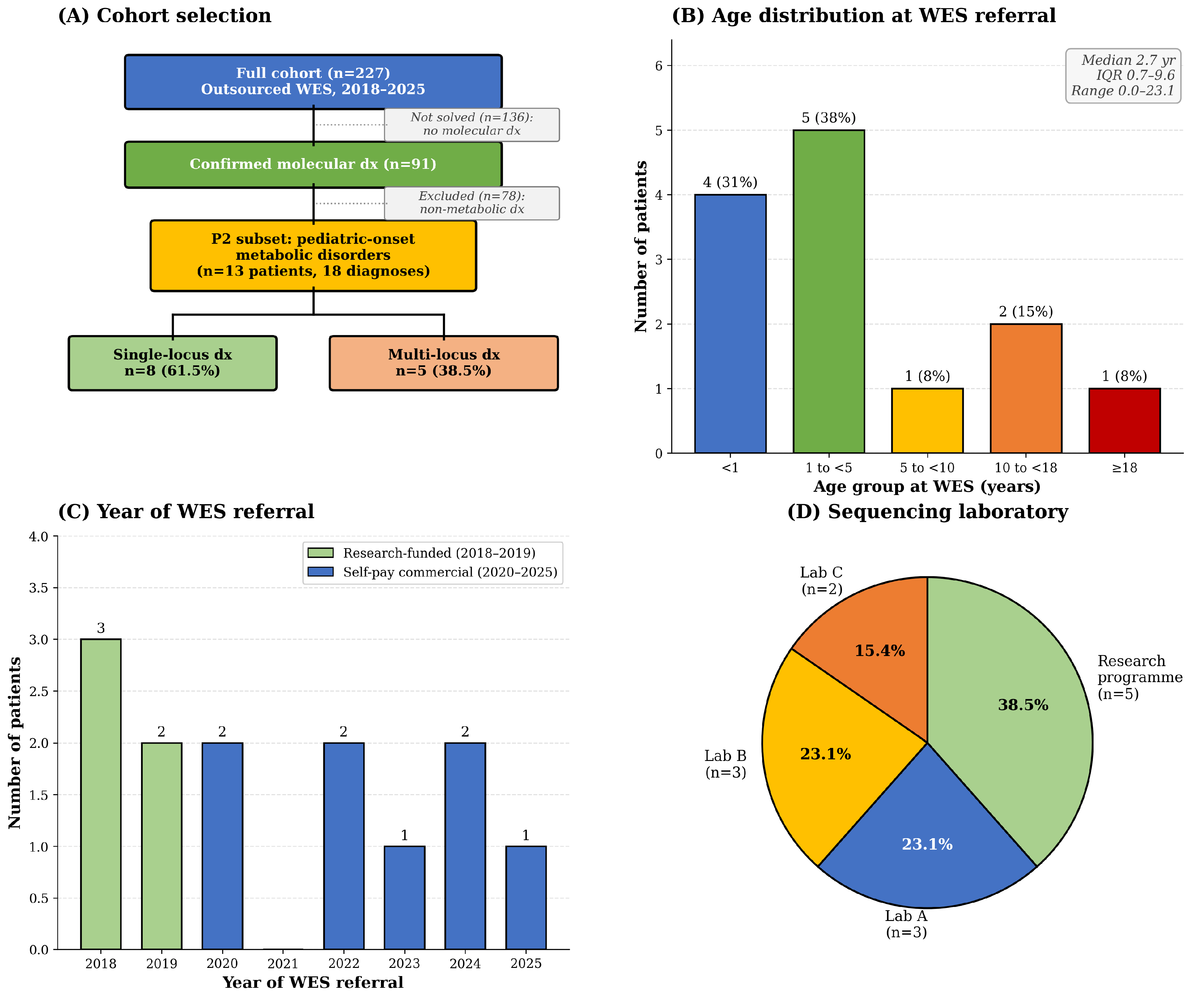
Figure 2.
Variant characterization: ACMG classification, inheritance pattern, and zygosity (A) ACMG classification of the 27 variants identified in the cohort. Pathogenic (6/27, 22.2%) and likely pathogenic (11/27, 40.7%) variants together accounted for 63.0% of all variants, whereas variants of uncertain significance (VUS) comprised the remaining 37.0% (10/27). VUSs were more frequently observed among secondary diagnoses in multi-locus cases (COG4, FLNA, and the pending autosomal dominant variant in P2-11) and among deep intronic variants identified by whole-genome sequencing rather than WES (GALNS c.423-862C>T). (B) Inheritance patterns of the 18 molecular diagnoses. Autosomal recessive inheritance predominated (9/18, 50.0%), consistent with the genetic architecture of classical inborn errors of metabolism. Autosomal dominant diagnoses (4/18, 22.2%) included two de novo variants (PTPN11 in P2-03 and ZSWIM6 in P2-10). X-linked recessive (3/18) and X-linked (2/18) diagnoses together accounted for 27.8%, reflecting the predominance of male patients in the cohort (10/13, 76.9%). (C) Zygosity of the 27 variants. Compound heterozygous variants were the most common (16/27, 59.3%). Heterozygous variants (6/27, 22.2%) included autosomal dominant diagnoses, secondary findings in multi-locus cases, the single reported HGSNAT allele in P2-06, and the X-linked G6PD variant in a female patient. Hemizygous variants (5/27, 18.5%) reflected X-linked findings in male patients, including both SLC25A5 variants in P2-12. No homozygous variants were identified, consistent with the non-consanguineous nature of the cohort. ACMG, American College of Medical Genetics and Genomics; AD, autosomal dominant; AR, autosomal recessive; LP, likely pathogenic; P, pathogenic; VUS, variant of uncertain significance; XL, X-linked; XLR, X-linked recessive.
Figure 2.
Variant characterization: ACMG classification, inheritance pattern, and zygosity (A) ACMG classification of the 27 variants identified in the cohort. Pathogenic (6/27, 22.2%) and likely pathogenic (11/27, 40.7%) variants together accounted for 63.0% of all variants, whereas variants of uncertain significance (VUS) comprised the remaining 37.0% (10/27). VUSs were more frequently observed among secondary diagnoses in multi-locus cases (COG4, FLNA, and the pending autosomal dominant variant in P2-11) and among deep intronic variants identified by whole-genome sequencing rather than WES (GALNS c.423-862C>T). (B) Inheritance patterns of the 18 molecular diagnoses. Autosomal recessive inheritance predominated (9/18, 50.0%), consistent with the genetic architecture of classical inborn errors of metabolism. Autosomal dominant diagnoses (4/18, 22.2%) included two de novo variants (PTPN11 in P2-03 and ZSWIM6 in P2-10). X-linked recessive (3/18) and X-linked (2/18) diagnoses together accounted for 27.8%, reflecting the predominance of male patients in the cohort (10/13, 76.9%). (C) Zygosity of the 27 variants. Compound heterozygous variants were the most common (16/27, 59.3%). Heterozygous variants (6/27, 22.2%) included autosomal dominant diagnoses, secondary findings in multi-locus cases, the single reported HGSNAT allele in P2-06, and the X-linked G6PD variant in a female patient. Hemizygous variants (5/27, 18.5%) reflected X-linked findings in male patients, including both SLC25A5 variants in P2-12. No homozygous variants were identified, consistent with the non-consanguineous nature of the cohort. ACMG, American College of Medical Genetics and Genomics; AD, autosomal dominant; AR, autosomal recessive; LP, likely pathogenic; P, pathogenic; VUS, variant of uncertain significance; XL, X-linked; XLR, X-linked recessive.
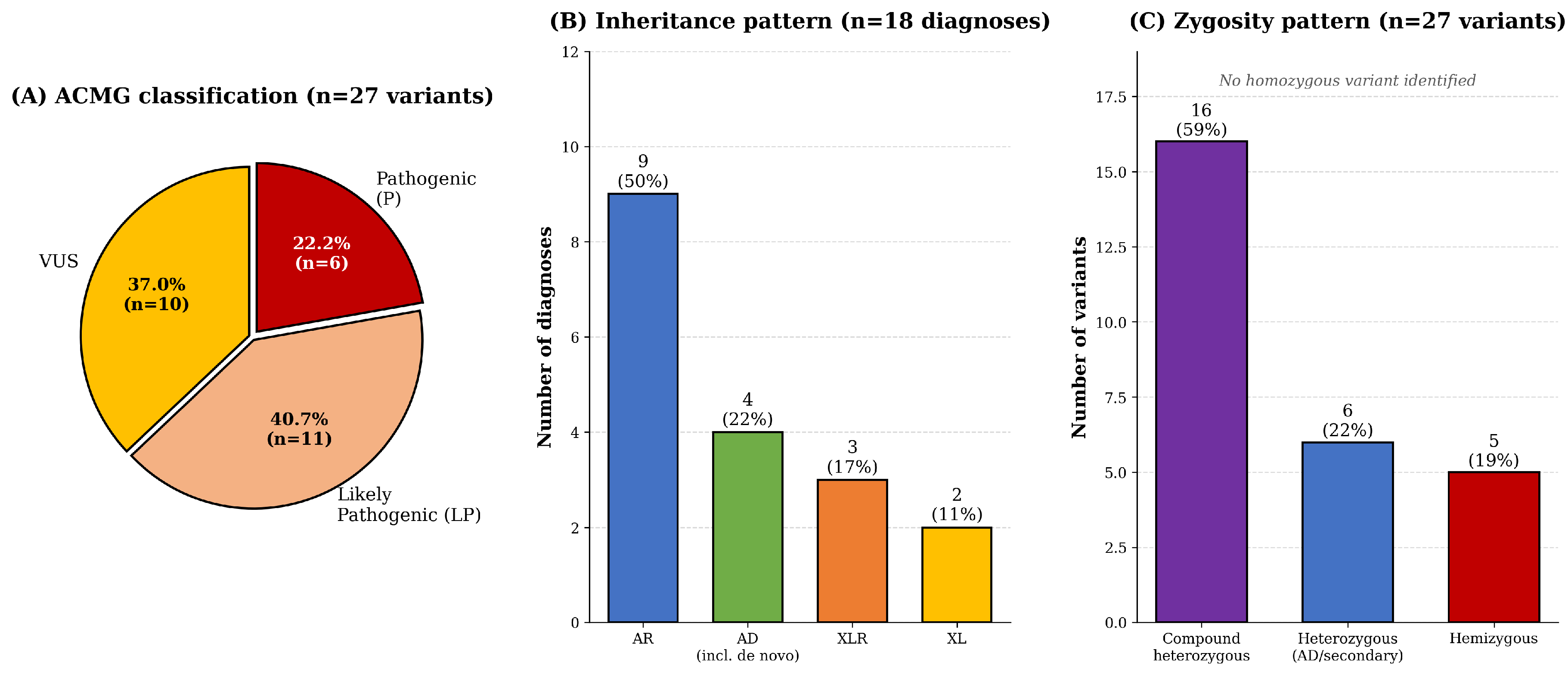
Figure 3.
Multi-locus diagnoses in pediatric-onset metabolic disorders (A) Prevalence of multi-locus diagnoses across cohorts. In this metabolic subset, five of 13 patients (38.5%; exact 95% confidence interval, 13.9–68.4) harbored two independent molecular diagnoses. This prevalence was 7.8-fold higher than the 4.9% reported by Posey et al. (N Engl J Med 2017;376:21–31) among 2,076 molecularly diagnosed patients from 7,374 consecutive WES cases and 3.9-fold higher than the 9.9% observed in our parallel full-cohort study (submitted). Although the confidence interval is wide because of the small sample size, its lower bound remains above both comparison cohorts. The higher prevalence likely reflects referral enrichment at a tertiary genetics center, extensive pre-WES evaluation, and the multisystem nature of inherited metabolic disorders. (B) Summary of the five patients with multi-locus diagnoses. The PIEZO2 variant in P2-11 remains a VUS, although the patient’s mild joint contracture, intellectual disability, and corrected esotropia are compatible with the reported PIEZO2 phenotype. P2-09 is not shown because the combination of a pathogenic and a likely benign ZNF469 variant represents carrier status rather than a second molecular diagnosis. NEJM, New England Journal of Medicine; NDD, neurodevelopmental disorder; VUS, variant of uncertain significance.
Figure 3.
Multi-locus diagnoses in pediatric-onset metabolic disorders (A) Prevalence of multi-locus diagnoses across cohorts. In this metabolic subset, five of 13 patients (38.5%; exact 95% confidence interval, 13.9–68.4) harbored two independent molecular diagnoses. This prevalence was 7.8-fold higher than the 4.9% reported by Posey et al. (N Engl J Med 2017;376:21–31) among 2,076 molecularly diagnosed patients from 7,374 consecutive WES cases and 3.9-fold higher than the 9.9% observed in our parallel full-cohort study (submitted). Although the confidence interval is wide because of the small sample size, its lower bound remains above both comparison cohorts. The higher prevalence likely reflects referral enrichment at a tertiary genetics center, extensive pre-WES evaluation, and the multisystem nature of inherited metabolic disorders. (B) Summary of the five patients with multi-locus diagnoses. The PIEZO2 variant in P2-11 remains a VUS, although the patient’s mild joint contracture, intellectual disability, and corrected esotropia are compatible with the reported PIEZO2 phenotype. P2-09 is not shown because the combination of a pathogenic and a likely benign ZNF469 variant represents carrier status rather than a second molecular diagnosis. NEJM, New England Journal of Medicine; NDD, neurodevelopmental disorder; VUS, variant of uncertain significance.
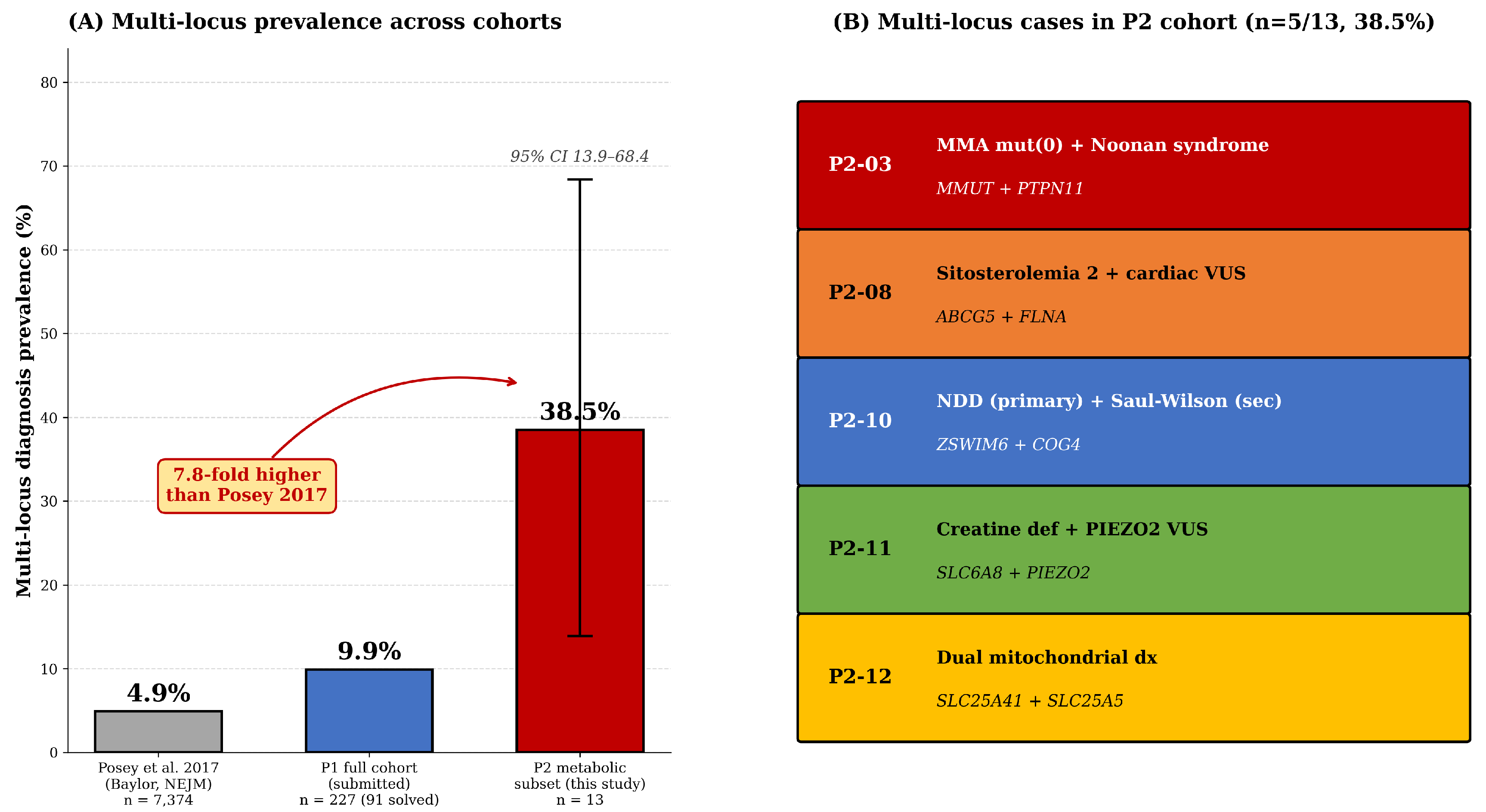
Figure 4.
Treatment impact: responses to enzyme replacement therapy and hematopoietic stem cell transplantation (A) Four-year follow-up of P2-05 receiving enzyme replacement therapy for MPS IVA (Morquio A syndrome). Urinary keratan sulfate (KS; red circles, left axis) and GALNS enzyme activity measured from dried blood spots (blue squares, right axis) were monitored from before initiation of elosulfase alfa (Q1 2021). Reference limits for KS (<7.9 µg/mL) and GALNS activity (>5.9 µmol/g/h) are shown as dashed lines. KS levels fluctuated around treatment cycles but showed an overall downward trend, whereas GALNS activity gradually increased but remained below the reference range. A transient increase in KS during Q2 2023 coincided with reduced treatment adherence during the COVID-19 pandemic, and neutralizing anti-drug antibodies developed during follow-up. (B) Biomarker response to combined therapy in P2-07 with severe MPS II (Hunter syndrome). The patient was identified through newborn screening at 0.4 yr of age, and the IDS c.1181-15C>A variant was confirmed by WES. Following enzyme replacement therapy (idursulfase, Q1 2025) and subsequent hematopoietic stem cell transplantation (Q3 2025) at an external center, urinary heparan sulfate decreased from 992.71 to 7.74 µg/mL (99% reduction) and remained low (35.91) after transplantation. The DMB ratio and dermatan sulfate levels also decreased. IDS enzyme activity increased modestly from 1.0 to 2.15 µmol/g protein/4 h (reference, 12.89–131.83). Post-transplant short tandem repeat analysis performed in Q4 2025 confirmed donor chimerism. DMB, dimethylmethylene blue; DS, dermatan sulfate; ERT, enzyme replacement therapy; GALNS, N-acetylgalactosamine-6-sulfatase; HS, heparan sulfate; HSCT, hematopoietic stem cell transplantation; IDS, iduronate-2-sulfatase; KS, keratan sulfate; MPS, mucopolysaccharidosis.
Figure 4.
Treatment impact: responses to enzyme replacement therapy and hematopoietic stem cell transplantation (A) Four-year follow-up of P2-05 receiving enzyme replacement therapy for MPS IVA (Morquio A syndrome). Urinary keratan sulfate (KS; red circles, left axis) and GALNS enzyme activity measured from dried blood spots (blue squares, right axis) were monitored from before initiation of elosulfase alfa (Q1 2021). Reference limits for KS (<7.9 µg/mL) and GALNS activity (>5.9 µmol/g/h) are shown as dashed lines. KS levels fluctuated around treatment cycles but showed an overall downward trend, whereas GALNS activity gradually increased but remained below the reference range. A transient increase in KS during Q2 2023 coincided with reduced treatment adherence during the COVID-19 pandemic, and neutralizing anti-drug antibodies developed during follow-up. (B) Biomarker response to combined therapy in P2-07 with severe MPS II (Hunter syndrome). The patient was identified through newborn screening at 0.4 yr of age, and the IDS c.1181-15C>A variant was confirmed by WES. Following enzyme replacement therapy (idursulfase, Q1 2025) and subsequent hematopoietic stem cell transplantation (Q3 2025) at an external center, urinary heparan sulfate decreased from 992.71 to 7.74 µg/mL (99% reduction) and remained low (35.91) after transplantation. The DMB ratio and dermatan sulfate levels also decreased. IDS enzyme activity increased modestly from 1.0 to 2.15 µmol/g protein/4 h (reference, 12.89–131.83). Post-transplant short tandem repeat analysis performed in Q4 2025 confirmed donor chimerism. DMB, dimethylmethylene blue; DS, dermatan sulfate; ERT, enzyme replacement therapy; GALNS, N-acetylgalactosamine-6-sulfatase; HS, heparan sulfate; HSCT, hematopoietic stem cell transplantation; IDS, iduronate-2-sulfatase; KS, keratan sulfate; MPS, mucopolysaccharidosis.
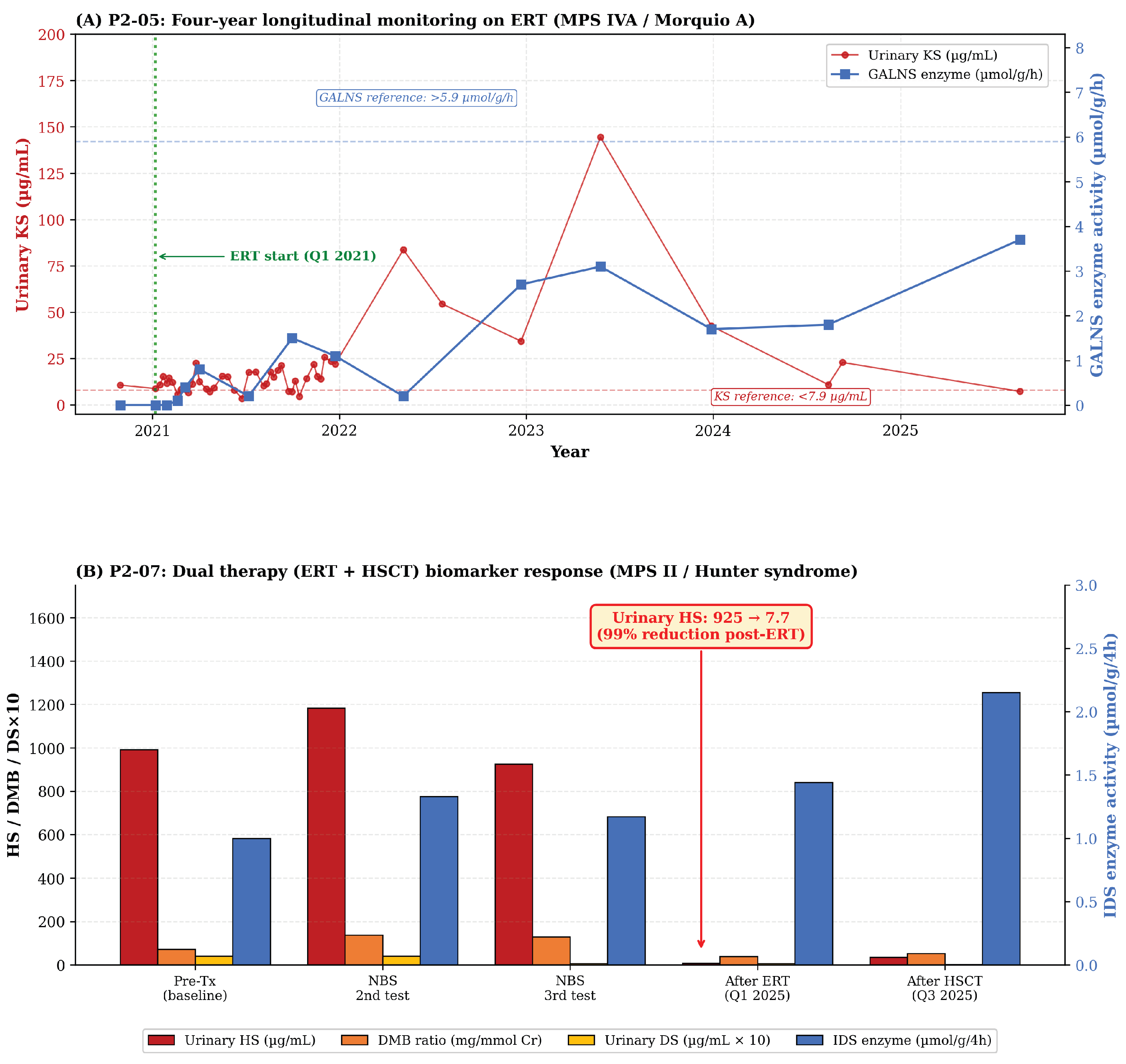

Table 1.
Baseline characteristics of the 13 patients in the metabolic-disorder subset (outsourced WES, 2018–2025).
Table 1.
Baseline characteristics of the 13 patients in the metabolic-disorder subset (outsourced WES, 2018–2025).
| Characteristic | Value |
| Total patients, n | 13 |
| Total molecular diagnoses, n | 18 |
| Total variants identified, n | 27 |
| Sex | |
| Male, n (%) | 10 (76.9) |
| Female, n (%) | 3 (23.1) |
| Age at WES referral | |
| Median (IQR), yr | 2.7 (0.7–9.6) |
| Range, yr | 0.0–23.1 |
| Age group at WES, n (%) | |
| <1 yr (infant) | 4 (30.8) |
| 1–<5 yr (toddler/preschool) | 5 (38.5) |
| 5–<10 yr (school age) | 1 (7.7) |
| 10–<18 yr (adolescent) | 2 (15.4) |
| ≥18 yr (adult age, pediatric-onset) | 1 (7.7) |
| WES year, n (%) | |
| 2018–2019 (research-funded) | 5 (38.5) |
| 2020–2025 (self-pay commercial) | 8 (61.5) |
| Sequencing laboratory, n (%) | |
| Research-funded program | 5 (38.5) |
| Commercial laboratory A | 3 (23.1) |
| Commercial laboratory B | 3 (23.1) |
| Commercial laboratory C | 2 (15.4) |
| WES type, n (%) | |
| Singleton | 9 (69.2) |
| Trio | 4 (30.8) |
| Phenotype category, n (%) | |
| Mucopolysaccharidosis (MPS) | 4 (30.8) |
| Mitochondrial disorders | 2 (15.4) |
| Organic acidemia | 1 (7.7) |
| Glycogen storage disease | 1 (7.7) |
| Amino-acid metabolism | 1 (7.7) |
| Creatine metabolism | 1 (7.7) |
| Lipid storage (sitosterolemia) | 1 (7.7) |
| Congenital disorder of glycosylation (CDG) | 1 (7.7) |
| Other IEM (G6PD deficiency) | 1 (7.7) |
| Molecular diagnosis outcome | |
| Single-locus diagnosis, n (%) | 8 (61.5) |
| Multi-locus diagnosis, n (%) | 5 (38.5) |
| Family ancestry | |
| Han Chinese (Taiwan) | 12 (92.3) |
| Mixed Taiwanese/European | 1 (7.7) |
Note. CDG, congenital disorder of glycosylation; IEM, inborn error of metabolism; IQR, interquartile range; MPS, mucopolysaccharidosis; WES, whole-exome sequencing. Demographic and procedural characteristics of the 13 unique patients with pediatric-onset inborn errors of metabolism who underwent outsourced whole-exome sequencing (WES) at our pediatric genetics center between 2018 and 2025. The cohort comprises five patients covered by a national research-funded sequencing program (2018–2019) and eight who underwent self-pay commercial WES (2020–2025); these categories describe funding rather than family structure. Four of the five research-funded exomes were sequenced as trios, whereas the fifth, referred from the neonatal intensive care unit, underwent proband-only sequencing, resulting in a cohort of nine singleton and four trio cases. Although one patient was 23.1 years old at the time of testing, all patients had pediatric-onset clinical symptoms; the cohort is therefore described as “pediatric-onset rare disease,” consistent with the broader cohort overview (anonymized authors, submitted). The 18 molecular diagnoses comprise one diagnosis in each of the eight single-locus patients and two diagnoses in each of the five multi-locus patients. These diagnoses are supported by 27 individual variants because a compound heterozygous diagnosis contributes two variants. Diagnoses are counted at the level of the gene–disease pair, and prevalence figures use patients as the denominator. Commercial Laboratories A, B, and C represent three independent clinical genetics testing providers contracted by our center during the study period. Patient identifiers (P2-01 through P2-13) are study identifiers assigned for this manuscript and cannot be linked to individual patients without access to the original master registry.
Table 2.
Detailed molecular diagnoses in 13 patients (18 diagnoses).
| Pt ID | Age at WES (yr) | Sex | Lab | Year | Disease | Gene | OMIM | HGVS variant(s) | Zygosity | Inheritance | ACMG | Multi-locus |
| P2-01 | 0.4 | F | Research | 2018 | G6PD deficiency | G6PD | 300908 | c.1466G>T p.Arg489Leu | Het | XLR | LP | No |
| P2-02 | 4.8 | F | C | 2020 | MPS VII (Sly) | GUSB | 253220 | c.104C>A p.Ser35Ter; c.1454C>T p.Ser485Phe | Compound het | AR | P/VUS* | No |
| P2-03 | 0.0 | F | B | 2022 | MMA mut(0) | MMUT | 251000 | c.1106G>A p.Arg369His; c.1677-1G>A | Compound het | AR | LP/LP | ⭐ Yes |
| P2-03 | 0.0 | F | B | 2022 | Noonan syndrome | PTPN11 | 163950 | c.794G>A p.Arg265Gln | Het | AD | P | (same pt) |
| P2-04 | 0.8 | M | B | 2022 | GSD Ib | SLC37A4 | 232220 | c.1042_1043delCT p.Leu348fs*53; c.898C>T p.Arg300Cys | Compound het | AR | VUS/LP | No |
| P2-05 | 2.7 | M | B | 2023 | MPS IVA (Morquio A) | GALNS | 253000 | c.953T>G p.Met318Arg; c.423-862C>T (intronic)† | Compound het | AR | P/VUS | No |
| P2-06 | 8.9 | M | A | 2024 | MPS IIIC (Sanfilippo C) | HGSNAT | 252930 | c.607C>T p.Arg203* | Het‡ | AR | P | No |
| P2-07 | 0.4 | M | A | 2024 | MPS II (Hunter, severe) | IDS | 309900 | c.1181-15C>A (intronic) | Hemi | XLR | VUS§ | No |
| P2-08 | 2.5 | M | A | 2025 | Sitosterolemia 2 | ABCG5 | 618666 | c.64C>T p.Gln22*; c.1166G>A p.Arg389His | Compound het | AR | P/VUS | ⭐ Yes |
| P2-08 | 2.5 | M | A | 2025 | (Cardiac VUS) | FLNA | 300017 | c.5479A>G p.Thr1827Ala | Hemi | XL | VUS | (same pt) |
| P2-09 | 11.7 | M | Research | 2018 | Glycine encephalopathy | SLC6A9 | 617301 | p.Leu28Pro; p.Tyr133Phe | Compound het | AR | LP | No‖ |
| P2-10 | 4.5 | M | Research | 2018 | Saul–Wilson (CDG II)—secondary | COG4 | 618150 | p.Ser236Trp | Het | AD | VUS | ⭐ Yes |
| P2-10 | 4.5 | M | Research | 2018 | NEDMAGA (NDD primary) | ZSWIM6 | 617865 | p.Ala161fs (de novo) | Het | AD | P | (same pt) |
| P2-11 | 14.8 | M | Research | 2019 | Cerebral creatine deficiency 1 | SLC6A8 | 300352 | p.Thr178Ser | Hemi | XLR | LP | ⭐ Yes |
| P2-11 | 14.8 | M | Research | 2019 | Marden–Walker/distal arthrogryposis spectrum | PIEZO2 | 613629 | p.Val1660Ala | Het | AD | VUS | (same pt) |
| P2-12 | 23.1 | M | Research | 2019 | Mito solute carrier #1 | SLC25A41 | N/A | p.Ala229Thr; p.Trp93Arg | Compound het | AR | LP | ⭐ Yes |
| P2-12 | 23.1 | M | Research | 2019 | Mito solute carrier #2 | SLC25A5 | 300150 | p.Phe271Leu; p.Asn277Ser | Hemi | XL | VUS | (same pt) |
| P2-13 | 1.9 | M | C | 2020 | Mitochondrial complex I deficiency | NDUFA10 | 618238 | c.404T>C p.Leu135Ser; c.1090_1110del | Compound het | AR | LP/LP | No¶ |
Note. AD, autosomal dominant; AR, autosomal recessive; CDG, congenital disorder of glycosylation; GSD, glycogen storage disease; HGVS, Human Genome Variation Society; LP, likely pathogenic; MMA, methylmalonic aciduria; MPS, mucopolysaccharidosis; NDD, neurodevelopmental disorder; NEDMAGA, neurodevelopmental disorder with movement abnormalities, abnormal gait, and autistic features; P, pathogenic; VUS, variant of uncertain significance; WES, whole-exome sequencing; XL, X-linked; XLR, X-linked recessive. Detailed molecular findings for the 13 unique patients (18 distinct diagnoses). Multi-locus cases are highlighted (cream shading) and indicated by ⭐ in the rightmost column; rows labeled “(same pt)” represent the second diagnosis in a multi-locus patient. “Research” denotes the national research-funded trio sequencing program; “A,” “B,” and “C” denote three independent commercial clinical laboratories. The second diagnosis in P2-11 is a heterozygous PIEZO2 variant of uncertain significance. Gain-of-function missense variants in this gene cause Marden–Walker syndrome (OMIM 248700) and distal arthrogryposis types 3 (OMIM 114300) and 5 (OMIM 108145). The gene entry number is cited because the patient’s phenotype does not clearly correspond to one of these three disorders. In P2-12, both SLC25A5 variants are recorded as hemizygous because the patient is male and the gene is X-linked; phasing was not established. P2-09 also carries two ZNF469 variants, p.Arg3090* (pathogenic) and p.Pro1363Arg (likely benign). Brittle cornea syndrome 1 is autosomal recessive and requires biallelic pathogenic variants; therefore, this combination represents carrier status rather than a molecular diagnosis. The characteristic ocular features were absent from the clinical record. The finding is reported in the Table S1 and Table S2 but is not counted here, and P2-09 is classified as a single-locus case.
Table 3.
Multi-locus molecular diagnoses (five patients with 10 diagnoses; 38.5% of the cohort).
| # | Pt ID | Age at WES | Lab | Primary diagnosis | Gene 1 | ACMG | Secondary diagnosis | Gene 2 | ACMG | Notes / clinical context |
| 1 | P2-03 | 0.0 yr (neonate) | B | MMA mut(0) (MMUT compound het) |
MMUT | LP/LP | Noonan syndrome (PTPN11 hotspot) |
PTPN11 | P | NICU presentation dominated by severe MMA (NH₃ 472, severe metabolic acidosis); ASD 0.368 cm noted on echocardiography. PTPN11 p.Arg265Gln is a hotspot variant typically de novo. Patient transferred to another tertiary center post-NICU; Noonan-related follow-up not available at our center. |
| 2 | P2-08 | 2.5 yr | A | Sitosterolemia 2 (ABCG5 compound het) |
ABCG5 | P/VUS | Cardiac VUS (filamin A; possible PAIVS contributor) |
FLNA | VUS | Xanthomas + severe hypercholesterolemia (TC 482, LDL 416 mg/dL) explained by ABCG5. Patient also has pulmonary atresia with intact ventricular septum (PAIVS) s/p valve perforation; FLNA hemizygous missense (SIFT/PROVEAN: Damaging) may explain cardiac phenotype. Parental Sanger pending. |
| 3 | P2-10 | 4.5 yr | Research | NEDMAGA (ZSWIM6, de novo) |
ZSWIM6 | P | Saul–Wilson syndrome (CDG II—IEM as secondary dx) |
COG4 | VUS | Primary clinical picture: global delay + autistic features + movement abnormalities—explained by ZSWIM6 (Palmer et al. AJHG 2017). COG4 is the IEM-relevant secondary finding (Saul–Wilson is a glycosylation disorder); VUS classification but a known disease gene. Treatment: CoQ10 + Vit B6 + Piracetam. |
| 4 | P2-11 | 14.8 yr | Research | Cerebral creatine deficiency 1 (SLC6A8, hemi) |
SLC6A8 | LP | Marden–Walker/distal arthrogryposis spectrum (PIEZO2, VUS) |
PIEZO2 | VUS | Pre-WES: complex chromosomal rearrangement noted on karyotype [46,XY .ish ins(2;6)(p15;q22.2q25), t(3;6;9)(p23;q25;q32), inv(7)(p15.3q31.2)]; array CGH negative. Parents both died of cancer in patient’s childhood; patient adopted by maternal aunt and institutionalized at a special-needs facility. Research-funded trio identified SLC6A8 plus a heterozygous PIEZO2 p.Val1660Ala VUS. Mild joint contracture, the cardinal feature of the PIEZO2 phenotypes, is recorded, alongside moderate intellectual disability and corrected esotropia, which mark the Marden–Walker and distal arthrogryposis type 5 ends of that spectrum. Both parents had died before testing, so segregation could not be assessed and the variant remains of uncertain significance. |
| 5 | P2-12 | 23.1 yr | Research | Mito solute carrier #1 (SLC25A41) |
SLC25A41 | LP | Mito solute carrier #2 (SLC25A5) |
SLC25A5 | VUS | Adult-age at WES (23.1 yr) but pediatric-onset PMR + seizures from infancy. Long diagnostic odyssey: Pompe and multiple sclerosis ruled out; minimal change myopathy noted on muscle biopsy (external center); decreased plasma CoQ10 levels. Dual mitochondrial solute-carrier finding is exceptionally rare; functional characterization needed. |
Note. AD, autosomal dominant; CDG, congenital disorder of glycosylation; EDS, Ehlers–Danlos syndrome; IEM, inborn error of metabolism; LP, likely pathogenic; MMA, methylmalonic aciduria; NEDMAGA, neurodevelopmental disorder with movement abnormalities, abnormal gait, and autistic features; P, pathogenic; PAIVS, pulmonary atresia with intact ventricular septum; PMR, psychomotor retardation; VUS, variant of uncertain significance; WES, whole-exome sequencing. Five patients in the metabolic disorder subset harbored two distinct molecular diagnoses, representing 38.5% of the 13-patient cohort (exact 95% confidence interval, 13.9–68.4). A sixth patient, P2-09, carries a pathogenic ZNF469 allele opposite a likely benign allele; because brittle cornea syndrome 1 is recessive, this represents carrier status rather than a diagnosis, and the patient is classified as single-locus. This prevalence is substantially higher than the 4.9% reported by Posey et al. (N Engl J Med. 2017;376:21–31) among 7,374 consecutive WES cases and higher than the 9.9% observed in our parallel full-cohort overview (submitted). In multi-locus patients, the primary diagnosis denotes the molecular diagnosis that best corresponds to the pre-WES referral indication, whereas the secondary diagnosis denotes the additional independent molecular finding. For P2-10, the primary clinical phenotype (neurodevelopmental disorder) corresponds to ZSWIM6 rather than the IEM-related secondary diagnosis (COG4/Saul–Wilson syndrome). The two diagnoses in P2-12 both involve mitochondrial solute-carrier genes.
Table 4.
WES turnaround time and post-WES management impact.
| A. WES turnaround time and age at referral | ||||
| Metric | Median (IQR) | Range | Notes | |
| WES turnaround time (sample dispatch → report), days | 35.5 (27.5–80.0) | 27–117 | n = 4; report dates were logged only from 2023 and not at all for the research program | |
| Age at WES referral, yr | 2.7 (0.7–9.6) | 0.0–23.1 | Adult-age outlier: P2-12 (pediatric-onset) | |
| B. Post-WES clinical management impact, n (%) | ||||
| Management change | n (%) | |||
| Initiation of disease-specific therapy (ERT, dietary, supplementation) | 8 (61.5) | |||
| Enzyme replacement therapy (ERT) | 3 (23.1) | |||
| Hematopoietic stem cell transplantation (HSCT) | 1 (7.7) | |||
| Family genetic counseling provided | 13 (100.0) | |||
| Parental Sanger validation completed or recommended | 11 (84.6) | |||
| Prenatal diagnosis offered (in subsequent pregnancy) | 2 (15.4) | |||
| Rare-disease registration/disability certification | 9 (69.2) | |||
| Referral to multidisciplinary specialist care | 13 (100.0) | |||
Note. Turnaround time and management impact for the 13 patients. Panel A: Turnaround time was defined as the interval from sample dispatch to release of the molecular report. It could be established for only four patients because our divisional registry did not record report dates before 2023 and the research-funded program did not record them; therefore, this estimate should be regarded as indicative rather than representative. The longest interval (117 days) occurred in the patient whose exome identified only a single GALNS allele and who subsequently underwent complementary whole-genome sequencing. Age at referral is shown for all 13 patients. Intervals from symptom onset to referral and from report to treatment are not presented because symptom-onset and treatment-start dates were not documented consistently throughout the eight-year study period. Panel B: Post-WES management changes attributable to the molecular diagnosis. Disease-specific therapy was initiated in eight of 13 patients (61.5%), including enzyme replacement therapy in three patients (P2-02, P2-05, and P2-07) and hematopoietic stem cell transplantation in one patient (P2-07, following transition from enzyme replacement therapy at an external center). The subcategories for specific dietary management and cofactor or vitamin supplementation are not presented because chart documentation did not allow every patient to be assigned unambiguously to a single category, and the resulting counts could not be reconciled with the patient-level summaries in the Table S1. Consequently, the subcategory totals do not equal the overall total. All 13 families received genetic counseling at our center. Quartiles follow the MedCalc default definition (rank = p × n + 0.5). ERT, enzyme replacement therapy; GALNS, N-acetylgalactosamine-6-sulfatase; HSCT, hematopoietic stem cell transplantation; IQR, interquartile range; WES, whole-exome sequencing.
Disclaimer/Publisher’s Note: The statements, opinions and data contained in all publications are solely those of the individual author(s) and contributor(s) and not of MDPI and/or the editor(s). MDPI and/or the editor(s) disclaim responsibility for any injury to people or property resulting from any ideas, methods, instructions or products referred to in the content. |
© 2026 by the authors. Licensee MDPI, Basel, Switzerland. This article is an open access article distributed under the terms and conditions of the Creative Commons Attribution (CC BY) license (http://creativecommons.org/licenses/by/4.0/).
Copyright: This open access article is published under a Creative Commons CC BY 4.0 license, which permit the free download, distribution, and reuse, provided that the author and preprint are cited in any reuse.