

Submitted:
18 September 2026
Posted:
18 September 2026
You are already at the latest version
Abstract
Type 2 diabetes mellitus (T2DM), a global metabolic disease, is fundamentally characterized by a vicious cycle of chronic low-grade inflammation and metabolic dysregulation. The complement system, a key effector of innate immunity, has recently been recognized as deeply involved in initiating and amplifying metabolic inflammation. Although current studies have revealed critical roles of complement components in pancreatic β-cell damage, insulin resistance, and adipose tissue inflammation, the interactive network between complement and metabolic inflammation in T2DM microvascular and macrovascular complications remains incompletely understood, and complement-targeted therapies still face specificity and safety challenges. This review systematically summarizes the molecular crosstalk between complement components and metabolic inflammation in T2DM and its complications, focusing on complement-mediated β-cell apoptosis, suppression of insulin signaling, adipose tissue macrophage polarization, and renal and vascular endothelial injury. We also discuss the prospects of complement-targeted drug development and barriers to clinical translation, aiming to offer new insights for precision treatment of T2DM and its complications.
Keywords:
complement system
; metabolic inflammation
; type 2 diabetes
; insulin resistance
; diabetic complications
; therapeutic targets
Introduction
The global prevalence of type 2 diabetes mellitus (T2DM) continues to rise, representing a major public health challenge of the 21st century. Recent epidemiological data reveal a marked shift in disease burden, with prevalence escalating rapidly, particularly among younger populations (diagnosed before age 40), where early-onset T2DM is associated with more aggressive disease progression, a higher burden of risk factors, and earlier, more severe complications[1]. The core pathological basis of T2DM is a chronic metabolic inflammatory state characterized by macrophage infiltration in adipose tissue, the liver, and pancreatic islets, which drives insulin resistance and β-cell dysfunction[2]. Furthermore, the interplay among inflammation, aging, and oxidative stress collectively exacerbates metabolic disturbances and insulin resistance[3].
As a cornerstone of innate immunity, the complement system has long been regarded as a key effector for pathogen clearance. However, recent studies have revealed its underappreciated regulatory role in metabolic homeostasis. Complement C3, a core component, has been consistently linked to T2DM risk: a 10-year prospective cohort study found that elevated serum C3 was independently associated with increased T2DM risk, with BMI potentially mediating approximately 41% of the effect[4]. A 7-year follow-up study demonstrated that changes in plasma C3 were positively correlated with indices of insulin resistance, and baseline C3 predicted the 7-year incidence of T2DM[5]; and in patients with rheumatoid arthritis, serum C3 was also an independent predictor of T2DM comorbidity[6]. In the metabolic inflammatory environment, complement components undergo extensive local synthesis and activation, modulating inflammatory cytokine secretion, immune cell polarization, and tissue fibrosis. Plasma levels of complement C1s, C3, C9, and ficolin-3 (FCN3) are significantly upregulated in T2DM patients, with FCN3 and C9 likely intensifying inflammation via complement activation[7]. In diabetic kidney disease (DKD), the urinary abundance of complement C3, C9, complement factor H (CFAH), C5, decay-accelerating factor (DAF), and CD59 is significantly associated with the rate of renal function decline and the risk of end-stage renal disease[8]; excessive C5 activation promotes DKD progression through the STAT3 pathway and the gut-kidney axis[9]. In diabetic retinopathy, serum C3 and C5 levels are independent risk factors, positively correlated with disease duration, HbA1c, and triglyceride (TG) levels[10]. Collectively, these findings indicate that the complement system—particularly C3—has transcended its conventional immune defense functions to become an important biomarker and potential mediator of metabolic disorders and T2DM complications.
A complex bidirectional regulatory network exists between the complement system and metabolic inflammation, with critical implications for T2DM development and its complications. Metabolic inflammatory conditions (e.g., obesity, hyperglycemia) activate the complement system, while complement activation products further amplify inflammation, creating a vicious cycle. For example, in T2DM patients, complement dysregulation is associated with aberrant cortisol and adrenocorticotropic hormone (ACTH) responses; notably, in patients with post-COVID-19 sequelae, T2DM exacerbates complement dysregulation and impairs endocrine hormone responsiveness[11]. This intricate regulatory interplay indicates that the complement system is not merely a passive participant but an active regulator of metabolic inflammation.
Targeting complement components or their receptors has yielded significant metabolic improvements in several preclinical models, offering novel therapeutic avenues for T2DM. In animal models of DKD, a C5a receptor antagonist (C5aRA) markedly improved renal function, attenuated glomerular extracellular matrix deposition, macrophage infiltration, and pro-inflammatory cytokine expression, and partially reversed the decline in gut microbiota diversity and short-chain fatty acid levels[9]. In addition, quantitative analysis of urinary complement proteins suggests that targeting complement pathways may slow DKD progression[8]. In diabetic retinopathy, serum C3 and C5 serve as predictive factors and provide potential biomarkers for the development of specific complement inhibitors[10]. These preclinical findings collectively suggest that blocking excessive complement activation may represent an effective strategy for treating T2DM and its complications.
This review aims to systematically delineate the mechanisms by which complement components participate in metabolic inflammation, clarify their relationship with T2DM and its complications, and provide a theoretical foundation for the translational application of complement-targeted therapies.
1. Composition, Activation Pathways, and Expression of the Complement System in Metabolic Tissues
1.1. The Three Activation Pathways and the Common Terminal Pathway
The complement system consists of multiple components that are activated through three distinct but convergent pathways, ultimately converging on a common terminal pathway. The classical pathway is initiated by the C1 complex, in which C1q acts as a pattern recognition molecule that binds to IgG/IgM in immune complexes or damage-associated molecular patterns (DAMPs), followed by sequential activation of C1r and C1s, which cleave C4 and C2 to form the C3 convertase[12]. The lectin pathway is triggered by mannose-binding lectin (MBL) and ficolins that recognize glycosylated ligands on pathogen surfaces, forming complexes with MBL-associated serine proteases (MASP-1 and MASP-2), which also cleave C4 and C2[13,14]. The alternative pathway is continuously activated at a low level through spontaneous hydrolysis of C3, forming C3 convertases on pathogen or damaged cell surfaces, and constitutes a powerful positive-feedback amplification loop[15]. Despite their different initiation mechanisms, all three pathways converge on a common terminal pathway: the formation of C5 convertase, which cleaves C5 into C5a and C5b. C5b sequentially assembles with C6, C7, C8, and multiple C9 molecules to form the C5b-9 membrane attack complex (MAC), which creates pores in the target cell membrane, leading to osmotic lysis[16,17]. During this process, anaphylatoxins such as C3a and C5a are released, acting as potent pro-inflammatory mediators that recruit and activate immune cells[18]. Notably, MAC formation does not always result in cell death; sublytic levels of MAC can trigger intracellular signaling pathways, such as activation of the NLRP3 inflammasome and promotion of IL-1β secretion, thereby modulating the inflammatory response[19]. The activity of the complement system is tightly regulated by a series of soluble and membrane-bound regulatory proteins to prevent excessive damage to self-tissues. For example, C7 can form complexes with the inhibitory regulatory protein clusterin in the circulation, potentially affecting MAC assembly and function[20]. In recent years, an important discovery has been the existence of an intracellular complement activation system, where complement components can be activated independently of the extracellular complement system and participate in the regulation of cellular metabolism, gene expression, and inflammatory responses, providing a novel perspective for understanding the role of the complement system in metabolic diseases.
1.2. Local Synthesis of Complement Components in Metabolic Tissues
The complement system has traditionally been viewed as a liver-derived serum protein network. However, recent studies have revealed that multiple metabolic tissues possess the capacity for local synthesis of complement components, reshaping our understanding of the complement system’s role in metabolic regulation. Adipose tissue is one of the most active sites of local complement synthesis, serving not only as a major source of C3 but also as the primary production site for factor D (adipsin). The expression of complement genes in adipose tissue is not static; rather, it undergoes significant adjustments in response to metabolic states. Under obese conditions, the complement gene expression profile in adipose tissue is markedly upregulated, particularly in white adipose tissue[21]. Locally produced complement components are not simply released into the circulation; instead, they act in a paracrine manner on neighboring adipocytes and infiltrating immune cells, forming a local complement activation network within the microenvironment. The active fragments generated by this local complement activation, such as the anaphylatoxins C3a and C5a, can modulate the metabolic functions of adipocytes, affecting lipid storage and insulin sensitivity[22]. Notably, the role of the complement system in adipose tissue is dual: on one hand, moderate activation of complement components such as C3 and factor B contributes to normal metabolic regulation of adipose tissue. On the other hand, under obese conditions, excessive activation of the complement system may exacerbate local inflammation and promote insulin resistance[23]. Furthermore, adipose tissue-derived complement components are closely linked to systemic metabolic status. Plasma levels of C3 and C4 are positively correlated with the risk of obesity and type 2 diabetes, suggesting that local complement synthesis in adipose tissue may participate in the pathogenesis of systemic metabolic disorders by influencing circulating complement levels[23].
The liver, as the primary organ for complement synthesis, also plays an important role in metabolic diseases through its local complement expression. In non-alcoholic fatty liver disease, the expression of complement components in the liver changes significantly, with C5 being shown to promote hepatic steatosis and inflammation[24]. Studies have demonstrated that C5 plays a key role in diet-induced fatty liver models, as C5-deficient mice exhibit less hepatic fat accumulation and inflammation after high-fat diet feeding, indicating that local complement activation in the liver is involved in the pathogenesis of metabolic liver disease[24]. In addition, complement C3 expression in the liver is regulated by metabolic status, and in a hypertensive nephropathy model, upregulated local C3 expression in the kidney is closely associated with the development of proteinuria[25]. Local complement synthesis in the liver not only affects the liver’s own metabolic functions but may also influence other metabolic tissues through paracrine or endocrine mechanisms, forming an inter-tissue complement signaling network.
The pancreatic islet, as a core organ for maintaining glucose homeostasis, undergoes fine regulation of its local complement synthesis and secretion. Studies have found that the expression of multiple complement components in the islet is regulated by glucose concentration and inflammatory factors, suggesting that the islet complement system may participate in the regulation of β-cell function[25]. In a diabetic nephropathy model, activation of the local complement system in the kidney has been identified as an important driver of renal injury, with significantly increased mRNA expression of complement components such as C3, C4, and C5, while the expression of regulatory factors such as CD55 and CD59 is suppressed, leading to an imbalance of local complement activation[25]. Skeletal muscle, as a major target tissue of insulin action, also possesses the capacity to synthesize complement components. Complement factor H and properdin are expressed in a variety of extrahepatic tissues, including skeletal muscle. This broad pattern of local synthesis suggests that the complement system may have regulatory functions in metabolic tissues that are independent of the systemic complement system[26]. Properdin, as the only positive regulator of the alternative pathway, is expressed in adipose tissue and is closely related to lipid metabolism. Properdin-deficient mice exhibit increased body weight and fat accumulation under high-fat diet, while supplementation with properdin suppresses insulin-mediated fatty acid uptake and triglyceride synthesis[22]. Collectively, these findings indicate that the local synthesis of complement components in metabolic tissues constitutes a complex regulatory network that participates in the maintenance of metabolic homeostasis through paracrine and autocrine mechanisms. Dysregulation of this network may promote the development and progression of type 2 diabetes and its complications.
1.3. Expression Patterns of Complement Regulatory Proteins in Metabolic Tissues
The expression patterns of complement regulatory proteins are key determinants of the local activation threshold and spatial distribution of the complement system in metabolic tissues. These regulatory proteins are broadly classified into membrane-bound forms (e.g., CD46, CD55, CD59) and soluble forms (e.g., factor H, C4b-binding protein, C1 inhibitor). Under normal physiological conditions, these regulators exhibit distinct expression profiles in metabolically active tissues such as adipose tissue, liver, pancreas, and skeletal muscle, collectively maintaining homeostatic balance of the complement system. For instance, podocytes, as critical components of the glomerular filtration barrier, have been shown to express multiple complement components and regulatory proteins, including C4bp, CD46, and CD59, suggesting that local complement regulatory networks play an important role in maintaining tissue microenvironment homeostasis[27]. C4BP, a major soluble complement regulatory protein with a plasma concentration of approximately 200 mg/L, effectively inhibits the formation of C3 convertases of the classical and lectin pathways by binding to C4b and serving as a cofactor for factor I, thereby controlling the complement activation cascade[28]. Moreover, C4BP is also expressed in pancreatic tissue, and its expression level can be regulated by inflammatory factors such as IL-1β, indicating that metabolic tissues possess the capacity to actively modulate local complement activation[29].
Under metabolic inflammatory conditions, the dysregulation of complement regulatory protein expression is a core mechanism leading to excessive local complement activation and exacerbated tissue damage. Obesity-associated chronic low-grade inflammation can significantly alter the expression profile of complement regulatory proteins in adipose tissue. Studies have shown that C4BP not only functions as a complement inhibitor but also exhibits a range of biological activities beyond classical complement regulation, including modulation of cell survival, influence on pathogen virulence, and participation in immune regulation[30,31]. In type 2 diabetes, aggregation of islet amyloid polypeptide (IAPP) is a key pathological feature of β-cell dysfunction. It has been found that C4BP can bind to IAPP and promote its conversion from cytotoxic oligomers to less toxic fibrillar forms, thereby protecting β-cells from IAPP-induced cytotoxicity [32,33]. However, this protective effect may be accompanied by local alterations in complement activation. Notably, different isoforms of C4BP exhibit distinct functions under inflammatory conditions. The α7β0 isoform (lacking the β-chain), which is overexpressed during the acute-phase response, can induce dendritic cells to enter a semi-mature, anti-inflammatory state, characterized by suppressed release of pro-inflammatory cytokines (e.g., IL-12, TNF-α) and increased secretion of IL-10, thereby exerting immunomodulatory effects[34]. This isoform-specific shift in expression patterns reflects the fine-tuning of the complement regulatory network by metabolic tissues under inflammatory stress.
Downregulation of factor H expression in adipose tissue is closely associated with obesity-related complement hyperactivation, providing a potential target for intervention. Factor H is a key soluble regulator of the alternative pathway, it binds to C3b and acts as a cofactor for factor I, accelerating the decay of C3 convertases. Under obese conditions, factor H expression levels in adipose tissue are significantly reduced, leading to excessive activation of the local alternative pathway, which in turn promotes the generation of anaphylatoxins such as C3a and C5a. These inflammatory mediators recruit macrophages and induce the release of pro-inflammatory cytokines, exacerbating adipose tissue inflammation and insulin resistance. C4BP and factor H exhibit functional synergy and complementarity. For example, in response to pathogen infection, certain bacteria such as Yersinia pestis can simultaneously recruit both factor H and C4BP via the outer membrane protein Ail, effectively inhibiting activation of all three complement pathways[35,36]. Similarly, in metabolic tissues, the synergistic action of C4BP and factor H is essential for maintaining local complement homeostasis. Studies have shown that C4BP can bind to TLR2 and negatively regulate TLR1/2-dependent pro-inflammatory cytokine production, suggesting that C4BP may participate in the regulation of metabolic inflammation through complement-independent mechanisms[37]. Furthermore, the high-affinity binding of C4BP to protein S not only directly links the complement system to the coagulation system but also enables C4BP to localize to negatively charged phospholipid membrane surfaces, thereby exerting local complement regulation at the cell membrane level[38]. Therefore, targeting the regulation of factor H or C4BP expression in adipose tissue may represent a novel strategy to restore complement homeostasis and alleviate metabolic inflammation.
2. The Role of the Complement System in Adipose Tissue Inflammation and Insulin Resistance
2.1. The Role of C3a/C3aR Signaling in Adipocyte Dysfunction
C3a, an anaphylatoxin generated upon complement activation, exerts a critical role in adipocyte dysfunction by binding to its specific G protein-coupled receptor, C3aR. Studies have demonstrated that the C3a/C3aR signaling axis directly modulates adipocyte metabolism and inflammatory status. In vitro, treatment of 3T3-L1 adipocytes with C3a (100 nM) exhibits insulin-like effects, promoting energy storage by increasing glucose and fatty acid uptake while inhibiting cAMP signaling and lipolysis, thereby facilitating lipid accumulation[39]. This mechanism is closely linked to the Gαi protein-coupled signaling pathway activated upon C3aR engagement, which suppresses adenylate cyclase activity, reduces intracellular cAMP levels, and consequently decreases hormone-sensitive lipase activity, ultimately inhibiting lipolysis. Furthermore, C3a stimulation significantly upregulates the expression of pro-inflammatory cytokines such as TNF-α and IL-6 in adipocytes, while impairing insulin signaling, as evidenced by reduced phosphorylation of insulin receptor substrate (IRS), thereby exacerbating insulin resistance[39].
Under obese conditions, C3aR expression in adipose tissue is markedly elevated. Animal studies have shown that in obese rats fed a high-carbohydrate, high-fat diet, C3aR and C5aR expression in visceral adipose tissue is significantly upregulated, associated with adipose tissue inflammation, glucose/insulin intolerance, and cardiovascular dysfunction[39]. Importantly, treatment with a C3aR-selective antagonist (30 mg/kg/day, oral) in obese mice significantly attenuated weight gain, reduced visceral fat accumulation, improved adipose tissue insulin sensitivity, and lowered pro-inflammatory adipokine levels[39]. Further investigations revealed that C3aR antagonist treatment also reduced macrophage infiltration into adipose tissue and altered the expression profile of inflammation-related genes, indicating that C3a/C3aR signaling not only directly affects adipocyte function but also participates in shaping the inflammatory microenvironment of adipose tissue by regulating immune cell recruitment and activation[39]. The activation mechanism of C3aR involves ligand-induced conformational changes; endogenous peptide ligands such as TLQP-21 bind to C3aR via a “fold-and-bind” mechanism, with specific C-terminal amino acid residues (e.g., R21) being critical for receptor activation[40].
Moreover, C3aR is expressed on multiple cell types, including adipocytes, macrophages, and mast cells, and its signaling exhibits cell-type specificity. In macrophages, C3a induces the release of prostaglandin E2 (PGE2), further amplifying the inflammatory response[39]. In mast cells, C3aR activation requires scaffold proteins such as NHERF1 and NHERF2, which, although not directly binding to C3aR, modulate downstream signaling pathways (e.g., NF-κB) to influence C3a-induced degranulation and chemokine production[41]. These findings collectively reveal the multifaceted mechanisms of C3a/C3aR signaling in adipocyte dysfunction, including direct promotion of lipid accumulation, induction of pro-inflammatory cytokine secretion, suppression of insulin signaling, and modulation of immune cell function, providing a strong rationale for developing C3aR-targeted therapeutic strategies for metabolic diseases.
2.2. C5a/C5aR1-Mediated Macrophage Polarization and Adipose Tissue Inflammation
The binding of C5a to C5aR1 plays a pivotal role in regulating macrophage polarization, promoting a shift toward the pro-inflammatory M1 phenotype while suppressing the anti-inflammatory M2 phenotype, thereby exacerbating adipose tissue inflammation. Studies have shown that C5a activates macrophages through its receptor C5aR1 (CD88), enhancing their phagocytic activity and the release of inflammatory cytokines such as TNF-α and IL-6 [42]. In obesity and T2DM, elevated C5a levels in adipose tissue drive macrophage polarization toward the M1 phenotype, accompanied by a significant reduction in the proportion of M2 macrophages [43]. This polarization imbalance leads to chronic low-grade inflammation in adipose tissue, which in turn promotes the development of insulin resistance. Furthermore, activation of C5aR1 also promotes the release of IL-1β and HMGB1 via the NLRP3 inflammasome pathway, further amplifying the inflammatory response[44]. In diabetic kidney disease (DKD) models, excessive C5a activation impairs the gut-kidney axis through the STAT3 signaling pathway, aggravating inflammation and tissue injury[9]. Additionally, C5aR1 expression on vascular endothelial cells is involved in regulating angiogenesis and inflammatory responses, suggesting a broad role in systemic metabolic inflammation[45]. Therefore, targeting the C5a-C5aR1 axis may disrupt the positive-feedback loop of inflammation, offering a novel therapeutic strategy for T2DM and its complications.
2.3. Local Complement Activation in Adipose Tissue and Its Interplay with Lipid Metabolism
Adipose tissue is not only a core organ for energy storage but also a key site where local complement activation intersects with metabolic regulation. Studies have shown that in obesity, the expression of complement genes in adipose tissue undergoes significant changes: components of the early complement pathway (e.g., C1q, C1s, C2) are upregulated, while terminal pathway components (e.g., C5, C6) are downregulated. This imbalanced complement activation profile is closely associated with adipose tissue inflammation and insulin resistance[46]. Moreover, complement C3 plays an important role in the interaction between adipocytes and tumor cells; lipid transfer from adipocytes to tumor cells induces upregulation of C3 and C5 proteins, thereby promoting an invasive tumor phenotype[47]. This finding suggests that complement activation not only drives local adipose tissue inflammation but may also influence pathological processes in distant tissues through the bridging role of lipid metabolism.
Adipose tissue-derived adipsin (factor D) is upregulated in obesity and participates in adipose tissue remodeling and fibrosis by promoting alternative pathway activation. Adipsin is a serine protease synthesized by adipocytes, its precursor form must be cleaved by MASP-3 to the mature form in order to cleave the complex of factor B and C3b and form the C3 convertase (C3bBb), a core step in alternative pathway activation and amplification[48]. In obese individuals, upregulated adipsin expression in adipose tissue leads to excessive alternative pathway activation, generating large amounts of anaphylatoxins such as C3a and C5a, which recruit immune cells and promote adipose tissue fibrosis. Notably, adipsin activity is tightly regulated; the ratio of precursor to mature form in the circulation is approximately 8-15%, and this balance may be disrupted in obesity, resulting in sustained complement activation[48]. Furthermore, in obese mouse models, C5 deficiency exacerbates glucose intolerance in male mice, suggesting a sex-dependent role of the terminal complement pathway in adipose tissue metabolic regulation[49].
Importantly, the C5 domain fragment of complement C5 (endotrophin) is generated via MMP-mediated cleavage in obese adipose tissue and promotes adipose tissue fibroinflammation and insulin resistance. A drug targeting the COL6A3-C5 domain (e.g., nigericin) effectively inhibits endotrophin production and improves insulin sensitivity[50]. These findings collectively reveal a complex network through which the complement system regulates adipocyte differentiation and lipid metabolism at multiple levels, providing new insights into the molecular mechanisms of metabolic inflammation.
3. Mechanisms of the Complement System in Pancreatic β-Cell Injury and Dysfunction
3.1. Complement Deposition in the Islet and β-Cell Injury
Local complement activation persists within pancreatic islets. Studies have confirmed that the aggregation and fibrillation of islet amyloid polypeptide (IAPP) are key factors in β-cell death in T2DM. IAPP fibrils can directly bind to the globular head domain of the complement initiation molecule C1q, triggering limited activation of the classical pathway, and C3d deposition has been observed in vitro[33]. Furthermore, immunohistochemical staining of pancreatic sections from T2DM patients reveals the presence of complement components within islets, with varying degrees of colocalization between IAPP fibrils and C1q or C3d, further confirming complement activation in the islet microenvironment[33]. This local complement deposition is not limited to IAPP-related pathology. In xenotransplantation models, porcine islet cells exhibit C3 deposition early after transplantation, whereas supporting cells with immune-privileged properties effectively inhibit C3 and membrane attack complex (MAC) deposition by highly expressing complement regulatory proteins such as CD46 and CD55, indirectly reflecting the susceptibility of islet cells to complement-mediated injury[51].
The mechanisms of complement-mediated β-cell injury are complex, involving both direct cell lysis induced by the C5b-9 and the triggering of apoptosis and dysfunction at sublytic doses. When a sufficient number of C5b-9 complexes are inserted into the cell membrane, they form transmembrane pores, leading to osmotic lysis and necrotic cell death, a process accompanied by elevated intracellular calcium levels and activation of death signals such as JNK, RIPK1, and RIPK3[52]. However, at sublytic doses, C5b-9 deposition does not immediately cause cell death but instead induces cell dysfunction through the activation of multiple signaling pathways. Studies have shown that in Barrett’s esophagus and esophageal adenocarcinoma cell lines, exposure to sublytic doses of C9 leads to C5b-9 complex formation, which is subsequently internalized and shed from the cell surface via extracellular vesicles, allowing cells to evade complement-mediated killing. However, this process may be accompanied by alterations in cell phenotype and function[53]. In pancreatic β-cells, similar sublytic C5b-9 deposition may activate signaling pathways such as MAPK, inducing endoplasmic reticulum stress and mitochondrial dysfunction, ultimately impairing insulin secretion and promoting apoptosis[54]. Additionally, the anaphylatoxins C3a and C5a generated by complement activation can bind to their respective receptors on the β-cell surface, exacerbating inflammatory responses and cell injury[55].
3.2. Potential Roles of Complement Regulatory Proteins in Islet Protection
Excessive activation of the complement system is a major contributor to β-cell injury, and complement regulatory proteins play a critical role in maintaining complement homeostasis and protecting β-cells from attack. CD59 is a widely expressed glycosylphosphatidylinositol (GPI)-anchored protein whose primary function is to inhibit the formation of the membrane attack complex (MAC), thereby protecting host cells from complement-mediated lysis. In pancreatic β-cells, CD59 expression levels are remarkably high, suggesting a particular importance in β-cell protection[56]. Further studies have revealed that the role of CD59 in β-cells extends beyond complement inhibition. In addition to the classical membrane-bound form, human pancreatic β-cells also contain non-GPI-anchored intracellular CD59 isoforms generated through alternative splicing, namely IRIS-1 and IRIS-2 (Isoforms Rescuing Insulin Secretion 1 and 2)[57]. These intracellular isoforms interact with SNARE proteins (e.g., VAMP2 and SNAP25) and directly participate in the exocytosis of insulin secretory granules, which is essential for normal insulin secretion[56,57]. Notably, the expression levels of IRIS-1 and IRIS-2 are significantly reduced in the islets of type 2 diabetes (T2D) patients, and high-glucose conditions (glucotoxicity) also lead to decreased IRIS-1 expression in primary human islets, suggesting that the downregulation of CD59 and its isoforms may be closely linked to β-cell dysfunction and impaired insulin secretion in T2D [57]. Therefore, CD59 not only provides protection by inhibiting MAC formation but also directly regulates insulin secretion through its intracellular isoforms, and maintaining its expression level is crucial for β-cell function.
Factor H (FH) is a key soluble regulatory protein of the alternative complement pathway, primarily functioning to accelerate the decay of C3 convertase and act as a cofactor for factor I in degrading C3b, thereby limiting excessive complement activation. In the islet microenvironment, the expression and function of factor H are equally important for protecting β-cells from complement-mediated injury. Studies have shown that factor H can be detected colocalizing with islet amyloid polypeptide (IAPP) fibrils in the islets of T2D patients[33]. IAPP aggregation and fibrillation are hallmark events of β-cell injury in T2D, and IAPP fibrils trigger limited complement activation involving both the classical and alternative pathways[33]. By binding to IAPP fibrils, factor H may help limit the extent of this local complement activation, thereby exerting a protective effect. Moreover, the recruitment of factor H is a strategy employed by various pathogens (e.g., Yersinia pestis, Leptospira) to evade host complement attack, indirectly underscoring its central role in controlling complement activation[35,58]. In diabetic states, if factor H expression levels are reduced or its function is impaired, local complement activation in the islet may become excessive, exacerbating β-cell injury and inflammation. Therefore, maintaining adequate factor H levels is potentially significant for limiting intra-islet complement activation and preserving β-cell function.
Given the critical roles of complement regulatory proteins in islet protection, strategies such as exogenous supplementation or enhancement of endogenous expression to increase their levels have emerged as potential therapeutic approaches to protect β-cells from complement attack. C4b-binding protein (C4BP) is a major soluble inhibitor of the classical and lectin pathways, acting by binding to C4b and serving as a cofactor for factor I to inhibit C3 convertase formation [59]. In the islet, C4BP exhibits multiple protective functions. First, C4BP can bind to IAPP, inhibiting IAPP-induced NLRP3 inflammasome activation and IL-1β production, thereby reducing IAPP-mediated β-cell cytotoxicity[29]. Second, C4BP directly protects β-cells from IAPP-induced cytotoxicity through mechanisms that include promoting IAPP fibril formation to reduce the concentration of toxic oligomers and, after internalization, protecting cells by enhancing cholesterol synthesis[32]. Additionally, C4BP inhibits IAPP-induced erythrocyte lysis [32,33]. These findings indicate that C4BP is a multifunctional protective protein. Therefore, exogenous supplementation of soluble C4BP or factor H, or overexpression of membrane-bound regulatory proteins such as CD59 in β-cells via gene therapy, may represent effective methods to enhance β-cell resistance to complement attack and delay diabetes progression. For example, the use of bispecific antibodies to recruit endogenous C4BP or factor H to specific cell surfaces to achieve localized, targeted complement inhibition is an emerging therapeutic concept[60]. These strategies provide important directions for the development of novel complement-based therapies for diabetes (Figure 1).
4. Complement Signaling Cross-Talk in Insulin Target Tissues
4.1. Hepatic Complement Expression and Regulation of Glucose and Lipid Metabolism
As the primary organ for the synthesis of circulating complement proteins, the liver undergoes significant changes in complement expression and function under type 2 diabetes (T2DM), profoundly affecting systemic glucose and lipid homeostasis. Studies have shown that in T2DM and related metabolic liver diseases, the capacity of hepatocytes to synthesize key complement components such as C3, C4, and factor B is markedly enhanced, leading to elevated circulating complement levels. This not only serves as a marker of systemic inflammation but also directly contributes to the dysregulation of metabolic networks[61]. For example, in patients with non-alcoholic steatohepatitis (NASH), the activation status of hepatic C3 (measured by the ratio of C3c to native C3) is significantly increased and positively correlated with liver inflammation scores, suggesting that local complement activation in the liver is a key driver of metabolic inflammation progression[61]. Furthermore, studies on alcoholic liver disease (ALD) have confirmed that ethanol exposure activates the hepatic complement system, resulting in markedly enhanced immunoreactivity of C1q, C3, and C5, which further exacerbates liver injury[62]. These findings collectively reveal the ubiquity of hepatic complement upregulation under metabolic stress and its pathological significance.
At the molecular level, the complement activation fragment C3a directly regulates the expression of key genes involved in glucose metabolism by activating intracellular signaling pathways in hepatocytes. Upon binding to its receptor C3aR, C3a activates the phosphatidylinositol 3-kinase/protein kinase B (PI3K/Akt) and extracellular signal-regulated kinase (ERK) signaling pathways[63]. Activation of these pathways not only affects cell survival and proliferation but also directly modulates the transcription of genes involved in glycogen synthesis and gluconeogenesis. For instance, PI3K/Akt pathway activation promotes the phosphorylation of glycogen synthase kinase-3 (GSK-3), thereby enhancing glycogen synthesis; meanwhile, ERK signaling may influence the expression of gluconeogenic enzymes such as phosphoenolpyruvate carboxykinase (PEPCK) and glucose-6-phosphatase (G6Pase) by regulating the activity of transcription factors including FOXO1[64]. Notably, the C5a-C5aR1 axis also exerts a protective role in hepatocytes. In a chronic ethanol feeding model, C5aR signaling on non-myeloid cells protects hepatocytes from ethanol- and TNFα-induced cytotoxicity by activating the Akt pathway, indicating that the role of complement signaling under metabolic stress is cell-type-specific and context-dependent[65].
4.2. Effects of Complement Signaling on Glucose Uptake in Skeletal Muscle
Skeletal muscle is the primary target organ for glucose metabolism and insulin action, and its normal glucose uptake capacity is essential for maintaining systemic glucose homeostasis. In recent years, accumulating evidence suggests that aberrant activation of the complement system can directly interfere with insulin signal transduction in skeletal muscle cells, thereby reducing glucose uptake. Skeletal muscle cells express multiple complement receptors, including C3aR and C5aR, which are key molecules through which complement activation products such as C3a and C5a exert their biological effects. Under pathological conditions such as metabolic inflammation, when the complement system is overactivated, anaphylatoxins like C5a bind to C5aR on skeletal muscle cells, subsequently activating downstream inflammatory signaling pathways[66]. Moreover, the association between the complement system and skeletal muscle pathology has been confirmed in neuromuscular diseases such as amyotrophic lateral sclerosis (ALS). In the tibialis anterior muscle of the hSOD1G93A mouse model, the expression levels of C1qB, C3, C5a, and C5aR1 increase with disease progression, and the C5a-C5aR1 signaling axis drives macrophage infiltration into skeletal muscle, further exacerbating neuromuscular junction denervation and muscle dysfunction[66]. In cardiac and skeletal muscle pathology, complement activation is considered a double-edged sword: under conditions such as ischemia, complement is recruited by damaged cells, and excessive activation exacerbates tissue injury, whereas moderate complement activation may be involved in waste clearance and regeneration processes[67]. Importantly, skeletal muscle tissue itself is also a significant source of complement components. A novel serum protein, MAP-1, encoded by the MASP1 gene, is highly expressed in cardiac and skeletal muscle tissues. This protein binds to MBL and ficolins, inhibiting complement activation, suggesting the existence of a fine-tuned complement regulatory network within skeletal muscle [68].
Exercise training is an effective non-pharmacological intervention for improving insulin sensitivity, and its mechanism may partially involve the regulation of the complement system in skeletal muscle. Studies have shown that regular physical activity significantly influences the activity status of the complement system, thereby improving the metabolic inflammatory environment. During exercise, the metabolic microenvironment of skeletal muscle undergoes dramatic changes, including energy metabolism shifts, local hypoxia, and mechanical stress, all of which may trigger complement activation[69]. However, long-term regular exercise training appears to induce an adaptive immune-metabolic response, reducing the expression levels of pro-inflammatory complement components in skeletal muscle. For example, in obesity and T2DM models, the expression of complement components such as acylation-stimulating protein (ASP, C3adesArg) is typically elevated in adipose tissue and skeletal muscle, whereas exercise training may indirectly downregulate complement gene expression in skeletal muscle by reducing adipose tissue inflammation and improving systemic metabolic status[42]. ASP itself has pro-inflammatory effects, inducing the production of cytokines such as IL-6 and TNF-α and promoting M1 macrophage infiltration, which in turn exacerbates insulin resistance in skeletal muscle[42]. By reducing levels of complement components such as ASP, exercise training may help break this vicious cycle between inflammation and insulin resistance. Additionally, exercise enhances the expression of complement regulatory proteins in skeletal muscle, such as MAP-1, which is highly expressed in skeletal muscle and effectively inhibits excessive complement activation[68]. This regulatory effect helps maintain the normal functions of the complement system in tissue repair and immune defense while preventing its overactivation from impairing insulin signaling. From a broader immuno-metabolic perspective, the regulation of the complement system by exercise training is a key component of its effect on improving systemic metabolic health. By reducing levels of central complement components such as C3, exercise alleviates the chronic low-grade inflammation associated with insulin resistance, metabolic syndrome, and T2DM[70]. Therefore, exercise training, by downregulating the expression of pro-inflammatory complement genes in skeletal muscle and enhancing complement regulatory mechanisms, may represent a key molecular mechanism underlying its improvement of skeletal muscle insulin sensitivity and glucose uptake capacity, providing an important theoretical basis for lifestyle interventions in the prevention and treatment of T2DM.
5. Complement System and Diabetic Nephropathy: From Inflammation to Fibrosis
5.1. Glomerular Complement Deposition and Podocyte Injury
In the early stages of diabetic nephropathy (DN), significant complement deposition is detectable within the glomerulus, a phenomenon closely associated with disease progression. Clinicopathological studies have confirmed that in renal biopsy tissues of DN patients, deposition of complement activation products such as C3 and C5b-9 is widespread in the glomerular mesangium and basement membrane, and the intensity of deposition is positively correlated with the level of proteinuria [71,72]. Moreover, C3c deposition is not only a marker of DN but also an independent risk factor for poor renal prognosis; patients with positive C3c deposition have a significantly higher risk of progression to end-stage renal disease[72,73]. These findings indicate that the complement system plays a central role in the pathogenesis of DN, with the degree of deposition directly reflecting the severity of glomerular injury.
Direct injury to podocytes by complement activation products is a key mechanism disrupting the glomerular filtration barrier. As terminally differentiated cells, podocytes are particularly susceptible to complement-mediated injury. Studies have shown that deposition of C5b-9 can directly induce podocyte apoptosis and foot process effacement, compromising the structural integrity of the glomerular filtration barrier[74]. In experimental models, increased complement deposition is closely associated with upregulation of podocyte injury markers such as desmin, accompanied by downregulation of slit diaphragm proteins including nephrin and podocin [75,76]. For example, in MYH9-related disorders, segmental foot process effacement is a common pathological feature, often accompanied by immunoglobulin and complement deposition[77]. Furthermore, in podocyte infolding glomerulopathy, dysregulation of the podocyte cytoskeletal protein α-actinin-4 (ACTN4) has been shown to participate in podocyte injury together with complement deposition[78]. These findings collectively demonstrate that complement activation products directly attack podocytes, leading to structural and functional abnormalities, thereby disrupting the filtration barrier and causing proteinuria.
The hyperglycemic environment is a key driver of excessive local complement activation in the glomerulus. Under diabetic conditions, hyperglycemia can directly upregulate the expression of complement receptors C3aR and C5aR on glomerular cells, thereby enhancing local sensitivity to complement signals and forming a vicious cycle[74]. Simultaneously, high glucose conditions can induce downregulation of complement regulatory proteins such as CD55/DAF on the surface of glomerular endothelial cells and podocytes, weakening the cell’s natural defense against complement activation[79,80]. For instance, endothelial-specific deletion of Krüppel-like factor 4 (KLF4) leads to reduced CD55 expression, thereby exacerbating glomerular complement deposition[80]. In addition, deposition of mannose-binding lectin (MBL) is significantly increased in diabetic glomeruli, suggesting that activation of the lectin pathway also participates in this process[81,82]. These findings collectively indicate that the hyperglycemic environment, by upregulating complement receptors and downregulating complement regulatory proteins, places the glomerulus in a state of complement susceptibility, thereby amplifying complement-mediated podocyte injury and accelerating DN progression.
5.2. The Role of C3a/C5a Signaling in Tubulointerstitial Inflammation
In chronic kidney diseases such as diabetic nephropathy, tubulointerstitial inflammation and fibrosis are key pathological processes leading to progressive loss of renal function. Proteinuria, as a hallmark of glomerular injury, not only reflects disruption of the filtration barrier but also acts as a pathogenic factor directly stimulating tubular epithelial cells. Studies have shown that under stress conditions such as proteinuria, tubular epithelial cells can express and secrete various complement components, including C3 and C5, thereby initiating a local complement activation cascade in the tubulointerstitium[83]. The locally produced complement activation products, particularly the anaphylatoxins C3a and C5a, bind to their respective receptors on tubular epithelial cells and infiltrating immune cells, playing a central role in amplifying interstitial inflammation and driving fibrosis[84].
C3a and C5a, by activating their specific receptors C3aR and C5aR1, can significantly promote epithelial-mesenchymal transition (EMT) in tubular epithelial cells. In vitro experiments have confirmed that exposure of the human renal tubular epithelial cell line HK-2 to C3a or C5a induces fibroblast-like morphological changes, loss of microvilli, upregulation of α-smooth muscle actin (α-SMA) and type I collagen (Col-I), and downregulation of the epithelial marker E-cadherin. These typical EMT features depend on activation of the transforming growth factor-β1 (TGF-β1)/connective tissue growth factor (CTGF) signaling pathway[85]. Further studies have revealed that C3a and C5a can also induce expression of β-catenin, a key transcriptional coactivator downstream of the TGF-β/Smad signaling pathway, whose upregulation promotes transcription of EMT-related genes[86]. In vivo, using a Crry (a C3 convertase inhibitor)-deficient mouse model, transplantation of Crry-/- C3-/- kidneys into C3aR- or C5aR-deficient recipients revealed that C3aR deficiency significantly reduced the infiltration of 7/4+F4/80+CD11b+ myelomonocytic cells and Thy1.2+ T cells in the tubulointerstitium, and attenuated interstitial deposition of collagen I and III, thereby preserving renal function; in contrast, C5aR deficiency did not show similar protective effects, suggesting that C3a and C5a play distinct but synergistic roles in tubulointerstitial injury[87].
Furthermore, C3a/C5a signaling exacerbates interstitial inflammation by regulating chemokine expression. Complement activation upregulates the expression of chemokines such as monocyte chemoattractant protein-1 (MCP-1) and regulated upon activation, normal T cell expressed and secreted (RANTES) in renal tissue, thereby promoting monocyte/macrophage recruitment to the renal interstitium[83]. In patients with IgA nephropathy (IgAN), glomerular deposition of C3a/C3aR and C5a/C5aR is significantly associated with lower baseline eGFR, higher 24-hour proteinuria, and a faster rate of eGFR decline. Moreover, deposition of these complement components can accelerate progression from advanced IgAN to end-stage renal disease[88]. In a respiratory syncytial virus (RSV)-induced IgAN mouse model, activation of the C5a-C5aR1 axis led to increased frequencies of Th1 and Th17 cells and decreased Treg cell frequencies, aggravating renal injury, while blockade of C5aR1 attenuated this immune imbalance and renal damage[89]. These findings collectively indicate that C3a/C5a signaling, by directly inducing EMT in tubular epithelial cells, promoting extracellular matrix deposition, and recruiting inflammatory cells, constitutes an important driving mechanism of tubulointerstitial inflammation and fibrosis. Targeting this signaling pathway may offer new therapeutic strategies for diseases such as diabetic nephropathy.
5.3. Experimental Progress of Complement-Targeted Therapy in Diabetic Nephropathy
Targeted therapies against the complement system have achieved significant progress in experimental studies of diabetic nephropathy (DN), with C5aR1 antagonists showing promising therapeutic potential. Studies have demonstrated that the C5a/C5aR signaling pathway plays a key role in the pathogenesis of DN. In the db/db mouse model, treatment with a novel C5a inhibitor NOX-D21 significantly reduced serum triglyceride levels and decreased the expression of diacylglycerol acyltransferase-1 and sterol regulatory element-binding protein-1 in the kidney, thereby reducing lipid accumulation[90]. Moreover, NOX-D21 treatment lowered serum urea nitrogen and creatinine levels, attenuated glomerular and tubulointerstitial injury, and reduced the expression of transforming growth factor-β1 (TGF-β1) by inhibiting the PI3K/Akt signaling pathway, thereby ameliorating tubulointerstitial fibrosis[90]. Another study also confirmed that a C5aR antagonist (C5aRA) alleviated renal dysfunction, extracellular matrix deposition, macrophage infiltration, and pro-inflammatory factor expression in db/db mice, with the mechanism possibly involving inhibition of the STAT3 pathway and improvement of the gut–kidney axis function[9]. These results collectively indicate that blocking C5aR signaling is an effective strategy for intervening in DN progression.
Anti-C5 monoclonal antibodies (e.g., eculizumab analogs) have also demonstrated renal protective effects in experimental DN models. In the streptozotocin-induced diabetic nephropathy model, C5 activation is considered a key step leading to renal injury. Studies have found that C5a expression levels are significantly elevated in renal biopsy tissues of diabetic nephropathy patients, and the intensity is positively correlated with disease progression[90]. By using C5 inhibitors such as eculizumab, C5 cleavage can be blocked, thereby inhibiting the generation of downstream C5a and C5b-9. The membrane attack complex, through causing cell injury and tissue inflammation in various kidney diseases, leads to sclerosis and fibrosis[91]. In diabetic nephropathy, C5b-9 deposition is closely associated with tubulointerstitial injury, and inhibiting its formation can attenuate renal inflammation and fibrotic processes[92]. In addition, a case report of de novo C3 glomerulonephritis after transplantation showed that treatment with eculizumab combined with adrenocorticotropic hormone led to near-complete remission of proteinuria in a short period, further supporting the efficacy of anti-C5 therapy in complement-mediated kidney diseases[93].
Factor B (CFB) inhibitors, by selectively blocking the activation of the alternative complement pathway, reduce renal injury while preserving the immune function of the classical pathway, exhibiting superior safety potential. Excessive activation of the alternative pathway is a major feature of complement dysregulation in DN. Studies have found that in diabetic nephropathy patients, staining for Bb, CFB, C3a, C5a, and C5b-9 is significantly enhanced in the tubulointerstitium[94]. In Cfb knockout diabetic mice, tubulointerstitial injury was significantly attenuated, and ceramide biosynthesis was inhibited[94]. Mechanistically, CFB deficiency suppressed activation of the NF-κB signaling pathway, thereby blocking the transcription of ceramide synthase, reducing ceramide synthesis, and protecting tubular cells from high glucose-induced endoplasmic reticulum stress, oxidative stress, and apoptosis[94]. Furthermore, a glomerular proteomics study in DN patients confirmed that alternative pathway-related proteins (e.g., CFB, CFH, etc.) are significantly upregulated in DN glomeruli and are positively correlated with glomerular pathological grade and proteinuria[95]. Therefore, targeting CFB to specifically inhibit the alternative pathway not only effectively alleviates renal injury in DN but also avoids comprehensive inhibition of the classical and lectin pathways, thereby reducing infection risk, representing a highly promising therapeutic strategy.
6. Complement System Involvement in Diabetic Retinopathy and Cardiovascular Complications
In the pathological progression of diabetic retinopathy (DR), aberrant activation of the complement system may be a key factor leading to retinal microvascular injury and disruption of the blood-retinal barrier (BRB). Clinical studies have confirmed the importance of complement activation in age-related macular degeneration (AMD), particularly in relation to the formation of membrane attack complexes in aging choriocapillaris[96]. This local accumulation of complement components directly triggers immune attack against retinal microvascular endothelial cells.
Furthermore, the complement system plays a critical role in the development of diabetic macroangiopathy and atherosclerosis. A large body of evidence has confirmed that complement components C3 and C5 are abundantly deposited in atherosclerotic plaques, and their activation products promote foam cell formation and plaque instability through multiple mechanisms. In patients with T2DM, elevated circulating C3 levels are closely associated with insulin resistance and cardiovascular disease risk, and this association is partially mediated by hepatic steatosis [97]. Moreover, C3 has been identified as a novel risk factor, and in the Atherosclerosis Risk in Communities (ARIC) study, it was shown to modestly improve risk prediction models for T2DM [98]. Within atherosclerotic plaques, the complement system is activated via the classical, lectin, and alternative pathways. Cholesterol crystals can be recognized by the lectin and classical pathways, leading to activation of C3 and C5 and the release of the potent inflammatory mediator C5a[99]. This complement activation interacts with the NLRP3 inflammasome–caspase-1 pathway, promoting IL-1β production and thereby exacerbating intraplaque inflammation[99]. Proteomic analysis of early atherosclerotic plaques has shown that complement system components are among the significantly upregulated proteins in early plaques, with C5 protein levels markedly increased in the intima of fibrolipid plaques and associated with in situ complement activation [100]. In clinical cohorts, plasma C5 levels are independently associated with the presence of systemic subclinical atherosclerosis and positively correlated with total plaque volume and coronary artery calcification, suggesting that C5 may serve as a novel circulating biomarker for subclinical atherosclerosis[100].
The complement system accelerates the progression of atherosclerosis by modulating the expression of adhesion molecules on endothelial cells, thereby promoting monocyte migration into the subendothelial space. Under diabetic conditions, hyperglycemia and oxidative stress activate vascular endothelial cells, upregulating the expression of intercellular adhesion molecule-1 (ICAM-1) and vascular cell adhesion molecule-1 (VCAM-1). Complement activation products, particularly C5a, further enhance the expression of these adhesion molecules on endothelial cells, promoting monocyte adhesion and transendothelial migration[101]. In experimental diabetic models, the assembly of the terminal complement complex (TCC) has been shown to be involved in early vascular inflammation and morphological changes. In C6-deficient rats, after short-term diabetes, the expression of VCAM-1 in mesenteric vessels, as well as leukocyte adhesion and extravasation, were significantly reduced[101]. In addition, deficiency of the complement regulatory protein CD59 accelerates diabetes-induced atherosclerosis. In ApoE-/- diabetic mice, CD59 deficiency led to increased aortic root plaque area, greater lipid deposition, enhanced MAC deposition, and increased macrophage infiltration[102]. This indicates that in the diabetic environment, dysregulation of complement regulatory mechanisms (e.g., glycation-induced inactivation of CD59) exacerbates complement-mediated vascular injury. Notably, mannose-binding lectin (MBL) shows a biphasic association with carotid intima-media thickness (cIMT) in T2DM patients, with both excessively high and low MBL levels being associated with increased cIMT, suggesting that MBL may have a complex dual role in diabetic macroangiopathy [103].
C5a contributes to thinning of the fibrous cap and plaque rupture by inducing smooth muscle cell proliferation and migration as well as matrix metalloproteinase secretion, which are key steps leading to plaque instability and acute cardiovascular events. As a potent chemotactic factor and inflammatory mediator generated by complement activation, C5a and its receptor C5aR1 are expressed on multiple cell types within atherosclerotic plaques, including smooth muscle cells, macrophages, and endothelial cells. Upon binding to C5aR1, C5a activates multiple signaling pathways, promoting the transition of vascular smooth muscle cells from a contractile to a synthetic phenotype, and enhancing their proliferative and migratory capacity[104]. Simultaneously, C5a stimulates smooth muscle cells and macrophages to secrete matrix metalloproteinases (MMPs), particularly MMP-2 and MMP-9, which degrade collagen and extracellular matrix components in the fibrous cap, leading to cap thinning and increased plaque vulnerability[104]. In patients with rheumatoid arthritis, C5a levels are positively correlated with carotid intima-media thickness, further supporting the role of C5a in vascular remodeling[105] (Figure 2).
7. Therapeutic Strategies Targeting the Complement System and Future Perspectives
Significant progress has been made in the development of complement-targeted drugs, with several agents approved or currently in clinical trials, targeting different components of the complement system. C5 inhibitors such as eculizumab and ravulizumab are the most widely used, blocking C5 cleavage to inhibit terminal complement pathway activation and thereby reducing the formation of C5b-9. Eculizumab, as the first approved complement inhibitor, has been successfully used to treat paroxysmal nocturnal hemoglobinuria (PNH) and atypical hemolytic uremic syndrome (aHUS)[106]. Ravulizumab, an improved version of eculizumab, extends the dosing interval from every 2 weeks to every 8 weeks by prolonging its half-life, significantly improving patient convenience, and has been shown to be non-inferior to eculizumab in multiple Phase III clinical trials[107,108]. In addition, the C3 inhibitor pegcetacoplan, by targeting C3, inhibits all complement activation pathways simultaneously and has demonstrated superior efficacy over C5 inhibitors in PNH patients, particularly in improving hemoglobin levels and reducing transfusion requirements [109,110]. The factor B inhibitor iptacopan, which inhibits the amplification loop of the alternative pathway, has also shown potential in PNH[111]. Complement-targeted drugs, owing to their potent immunomodulatory effects, exhibit unique advantages in the systemic treatment of metabolic diseases. For example, complement C3 also participates in the prothrombotic state in T2DM patients by affecting the fibrinolytic system [112,113]. Therefore, targeting the complement system may simultaneously ameliorate metabolic disturbances and inflammatory responses, offering new therapeutic strategies for T2DM and its complications.
The combination of complement-targeted therapy with conventional glucose-lowering drugs theoretically holds significant synergistic potential, enabling simultaneous improvement of metabolic control and inflammatory status. Metformin, a first-line drug for type 2 diabetes, primarily improves insulin sensitivity by activating the AMPK pathway, while GLP-1 receptor agonists lower blood glucose by promoting insulin secretion and inhibiting glucagon release. However, these drugs have limited effects on modulating chronic low-grade inflammation. Studies have shown that the complement system plays a central role in metabolic inflammation, with anaphylatoxins such as C3a and C5a promoting adipose tissue macrophage infiltration and insulin resistance[114]. Combining complement-targeted drugs (e.g., C5aR antagonists) with metformin may produce effects beyond those of monotherapy by simultaneously inhibiting complement-mediated inflammatory pathways and improving metabolic signaling.
However, although these drugs have shown good efficacy in complement-mediated kidney diseases and hematologic disorders, strategies targeting a single complement component alone may yield limited benefits in metabolic diseases. The pathogenesis of metabolic diseases such as T2DM and its complications involves multiple components of the complement system and a complex inflammatory network. For example, elevated complement C3 levels are closely associated with insulin resistance, fatty liver disease, and cardiovascular disease risk[97,115]. Furthermore, C5a promotes diabetic nephropathy progression by activating the STAT3 pathway and disrupting the gut–kidney axis[9]. Therefore, blocking only C5 or C3 may not fully suppress the excessive complement activation in metabolic inflammation and may overlook the pathogenic roles of other complement components such as C1q and MBL. For instance, low MBL genotypes are associated with an increased risk of cardiovascular events in T2DM patients, suggesting that the MBL pathway plays a distinct role in metabolic vascular complications[116].
At the same time, future efforts should explore tissue-specific delivery systems tailored to metabolic tissues to maximize efficacy and minimize side effects associated with systemic immunosuppression. Currently approved complement inhibitors are mostly administered systemically, which may increase the risk of immunosuppression, such as a higher risk of infections[117]. For metabolic diseases such as non-alcoholic fatty liver disease (NAFLD) or diabetic nephropathy, developing delivery systems targeting the liver, adipose tissue, or kidneys (e.g., nanoparticles or antibody-drug conjugates) may achieve local complement inhibition while preserving systemic immune defense functions. Furthermore, incorporating biomarkers (e.g., complement C3, C5a levels) to guide personalized treatment is expected to further improve efficacy and reduce side effects[118]. As our understanding of the mechanisms of the complement system in metabolic diseases deepens, these innovative strategies will open new avenues for the treatment of T2DM and its complications.
Conclusions
The role of the complement system in T2DM and its complications has expanded from traditional immune defense functions to a central hub of metabolic regulation—a paradigm shift that marks a deeper molecular understanding of T2DM pathophysiology. Based on current evidence, complement activation products do not simply promote or inhibit disease progression in a unidirectional manner; rather, they exert bidirectional regulatory effects across multiple dimensions, including insulin resistance, β-cell dysfunction, inflammasome activation, and tissue fibrosis, in a dose-dependent, tissue-specific, and cell-type-dependent manner. For instance, C3a and C5a facilitate inflammatory clearance in the acute phase but exacerbate tissue damage under chronic metabolic stress, while local expression of C1q and mannose-binding lectin in adipose tissue exhibits a non-linear association with insulin sensitivity. This complexity necessitates that, when interpreting findings from different studies, we distinguish between circulating complement levels and local tissue complement activation, and consider the interactive feedback between the complement system, insulin signaling, lipid metabolism, and the gut microbiota. From a translational perspective, complement components hold significant potential as biomarkers. However, fluctuations in a single complement indicator often lack specificity. Future efforts should focus on constructing combined models of multi-component complement profiles and metabolic parameters to improve the accuracy of T2DM risk stratification and prediction of treatment response. In terms of therapeutic strategies, inhibitors targeting the C5a receptor or C3 have shown promise in animal models for improving insulin resistance and delaying nephropathy progression. Nevertheless, caution is warranted regarding the potential risks of infection and negative impacts on tissue repair associated with long-term complement inhibition. Therefore, future research should prioritize the exploration of locally targeted delivery systems or intermittent dosing regimens to balance efficacy and safety. Ultimately, a deep dissection of the complement–metabolic network will drive the transition of T2DM management from a unified approach based on glycemic control toward precision interventions guided by immunometabolic subtypes. This will require the integration of multi-omics data, dynamic monitoring of complement activation status, and the inclusion of complement-related endpoints in clinical trials, thereby achieving a true translational loop from mechanism to clinic.
Author Contributions
Y.X., W.Y. and X.W.; writing—original draft preparation, Y.X., W.Y. and X.W.; writing—review and editing, B.Z and J.Z.; supervision, B.Z and J.Z.; project administration. All authors have read and agreed to the published version of the manuscript.
Funding
This work was funded by the National Natural Science Fundation of China (82204824) and Non communicable Chronic Diseases-National Science and Technology Major Project(2023ZD0508704).
Institutional Review Board Statement
Not applicable.
Informed Consent Statement
Not applicable.
Data Availability Statement
No new data were created or analyzed in this study. Data sharing is not applicable to this article.
Acknowledgments
During the preparation of this manuscript, the authors used a generative artificial intelligence tool to assist with English language polishing and improve the clarity and readability of the text. The tool was not used to generate scientific conclusions, interpret data, or develop the substantive scientific content of the review. All AI-assisted text was carefully reviewed and revised by the authors, who take full responsibility for the accuracy and integrity of the final manuscript.
Conflicts of Interest
The authors declare no conflicts of interest.
References
- Davies, M.J.; Lim, S.; Slater, T.; Goldney, J.; Philis-Tsimikas, A.; Franco, D.R.; Lamptey, R.; Yates, T.; Tankova, T.; Lingvay, I. Type 2 Diabetes Mellitus. Nat. Rev. Dis. Primer 2026, 12, 13. [Google Scholar] [CrossRef] [PubMed]
- Richardson, V.R.; Smith, K.A.; Carter, A.M. Adipose Tissue Inflammation: Feeding the Development of Type 2 Diabetes Mellitus. Immunobiology 2013, 218, 1497–1504. [Google Scholar] [CrossRef] [PubMed]
- Halim, M.; Halim, A. The Effects of Inflammation, Aging and Oxidative Stress on the Pathogenesis of Diabetes Mellitus (Type 2 Diabetes). Diabetes Metab. Syndr. Clin. Res. Rev. 2019, 13, 1165–1172. [Google Scholar] [CrossRef] [PubMed]
- Jiang, J.; Wang, H.; Liu, K.; He, S.; Li, Z.; Yuan, Y.; Yu, K.; Long, P.; Wang, J.; Diao, T.; et al. Association of Complement C3 With Incident Type 2 Diabetes and the Mediating Role of BMI: A 10-Year Follow-Up Study. J. Clin. Endocrinol. Metab. 2023, 108, 736–744. [Google Scholar] [CrossRef] [PubMed]
- Wlazlo, N.; Van Greevenbroek, M.M.J.; Ferreira, I.; Feskens, E.J.M.; Van Der Kallen, C.J.H.; Schalkwijk, C.G.; Bravenboer, B.; Stehouwer, C.D.A. Complement Factor 3 Is Associated With Insulin Resistance and With Incident Type 2 Diabetes Over a 7-Year Follow-up Period: The CODAM Study. Diabetes Care 2014, 37, 1900–1909. [Google Scholar] [CrossRef] [PubMed]
- Ursini, F.; D`Angelo, S.; Russo, E.; Ammerata, G.; Abenavoli, L.; Mauro, D.; Bruno, C.; Naty, S.; Grembiale, R.D.; Olivieri, I.; et al. Serum Complement C3 and Type 2 Diabetes in Rheumatoid Arthritis: A Case-Control Study. Rev. Recent Clin. Trials 2018, 13, 215–221. [Google Scholar] [CrossRef] [PubMed]
- Shang, Q.; Feng, L.; Yu, W.; Xu, J.; Liu, X.; Wang, J. Proteomics study on ficolin 3 in the human plasma of type 2 diabetics. Wei Sheng Yan Jiu 2016, 45, 8–13. [Google Scholar] [PubMed]
- Zhao, L.; Zhang, Y.; Liu, F.; Yang, H.; Zhong, Y.; Wang, Y.; Li, S.; Su, Q.; Tang, L.; Bai, L.; et al. Urinary Complement Proteins and Risk of End-Stage Renal Disease: Quantitative Urinary Proteomics in Patients with Type 2 Diabetes and Biopsy-Proven Diabetic Nephropathy. J. Endocrinol. Invest. 2021, 44, 2709–2723. [Google Scholar] [CrossRef] [PubMed]
- Li, L.; Wei, T.; Liu, S.; Wang, C.; Zhao, M.; Feng, Y.; Ma, L.; Lu, Y.; Fu, P.; Liu, J. Complement C5 Activation Promotes Type 2 Diabetic Kidney Disease via Activating STAT3 Pathway and Disrupting the Gut-kidney Axis. J. Cell. Mol. Med. 2021, 25, 960–974. [Google Scholar] [CrossRef] [PubMed]
- Wang, G.; Guo, Y. Correlation of Circulating Complement Levels with Clinical Characteristics of Patients with Diabetic Retinopathy. Int. J. Gen. Med. 2024, Volume 17, 5581–5591. [Google Scholar] [CrossRef] [PubMed]
- Ji, W.; Xie, X.; Bai, G.; Fan, Y.; He, Y.; Zhang, L.; Zhou, H.; Li, L.; Qiang, D.; Li, H. Type 2 Diabetes Mellitus Aggravates Complement Dysregulation and Affects Cortisol Response in Patients with Post-COVID-19. Diabetes Metab. Syndr. Obes. 2024, Volume 17, 3849–3861. [Google Scholar] [CrossRef] [PubMed]
- Mortensen, S.A.; Sander, B.; Jensen, R.K.; Pedersen, J.S.; Golas, M.M.; Jensenius, J.C.; Hansen, A.G.; Thiel, S.; Andersen, G.R. Structure and Activation of C1, the Complex Initiating the Classical Pathway of the Complement Cascade. Proc. Natl. Acad. Sci. 2017, 114, 986–991. [Google Scholar] [CrossRef] [PubMed]
- Dobó, J.; Kocsis, A.; Farkas, B.; Demeter, F.; Cervenak, L.; Gál, P. The Lectin Pathway of the Complement System—Activation, Regulation, Disease Connections and Interplay with Other (Proteolytic) Systems. Int. J. Mol. Sci. 2024, 25, 1566. [Google Scholar] [CrossRef] [PubMed]
- Beltrame, M.H.; Catarino, S.J.; Goeldner, I.; Boldt, A.B.W.; De Messias-Reason, I.J. The Lectin Pathway of Complement and Rheumatic Heart Disease. Front. Pediatr. 2015, 2. [Google Scholar] [CrossRef] [PubMed]
- Shait Mohammed, M.R.; Krishnan, S.; Amrathlal, R.S.; Jayapal, J.M.; Namperumalsamy, V.P.; Prajna, L.; Kuppamuthu, D. Local Activation of the Alternative Pathway of Complement System in Mycotic Keratitis Patient Tear. Front. Cell. Infect. Microbiol. 2020, 10, 205. [Google Scholar] [CrossRef] [PubMed]
- Lamb, E.R.; Criss, A.K. Terminal Complement Complexes with or without C9 Potentiate Antimicrobial Activity against. Neisseria Gonorrhoeae 2025. [Google Scholar] [CrossRef] [PubMed]
- Majeranowski, A.; Stasiłojć, G.; Panasiuk, N.; Okrój, M. Selection of Laboratory Assays for Reliable Assessment of Complement-Dependent Cytotoxicity: Impact of Assay Choice on CDC Quantification. Front. Immunol. 2026, 17, 1786368. [Google Scholar] [CrossRef] [PubMed]
- Kościelska-Kasprzak, K.; Bartoszek, D.; Myszka, M.; Żabińska, M.; Klinger, M. The Complement Cascade and Renal Disease. Arch. Immunol. Ther. Exp. (Warsz.) 2014, 62, 47–57. [Google Scholar] [CrossRef] [PubMed]
- Diaz-del-Olmo, I.; Worboys, J.; Martin-Sanchez, F.; Gritsenko, A.; Ambrose, A.R.; Tannahill, G.M.; Nichols, E.-M.; Lopez-Castejon, G.; Davis, D.M. Internalization of the Membrane Attack Complex Triggers NLRP3 Inflammasome Activation and IL-1β Secretion in Human Macrophages. Front. Immunol. 2021, 12, 720655. [Google Scholar] [CrossRef] [PubMed]
- Massri, M.; Toonen, E.J.M.; Sarg, B.; Kremser, L.; Grasse, M.; Fleischer, V.; Torres-Quesada, O.; Hengst, L.; Skjoedt, M.-O.; Bayarri-Olmos, R.; et al. Complement C7 and Clusterin Form a Complex in Circulation. Front. Immunol. 2024, 15, 1330095. [Google Scholar] [CrossRef] [PubMed]
- Errafii, K.; Boujraf, S.; Chikri, M. Transcriptomic Analysis from Normal Glucose Tolerance to T2D of Obese Individuals Using Bioinformatic Tools. Int. J. Mol. Sci. 2023, 24, 6337. [Google Scholar] [CrossRef] [PubMed]
- Gauvreau, D.; Roy, C.; Tom, F.-Q.; Lu, H.; Miegueu, P.; Richard, D.; Song, W.-C.; Stover, C.; Cianflone, K. A New Effector of Lipid Metabolism: Complement Factor Properdin. Mol. Immunol. 2012, 51, 73–81. [Google Scholar] [CrossRef] [PubMed]
- Goff, L.M.; Davies, K.; Zelek, W.M.; Kodosaki, E.; Hakim, O.; Lockhart, S.; O’Rahilly, S.; Morgan, B.P. Ethnic Differences in Complement System Biomarkers and Their Association with Metabolic Health in Men of Black African and White European Ethnicity. Clin. Exp. Immunol. 2023, 212, 52–60. [Google Scholar] [CrossRef] [PubMed]
- Bavia, L.; Cogliati, B.; Dettoni, J.B.; Ferreira Alves, V.A.; Isaac, L. The Complement Component C5 Promotes Liver Steatosis and Inflammation in Murine Non-Alcoholic Liver Disease Model. Immunol. Lett. 2016, 177, 53–61. [Google Scholar] [CrossRef] [PubMed]
- Bode, M.; Diemer, J.N.; Luu, T.V.; Ehnert, N.; Teigeler, T.; Wiech, T.; Lindenmeyer, M.T.; Herrnstadt, G.R.; Bülow, J.; Huber, T.B.; et al. Complement Component C3 as a New Target to Lower Albuminuria in Hypertensive Kidney Disease. Br. J. Pharmacol. 2023, 180, 2412–2435. [Google Scholar] [CrossRef] [PubMed]
- Kouser, L.; Abdul-Aziz, M.; Nayak, A.; Stover, C.M.; Sim, R.B.; Kishore, U. Properdin and Factor H: Opposing Players on the Alternative Complement Pathway “See-Saw.”. Front. Immunol. 2013, 4. [Google Scholar] [CrossRef] [PubMed]
- Li, X.; Ding, F.; Zhang, X.; Li, B.; Ding, J. The Expression Profile of Complement Components in Podocytes. Int. J. Mol. Sci. 2016, 17, 471. [Google Scholar] [CrossRef] [PubMed]
- Mohlin, F.C.; Blom, A.M. Purification and Functional Characterization of C4b-Binding Protein (C4BP). In The Complement System; Methods in Molecular Biology; Gadjeva, M., Ed.; Humana Press: Totowa, NJ, 2014; Vol. 1100, pp. 169–176. ISBN 978-1-62703-723-5. [Google Scholar]
- Kulak, K.; Westermark, G.T.; Papac-Milicevic, N.; Renström, E.; Blom, A.M.; King, B.C. The Human Serum Protein C4b-Binding Protein Inhibits Pancreatic IAPP-Induced Inflammasome Activation. Diabetologia 2017, 60, 1522–1533. [Google Scholar] [CrossRef] [PubMed]
- Werner, L.M.; Criss, A.K. Diverse Functions of C4b-Binding Protein in Health and Disease. J. Immunol. 2023, 211, 1443–1449. [Google Scholar] [CrossRef] [PubMed]
- Ermert, D.; Blom, A.M. C4b-Binding Protein: The Good, the Bad and the Deadly. Novel Functions of an Old Friend. Immunol. Lett. 2016, 169, 82–92. [Google Scholar] [CrossRef] [PubMed]
- Sjölander, J.; Byman, E.; Kulak, K.; Nilsson, S.C.; Zhang, E.; Krus, U.; Westermark, G.T.; Storm, P.; King, B.C.; Renström, E.; et al. C4b-Binding Protein Protects β-Cells from Islet Amyloid Polypeptide-Induced Cytotoxicity. J. Biol. Chem. 2016, 291, 21644–21655. [Google Scholar] [CrossRef] [PubMed]
- Sjölander, J.; Westermark, G.T.; Renström, E.; Blom, A.M. Islet Amyloid Polypeptide Triggers Limited Complement Activation and Binds Complement Inhibitor C4b-Binding Protein, Which Enhances Fibril Formation. J. Biol. Chem. 2012, 287, 10824–10833. [Google Scholar] [CrossRef] [PubMed]
- Olivar, R.; Luque, A.; Naranjo-Gómez, M.; Quer, J.; García De Frutos, P.; Borràs, F.E.; Rodríguez De Córdoba, S.; Blom, A.M.; Aran, J.M. The A7β0 Isoform of the Complement Regulator C4b-Binding Protein Induces a Semimature, Anti-Inflammatory State in Dendritic Cells. J. Immunol. 2013, 190, 2857–2872. [Google Scholar] [CrossRef] [PubMed]
- Ho, D.K.; Riva, R.; Skurnik, M.; Meri, S. The Yersinia Pseudotuberculosis Outer Membrane Protein Ail Recruits the Human Complement Regulatory Protein Factor H. J. Immunol. 2012, 189, 3593–3599. [Google Scholar] [CrossRef] [PubMed]
- Thomson, J.J.; Plecha, S.C.; Krukonis, E.S. Ail Provides Multiple Mechanisms of Serum Resistance to Yersinia Pestis. Mol. Microbiol. 2019, 111, 82–95. [Google Scholar] [CrossRef] [PubMed]
- Morita, N.; Yamai, I.; Takahashi, K.; Kusumoto, Y.; Shibata, T.; Kobayashi, T.; Nonaka, M.I.; Ichimonji, I.; Takagi, H.; Miyake, K.; et al. C4b Binding Protein Negatively Regulates TLR1/2 Response. Innate Immun. 2017, 23, 11–19. [Google Scholar] [CrossRef] [PubMed]
- Dahlbäck, B. C4b-Binding Protein: A Forgotten Factor in Thrombosis and Hemostasis. Semin. Thromb. Hemost. 2011, 37, 355–361. [Google Scholar] [CrossRef] [PubMed]
- Lim, J.; Iyer, A.; Suen, J.Y.; Seow, V.; Reid, R.C.; Brown, L.; Fairlie, D.P. C5aR and C3aR Antagonists Each Inhibit Diet-induced Obesity, Metabolic Dysfunction, and Adipocyte and Macrophage Signaling. FASEB J. 2013, 27, 822–831. [Google Scholar] [CrossRef] [PubMed]
- Cero, C.; Vostrikov, V.V.; Verardi, R.; Severini, C.; Gopinath, T.; Braun, P.D.; Sassano, M.F.; Gurney, A.; Roth, B.L.; Vulchanova, L.; et al. The TLQP-21 Peptide Activates the G-Protein-Coupled Receptor C3aR1 via a Folding-upon-Binding Mechanism. Structure 2014, 22, 1744–1753. [Google Scholar] [CrossRef] [PubMed]
- Subramanian, H.; Gupta, K.; Ali, H. Roles for NHERF1 and NHERF2 on the Regulation of C3a Receptor Signaling in Human Mast Cells. PLoS ONE 2012, 7, e51355. [Google Scholar] [CrossRef] [PubMed]
- Fisette, A.; Poursharifi, P.; Oikonomopoulou, K.; Munkonda, M.N.; Lapointe, M.; Cianflone, K. Paradoxical Glucose-Sensitizing yet Proinflammatory Effects of Acute ASP Administration in Mice. Mediat. Inflamm. 2013, 2013, 1–9. [Google Scholar] [CrossRef] [PubMed]
- Bohlson, S.S.; Oâ€TMConner, S.D.; Hulsebus, H.J.; Ho, M.-M.; Fraser, D.A. Complement, C1q, and C1q-Related Molecules Regulate Macrophage Polarization. Front. Immunol. 2014, 5. [Google Scholar] [CrossRef] [PubMed]
- Yu, S.; Wang, D.; Huang, L.; Zhang, Y.; Luo, R.; Adah, D.; Tang, Y.; Zhao, K.; Lu, B. The Complement Receptor C5aR2 Promotes Protein Kinase R Expression and Contributes to NLRP3 Inflammasome Activation and HMGB1 Release from Macrophages. J. Biol. Chem. 2019, 294, 8384–8394. [Google Scholar] [CrossRef] [PubMed]
- Cao, D.; Strainic, M.G.; Counihan, D.; Sridar, S.; An, F.; Hussain, W.; Schmaier, A.H.; Nieman, M.; Medof, M.E. Vascular Endothelial Cells Produce Coagulation Factors That Control Their Growth via Joint Protease-Activated Receptor and C5a Receptor 1 (CD88) Signaling. Am. J. Pathol. 2022, 192, 361–378. [Google Scholar] [CrossRef] [PubMed]
- Kaye, S.; Lokki, A.I.; Hanttu, A.; Nissilä, E.; Heinonen, S.; Hakkarainen, A.; Lundbom, J.; Lundbom, N.; Saarinen, L.; Tynninen, O.; et al. Upregulation of Early and Downregulation of Terminal Pathway Complement Genes in Subcutaneous Adipose Tissue and Adipocytes in Acquired Obesity. Front. Immunol. 2017, 8, 545. [Google Scholar] [CrossRef] [PubMed]
- Valdivia, A.; Isac, A.M.; Cardenas, H.; Zhao, G.; Zhang, Y.; Huang, H.; Wei, J.-J.; Cuello-Fredes, M.; Kato, S.; Gómez-Valenzuela, F.; et al. Complement Activation at the Interface between Adipocytes and Cancer Cells Drives Tumor Progression. JCI Insight 2025, 10, e184935. [Google Scholar] [CrossRef] [PubMed]
- Henriksen, M.L.; Nielsen, C.; Pedersen, D.; Andersen, G.R.; Thiel, S.; Palarasah, Y.; Hansen, S.W.K. Quantification of the Pro-Form of Human Complement Component Factor D (Adipsin). J. Immunol. Methods 2022, 507, 113295. [Google Scholar] [CrossRef] [PubMed]
- Winn, N.C.; Patel, V.S.; Blair, J.A.; Rodriguez, A.; Garcia, J.N.; Yang, T.S.; Hasty, A.H. Deletion of Complement Factor 5 Amplifies Glucose Intolerance in Obese Male but Not Female Mice. Am. J. Physiol.-Endocrinol. Metab. 2023, 325, E325–E335. [Google Scholar] [CrossRef] [PubMed]
- Kim, C.-S.; Jo, W.; Yoo, J.; Kim, M.; An, Ji.-P.; Oh, W.-K.; Park, J. Targeting COL6A3-C5 with Nigericin Suppresses Endotrophin Formation and Enhances Insulin Sensitivity in Obesity. Exp. Mol. Med. 2026, 58, 768–781. [Google Scholar] [CrossRef] [PubMed]
- Wright, K.; Dziuk, R.; Mital, P.; Kaur, G.; Dufour, J.M. Xenotransplanted Pig Sertoli Cells Inhibit Both the Alternative and Classical Pathways of Complement-Mediated Cell Lysis While Pig Islets Are Killed. Cell Transplant. 2016, 25, 2027–2040. [Google Scholar] [CrossRef] [PubMed]
- Fishelson, Z.; Kirschfink, M. Complement C5b-9 and Cancer: Mechanisms of Cell Damage, Cancer Counteractions, and Approaches for Intervention. Front. Immunol. 2019, 10, 752. [Google Scholar] [CrossRef] [PubMed]
- Kolka, C.M.; Webster, J.; Lepletier, A.; Winterford, C.; Brown, I.; Richards, R.S.; Zelek, W.M.; Cao, Y.; Khamis, R.; Shanmugasundaram, K.B.; et al. C5b-9 Membrane Attack Complex Formation and Extracellular Vesicle Shedding in Barrett’s Esophagus and Esophageal Adenocarcinoma. Front. Immunol. 2022, 13, 842023. [Google Scholar] [CrossRef] [PubMed]
- Rongrong, C.; Xueting, Y.; Lian, L.; Qiang, W.; Guangjun, J.; Ying, L.; Chen, Y.; Yanling, M.; Qingqiang, Y.; Yan, L.; et al. Study on the Mechanism and Pharmacokinetics of HB-NC4 Based on C5b-9 Target in the Treatment of Osteoarthritis. Biochim. Biophys. Acta BBA-Mol. Basis Dis. 2023, 1869, 166699. [Google Scholar] [CrossRef] [PubMed]
- Afshar-Kharghan, V. The Role of the Complement System in Cancer. J. Clin. Invest. 2017, 127, 780–789. [Google Scholar] [CrossRef] [PubMed]
- Krus, U.; King, B.C.; Nagaraj, V.; Gandasi, N.R.; Sjölander, J.; Buda, P.; Garcia-Vaz, E.; Gomez, M.F.; Ottosson-Laakso, E.; Storm, P.; et al. The Complement Inhibitor CD59 Regulates Insulin Secretion by Modulating Exocytotic Events. Cell Metab. 2014, 19, 883–890. [Google Scholar] [CrossRef] [PubMed]
- Golec, E.; Ekström, A.; Noga, M.; Omar-Hmeadi, M.; Lund, P.-E.; Villoutreix, B.O.; Krus, U.; Wozniak, K.; Korsgren, O.; Renström, E.; et al. Alternative Splicing Encodes Functional Intracellular CD59 Isoforms That Mediate Insulin Secretion and Are Down-Regulated in Diabetic Islets. Proc. Natl. Acad. Sci. 2022, 119, e2120083119. [Google Scholar] [CrossRef] [PubMed]
- Castiblanco-Valencia, M.M.; Fraga, T.R.; Silva, L.B.D.; Monaris, D.; Abreu, P.A.E.; Strobel, S.; Józsi, M.; Isaac, L.; Barbosa, A.S. Leptospiral Immunoglobulin-like Proteins Interact With Human Complement Regulators Factor H, FHL-1, FHR-1, and C4BP. J. Infect. Dis. 2012, 205, 995–1004. [Google Scholar] [CrossRef] [PubMed]
- Xie, H.-G.; Jiang, L.-P.; Tai, T.; Ji, J.-Z.; Mi, Q.-Y. The Complement System and C4b-Binding Protein: A Focus on the Promise of C4BPα as a Biomarker to Predict Clopidogrel Resistance. Mol. Diagn. Ther. 2024, 28, 189–199. [Google Scholar] [CrossRef] [PubMed]
- Wang, H.; Van De Bovenkamp, F.S.; Dijkstra, D.J.; Abendstein, L.; Borggreven, N.V.; Pool, J.; Zuijderduijn, R.; Gstöttner, C.; Gelderman, K.A.; Damelang, T.; et al. Targeted Complement Inhibition Using Bispecific Antibodies That Bind Local Antigens and Endogenous Complement Regulators. Front. Immunol. 2024, 15, 1288597. [Google Scholar] [CrossRef] [PubMed]
- Segers, F.M.; Verdam, F.J.; De Jonge, C.; Boonen, B.; Driessen, A.; Shiri-Sverdlov, R.; Bouvy, N.D.; Greve, J.W.M.; Buurman, W.A.; Rensen, S.S. Complement Alternative Pathway Activation in Human Nonalcoholic Steatohepatitis. PLoS ONE 2014, 9, e110053. [Google Scholar] [CrossRef] [PubMed]
- Shen, H.; French, B.A.; Liu, H.; Tillman, B.C.; French, S.W. Increased Activity of the Complement System in the Liver of Patients with Alcoholic Hepatitis. Exp. Mol. Pathol. 2014, 97, 338–344. [Google Scholar] [CrossRef] [PubMed]
- Desai, J.V.; Kumar, D.; Freiwald, T.; Chauss, D.; Johnson, M.D.; Abers, M.S.; Steinbrink, J.M.; Perfect, J.R.; Alexander, B.; Matzaraki, V.; et al. C5a-Licensed Phagocytes Drive Sterilizing Immunity during Systemic Fungal Infection. Cell 2023, 186, 2802–2822.e22. [Google Scholar] [CrossRef] [PubMed]
- Wang, X.; Chen, X.; Zhang, X.; Su, C.; Yang, M.; He, W.; Du, Y.; Si, S.; Wang, L.; Hong, B. A Small-Molecule Inhibitor of PCSK9 Transcription Ameliorates Atherosclerosis through the Modulation of FoxO1/3 and HNF1α. EBioMedicine 2020, 52, 102650. [Google Scholar] [CrossRef] [PubMed]
- McCullough, R.L.; McMullen, M.R.; Das, D.; Roychowdhury, S.; Strainic, M.G.; Medof, M.E.; Nagy, L.E. Differential Contribution of Complement Receptor C5aR in Myeloid and Non-Myeloid Cells in Chronic Ethanol-Induced Liver Injury in Mice. Mol. Immunol. 2016, 75, 122–132. [Google Scholar] [CrossRef] [PubMed]
- Wang, H.A.; Lee, J.D.; Lee, K.M.; Woodruff, T.M.; Noakes, P.G. Complement C5a-C5aR1 Signalling Drives Skeletal Muscle Macrophage Recruitment in the hSOD1G93A Mouse Model of Amyotrophic Lateral Sclerosis. Skelet. Muscle 2017, 7, 10. [Google Scholar] [CrossRef] [PubMed]
- Syriga, M.; Mavroidis, M. Complement System Activation in Cardiac and Skeletal Muscle Pathology: Friend or Foe? In Complement Therapeutics. In Advances in Experimental Medicine and Biology; Lambris, J.D., Holers, V.M., Ricklin, D., Eds.; Springer US: Boston, MA, 2013; Vol. 735, pp. 207–218. ISBN 978-1-4614-4117-5. [Google Scholar]
- Skjoedt, M.-O.; Hummelshoj, T.; Palarasah, Y.; Honore, C.; Koch, C.; Skjodt, K.; Garred, P. A Novel Mannose-Binding Lectin/Ficolin-Associated Protein Is Highly Expressed in Heart and Skeletal Muscle Tissues and Inhibits Complement Activation. J. Biol. Chem. 2010, 285, 8234–8243. [Google Scholar] [CrossRef] [PubMed]
- Wojciuk, B.; Frulenko, I.; Brodkiewicz, A.; Kita, D.; Baluta, M.; Jędrzejczyk, F.; Budkowska, M.; Turkiewicz, K.; Proia, P.; Ciechanowicz, A.; et al. The Complement System as a Part of Immunometabolic Post-Exercise Response in Adipose and Muscle Tissue. Int. J. Mol. Sci. 2024, 25, 11608. [Google Scholar] [CrossRef] [PubMed]
- Hertle, E.; Stehouwer, C.D.A.; Van Greevenbroek, M.M.J. The Complement System in Human Cardiometabolic Disease. Mol. Immunol. 2014, 61, 135–148. [Google Scholar] [CrossRef] [PubMed]
- Sun, Z.-J.; Li, X.-Q.; Chang, D.-Y.; Wang, S.-X.; Liu, G.; Chen, M.; Zhao, M.-H. Complement Deposition on Renal Histopathology of Patients with Diabetic Nephropathy. Diabetes Metab. 2019, 45, 363–368. [Google Scholar] [CrossRef] [PubMed]
- Jiang, S.; Di, D.; Jiao, Y.; Zou, G.; Gao, H.; Li, W. Complement Deposition Predicts Worsening Kidney Function and Underlines the Clinical Significance of the 2010 Renal Pathology Society Classification of Diabetic Nephropathy. Front. Immunol. 2022, 13, 868127. [Google Scholar] [CrossRef] [PubMed]
- Li, M.; Sun, Z.; Chang, D.; Yu, X.; Wang, S.; Chen, M.; Zhao, M. C3c Deposition Predicts Worse Renal Outcomes in Patients with Biopsy-proven Diabetic Kidney Disease in Type 2 Diabetes Mellitus. J. Diabetes 2022, 14, 291–297. [Google Scholar] [CrossRef] [PubMed]
- Wang, H.; Vinnikov, I.; Shahzad, K.; Bock, F.; Ranjan, S.; Wolter, J.; Kashif, M.; Oh, J.; Bierhaus, A.; Nawroth, P.; et al. The Lectin-like Domain of Thrombomodulin Ameliorates Diabetic Glomerulopathy via Complement Inhibition. Thromb. Haemost. 2012, 108, 1141–1153. [Google Scholar] [CrossRef] [PubMed]
- Talsma, D.T.; Katta, K.; Ettema, M.A.B.; Kel, B.; Kusche-Gullberg, M.; Daha, M.R.; Stegeman, C.A.; Van Den Born, J.; Wang, L. Endothelial Heparan Sulfate Deficiency Reduces Inflammation and Fibrosis in Murine Diabetic Nephropathy. Lab. Invest. 2018, 98, 427–438. [Google Scholar] [CrossRef] [PubMed]
- Duann, P.; Lin, P.-H.; Lianos, E.A. Data on Characterization of Metalloporphyrin-Mediated HO-1 and DAF Induction in Rat Glomeruli and Podocytes. Data Brief. 2019, 22, 279–285. [Google Scholar] [CrossRef] [PubMed]
- Nakatani, R.; Miura, K.; Shirai, Y.; Ohtsuka, Y.; Ohwada, Y.; Konomoto, T.; Morohashi, T.; Tsugawa, K.; Taneda, S.; Honda, K.; et al. Kidney Pathological Findings of MYH9-Related Disease: A Cross-Sectional Nationwide Survey in Japan. Pediatr. Nephrol. 2025, 40, 3201–3209. [Google Scholar] [CrossRef] [PubMed]
- Hong, L.; Wang, L.; Wang, H.; Wang, Q.; Yang, S.; Tian, T.; Cui, T.; Yue, S.; Hou, X.; Zheng, Z.; et al. Podocyte Infolding Glomerulopathy: A Special Morphology of Podocyte Injury Caused by Heterogeneous Diseases. Kidney Int. Rep. 2023, 8, 2742–2753. [Google Scholar] [CrossRef] [PubMed]
- Detsika, M.G.; Duann, P.; Atsaves, V.; Papalois, A.; Lianos, E.A. Heme Oxygenase 1 Up-Regulates Glomerular Decay Accelerating Factor Expression and Minimizes Complement Deposition and Injury. Am. J. Pathol. 2016, 186, 2833–2845. [Google Scholar] [CrossRef] [PubMed]
- Karpman, D.; Loos, S. A Link between Krüppel-like Factor 4, Complement Activation, and Kidney Damage. Kidney Int. 2022, 102, 14–16. [Google Scholar] [CrossRef] [PubMed]
- Østergaard, J.A.; Ruseva, M.M.; Malik, T.H.; Hoffmann-Petersen, I.T.; Pickering, M.C.; Thiel, S.; Hansen, T.K. Increased Autoreactivity of the Complement-Activating Molecule Mannan-Binding Lectin in a Type 1 Diabetes Model. J. Diabetes Res. 2016, 2016, 1–7. [Google Scholar] [CrossRef] [PubMed]
- Dørflinger, G.H.; Holt, C.B.; Thiel, S.; Bech, J.N.; Østergaard, J.A.; Bjerre, M. Mannan-Binding Lectin Is Associated with Inflammation and Kidney Damage in a Mouse Model of Type 2 Diabetes. Int. J. Mol. Sci. 2024, 25, 7204. [Google Scholar] [CrossRef] [PubMed]
- Buelli, S.; Imberti, B.; Morigi, M. The Complement C3a and C5a Signaling in Renal Diseases: A Bridge between Acute and Chronic Inflammation. Nephron 2024, 148, 712–723. [Google Scholar] [CrossRef] [PubMed]
- Ma, J.; Yiu, W.H.; Tang, S.C.W. Complement Anaphylatoxins: Potential Therapeutic Target for Diabetic Kidney Disease. Diabet. Med. 2025, 42, e15427. [Google Scholar] [CrossRef] [PubMed]
- Liu, F.; Gou, R.; Huang, J.; Fu, P.; Chen, F.; Fan, W.-X.; Huang, Y.-Q.; Zang, L.; Wu, M.; Qiu, H.-Y.; et al. Effect of Anaphylatoxin C3a, C5a on the Tubular Epithelial-Myofibroblast Transdifferentiation in Vitro. Chin. Med. J. (Engl.) 2011, 124, 4039–4045. [Google Scholar] [PubMed]
- Liu, F.; Gou, R.; Huang, J.; Fu, P. [The effect of C3a, C5a and their receptor antagonists on the expression of beta-catenin in renal tubular cells]. Sichuan Da Xue Xue Bao Yi Xue Ban. 2011, 42, 74–77. [Google Scholar] [PubMed]
- Bao, L.; Wang, Y.; Haas, M.; Quigg, R.J. Distinct Roles for C3a and C5a in Complement-Induced Tubulointerstitial Injury. Kidney Int. 2011, 80, 524–534. [Google Scholar] [CrossRef] [PubMed]
- Wang, Y.; Shang, S.; Jiang, S.; Zou, G.; Gao, H.; Li, W. Complement C3a/C3aR and C5a/C5aR Deposits Accelerate the Progression of Advanced IgA Nephropathy to End-Stage Renal Disease. Clin. Exp. Med. 2024, 24, 139. [Google Scholar] [CrossRef] [PubMed]
- Hu, X.; Feng, J.; Zhou, Q.; Luo, L.; Meng, T.; Zhong, Y.; Tang, W.; Deng, S.; Li, X. Respiratory Syncytial Virus Exacerbates Kidney Damages in IgA Nephropathy Mice via the C5a-C5aR1 Axis Orchestrating Th17 Cell Responses. Front. Cell. Infect. Microbiol. 2019, 9, 151. [Google Scholar] [CrossRef] [PubMed]
- Yiu, W.H.; Li, R.X.; Wong, D.W.L.; Wu, H.J.; Chan, K.W.; Chan, L.Y.Y.; Leung, J.C.K.; Lai, K.N.; Sacks, S.H.; Zhou, W.; et al. Complement C5a Inhibition Moderates Lipid Metabolism and Reduces Tubulointerstitial Fibrosis in Diabetic Nephropathy. Nephrol. Dial. Transplant. 2018, 33, 1323–1332. [Google Scholar] [CrossRef] [PubMed]
- Koopman, J.J.E.; Van Essen, M.F.; Rennke, H.G.; De Vries, A.P.J.; Van Kooten, C. Deposition of the Membrane Attack Complex in Healthy and Diseased Human Kidneys. Front. Immunol. 2021, 11, 599974. [Google Scholar] [CrossRef] [PubMed]
- Jiang, X.; Sun, Z. The Role of Complement Protein and Complement Regulatory Protein in Diabetic Nephropathy. Hum. Immunol. 2026, 87, 111686. [Google Scholar] [CrossRef] [PubMed]
- Naseer, M.S.; Singh, A.; Singh, N. Repository Corticotropin in Treating de Novo C3 Glomerulonephritis after Transplantation. Glomerular Dis. 2021, 2, 100–105. [Google Scholar] [CrossRef] [PubMed]
- Sun, Z.; Chang, D.; Chen, M.; Zhao, M. Deficiency of CFB Attenuates Renal Tubulointerstitial Damage by Inhibiting Ceramide Synthesis in Diabetic Kidney Disease. JCI Insight 2022, 7, e156748. [Google Scholar] [CrossRef] [PubMed]
- Yang, Y.; Zhang, Y.; Li, Y.; Zhou, X.; Honda, K.; Kang, D.; Wang, M.; Yang, J.-H.; Xia, Z.; Wei, Y.; et al. Complement Classical and Alternative Pathway Activation Contributes to Diabetic Kidney Disease Progression: A Glomerular Proteomics on Kidney Biopsies. Sci. Rep. 2025, 15, 495. [Google Scholar] [CrossRef] [PubMed]
- Whitmore, S.S.; Sohn, E.H.; Chirco, K.R.; Drack, A.V.; Stone, E.M.; Tucker, B.A.; Mullins, R.F. Complement Activation and Choriocapillaris Loss in Early AMD: Implications for Pathophysiology and Therapy. Prog. Retin. Eye Res. 2015, 45, 1–29. [Google Scholar] [CrossRef] [PubMed]
- Van Greevenbroek, M.M.J.; Jacobs, M.; Van Der Kallen, C.J.H.; Vermeulen, V.M.M.-J.; Jansen, E.H.J.M.; Schalkwijk, C.G.; Ferreira, I.; Feskens, E.J.M.; Stehouwer, C.D.A. The Cross-Sectional Association between Insulin Resistance and Circulating Complement C3 Is Partly Explained by Plasma Alanine Aminotransferase, Independent of Central Obesity and General Inflammation (the CODAM Study): INSULIN RESISTANCE, COMPLEMENT C3 AND ALT. Eur. J. Clin. Invest. 2011, 41, 372–379. [Google Scholar] [CrossRef] [PubMed]
- Raynor, L.A.; Pankow, J.S.; Duncan, B.B.; Schmidt, M.I.; Hoogeveen, R.C.; Pereira, M.A.; Young, J.H.; Ballantyne, C.M. Novel Risk Factors and the Prediction of Type 2 Diabetes in the Atherosclerosis Risk in Communities (ARIC) Study. Diabetes Care 2013, 36, 70–76. [Google Scholar] [CrossRef] [PubMed]
- Niyonzima, N.; Halvorsen, B.; Sporsheim, B.; Garred, P.; Aukrust, P.; Mollnes, T.E.; Espevik, T. Complement Activation by Cholesterol Crystals Triggers a Subsequent Cytokine Response. Mol. Immunol. 2017, 84, 43–50. [Google Scholar] [CrossRef] [PubMed]
- Martínez-López, D.; Roldan-Montero, R.; García-Marqués, F.; Nuñez, E.; Jorge, I.; Camafeita, E.; Minguez, P.; Rodriguez De Cordoba, S.; López-Melgar, B.; Lara-Pezzi, E.; et al. Complement C5 Protein as a Marker of Subclinical Atherosclerosis. J. Am. Coll. Cardiol. 2020, 75, 1926–1941. [Google Scholar] [CrossRef] [PubMed]
- Fischetti, F.; Candido, R.; Toffoli, B.; Durigutto, P.; Bernardi, S.; Carretta, R.; Tedesco, F.; Fabris, B. Innate Immunity, through Late Complement Components Activation, Contributes to the Development of Early Vascular Inflammation and Morphologic Alterations in Experimental Diabetes. Atherosclerosis 2011, 216, 83–89. [Google Scholar] [CrossRef] [PubMed]
- Liu, F.; Sahoo, R.; Ge, X.; Wu, L.; Ghosh, P.; Qin, X.; Halperin, J.A. Deficiency of the Complement Regulatory Protein CD59 Accelerates the Development of Diabetes-Induced Atherosclerosis in Mice. J. Diabetes Complicat. 2017, 31, 311–317. [Google Scholar] [CrossRef] [PubMed]
- Káplár, M.; Sweni, S.; Kulcsár, J.; Cogoi, B.; Esze, R.; Somodi, S.; Papp, M.; Oláh, L.; Magyar, M.T.; Szabó, K.; et al. Mannose-Binding Lectin Levels and Carotid Intima-Media Thickness in Type 2 Diabetic Patients. J. Diabetes Res. 2016, 2016, 1–8. [Google Scholar] [CrossRef] [PubMed]
- Kiss, M.G.; Binder, C.J. The Multifaceted Impact of Complement on Atherosclerosis. Atherosclerosis 2022, 351, 29–40. [Google Scholar] [CrossRef] [PubMed]
- Hernández-Díaz, M.; Rodríguez-González, D.; Heras-Recuero, E.; Gómez-Bernal, F.; Quevedo-Abeledo, J.C.; González-Rivero, A.F.; González-López, E.; Ocejo-Vinyals, J.G.; Jimenez-Sosa, A.; González-Gay, M.Á.; et al. The Relationship between the Complement System and Subclinical Carotid Atherosclerosis in Patients with Rheumatoid Arthritis. Arthritis Res. Ther. 2024, 26, 127. [Google Scholar] [CrossRef] [PubMed]
- Gavriilaki, E.; De Latour, R.P.; Risitano, A.M. Advancing Therapeutic Complement Inhibition in Hematologic Diseases: PNH and Beyond. Blood 2022, 139, 3571–3582. [Google Scholar] [CrossRef] [PubMed]
- Kulasekararaj, A.G.; Hill, A.; Rottinghaus, S.T.; Langemeijer, S.; Wells, R.; Gonzalez-Fernandez, F.A.; Gaya, A.; Lee, J.W.; Gutierrez, E.O.; Piatek, C.I.; et al. Ravulizumab (ALXN1210) vs Eculizumab in C5-Inhibitor–Experienced Adult Patients with PNH: The 302 Study. Blood 2019, 133, 540–549. [Google Scholar] [CrossRef] [PubMed]
- Lee, J.W.; Sicre De Fontbrune, F.; Wong Lee Lee, L.; Pessoa, V.; Gualandro, S.; Füreder, W.; Ptushkin, V.; Rottinghaus, S.T.; Volles, L.; Shafner, L.; et al. Ravulizumab (ALXN1210) vs Eculizumab in Adult Patients with PNH Naive to Complement Inhibitors: The 301 Study. Blood 2019, 133, 530–539. [Google Scholar] [CrossRef] [PubMed]
- Bhak, R.H.; Mody-Patel, N.; Baver, S.B.; Kunzweiler, C.; Yee, C.W.; Sundaresan, S.; Swartz, N.; Duh, M.S.; Krishnan, S.; Sarda, S.P. Comparative Effectiveness of Pegcetacoplan versus Ravulizumab in Patients with Paroxysmal Nocturnal Hemoglobinuria Previously Treated with Eculizumab: A Matching-Adjusted Indirect Comparison. Curr. Med. Res. Opin. 2021, 37, 1913–1923. [Google Scholar] [CrossRef] [PubMed]
- Wong, R.; Fishman, J.; Wilson, K.; Yeh, M.; Al-Adhami, M.; Zion, A.; Yee, C.W.; Huynh, L.; Duh, M.S. Comparative Effectiveness of Pegcetacoplan Versus Ravulizumab and Eculizumab in Complement Inhibitor-Naïve Patients with Paroxysmal Nocturnal Hemoglobinuria: A Matching-Adjusted Indirect Comparison. Adv. Ther. 2023, 40, 1571–1589. [Google Scholar] [CrossRef] [PubMed]
- Füreder, W.; Thalhammer, R.; Valent, P. Resolution of Extravascular Hemolysis with Oral Iptacopan Monotherapy in a Patient with Treatment Experienced Paroxysmal Nocturnal Hemoglobinuria (PNH). Wien. Klin. Wochenschr. 2024, 136, 472–475. [Google Scholar] [CrossRef] [PubMed]
- Hess, K.; Alzahrani, S.H.; Price, J.F.; Strachan, M.W.; Oxley, N.; King, R.; Gamlen, T.; Schroeder, V.; Baxter, P.D.; Ajjan, R.A. Hypofibrinolysis in Type 2 Diabetes: The Role of the Inflammatory Pathway and Complement C3. Diabetologia 2014, 57, 1737–1741. [Google Scholar] [CrossRef] [PubMed]
- Bryk-Wiązania, A.H.; Undas, A. Hypofibrinolysis in Type 2 Diabetes and Its Clinical Implications: From Mechanisms to Pharmacological Modulation. Cardiovasc. Diabetol. 2021, 20, 191. [Google Scholar] [CrossRef] [PubMed]
- Vlaicu, S.I.; Tatomir, A.; Boodhoo, D.; Vesa, S.; Mircea, P.A.; Rus, H. The Role of Complement System in Adipose Tissue-Related Inflammation. Immunol. Res. 2016, 64, 653–664. [Google Scholar] [CrossRef] [PubMed]
- Onat, A.; Can, G.; Rezvani, R.; Cianflone, K. Complement C3 and Cleavage Products in Cardiometabolic Risk. Clin. Chim. Acta 2011, 412, 1171–1179. [Google Scholar] [CrossRef] [PubMed]
- Siezenga, M.A.; Chandie Shaw, P.K.; Daha, M.R.; Rabelink, T.J.; Berger, S.P. Low Mannose-Binding Lectin (MBL) Genotype Is Associated with Future Cardiovascular Events in Type 2 Diabetic South Asians. a Prospective Cohort Study. Cardiovasc. Diabetol. 2011, 10, 60. [Google Scholar] [CrossRef] [PubMed]
- Mathew, G.G. Complement System Inhibitors in Nephrology: A Comprehensive Review. G. Ital. Nefrol. 2026, 43, 1. [Google Scholar] [CrossRef] [PubMed]
- Lee, Y.H.; Seo, J.-W.; Kim, M.; Tae, D.; Seok, J.; Kim, Y.G.; Lee, S.-H.; Kim, J.S.; Hwang, H.S.; Jeong, K.-H.; et al. Urinary mRNA Signatures as Predictors of Renal Function Decline in Patients With Biopsy-Proven Diabetic Kidney Disease. Front. Endocrinol. 2021, 12, 774436. [Google Scholar] [CrossRef] [PubMed]
Figure 1.
Complement-mediated pancreatic β-cell injury and dysfunction in T2DM. IAPP aggregation promotes complement activation and generation of C3a, C5a, and C5b-9. High levels of MAC induce membrane pore formation, calcium influx, and necrotic cell death, whereas sublytic MAC activates MAPK-related signaling, ER stress, mitochondrial dysfunction, impaired insulin secretion, and apoptosis. C3a/C3aR and C5a/C5aR signaling further amplify local inflammatory responses.
Figure 1.
Complement-mediated pancreatic β-cell injury and dysfunction in T2DM. IAPP aggregation promotes complement activation and generation of C3a, C5a, and C5b-9. High levels of MAC induce membrane pore formation, calcium influx, and necrotic cell death, whereas sublytic MAC activates MAPK-related signaling, ER stress, mitochondrial dysfunction, impaired insulin secretion, and apoptosis. C3a/C3aR and C5a/C5aR signaling further amplify local inflammatory responses.
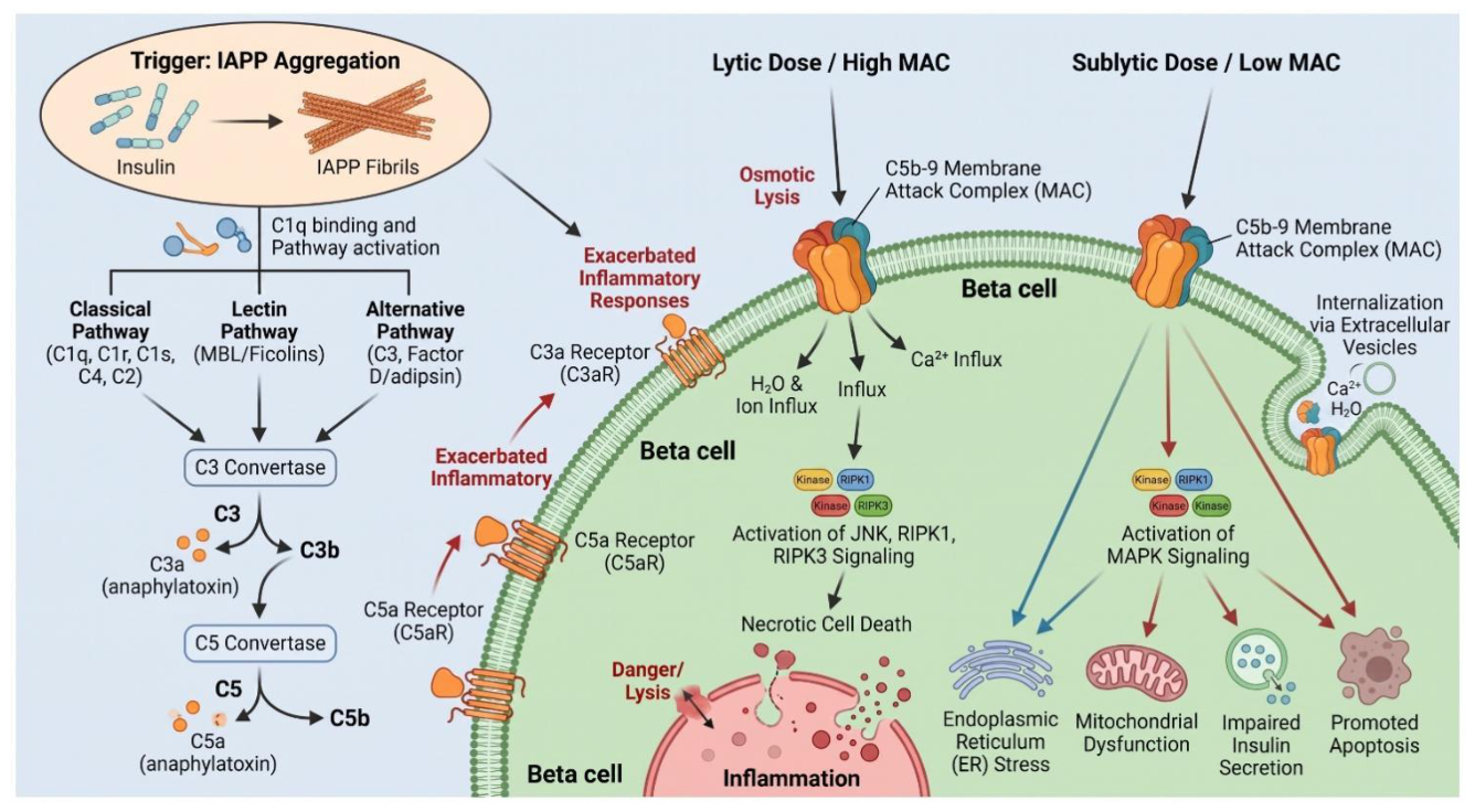
Figure 2.
Complement–metabolic inflammation crosstalk in T2DM and its complications. Metabolic stress promotes systemic and tissue-specific complement activation, which contributes to pancreatic β-cell dysfunction, hepatic metabolic dysregulation, adipose tissue inflammation and insulin resistance, and diabetic kidney injury. Complement-mediated inflammatory signaling also participates in diabetic retinopathy and cardiovascular complications, highlighting potential therapeutic targets including C3, C5, and alternative pathway components.
Figure 2.
Complement–metabolic inflammation crosstalk in T2DM and its complications. Metabolic stress promotes systemic and tissue-specific complement activation, which contributes to pancreatic β-cell dysfunction, hepatic metabolic dysregulation, adipose tissue inflammation and insulin resistance, and diabetic kidney injury. Complement-mediated inflammatory signaling also participates in diabetic retinopathy and cardiovascular complications, highlighting potential therapeutic targets including C3, C5, and alternative pathway components.
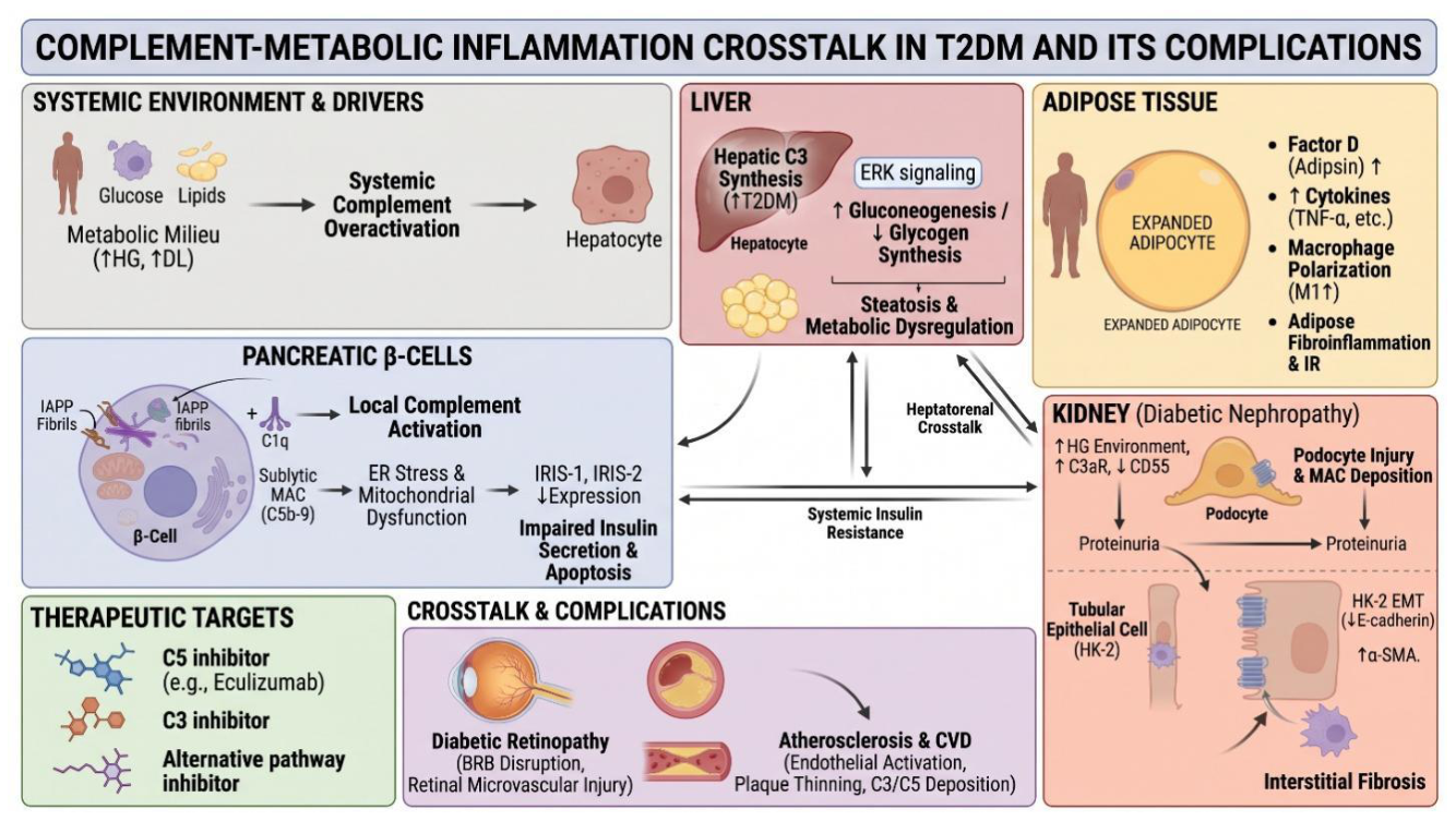
Disclaimer/Publisher’s Note: The statements, opinions and data contained in all publications are solely those of the individual author(s) and contributor(s) and not of MDPI and/or the editor(s). MDPI and/or the editor(s) disclaim responsibility for any injury to people or property resulting from any ideas, methods, instructions or products referred to in the content. |
© 2026 by the authors. Licensee MDPI, Basel, Switzerland. This article is an open access article distributed under the terms and conditions of the Creative Commons Attribution (CC BY) license (http://creativecommons.org/licenses/by/4.0/).
Copyright: This open access article is published under a Creative Commons CC BY 4.0 license, which permit the free download, distribution, and reuse, provided that the author and preprint are cited in any reuse.