

Submitted:
21 September 2026
Posted:
22 September 2026
You are already at the latest version
Abstract
This paper examines the inferential framework used to attribute the positive association between acid-suppressive therapy (AS) and esophageal adenocarcinoma (EAC) to confounding by gastroesophageal reflux disease (GERD), a common indication for AS. The experimental literature has proposed several hypotheses by which AS may increase EAC risk, and reviews of the in vivo literature have suggested that the AS--EAC association may be causal. At the same time, clinical guidelines maintain that the AS--EAC association likely reflects confounding by GERD and cite the findings of decades-old observational studies that either estimate the AS--EAC association among those without reflux-related indications or estimate a common treatment OR after adjusting for the presence of GERD as a main-effect covariate. This paper first examines the causal hypotheses described in the in vivo literature and establishes that the patient's underlying reflux burden is a presupposed effect modifier under each of these hypotheses. It then argues that the epidemiologic findings presented to clinicians as reassuring evidence against causality do not distinguish confounding by GERD from any of these hypotheses.
Keywords:
acid suppression
; gastroesophageal reflux disease
; GERD
; Barrett’s esophagus
; esophageal adenocarcinoma
; proton pump inhibitors
; H2-receptor antagonists
; confounding by indication
; effect modification
; causal inference
1. Introduction
The annual incidence of esophageal adenocarcinoma (EAC) increased in the United States by approximately 767% between 1973 and 2017, the largest growth rate of any epithelial malignancy during that period; melanoma ranked second, at approximately 274% [1]. EAC is thought to arise from chronic exposure of the distal esophagus to refluxed gastric and duodenal contents (e.g. acid, bile), and chronic esophageal reflux (GERD) is an established risk factor in EAC etiology [2]. Approximately 10% of those with reflux symptoms who undergo upper endoscopy are found to have Barrett’s esophagus (BE), the precursor lesion to EAC [3], and 0.1-0.5% of those with BE will progress to EAC in any given year, with a higher progression rate among those with dysplasia [4]. Though trends in obesity are thought to partially explain the increased prevalence of GERD [5], it is generally unclear why certain GERD patients develop BE/EAC while others do not [3].
Symptoms of GERD are commonly treated with acid-suppressive therapy (AS), traditionally through the use of proton pump inhibitors (PPIs) and H2-receptor antagonists (H2RAs) [6]. Despite evidence that acid can damage DNA [7], the experimental literature has proposed several hypotheses by which the use of AS may nevertheless increase the risk of EAC. These hypotheses suggest that by increasing gastric pH, AS modulates the composition [8,9] and solubility [8,10,11,12] of specific reflux constituents implicated in EAC pathogenesis [9,10,11,13], and separately promotes the progression to EAC by altering the signaling in reflux-conditioned tissue [14,15,16,17]. There is moreover a cluster of in vivo studies observing higher rates of EAC, dysplasia, or BE under less acidic conditions [9,10,14,18,19,20], and although the results of these studies are heterogeneous [21,22], reviews of the animal literature have suggested that the use of AS may explain the rise in EAC incidence [11,23].
Observational studies generally report a positive association between EAC and the use of AS [24,25,26,27], but clinical guidelines maintain that this association likely reflects confounding by indication, whereby acidic reflux promotes BE/EAC and the use of AS is a benign marker of risk [28,29]. This interpretation appears to be largely informed by a collection of observational studies that either estimate the AS–EAC association among those without reflux-related indications [27] or estimate a common treatment OR after adjusting for the presence of GERD as a main-effect covariate [25,26,27,30]. In either case, the statistical non-significance of these estimates [25,26,27] has been widely read as affirmative evidence that the AS–EAC association is non-causal [28,29,31], including in current clinical guidelines on the management of GERD [29].
This paper does not argue that AS contributes to EAC risk in human populations but rather that the findings of these observational studies have been interpreted within a framework that is misaligned with the experimental literature. The statistical non-significance of the estimates reported in these studies may be read [28,29,31] as evidence for the absence of a GERD-independent causal effect under a liberal decision rule. However, the hypotheses of the experimental literature do not posit the existence of such an effect: they presuppose that the patient’s underlying reflux burden determines the duration of esophageal exposure to refluxate that is modified by AS, or that it determines the cellular context (e.g. mutational burden, receptor expression) in which the hormonal effects of AS are exerted. In this setting, the findings of these observational studies may materially misrepresent the risk associated with treatment among those with GERD, and they cannot therefore be used to ascribe the AS–EAC association to confounding by GERD.
2. Reflux Burden as an Effect Modifier
In a 2015 review of rat reflux models, Greene et al. [11] conclude that the suppression of gastric acidity in rats appears to “potentiate the development of esophageal adenocarcinoma" [11] and point to two mechanisms articulated in the reviewed studies. The first concerns the pH-dependent toxicity of bile [9,10], a reflux constituent that is suspected to be a central driver of BE/EAC pathogenesis [32,33]: while many bile acids precipitate irreversibly in normal gastric acidity [34,35], weakly acidic to alkaline conditions maintain many bile species in a soluble state from which they can passively diffuse through cell membranes [12,36,37]. The second mechanism concerns the bacterial reduction of nitrate to nitrite, which Fein et al. [9] had speculated to facilitate the formation of carcinogenic N-nitroso compounds.
These hypothesized mechanisms are illustrated in Figure 1. Under these mechanisms, the use of AS alters the carcinogenicity of refluxate, but (treated) reflux burden determines the extent of esophageal exposure to that refluxate. As such, no causal effect exists in the absence of reflux—regardless of how carcinogenic that refluxate would be—and the magnitude of this effect on EAC risk generally increases with (treated) reflux burden. Several other proposed mechanisms can be represented similarly [8,23,38].
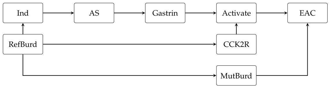
Next, consider the subsequent mouse model of Lee et al. [14], where the authors examine the hypothesis that PPI-induced hypergastrinemia could promote the progression from BE to EAC [16,17]. The use of AS promotes secondary hypergastrinemia [39], and gastrin can function as a pro-oncogenic stimulus [15,16,40]. Separately, reflux-related inflammation damages DNA [41] and appears to enhance tissue responsiveness to gastrin through upregulation of the receptor CCK2R [14,17]. Consistent with an interactive effect, Lee et al. observe that PPI-induced hypergastrinemia promotes the development and dysplastic progression of Barrett’s-like metaplasia in chronically inflamed mouse esophagi, but do not observe any metaplastic alteration in mouse esophagi that lack the inflammatory stimulus [14].
This hypothesized mechanism is illustrated in Figure 2. Under this mechanism, the use of AS promotes hypergastrinemia, but the carcinogenic susceptibility to this pro-oncogenic stimulus depends on the prior accumulation of reflux-related cellular changes, namely CCK2R overexpression and mutational burden. Accordingly, the effect on EAC risk generally increases with (historical) reflux burden, and unless there is a widespread alternative source of esophageal injury that mimics the cellular context produced by chronic reflux—a source that does not appear to exist [11,42]—then virtually no effect on risk manifests in a patient who has not experienced reflux.
3. Conditioning on Reflux Burden
The causal hypotheses proposed in the experimental literature presuppose that the effect of AS on EAC risk would depend integrally on reflux burden, yet the source of the AS–EAC association seems to have been adjudicated within a framework that omits the possibility of any such dependency. In what appears to be considered the single most probative observational study on the source of the AS–EAC association [28,29,31], Garcia Rodriguez et al. [27] make this framework explicit: “Long term pharmacological gastric acid suppression is a marker of increased risk... However these associations are most likely explained by the underlying treatment indications being a risk factor for the cancer rather than an independent harmful effect of these agents per se." Within this framework, it suffices to observe evidence for the absence of an independent causal effect in order to infer the presence of confounding by GERD.
Consider first Garcia Rodriguez et al. [27], in which the authors distinguish long-term (3+ years) AS users with esophageal indications (e.g. reflux symptoms, esophagitis, BE) from long-term users with non-esophageal indications (e.g. peptic ulcer disease, gastritis) and estimate separate ORs for each group computed against a common referent of non-users. The authors observe that the OR is statistically significant for the esophageal group (OR: 5.42; 95% CI: [3.13, 9.39]) and statistically non-significant for the non-esophageal group (OR: 1.74; 95% CI: [0.90, 3.34]), and Spechler et al. [28] present this finding in a technical review for the American Gastroenterological Association:
PPI therapy also has effects that, conceivably, might promote the development of cancer in Barrett’s esophagus. For example, use of PPIs often is associated with an increase in the serum levels of gastrin, a hormone that has been shown to increase proliferation in Barrett’s epithelium... Garcia Rodriguez et al. found that patients who were treated with acid suppression for an `esophageal indication’ such as GERD had a significantly increased risk of developing esophageal adenocarcinoma… In contrast, for patients who were treated with acid suppression for a `gastroduodenal indication’ such as peptic ulcer disease, there was no significantly increased risk of adenocarcinoma... The lack of an association with cancer in patients taking PPIs for gastroduodenal disease suggests that the positive association in the patients with esophageal disease resulted from confounding by indication. In other words, it was likely the GERD, not the GERD treatment, that increased the incidence of cancer.
Spechler et al. appear to recognize what they consider to be evidence for the absence of an independent causal effect and subsequently infer the presence of confounding by GERD. However, this inference does not follow from the premise that the authors themselves articulate: if AS-induced hypergastrinemia promotes the progression from BE to EAC by increasing proliferation in BE [17,40]—a tissue context characterized by heightened responsiveness to gastrin [14,40] and a substantial mutational burden [43]—then one would only expect to observe an effect among those with BE or perhaps the GERD that causes it [3]—both are esophageal indications [27].
More generally, the causal hypotheses articulated in the in vivo literature need not predict any observable effect in patients who lack a pathologically large reflux burden. BE/EAC has long been believed to arise from chronic injury [3,44] such that any causal effect of AS on EAC risk would reasonably exhibit convex or threshold-like behavior. The OR observed for the non-esophageal group is therefore plainly compatible with these hypotheses. Moreover, because the ORs for each group are computed against a common odds baseline and the baseline risk for the esophageal group presumably exceeds that of the non-esophageal group, these causal hypotheses would each predict a larger OR for the esophageal group. We cannot therefore adjudicate the source of the esophageal group’s OR based on the magnitude of that observed for the non-esophageal group.
A similar interpretive constraint applies in the context of main-effect regression adjustment, where observational studies have reported a common reflux-adjusted treatment OR that is estimated across levels of reflux burden [24,25,27,30,45]. Though effect modification is scale-dependent, the plausibility of a convex or threshold-like effect on risk suggests that the predicted treatment OR at a large reflux burden may substantially exceed that which would be predicted at a small reflux burden. Assuming that these ORs are approximately collapsible (EAC is rare [1]), the common adjusted treatment OR would then lie below the treatment OR of some high-reflux stratum.
Consider Chow et al. [25] as an example. The authors report a common reflux-adjusted treatment OR of 1.5 (95% CI: [0.4, 5.4]) while also reporting “no excess risk" among those without documented reflux-related conditions. Both the authors and the AGA technical review described above present these findings as affirmative evidence against causality [28], but this would only be the case if GERD-independent causation were the relevant causal hypothesis. If we take the authors’ words to imply that the treatment OR within this non-predisposed group is below 1.5, it would not by itself weigh against the hypotheses discussed in the in vivo literature, and it would also suggest that the treatment OR in at least some subset of predisposed individuals exceeds 1.5. We are therefore similarly unable to attribute the AS–EAC association to confounding by GERD based on the observed magnitude of the reported treatment OR.
4. Discussion
In this context, confounding and causality need only be distinguishable among those with the largest underlying reflux burdens. While the treatment OR within a high-reflux stratum may therefore have more probative value than that of a low-reflux stratum, its interpretability is still practically constrained by the available proxy for reflux burden. For example, a GERD diagnosis can itself correspond to a wide range of underlying reflux burdens [46] such that a near-null association among those with GERD could mask an effect that is concentrated among those with the most severe underlying disease, while a positive association could still be attributable to confounding by GERD. Self-reported symptom severity can instead be stratified finely [26], but the subjective symptoms of treated patients are inherently influenced by treatment [26,47]. This point is highlighted by Nason et al. [47], where the authors find that, among PPI users, those who reported the least frequent reflux symptoms were at the highest risk of developing BE or EAC. This point may also explain the interaction analysis of Farrow et al. [26] where, among H2RA users, those with the fewest severe reflux episodes per month were at the highest risk of developing EAC.
A distinct but closely related subliterature examines whether PPIs may be chemopreventive among those who have already progressed to BE. This evidence base consists primarily of observational studies that compare PPI users to non-users (including H2RA users) and a randomized controlled trial (AspECT) [48] that compares high and low doses of esomeprazole [49,50]. Whether epidemiologic evidence concerning PPI chemoprevention among those with BE can adjudicate the source of the AS–EAC association observed in the general population requires distinguishing three questions: first, whether the evidence genuinely supports a chemopreventive effect of PPIs among those with BE; second, whether any such protectiveness would generalize to those who have not yet developed BE; and third, whether such evidence bears on the safety or protectiveness of H2RAs.
Regarding the first question, a recent meta-analysis reported an adjusted OR of 0.46 (95% CI: [0.25, 0.86]) for PPI use among those with BE, but it also reported significant heterogeneity () and determined that the results of each study were at risk of either serious or critical bias (ROBINS-I) [49]. Several of the included studies also condition on predictors of reflux burden as main-effect covariates (e.g. hiatal hernia [51,52], esophagitis [53]). While the AspECT trial observed a trend towards superior EAC-related outcomes within the high-dose group [48], this would only necessarily constitute evidence of chemoprevention (and safety) if the safety of the lower dose is established elsewhere or if the PPI dose-risk curve is monotonic. This monotonicity is not guaranteed because the toxicity of bile acids is non-monotonic across the acidic pH spectrum [36,37] and PPIs possess pleiotropic effects [21,54,55,56]. PPI pleiotropy has been discussed within the in vivo literature [11,21] and could plausibly explain why the AspECT trial’s primary endpoint was substantially driven by non-EAC mortality, which was lower in the high-dose group [48].
As for the second question, if PPIs were to exert a protective effect via increased gastric pH [28] or reduced refluxate volume [10], then there would not be an obvious reason as to why this protectiveness would be stage specific. If this protectiveness instead reflected a pleiotropic effect that manifests at a later stage of carcinogenesis [55,56], then protectiveness among those with BE would still be compatible with PPIs contributing to BE pathogenesis and marginal EAC risk. To the third and final question, the protectiveness of PPIs would not speak to the safety of H2RAs insofar as this protective effect could reflect PPI-specific pleiotropy [21,54,55], a reduction in refluxate volume that is not achieved by H2RAs [57], or a more complete and prolonged suppression of gastric acidity [58] that maintains certain bile species in an ionized state [8,36] for a greater duration than do H2RAs.
To conclude, this paper does not argue that AS contributes to EAC in human populations but that the presumption to the contrary may be unwarranted. The in vivo literature establishes the biological plausibility of a causal role of AS in EAC etiology [10,11,14,18], and certain epidemiologic findings that had been thought to adjudicate the causal question do not do so [25,27]. Until the risk associated with untreated reflux has been adequately separated from the treatment that it precipitates, the AS–EAC association should not be dismissed as spurious.
Funding
The author received no funding for this work.
Competing Interests
The author declares no competing interests.
References
- Hang, T.V.P.; Spiritos, Z.; Gamboa, A.M.; Chen, Z.; Force, S.; Patel, V.; Chawla, S.; Keilin, S.; Saba, N.F.; El-Rayes, B.; et al. Epidemiology of Early Esophageal Adenocarcinoma. Clinical Endoscopy 2022, 55, 372–380. [CrossRef]
- Iijima, K. Etiologic factors for Barrett’s esophagus: toward countermeasures in Asia. Expert Review of Gastroenterology & Hepatology 2024, 18, 407–420. [CrossRef]
- Souza, R.F.; Spechler, S.J. Mechanisms and pathophysiology of Barrett oesophagus. Nature Reviews Gastroenterology & Hepatology 2022, 19, 605–620. [CrossRef]
- Kota, L.; Tyberg, A. Risk of Barrett’s esophagus progression to esophageal adenocarcinoma. Clinical Endoscopy 2024, 57, 763–764. [CrossRef]
- Mao, Y.; Zhang, Y.; Hu, L.; Zhang, W.; An, Y.; Zhao, Y.; Tang, X. Association between body mass index at different levels and risk of gastroesophageal reflux disease: a systematic review with dose-response meta-analysis. Frontiers in Physiology 2025, 16, 1675457. [CrossRef]
- Al-Frejat, Z.; Martini, N.; Esper, A.; Al-Frejat, D.; Younes, S.; Hanna, M. GERD: Latest update on acid-suppressant drugs. Current Research in Pharmacology and Drug Discovery 2024, 7, 100198. [CrossRef]
- Jolly, A.J.; Wild, C.P.; Hardie, L.J. Acid and bile salts induce DNA damage in human oesophageal cell lines. Mutagenesis 2004, 19, 319–324. [CrossRef]
- Theisen, J.; Nehra, D.; Citron, D.; Johansson, J.; Hagen, J.A.; Crookes, P.E.; DeMeester, S.R.; Bremner, C.G.; DeMeester, T.R.; Peters, J.H. Suppression of gastric acid secretion in patients with gastroesophageal reflux disease results in gastric bacterial overgrowth and deconjugation of bile acids. Journal of Gastrointestinal Surgery 2000, 4, 50–54. [CrossRef]
- Fein, M.; Peters, J.H.; Chandrasoma, P.; Ireland, A.P.; Oberg, S.; Ritter, M.P.; Bremner, C.G.; Hagen, J.A.; DeMeester, T.R. Duodenoesophageal reflux induces esophageal adenocarcinoma without exogenous carcinogen. Journal of Gastrointestinal Surgery 1998, 2, 260–268. [CrossRef]
- Nasr, A.O.; Dillon, M.F.; Conlon, S.; Downey, P.; Chen, G.; Ireland, A.; Leen, E.; Bouchier-Hayes, D.; Walsh, T.N. Acid suppression increases rates of Barrett’s esophagus and esophageal injury in the presence of duodenal reflux. Surgery 2012, 151, 382–390. [CrossRef]
- Greene, C.L.; Worrell, S.G.; DeMeester, T.R. Rat Reflux Model of Esophageal Cancer and Its Implication in Human Disease. Annals of Surgery 2015, 262, 910–924. [CrossRef]
- Kauer, W.K.H.; Stein, H.J. Emerging concepts of bile reflux in the constellation of gastroesophageal reflux disease. Journal of Gastrointestinal Surgery 2010, 14, S9–S16. [CrossRef]
- Fujimura, T.; Oyama, K.; Sasaki, S.; Nishijima, K.; Miyashita, T.; Ohta, T.; Miwa, K.; Hattori, T. Inflammation-Related Carcinogenesis and Prevention in Esophageal Adenocarcinoma Using Rat Duodenoesophageal Reflux Models. Cancers 2011, 3, 3206–3224. [CrossRef]
- Lee, Y.; Urbanska, A.M.; Hayakawa, Y.; Wang, H.; Au, A.S.; Luna, A.M.; Chang, W.; Jin, G.; Bhagat, G.; Abrams, J.A.; et al. Gastrin stimulates a cholecystokinin-2-receptor-expressing cardia progenitor cell and promotes progression of Barrett’s-like esophagus. Oncotarget 2017, 8, 203–214. [CrossRef]
- Harris, J.C.; Clarke, P.A.; Awan, A.; Jankowski, J.; Watson, S.A. An antiapoptotic role for gastrin and the gastrin/CCK-2 receptor in Barrett’s esophagus. Cancer Research 2004, 64, 1915–1919. [CrossRef]
- Haigh, C.R.; Attwood, S.E.A.; Thompson, D.G.; Jankowski, J.A.; Kirton, C.M.; Pritchard, D.M.; Varro, A.; Dimaline, R. Gastrin Induces Proliferation in Barrett’s Metaplasia Through Activation of the CCK2 Receptor. Gastroenterology 2003, 124, 615–625. [CrossRef]
- Quante, M.; Abrams, J.A.; Lee, Y.; Wang, T.C. Barrett esophagus: What a mouse model can teach us about human disease. Cell Cycle 2012, 11, 4328–4338. [CrossRef]
- Dall’Olmo, L.; Fassan, M.; Dassie, E.; Scarpa, M.; Realdon, S.; Cavallin, F.; Cagol, M.; Battaglia, G.; Pizzi, M.; Guzzardo, V.; et al. Role of Proton Pump Inhibitor on Esophageal Carcinogenesis and Pancreatic Acinar Cell Metaplasia Development: An Experimental In Vivo Study. PLOS ONE 2014, 9, e112862. [CrossRef]
- Ireland, A.P.; Peters, J.H.; Smyrk, T.C.; DeMeester, T.R.; Clark, G.W.B.; Mirvish, S.S.; Adrian, T.E. Gastric Juice Protects Against the Development of Esophageal Adenocarcinoma in the Rat. Annals of Surgery 1996, 224, 358–371. [CrossRef]
- Cheng, P.; Li, J.S.; Gong, J.; Zhang, L.F.; Chen, R.Z. Effects of refluxate pH values on duodenogastroesophageal reflux-induced esophageal adenocarcinoma. World Journal of Gastroenterology 2011, 17, 3060–3065. [CrossRef]
- Miyashita, T.; Shah, F.A.; Marti, G.P.; Wang, J.; Bonde, P.; Gibson, M.K.; Ohta, T.; Montgomery, E.A.; Duncan, M.; Harmon, J.W. Rabeprazole impedes the development of reflux-induced esophageal cancer in a surgical rat model. Digestive Diseases and Sciences 2011, 56, 1309–1314. [CrossRef]
- Majka, J.; Wierdak, M.; Szlachcic, A.; Magierowski, M.; Targosz, A.; Urbanczyk, K.; Krzysiek-Maczka, G.; Ptak-Belowska, A.; Bakalarz, D.; Magierowska, K.; et al. Interaction of epidermal growth factor with COX-2 products and peroxisome proliferator-activated receptor-γ system in experimental rat Barrett’s esophagus. American Journal of Physiology-Gastrointestinal and Liver Physiology 2020, 318, G375–G389. [CrossRef]
- Mirvish, S.S. Studies on experimental animals involving surgical procedures and/or nitrosamine treatment related to the etiology of esophageal adenocarcinoma. Cancer Letters 1997, 117, 161–174. [CrossRef]
- Lagergren, J.; Bergström, R.; Lindgren, A.; Nyrén, O. Symptomatic gastroesophageal reflux as a risk factor for esophageal adenocarcinoma. New England Journal of Medicine 1999, 340, 825–831.
- Chow, W.H.; Finkle, W.D.; McLaughlin, J.K.; Frankl, H.; Ziel, H.K.; Fraumeni, Jr., J.F. The Relation of Gastroesophageal Reflux Disease and Its Treatment to Adenocarcinomas of the Esophagus and Gastric Cardia. JAMA 1995, 274, 474–477.
- Farrow, D.C.; Vaughan, T.L.; Sweeney, C.; Gammon, M.D.; Chow, W.H.; Risch, H.A.; Stanford, J.L.; Hansten, P.D.; Mayne, S.T.; Schoenberg, J.B.; et al. Gastroesophageal reflux disease, use of H2 receptor antagonists, and risk of esophageal and gastric cancer. Cancer Causes & Control 2000, 11, 231–238. [CrossRef]
- García Rodríguez, L.A.; Lagergren, J.; Lindblad, M. Gastric acid suppression and risk of oesophageal and gastric adenocarcinoma: a nested case control study in the UK. Gut 2006, 55, 1538–1544. [CrossRef]
- Spechler, S.J.; Sharma, P.; Souza, R.F.; Inadomi, J.M.; Shaheen, N.J.; Association, A.G. American Gastroenterological Association technical review on the management of Barrett’s esophagus. Gastroenterology 2011, 140, e18–e52. [CrossRef]
- Katz, P.O.; Dunbar, K.B.; Schnoll-Sussman, F.H.; Greer, K.B.; Yadlapati, R.; Spechler, S.J. ACG Clinical Guideline for the Diagnosis and Management of Gastroesophageal Reflux Disease. The American Journal of Gastroenterology 2022, 117, 27–56. [CrossRef]
- Duan, L.; Wu, A.H.; Sullivan-Halley, J.; Bernstein, L. Antacid drug use and risk of esophageal and gastric adenocarcinomas in Los Angeles County. Cancer Epidemiology, Biomarkers & Prevention 2009, 18, 526–533. [CrossRef]
- Dunbar, K.B.; Spechler, S.J. The Effect of Proton Pump Inhibitors on Barrett’s Esophagus. Gastroenterology Clinics of North America 2015, 44, 415–424. [CrossRef]
- Hu, Y.; Ye, X.; Wang, R.; Poon, K. Current research progress in the role of reactive oxygen species in esophageal adenocarcinoma. Translational Cancer Research 2021, 10, 1568–1577. [CrossRef]
- Iwaya, Y.; Goda, K.; Kako, S.; Hattori, H.; Miyazawa, T.; Hara, D.; Hashigami, K.; Hirayama, A.; Okamura, T.; Nagaya, T.; et al. Association between endoscopic evidence of bile reflux and Barrett’s esophagus: A large-scale case-control study. Digestive and Liver Disease 2024. [CrossRef]
- Barthlen, W.; Liebermann-Meffert, D.; Feussner, H.; Stein, H.J. Influence of pH on bile acid concentration in human, pig and commercial bile: Implications for `alkaline’ gastro-esophageal reflux. Diseases of the Esophagus 1994, 7, 127–130. [CrossRef]
- Armstrong, D.; Bennett, J.R.; Blum, A.L.; Dent, J.; De Dombal, F.T.; Galmiche, J.P.; Lundell, L.; Margulies, M.; Richter, J.E.; Spechler, S.J. Gastric mucosal toxicity of duodenal juice constituents in the rat. Digestive Diseases and Sciences 1994. [CrossRef]
- Bus, P.; Siersema, P.D.; van Baal, J.W.P.M. Cell culture models for studying the development of Barrett’s esophagus: a systematic review. Cellular Oncology 2012, 35, 149–161. [CrossRef]
- Nehra, D.; Howell, P.; Williams, C.P.; Pye, J.K.; Beynon, J. Toxic bile acids in gastro-oesophageal reflux disease: influence of gastric acidity. Gut 1999, 44, 598–602. [CrossRef]
- Feagins, L.A.; Zhang, H.Y.; Hormi-Carver, K.; Quinones, M.H.; Thomas, T.; Zhang, X.; Terada, L.S.; Spechler, S.J.; Souza, R.F. Acid has antiproliferative effects in nonneoplastic Barrett’s epithelial cells. The American Journal of Gastroenterology 2007, 102, 10–20. [CrossRef]
- Dacha, S.; Razvi, M.; Massaad, J.; Cai, Q.; Wehbi, M. Hypergastrinemia. Gastroenterology Report 2015, 3, 201–208. [CrossRef]
- Abdalla, S.I.; Lao-Sirieix, P.; Novelli, M.R.; Lovat, L.B.; Sanderson, I.R.; Fitzgerald, R.C. Gastrin-induced cyclooxygenase-2 expression in Barrett’s carcinogenesis. Clinical Cancer Research 2004, 10, 4784–4792. [CrossRef]
- Gokulan, R.C.; Garcia-Buitrago, M.T.; Zaika, A.I. From Genetics to Signaling Pathways: Molecular Pathogenesis of Esophageal Adenocarcinoma. Biochimica et Biophysica Acta (BBA) - Reviews on Cancer 2019, 1872, 37–48. [CrossRef]
- Zamani, S.A.; Wu, L.; Black, E.L.; Bartram, A.; Ng, A.W.T.; Secrier, M.; Perelman, J.D.; Ustaoglu, A.; Ococks, E.; Jacobson, D.; et al. Integrated epidemiological and molecular data inform the relationship between precancer and cancer states of esophageal adenocarcinoma. Nature Medicine 2026, 32, 1805–1816. [CrossRef]
- Gregson, E.M.; Bornschein, J.; Fitzgerald, R.C. Genetic progression of Barrett’s oesophagus to oesophageal adenocarcinoma. British Journal of Cancer 2016, 115, 403–410. [CrossRef]
- Stein, H.J.; Siewert, J.R. Barrett’s Esophagus: Pathogenesis, Epidemiology, Functional Abnormalities, Malignant Degeneration, and Surgical Management. Dysphagia 1993, 8, 276–288. [CrossRef]
- Arai, J.; Niikura, R.; Hayakawa, Y.; Kawahara, T.; Honda, T.; Hasatani, K.; Yoshida, N.; Nishida, T.; Sumiyoshi, T.; Kiyotoki, S.; et al. Chemoprevention of Oesophageal Squamous-Cell Carcinoma and Adenocarcinoma: A Multicentre Retrospective Cohort Study. Digestion 2022, 103, 192–204. [CrossRef]
- Horton, A.J.; Clayton, S.B. Optimization of impedance-measured reflux events in GORD utilizing acid exposure time. BMC Gastroenterology 2020, 20, 179. [CrossRef]
- Nason, K.S.; Wichienkuer, P.P.; Awais, O.; Schuchert, M.J.; Luketich, J.D.; O’Rourke, R.W.; Hunter, J.G.; Morris, C.D.; Jobe, B.A. Gastroesophageal Reflux Disease Symptom Severity, Proton Pump Inhibitor Use, and Esophageal Carcinogenesis. Archives of Surgery 2011, 146, 851–858. [CrossRef]
- Jankowski, J.A.Z.; de Caestecker, J.; Love, S.B.; Reilly, G.; Watson, P.; Sanders, S.; Ang, Y.; Morris, D.; Bhandari, P.; Brooks, C.; et al. Esomeprazole and aspirin in Barrett’s oesophagus (AspECT): a randomised factorial trial. Lancet 2018, 392, 400–408. [CrossRef]
- Ko, M.T.; Rizwanullah, A.; Jafri, Z.; Fung, A.; Alexandre, L. Chemoprevention of Barrett’s Esophagus: a Systematic Review and Comprehensive Assessment of Bias. Diseases of the Esophagus 2025, 38, doaf062. [CrossRef]
- Hu, Q.; Sun, T.T.; Hong, J.; Fang, J.Y.; Xiong, H.; Meltzer, S.J. Proton Pump Inhibitors Do Not Reduce the Risk of Esophageal Adenocarcinoma in Patients with Barrett’s Esophagus: A Systematic Review and Meta-Analysis. PLOS ONE 2017, 12, e0169691. [CrossRef]
- Krishnamoorthi, R.; Borah, B.; Heien, H.; Das, A.; Chak, A.; Iyer, P.G. Rates and predictors of progression to esophageal carcinoma in a large population-based Barrett’s esophagus cohort. Gastrointestinal Endoscopy 2016, 84, 40–46.e7. [CrossRef]
- Thota, P.N.; Hajifathalian, K.; Benjamin, T.; Runkana, A.; Lopez, R.; Sanaka, M.R. Lack of incremental effect of histamine receptor antagonists over proton pump inhibitors on the risk of neoplastic progression in patients with Barrett’s esophagus: a cohort study. Journal of Digestive Diseases 2017, 18, 143–150. [CrossRef]
- Kastelein, F.; Spaander, M.C.W.; Steyerberg, E.W.; Biermann, K.; Valkhoff, V.E.; Kuipers, E.J.; Bruno, M.J. Proton Pump Inhibitors Reduce the Risk of Neoplastic Progression in Patients With Barrett’s Esophagus. Clinical Gastroenterology and Hepatology 2013, 11, 382–388. [CrossRef]
- Huo, X.; et al. In oesophageal squamous cells exposed to acidic bile salt medium, omeprazole inhibits IL-8 expression through effects on NF-κB and AP-1 that are independent of acid inhibition. Gut 2014, 63, 1042–1050. [CrossRef]
- Miyashita, T.; Shah, F.A.; Harmon, J.W.; Marti, G.P.; Matsui, D.; Okamoto, K.; Makino, I.; Hayashi, H.; Oyama, K.; Nakagawara, H.; et al. Do proton pump inhibitors protect against cancer progression in GERD? Surgery Today 2013, 43, 831–837. [CrossRef]
- Chueca, E.; Apostolova, N.; Esplugues, J.V.; García-González, M.A.; Lanas, Á.; Piazuelo, E. Proton Pump Inhibitors Display Antitumor Effects in Barrett’s Adenocarcinoma Cells. Frontiers in Pharmacology 2016, 7, 452. [CrossRef]
- Navas, E.; Luis-Navarro, J.C. Utility of premedication with different regimens of omeprazole and ranitidine in the prevention of acid broncho-aspiration syndrome. Revista Espanola de Anestesiologia y Reanimacion 1992, 39, 337–340.
- Abrahami, D.; McDonald, E.G.; Schnitzer, M.; Azoulay, L. Trends in acid suppressant drug prescriptions in primary care in the UK: a population-based cross-sectional study. BMJ Open 2020, 10, e041529. [CrossRef]
Figure 1.
Directed acyclic graph of refluxate-carcinogenicity hypotheses. Abbreviations: Ind, treatment indication; AS, acid suppression; RefCarc, refluxate carcinogenicity; EsoExp, esophageal exposure to refluxate; EAC, esophageal adenocarcinoma; RefBurd, reflux burden.
Figure 1.
Directed acyclic graph of refluxate-carcinogenicity hypotheses. Abbreviations: Ind, treatment indication; AS, acid suppression; RefCarc, refluxate carcinogenicity; EsoExp, esophageal exposure to refluxate; EAC, esophageal adenocarcinoma; RefBurd, reflux burden.

Figure 2.
Directed acyclic graph of hypergastrinemia hypothesis. Abbreviations: Ind, treatment indication; AS, acid suppression; Gastrin, serum gastrin concentration; Activate, CCK2R activation; EAC, oesophageal adenocarcinoma; RefBurd, reflux burden; CCK2R, CCK2R expression; MutBurd, mutational burden.
Figure 2.
Directed acyclic graph of hypergastrinemia hypothesis. Abbreviations: Ind, treatment indication; AS, acid suppression; Gastrin, serum gastrin concentration; Activate, CCK2R activation; EAC, oesophageal adenocarcinoma; RefBurd, reflux burden; CCK2R, CCK2R expression; MutBurd, mutational burden.
Disclaimer/Publisher’s Note: The statements, opinions and data contained in all publications are solely those of the individual author(s) and contributor(s) and not of MDPI and/or the editor(s). MDPI and/or the editor(s) disclaim responsibility for any injury to people or property resulting from any ideas, methods, instructions or products referred to in the content. |
© 2026 by the authors. Licensee MDPI, Basel, Switzerland. This article is an open access article distributed under the terms and conditions of the Creative Commons Attribution (CC BY) license (http://creativecommons.org/licenses/by/4.0/).
Copyright: This open access article is published under a Creative Commons CC BY 4.0 license, which permit the free download, distribution, and reuse, provided that the author and preprint are cited in any reuse.