
Submitted:
17 September 2026
Posted:
18 September 2026
You are already at the latest version
Abstract
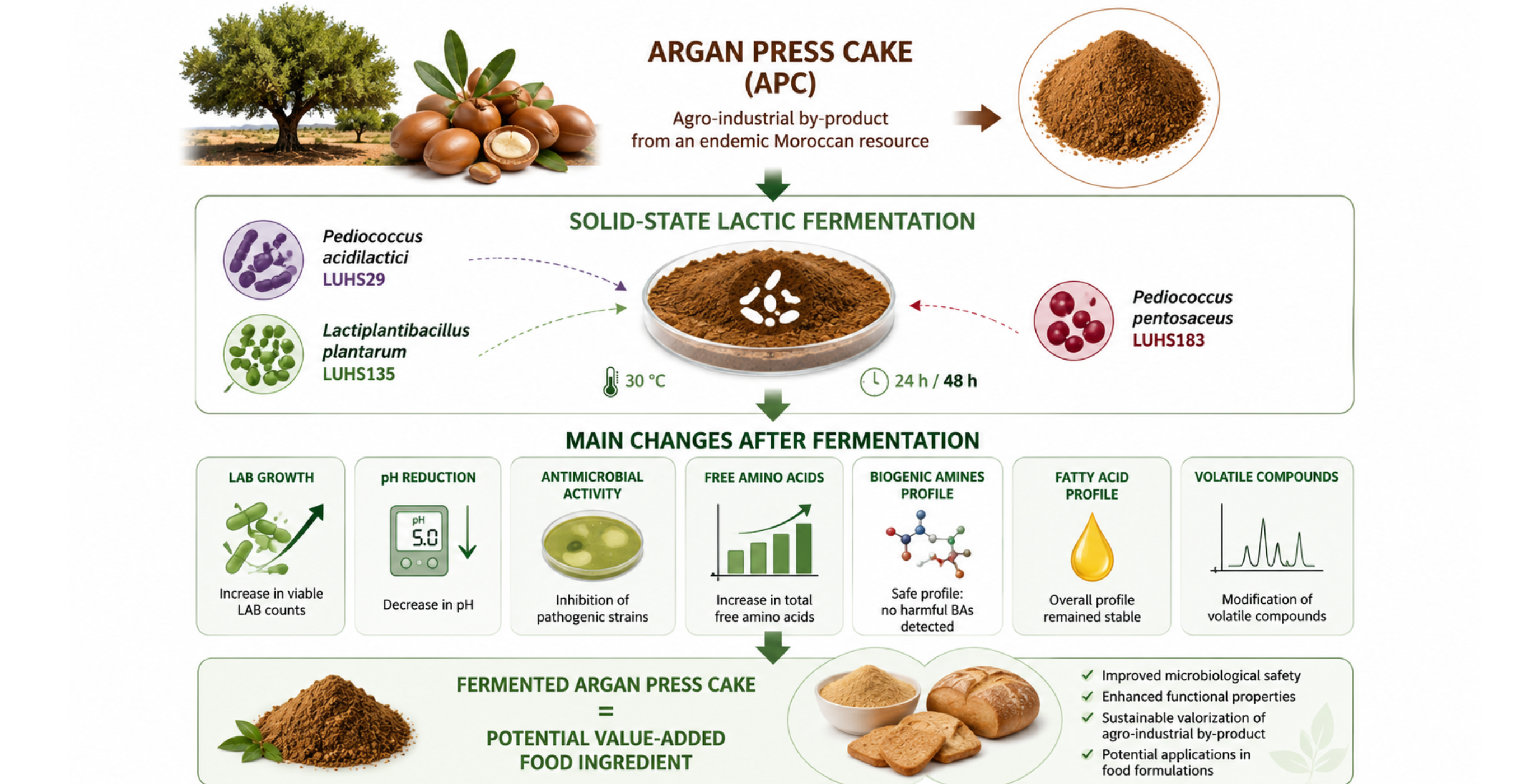
Argan oil production generates large amounts of press cake that remain underexploited despite its potential as a functional ingredient. The objective of this study therefore aimed to evaluate the effects of solid-state fermentation (24h and 48h at 30 °C) using three strains of lactic acid bacteria (LAB)—Pediococcus pentosaceus, Pediococcus acidilactici, and Lactiplantibacillus plantarum on the functional properties of argan press cake (APC). The effects of fermentation were assessed by determining pH, LAB viable counts, free amino acid (FAA) profiles (including γ-aminobutyric acid), biogenic amine (BA) concentrations, fatty acid (FA) and volatile compound (VC) profiles. Additionally, the antimicrobial activity were evaluated against 15 pathogenic strains. The results showed growth of LAB, followed by a decrease in pH, which correlated with antimicrobial activity against pathogens in the fermented samples. As for free amino acids, their bioavailability increased in the fermented samples. Only two biogenic amines namely spermidine (29.4 ± 2.29 mg/kg) and spermine (29.7 ± 3.66 mg/kg) were identified in all argan cake samples. Furthermore, no changes were observed in the fatty acid profile. Overall, solid-state fermentation improved the biochemical and antimicrobial properties of argan press cake without compromising its lipid quality or safety.
Keywords:
argan press cake
; fermentation
; antimicrobial activity
; amino acids
; biogenic amines
; fatty acids
; volatile compounds
1. Introduction
Argania spinosa, known locally as argan in Morocco, is an endemic species of the Sapotaceae family occupying approximately 800,000 ha [1]. Its economic importance is mainly attributed to the oil extracted from its kernels, which is widely recognized for its nutritional, therapeutic, and cosmetic properties [2,3,4,5]. On average, from 100 kg of dried fruits, approximately 6 kg of kernels are obtained, producing about 3 L of argan oil, while the remaining biomass consists mainly of pulp, nutshells and press cake [6]. However, argan oil production generates significant amount of byproducts, including fruit pulp, nutshells, and argan press cake (APC). Among these by-products, APC represents one of the major residues generated after oil extraction and remains largely underutilized despite its nutritional potential. Locally, it is commonly used as animal feed due to its high fiber and protein content. APC contains significant amounts of protein, fiber, residual oil, and bioactive compounds, which make it a valuable resource for value-added food applications. However, the APC also contains significant levels of antinutritional factors, particularly saponins, which may limit its nutritional value. Consequently, it is necessary to implement strategies aimed at improving the functional characteristics of APC while reducing limiting factors in order to facilitate its incorporation into food products.
Solid-state fermentation (SSF) is a biotechnological process carried out on moist solid substrates with little or no free water, in which microorganisms grow directly on the substrate and modify its biochemical composition. During solid-state fermentation, the substrate is almost totally free of liquid, and must enable both the survival and development of micro-organisms [7]. This is a process that can improve some biological properties, such as antioxidant capacity, digestibility/bioavailability, antimicrobial activity and antinutrient reduction [8,9,10,11]. Compared to fermentation in liquid phase, it is more economically effective and more sustainable (lower energy use, reduced water consumption, minimal wastewater) [12]. Fermentation by LAB is increasingly applied due to their ability to improve the nutritional and antimicrobial properties of products [13]. In addition, the use of LABs constitutes both an efficient and sustainable process for producing bioactive substances [10]. Our previous studies showed that Pediococcus pentosaceus (LUSH183), Pediococcus acidilactici (LUSH29), and Lactiplantibacillus plantarum (LUSH135) possess desirable antimicrobial activity against a variety of pathogenic and opportunistic strains [14]. Therefore, these strains were selected as potential starter cultures for APC fermentation.
Value-added techniques and the chemical composition of APC have been reported in several studies, highlighting its high content of protein, carbohydrates, residual oil, and bioactive compounds [6,15]. Furthermore, argan cake has been the subject of studies to assess its potential applications in animal feed, residual oil extraction, and the recovery of valuable bioactive compounds [16,17]. Despite its nutritional potential, studies on the biotransformation of APC via lactic acid fermentation remain very limited. Previous research has primarily focused on the influence of fermentation on antioxidant properties [18], while information regarding the effects of LAB on antimicrobial activity, free amino acid composition, γ-aminobutyric acid production, the formation of biogenic amines, the fatty acid profile, and volatile compounds in APC remains limited. To our knowledge, no previous study has comprehensively evaluated the effects of lactic acid fermentation on the antimicrobial activity, free amino acid profile, biogenic amine content, and volatile compounds profile of APC.
Therefore, this study aimed to evaluate the influence of solid-state lactic acid fermentation with P. pentosaceus (LUSH183), P. acidilactici (LUSH29), and Lb. plantarum (LUSH135) on the antimicrobial and physicochemical properties of APC. The antimicrobial properties of fermented APC against a panel of opportunistic pathogenic microorganisms were tested. In addition, the changes in pH, lactic acid bacteria (LAB) viable counts, free amino acid (FAA) profiles, γ-aminobutyric acid (GABA), biogenic amine (BA), fatty acid (FA), and volatile compound (VC) profiles were investigated to evaluate the impact of fermentation on the functional characteristics and valorization potential of APC for food applications.
2. Materials and Methods
2.1. Materials Used for Experiment and Solid-State Fermentation Conditions
Argan fruits were collected in the Sous Massa region of Agadir (Morocco), and then stored in a dark place. After collection, they were dried and the pulp removed. The nut was then broken by hand to release the kernels. The kernels were then mechanically pressed using an automatic cold-oil press (model T91, China). In this study, the freshly produced APC was obtained from the cooperative, stored and protected from light, and used for the fermentation experiments within one week of collection.
The LAB strains P. pentosaceus, P. acidilactici, Lp. Plantarum were acquired from the Lithuanian University of Health Sciences collection (LSMU, Kaunas, Lithuania). These strains were isolated from a spontaneous rye sourdough starter and identified by sequencing the 16S rRNA gene. They are part of an internal collection at LSMU. These strains were selected according to their inhibitory properties against pathogenic and opportunistic bacterial strains [19,20,21]. Before the experiment, LAB strains were incubated and multiplied in De Man, Rogosa, and Sharpe (MRS) broth culture medium (Biolife, Milano, Italy) at 30 ºC under anaerobic conditions for 24 h and 48 h. A total of 3 ml of LAB inoculum cultured on MRS broth (average cell concentration of 8.6 log10 CFU/ml) were inoculated into 100 g of argan press cake (APC-to-water ratio of 2.22:1, w/w) in sterilized glass jars. The jars were then immediately sealed. The APC samples were fermented in a non-agitated incubation chamber (Memmert GmbH Co. KG, Schwabach, Germany) for 24 h and 48 h at 30 °C under anaerobic conditions. The control sample (unfermented APC) mixed with water (APC-to-water ratio of 2.22:1, w/w). The experimental design included seven APC samples: one control and six solid-state fermented samples obtained after 24 or 48 h of fermentation using Pediococcus pentosaceus, Pediococcus acidilactici, and Lactiplantibacillus plantarum.
The antimicrobial properties of control and fermented samples of APC were tested against 15 pathogenic opportunistic strains (Klebsiella pneumoniae LT001, Salmonella enterica 24SPn06, Pseudomonas aeruginosa 17-331, Acinetobacter baumannii 17-380, Proteus mirabilis LT006, Methicillin-resistant Staphylococcus aureus (MRSA) FOX96, Enterococcus faecalis 86, Enterococcus faecium 103, Bacillus cereus 18 01, Streptococcus mutans D04, Enterobacter cloacae LT003, Citrobacter freundii LT008, Staphylococcus epidermidis 24-150, Staphylococcus haemolyticus 17-15, Pasteurella multocida 17) identified using MALDI-TOF mass spectrometry in combination with biochemical methods. These strains were field isolates previously collected from clinical samples of diseased domestic animals and obtained from the microorganism collection of the Institute of Microbiology and Virology at the Lithuanian University of Health Sciences (Kaunas, Lithuania). Likewise, the changes in pH, LAB viable counts, free amino acid (FAA) profiles, γ-aminobutyric acid (GABA), biogenic amine (BA), fatty acid (FA), and volatile compound (VC) profiles in APC were assessed.
2.2. Evaluation of Antimicrobial Activity of APC Against Pathogenic Opportunistic Strains
The pathogenic and opportunistic strains were acquired from the Lithuanian University of Health Sciences collection (Kaunas, Lithuania). An agar well diffusion assay was used for testing the antimicrobial activity of APC. For this purpose, 0.5 McFarland turbidity suspension of each pathogenic opportunistic strain was inoculated onto the surface of cooled Mueller Hinton Agar (Oxoid, UK) using sterile cotton swabs. Wells with a 6 mm diameter were punched in the agar and filled with the fermented APC. The antimicrobial activity against the tested pathogenic and opportunistic strains was determined by measuring the DIZ (mm). The experiments were repeated three times, and the average value of DIZ (mm) was calculated.
2.3. Microbiological Analyses of APC Samples
The assessment of microbiological parameters in non-fermented and solid-state fermented by-product samples encompasses the quantification of LAB (Lactobacillus) counts, TBC (Total Bacteria Count), and M/Y (Total Molds/Yeast) counts. The LAB counts were determined on MRS agar with Tween-80 (Biolife, Milano, Italy); TBC was determined on plate count agar (Biolife, Milan, Italy); M/Y counts were determined on Dichloran rose Bengal chloramphenicol agar (Liofilchem, Milan, Italy). For LAB counts determination ISO 15214:1998 [22], for TBC assessment ISO 4833-2 [23] and for M/Y evaluation ISO 21527-2 [24] method was used.
2.4. Analysis of Free Amino Acid Profile and Gamma-Aminobutyric Acid Content
Sample preparation was performed according to Kaspar, et al. [25] method with some modifications. Concentrations of free amino acids (FAA) were determined using GCMS-QP2010 (Shimadzu, Japan) gas chromatograph with a mass spectrometer. Fermented and unfermented APC samples were stored at −20 °C until analysis. Concentration of individual analytes were determined using calibration curve. Calibration curves were prepared for each analyte using derived standard solutions. Norleucine was used as an internal standard for quantification. The samples were prepared by extracting approximately 1 g of APC with 10 mL of 0.1 M HCl for 60 minutes using an orbital shaker. The acid extract was then separated by centrifugation at 4,000 rpm for 10 minutes and subsequently used for derivatization and analysis. To 100 µl of the sample solution 50 µl of internal standard (~500 µM of norleucine), 150 µl of 0.1 M HCl, 40 µl of 2M NaOH, 200 µl of methanol – pyridine mixture (MeOH:Pyridine – 4:1) and 500 µl of chloroform were added. FAA and GABA, present in the acidic extract, were derivatized before GC-MS analysis. Derivatization were performed using 50 µl of isobutylchloroformate, according to method of Matsumura, et al. [26]. The mixture was centrifuged at 13,2000 rpm and organic layer was dried with anhydrous sodium sulfate before the analysis. Capillary Rxi®-5MS column (Restek, USA) (length 30 m, coating thickness 0.25 µm, the inner diameter 0.25 mm) was used for the analysis. Mass spectrometer operated in single-ion monitoring mode. Analyte was injected in splitless mode. The following parameters were used: MS ion source temperature: 220 °C, MS interface temperature 300 °C, helium (carrier gas) flow: 0,99 ml/min, injector: 250 °C, oven temperature 100 °C (0.5 min), 10 °C / min to 310 °C (4 min).
2.5. Biogenic Amines Evaluation of APC
Biogenic amines were extracted, derivatized, and quantified using the method described by Ben-Gigirey, et al. [27]. Briefly, biogenic amine (BA) standard solutions were prepared by dissolving accurately weighed amounts of each BA, including the internal standard (1,7-diaminoheptane), in 20 mL of deionized water. For sample extraction, 250 μL of the internal standard solution and 10 mL of 0.4 mol/L perchloric acid were added to 5 g of the sample. The mixture was homogenized and centrifuged at 4,000 rpm for 10 min. The extraction procedure was repeated twice, and the combined supernatants were adjusted to a final volume of 25 mL with 0.4 mol/L perchloric acid. Derivatization of both samples supernatants and BA standard solutions was performed using dansyl chloride (10 mg/mL in acetonitrile). Briefly, 0.5 mL of the sample supernatant was mixed with 100 μL of 2 mol/L sodium hydroxide and 150 μL of saturated sodium bicarbonate solution. After vortex mixing, 1 mL of the dansyl chloride solution was added, and the reaction mixture was incubated at 40 °C for 45 min. Excess dansyl chloride was neutralized by adding 50 μL of 25% ammonium hydroxide, followed by incubation at room temperature for 30 min. Subsequently, 3.2 mL of a 1:1 (v/v) mixture of 0.1 mol/L ammonium acetate and acetonitrile was added, and the solution was thoroughly mixed. The derivatized samples were filtered through 0.45 μm syringe filters (Q-Max RR, Frisenette, Denmark) and transferred to chromatographic vials for analysis. The chromatographic analyses were carried out using a Varian ProStar HPLC system (Varian Corp., Palo Alto, California, USA) controlled with Galaxy software (Agilent, Santa Clara, California, USA). For the separation of BA, a Discovery ® HS C18 column (150 × 4.6 mm, 5 μm; SupelcoTM Analytical, Bellefonte, Pennsylvania, USA) was used. The eluents were ammonium acetate (A) and acetonitrile (B), and the elution program consisted of a gradient system with a 0.8 ml/min flow rate. The detection wavelength was set to 254 nm, the oven temperature was 40 °C, and samples were injected in 20 μl aliquots. The target compounds were identified based on their retention times in comparison to their corresponding standards. Each BA concentration was determined using a calibration curve prepared from BA standards, and results were expressed as mg/kg. The limit of quantification (LOQ) was 2.5 mg/kg.
2.6. Analysis of Argan Press Cake Fatty Acid Profile
The protocol used to perform FA profile analysis was described by Pérez-Palacios, et al. [28] with some modifications. Fatty acid (FA) composition of the samples were determined using GCMS-QP2010 (Shimadzu, Japan) gas chromatograph with a mass spectrometer. Concentrations of Fatty Acid Methyl Esters (FAME) were determined by internal standardization using analyte-specific calibration curves, with heptadecanoic acid (C17:0) as the internal standard. The sample was prepared by extracting 1 g of the homogeneous sample with 10 mL of extraction solution (chloroform: methanol = 2:1, containing 400 mg/L of internal standard heptadecanoic acid) under agitation for 1 h. Afterwards, the mixture was centrifuged at 4000 rpm for 5 minutes, and 5 mL of organic phase were washed with 1 mL of 0.9% aqueous sodium chloride solution. The resulting mixture was centrifuged at 4000 rpm for 5 minutes, and 0.1 mL of the organic phase was evaporated using nitrogen gas. The resulting residue was reacted with 300 µl of methylation reagent (6% H2SO4 in methanol) in a laboratory oven at 80 °C for 60 minutes. The analytes were extracted with 1 mL of hexane, centrifuged at 13,2000 rpm, and the upper supernatant was used for the analysis. The capillary column was an SP-2560, 100 m, 0.25 mm i.d., 0.20 µm film thickness. Mass spectrometer operated at single ion scan mode. Analyte was injected in split mode at 1:10 split ratio. The following parameters were used: MS ion source temperature: 240 °C, MS interface temperature 240 °C, helium (carrier gas) flow: 2 ml/min, injector: 250 °C, oven temperature 100 °C (4 min), 12 °C / min to 200 °C (10 min hold), 5 °C / min to 250 °C (6 min hold). The identification of FAMEs was based on retention times and characteristic ion ratios.
2.7. Analysis of Argan Press Cake Volatile Compound Profile
The volatile compounds (VC) of samples were analysed by gas chromatography-mass spectrometry (GC-MS). A solid-phase microextraction (SPME) device with Stableflex™ fibre coated with a 50 μm PDMS-DVB-Carboxen™ layer (Supelco, USA) was used for analysis. For headspace extraction of argan press cake samples, 2 g of sample, 50 µl of internal standard (1mg/ml valeric acid) and 10 mL of 1M phosphate buffer (pH = 3) were transferred to the 20 mL extraction vial, mixed, sealed with a polytetrafluoroethylene septum, and thermostatted at 60 °C for 30 min before exposing the fibre in the headspace. The fibre was exposed to the headspace of the vial for 10 min and desorbed in an injector liner for 2 min (splitless injection mode). Prepared samples were analysed with a GCMS-QP2010 (Shimadzu, Japan) gas chromatograph and mass spectrometer. The following conditions were used for analysis: injector temperature 250 °C, ion source temperature 220 °C and interface temperature 260 °C. Helium was used as a carrier gas at 0.65 mL/min flowrate. For separation of VC, a low-polarity Rxi®-5MS column (Restek, USA) (length 30 m, coating thickness 0.25 μm-ϕ, inner diameter of 0.25 mm-ϕ) was used. The temperature gradient was programmed from starting at 40 °C (3 min hold) to 220 °C (5 °C/min) up to 310 °C (15°/min) (6 min hold). The VC were identified according to mass spectrum libraries (NIST11, FFNSC2) and corresponding retentions indices. Results were expressed as relative peak area (%).
2.8. Statistical Analysis
The results were presented as the mean ± standard error (SE), based on two or three independent determinations depending on the analysis. Data were analysed using multivariate analysis of variance (MANOVA), followed by univariate analysis (ANOVA-Tests of Between-Subjects Effects) and Tukey’s-honest significant difference (Tukey-HSD) as post hoc tests using IBM SPSS® Statistics 29 (IBM Corp., Armonk, New York, NY, USA). Differences were recognized as statistically significant at p ≤ 0.05.
3. Results
3.1. Microbiological and Acidity Parameters, Antimicrobial Properties of APC
Table 1 highlighted the results of the microbiological and acid profile analyses performed on APC. MANOVA confirmed a significant effect of fermentation on the microbiological parameters of APC (TBC and LAB). The unfermented control had a TBC of 5.79 log₁₀ CFU/g. After 24 hours, TBC values increased slightly (6.24-6.30 log₁₀ CFU/g), though not significantly from the control (p > 0.05). In contrast, after 48 hours, TBC values rose significantly to 7.33–7.40 log₁₀ CFU/g, as observed (p < 0.05), indicating active bacterial proliferation. Similar trends have been observed during the fermentation of plant-based substrates and agro-industrial byproducts, where LAB efficiently utilize available nutrients and rapidly colonize the fermentation medium [29,30,31]. Some studies have demonstrated that plant-based agro-industrial waste provides nutrient-rich substrates that effectively promote microbial fermentation, resulting in the growth of LAB and the production of value-added metabolites [32,33].
Regarding LAB count, the control was free; however, all fermented samples had LAB value ranging from 6.15 to 7.30 log₁₀ CFU/g. Although, there was no significant between fermented samples in 24h and 48h according to Tukey’s HSD test. These results showed that Pediococcus pentosaceus, Pediococcus acidilactici and Lactiplantibacillus plantarum were all able to colonize and ferment APC under the experimental conditions used.
The observed LAB proliferation was associated by changes in acidity. Concerning acidity, a one-way analysis of variance (F (9, 20) = 2,405.214, p < 0.001) showed a significantly affects on pH values during fermentation. Samples fermented for 48 hours (F48-LUHS183, F48-LUHS29, and F48-LUHS135) showed a decrease in pH from the control sample’s value of 5.79 to values ranging from 4.22 to 4.40. Furthermore, intermediate values were observed for fermented groups at 24h. The gradual decrease in pH during fermentation is attributed to LAB metabolism, during which fermentable carbohydrates are converted into lactic acid and other organic acids [34,35]. There was a significant difference in pH values between all 24h and 48h fermented samples according to Tukey HSD post hoc test.
For M/Y, it was not detected in either the control and fermented APC. This finding suggested that fermentation does not lead to the growth of fungal microorganisms within the experimental conditions used. Progressive acidification of the APC samples can explain the consistent absence of molds and yeasts throughout fermentation. Similar findings have been reported for natural substrates fermented by LAB, where acidic conditions help maintain the product’s microbiological quality [31,34,35].
Overall, our results are consistent with those reported by Goto, Kuda, Shikano, Charrouf, Yamauchi, Yokozawa, Takahashi and Kimura [18], their results indicated that the fermentation of APC suspension using Lactiplantibacillus plantarum led to an increase in LAB populations of approximately 6 to 8 log₁₀ CFU/g within 24 hours, while reducing the pH from 6.4 to around 4.0. However, slight differences were observed between our two studies in terms of microbial count and pH value. These variations may be explained by the properties of the inoculum, the fermentation conditions, and the metabolic activities specific to each strain. Nevertheless, both studies clearly demonstrate that APC is a suitable substrate for LAB growth and for effective acidification.
The APC samples tested against opportunistic pathogen strains showed promising results, as shown in Table 2.
MANOVA indicated that fermentation significantly affected the antimicrobial properties of APC samples (Pillai Trace = 4.372, F (54, 66) = 3.283, p < 0.001). Univariate analysis showed that inhibitory activity varied significantly across samples for Methicillin-resistant Staphylococcus aureus (MRSA) FOX96, Klebsiella pneumoniae LT001, Proteus mirabilis LT006, Pseudomonas aeruginosa 17-33, Streptococcus mutans D04, Bacillus cereus 18 01 and Pasteurella multocida 17 (p < 0.001), while a weaker but significant effect was observed for Enterobacter cloacae LT003 (p < 0.05). In contrast, no significant difference was observed for Salmonella enterica 24SPn06, Acinetobacter baumannii 17-380, Enterococcus faecalis 86, Enterobacter cloacae LT003, Citrobacter freundii LT008, Staphylococcus epidermidis 24-150, Staphylococcus haemolyticus 17-15 (p > 0.05).
Unfermented APC showed no inhibitory activity against any of the tested pathogens, whereas fermentation produced antimicrobial activity. Overall, samples fermented for 48 hours exhibited higher inhibitory activity than those fermented for 24 hours. The strongest inhibition was observed against Methicillin-resistant Staphylococcus aureus (MRSA) FOX96, with inhibition zones ranging from 20.7 to 22.3 mm in all fermented samples, indicating a high sensitivity of this pathogen to the antimicrobial metabolites produced during fermentation. Streptococcus mutans D04, Proteus mirabilis LT006, and Pasteurella multocida 17 were also consistently inhibited after fermentation. Meanwhile, Pseudomonas aeruginosa 17-331 was inhibited only after 24 hours of fermentation, while Klebsiella pneumoniae LT001 was inhibited only after 48 hours, suggesting that the antimicrobial spectrum changed during fermentation. No inhibitory activity was observed against Salmonella enterica 24SPn06, Acinetobacter baumannii 17-380, Enterobacter cloacae LT003, Citrobacter freundii LT008, Staphylococcus epidermidis 24-150, Staphylococcus haemolyticus 17-15. Among the LAB strains tested, Pediococcus acidilactici generally exhibited the broadest and strongest antimicrobial activity, particularly after 48 hours of fermentation. Pediococcus pentosaceus exhibited a comparable inhibition spectrum, while Lactiplantibacillus plantarum generally produced smaller inhibition zones for several susceptible pathogens. These results indicate that the antimicrobial properties of fermented APC depend on the strain.
Fermented APC’s antimicrobial properties are likely linked to the production of antimicrobial metabolites by LAB, including organic acids, hydrogen peroxide, and bacteriocins, which together inhibit the growth of susceptible microorganisms [36]. The strong antimicrobial activity observed after 48 hours suggests that prolonged fermentation promoted the accumulation of these antimicrobial compounds, which is consistent with previous studies on plant-derived substrates [37].
Furthermore, the differences between the three LAB strains are also consistent with each strain’s specific production of antimicrobial metabolites, particularly bacteriocins [38]. The antimicrobial activity observed in this study provides new insights into the potential of fermented APC as a natural antimicrobial ingredient. Previous studies on Argania spinosa were mainly carried out on the antimicrobial properties of argan oil, leaves, and other plant-derived extracts rather than fermented press cake [39,40,41,42].
3.2. Changes in Argan Press Cake Production of Free Amino Acid Profile
Amino acids are essential components of many biological functions. Table 3 showed the results of amino acids in APC sample expressed in μmol/kg. A total of 17 amino acids were identified, including 9 essential amino acids (EAA) and 8 non-essential amino acids (NEAA), together with GABA known as an inhibitory neurotransmitter in the central nervous system. Among these amino acids, asparagine, proline, alanine, aspartic acid, glutamic acid, histidine and serine were predominant in all samples. NEAAs were present in higher concentrations than EAAs, regardless of the fermentation conditions.
MANOVA revealed a significant global effect of fermentation on the free amino acid profile (Pillai trace = 5.140, F (42, 36) = 5.126, p < 0.001). Fermentation affected the bioavailability of amino acids in a strain- and time-dependent manner (Table 3). Univariate analysis showed that all individual amino acids were significantly affected by fermentation (p < 0.05), with the exception of proline, glutamic acids and GABA, which remained statistically unchanged (p > 0.05). However, the magnitude and significance of these changes depended on the amino acid and the fermentation conditions.
After 24 hours of fermentation, several amino acids—including leucine, isoleucine, methionine, phenylalanine, lysine, and threonine—had lower concentrations than those in the unfermented APC. In contrast, after 48 h, the concentrations of many amino acids were significantly increased, especially in the samples fermented with Pediococcus acidilactici LUHS29 and Pediococcus pentosaceus LUHS183. Similarly, glycine, histidine, tyrosine, and aspartic acid reached significantly higher concentrations in some of the samples fermented for 48 hours than in the control. Consequently, the total EAA content recovered after 48 hours, reaching levels comparable to or even higher than those of the control in the samples fermented with LUHS29 and LUHS183, while the lowest EAA concentration was observed after 24 hours of fermentation. A similar trend was also observed for total NEAs.
The change observed in FAA concentrations suggests that LAB initially utilized amino acids during their active growth phase, followed by their release through the gradual proteolysis of APC proteins as fermentation progressed. Indeed, the proteolytic systems of LAB, in which extracytoplasmic proteases—in association with other biological components—hydrolyze storage proteins into peptides and free amino acids, while certain amino acids are simultaneously consumed for bacterial metabolism [43,44,45,46]. Some differences were also observed among the 3 LAB strains. Overall, Lactiplantibacillus plantarum showed the greatest improvement in the overall availability of free amino acids, while Pediococcus pentosaceus promoted the most significant increases in several essential amino acids, including valine, leucine, methionine, phenylalanine, and histidine. These differences can be attributed to the proteolytic nature of LAB as well as to the metabolism of free amino acids. These strain-specific responses are likely attributable to differences in proteolytic activity and amino acid metabolism, which ultimately determine the release and utilization of free amino acids during fermentation [43,44,45].
With regard to GABA, no significant differences were observed among the samples as a group, with the exception of sample F48-LUHS29, for which a significant increase in concentration was observed. This outcome suggests that GABA’s biosynthesis was strain-dependent and that the glutamate decarboxylase pathways were more active in samples fermented with Pediococcus acidilactici.
To the best of our knowledge, no studies have been reported on the effect of lactic fermentation on the amino acid profile of APC. According to the literature, few studies have reported the amino acid profile of unfermented argan press cake. In a study carried out by Taarji, et al. [47] on unfermented argan press cake, the results showed 17 amino acids, which is similar to our results. However, there was a difference in terms of detected amino acid composition, notably the presence arginine and cysteine in the present study, whereas they reported asparagine and tryptophan. Moreover, the reported concentrations of amino acids in this study are all higher than those reported by them, with the exception of methionine. Our findings are almost consistent with theirs in terms of the total number of essential amino acids identified, 8 versus 9 (tryptophan was not identified in their samples) [44,48]. Furthermore, similar increases in free amino acids following lactic acid fermentation have been reported from rapeseek of colza [44], on corn gluten-wheat [45].
3.3. Biogenic Amines Concentration in Argan Press Cake
Biogenic amines (BAs) are low-molecular-weight organic bases produced during fermentation by microbial decarboxylation of amino acids to create the corresponding amine and CO₂, or by amination and transamination of ketones and aldehydes [49]. Biogenic amine concentrations detected in APC samples are indicated in Table 4.
The results showed the absence of six biogenic amines, notably TRY, PHE, PUTR, CAD, HIS, and TYR in all samples, mainly derived from microbial decarboxylation of free amino acids. This absence of these BAs is also consistent with the fermentation characteristics observed in this study. Rapid acidification, combined with the predominance of LAB, may have limited the growth of decarboxylase-positive microorganisms and, consequently, reduced the formation of toxic biogenic amines. Moreover, it could result either from the lack or low enzymatic activity of decarboxylase enzymes, or from conditions (pH, time, water activity) not optimal for their activity [50]. Meanwhile, spermidine and spermine were detected at low concentrations in both fermented and unfermented samples. The spermidine and spermine concentrations in the control were 29.4 ± 2.29 mg/kg and 29.7 ± 3.66 mg/kg, respectively. Their detection in all samples may result from endogenous presence, as these two biogenic amines are polyamines that are ubiquitous in all living cells [51]. Statistical analyses MANOVA revealed a significant effect of sample on the combined biogenic amine profile. However, univariate analyses revealed no significant differences between samples. Although multivariate analysis highlighted an overall fermentation effect on the BAs profile, neither individual amines nor their concentrations showed significant differences among the samples. Overall, these findings showed that fermentation carried out by the selected LAB did not compromise safety regarding BAs and support the possible use of these strains for APC recovery. The absence of toxic biogenic amines in fermented APC is an important safety finding, complementing the novel data presented in this study.
3.4. Fatty Acid Profile of Argan Press Cake
The results of the fatty acid profile of APC are summarized in Table 5. A total of 36 fatty acids compounds were identified across all samples, with polyunsaturated fatty acids (PUFA) being the most abundant, followed by monounsaturated (MUFA) and saturated fatty acids (SFA). The main fatty acids were linoleic acid (44.7–45.2%), oleic acid (30.1–31.5%), palmitic acid (14.5–14.7%), and cis-10-heptadecenoic acid (6.25–6.57%). All other fatty acids were present at concentrations below 1% (Table 5).
Although many fatty acids have been identified, statistical analysis indicated that fermentation did not significantly alter the overall fatty acid composition of the APC. MANOVA revealed no significant effect of fermentation on the fatty acid profile. Consistently, univariate ANOVA showed that none of the individual fatty acids were significantly affected by fermentation (p > 0.05 for all).
To our knowledge, no studies have been reported on the effect of fermentation on the fatty acid profile of argan cake. Previous studies have shown results consistent with the present study regarding the predominance of linoleic, oleic, and palmitic acids observed in both fermented APC and control samples, despite slight differences in their relative proportions [15,52,53].
Similarly, several studies on the fatty acid profile of argan oil have identified the same major lipid components, namely linoleic acid, oleic acid, and palmitic acid, thereby confirming that the lipid fraction remaining in APC retains the characteristic fatty acid composition of argan kernels [54,55,56,57,58]. In contrast to our findings, fermentation-induced changes in the content of fatty acids have been reported in plant-based substrates, including fermented beverages derived from nuts and beans [59,60]. These differences may be due to the type of substrate or the limited amount of residual lipid in APC. They may also be explained by the low lipolytic activity of selected LAB strains [61,62].
3.5. Volatile Compounds Formation During the Solid-State Fermentation of Argan Press Cake
The results of the analysis of volatile compounds in APC samples are presented in Table 6 in percentage area (%A). GC-MS analysis revealed that lactic fermentation induced changes in the profile of volatile compounds, increasing their diversity and altering the relative abundance of several major metabolites.
In the unfermented sample, a total of 24 compounds were identified, compared to 28-29 in the fermented APCs. Several compounds, notably acetoin, 1-heptanol, guaiacol, phenethyl alcohol, and methyl 2-hydroxy-3-methylpentanoate, appeared only after fermentation. Despite these observed variations, the volatile compound profile in all samples remained primarily dominated by eucalyptol (8.04-18.4%A), α-thujone (5.36-15.2%A), camphor (3.54-8.79%A), 2-ethyl-1-hexanol (4.51-8.53), hexanoic acid (3.95-6.41%A), β-thujone (2.93-6.21%A), benzaldehyde (1.84-5.63%A), γ-terpinene (2.16-4.35%A) and p-cymene (2.04-4.13%A). Apart from increasing the number of compounds detected, fermentation also significantly influenced the relative abundance of some volatile components.
The control sample was dominated by eucalyptol, followed by acetic acid, which became the predominant compound in the fermented sample. However, an exception was noticed in sample F24-LUHS183, in which methyl 2-hydroxy-3-methylpentanoate (17.3 ± 0.132%A) was the predominant volatile compound.
The overall effect observed in fermented APC was supported by MANOVA, which showed an influence of the sample on the volatile profile (p < 0.001). Moreover, Tukey’s HSD test revealed that acetic acid, 1-hexanol, hexanoic acid, and 1-nonanol were significantly higher in fermented samples than in the control. These differences are correlated with the metabolic activity of LAB, which convert fermentable carbohydrates into organic acids and promote the production of alcohols and associated oxidation products through amino acid and lipid metabolism. Similar increases in the concentrations of acetic acid, C6 alcohols, and other volatile compounds resulting from fermentation have been reported following the fermentation of plant-based matrices—including mango and elderberry juices—by LAB [63,64,65].
In contrast, many aromatic compounds, including p-cymene, eucalyptol, phenylacetaldehyde, γ-terpinene, linalool, nonanal, α-thujone, β-thujone, camphor, menthol, benzaldehyde, caryophyllene, and isobornyl acetate, showed a significant decrease in their concentrations following fermentation, whereas those of tridecane and octadecane remained unchanged. The decrease in these compounds is likely due to microbial biotransformation, including the reduction of aldehydes to alcohols, the hydrolysis of esters, and the metabolic conversion of terpenes during fermentation by LAB, as has previously been reported for fermented plant products [64,65]. Furthermore, the increase in organic acids and alcohols observed in the fermented samples is consistent with the metabolic activity of LAB [63].
4. Conclusions
Overall, solid-state fermentation of APC using LAB strains improves the physicochemical and biochemical characteristics of APC while maintaining its microbiological safety. The process successfully increased LAB populations and lowered pH levels, which was associated with the development of antimicrobial activity. Moreover, fermentation enriched the volatile compounds profile through the formation of acid and alcohol compounds, while maintaining the fatty acid composition and preventing the accumulation of harmful biogenic amines. These outcomes highlight the potential of fermented APC as a value-added functional ingredient for future applications in the food sector.
Author Contributions
Conceptualization, methodology E.B. and N.E.A.; formal analysis M.S., V.S., D.K., E.M., E.T. and E.M.; investigation, M.R., M.S., V.S., D.K., E.M., E.T. and E.M.; Software E.M and M.S.; writing—original draft preparation, M.R., M.S., V.S., D.K., E.M., E.T. and E.M.; writing—review and editing M.S., N.E.A, and E.B.; supervision, N.E.A. and E.B. All authors (M.S., M.R., E.B., N.E.A., V.S., D.K., E.M., E.T. and E.M.) have read and agreed to the published version of the manuscript.
Funding
This research was funded by Lithuanian University of Health Sciences Grant Agreement for Erasmus+ Staff Mobility for Teaching Between Programme and Partner Countries: Lithuanian University of Health Sciences and Abdelmalek Essaadi University.
Institutional Review Board Statement
Not applicable.
Informed Consent Statement
Not applicable.
Data Availability Statement
The original contributions presented in this study are included in the article. Further inquiries can be directed to the corresponding author.
Acknowledgments
The authors acknowledge the support of Erasmus plus Mobility (K171) university of Kaunas and University Abdlemalek Essaadi, and also CostAction CA22134 (Sustainable Network for agrofood loss and waste prevention, management, quantification and valorisation -FoodWaStop).
Conflicts of Interest
The authors declare no conflicts of interest.
Abbreviations
The following abbreviations are used in this manuscript:
| APC | Argan press cake |
| LAB | Lactic acid bacteria |
| FAA | Free amino acids |
| GABA | γ-Aminobutyric acid |
| BA | Biogenic amines |
| FA | Fatty acids |
| VC | Volatile compounds |
| SFA | Saturated fatty acids |
| MUFA | Monounsaturated fatty acids |
| PUFA | Polyunsaturated fatty acids |
| CFU | Colony-forming units |
| GC-MS | Gas chromatography–mass spectrometry |
| HPLC | High-performance liquid chromatography |
| SFA | Saturated fatty acids |
References
- Msanda, F.; El Aboudi, A.; Peltier, J.-P. Biodiversité et biogéographie de l’arganeraie marocaine. Cahiers Agricultures 2005, 14, 357–364 (351).
- Rammal, H.; Bouayed, J.; Younos, C.; Soulimani, R. Notes ethnobotanique et phytopharmacologique d’Argania spinosa L. Phytothérapie 2009, 7, 157–160.
- Asbbane, A.; Ibourki, M.; Hallouch, O.; Oubannin, S.; El Boukhari, A.; Bouyahya, A.; Goh, K.W.; Al Abdulmonem, W.; Ait Aabd, N.; Guillaume, D. A comparative evaluation of the physico-and bio-chemical characteristics and antioxidant activities of six Argan (Argania spinosa (L.) Skeels) varieties. Journal of Agriculture and Food Research 2025, 19, 101582.
- Amssayef, A.; Elbouny, H.; Soulaimani, B.; Abdessadak, O.; Chihab, H.; El Hilaly, J.; Eddouks, M. The protective effect of Argan oil and its Main constituents against xenobiotics-induced toxicities. Fitoterapia 2024, 106325.
- Bennani, H.; Drissi, A.; Giton, F.; Kheuang, L.; Fiet, J.; Adlouni, A. Antiproliferative effect of polyphenols and sterols of virgin argan oil on human prostate cancer cell lines. Cancer detection and prevention 2007, 31, 64–69.
- Hallouch, O.; Ibourki, M.; Bijla, L.; Oubannin, S.; Asbbane, A.; Mazar, A.; Devkota, K.P.; Guillaume, D.; Goh, K.W.; Bouyahya, A. A review on the utilization of the by-products generated during the production of Argan oil. Journal of Agriculture and Food Research 2025, 20, 101770.
- Erskine, E.; Ozkan, G.; Lu, B.; Capanoglu, E. Effects of fermentation process on the antioxidant capacity of fruit byproducts. ACS omega 2023, 8, 4543–4553.
- do Prado, F.G.; Pagnoncelli, M.G.B.; de Melo Pereira, G.V.; Karp, S.G.; Soccol, C.R. Fermented soy products and their potential health benefits: A review. Microorganisms 2022, 10, 1606.
- Sangkaew, O.; Yompakdee, C. Rice-based fermented products: the functional properties of the microorganisms in the defined starter contributing to melanogenesis inhibition activity. FEMS Yeast Research 2023, 23, foad030.
- Alu’datt, M.H.; Al-u’datt, D.a.G.; Alhamad, M.N.; Tranchant, C.C.; Rababah, T.; Gammoh, S.; Althnaibat, R.M.; Daradkeh, M.G.; Kubow, S. Characterization and biological properties of peptides isolated from dried fermented cow milk products by RP-HPLC: Amino acid composition, antioxidant, antihypertensive, and antidiabetic properties. Journal of Food Science 2021, 86, 3046–3060.
- Şanlier, N.; Gökcen, B.B.; Sezgin, A.C. Health benefits of fermented foods. Critical reviews in food science and nutrition 2019, 59, 506–527.
- Kalaiselvan, P.; Devi, N.C.; Deepti, M.; Devi, A.A.; Akamad, K.; Dheeran, P.; Debbarma, S.; Vadivel, D.; Rajesh, D. Solid-state fermentation—a sustainable future technology in aquafeeds? Frontiers in Marine Science 2025, 12, 1669719.
- Petrova, P.; Petrov, K. Lactic acid fermentation of cereals and pseudocereals: Ancient nutritional biotechnologies with modern applications. Nutrients 2020, 12, 1118.
- Bartkiene, E.; Lele, V.; Ruzauskas, M.; Domig, K.J.; Starkute, V.; Zavistanaviciute, P.; Bartkevics, V.; Pugajeva, I.; Klupsaite, D.; Juodeikiene, G. Lactic acid bacteria isolation from spontaneous sourdough and their characterization including antimicrobial and antifungal properties evaluation. Microorganisms 2019, 8, 64.
- Mouahid, A.; Claeys-Bruno, M.; Bombarda, I.; Amat, S.; Ciavarella, A.; Myotte, E.; Nisteron, J.-P.; Crampon, C.; Badens, E. Valorization of handmade argan press cake by supercritical CO2 extraction. Food and Bioproducts Processing 2023, 137, 168–176.
- Gebrai, Y.; Naughton, C.C.; Sánchez, K.D.; Bargach, J.; Deubel, T.F. Environmental and social impacts of women’s argan oil production in Morocco. The International Journal of Life Cycle Assessment 2025, 30, 1415–1434.
- Hilali, M.; Bey, M.; Oubarka, S.; Barami, K.; El Monfalouti, H.; El Hammari, L.; Kartah, B.E.; Rifi, E.; Lebkiri, A. Effects of argan cake (Argania spinosa (L.), Saptaceae) substitution on the growth performance, nutritional value, and economic efficacy of broiler chickens. 2022.
- Goto, M.; Kuda, T.; Shikano, A.; Charrouf, Z.; Yamauchi, K.; Yokozawa, M.; Takahashi, H.; Kimura, B. Induction of superoxide anion radical-scavenging capacity in an argan press cake-suspension by fermentation using Lactobacillus plantarum Argan-L1. LWT 2019, 100, 56–61.
- Bartkiene, E.; Lele, V.; Sakiene, V.; Zavistanaviciute, P.; Ruzauskas, M.; Bernatoniene, J.; Jakstas, V.; Viskelis, P.; Zadeike, D.; Juodeikiene, G. Improvement of the antimicrobial activity of lactic acid bacteria in combination with berries/fruits and dairy industry by-products. Journal of the Science of Food and Agriculture 2019, 99, 3992–4002.
- Bartkiene, E.; Bartkevics, V.; Krungleviciute, V.; Juodeikiene, G.; Zadeike, D.; Baliukoniene, V.; Bakutis, B.; Zelvyte, R.; Santini, A.; Cizeikiene, D. Application of hydrolases and probiotic Pediococcus acidilactici BaltBio01 strain for cereal by-products conversion to bioproduct for food/feed. International Journal of Food Sciences and Nutrition 2018, 69, 165–175.
- Bartkiene, E.; Bartkevics, V.; Ikkere, L.E.; Pugajeva, I.; Zavistanaviciute, P.; Lele, V.; Ruzauskas, M.; Bernatoniene, J.; Jakstas, V.; Klupsaite, D. The effects of ultrasonication, fermentation with Lactobacillus sp., and dehydration on the chemical composition and microbial contamination of bovine colostrum. Journal of dairy science 2018, 101, 6787–6798.
- ISO.15214. Microbiology of Food and Animal Feeding Stuffs—Horizontal Method for the Enumeration of Mesophilic Lactic Acid Bacteria—Colony-Count Technique at 30 °C. 1998.
- ISO.4833-2. Microbiology of the food chain — Horizontal method for the enumeration of microorganisms. 2013.
- ISO.21527-2. Microbiology of Food and Animal Feeding Stuffs — Horizontal Method for the Enumeration of Yeasts and Moulds — Part 2: Colony Count Technique in Products with Water Activity Less than or Equal to 0,95 2008.
- Kaspar, H.; Dettmer, K.; Gronwald, W.; Oefner, P.J. Automated GC–MS analysis of free amino acids in biological fluids. Journal of Chromatography B 2008, 870, 222–232.
- Matsumura, S.; Kataoka, H.; Makita, M. Capillary gas chromatographic analysis of protein amino acids as their N (O, S)-isobutoxycarbonyl methyl ester derivatives. Biomedical Chromatography 1995, 9, 205–210.
- Ben-Gigirey, B.; De Sousa, J.M.V.B.; Villa, T.G.; Barros-Velazquez, J. Histamine and cadaverine production by bacteria isolated from fresh and frozen albacore (Thunnus alalunga). Journal of Food Protection 1999, 62, 933–939.
- Pérez-Palacios, T.; Ruiz, J.; Ferreira, I.; Petisca, C.; Antequera, T. Effect of solvent to sample ratio on total lipid extracted and fatty acid composition in meat products within different fat content. Meat science 2012, 91, 369–373.
- Salsabila, W.; Wikandari, P.R. The Effect of Fermented Jicama Extract with Lactobacillus plantarum B1765 as the Culture Starter on the Product Quality and Flavonoid Contents. Jurnal Pijar Mipa 2024, 19, 319–325.
- Starkute, V.; Zokaityte, E.; Klupsaite, D.; Mockus, E.; Zokaityte, G.; Tusas, S.; Miseikiene, R.; Stankevicius, R.; Rocha, J.M.; Bartkiene, E. Influence of lactic acid fermentation on the microbiological parameters, biogenic amines, and volatile compounds of bovine colostrum. Journal of Dairy Science 2023, 106, 8389–8403.
- Bartkiene, E.; Zokaityte, E.; Lele, V.; Starkute, V.; Zavistanaviciute, P.; Klupsaite, D.; Cernauskas, D.; Ruzauskas, M.; Bartkevics, V.; Pugajeva, I. Combination of extrusion and fermentation with Lactobacillus plantarum and L. uvarum strains for improving the safety characteristics of wheat bran. Toxins 2021, 13, 163.
- Yafetto, L.; Odamtten, G.T.; Wiafe-Kwagyan, M. Valorization of agro-industrial wastes into animal feed through microbial fermentation: A review of the global and Ghanaian case. Heliyon 2023, 9.
- Astudillo, Á.; Rubilar, O.; Briceño, G.; Diez, M.C.; Schalchli, H. Advances in agroindustrial waste as a substrate for obtaining eco-friendly microbial products. Sustainability 2023, 15, 3467.
- Xiang, H.; Sun-Waterhouse, D.; Waterhouse, G.I.; Cui, C.; Ruan, Z. Fermentation-enabled wellness foods: A fresh perspective. Food Science and Human Wellness 2019, 8, 203–243.
- Mangieri, N.; Rosciano, G.; Porcellato, D.; Winther, A.R.; De Noni, I.; Fracassetti, D.; Foschino, R.; Vigentini, I. Sustainability of food side streams: a case study of fermented blends made with sour whey and sunflower press cake powder using the back-slopping technique. Frontiers in Sustainable Food Systems 2023, 7, 1166002.
- Chen, X.; Bai, H.; Mo, W.; Zheng, X.; Chen, H.; Yin, Y.; Liao, Y.; Chen, Z.; Shi, Q.; Zuo, Z. Lactic Acid Bacteria Bacteriocins: Safe and Effective Antimicrobial Agents. International Journal of Molecular Sciences 2025, 26, 4124.
- Yousefi, H.; Moosavi-Nasab, M.; Soleimanian-Zad, S.; Golmakani, M.-T.; Majdinasab, M. Antibacterial metabolites production by Lactobacillus plantarum PTCC 1896 in fermented whey and optimization of fermentation conditions for maximum production using RSM. International Dairy Journal 2024, 152, 105882.
- Hu, C.H.; Ren, L.Q.; Zhou, Y.; Ye, B.C. Characterization of antimicrobial activity of three Lactobacillus plantarum strains isolated from Chinese traditional dairy food. Food science & nutrition 2019, 7, 1997–2005.
- Almuhayawi, M.S.; Alruhaili, M.H.; Gattan, H.S.; Alharbi, M.T.; Nagshabandi, M.K.; Hagagy, N.; Almuhayawi, S.M.; Al Jaouni, S.K.; Selim, S.; Mostafa, E.M. In vitro and in silico biopotentials of phytochemical compositions and antistaphylococcal and antipseudomonal activities of volatile compounds of Argania spinosa (L.) seed oil. Frontiers in Bioengineering and Biotechnology 2024, 12, 1348344.
- Nafis, A.; Hassani, L.; Marraiki, N.; Al-Rashed, S.; Elgorban, A.M.; Syed, A.; Iriti, M. Antimicrobial and synergistic effect of Moroccan native Argania spinosa essential oil for modulating of antibiotics resistance. Natural Product Research 2021, 35, 6078–6082.
- Dakiche, H.; Khali, M.; Khaled Abu-el-Haija, A.; Al-Maaytah, A.; Ali Al-Balas, Q. Biological activities and phenolic contents of Argania spinosa L (Sapotaceae) leaf extract. Tropical journal of pharmaceutical research 2016, 15.
- Alaoui, A.; Sahri, N.; Mahdi, I.; Fahsi, N.; Sobeh, M. Argan: Phytochemical profiling and evaluation of the antioxidant, hypoglycemic, and antibacterial properties of its fruit pulp extracts. Heliyon 2024, 10.
- Taarji, N.; Lyamlouli, K.; Barakat, A.; Tominaga, K.; Isoda, H.; Nakajima, M. Biochemical and techno-functional investigation of argan press-cake proteins foreseeing food application. LWT 2024, 192, 115734.
- Song, Y.; Sun, L.; Zhang, S.; Fan, K.; Wang, H.; Shi, Y.; Shen, Y.; Wang, W.; Zhang, J.; Han, X. Enzymes and microorganisms jointly promote the fermentation of rapeseed cake. Frontiers in Nutrition 2022, 9, 989410.
- Jiang, X.; Liu, X.; Xu, H.; Sun, Y.; Zhang, Y.; Wang, Y. Improvement of the nutritional, antioxidant and bioavailability properties of corn gluten-wheat bran mixture fermented with lactic acid bacteria and acid protease. Lwt 2021, 144, 111161.
- Lin, J.; Hua, B.; Xu, Z.; Li, S.; Ma, C. The impact of proteolytic pork hydrolysate on microbial, flavor and free amino acids compounds of yogurt. Korean journal for food science of animal resources 2016, 36, 558.
- Taarji, N.; Lyamlouli, K.; Barakat, A.; Tominaga, K.; Isoda, H.; Nakajima, M. Biochemical and techno-functional investigation of argan press-cake proteins foreseeing food application. LWT-Food Science and Technology 2024, 192, 115734.
- Aung, T.; Park, S.-S.; Kim, M.-J. Influence of Lactobacillus (LAB) fermentation on the enhancement of branched chain amino acids and antioxidant properties in bran among wheat by-products. Fermentation 2022, 8, 732.
- Turna, N.S.; Chung, R.; McIntyre, L. A review of biogenic amines in fermented foods: Occurrence and health effects. Heliyon 2024, 10.
- Gardini, F.; Özogul, Y.; Suzzi, G.; Tabanelli, G.; Özogul, F. Technological factors affecting biogenic amine content in foods: A review. Frontiers in microbiology 2016, 7, 1218.
- Handa, A.K.; Fatima, T.; Mattoo, A.K. Polyamines: bio-molecules with diverse functions in plant and human health and disease. Frontiers in chemistry 2018, 6, 10.
- Zeghlouli, J.; Guendouz, A.; Duchez, D.; El Modafar, C.; Michaud, P.; Delattre, C. Valorization of co-products generated by argan oil extraction process: application to biodiesel production. Biofuels 2022, 13, 771–777.
- HALLOUCH, O.; IBOURKI, M.; ASBBANE, A.; DEVKOTA, K.; MARIA GIUFFRÈ, A.; MAJOURHAT, K.; GHARBY, S. Exploring the Bioactive Potential of Argan Oil Cake: A Comprehensive Physicochemical Comparison with various Seeds Cakes. Current Research in Nutrition & Food Science 2025, 13.
- Miklavčič, M.B.; Taous, F.; Valenčič, V.; Elghali, T.; Podgornik, M.; Strojnik, L.; Ogrinc, N. Fatty acid composition of cosmetic argan oil: provenience and authenticity criteria. Molecules 2020, 25, 4080.
- El Monfalouti, H.; Guillaume, D.; Denhez, C.; Charrouf, Z. Therapeutic potential of argan oil: a review. Journal of Pharmacy and Pharmacology 2010, 62, 1669–1675.
- Charrouf, Z.; Guillaume, D. Argan oil: Occurrence, composition and impact on human health. European Journal of Lipid Science and Technology 2008, 110, 632–636.
- El Abbassi, A.; Khalid, N.; Zbakh, H.; Ahmad, A. Physicochemical characteristics, nutritional properties, and health benefits of argan oil: A review. Critical reviews in food science and nutrition 2014, 54, 1401–1414.
- Simões, T.; Ferreira, J.; Lemos, M.F.; Augusto, A.; Félix, R.; Silva, S.F.; Ferreira-Dias, S.; Tecelão, C. Argan oil as a rich source of linoleic fatty acid for dietetic structured lipids production. Life 2021, 11, 1114.
- Ziarno, M.; Bryś, J.; Parzyszek, M.; Veber, A. Effect of lactic acid bacteria on the lipid profile of bean-based plant substitute of fermented milk. Microorganisms 2020, 8, 1348.
- Fiorino, G.M.; Tlais, A.Z.A.; Losito, I.; Filannino, P.; Gobbetti, M.; Di Cagno, R. Triacylglycerols hydrolysis and hydroxy-and epoxy-fatty acids release during lactic fermentation of plant matrices: An extensive study showing inter-and intra-species capabilities of lactic acid bacteria. Food Chemistry 2023, 412, 135552.
- Medina, R.B.; Katz, M.B.; González, S.; Oliver, G. Determination of esterolytic and lipolytic activities of lactic acid bacteria. In Public Health Microbiology: Methods and Protocols; Springer: 2004; pp. 465–470.
- Katz, M.; Medina, R.; Gonzalez, S.; Oliver, G. Esterolytic and lipolytic activities of lactic acid bacteria isolated from ewe’s milk and cheese. Journal of food protection 2002, 65, 1997–2001.
- Gomes, A.L.M.; Bueno, A.V.I.; Osmari, M.P.; Machado, J.; Nussio, L.G.; Jobim, C.C.; Daniel, J.L.P. Effects of obligate heterofermentative lactic acid bacteria alone or in combination on the conservation of sugarcane silage. Frontiers in microbiology 2021, 12, 643879.
- Mandha, J.; Shumoy, H.; Devaere, J.; Schouteten, J.J.; Gellynck, X.; De Winne, A.; Matemu, A.O.; Raes, K. Effect of lactic acid fermentation on volatile compounds and sensory characteristics of mango (Mangifera indica) juices. Foods 2022, 11, 383.
- Ricci, A.; Cirlini, M.; Levante, A.; Dall’Asta, C.; Galaverna, G.; Lazzi, C. Volatile profile of elderberry juice: Effect of lactic acid fermentation using L. plantarum, L. rhamnosus and L. casei strains. Food Research International 2018, 105, 412–422.

Table 1.
Microbiological parameters and acidity of APC samples.
| Microorganisms count, log10 CFU/g | Acidity | |||||
| APC samples | TBC | LAB | M/Y | Sample | pH | |
| Control | 5.79±0.340a | nd | nd | Control | 5.79 ± 0.031g,f | |
| F24-LUHS29 | 6.24±0.390ab | 6.15±0.410 | nd | CLAB-LUHS29 | 5.80 ± 0.010g,f | |
| F24-LUHS135 | 6.30±0.420ab | 6.28±0.370 | nd | CLAB-LUHS135 | 5.76 ± 0.010g,f | |
| F24-LUHS183 | 6.29±0.480ab | 6.26±0.430 | nd | CLAB-LUHS183 | 5.72 ± 0.007f | |
| F48-LUHS29 | 7.39±0.610b | 7.26±0.600 | nd | F24-LUHS29 | 4.56 ± 0.032c | |
| F48-LUHS135 | 7.33±0.590b | 7.30±0.570 | nd | F24-LUHS135 | 5.02 ± 0.026e | |
| F48-LUHS183 | 7.40±0.630b | 7.29±0.450 | nd | F24-LUHS183 | 4.76 ± 0.010d | |
| APC – argan press cake; Control – unfermented sample; F24 – fermented 24 hours; F48 – fermented 48 hours; TBC – total bacteria count; LAB – total lactic acid bacteria count; M/Y – total molds and yeast count; CLAB –control with LAB at t=0; LUHS29 – Pediococcus acidilactici; LUHS135 – Lactiplantibacillus plantarum; LUHS183 – Pediococcus pentosaceus; nd – not detected. Data are represented as means (n = 3) ± SE. nd – not detected; Means with different superscript letters (a–g) within the same column are significantly different (p ≤ 0.05) | F48-LUHS29 | 4.40 ± 0.015b | ||||
| F48-LUHS135 | 4.22 ± 0.031a | |||||
| F48-LUHS183 | 4.34 ± 0.037b | |||||
Table 2.
Diameters of inhibition zone of pathogenic strains by APC samples.
| Diameter of Inhibition Zone (mm) | |||||||
| PaS | APC Sample | ||||||
| Control | F24-LUHS29 | F24-LUHS135 | F24-LUHS183 | F48-LUHS29 | F48-LUHS135 | F48-LUHS183 | |
| 1 | nd | nd | nd | nd | 18 ± 3.46c | 14.3 ± 0.577b | 15.3 ± 0.577b,c |
| 2 | nd | nd | nd | nd | nd | nd | nd |
| 3 | nd | 10 ± 2c | 8.7 ± 1.16c | 8.7 ± 2.31c | 3.33 ± 1.16a,b | 4.68 ± 1.16b | 4 ± 2b |
| 4 | nd | nd | nd | nd | nd | nd | nd |
| 5 | nd | 16.3 ± 0.577c | 16.7 ± 1.20c | 16.3 ± 1.53c | 17.7 ± 1.53c | 12 ± 1b | 16.7 ± 2.08c |
| 6 | nd | 20.7 ± 2.31b | 21.7 ± 1.16b | 22 ± 1b | 20.7 ± 1.26b | 22.3 ± 1.53b | 21.3 ± 1.53b |
| 7 | nd | 4 ± 6.93a,b | nd | nd | 12.3 ± 2.08b | nd | 4.33 ± 7.51a,b |
| 8 | nd | 6 ± 5.20 | nd | 7.67 ± 6.66 | 7 ± 6.08 | nd | 6 ± 5.29 |
| 9 | nd | 3.67 ± 0.577 | 3.67 ± 0.577 | 5.30 ± 1.16 | 3.67 ± 0.577 | 3.33 ± 1.16 | 3.33 ± 1.16 |
| 10 | nd | 11.3 ± 1.16 | 9.67 ± 0.577 | 10 ± 2 | 9.67 ± 0.577 | 9.33 ± 1.16 | 11.7 ± 0.577 |
| 11 | nd | nd | nd | nd | nd | nd | nd |
| 12 | nd | nd | nd | nd | nd | nd | nd |
| 13 | nd | nd | nd | nd | nd | nd | nd |
| 14 | nd | nd | nd | nd | nd | nd | nd |
| 15 | nd | 14 ± 1 | 16 ± 2.60 | 16 ± 1 | 14.7 ± 2.08 | 15.7 ± 2.52 | 18 ± 2.65 |
| APC – argan press cake; PaS – Pathogens strains; Control – unfermented; F24 – fermented 24 hours; F48 – fermented 48 hours; LUHS29 – Pediococcus acidilactici; LUHS135 – Lactiplantibacillus plantarum; LUHS183 – Pediococcus pentosaceus; 1-Klebsiella pneumonia LT001; 2–Salmonella enterica 24SPn06; 3– Pseudomonas aeruginosa 17-331; 4–Acinetobacter baumanni 17-380; 5–Proteus mirabilis LT006; 6– Methicillin-resistant Staphylococcus aureus (MRSA) FOX96; 7–Enterococcus faecalis 86; 8– Enterococcus faecium 103; 9–Bacillus cereus 18-01; 10–Streptococcus mutans DO4; 11–Enterobacter cloacae LT003; 12–Citrobacter freundii LT008; 13–Staphylococcus epidermidis 24-150; 14–Staphylococcus haemolyticus 17-15; 15–Pasteurella multocida 17. Means with different superscript letters (a–c) within the same row are significantly different (p ≤ 0.05). Data are represented as means (n = 3) ± SE. nd – not detected; | |||||||
Table 3.
Free amino acid and gamma-aminobutyric acid (GABA) content in APC samples.
| APC samples | Essential free amino acid (EAA) content, µmol/kg | Total | ||||||||
| Val | Leu | Ileu | Thr | Met | Phe | Lys | His | Trp | EAA | |
| Control | 885.4 ± 5.80c | 718.4 ± 5.62b | 347.9 ± 2.76d | 895.2 ± 20.5b | 200.4 ± 1.53b | 799.9 ± 1.88c | 1003 ± 12.9c | 1328 ± 46.2a | 494.7 ± 7.55a,b | 6674 ± 58.3b |
| F24-LUHS29 | 450.3 ± 16.5a | 312.9 ± 22.8a | 66.5 ± 3.23a | 459.2 ± 12.5a | 67.4 ± 2.60a | 451.9 ± 14.2a | 459.6 ± 0.640a | 1309 ± 30.4a | 440.9 ± 33.3a | 4017 ± 68.9a |
| F24-LUHS135 | 446.7 ± 21.5a | 178.9 ± 8.01a | 59.8 ± 2.25a | 490.1 ± 30.3a | 64 ± 0.759a | 429 ± 0.136a | 542.4 ± 70.8a,b | 1307 ± 230.5a | 441.8 ± 10.9a | 3960 ± 351.7a |
| F24-LUHS183 | 554.5 ± 19.1a,b | 367.5 ± 18.4a | 73.3 ± 5.03a | 558.2 ± 29.4a | 47.2 ± 2.08a | 608.8 ± 10.4b | 427.9 ± 83.5a | 1172 ± 0.170a | 432.3 ± 9.4a | 4242 ± 96.5a |
| F48-LUHS29 | 947.7 ± 28.7c,d | 1457 ± 43.6d | 266.2 ± 8.96c | 921.6 ± 96.1b | 295.7 ± 15.2d | 637.3 ± 31.8b | 888.9 ± 154.2b,c | 1995 ± 130b | 580.5 ± 33b | 7990 ± 133.9c |
| F48-LUHS135 | 702.8 ± 78.8b | 1089 ± 120.4c | 184.7 ± 27.3b | 819.8 ± 93.3b | 278.8 ± 23.9c,d | 630.4 ± 64.3b | 719.9 ± 130.2a,b,c | 1638 ± 107.7a,b | 495.9 ± 35.1a,b | 6559 ± 681.0b |
| F48-LUHS183 | 1049 ± 45.1d | 1363 ± 33.3d | 288.6 ± 16.1c | 941.2 ± 85.7b | 246.3 ± 0.149c | 976.5 ± 3.20d | 614.7 ± 100.9a,b | 1922 ± 180.8b | 572.2 ± 33.3b | 7974 ± 63.9c |
| APC samples | Nonessential free amino acid (NEAA) and GABA content, µmol/kg | Total | ||||||||
| Ala | Gly | Ser | Pro | Asn | Aps | Glu | Tyr | GABA | NEAA | |
| Control | 3993 ± 4.45c | 1211 ± 4.97a | 1709 ± 45.9c | 7883 ± 51.8 | 9507 ± 107.2b | 3022 ± 5.65a | 2649 ± 133.4 | 742.4 ± 16.6b,c | 924.5 ± 50.3a | 30717 ± 164.6b |
| F24-LUHS29 | 2238 ± 72.5a | 1156 ± 50.8a | 408.0 ± 11.9a | 6988 ± 270.9 | 4194 ± 8.66a | 4185 ± 188.4a,b,c | 2570 ± 184.3 | 440.5 ± 4.34a | 1087 ± 125.1a,b | 22181 ± 605.7a |
| F24-LUHS135 | 3159 ± 183.2b | 1344 ± 51.6ab | 793.5 ± 116.5a | 7073 ± 239.8 | 5321 ± 636.3a | 3278 ± 221.9a,b | 2697 ± 344 | 554.8 ± 36.5a,b | 974.6 ± 157.7a,b | 24221 ± 1142a,b |
| F24-LUHS183 | 2211 ± 27.5a | 1145 ± 5.02a | 462.4 ± 36a | 7648 ± 274.2 | 4635 ± 726.1a | 5037 ± 274.6c | 2642 ± 462.2 | 751.4 ± 84.2b,c | 1142 ± 219.6a,b | 24532 ± 1889.8a,b |
| F48-LUHS29 | 3149 ± 214.4b | 1605 ± 2.21c,d | 710.2 ± 104.9a | 7754 ± 356.4 | 4388 ± 719.2a | 4902 ± 870.6b,c | 3763 ± 603.3 | 549.3 ± 80.6a,b | 1459 ± 149.6b | 26821 ± 2947a,b |
| F48-LUHS135 | 4032 ± 242.3c | 1835 ± 124.9d | 1222 ± 205.8b | 7186 ± 524.1 | 5505 ± 614.4a | 3829 ± 367.9a,b,c | 3201 ± 177.8 | 674.9 ± 82.4a,b,c | 1078 ± 51.6a,b | 27485 ± 2339.7a,b |
| F48-LUHS183 | 2699 ± 231.4a,b | 1588 ± 74.8b,c | 755 ± 89.7a | 7306 ± 187.6 | 3633 ± 558.8a | 4722 ± 517.8a,b,c | 3456 ± 506.4 | 868.2 ± 86.6c | 1127 ± 99.5a,b | 25028 ± 1240.4a,b |
| Val – valine; Leu – leucine; Ile – isoleucine; Thr – threonine; Met – methionine; Phe – phenylalanine; Lys – lysine; His – histidine; Trp – tryptophan; Ala – alanine; Gly – glycine; Ser – serine; Pro – proline; Asp – asparagine; Glu – glutamic acid; Tyr – tyrosine; GABA - gamma-aminobutyric acid; EAA – essential amino acids; NEAA – non essential amino acids. APC – argan press cake; Control – unfermented; F24 – fermented 24 hours; F48 – fermented 48 hours; LUHS29 – Pediococcus acidilactici; LUHS135 – Lactiplantibacillus plantarum; LUHS183 – Pediococcus pentosaceus. Data are represented as means (n = 2) ± SE. nd – not detected; Means with different superscript letters (a–d) within the same column are significantly different (p ≤ 0.05) | ||||||||||
Table 4.
Biogenic amine content of APC samples.
| APC Samples | Biogenic amine concentrations (mg/kg) | |||||||
| TRY | PHE | PUTR | CAD | HIS | TYR | SPRMD | SPRM | |
| Control | nd | nd | nd | nd | nd | nd | 29.4 ± 2.29 | 29.7 ± 3.66 |
| F24-LUHS29 | nd | nd | nd | nd | nd | nd | 29.8 ± 0.007 | 27.9 ± 0.240 |
| F24-LUHS135 | nd | nd | nd | nd | nd | nd | 24.9 ± 1.20 | 26.7 ± 0.990 |
| F24-LUHS183 | nd | nd | nd | nd | nd | nd | 27.1 ± 4.82 | 25.1 ± 4.49 |
| F48-LUHS29 | nd | nd | nd | nd | nd | nd | 25.1 ± 2.57 | 23.0 ± 2.57 |
| F48-LUHS135 | nd | nd | nd | nd | nd | nd | 20.9 ± 1.31 | 22.7 ± 1.05 |
| F48-LUHS183 | nd | nd | nd | nd | nd | nd | 28.7 ± 0.849 | 25.2 ± 0.820 |
| APC – argan press cake; Control – unfermented; F24 – fermented 24 hours; F48 – fermented 48 hours; LUHS29 – Pediococcus acidilactici; LUHS135 – Lactiplantibacillus plantarum; LUHS183 – Pediococcus pentosaceus; tryptamine (TRY), phenylethylamine (PHE), putrescine (PUTR), cadaverine (CAD), histamine (HIS), tyramine (TYR), spermidine (SPRMD), and spermine (SPRM). Data are represented as means (n = 2) ± SE. nd – not detected; | ||||||||
Table 5.
Fatty acids compounds in APC samples.
| Fatty acids (%) | APC samples | ||||||
| Control | F24-LUHS29 | F24-LUHS135 | F24-LUHS183 | F48-LUHS29 | F48-LUHS135 | F48-LUHS183 | |
| Butyric acid | nd | nd | nd | nd | nd | nd | nd |
| Hexanoic acid | nd | nd | nd | nd | nd | nd | nd |
| Octanoic acid | 0.104 ± 0.001 | 0.086 ± 0.007 | 0.092 ± 0.004 | 0.097 ± 0.003 | 0.104 ± 0.012 | 0.091 ± 0.001 | 0.095 ± 0.001 |
| Decanoic acid | 0.097 ± 0.003 | 0.078 ± 0.005 | 0.088 ± 0.011 | 0.093 ± 0.004 | 0.105 ± 0.02 | 0.084 ± 0.001 | 0.09 ± 0.003 |
| Undecanoic acid | nd | nd | nd | nd | nd | nd | nd |
| Lauric acid | 0.183 ± 0.0005 | 0.162 ± 0.011 | 0.171 ± 0.009 | 0.177 ± 0.007 | 0.196 ± 0.013 | 0.165 ± 0.003 | 0.173 ± 0.004 |
| Tridecanoic acid | nd | nd | nd | nd | nd | nd | nd |
| Tetradecanoic acid | 0.336 ± 0.008 | 0.355 ± 0.02 | 0.341 ± 0.044 | 0.358 ± 0.037 | 0.414 ± 0.03 | 0.367 ± 0.036 | 0.363 ± 0.003 |
| Myristoleic acid | nd | nd | nd | nd | nd | nd | nd |
| Pentadecanoic acid | 0.121 ± 0.005 | 0.112 ± 0.004 | 0.115 ± 0.004 | 0.116 ± 0.003 | 0.121 ± 0.005 | 0.114 ± 0.001 | 0.122 ± 0.008 |
| cis-10-pentadecenoic acid | nd | nd | nd | nd | nd | nd | nd |
| Palmitic acid | 14.6 ± 0.318 | 14.8 ± 0.007 | 14.7 ± 0.074 | 14.7 ± 0.039 | 14.7 ± 0.001 | 14.6 ± 0.021 | 14.8 ± 0.007 |
| Palmitoleic acid | 0.163 ± 0.003 | 0.153 ± 0 | 0.155 ± 0.002 | 0.155 ± 0.001 | 0.169 ± 0.002 | 0.168 ± 0.011 | 0.159 ± 0.008 |
| cis-10-heptadecenoic acid | nd | nd | nd | nd | nd | nd | nd |
| Stearic acid | 6.25 ± 0.121 | 6.57 ± 0.03 | 6.31 ± 0.041 | 6.36 ± 0.019 | 6.52 ± 0.133 | 6.45 ± 0.059 | 6.48 ± 0.011 |
| Elaidic acid | nd | nd | nd | nd | nd | nd | nd |
| Oleic acid | 31.5 ± 0.234 | 31.2 ± 0.018 | 31.1 ± 0.246 | 31.2 ± 0.049 | 31.2 ± 0.233 | 31.2 ± 0.067 | 30.9 ± 0.077 |
| linolelaidic acid | nd | nd | nd | nd | nd | nd | nd |
| Linoleic acid | 44.9 ± 0.217 | 44.7 ± 0.032 | 45.2 ± 0.351 | 45.1 ± 0.035 | 44.7 ± 0.061 | 45 ± 0.015 | 45.1 ± 0.061 |
| eicosanoic acid | 0.548 ± 0.002 | 0.547 ± 0.01 | 0.522 ± 0.001 | 0.533 ± 0.008 | 0.558 ± 0.006 | 0.549 ± 0.012 | 0.552 ± 0.002 |
| γ-linolenic acid | nd | nd | nd | nd | nd | nd | nd |
| cis-11-eicosenoic acid | 0.378 ± 0.002 | 0.393 ± 0.001 | 0.381 ± 0.007 | 0.383 ± 0.003 | 0.379 ± 0.015 | 0.386 ± 0.007 | 0.381 ± 0.007 |
| α-linolenic acid | 0.245 ± 0.002 | 0.249 ± 0.002 | 0.287 ± 0.052 | 0.249 ± 0.007 | 0.313 ± 0.076 | 0.239 ± 0.001 | 0.248 ± 0.019 |
| Heneicosanoic acid | 0.079 ± 0.002 | 0.067 ± 0.005 | 0.068 ± 0.0004 | 0.068 ± 0.005 | 0.074 ± 0.003 | 0.071 ± 0.0005 | 0.072 ± 0.001 |
| cis-11,14-eicosadienoic acid | nd | nd | nd | nd | nd | nd | nd |
| Docosanoic acid | 0.392 ± 0.003 | 0.39 ± 0.011 | 0.368 ± 0.004 | 0.38 ± 0.009 | 0.399 ± 0.006 | 0.393 ± 0.011 | 0.396 ± 0.002 |
| cis-8,11,14-eicosatrienoic acid | nd | nd | nd | nd | nd | nd | nd |
| cis-13-docosenoic acid | nd | nd | nd | nd | nd | nd | nd |
| cis-11,14,17-eicosatrienoic acid | nd | nd | nd | nd | nd | nd | nd |
| Tricosanoic acid | 0.101 ± 0.001 | 0.088 ± 0.007 | 0.089 ± 0.001 | 0.091 ± 0.006 | 0.096 ± 0.003 | 0.093 ± 0.002 | 0.094 ± 0.001 |
| cis-5,8,11,14-eicosatetraenoic acid | nd | nd | nd | nd | nd | nd | nd |
| cis-13,16-docosadienoic acid | nd | nd | nd | nd | nd | nd | nd |
| Tetracosanoic acid | nd | nd | nd | nd | nd | nd | nd |
| cis-5,8,11,14,17-eicosapentanoic acid | nd | nd | nd | nd | nd | nd | nd |
| cis-15-tetracosenoic acid | nd | nd | nd | nd | nd | nd | nd |
| all cis-4,7,10,13,16,19-docosahexanoic acid | nd | nd | nd | nd | nd | nd | nd |
| SFA | 22.8 ± 0.451 | 23.2 ± 0.231 | 23 ± 0.063 | 23.2 ± 0.017 | 22.9 ± 0.006 | 22.8 ± 0.048 | 23.3 ± 0.017 |
| MUFA | 32 ± 0.232 | 31.7 ± 0.246 | 31.7 ± 0.049 | 31.8 ± 0.063 | 31.8 ± 0.048 | 31.6 ± 0.251 | 31.8 ± 0.016 |
| PUFA | 45.2 ± 0.219 | 45.1 ± 0.015 | 45.3 ± 0.014 | 45.3 ± 0.08 | 45.3 ± 0.042 | 45.5 ± 0.299 | 44.9 ± 0.034 |
| omega 3 | 0.245 ± 0.002 | 0.313 ± 0.076 | 0.239 ± 0.001 | 0.248 ± 0.019 | 0.249 ± 0.007 | 0.287 ± 0.052 | 0.249 ± 0.002 |
| omega 6 | 44.9 ± 0.217 | 44.7 ± 0.1 | 45 ± 0.015 | 45.1 ± 0.061 | 45.1 ± 0.035 | 45.2 ± 0.351 | 44.7 ± 0.032 |
| omega 9 | 32 ± 0.232 | 31.7 ± 0.3 | 31.7 ± 0.049 | 31.5 ± 0.063 | 31.8 ± 0.048 | 31.7 ± 0.251 | 31.8 ± 0.016 |
| APC – argan press cake; Control – unfermented; F24 – fermented 24 hours; F48 – fermented 48 hours; LUHS29 – Pediococcus acidilactici; LUHS135 – Lactiplantibacillus plantarum; LUHS183 – Pediococcus pentosaceus; SFA – saturated fatty acids; MUFA – monounsaturated fatty acids; PUFA – polyunsaturated fatty acids (PUFA). Data are represented as means (n = 2) ± SE. nd – not detected; | |||||||
Table 6.
Volatile compounds profile of APC samples.
| RT | RI | Volatile compounds | Argan Press Cake-%area (%A) | ||||||
| Control | F24-LUHS29 | F24-LUHS135 | F24-LUHS183 | F48-LUHS29 | F48-LUHS135 | F48-LUHS183 | |||
| 2,675 | 691 | Acetic acid | 0.927 ± 0.070a | 13.6 ± 0.097b | 14.9 ± 0.301b,c,d | 13.8 ± 0.094b,c | 19 ± 4.20b,c,d | 21.0 ± 1.80c,d | 21.5 ± 1.70d |
| 4.2 | 738 | Acetoin | nd | 1.45 ± 0.228b | 3.67 ± 0.184c | nd | 1.11 ± 0.023b | 3.36 ± 0.043c | nd |
| 8.515 | 869 | 1-Hexanol | 1.47 ± 0.214a | 6.90 ± 0.144c | 5.83 ± 0.204c | 6.40 ± 0.007c | 6.25 ± 0.802c | 3.65 ± 0.128b | 6.94 ± 0.069c |
| 11.32 | 955 | (Z)- 2-Heptenal | 1.32 ± 0.178a | 1.20 ± 0.068a | 1.33 ± 0.206a | 2.28 ± 0.045b | 1.36 ± 0.132a | 1.29 ± 0.263a | 1.21 ± 0.197a |
| 11.44 | 959 | Benzaldehyde | 5.63 ± 0.329c | 2.59 ± 0.077a,b | 3.13 ± 0.352b | 3.21 ± 0.144b | 1.95 ± 0.274a | 2.76 ± 0.168a,b | 1.84 ± 0.304a |
| 11.79 | 969 | 1-Heptanol | Nd | 1.69 ± 0.095b | 1.48 ± 0.003b | 1.63 ± 0.059b | 1.55 ± 0.108b | 1.66 ± 0.078b | 1.44 ± 0.144b |
| 12.18 | 981 | Hexanoic acid | 3.95 ± 0.053a | 6.23 ± 0.789b | 6.18 ± 0.219b | 6.41 ± 0.132b | 6.06 ± 0.135b | 6.57 ± 0.102b | 5.45 ± 0.247b |
| 12.53 | 992 | Methyl 2-hydroxy-3-methylpentanoate | Nd | 16.6 ± 0.331d,e | 12.3 ± 0.443b | 17.3 ± 0.132e | 14.6 ± 1.12c,d | 12.5 ± 0.734b,c | 14.2 ± 0.202b,c |
| 13.55 | 1027 | p-Cymene | 4.13 ± 0.368b | 2.27 ± 0.151a | 2.25 ± 0.122a | 2.54 ± 0.038a | 2.37 ± 0.306a | 2.04 ± 0.017a | 2.19 ± 0.052a |
| 13.68 | 1032 | 2-ethyl-1-Hexanol | 8.53 ± 0.046c | 4.87 ± 0.503a,b | 4.99 ± 0.183a,b | 4.92 ± 0.202a,b | 3.89 ± 0.382a | 4.51 ± 0.250a,b | 4.54 ± 0.058a,b |
| 13.75 | 1035 | Eucalyptol | 18.5 ± 0.110d | 9.08 ± 0.069b,c | 9.23 ± 0.070c | 9.37 ± 0.044c | 7.37 ± 0.548a | 7.82 ± 0.426a | 8.03 ± 0.017a,b |
| 14.17 | 1050 | Phenylacetaldehyde | 1.62 ± 0.035d | 0.313 ± 0.000a,b | 0.345 ± 0.071a,b | 0.299 ± 0.083a | 0.779 ± 0.090c | 0.463 ± 0.045a,b | 0.541 ± 0.001b |
| 14.66 | 1067 | γ-Terpinene | 4.35 ± 0.223b | 2.57 ± 0.251a | 2.39 ± 0.094a | 2.62 ± 0.047a | 2.32 ± 0.419a | 2.16 ± 0.103a | 2.19 ± 0.045a |
| 15.03 | 1081 | 1-Octanol | 2.31 ± 0.230 | 3.01 ± 0.579 | 2.75 ± 0.013 | 2.84 ± 0.164 | 2.94 ± 0.210 | 3.09 ± 0.245 | 2.98 ± 0.369 |
| 15.66 | 1103 | Guaiacol | nd | 1.71 ± 0.155c | 1.91 ± 0.060c | 0.887 ± 0.129b | 1.60 ± 0.187c | 1.97 ± 0.054c | 0.950 ± 0.041b |
| 15.95 | 1114 | Linalool | 1.49 ± 0.036d | 0.866 ± 0.004b,c | 0.932 ± 0.051c | 0.768 ± 0.028a,b | 0.689 ± 0.007a | 0.690 ± 0.033a | 0.692 ± 0.053a |
| 16.07 | 1118 | Nonanal | 5.89 ± 0.159b | 1.78 ± 0.078a | 1.92 ± 0.108a | 1.67 ± 0.067a | 1.59 ± 0.338a | 1.52 ± 0.088a | 1.29 ± 0.070a |
| 16.14 | 1120 | α-Thujone | 15.2 ± 0.301b | 6.16 ± 0.364a | 6.75 ± 0.313a | 6.24 ± 0.013a | 6.23 ± 1.19a | 5.36 ± 0.164a | 6.09 ± 0.737a |
| 16.4 | 1129 | Phenethyl alcohol | nd | 2.90 ± 0.294b,c | 2.80 ± 0.228b,c | 2.21 ± 0.004b | 3.60 ± 0.230c | 3.36 ± 0.332c | 3.30 ± 0.040c |
| 16.48 | 1131 | β-Thujone | 6.21 ± 0.126b | 3.08 ± 0.197a | 3.63 ± 0.051a | 3.16 ± 0.021a | 3.56 ± 0.628a | 2.93 ± 0.097a | 3.11 ± 0.373a |
| 17.36 | 1160 | Camphor | 8.79 ± 0.703b | 3.814 ± 0.284a | 4.07 ± 0.215a | 3.86 ± 0.052a | 3.62 ± 0.075a | 3.54 ± 0.046a | 3.68 ± 0.074a |
| 18.12 | 1185 | 1-Nonanol | 0.630 ± 0.110a | 2.25 ± 0.208c | 1.73 ± 0.079b | 2.22 ± 0.145b,c | 2.84 ± 0.104d | 2.23 ± 0.110b,c | 2.74 ± 0.107c,d |
| 18.21 | 1188 | Menthol | 4.98 ± 0.274b | 2.40 ± 0.087a | 2.67 ± 0.088a | 2.64 ± 0.105a | 2.49 ± 0.188a | 2.34 ± 0.015a | 2.31 ± 0.083a |
| 18.97 | 1213 | Dodecane | 0.861 ± 0.025b | 0.536 ± 0.101a | 0.589 ± 0.017a | 0.60 ± 0.044a | 0.421 ± 0.026a | 0.581 ± 0.013a | 0.482 ± 0.129a |
| 21.28 | 1282 | 4,6-Dimethyldodecane | 0.643 ± 0.093d | 0.371 ± 0.059a,b,c | 0.417 ± 0.033a,b,c | 0.320 ± 0.028a,b | 0.270 ± 0.054a | 0.607 ± 0.118b,c | 0.406 ± 0.115a,b,c |
| 21.51 | 1289 | Isobornyl acetate | 0.744 ± 0.055b | 0.408 ± 0.004a | 0.377 ± 0.033a | 0.365 ± 0.047a | 0.375 ± 0.038a | 0.534 ± 0.041a | 0.450 ± 0.079a |
| 21.82 | 1299 | Tridecane | 0.496 ± 0.105 | 0.270 ± 0.030 | 0.344 ± 0.059 | 0.364 ± 0.070 | 0.259 ± 0.013 | 0.414 ± 0.097 | 0.422 ± 0.002 |
| 24.49 | 1411 | Heptadecane | 0.869 ± 0.025 | 0.684 ± 0.157 | 0.802 ± 0.121 | 0.828 ± 0.192 | 0.625 ± 0.010 | 0.718 ± 0.090 | 0.753 ± 0.073 |
| 25.21 | 1439 | Caryophyllene | 0.547 ± 0.046b | 0.307 ± 0.014a | 0.311 ± 0.017 | 0.260 ± 0.021a | 0.249 ± 0.017a | 0.289 ± 0.031a | 0.263 ± 0.038a |
| APC – argan press cake; Control – unfermented; F24 – fermented 24 hours; F48 – fermented 48 hours; LUHS29 – Pediococcus acidilactici; LUHS135 – Lactiplantibacillus plantarum; LUHS183 – Pediococcus pentosaceus; RT – retention time; RI rentention. Data are represented as means (n = 2) ± SE. nd – not detected; Means with different superscript letters (a–e) within the same row are significantly different (p ≤ 0.05). | |||||||||
Disclaimer/Publisher’s Note: The statements, opinions and data contained in all publications are solely those of the individual author(s) and contributor(s) and not of MDPI and/or the editor(s). MDPI and/or the editor(s) disclaim responsibility for any injury to people or property resulting from any ideas, methods, instructions or products referred to in the content. |
© 2026 by the authors. Licensee MDPI, Basel, Switzerland. This article is an open access article distributed under the terms and conditions of the Creative Commons Attribution (CC BY) license (http://creativecommons.org/licenses/by/4.0/).
Copyright: This open access article is published under a Creative Commons CC BY 4.0 license, which permit the free download, distribution, and reuse, provided that the author and preprint are cited in any reuse.