

Submitted:
17 September 2026
Posted:
18 September 2026
You are already at the latest version
Abstract
Most existing reviews treat endoplasmic reticulum (ER) stress as one mechanism among several in leptin resistance. This review advances a different claim: ER proteostasis may act as a central convergence node, translating metabolic, inflammatory, developmental, and nutrient-derived signals into a shared effector signature. In common obesity, leptin responsiveness is blunted despite hyperleptinaemia—the principal obstacle to leptin-based therapy. Leptin resistance is examined at the intersection of intracellular signalling networks (JAK2/STAT3, PI3K/Akt, MAPK, AMPK, mTOR, FoxO1, SOCS3, PTP1B) and hypothalamic ER stress and the unfolded protein response (UPR). This narrative synthesis incorporates 22 primary studies, with PRISMA 2020 principles used to document study selection transparently. The ER shapes leptin signalling at three levels: receptor biogenesis (SEL1L–HRD1 ERAD), receptor bioavailability (CHOP-mediated soluble receptor), and post-receptor inhibition (SOCS3/PTP1B). Distinct stressors converge on a narrow effector set (CHOP, SOCS3, PTP1B), and receptor quality control can operate independently of ER stress, demonstrating that the ER's role is not confined to a stress response. The framework yields three testable hypotheses with an explicit falsification criterion, and proposes that leptin resistance comprises phenotypically distinct subtypes. The evidence base is entirely preclinical (17 rodent in vivo; 5 in vitro studies); translation to humans remains untested.
Keywords:
leptin resistance
; endoplasmic reticulum stress
; ER proteostasis
; unfolded protein response
; CHOP
; SOCS3
; PTP1B
; JAK2/STAT3
; hypothalamus
; obesity
1. Introduction
The signal linking adipose mass to feeding behaviour was predicted by Coleman’s parabiosis experiments [1] and identified molecularly with the cloning of the obese (ob) gene [2]. That recombinant leptin reduces food intake [3,4], that the receptor was subsequently cloned [5], and that loss-of-function mutations in the leptin gene cause early-onset obesity reversible by replacement therapy [6,7] together established the hormone’s role in energy balance. Leptin acts by binding LepRb on POMC and AgRP neurons of the arcuate nucleus, thereby suppressing food intake [8,9].
What is seen in common obesity is hyperleptinaemia rather than leptin deficiency: affected individuals fail to mount the expected anorexigenic response to leptin. This state, termed leptin resistance, distinguishes rare type 1 (leptin-deficient) obesity from the far more common type 2 (leptin-resistant) form [8]. Roughly one in eight people worldwide now lives with obesity, and the prevalence is projected to keep rising through 2050 [10,11]. Leptin resistance is both the principal barrier to the therapeutic use of leptin [12,13,14] and a contributor to cardiometabolic complications [9].
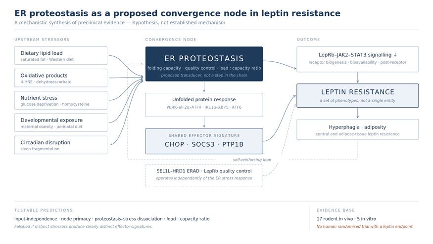
This review conceptualises ER proteostasis as a candidate regulatory hub at which metabolic, inflammatory, developmental and nutritional inputs intersect and are routed towards a leptin-resistant phenotype. The framework rests on three testable hypotheses — input independence, node primacy, and the proteostasis–stress distinction — and further argues that leptin resistance may comprise several phenotypes differing in their dominant mechanism. The mechanisms are presented not as an established model but as an explicitly falsifiable framework.
2. Materials and Methods
Literature Search Strategy
This is a narrative review. The reporting principles of the PRISMA 2020 statement [15] were applied not to conduct a systematic review but to document study selection transparently. Searches of PubMed/MEDLINE and Scopus were completed on 19 July 2026.
The following search string was applied identically to both databases:
( "leptin resistance" OR "leptin signaling" ) AND ( JAK2 OR STAT3 OR PI3K OR Akt OR MAPK OR ERK OR AMPK OR mTOR OR FoxO1 OR SOCS3 OR PTP1B OR "ER stress" OR "endoplasmic reticulum stress" OR "unfolded protein response" OR UPR OR PERK OR IRE1 OR ATF6 OR CHOP OR XBP1 )
Only studies published on or after 1 January 2015 were eligible. Landmark pre-2015 work is cited for historical context in the Introduction and Section 3 but was not entered into the evidence table. Studies were included if they reported original research, addressed leptin resistance or leptin signal transduction as a primary topic, linked at least one ER stress or UPR component (PERK, IRE1α, ATF6, CHOP, XBP1, GRP78/BiP, eIF2α, ERAD) mechanistically to leptin signalling, and were available in English as full text. Studies treating leptin only superficially were excluded, as were records without abstracts, conference proceedings, case reports and retracted publications.
Of 421 records retrieved (PubMed/MEDLINE, n = 244; Scopus, n = 177), 83 duplicates were removed. Of the 338 unique records, 58 were excluded at title and abstract screening and a further 258 at full-text assessment (target mechanism not the primary focus, n = 189; signalling studies with no ER stress component, n = 54; reviews, n = 15). Twenty-two studies entered the synthesis and the evidence table, and the animal studies were classified on the basis of the SYRCLE tool [16] (Figure 1; Table 1).
During the preparation of this manuscript, a large language model (Claude, Anthropic; model claude-opus-5) was used solely for language editing and academic style refinement of the English text. No scientific content, data, interpretations, or conclusions were generated by the tool. The corresponding author reviewed and approved all language-related edits and takes full responsibility for the content of the manuscript.
3. Leptin Physiology
Leptin is a 167-amino-acid polypeptide encoded by the ob gene and produced mainly by mature adipocytes of white adipose tissue; circulating concentrations correlate strongly with total fat mass [2,8]. Of the receptor isoforms, only LepRb carries a full-length intracellular domain and can therefore transmit JAK2/STAT3 signalling; it is densely expressed in POMC and AgRP neurons. The soluble isoform sOb-R determines the size of the free leptin pool [5,17,18]. Because canonical leptin physiology has been reviewed extensively elsewhere [8,9], only three features that intersect with the ER are emphasised here.
First, the functional competence of the receptor depends not only on its expression level but on correct folding within the ER. Newly synthesised LepRb is prone to misfolding, is a substrate of the SEL1L–HRD1 ERAD complex, and loss of SEL1L in POMC neurons attenuates leptin signalling; notably, retention of the receptor within the ER occurs independently of ER stress [19]. Second, receptor expression can fall under pathological conditions: in a model of congenital hypothyroidism, hypothalamic OB-R expression decreased by 45–63%, pSTAT3 by 51–56% and pERK by 48–50%, while SOCS3 rose [20]. Third, leptin resistance is not exclusively a central phenomenon: mice fed a high-fat diet develop local ER stress and leptin resistance within adipose tissue, and α-melanocyte-stimulating hormone (αMSH) reverses this picture by lowering GRP78 and CHOP [21].
4. Canonical Leptin Signalling
Binding of leptin to LepRb activates JAK2, which phosphorylates tyrosine residues on the cytoplasmic tail of the receptor and thereby creates docking sites for partially dissociable parallel arms (Figure 2).
The JAK2/STAT3 pathway, one of the principal mediators of leptin’s anorexigenic effects, is frequently used as a readout of leptin resistance. Yet stimuli as different as selenoprotein M deficiency, homocysteine, 4-HNE and glucose deprivation all lower pSTAT3, which shows that the marker is sensitive but not mechanism-specific. In diet-induced obese mice, moreover, STAT3 DNA-binding activity increases, and pharmacological reduction of pSTAT3 facilitates leptin-dependent POMC expression [22] — evidence that reduced pSTAT3 does not correspond to leptin resistance under all conditions.
PI3K/Akt is among the principal pathways at which leptin and insulin signalling intersect within the hypothalamus: mice carrying a kinase-domain mutation in PI3K-C2α develop leptin resistance, age-dependent insulin resistance and obesity together [23]. Perinatal nutritional interventions have also been reported to alter hypothalamic IRS1 and AMPK phosphorylation and thereby to affect leptin signalling permanently [24].
Evidence for a role of the MAPK/ERK arm in leptin resistance is limited and largely observational [20]. AMPK is suppressed by leptin; in a glucose-deprivation model, flurbiprofen reversed the suppression of pSTAT3 without altering UPR activation, which suggests that UPR activation alone may not fully account for leptin resistance [25].
mTOR is linked to ER proteostasis through its role as a negative regulator of autophagy, but the supporting studies are confined to cardiac and hepatic tissue [26,27,28]. FoxO1, for its part, competes with STAT3 at the POMC promoter and so shapes the transcriptional balance [22,29], raising the possibility that leptin resistance also reflects a shift in the balance between opposing transcriptional regulators. No primary study has examined the relationship between PI3K/Akt, mTOR or FoxO1 and ER stress directly in the context of leptin resistance; these links remain inferential (Table 2).
5. Molecular Basis of Leptin Resistance
For leptin to exert its central effects it must cross from the circulation into the brain, and the saturable nature of this transport may create an early bottleneck under hyperleptinaemic conditions [12]. Tanycytes surrounding the mediobasal hypothalamus have been shown to form an ERK-dependent gate for leptin, and this mechanism is disrupted in diet-induced obesity [30].
Upstream of this step lies the soluble receptor (sOb-R), which regulates the free leptin pool. Pharmacological and genetic modulation of hepatic CB1R alters sOb-R levels and hepatic leptin resistance; peripheral CB1R blockade is ineffective in CHOP-deficient mice; and CHOP regulates the sOb-R promoter directly [18]. ER stress can therefore interfere with leptin signalling through the circulating leptin pool, before the target cell is reached at all.
Impairment at the level of the receptor can arise in two ways. Direct evidence for reduced receptor expression comes from the congenital hypothyroidism model [20]. The more fundamental mechanism concerns receptor folding and maturation: LepRb has been identified as a substrate of the SEL1L–HRD1 ERAD complex, and loss of SEL1L in POMC neurons leads to accumulation of the receptor within the ER and to leptin resistance [19]. That this effect arises independently of classical ER stress suggests that the ER acts as a basal regulator of receptor biogenesis and quality control, and it constitutes the strongest support for the framework proposed here.
SOCS3 is among the principal negative-feedback regulators of leptin signalling: induced by STAT3, it binds Tyr985 of LepRb and suppresses JAK2 activity [31]. Sustained activation of this loop may contribute to leptin resistance, and elevated SOCS3 has been demonstrated in models of congenital hypothyroidism, high-fat feeding and cafeteria diet [20,21,32]. In the only study to address the relationship with ER stress directly, elevated CHOP and spliced XBP1 in pancreatic islets of ob/ob mice approached control levels after phenylbutyric acid (PBA), with a parallel change in SOCS3 expression [33]; the evidence is nonetheless confined to a single tissue and model.
PTP1B is a tyrosine phosphatase that terminates leptin signalling by dephosphorylating JAK2 and is localised to the ER membrane — a position that makes it one of the most direct links between ER proteostasis and leptin signalling. In a model of chronic sleep fragmentation, increased food intake was accompanied by hypothalamic UPR activation, unchanged ObR expression and reduced STAT3 phosphorylation; the picture was attributed to PTP1B, and both TUDCA and CHOP haploinsufficiency were protective [34]. PTP1B has also been shown to be modifiable by diet [32,35].
These four mechanisms do not rest on evidence of equal strength. The strongest causal findings come from receptor quality control and from mechanisms upstream of the UPR, where conditional genetic models link molecular changes to physiological endpoints [19,34,36]. Much of the evidence for SOCS3 and PTP1B, by contrast, is correlative: hypothalamic SOCS3 knockdown experiments are lacking [20], the ER stress–PTP1B relationship has not been tested with neuron-specific deletion, and the effect of PBA on SOCS3 suggests that the relationship may vary by tissue [33]. No mechanism, including tanycyte-mediated transport, is supported by human data [30].
6. Endoplasmic Reticulum Stress
6.1. UPR Activation
This review distinguishes ER proteostasis, ER stress and the UPR. ER proteostasis denotes the basal capacity of the ER to sustain protein folding, quality control and degradation; ER stress denotes the point at which the unfolded protein load exceeds that capacity; and the UPR denotes the cellular programme that responds through the PERK–eIF2α–ATF4, IRE1α–XBP1 and ATF6 arms — initially adaptive, but capable of impairing leptin signalling once it becomes chronic. The distinction matters, because retention of LepRb following loss of ERAD can produce leptin resistance in the absence of ER stress, showing that disrupted proteostasis can affect signalling independently of the UPR [19].
Findings that all three UPR arms are activated together in the context of leptin resistance are consistent across models: sleep fragmentation [34], diet-induced obese rats [35], activation reversible by phenethyl isothiocyanate [37], and increased hypothalamic ER stress gene expression in neonates in a maternal obesity model [38]. The foundation of this framework is the study establishing the role of the IRE1α–XBP1 pathway in POMC neurons [39].
Regulators upstream of the UPR indicate that the mechanism can be situated within a broader proteostasis network: deletion of TAK1 (MAP3K7) increases SREBP-mediated lipogenesis and ER capacity and thereby prevents ER-stress-driven leptin resistance and hyperphagic obesity under a high-fat diet [36].
6.1.1. Lipid Overload
Western-type diets rich in saturated fat are thought to trigger the UPR by altering ER membrane composition and protein-folding capacity. In apolipoprotein E-deficient mice, a Western diet raised markers of ER stress, whereas diallyl disulfide lowered these markers together with hypercholesterolaemia [40]; indole-3-carbinol has been reported to reduce plasma leptin and thereby to attenuate ER-stress-associated hyperleptinaemia [41].
These findings appear at first sight to conflict with the TAK1 study: in ApoE−/− models, reducing lipid load was associated with improvement in ER stress markers, whereas in TAK1 deletion the increase in lipogenesis was interpreted as an expansion of ER capacity [36]. The discrepancy suggests that what matters is less the absolute lipid load than the balance between that load and the processing capacity of the ER — an interpretation that has not been tested directly.
6.1.2. Oxidative ER Stress
Because the ER lumen is an oxidising environment, shifts in redox balance can affect protein-folding capacity. The lipid peroxidation product 4-HNE has been reported to reduce leptin-stimulated STAT3 phosphorylation in a dose-dependent manner through the eIF2α–CHOP pathway; global tyrosine phosphorylation was unaffected, suggesting relative selectivity for leptin signalling [42]. Dehydroascorbic acid similarly raised GRP78, CHOP and spliced XBP1 while lowering pSTAT3 [43], and redox disturbance was shown to affect leptin signalling in selenoprotein M deficiency as well [44].
6.1.3. Nutrient-Derived ER Stress
Nutrient excess is not the only relevant input; nutrient deprivation and specific metabolites also affect ER stress and leptin signalling. Glucose deprivation activates the UPR by increasing GRP78 and CHOP expression while reducing pSTAT3 and pSTAT5 [25], and elevated homocysteine selectively suppresses STAT3 activity [45]. From a developmental perspective, protein restriction combined with a high-fat diet has been associated with hypothalamic leptin resistance, a response attenuated by taurine supplementation [46].
Findings that the several UPR arms are activated together across independent models are consistent, and CNS-specific Tak1 deletion offers causal support [36]; the load-versus-capacity interpretation, by contrast, is an inference drawn from the available data.
6.2. Molecular Convergence of Distinct Stressors on Leptin Resistance Effectors
One of the patterns that stands out in this review is that stressors as varied as sleep fragmentation, homocysteine, 4-HNE, glucose deprivation, Western-type diet and maternal obesity all appear to act through a limited set of shared effectors — SOCS3, PTP1B and CHOP. Of these, CHOP is the strongest candidate integrating mechanism: CHOP haploinsufficiency protects against hypothalamic leptin resistance [34], CHOP participates directly in regulation of the sOb-R promoter [18], and the effects of 4-HNE are mediated through the eIF2α–CHOP axis [42].
The localisation of PTP1B to the ER membrane provides a structural basis for considering SOCS3, PTP1B and ER-stress-dependent effectors within a common proteostatic context. That α-MSH lowers GRP78 and CHOP while suppressing SOCS3 and raising pSTAT3 further suggests that these mechanisms interact within a connected regulatory network rather than operating as independent processes [21]. The therapeutic hypothesis that follows is that modulating the mechanisms at which stressors converge might achieve broader efficacy than targeting each stressor separately; this approach has not yet been tested directly.
This shared pattern suggests that particular regulatory mechanisms may be common to distinct pathophysiological inputs. For CHOP, genetic findings support a causal role; for SOCS3 and PTP1B the evidence is weaker. Because the stressors have not been assessed within the same model under comparable conditions, it remains difficult to determine whether the observed convergence is a general biological property or an apparent pattern arising from similarities between models (Table 3). Molecular convergence is therefore a hypothesis requiring direct comparative testing.
6.3. A Possible ER Stress-Mediated Link Between Leptin and Insulin Resistance
The metabolic consequences of ER stress may not be confined to leptin signalling; similar mechanisms may also contribute to insulin resistance, placing the two disorders on a shared cellular footing. That loss of SEL1L in POMC neurons is associated with hepatic steatosis, glucose intolerance and insulin resistance as well as leptin resistance [19], and that TUDCA improved findings related to both forms of resistance in a maternal obesity model [38], lend support to this possibility. A plausible molecular basis is that PTP1B dephosphorylates both JAK2 and the insulin receptor, and that PI3K/Akt is a signalling component shared by the two hormones. No direct human evidence shows that correcting ER proteostasis improves both forms of resistance together. Table 4 compares the mechanisms discussed in Section 5 and Section 6 by the UPR arm affected, the point at which each acts, and the strength of the evidence supporting it.
6.4. The Causality Problem: Separating Direct Effects from Weight Loss
Most of the intervention studies summarised here share a common interpretive difficulty: nearly all report reductions in body weight and/or adiposity alongside the intervention (Table 5). Because weight loss by itself reduces hypothalamic ER stress and leptin resistance, it cannot be determined whether the observed improvement reflects a direct effect of the intervention or the accompanying weight loss.
The only study whose design overcomes this difficulty is the Roux-en-Y gastric bypass (RYGB) study of Chen et al. [47], which used a weight-matched caloric restriction control and showed that improvements in hypothalamic ER stress and leptin sensitivity were specific to RYGB. It is thus the sole direct basis for the claim that reducing ER stress can restore leptin sensitivity independently of weight loss, and it awaits independent replication; wider adoption of weight-matched controls in this field is needed.
A second difficulty concerns the target specificity of chemical chaperones: beyond reducing ER stress, PBA and TUDCA act on numerous targets, including histone deacetylase inhibition and bile acid signalling. Findings obtained with these agents therefore provide supportive but limited evidence, whereas findings based on CHOP genetics constitute a firmer basis.
Third, findings obtained solely in vitro warrant additional caution: four of these studies used SH-SY5Y neuroblastoma cells transfected to overexpress Ob-Rb, a tumour line that does not carry hypothalamic neuronal identity, and both receptor density and stressor concentrations lie outside the physiological range. Such studies are valuable for generating hypotheses but are not sufficient for inference about leptin resistance in vivo.
7. Intercellular Interactions
Leptin resistance is less a cell-autonomous event within an isolated neuron than the product of continuous interaction among hypothalamic neurons, microglia, astrocytes and peripheral tissues. The low-grade inflammatory response that overnutrition induces in the hypothalamus is accompanied by ER stress induction and defective autophagy, and the process is mediated through interaction with non-neuronal cells [35,47]. A comparable relationship has been described at the level of sympathetic output: silencing Sirt1 or FoxO1 in the arcuate nucleus reduces leptin-mediated inflammation and sympathetic nerve activity [29].
The strongest evidence that this interaction can be reversed comes from bariatric surgery: RYGB improved hypothalamic gliosis, inflammatory signalling and ER stress in diet-induced obese rats, the improvement was associated with increased central leptin responsiveness, and it was not observed in the weight-matched control [47] (Section 6.4).
Autophagy represents a further node in this network: a berberine-containing herbal preparation attenuated hypothalamic ER stress and leptin resistance through modulation of autophagy, with the findings confirmed in both microglial and neuronal cell models [35]. In the periphery, αMSH attenuates ER-stress-induced leptin resistance in adipose tissue, whereas ER stress induced by tunicamycin or thapsigargin reduces leptin sensitivity [21] — pointing to a link between central melanocortin signalling and peripheral adipose expansion that operates through ER stress.
8. Developmental Programming and Epigenetic Regulation
Evidence that leptin resistance can be permanently programmed by early-life conditions continues to accumulate.
8.1. Developmental Programming
Maternal obesity has been reported to disrupt the development of melanocortin circuits in offspring, producing neonatal hyperleptinaemia and leptin resistance, to upregulate ER-stress-related genes in the hypothalamus, and to be corrected — in both its metabolic and its neurodevelopmental components — by neonatal TUDCA [38]. That an intervention delivered within a critical developmental window can alter an otherwise persistent phenotype points to a causal role for early-life ER stress.
Programming has been shown to begin at the level of the placenta: maternal obesity activates placental insulin/IGF-I/mTOR and leptin pathways [49], and placental dysfunction is associated with increased SOCS3 and PTPN2 expression [50]. The same axis operates postnatally, where perinatal diet composition and gestational thyroid hormone insufficiency produce persistent and partly sex-specific hypothalamic leptin signalling defects in adulthood [20,24,46].
The most direct evidence that this programming is reversible comes from silencing maternal SOCS3: a Socs3-shRNA lentivirus administered in early gestation to obese dams attenuated postnatal obesity in the offspring [48]. This suggests that increased hypothalamic SOCS3 is not merely a marker of early-life programming but also a mediator of it.
8.2. Epigenetic and Post-Translational Mediators
The first layer of epigenetic regulation comprises histone modification and the acetylation balance. Hypothalamic leptin action has been shown to be mediated by histone deacetylase 5 (HDAC5), which shapes the transcriptional output of leptin signalling at the chromatin level [51]. In parallel, hypothalamic expression of the nuclear receptor Nur77 falls in obese mice, leptin fails to reduce body weight in Nur77 knockout animals, and Nur77 recruits p300 and dissociates HDAC1, thereby increasing STAT3 acetylation and transcriptional activity [52].
The second layer has been defined at the level of DNA methylation and non-coding RNAs, and it includes human data: adiposity-associated DNA methylation signatures in adolescents have been related to leptin concentrations and perinatal factors [53], and circulating microRNAs in children with obesity have been associated with leptin resistance [54]. Experimentally, a maternal high-fat diet impairs leptin signalling in the neonatal hypothalamus and upregulates the type-1 cannabinoid receptor through sex-specific epigenetic changes [55].
The two layers do not rest on evidence of equal strength. Findings on developmental programming carry relatively strong causal support because they are reversible by intervention within a critical window [38,48], whereas the DNA methylation and microRNA findings are cross-sectional and correlational. More importantly, no direct mechanistic link between ER stress and epigenetic marks has been demonstrated; the fact that these two literatures remain unintegrated is the most conspicuous gap in the framework proposed here.
9. A Unified Mechanistic Model and the Proposed Conceptual Framework
9.1. Integrating the Findings into a Single Mechanistic Model
The findings summarised in this review can be read not as a series of independent observations but as a single mechanistic route (Figure 3). In the proposed model, stressors that appear unrelated — lipid overload, oxidative products, nutrient-derived stressors, maternal obesity and sleep fragmentation — converge on hypothalamic ER stress; ER stress activates all three arms of the UPR; and that activation converges on a narrow set of effectors (CHOP, SOCS3, PTP1B). These effectors can be argued to suppress LepRb–JAK2–STAT3 signalling and so to produce leptin resistance, which in turn drives hyperphagia and increased adiposity, feeds back onto ER stress, and closes a self-reinforcing loop. No single study has demonstrated the route in its entirety.
A second arm runs parallel to this route: LepRb quality control mediated by the SEL1L–HRD1 ERAD system and occurring independently of ER stress [19]. On this view, the ER can shape leptin signalling through both its stress response and its basal proteostatic function.
9.2. ER Proteostasis as a Convergence Node
On this basis, the following testable framework is proposed:
Endoplasmic reticulum proteostasis may function as a central convergence node that translates metabolic, inflammatory, developmental and nutrient-derived signals into leptin resistance.
What distinguishes the framework is that it positions the ER not as one step in a chain leading to leptin resistance but as a regulatory hub converting different classes of input into a common output. Three hypotheses follow, each corresponding to a directly testable experimental design.
The first is that the effector signature is independent of the input: whatever the stressor, a similar effector pattern encompassing CHOP, SOCS3 and PTP1B would be expected. The available data appear consistent with this expectation, but no study has tested different stressors within the same model under comparable conditions.
The second is that intervening at the shared effector node will prove more effective than targeting upstream stressors individually. Work on chemical chaperones and CHOP genetics offers partial support, but no study has compared node-level intervention with upstream intervention within a single model.
The third is that disrupted ER proteostasis can produce leptin resistance even when the ER stress response is not activated. The SEL1L–HRD1 ERAD finding of Mao et al. [19] is the only direct evidence supporting this hypothesis and represents the most original aspect of the framework; were it confirmed, it would follow that studies measuring only UPR markers are missing part of the leptin resistance phenotype.
The framework requires the ER stress and ER proteostasis literatures to be brought together, and it carries an explicit falsification criterion: a demonstration that different stressors produce markedly different effector signatures would invalidate it.
Table 3 lists these three hypotheses, together with two further hypotheses concerning the load-to-capacity ratio and translatability to humans, in terms of the expected outcome, the proposed experimental design, the falsifying finding and the current state of testing.
10. Clinical Implications
Detail about molecular mechanism acquires clinical meaning only to the extent that it can be translated into patient care. Figure 4 sets out the therapeutic approaches considered in this section by level of intervention, together with the nature of the evidence available for each.
10.1. Clinical Outcomes of Leptin Replacement Therapy
The therapeutic potential of leptin has been tested in a single clinical trial of the hormone itself, with largely negative results: in a randomised, double-blind, placebo-controlled dose-escalation study, 73 obese and 54 lean participants received daily subcutaneous recombinant leptin, and placebo-adjusted weight loss in the obese group remained around 3% even at the highest doses [58].
Within the proposed framework this is an expected result: if leptin resistance is generated simultaneously at the receptor, post-receptor and bioavailability levels, raising circulating ligand will correct none of these steps. In common obesity the aim is therefore not to administer leptin but to repair the response to it. States of genuine deficiency, such as congenital leptin deficiency and lipodystrophy, fall outside this assessment.
10.2. Human Data on Chemical Chaperones
Chemical chaperones have been tested in humans, but leptin endpoints were not assessed. Kars et al. [56] gave TUDCA (1,750 mg/day) or placebo to 20 participants with obesity for four weeks and reported an approximately 30% increase in hepatic and muscle insulin sensitivity in the TUDCA arm. Xiao et al. [57] showed that two weeks of oral sodium phenylbutyrate (7.5 g/day) partly attenuated lipid infusion-induced insulin resistance.
These two trials point in two directions at once. Interventions targeting ER stress are feasible in humans, are tolerated, and can produce measurable metabolic benefit, and the interest is extending to other metabolic diseases [59,60]. Against this, both trials measured only insulin endpoints, the samples were small (n = 20 and n = 8) and the exposures brief; the assumption that chemical chaperones correct leptin resistance in humans therefore remains untested and represents the most concrete gap in the field.
10.3. PTP1B as a Therapeutic Target
PTP1B is the member of the proposed effector node that appears most amenable to drug development [61]. The aminosterol trodusquemine was reported to suppress appetite and produce fat-specific weight loss in diet-induced obese mice [62] and was well tolerated in phase 1 trials, yet the development programme for the obesity indication was not pursued.
The reason is not inefficacy alone but the structural nature of the target: the catalytic site of PTP1B is highly polar and charged, molecules that bind it remain too polar to cross the cell membrane, and structural similarity to other members of the phosphatase family creates a selectivity problem. Current work has therefore turned to allosteric and natural product-derived inhibitors [63]. Correctly identifying an effector node, in other words, does not make that node druggable.
10.4. Bariatric Surgery and Incretin-Based Therapies
The mechanism of bariatric surgery bears directly on the proposed framework (Section 6.4 and Section 7): that RYGB improves hypothalamic ER stress and inflammation, that this is associated with increased central leptin sensitivity, and that it is not seen in a weight-matched control [47] offers a mechanistic explanation for why surgically induced weight loss is more durable than equivalent loss achieved by diet.
GLP-1 receptor agonists raise a different question: liraglutide has been reported to attenuate the fall in free plasma leptin during maintenance of weight loss [64]. Since weight loss normally lowers free leptin and thereby facilitates weight regain, this effect may contribute to the drug’s efficacy. The study did not, however, assess sensitivity, and the effect of GLP-1 receptor agonists on hypothalamic ER stress has not been examined in humans. Table 6 summarises the interventions that target this axis, together with the current state of human data for each.
10.5. Molecular Heterogeneity of Leptin Resistance and a Phenotyping Framework
One of the most direct clinical implications of this review is that leptin resistance is not a single entity: the findings in Section 5 and Section 6 show that it can arise at different molecular levels and that a set of phenotypes distinguished by dominant mechanism exists.
Table 7 proposes a phenotyping framework on this basis, linking five questions: which patient, which dominant mechanism, which biomarker, which intervention and which primary endpoint. Because an intervention effective in only one subgroup will appear ineffective overall in an unstratified trial, establishing this chain is a precondition for making the axis clinically testable. The history of the leptin field illustrates the risk: recombinant leptin proved ineffective in an unselected obese population [58] but markedly effective in the leptin-deficient phenotype [7].
The status of this framework should be stated plainly: none of the proposed phenotypes has been confirmed in humans. The classification is a hypothesis-generating structure derived from animal and laboratory findings, and the biomarkers listed are candidates. Its purpose is not to offer a clinical algorithm but to define a testable stratification hypothesis.
The weakest link in the framework is measurement: hypothalamic ER stress cannot be measured directly in humans, so distinguishing an ER-dominant phenotype depends on unvalidated indirect markers. The second weak link is the endpoint, since no standard method exists for measuring leptin sensitivity in a clinical setting. Until both gaps are addressed, the pairings in Table 7 cannot be tested (Table 2).
10.6. Determinants of Individual Response Variability
The framework assumes that individuals do not respond identically to the same metabolic load — the founding premise of the personalised nutrition literature [66], which has empirical support [67]. A plausible molecular counterpart is that, where ER folding capacity differs between individuals, the same dietary lipid load will generate different degrees of ER stress [36] (Section 6.1.1); this inference remains conceptual.
The first source of variability is genotype. The most direct human evidence comes from the LEPR Q223R (rs1137101) variant, which reduces leptin binding and predicts annual weight gain in childhood obesity [68]. Rare variants illustrate the same principle at the level of proteostasis: the Cys604Ser variant of LepRb is prone to misfolding and is processed as an ERAD substrate [19]. Because no study has shown that a genotype-tailored intervention alters outcome, the evidence remains at the level of hypothesis [69].
The second source is developmental windows; the determinant of response variability with the strongest evidence base is early-life exposure (Section 8). Its clinical implication is twofold: the timing of an intervention may determine its efficacy, and sex should be treated as an independent response modifier [24,38,48,55].
The third source is diet-derived modulators, and it is notable that most of the compounds found effective along this axis are dietary in origin: phenethyl isothiocyanate, indole-3-carbinol, diallyl disulfide, proanthocyanidins, biochanin A, berberine, taurine and a Rubisco-derived plant peptide [32,35,37,40,41,46,70,71] (Table 5). Three caveats apply: the studies were conducted almost entirely in animal models and at doses above the range attainable through diet; most reported concurrent weight loss, so direct effects cannot be separated (Section 6.4); and differences in bioavailability and microbiota-mediated metabolism may alter efficacy [72].
10.7. Implications for Clinical Practice
As far as the current evidence allows, the clinical implications can be grouped under four points.
The first concerns the interpretation of hyperleptinaemia: a high leptin concentration in a patient with obesity reflects a defective response rather than an excess of hormone, and leptin measurement does not currently function as a decision tool in routine practice.
The second concerns diet composition: although most of the compounds of interest are dietary in origin, the findings remain at the level of animal models and do not support supplementation. They provide mechanistic support only for existing advice to reduce saturated fat and excess caloric load.
The third concerns the nature of weight loss: surgical data suggest that the same amount of weight loss may have different effects on the central leptin response depending on how it is achieved, so that the method as well as the magnitude of loss deserves attention.
The fourth concerns the prospect of phenotype-based stratification: adiposity-associated DNA methylation signatures and circulating microRNAs [53,54] may represent measurable molecular correlates of individual differences in response. These findings are correlational and unvalidated, however, and provide no basis for any clinical decision at present.
The clinical value of this review lies not in offering a treatment recommendation but in explaining mechanistically why current treatments do and do not work, and in defining a concrete research question ready for testing: can agents targeting ER proteostasis restore leptin sensitivity in humans?
11. Conclusions
This review has examined leptin resistance at the intersection of intracellular signalling networks and endoplasmic reticulum stress, synthesising 22 core primary studies together with supporting literature. The aim was not to restate known mechanisms but to unify them under a single testable framework and to set out explicitly the points at which that framework could be falsified.
11.1. Principal Conclusions of the Synthesis
Three principal conclusions emerge. First, the ER appears to shape leptin signalling at the levels of receptor biogenesis, receptor bioavailability and post-receptor inhibition; that the first of these layers operates independently of ER stress, and rests on conditional genetic designs, makes it the best-supported claim in this review. Second, distinct stressors appear to converge on a narrow set of effectors, although the causal role of CHOP is supported by genetic evidence while the evidence for SOCS3 and PTP1B remains correlative. Third, this node appears pharmacologically accessible in experimental models, yet direct effects have been separated from weight-loss-dependent effects in only one weight-matched study [47].
11.2. Limitations of the Evidence Base
All of these conclusions must be read against a substantial constraint: 17 of the core evidence base rests on rodent in vivo models and 5 on cell culture alone, and no study has been conducted in human participants. Studies using the human-derived SH-SY5Y line provide data from a human cell line, not human data.
This structure determines which claims the evidence supports. Animal models strongly support the proposition that ER stress can impair hypothalamic leptin signalling and that the impairment is reversible. The same evidence base does not support the proposition that targeting ER proteostasis should be used as an obesity treatment in humans; such a proposal is at best a hypothesis worth testing.
The distinction deserves emphasis, because the history of the leptin field is itself instructive: recombinant leptin was markedly effective in rodent models yet failed in common human obesity, and there is no basis for assuming that strategies targeting ER stress will avoid a comparable discontinuity.
Further limitations are as follows: (i) the great majority of studies used male animals, and sex differences have not been examined systematically, with El-Sehrawy et al. [65] an exception; (ii) phytochemical doses exceed the range attainable through diet in humans; (iii) effect sizes vary widely across models; (iv) study durations are generally limited to 6–12 weeks; and (v) although leptin resistance manifests with different molecular signatures in different tissues, most studies treat it as a single entity.
11.3. Research Gaps
The principal research gaps and the methodological approaches required to address them are summarised in Table 2; the most important is the absence of a randomised controlled trial testing whether an intervention targeting ER stress restores leptin sensitivity in humans.
11.4. Methodological Requirements and Future Directions
Closing these gaps will require several methodological advances: single-cell and spatial transcriptomics could resolve the cell-type-specific UPR activation that tissue homogenates obscure; human iPSC-derived hypothalamic organoids could overcome the limitations of the SH-SY5Y line; multi-omic integration could be used to identify circulating biomarkers; and computational drug discovery could contribute to the design of selective PTP1B and UPR modulators [63].
How the response varies between individuals, meanwhile, is unknown. Studies stratified by genotype, epigenetic profile, microbiota composition and baseline metabolic state offer the most realistic route to testing these compounds in humans, but this is a research programme that first requires phenotyping tools to be developed.
The field should also adopt two design standards: the use of weight-matched control groups in intervention studies, and the inclusion of both sexes.
11.5. Concluding Assessment
In rodent models the endoplasmic reticulum appears not merely as a waypoint in leptin resistance but as its regulatory centre, and its multi-layered role — extending from receptor quality control to induction of negative regulators — is supported by conditional genetic designs. Recasting that observation as a convergence-node framework, however, remains at the level of hypothesis.
The most important consequence of this multi-layered organisation is that leptin resistance is not a uniform disorder; the phenotypes arising at different levels (Table 7) offer a molecular basis for explaining why the same intervention produces different outcomes in different individuals: Resolving the molecular heterogeneity of leptin resistance may be necessary before interventions aimed at restoring leptin sensitivity can be tested in the appropriate patient subgroups.
The conditional phrasing is deliberate: the evidence base consists largely of animal and cell culture studies, and in humans neither can leptin resistance phenotypes be distinguished nor can hypothalamic ER stress be measured. The stratification proposed here is therefore not a clinical strategy but a mechanistically justified research programme.
The priority for the field is not to generate further rodent studies but to take three concrete steps: (i) developing and validating circulating biomarkers capable of distinguishing leptin resistance phenotypes in humans; (ii) testing agents already administered in humans, such as TUDCA and 4-PBA, in weight-matched trials with leptin sensitivity as the primary endpoint; and (iii) designing studies in which interventions are stratified by genotype, epigenetic profile and microbiota composition.
Author Contributions
Conceptualization, S.A.; methodology, S.A.; writing—original draft preparation, S.A.; writing—review and editing, S.A., N.Y. and H.T.B.; visualization, S.A.; supervision, H.T.B. All authors have read and agreed to the published version of the manuscript.
Funding
This work received no specific grant from any funding agency in the public, commercial, or not-for-profit sectors.
Institutional Review Board Statement
Not applicable. This study is a narrative review of published research and did not involve human participants, animals, or new data collection.
Informed Consent Statement
Not applicable.
Data Availability Statement
Data sharing is not applicable to this article, as no new data were created or analysed in this study. All studies synthesised are published and are listed in the reference list; the search strategy and selection criteria are reported in full in Section 2.
Conflicts of Interest
The authors declare no conflicts of interest.
Generative AI Statement
A large language model (Claude, Anthropic; model claude-opus-5) was used solely for language editing and academic style refinement. No scientific content, data, interpretations, or conclusions were generated by the tool. The authors reviewed and approved all language-related edits.
References
- Coleman, D.L. Effects of parabiosis of obese with diabetes and normal mice. Diabetologia 1973, 9, 294–298. [Google Scholar] [CrossRef] [PubMed]
- Zhang, Y.; Proenca, R.; Maffei, M.; Barone, M.; Leopold, L.; Friedman, J.M. Positional cloning of the mouse obese gene and its human homologue. Nature 1994, 372, 425–432. [Google Scholar] [CrossRef] [PubMed]
- Halaas, J.L.; Gajiwala, K.S.; Maffei, M.; Cohen, S.L.; Chait, B.T.; Rabinowitz, D.; Lallone, R.L.; Burley, S.K.; Friedman, J.M. Weight-reducing effects of the plasma protein encoded by the obese gene. Science 1995, 269, 543–546. [Google Scholar] [CrossRef] [PubMed]
- Pelleymounter, M.A.; Cullen, M.J.; Baker, M.B.; Hecht, R.; Winters, D.; Boone, T.; Collins, F. Effects of the obese gene product on body weight regulation in ob/ob mice. Science 1995, 269, 540–543. [Google Scholar] [CrossRef] [PubMed]
- Tartaglia, L.A.; Dembski, M.; Weng, X.; Deng, N.; Culpepper, J.; Devos, R.; Richards, G.J.; Campfield, L.A.; Clark, F.T.; Deeds, J.; et al. Identification and expression cloning of a leptin receptor, OB-R. Cell 1995, 83, 1263–1271. [Google Scholar] [CrossRef] [PubMed]
- Montague, C.T.; Farooqi, I.S.; Whitehead, J.P.; Soos, M.A.; Rau, H.; Wareham, N.J.; Sewter, C.P.; Digby, J.E.; Mohammed, S.N.; Hurst, J.A.; et al. Congenital leptin deficiency is associated with severe early-onset obesity in humans. Nature 1997, 387, 903–908. [Google Scholar] [CrossRef] [PubMed]
- Farooqi, I.S.; Jebb, S.A.; Langmack, G.; Lawrence, E.; Cheetham, C.H.; Prentice, A.M.; Hughes, I.A.; McCamish, M.A.; O'Rahilly, S. Effects of recombinant leptin therapy in a child with congenital leptin deficiency. N. Engl. J. Med. 1999, 341, 879–884. [Google Scholar] [CrossRef] [PubMed]
- Friedman, J.M. Leptin and the endocrine control of energy balance. Nat. Metab. 2019, 1, 754–764. [Google Scholar] [CrossRef] [PubMed]
- Obradovic, M.; Sudar-Milovanovic, E.; Soskic, S.; Essack, M.; Arya, S.; Stewart, A.J.; Gojobori, T.; Isenovic, E.R. Leptin and obesity: role and clinical implication. Front. Endocrinol. 2021, 12, 585887. [Google Scholar] [CrossRef] [PubMed]
- World Health Organization. Obesity and Overweight. Available online: https://www.who.int/news-room/fact-sheets/detail/obesity-and-overweight (accessed on 19 July 2026).
- GBD 2021 Adult BMI Collaborators. Global, regional, and national prevalence of adult overweight and obesity, 1990–2021, with forecasts to 2050: a forecasting study for the Global Burden of Disease Study 2021. Lancet 2025, 405, 813–838. [Google Scholar] [CrossRef] [PubMed]
- Myers, M.G.; Leibel, R.L.; Seeley, R.J.; Schwartz, M.W. Obesity and leptin resistance: distinguishing cause from effect. Trends Endocrinol. Metab. 2010, 21, 643–651. [Google Scholar] [CrossRef] [PubMed]
- Izquierdo, A.G.; Crujeiras, A.B.; Casanueva, F.F.; Carreira, M.C. Leptin, obesity, and leptin resistance: where are we 25 years later? Nutrients 2019, 11, 2704. [Google Scholar] [CrossRef] [PubMed]
- Hu, W.; Zhu, H.; Gong, F. Leptin and leptin resistance in obesity: current evidence, mechanisms and future directions. Endocr. Connect. 2025, 14, e250521. [Google Scholar] [CrossRef] [PubMed]
- Page, M.J.; McKenzie, J.E.; Bossuyt, P.M.; Boutron, I.; Hoffmann, T.C.; Mulrow, C.D.; Shamseer, L.; Tetzlaff, J.M.; Akl, E.A.; Brennan, S.E.; et al. The PRISMA 2020 statement: an updated guideline for reporting systematic reviews. BMJ 2021, 372, n71. [Google Scholar] [CrossRef] [PubMed]
- Hooijmans, C.R.; Rovers, M.M.; de Vries, R.B.M.; Leenaars, M.; Ritskes-Hoitinga, M.; Langendam, M.W. SYRCLE's risk of bias tool for animal studies. BMC Med. Res. Methodol. 2014, 14, 43. [Google Scholar] [CrossRef] [PubMed]
- Saxton, R.A.; Caveney, N.A.; Moya-Garzon, M.D.; Householder, K.D.; Rodriguez, G.E.; Burdsall, K.A.; Long, J.Z.; Garcia, K.C. Structural insights into the mechanism of leptin receptor activation. Nat. Commun. 2023, 14, 1797. [Google Scholar] [CrossRef] [PubMed]
- Drori, A.; Gammal, A.; Azar, S.; Hinden, L.; Hadar, R.; Wesley, D.; Nemirovski, A.; Szanda, G.; Salton, M.; Tirosh, B.; et al. CB1R regulates soluble leptin receptor levels via CHOP, contributing to hepatic leptin resistance. eLife 2020, 9, e60771. [Google Scholar] [CrossRef] [PubMed]
- Mao, H.; Kim, G.H.; Pan, L.; Qi, L. Regulation of leptin signaling and diet-induced obesity by SEL1L-HRD1 ER-associated degradation in POMC expressing neurons. Nat. Commun. 2024, 15, 8435. [Google Scholar] [CrossRef] [PubMed]
- Aiceles, V.; Gombar, F.; Cavalcante, F.; Ramos, C. Congenital hypothyroidism is associated with impairment of the leptin signaling pathway in the hypothalamus in male Wistar animals in adult life. Horm. Metab. Res. 2019, 51, 330–335. [Google Scholar] [CrossRef] [PubMed]
- Gan, L.; Liu, Z.; Wu, T.; Feng, F.; Sun, C. αMSH promotes preadipocyte proliferation by alleviating ER stress-induced leptin resistance and by activating Notch1 signal in mice. Biochim. Biophys. Acta Mol. Basis Dis. 2017, 1863, 231–238. [Google Scholar] [CrossRef] [PubMed]
- Xu, L.; Li, H.; Zhou, G.; Lu, W.; Yang, R.; Liu, H.; Yang, G. DNA-binding activity of STAT3 increased in hypothalamus of DIO mice; the reduction of STAT3 phosphorylation may facilitate leptin signaling. Biochem. Biophys. Res. Commun. 2018, 505, 229–235. [Google Scholar] [CrossRef] [PubMed]
- Alliouachene, S.; Bilanges, B.; Chaussade, C.; Pearce, W.; Foukas, L.C.; Scudamore, C.L.; Moniz, L.S.; Vanhaesebroeck, B. Inactivation of class II PI3K-C2α induces leptin resistance, age-dependent insulin resistance and obesity in male mice. Diabetologia 2016, 59, 1503–1512. [Google Scholar] [CrossRef] [PubMed]
- Rivera, P.; Ramírez-López, M.T.; Vargas, A.; Decara, J.; Vázquez, M.; Arco, R.; Gómez de Heras, R.; Argente, J.; Rodríguez de Fonseca, F.; Chowen, J.A.; et al. Perinatal free-choice of a high-calorie low-protein diet affects leptin signaling through IRS1 and AMPK dephosphorylation in the hypothalami of female rat offspring in adulthood. Acta Physiol. 2019, 226, e13244. [Google Scholar] [CrossRef] [PubMed]
- Hosoi, T.; Suyama, Y.; Kayano, T.; Ozawa, K. Flurbiprofen ameliorates glucose deprivation-induced leptin resistance. Front. Pharmacol. 2016, 7, 354. [Google Scholar] [CrossRef] [PubMed]
- Rubio, B.; Mora, C.; Pintado, C.; Mazuecos, L.; Fernández, A.; López, V.; Andrés, A.; Gallardo, N. The nutrient sensing pathways FoxO1/3 and mTOR in the heart are coordinately regulated by central leptin through PPARβ/δ: implications in cardiac remodeling. Metabolism 2021, 115, 154453. [Google Scholar] [CrossRef] [PubMed]
- Gogiraju, R.; Hubert, A.; Fahrer, J.; Straub, B.K.; Brandt, M.; Wenzel, P.; Münzel, T.; Konstantinides, S.; Hasenfuss, G.; Schäfer, K. Endothelial leptin receptor deletion promotes cardiac autophagy and angiogenesis following pressure overload by suppressing Akt/mTOR signaling. Circ. Heart Fail. 2019, 12, e005622. [Google Scholar] [CrossRef] [PubMed]
- An, X.; Liu, J.; Li, Y.; Dou, Z.; Li, N.; Suo, Y.; Ma, Y.; Sun, M.; Tian, Z.; Xu, L. Chemerin/CMKLR1 ameliorates nonalcoholic steatohepatitis by promoting autophagy and alleviating oxidative stress through the JAK2-STAT3 pathway. Peptides 2021, 135, 170422. [Google Scholar] [CrossRef] [PubMed]
- Liu, X.; Zheng, H. Modulation of Sirt1 and FoxO1 on hypothalamic leptin-mediated sympathetic activation and inflammation in diet-induced obese rats. J. Am. Heart Assoc. 2021, 10, e020667. [Google Scholar] [CrossRef] [PubMed]
- Balland, E.; Dam, J.; Langlet, F.; Caron, E.; Steculorum, S.; Messina, A.; Rasika, S.; Falluel-Morel, A.; Anouar, Y.; Dehouck, B.; et al. Hypothalamic tanycytes are an ERK-gated conduit for leptin into the brain. Cell Metab. 2014, 19, 293–301. [Google Scholar] [CrossRef] [PubMed]
- Bjørbaek, C.; Elmquist, J.K.; Frantz, J.D.; Shoelson, S.E.; Flier, J.S. Identification of SOCS-3 as a potential mediator of central leptin resistance. Mol. Cell 1998, 1, 619–625. [Google Scholar] [CrossRef] [PubMed]
- Ibars, M.; Ardid-Ruiz, A.; Suárez, M.; Muguerza, B.; Bladé, C.; Aragonès, G. Proanthocyanidins potentiate hypothalamic leptin/STAT3 signalling and Pomc gene expression in rats with diet-induced obesity. Int. J. Obes. 2017, 41, 129–136. [Google Scholar] [CrossRef] [PubMed]
- Baba, B.; Caliskan, M.; Boyuk, G.; Hacisevki, A. Chemical chaperone PBA attenuates ER stress and upregulates SOCS3 expression as a regulator of leptin signaling. Biochemistry (Mosc.) 2021, 86, 480–488. [Google Scholar] [CrossRef] [PubMed]
- Hakim, F.; Wang, Y.; Carreras, A.; Hirotsu, C.; Zhang, J.; Peris, E.; Gozal, D. Chronic sleep fragmentation during the sleep period induces hypothalamic endoplasmic reticulum stress and PTP1b-mediated leptin resistance in male mice. Sleep 2015, 38, 31–40. [Google Scholar] [CrossRef] [PubMed]
- Hua, T.; Wu, Q.; Huang, Z.; Cai, J. Fufang Zhenzhu Tiaozhi capsule alleviates hypothalamic endoplasmic reticulum stress and leptin resistance through autophagy modulation in DIO rats. J. Ethnopharmacol. 2025, 351, 120056. [Google Scholar] [CrossRef] [PubMed]
- Sai, K.; Morioka, S.; Takaesu, G.; Muthusamy, N.; Ghashghaei, H.T.; Hanafusa, H.; Matsumoto, K.; Ninomiya-Tsuji, J. TAK1 determines susceptibility to endoplasmic reticulum stress and leptin resistance in the hypothalamus. J. Cell Sci. 2016, 129, 1855–1865. [Google Scholar] [CrossRef] [PubMed]
- Xu, J.; Ma, C.; Zhang, L.; Gao, H.; Huang, Q.; Deng, Q.; Xiang, X.; Li, W.; Huang, F. Phenethyl isothiocyanate ameliorates high-fat diet-induced obesity by antagonizing hypothalamic leptin resistance. Phytomedicine 2026, 156, 158234. [Google Scholar] [CrossRef] [PubMed]
- Park, S.; Jang, A.; Bouret, S.G. Maternal obesity-induced endoplasmic reticulum stress causes metabolic alterations and abnormal hypothalamic development in the offspring. PLoS Biol. 2020, 18, e3000296. [Google Scholar] [CrossRef] [PubMed]
- Williams, K.W.; Liu, T.; Kong, X.; Fukuda, M.; Deng, Y.; Berglund, E.D.; Deng, Z.; Gao, Y.; Liu, T.; Sohn, J.W.; et al. Xbp1s in Pomc neurons connects ER stress with energy balance and glucose homeostasis. Cell Metab. 2014, 20, 471–482. [Google Scholar] [CrossRef] [PubMed]
- Kim, H.J.; Kim, M. Diallyl disulfide alleviates hypercholesterolemia induced by a western diet by suppressing endoplasmic reticulum stress in apolipoprotein E-deficient mice. BMC Complement. Med. Ther. 2023, 23, 141. [Google Scholar] [CrossRef] [PubMed]
- Kim, H.J. Indole-3-carbinol ameliorates ER stress-mediated hyperleptinemia in western diet-fed apoE−/− mice. Food Nutr. Res. 2026, 70, 12263. [Google Scholar] [CrossRef] [PubMed]
- Hosoi, T.; Kuwamura, A.; Thon, M.; Tsuchio, K.; Abd El-Hafeez, A.A.; Ozawa, K. Possible involvement of 4-hydroxy-2-nonenal in the pathogenesis of leptin resistance in obesity. Am. J. Physiol. Cell Physiol. 2019, 316, C641–C648. [Google Scholar] [CrossRef] [PubMed]
- Thon, M.; Hosoi, T.; Ozawa, K. Dehydroascorbic acid-induced endoplasmic reticulum stress and leptin resistance in neuronal cells. Biochem. Biophys. Res. Commun. 2016, 478, 716–720. [Google Scholar] [CrossRef] [PubMed]
- Gong, T.; Hashimoto, A.C.; Sasuclark, A.R.; Khadka, V.S.; Gurary, A.; Pitts, M.W. Selenoprotein M promotes hypothalamic leptin signaling and thioredoxin antioxidant activity. Antioxid. Redox Signal. 2021, 35, 775–787. [Google Scholar] [CrossRef] [PubMed]
- Preninka, A.I.; Kuriya, K.; Yazawa, K.; Yoshii, M.; Yanase, Y.; Jockers, R.; Dam, J.; Hosoi, T.; Ozawa, K. Homocysteine causes neuronal leptin resistance and endoplasmic reticulum stress. PLoS ONE 2022, 17, e0278965. [Google Scholar] [CrossRef] [PubMed]
- Camargo, R.L.; Batista, T.M.; Ribeiro, R.A.; Branco, R.C.S.; Da Silva, P.M.R.; Izumi, C.; Araujo, T.R.; Greene, L.J.; Boschero, A.C.; Carneiro, E.M. Taurine supplementation preserves hypothalamic leptin action in normal and protein-restricted mice fed on a high-fat diet. Amino Acids 2015, 47, 2419–2435. [Google Scholar] [CrossRef] [PubMed]
- Chen, J.; Haase, N.; Haange, S.B.; Sucher, R.; Münzker, J.; Jäger, E.; Schischke, K.; Seyfried, F.; von Bergen, M.; Hankir, M.K.; et al. Roux-en-Y gastric bypass contributes to weight loss-independent improvement in hypothalamic inflammation and leptin sensitivity through gut-microglia-neuron-crosstalk. Mol. Metab. 2021, 48, 101214. [Google Scholar] [CrossRef] [PubMed]
- Zhu, F.; Zhang, D.; Shen, F.; Xu, K.; Huang, X.; Liu, J.; Zhang, J.; Teng, Y. Maternal Socs3 knockdown attenuates postnatal obesity caused by an early life environment of maternal obesity and intrauterine overnutrition in progeny mice. IUBMB Life 2021, 73, 1210–1221. [Google Scholar] [CrossRef] [PubMed]
- Rosario, F.J.; Powell, T.L.; Jansson, T. Activation of placental insulin and mTOR signaling in a mouse model of maternal obesity associated with fetal overgrowth. Am. J. Physiol. Regul. Integr. Comp. Physiol. 2016, 310, R87–R93. [Google Scholar] [CrossRef] [PubMed]
- Walewska, E.; Makowczenko, K.G.; Witek, K.; Laniecka, E.; Molcan, T.; Alvarez-Sanchez, A.; Kelsey, G.; Perez-Garcia, V.; Galvão, A.M. Fetal growth restriction and placental defects in obese mice are associated with impaired decidualisation: the role of increased leptin signalling modulators SOCS3 and PTPN2. Cell. Mol. Life Sci. 2024, 81, 329. [Google Scholar] [CrossRef] [PubMed]
- Kabra, D.G.; Pfuhlmann, K.; Garcia-Caceres, C.; Schriever, S.C.; Casquero Garcia, V.; Kebede, A.F.; Fuente-Martin, E.; Trivedi, C.; Heppner, K.; Uhlenhaut, N.H.; et al. Hypothalamic leptin action is mediated by histone deacetylase 5. Nat. Commun. 2016, 7, 10782. [Google Scholar] [CrossRef] [PubMed]
- Chen, Y.; Wu, R.; Chen, H.Z.; Xiao, Q.; Wang, W.J.; He, J.P.; Li, X.X.; Yu, X.W.; Li, L.; Wang, P.; et al. Enhancement of hypothalamic STAT3 acetylation by nuclear receptor Nur77 dictates leptin sensitivity. Diabetes 2015, 64, 2069–2081. [Google Scholar] [CrossRef] [PubMed]
- Huang, R.C.; Melton, P.E.; Burton, M.A.; Beilin, L.J.; Clarke-Harris, R.; Cook, E.; Godfrey, K.M.; Burdge, G.C.; Mori, T.A.; Anderson, D.; et al. Adiposity associated DNA methylation signatures in adolescents are related to leptin and perinatal factors. Epigenetics 2022, 17, 819–836. [Google Scholar] [CrossRef] [PubMed]
- Altinkilic, E.M.; Bayrakdar, S.; Seymen Karabulut, G.; Haliloglu, B.; Attar, R. The role of circulating miRNAs in leptin resistance in obese children. J. Pediatr. Endocrinol. Metab. 2022, 35, 761–766. [Google Scholar] [CrossRef] [PubMed]
- Almeida, M.M.; Dias-Rocha, C.P.; Reis-Gomes, C.F.; Wang, H.; Atella, G.C.; Cordeiro, A.; Pazos-Moura, C.C.; Joss-Moore, L.; Trevenzoli, I.H. Maternal high-fat diet impairs leptin signaling and up-regulates type-1 cannabinoid receptor with sex-specific epigenetic changes in the hypothalamus of newborn rats. Psychoneuroendocrinology 2019, 103, 306–315. [Google Scholar] [CrossRef] [PubMed]
- Kars, M.; Yang, L.; Gregor, M.F.; Mohammed, B.S.; Pietka, T.A.; Finck, B.N.; Patterson, B.W.; Horton, J.D.; Mittendorfer, B.; Hotamisligil, G.S.; et al. Tauroursodeoxycholic acid may improve liver and muscle but not adipose tissue insulin sensitivity in obese men and women. Diabetes 2010, 59, 1899–1905. [Google Scholar] [CrossRef] [PubMed]
- Xiao, C.; Giacca, A.; Lewis, G.F. Sodium phenylbutyrate, a drug with known capacity to reduce endoplasmic reticulum stress, partially alleviates lipid-induced insulin resistance and beta-cell dysfunction in humans. Diabetes 2011, 60, 918–924. [Google Scholar] [CrossRef] [PubMed]
- Heymsfield, S.B.; Greenberg, A.S.; Fujioka, K.; Dixon, R.M.; Kushner, R.; Hunt, T.; Lubina, J.A.; Patane, J.; Self, B.; Hunt, P.; et al. Recombinant leptin for weight loss in obese and lean adults: a randomized, controlled, dose-escalation trial. JAMA 1999, 282, 1568–1575. [Google Scholar] [CrossRef] [PubMed]
- Congur, I.; Mingrone, G.; Guan, K. Targeting endoplasmic reticulum stress as a potential therapeutic strategy for diabetic cardiomyopathy. Metabolism 2025, 162, 156062. [Google Scholar] [CrossRef] [PubMed]
- Pena-Leon, V.; Perez-Lois, R.; Villalon, M.; Prida, E.; Munoz-Moreno, D.; Ferno, J.; Quinones, M.; Al-Massadi, O.; Seoane, L.M. Novel mechanisms involved in leptin sensitization in obesity. Biochem. Pharmacol. 2024, 223, 116129. [Google Scholar] [CrossRef] [PubMed]
- Bence, K.K.; Delibegovic, M.; Xue, B.; Gorgun, C.Z.; Hotamisligil, G.S.; Neel, B.G.; Kahn, B.B. Neuronal PTP1B regulates body weight, adiposity and leptin action. Nat. Med. 2006, 12, 917–924. [Google Scholar] [CrossRef] [PubMed]
- Lantz, K.A.; Hart, S.G.E.; Planey, S.L.; Roitman, M.F.; Ruiz-White, I.A.; Wolfe, H.R.; McLane, M.P. Inhibition of PTP1B by trodusquemine (MSI-1436) causes fat-specific weight loss in diet-induced obese mice. Obesity 2010, 18, 1516–1523. [Google Scholar] [CrossRef] [PubMed]
- Kim, J.H.; Lee, S.S.; Lee, S.H.; Bang, K.H.; Yu, J.; Kim, Y.G.; Kim, S.Y.; Lee, I.S.; Yoo, Y.C. Cannabisin A and B from hemp seed hulls improve glucose homeostasis by re-engaging insulin, leptin, and AMPK pathways via selective PTP1B inhibition. Phytomedicine 2026, 154, 158054. [Google Scholar] [CrossRef] [PubMed]
- Iepsen, E.W.; Lundgren, J.; Dirksen, C.; Jensen, J.E.B.; Pedersen, O.; Hansen, T.; Madsbad, S.; Holst, J.J.; Torekov, S.S. Treatment with a GLP-1 receptor agonist diminishes the decrease in free plasma leptin during maintenance of weight loss. Int. J. Obes. 2015, 39, 834–841. [Google Scholar] [CrossRef] [PubMed]
- El-Sehrawy, A.A.M.A.; Zandi, E.; Yazdi, F.; Rafiei, S.; Sapaev, I.B.; Karkon Shayan, S.; Ali Khiavi, P.; Salajegheh, P.; Shadi, M.; et al. Selective ERα attenuates hypothalamic ER stress and regulates energy homeostasis in ovariectomized mice fed with high-fat diet. J. Biochem. Mol. Toxicol. 2025, 39, e70446. [Google Scholar] [CrossRef] [PubMed]
- de Toro-Martín, J.; Arsenault, B.J.; Despres, J.P.; Vohl, M.C. Precision nutrition: a review of personalized nutritional approaches for the prevention and management of metabolic syndrome. Nutrients 2017, 9, 913. [Google Scholar] [CrossRef] [PubMed]
- Zeevi, D.; Korem, T.; Zmora, N.; Israeli, D.; Rothschild, D.; Weinberger, A.; Ben-Yacov, O.; Lador, D.; Avnit-Sagi, T.; Lotan-Pompan, M.; et al. Personalized nutrition by prediction of glycemic responses. Cell 2015, 163, 1079–1094. [Google Scholar] [CrossRef] [PubMed]
- Marcos-Pasero, H.; Aguilar-Aguilar, E.; Colmenarejo, G.; Ramirez de Molina, A.; Reglero, G.; Loria-Kohen, V. The Q223R polymorphism of the leptin receptor gene as a predictor of weight gain in childhood obesity and the identification of possible factors involved. Genes 2020, 11, 560. [Google Scholar] [CrossRef] [PubMed]
- Wang, L.; Liu, Y.; Stratigopoulos, G.; Panigrahi, S.; Sui, L.; Zhang, Y.; Leduc, C.A.; Glover, H.J.; De Rosa, M.C.; Burnett, L.C.; et al. Bardet-Biedl syndrome proteins regulate intracellular signaling and neuronal function in patient-specific iPSC-derived neurons. J. Clin. Invest. 2021, 131, e146287. [Google Scholar] [CrossRef] [PubMed]
- Horiuchi, K.; Tsuchio, K.; Matsushima, G.; Thon, M.; Hosoi, T.; Ozawa, K. The possible role of biochanin A in ameliorating endoplasmic reticulum stress-induced leptin resistance. Neuroreport 2021, 32, 983–987. [Google Scholar] [CrossRef] [PubMed]
- Kaneko, K.; Takekuma, Y.; Goto, T.; Ohinata, K. An orally active plant Rubisco-derived peptide increases neuronal leptin responsiveness. Sci. Rep. 2022, 12, 8599. [Google Scholar] [CrossRef] [PubMed]
- Anachad, O.; Taouil, A.; Taha, W.; Bennis, F.; Chegdani, F. The implication of short-chain fatty acids in obesity and diabetes. Microbiol. Insights 2023, 16, 11786361231162720. [Google Scholar] [CrossRef] [PubMed]
Figure 1.
Study Selection Flow (PRISMA 2020 format). Flow of study selection for the core evidence base; the diagram follows the PRISMA 2020 format.
Figure 1.
Study Selection Flow (PRISMA 2020 format). Flow of study selection for the core evidence base; the diagram follows the PRISMA 2020 format.
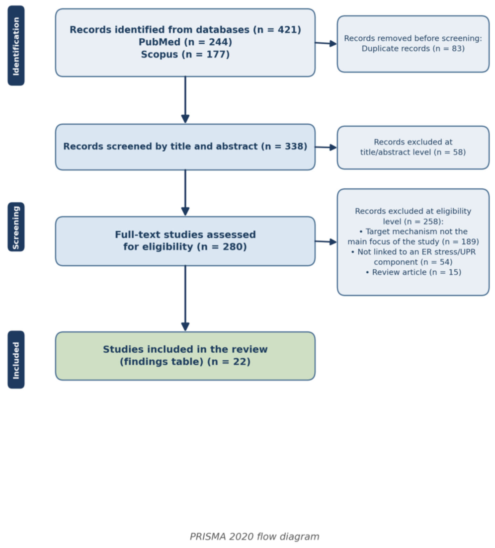
Figure 2.
Leptin signal transduction under physiological conditions. Parallel signalling arms emerging from the leptin–LepRb–JAK2 axis and their net effect on hypothalamic POMC/AgRP neurons.
Figure 2.
Leptin signal transduction under physiological conditions. Parallel signalling arms emerging from the leptin–LepRb–JAK2 axis and their net effect on hypothalamic POMC/AgRP neurons.
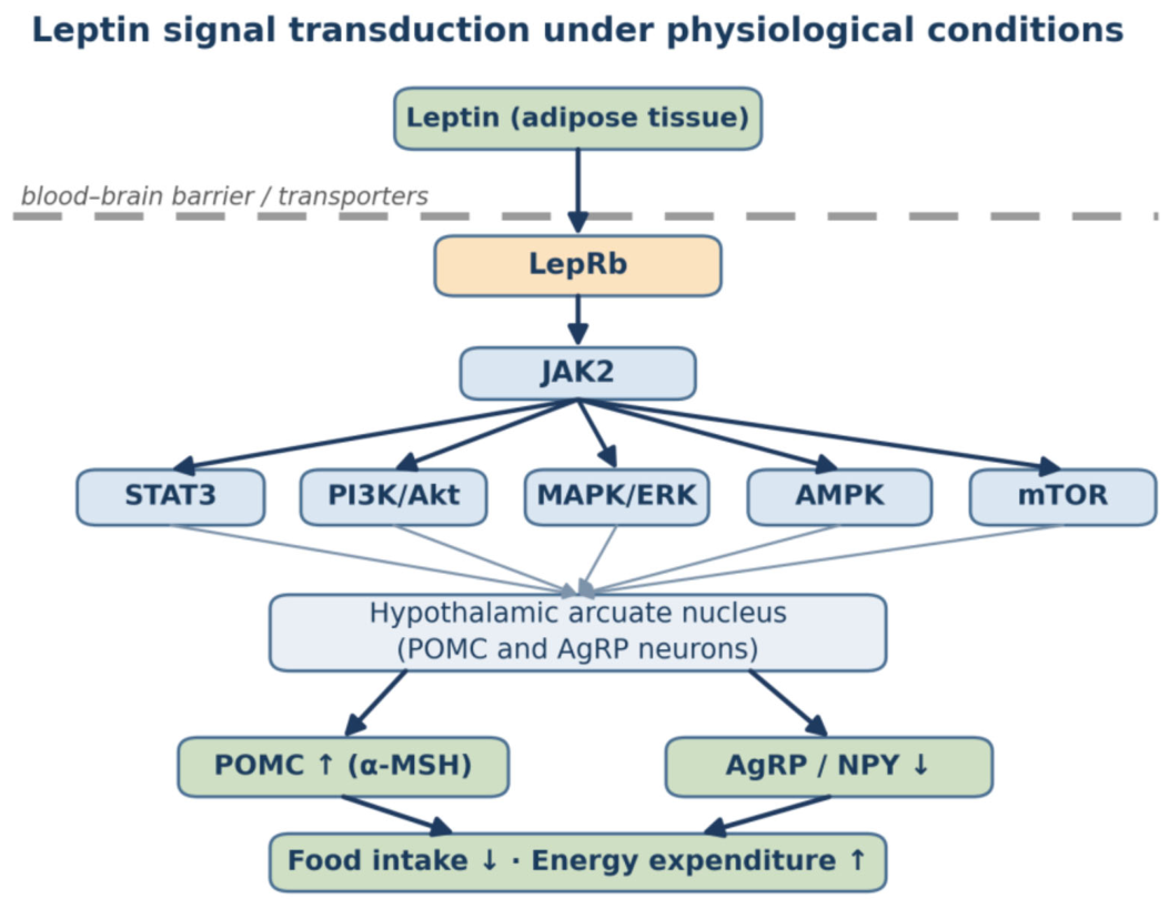
Figure 3.
ER stress-mediated leptin resistance: a unified mechanistic model. Convergence of distinct stressors on hypothalamic ER stress, their subsequent convergence through the three UPR arms onto a narrow effector set (CHOP, SOCS3, PTP1B), and the production of leptin resistance within a self-reinforcing loop. The SEL1L–HRD1 ERAD arm on the left operates independently of ER stress.
Figure 3.
ER stress-mediated leptin resistance: a unified mechanistic model. Convergence of distinct stressors on hypothalamic ER stress, their subsequent convergence through the three UPR arms onto a narrow effector set (CHOP, SOCS3, PTP1B), and the production of leptin resistance within a self-reinforcing loop. The SEL1L–HRD1 ERAD arm on the left operates independently of ER stress.
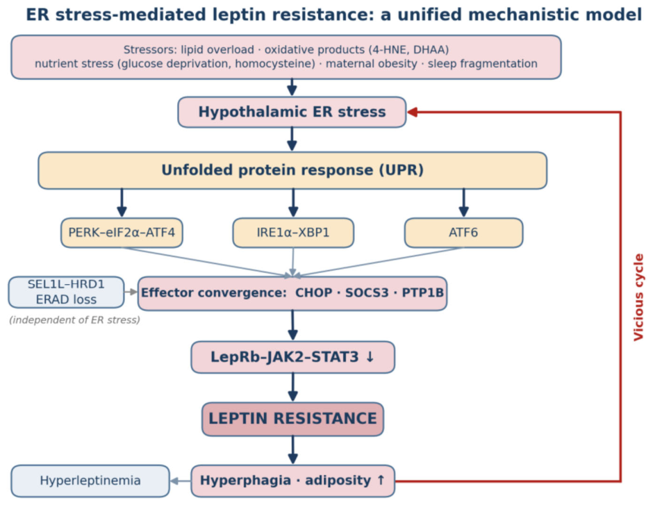
Figure 4.
Therapeutic targets and levels of evidence. Therapeutic approaches by level of intervention and the nature of the evidence available for each.
Figure 4.
Therapeutic targets and levels of evidence. Therapeutic approaches by level of intervention and the nature of the evidence available for each.
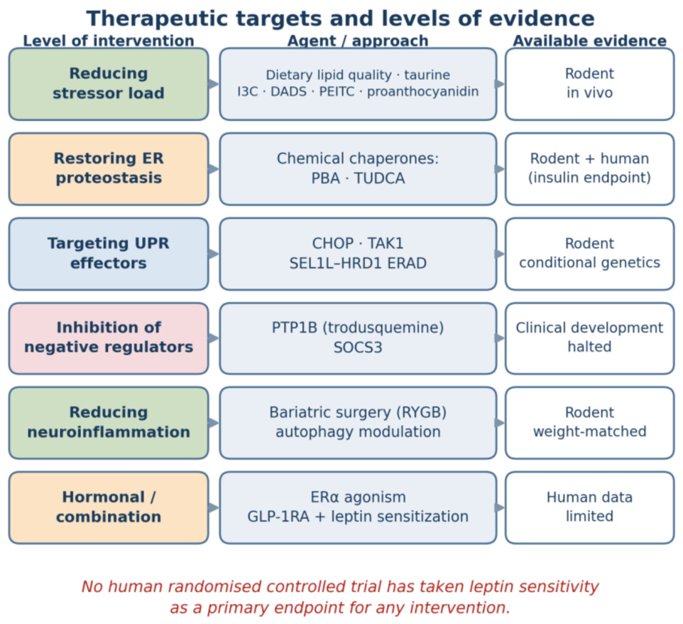

Table 1.
Summary of Findings. Summary of the 22 primary studies addressing the relationship between ER stress and intracellular signal transduction in leptin resistance (in descending order of publication year). The set contains no human studies and no randomised controlled trials (animal in vivo n = 17; laboratory studies only n = 5). The evidence column is colour-coded by model system: green = human data, yellow = animal study, red = laboratory (in vitro) study; because no human study is included, the green category is unrepresented.
Table 1.
Summary of Findings. Summary of the 22 primary studies addressing the relationship between ER stress and intracellular signal transduction in leptin resistance (in descending order of publication year). The set contains no human studies and no randomised controlled trials (animal in vivo n = 17; laboratory studies only n = 5). The evidence column is colour-coded by model system: green = human data, yellow = animal study, red = laboratory (in vitro) study; because no human study is included, the green category is unrepresented.
| Study (author, reference) | Model / System | Level of Evidence | ER Stress / UPR and Associated Pathways | Key Finding |
|---|---|---|---|---|
| Kim (41) | Mouse (ApoE−/−, in vivo) | Animal study – dietary intervention | ER stress/UPR | Objective: Indole-3-carbinol (I3C) improves metabolic dysfunction in diet-induced obesity; however, its role in protecting against ER stress-induced hyperleptinemia remains unclear. |
| Xu (37) | Mouse (C57BL/6J, in vivo) | Animal study – dietary intervention | JAK2/STAT3, ER stress/UPR | PEITC suppressed hypothalamic neuroinflammation and attenuated ER stress across all three unfolded protein response branches. |
| El-Sehrawy (65) | Mouse (OVX, in vivo) | Animal study – pharmacological | JAK2/STAT3, ER stress/UPR | We conclude that ERα activation may reduce hypothalamic ER stress and improve leptin sensitivity, thereby regulating energy intake and body weight. |
| Hua (35) | Rat (DIO) + BV2/GT1-7 cell (in vitro) | Animal study + laboratory (in vitro) | JAK2/STAT3, SOCS3, PTP1B, ER stress/UPR | Glycolipid indicators, autophagy, ER stress, and leptin resistance markers were evaluated using biochemical assays, qPCR, Western blotting, and immunofluorescence. |
| Mao (19) | Mouse (POMC-specific SEL1L-KO, in vivo) | Animal study – conditional genetics | ER stress/UPR | This study uncovers an important role of SEL1L-HRD1 ERAD in the pathogenesis of central leptin resistance and leptin signaling. |
| Kim & Kim (40) | Mouse (ApoE−/−, in vivo) | Animal study – dietary intervention | ER stress/UPR | Background: The endoplasmic reticulum (ER) plays a pivotal role in maintaining cellular metabolic homeostasis. |
| Preninka (45) | SH-SY5Y-ObRb human cell line (in vitro) | Laboratory only (in vitro) | JAK2/STAT3, ER stress/UPR | These effects may be specific to homocysteine and to the leptin pathway, as other homocysteine-related compounds, namely methionine and cysteine, have weak effect on leptin-induced inhibition of STAT3 phosphorylation, and homocysteine has no impact on IL-6-induced activation of STAT3. |
| Baba (33) | Mouse (ob/ob), isolated pancreatic islet (ex vivo) | Animal study – pharmacological | SOCS3, ER stress/UPR | It is suggested that chronic ER stress is associated with obesity and leptin resistance. |
| Chen (47) | Rat (in vivo, RYGB surgery) | Animal study – weight-matched control | ER stress/UPR | Mechanistically, we demonstrate that RYGB interferes with hypothalamic ER stress and toll-like receptor 4 (TLR4) signaling to restore the anorexigenic action of leptin, which most likely results from modulation of a circulating factor derived from the altered gut microbial environment upon RYGB surgery. |
| Gong (44) | Mouse (in vivo) + mHypoE-44 neuron (in vitro) | Animal study – causal genetic | JAK2/STAT3, ER stress/UPR | Innovation and Results: Our initial experiment in vivo demonstrated that (i) leptin promotes hypothalamic expression of SELENOM and (ii) leptin-induced STAT3 phosphorylation is impeded by SELENOM deficiency. |
| Horiuchi (70) | Neuronal cell line (in vitro) | Laboratory only (in vitro) | ER stress/UPR | We showed that biochanin A, a family of isoflavones, strongly attenuated cell death induced by ER stress in neuronal cells, improved ER stress-induced impairments in leptin signaling, and suppressed ER stress-induced expression of glucose-regulated protein 78. |
| Drori (18) | Mouse (CHOP-KO) + hepatosit (in vitro) | Animal study – causal genetic | ER stress/UPR | Our findings highlight a novel molecular aspect by which the hepatic eCB/CB1R system is involved in the development of hepatic leptin resistance and in the regulation of sOb-R levels via CHOP. |
| Park (38) | Mouse (maternal obezite, in vivo) | Animal study – pharmacological | ER stress/UPR | Neonatal treatment with the ER stress-relieving drug tauroursodeoxycholic acid improved metabolic and neurodevelopmental deficits and reversed leptin resistance in the offspring of obese dams. |
| Aiceles (20) | Rat (in vivo) | Animal study – observational / model | JAK2/STAT3, SOCS3, ER stress/UPR | We can conclude that hypothyroxinemia condition in rats on adulthood results in impairment of the leptin signaling pathway via ObRb-STAT3 in the hypothalamus, which is likely to be involved in the leptin resistance. |
| Hosoi (42) | Neuronal cell line (in vitro) | Laboratory only (in vitro) | JAK2/STAT3, ER stress/UPR | Overall, these results suggest that 4-HNE may partly affect endoplasmic reticulum stress-induced unfolded protein response signaling and may be involved in the pathogenesis of leptin resistance. |
| Gan (21) | Mouse (in vivo) + preadiposit (in vitro) | Animal study + laboratory (in vitro) | JAK2/STAT3, SOCS3, ER stress/UPR | In summary, our data revealed αMSH enhanced leptin sensitivity and preadipocyte proliferation, meanwhile inhibited ER stress of preadipocytes by activating Notch1 signal. |
| Ibars (32) | Rat (Wistar, in vivo) | Animal study – dietary intervention | JAK2/STAT3, SOCS3, PTP1B, ER stress/UPR | The effects of the GSPE treatment in the peripheral tissues were not as evident as those in the hypothalamus, although the GSPE treatment significantly restored the mRNA levels of Socs3 and Ptp1b in the skeletal muscle. |
| Hosoi (25) | Neuronal cell line (in vitro) | Laboratory only (in vitro) | JAK2/STAT3, ER stress/UPR | The present study showed that glucose deprivation inhibited leptin-induced phosphorylation of signal transducer and activator of transcription 3 (STAT3) and signal transducer and activator of transcription 5 (STAT5) in neuronal cells. |
| Sai (36) | Mouse (CNS-specific Tak1-KO) + cell (in vitro) | Animal study – conditional genetics | ER stress/UPR | In the in vivo setting, central nervous system (CNS)-specific Tak1 deletion upregulated SREBP-target lipogenic genes and blocked ER stress in the hypothalamus. |
| Thon (43) | SH-SY5Y-ObRb human cell line (in vitro) | Laboratory only (in vitro) | JAK2/STAT3, ER stress/UPR | However, most obese subjects are in a state of leptin resistance, and endoplasmic reticulum (ER) stress is suggested to be involved in the pathophysiology of leptin resistance. |
| Camargo (46) | Mouse (in vivo) | Animal study – dietary intervention | ER stress/UPR | Global alterations in the expressions of genes involved in hypothalamic metabolism, cellular defense, apoptosis and endoplasmic reticulum stress pathways were induced by dietary manipulations and Tau treatment. |
| Hakim (34) | Mouse (CHOP+/− transgenic, in vivo) | Animal study – causal genetic | JAK2/STAT3, SOCS3, PTP1B, ER stress/UPR | SF-induced effects were reversed following TUDCA treatment and were absent in CHOP -/+ mice.; Conclusions: Sleep fragmentation (SF) induces hyperphagic behaviors and reduced leptin signaling in hypothalamus that are mediated by activation of endoplasmic reticulum (ER) stress, and ultimately lead to increased PTP1B acti… |
Abbreviations: UPR, unfolded protein response; ER, endoplasmic reticulum; JAK2, Janus kinase 2; STAT3, signal transducer and activator of transcription 3; SOCS3, suppressor of cytokine signalling 3; PTP1B, protein tyrosine phosphatase 1B.
Table 2.
Knowledge Gaps and Proposed Research Approaches. The principal gaps identified by this review and the methodological approach required for each.
Table 2.
Knowledge Gaps and Proposed Research Approaches. The principal gaps identified by this review and the methodological approach required for each.
| Gap area | Current situation | Approach required |
|---|---|---|
| Human intervention studies | Agents targeting ER stress have been tested in humans against insulin endpoints only | Randomised controlled trial with leptin sensitivity as the primary endpoint |
| PI3K/Akt – ER stress link | No primary study links the two processes directly | Simultaneous UPR and PI3K measurement in conditional genetic models |
| mTOR – autophagy – ER stress | Current evidence rests on extrapolation from peripheral tissues | Tissue-specific studies with hypothalamic endpoints |
| FoxO1 – UPR interaction | No primary study addresses the question directly | Examination of the STAT3/FoxO1 promoter balance under ER stress |
| Sex differences | The great majority of studies were conducted in male animals | Inclusion of both sexes; sex-stratified analysis |
| Intestinal permeability and microbiota | RYGB data point to gut-derived signals, but the mechanism is unresolved | Gnotobiotic models; hypothalamic UPR measurement after microbiota transfer |
| Epigenetic mediators | Correlational human data exist for DNA methylation and miRNA; no link to ER stress has been established | Integration of the ER stress and epigenome layers within the same model |
| Cellular resolution | Measurements rest on tissue homogenates | Single-cell RNA sequencing; spatial transcriptomics |
| Absence of biomarkers | Hypothalamic ER stress cannot be measured in humans | Identification of circulating biomarkers through multi-omics |
| Nutrigenomics / precision nutrition | Individual response variability is unknown | Nutrition studies stratified by genotype and microbiota |
Table 3.
Testable Hypotheses of the Proposed Framework. Each row gives a hypothesis, the outcome expected if it holds, an experimental design capable of testing it, the finding that would falsify it, and the extent to which it has been tested. The final column is excluded from the colour coding because it reports the state of testing rather than the model system from which the evidence derives.
Table 3.
Testable Hypotheses of the Proposed Framework. Each row gives a hypothesis, the outcome expected if it holds, an experimental design capable of testing it, the finding that would falsify it, and the extent to which it has been tested. The final column is excluded from the colour coding because it reports the state of testing rather than the model system from which the evidence derives.
| Hypothesis | Expected outcome | Proposed experiment | Falsifying finding | State of testing |
|---|---|---|---|---|
| Hypothesis 1 — Input independence | Whatever the stressor, the effector signature (CHOP↑, SOCS3↑, PTP1B↑) is similar. | Parallel administration of five stressors (saturated lipid load, 4-HNE, homocysteine, glucose deprivation, sleep fragmentation) in the same strain, at the same age and time point, with effector signatures compared by hypothalamic single-cell RNA-seq. | Stressors producing discrete, non-overlapping effector signatures. | Indirectly consistent; no direct comparative study |
| Hypothesis 2 — Node primacy | Intervening at the shared effector node is more effective than targeting upstream stressors individually. | Direct comparison, within the same DIO model, of CHOP silencing against three upstream interventions (antioxidant, anti-inflammatory, lipid reduction); primary endpoint weight-matched leptin sensitivity. | Any upstream intervention matching or exceeding the node intervention. | Partially supported (CHOP+/−; chemical chaperones) |
| Hypothesis 3 — Proteostasis–stress distinction | Disrupted ER proteostasis can produce leptin resistance without any UPR activation. | Measurement of leptin sensitivity in POMC-specific SEL1L/HRD1 deletion while UPR markers (BiP, sXBP1, CHOP) remain unchanged; proteostatic capacity monitored independently of the UPR. | Leptin resistance arising only when it accompanies UPR activation. | Supported (19) — the most original aspect of the framework |
| Hypothesis 4 — Load-to-capacity ratio | What matters is not absolute lipid load but its ratio to ER folding capacity. | Measurement of the effect of the same high-fat load on leptin sensitivity in mice with genetically increased ER folding capacity (POMC-XBP1s). | Capacity-enhanced animals developing the same degree of leptin resistance at the same dietary load. | Indirect support (36, 39) |
| Hypothesis 5 — Translatability to humans | Agents targeting ER proteostasis improve leptin sensitivity in humans. | Randomised, placebo-controlled, weight-matched trial of TUDCA or 4-PBA; primary endpoint a standardised measure of leptin sensitivity. | No difference in leptin sensitivity versus placebo once weight is matched. | Untested — Kars et al. (56) and Xiao et al. (57) measured no leptin endpoint |
Table 4.
Comparison of Molecular Mechanisms. Comparison of the mechanisms leading to leptin resistance by UPR arm affected, point of intervention and level of evidence. The evidence column is colour-coded by the model system in which each mechanism was demonstrated: green = human data, yellow = animal study, red = laboratory (in vitro) study. Because none of the mechanisms listed is supported by human studies, the green category is unrepresented.
Table 4.
Comparison of Molecular Mechanisms. Comparison of the mechanisms leading to leptin resistance by UPR arm affected, point of intervention and level of evidence. The evidence column is colour-coded by the model system in which each mechanism was demonstrated: green = human data, yellow = animal study, red = laboratory (in vitro) study. Because none of the mechanisms listed is supported by human studies, the green category is unrepresented.
| Mechanism / level | UPR arm | Point of action | Key studies | Level of evidence (model system) |
|---|---|---|---|---|
| LepRb quality control (ERAD) | Independent of ER stress | Receptor biogenesis; retention of LepRb in the ER | (19) | Animal study — tissue-specific conditional genetics |
| CHOP-mediated regulation of sOb-R | PERK–eIF2α–CHOP | Receptor bioavailability; circulating free leptin | (18) | Animal study — genetic (CHOP-KO) + pharmacological |
| PTP1B-mediated JAK2 dephosphorylation | All three arms | Post-receptor signal termination | (32, 34, 35) | Animal study — genetic (CHOP+/−) + correlative |
| SOCS3-mediated feedback inhibition | PERK and IRE1α | LepRb Tyr985; suppression of JAK2 | (20, 21, 33) | Animal study — largely correlative |
| Redox / oxidative ER stress | PERK–eIF2α–CHOP | Selective inhibition of STAT3 phosphorylation | (42–44) | Predominantly laboratory (in vitro) — limited animal support |
| Lipid load / ER capacity balance | IRE1α–XBP1; ATF6 | ER membrane capacity; SREBP-mediated expansion | (36, 40, 41) | Animal study — conditional genetics + diet |
| Neuroinflammation–autophagy axis | All three arms | Microglia–neuron interaction; autophagic flux | (35, 47) | Animal study — weight-matched control |
| Developmental programming | PERK–eIF2α–CHOP | Melanocortin circuit development; sustained SOCS3 elevation | (38, 46, 48) | Animal study — reversible by intervention |
Table 5.
Molecular Target – Dietary/Pharmacological Modulator – Level of Evidence. Molecular targets identified in leptin resistance and their corresponding dietary or pharmacological modulators. The evidence column indicates the model system in which each pairing was demonstrated and is colour-coded accordingly: green = human data, yellow = animal study, red = laboratory (in vitro) study.
Table 5.
Molecular Target – Dietary/Pharmacological Modulator – Level of Evidence. Molecular targets identified in leptin resistance and their corresponding dietary or pharmacological modulators. The evidence column indicates the model system in which each pairing was demonstrated and is colour-coded accordingly: green = human data, yellow = animal study, red = laboratory (in vitro) study.
| Molecular Target | Nutritional Strategy | Model / System | Level of Evidence |
|---|---|---|---|
| ER stress / UPR (general) | Reduction of saturated fat and excess caloric load | Mouse, ApoE−/− (40, 41) | Animal study — dietary intervention |
| GRP78, CHOP, XBP1 | Indole-3-carbinol (cruciferous vegetables) | Mouse, ApoE−/− (41) | Animal study — dietary intervention |
| ER stress + JAK2/STAT3 | Phenethyl isothiocyanate (cruciferous vegetables) | Mouse, C57BL/6J (37) | Animal study — dietary intervention |
| ER stress + cholesterol | Diallyl disulfide (garlic) | Mouse, ApoE−/− (40) | Animal study — dietary intervention |
| STAT3 / SOCS3 / PTP1B | Proanthocyanidins (grape seed) | Rat, cafeteria diet (32) | Animal study — dietary intervention |
| ER stress + autophagy | Berberine (FTZ preparation) | Rat, DIO + BV2/GT1-7 (35) | Animal study + laboratory (in vitro) |
| Hypothalamic ER stress | Taurine supplementation | Mouse, protein restriction + HFD (46) | Animal study — dietary intervention |
| GRP78 / ER-derived cell death | Biochanin A (isoflavone) | Neuronal cell line (70) | Laboratory (in vitro) only |
| PTP1B + AMPK | Cannabisin A and B (hemp seed) | In vitro + computational (63) | Laboratory (in vitro) only |
| Neuronal leptin responsiveness | Rubisco-derived plant peptide | Rodent, oral administration (71) | Animal study — oral administration |
| Gut–hypothalamus axis | Microbiota-derived short-chain fatty acids | Human/animal review (72); rat RYGB (47) | Animal study — indirect; mechanism untested |
| Developmental programming (SOCS3) | Modulation of maternal diet quality | Mouse/rat (24, 38, 48) | Animal study — critical-window intervention |
| ER proteostasis (chaperone) | Pharmacological: TUDCA / 4-PBA | Human (56, 57) | Human data — but insulin endpoint |
Table 6.
Therapeutic Interventions and the State of Human Data. Interventions targeting the ER stress–leptin resistance axis; the human data column indicates the current limit of clinical translation. The column is colour-coded by the model system in which the intervention was evaluated: green = human data, yellow = animal study, red = laboratory (in vitro) study.
Table 6.
Therapeutic Interventions and the State of Human Data. Interventions targeting the ER stress–leptin resistance axis; the human data column indicates the current limit of clinical translation. The column is colour-coded by the model system in which the intervention was evaluated: green = human data, yellow = animal study, red = laboratory (in vitro) study.
| Agent / approach | Class | Model | Key endpoint | Human data |
|---|---|---|---|---|
| TUDCA | Chemical chaperone | Mouse (34, 38); human (56) | Leptin sensitivity ↑ (rodent); hepatic/muscle insulin sensitivity +30% (human) | Yes — but no leptin endpoint (n = 20, 4 weeks) |
| 4-PBA (sodium phenylbutyrate) | Chemical chaperone | Mouse (33); human (57) | UPR markers ↓, SOCS3 modulation; lipid-induced insulin resistance ↓ | Yes — no leptin endpoint (n = 8, 2 weeks) |
| Recombinant leptin | Hormone replacement | Human RCT (58) | ~3% placebo-adjusted weight loss in obesity | Yes — largely ineffective |
| Trodusquemine (MSI-1436) | PTP1B inhibitor | Mouse (62); phase 1/1b | Fat-specific weight loss; appetite suppression | Phase 1 completed; obesity programme not pursued |
| GLP-1RA (liraglutide) | Incretin analogue | Human RCT (64) | Prevention of the fall in free leptin during weight-loss maintenance | Yes — leptin level measured, sensitivity not |
| Roux-en-Y gastric bypass | Surgical | Rat (47) | Hypothalamic ER stress ↓, leptin sensitivity ↑ (independent of weight loss) | Indirect — mechanism untested in humans |
| PEITC, I3C, DADS, proanthocyanidins, berberine, taurine, biochanin A | Diet-derived phytochemical | Rodent in vivo / in vitro | ER stress markers ↓, pSTAT3 ↑, weight ↓ | None — doses above the range attainable through diet |
| ERα agonism (PPT) | Hormonal | Mouse, ovariectomised (65) | Hypothalamic ER stress ↓, leptin signalling ↑ | None |
Table 7.
Proposed Leptin Resistance Phenotypes and the Chain of Clinical Testing. The chain of which patient – which dominant mechanism – which candidate biomarker – which intervention – which primary endpoint. The phenotypes are derived from animal and laboratory studies; none has been confirmed in humans. The final column is colour-coded by the model system on which the phenotype rests: green = human data, yellow = animal study, red = laboratory (in vitro) study.
Table 7.
Proposed Leptin Resistance Phenotypes and the Chain of Clinical Testing. The chain of which patient – which dominant mechanism – which candidate biomarker – which intervention – which primary endpoint. The phenotypes are derived from animal and laboratory studies; none has been confirmed in humans. The final column is colour-coded by the model system on which the phenotype rests: green = human data, yellow = animal study, red = laboratory (in vitro) study.
| Proposed phenotype | Dominant mechanism | Candidate biomarker (human) | Expected response to intervention | Proposed primary endpoint | Level of evidence |
|---|---|---|---|---|---|
| Leptin-sensitive obesity (reference) | No dominant signalling defect; energy balance load predominates | Leptin within the range expected for fat mass; normal sOb-R | Expected response to caloric restriction and GLP-1 RA | Weight loss; hunger/satiety scores | Human data — clinical observation |
| ER-dominant (proteostatic) phenotype | Retention of LepRb in the ER, SEL1L–HRD1 ERAD defect; CHOP-mediated regulation of sOb-R | sOb-R ↑ / free leptin index ↓; LEPR misfolding variants (e.g. Cys604Ser) | Group expected to benefit most from chemical chaperones (TUDCA, 4-PBA) | Weight-matched leptin sensitivity | Animal study (conditional genetics) + a single human variant report |
| Inflammatory phenotype | Hypothalamic low-grade inflammation; SOCS3 induction | hsCRP, IL-6, TNF-α; neuroinflammation imaging markers | Anti-inflammatory dietary pattern; RYGB (weight-independent effect) | hsCRP + weight-matched leptin sensitivity | Animal study; indirect in humans (47) |
| Oxidative phenotype | Lipid peroxidation products (4-HNE); loss of redox protection (selenoprotein M) | Plasma 4-HNE adducts; oxidised LDL; selenium status | Redox-targeted intervention; reduction of saturated fat load | 4-HNE adduct level + food intake | Animal study + laboratory (in vitro) |
| Post-receptor inhibition (PTP1B-dominant) | PTP1B-mediated JAK2 dephosphorylation; localisation to the ER membrane | No direct marker; indirectly, indices of insulin resistance | PTP1B modulation — unconfirmed clinically | HOMA-IR + leptin sensitivity | Animal study; human trial inconclusive (trodusquemine) |
| Developmental / epigenetic phenotype | Melanocortin circuit programming driven by maternal obesity | Adiposity-associated DNA methylation signatures; circulating miRNA profiles | Intervention within the pregnancy and early childhood window | Childhood adiposity trajectory | Human data (correlational) + animal study (53, 54) |
| Transcriptional balance phenotype | STAT3/FoxO1 balance and STAT3 acetylation status | No known circulating biomarker | No benefit expected from chemical chaperones | Not defined | Animal study; not directly tested |
Disclaimer/Publisher’s Note: The statements, opinions and data contained in all publications are solely those of the individual author(s) and contributor(s) and not of MDPI and/or the editor(s). MDPI and/or the editor(s) disclaim responsibility for any injury to people or property resulting from any ideas, methods, instructions or products referred to in the content. |
© 2026 by the authors. Licensee MDPI, Basel, Switzerland. This article is an open access article distributed under the terms and conditions of the Creative Commons Attribution (CC BY) license (http://creativecommons.org/licenses/by/4.0/).
Copyright: This open access article is published under a Creative Commons CC BY 4.0 license, which permit the free download, distribution, and reuse, provided that the author and preprint are cited in any reuse.