

Submitted:
17 September 2026
Posted:
18 September 2026
You are already at the latest version
Abstract
Specific cell tracking or targeted modulation of cellular responses are often limited by the availability, specificity and effector functions of endogenous cell-surface receptors. Synthetic receptors with customizable functionality can overcome these limitations. Based on a qualitative survey of the literature, this narrative review describes the design, the functionality and the applications of nanobody based synthetic receptor/ligand systems that employ either GFP/mCherry or the clinically approved humanized antibody Palivizumab as ligands. Depending on their molecular architecture, these systems can mediate cargo internalization for cell tracking or induce defined cellular responses through engineered interleukin, interferon, or Fas signaling pathways. We position these platforms within the broader field of synthetic receptors by discussing engineered GPCR- and Notch-based systems and briefly outline the development and clinical application of CAR T cells to illustrate the translational potential of synthetic receptor systems. Finally, we discuss the opportunities and limitations of GFP/mCherry- and Palivizumab-based systems, with particular emphasis on immunogenicity, in vivo applicability, manufacturability, scalability, and clinical translation. By linking modular receptor design to distinct functional outputs, this review highlights both the established value of GFP/mCherry- and Palivizumab-based synthetic receptor–ligand systems as experimental tools and their potential for future applications in molecular imaging and controlled signal transduction.
Keywords:
synthetic receptors
; molecular engineering
; signal transduction
; cell targeting
; endocytosis
; CAR T cell
; green fluorescent protein
; mCherry
; Palivizumab
; nanobody
1. Scope and Methodology
Tracking of specific cell-types or site-directed delivery of drugs via endogenous cell surface receptors is sometimes limited by a lack in availability, specificity, and effector functionality. The development of synthetic ligand-/receptor-pairs could overcome these limitations and enable the active targeting for specific labeling of individual cell types and for the targeted regulation of biological processes. These synthetic ligand-receptor pairs are normally absent in humans or animals and are therefore highly specific. Importantly, their composition can be designed to facilitate effector functions such as internalization for cell-labeling or the activation of defined signal transduction pathways.
In the present review article, we describe the design, functionality and utilization of GFP/mCherry (Chapter 3) and Palivizumab (Chapter 4) based synthetic ligand/receptor systems. These systems have been used in the past for cell imaging [1] or induction of cell-signaling for functional modulation of the target cells [2,3,4]. In the first part of the review (Chapter 2) we briefly introduce synthetic receptors in general, which helps the reader to compare the GFP/mCherry and Palivizumab receptor/ligand systems to other synthetic receptor/ligand systems. Furthermore, we highlight CAR T cells (Chapter 2.1) to demonstrate the translational potential and the impact that such systems can have for patients. In the discussion and outlook section (Chapter 5), we do also describe challenges and opportunities for a clinical application of these receptors.
This manuscript is a narrative review and information was collected by an extensive search of the literature between 2015-2026. The primary databases searched were „Pubmed“ and „Google Scholar“, and we additionally screened selected journals (e.g. Nature Nanotechnology or ACS journals). The following key-words were used for the search: „Synthetic receptors“; „artificial receptors“; specific search for selected most known/used synthetic receptors (e.g. „chimeric antigen receptor“, „synthetic cytokine receptor“, „MESA receptor“, „synthetic Notch receptor“); „CAR T cell“; „CAR NK cell“; „nanobody”; „nanobody cancer“; „nanobody synthetic receptor“; „cell labeling“; „fluorescence labeling“; „radiolabeling“; „magnetic beads“; „protein tags“; „GFP“; „bioorthogonal functionalization“; „cysteine additions“; „lysine additions“.
There was no predefined study protocol with formal criteria for inclusion of studies. Exclusion criteria were also qualitative. After screening the results, we decided to focus on receptors for GFP/mCherry and Palivizumab, for engineering of T-cells, programmable cell therapies, protein- or nanobody-based transmembrane receptors, synthetic cytokine receptors, and membrane receptors for cell therapy. We omitted publications dealing with: olfactory receptors, photoreceptors, sensory neurons, natural receptors or ion channels. Furthermore, no statistical methods were used to summarize or quantitatively evaluate study data or to assess publication counts on specific topics.
2. A Brief Introduction Into the Design and Application of Synthetic Receptors
Synthetic receptors are engineered proteins or protein complexes that in this form do not occur in nature and are designed to interact with defined biological or chemical signals to trigger a programmed biological output [5,6]. These receptors can be designed to label cells with contrast agents for cell imaging studies, or to precisely control cellular functions such as triggering specific signal transduction pathways. This opens new ways to visualize or modulate cells and may also enhance specificity, reduce off-target effects and therefore increases the safety and efficacy of cell specific delivery of contrast agents and pharmacological drugs.
2.1. Synthetic Receptor/Ligand Systems Based on Natural Receptors
There are several types of synthetic receptors, which are for example based on G-protein-coupled receptors (GPCR), Notch receptors or T-cell receptors. GPCRs are the largest group of human membrane proteins with about 800 members that bind a variety of different molecules and stimulate intracellular signaling pathways. GPCRs consist of seven transmembrane domains, which are connected by three extracellular loops and three intracellular loops [7] (Figure 1A). After ligand binding the receptor changes its conformation that enables the interaction with intracellular G proteins and initiates signal transduction into the cell. By introduction of point mutations, GPCRs were made insensitive to their native ligands, but they became selectively responsive to small molecules like Clozapine-N-oxide or C21 [8]. These GPCRs were termed DREADDs (designer receptors exclusively activated by designer drugs) that have been utilized in neuroscience to activate or silence neuronal activity [8,9]. Based on the design of DREADDs, one group has developed synthetic receptors that stimulate cellular processes in response to a defined antigen [10]. They fused a GFP-nanobody and a peptide antagonist to the N-terminus of a GPCR, which blocked the ligand binding site. Addition of GFP or proteolytic cleavage of the autoinhibitory domain enabled binding of the ligand, receptor activation and induction of biological responses such as gene expression, G-protein activation, but also macrophage activation or T-cell killing. These receptor-systems were called PAGERs for programmable antigen-gated G-protein-coupled engineered receptors [10]. Another study achieved the stimulation of pathway selective signaling by fusing a peptide ligand against the type-1 parathyroid receptor (PTH1-11) to a nanobody (Nb) that binds to a separate site on the receptor [11]. Interestingly, the PTH1-11-Nb conjugate was highly selective for the Gαs/cAMP pathway whereas PTH1-11 alone activated all signaling pathways.
Another group of synthetic receptors is based on the native Notch receptor and are called MESA (modular extracellular sensor architecture, Figure 1B), synNotch (synthetic Notch, Figure 1C) or SNIPR (synthetic intramembrane proteolysis receptor).
Notch receptors are transmembrane receptors that mediate communication between directly neighboring cells. They primarily regulate cell fate, differentiation, proliferation, and cell death [12]. Unlike many other receptors, they do not activate a classical intracellular kinase or second-messenger cascade. Instead, they undergo proteolytic cleavage after ligand binding, thereby releasing an intracellular transcriptional regulatory fragment known as NICD (Notch intracellular domain). NICD translocates to the nucleus, where it binds to the DNA-bound transcription factor CSL and to coactivators such as MAML (mastermind-like) [13], that activate Notch target genes.
The synthetic receptors MESA, synNotch and SNIPR have the same function as the native Notch receptors. After ligand binding, they trigger the proteolytic release of an intracellular transcription factor. The main difference is how ligand binding is translated into proteolysis and whether the receptor consists of one or two chains. MESA receptors are two-chain-based systems with a protease chain and target chain that detect soluble ligands through ligand-induced receptor dimerization (Figure 1B) [14]. The synNotch receptors are single-chain, Notch-derived receptors that detect cell-surface antigens and release a synthetic transcription factor upon proteolytic cleavage (Figure 1C) [15]. SNIPRs are a modified variant of synNotch based on regulated intramembrane proteolysis and the single-chain that retain the Notch cleavage mechanism [16] but can be adapted to soluble ligand detection instead of cell-surface antigens (Figure 1D). Piraner et al. expanded the SNIPR platform to enable robust detection of soluble ligands [17]. Ligand-induced receptor clustering and endocytosis promoted pH-dependent proteolytic activation, releasing a synthetic transcription factor that reprogrammed cellular gene expression. The authors applied this approach to tumor-associated soluble factors, including TGF-β and VEGF, and demonstrated that, after recognition, a therapeutic gene program or a CAR can be activated. It has also been demonstrated that MESA can be developed to bind a soluble factor such as VEGF and produce the immunostimulatory factor IL-2. This receptor was introduced into human T cells to enable ligand-dependent regulation of their function [18]. In summary, these receptor systems are used, among other applications, to develop safer and more precise CAR T-cell therapies.
2.2. Chimeric Antigen Receptor T-Cells (CAR T Cells)
The first synthetic receptor developed for immunotherapy is the chimeric antigen receptor (CAR), which has been further developed over the years and is applied in the treatment of various diseases, most notably through targeting CD19 in B-cell malignancies [19,20]. CARs represent a groundbreaking immunotherapeutic strategy in cancer treatment that combines synthetic biology with genetic engineering [21,22,23,24]. CAR T cells are T-lymphocytes that have been genetically modified to express synthetic receptors, allowing them to recognize and eliminate cancer cells with high specificity. These chimeric antigen receptors combine the antigen-binding capacity of monoclonal antibodies with the cytotoxic functions of T-cells, enabling targeted and potent anti-tumor activity.
The CAR molecule consists of four key domains (Figure 2, left): an extracellular antigen recognition domain, a hinge region, a transmembrane domain, and one or more intracellular signaling domains [25,26,27]. The antigen recognition domain often consists of single-chain variable fragments (scFvs) derived from antibodies (Figure 2, left), but it can also be composed of peptides or full extracellular domains [28,29]. The signal domain of CARs is similar to the domains of the native T-cell receptor (TCR; Figure 2). This modular design allows the CAR T cell to recognize tumor cell surface antigens independently of major histocompatibility complex (MHC) presentation, which differs fundamentally from the physiology of native TCRs [30].
Over time, the CAR technology has undergone significant advancements, resulting in the delineation of five distinct receptor generations. Figure 2 gives an overview on the structural and functional developments of the generations of CARs. First-generation CARs, introduced in 1993, comprise an extracellular ligand-binding or scFv domain in conjunction with an intracellular CD3ζ signaling motif, reflective of the native TCR architecture. Second-generation CARs emerged in 2002 and integrate an scFv recognition domain with a single co-stimulatory signaling element, most frequently derived from CD137 or CD28, which are co-stimulation molecules and have an important role in the activation and function of T-cells. Third-generation CARs, developed between 2005 and 2010, are defined by the inclusion of two co-stimulatory domains (typically CD137 and CD28) to further augment T-cell activation. Fourth-generation CARs are designated as T-cells redirected for universal cytokine-mediated killing (TRUCKs). These cell-types are genetically modified to express transgenes encoding cytokines or immune-modulatory molecules, thus enabling modulation of the tumor microenvironment. Fifth-generation CARs, introduced in 2018, are distinguished by the incorporation of three synergistic co-stimulatory modules, further enhancing T-cell functionality, persistence, and therapeutic potential [24,31].
CAR T-cell therapy targeting the B-cell antigen CD19 has emerged as the most widely employed strategy for the treatment of B-cell malignancies [33]. In addition to CD19 alternative antigenic targets have been explored to expand the application of CARs to diverse malignancies, including hematological diseases, solid tumors and autoimmune diseases [30,34]. For example CAR T cells can be engineered to target fibroblast activation protein (FAP) within the tumor stroma [35,36]. FAP is a membrane protease that is highly and specifically expressed on cancer-associated fibroblasts (CAFs). CAFs remodel the extracellular matrix (ECM), creating a dense stromal barrier that physically restricts infiltration of therapeutic immune cells such as CAR T cells and also limits drug penetration. By selectively targeting FAP-expressing CAFs, CAR T cells can disrupt these physical and immunosuppressive barriers, facilitating enhanced immune cell access and activation within the tumor.
Although CARs were originally engineered for T lymphocytes, recent advances have enabled their adaptation to other immune cells, notably natural killer (NK) cells [37,38] and macrophages [39,40]. The therapeutic efficacy and mechanistic properties of CAR-modified T-cells, NK cells, and macrophages have been systematically evaluated in glioma (brain tumor) models, including both in vitro assays and in vivo murine experiments. These studies reveal that each CAR-engineered immune cell subset exhibits distinct antitumor mechanisms and behavioral profiles within the glioma microenvironment. Importantly, the antitumor activities of all CAR cell types were significantly augmented in the presence of pro-inflammatory cytokines, underscoring the potential benefit of combinatorial cellular engineering strategies to overcome tumor-associated immunosuppression [41].
One of the major advantages of CAR T cells is their ability to persist and expand in vivo, providing durable responses. However, major challenges remain, including severe toxicities (such as cytokine release syndrome and neurotoxicity), limited efficacy against solid tumors, and mechanisms of tumor resistance like antigen escape. Antigen escape means that the tumor can evade attack by the immune system by causing the target antigen disappear or become harder to detect. To address these challenges, advanced CAR architectures have been developed, including dual/multi-antigen receptors, ON- and OFF-switch systems, and incorporation of co-stimulatory domains to enhance specificity, safety, and efficacy [34,42,43,44].
A further limitation is that CAR T-cell therapies must be produced from the patient’s own T-cells. This is necessary because using T-cells from other individuals (allogeneic T-cells) can lead to serious immune complications, like graft-vs.-host disease (GvHD), due to mismatched tissue markers known as human leukocyte antigens (HLA) [45] that limits the transferability to other patients. In contrast, NK cells, including CAR-NK cells, from healthy donors can also be used allogeneically and as an “off-the-shelf” therapy for a broad group of patients [46,47,48,49]. NK cells work differently than T-cells. They recognize target cells primarily through the absence of the body's own HLA molecules and do not have a T-cell receptor that requires strict HLA compatibility. As a result, they cause significantly less risk of GvHD and can be scaled and used as universal cell therapy [50].
3. GFP-/mCherry-Based receptor/Ligand Systems
3.1. GFP-Based Cargo Internalization Receptors for Cell Tracking
Targeting of endogenous cell-surface receptors can be challenging because many receptors are not truly cell-specific, are not efficiently internalized after ligand binding or execute unwanted cell-signaling cascades that alter the functionality of the targeted cells. To overcome these limitations, we designed synthetic cargo internalization receptors (CIRs) that show efficient binding, rapid internalization and do not induce cell signaling [1]. These receptors were based on the sequence of a GFP-specific nanobody (GFP-VHH) located on the extracellular side followed by a short linker, a transmembrane region and three different cytoplasmic tails. Figure 3A shows an overview of the structure of the three receptors. An additional Myc-tag (small polypetide-tag of ten amino acids: EQKLISEEDL ; derived from the human c-Myc gene) was inserted at the N-terminal part of the receptors to enable the detection of the receptor independent of GFP. CIR1 has the transmembrane (TMD) and cytoplasmic domains (CTD) of the human IL-6 receptor α (IL-6Rα), CIR2 contains the TMD of IL-6Rα and CTD of Endo180, a member of the mannose receptor family, and CIR3 was constructed with the TMD/CTD of the phagocytic FcγRIIA receptor [51]. The endocytic motifs of these receptors allow for a controlled and rapid internalization GFP or GFP-labeled nanoparticles from the cell surface into the endosomal system without induction of cell-signaling.
Incubation of cells that express the CIRs on the cell surface results in specific binding only to cells that display CIR-1, -2 or -3 on the cell surface and not to cells without the receptor (Figure 3B). Internalization from the cell surface into the vesicular endosomal system could also be visualized by confocal fluorescence microscopy. CIR-expressing cells were labeled with GFP at 4 °C for binding to the cell surface or treated at 37 °C to allow internalization, which resulted in a vesicular distribution within the cells. Of note, the CIRs show different internalization kinetics after ligand binding with the lowest internalization rate for CIR-1 because of the absence of specific internalization motifs. In contrast, CIR-2 showed rapid internalization, which remained on a constant level after ~30 min and CIR-3 had a slower internalization rate, but the magnitude of internalized receptors was higher compared to CIR-2.
3.2. Imaging of CIR-Expressing Cells by Combined 1H/19F MRI
There are numerous ways to visualize certain cell-types by fluorescent or radioactive tracers that are tagged to molecules or ligands that allows for tracking cells (Supplementary information section 1 provides an overview about cell-imaging by non-specific protein labeling). To utilize the GFP/CIR-System for 19F MRI-based imaging of CIR-expressing cells, GFP was conjugated to the surface of perfluorocarbon nanoemulsions (PFCs). PFCs are a specialized form of lipid nanoparticles that contain a high amount of fluorinated organic molecules called perfluorocarbons, which are encapsulated by a single layer of lipids [52]. Perfluorocarbons are chemically and biologically inert, but can be detected by 19F magnetic resonance imaging (MRI) or NMR spectroscopy [52]. Perfluorocarbons are not miscible with water and have to be emulsified with lipids to generate PFCs or are encapsulated in poly(lactide-co-glycolide) (PLGA) or silica to obtain nanoparticles for biological applications [53,54,55]. Several years ago, it was found that unmodified PFCs are avidly taken up by phagocytic immune cells, which infiltrate inflammatory lesions enabling the noninvasive detection of monocytes/macrophages in the heart and the brain after ischemia reperfusion injury in mice [56]. Of note, due to the fact that 19F has no soft-tissue background and occurs only in small amounts in bones and teeth, the obtained 19F signals are highly specific and nearly background-free [57]. Furthermore, the signal strength is directly proportional to the number of 19F atoms and therefore it is not only possible to determine the precise anatomical location, but also to quantify the magnitude of the accumulation of monocytes and macrophages [58].
We and others have utilized PFCs for the imaging of inflammation in a variety of different clinical disease models [56,59,60] and we have also expanded the passive targeting of phagocytic immune cells and developed a platform for the active targeting of PFCs [61]. To this end, we modified the PFCs with polyethylene glycol (PEG) to suppress the passive uptake and in parallel by conjugation of specific ligands to the lipid surface of the nanodroplets [61]. This enabled us to visualize thrombi [62], activated platelets [63], specialized stem cells of the heart [64] and more recently, we were also capable to follow the migration of murine neutrophils from the bone marrow into the infarcted heart [65].
Based on this platform for the active targeting of PFCs, GFP was conjugated to the surface of PEGylated PFCs by a maleimide/thiol click reaction. First, the lysine residues on the surface of the GFP were modified with Traut’s reagent to introduce free SH-groups. Afterwards, the SH-groups of the GFP form a stable thioether with the maleimide groups on the surface of the PFCs. The uptake of GFP-PFCs was then evaluated by cells that express CIR-1, CIR-2 or CIR-3 using flow cytometry and microscopy or 19F MRI (Figure 4). To this end, cells were intensively washed after incubation to remove any PFCs that were not internalized. Next, the cells were subjected to density gradient centrifugation (percoll gradient) for further separation of cells and unbound PFCs, which resulted in a cellular layer that could be visualized by T2-weigthed 1H MRI (Figure 4A). Subsequent acquisition of the 19F signal from the same field of view revealed that the 19F signal exactly matched the location of the cell layer. Quantification of the 19F signals in CIR-expressing cells showed that in particular CIR-2 and CIR-3 specifically and efficiently bound, internalized and accumulated GFP-PFCs. Furthermore, cells that express CIR-2 and CIR-3 outcompete even highly phagocytic monocytes and macrophages obtained from a subcutaneous inflammatory lesion in mice. In this model, fluid Matrigel is mixed with lipopolysaccharide (LPS) and subsequently implanted subcutaneously into the neck of mice, which triggers an local inflammatory reaction [66].
One critical aspect of each cell-tracking approach is that the engagement of cell-surface receptors can result in the induction of unwanted cell-signaling cascades that alters the functionality of the target cells. However, the intracellular domains of CIR1-3 do not contain any signal transduction motifs and therefore targeting cell surface exposed CIRs by GFP-cargo should not activate major cell signaling cascades. Indeed, treatment of CIR-expressing Ba/F3-gp130 cells (derived from the murine pro B-cell line Ba/F3 that were genetically modified with cDNA coding for gp130 [67]) with multivalent GFP-PFCs that leads to receptor clustering followed by transcriptomic analyses revealed an altered expression level only in seven genes in each cell line [1]. In contrast, incubation of Ba/F3-gp130 cells with Hyper-IL-6, which activates the STAT3 pathway upregulated the expression of 151 genes.
3.3. GFP/mCherry-Based Synthetic Cytokine Signaling
Nanobody-based synthetic receptor systems can also be used to induce background-free and cell-type–specific cytokine signaling with non-physiological ligands [2]. To this end, fluorescent proteins like the green fluorescent protein (GFP) derived from Aequorea victoria jellyfish or mCherry derived from DsRed of Discosoma sea anemones can be used as ligands. Synthetic cytokine receptors (SyCyRs) are then designed by replacing the extracellular domains of native cytokine receptors with nanobodies that bind with a high affinity to GFP or mCherry (Figure 5, A-C, left).
Engelowski et al. [2] generated heterodimeric and homodimeric SyCyRs with GFP- and mCherry-nanobodies fused to transmembrane and intracellular domains of cytokine receptors such as gp130, IL-12Rβ1 and IL-23R to mimic interleukin (IL)-6 and IL-23 signaling (Figure 5A, left). IL-23 is a pro-inflammatory cytokine consisting of a p19 and p40 subunit [68], and is a member of the IL-12 cytokine family [69]. The receptor for IL-23 consists of IL-12Rβ1 and a unique IL-23R chain [69]. IL-6 is also a pro-inflammatory cytokine [70,71] that is recognized by IL-6Rα, which is part of a signaling complex together with gp130 [72]. Binding of these cytokines to their receptors results in dimerization of the receptor components and recruitment of Janus kinases (JAK) that activate the STAT (Signal Transducers and Activators of Transcription) pathway [73]. After binding of GFP-mCherry fusion proteins to SyCyRs based on IL-23R or IL-12Rβ1 signaling was induced that led to proliferation of Ba/F3-gp130 cells (Fig. 5A, right). The synthetic receptor complex mimicking IL-6-signaling was activated by homodimeric synthetic GFP-Fc-ligands. Activation of IL-6-signaling by trimeric GFP could also be shown in the liver of mice after hydrodynamic injection of plasmids that encode the corresponding SyCyRs [2].
Previously, homo- and heterodimeric GFP-mCherry fusion proteins were produced in Escherichia coli bacteria or CHO-K1 cells that was associated with limited stability of the recombinant proteins or low protein amount. To increase yield and stability, Mossner et al. applied two alternative multimerization strategies and achieved immunoglobulin Fc-mediated dimeric (Fig. 5B, left) and coiled-coil GCN4pII-mediated trimeric assemblies [74]. GCN4pII is a trimerization motif that was fused to the C-terminus of the fusion proteins (GFP or mCherry), causing these proteins to form trimers [75]. GFP- and/or mCherry-Fc activated synthetic gp130 cytokine receptors that resulted in proliferation (Figure 5B, middle) and phosphorylation of STAT3 and ERK (= extracellular signal-regulated kinase; Figure 5B, right).
Zoellner et al. and Zoler et al. generated SyCyR mimicking Type I interferons (IFNs), which are potent inhibitors of viral replication (Figure 5C) [76,77]. IFNs initiate signaling by binding to the heterodimeric receptor complex, IFNAR, which consist of the subunits IFNAR1 and IFNAR2. This binding stimulates the cross-activation of those tyrosine kinases associated with the receptor subunits, TYK2 (tyrosine kinase 2) and JAK1. The activated kinases lead to tyrosine phosphorylation of IFNAR and STAT proteins, which mediate transcriptional responses [78]. The synthetic IFNARs were activated by multimeric GFP/mCherry (G/C)-Fc ligands instead of IFNs [76,77]. For example, cells expressing human IFNAR1/2 variants stimulated with GCCG, CC or GC led to phosphorylation of STAT1 and STAT2 (Fig. 5C, middle). Furthermore, expression and stimulation of these receptors did also induce antiviral activity. MC57 cells (mouse fibrosarcoma cell line), which expressed the murine IFNAR1/2 variants and were treated with GC, had lower virus titer after infection with vesicular stomatitis virus (VSV) (MOI = 0.1 or 0.01.; MOI = multiplicity of infection) compared to controls (Fig. 5C, right).
Further examples and applications of synthetic cytokine receptors that contain a ligand-binding domain composed of GFP/mCherry receptors is found as supplementary information (Supplementary Figure S2).
4. Palivizumab and Corresponding Anti-Idiotypic Nanobodies as Novel Receptor/Ligand Pair
4.1. A Short Overview on Palivizumab and Palivizumab-Based Receptors
GFP-based cargo internalization receptors and synthetic cytokine receptors can work effectively for targeted delivery or for modulating cellular functions, but they have the disadvantage of being immunogenic in humans [79]. GFP is a foreign protein from jellyfish that is not naturally present in humans or mice [79,80]. Processed peptides derived from GFP that are presented by the major histocompatibility complex on the cell surface could potentially trigger T-cell immune responses against GFP-positive cells [81]. Other side effects may include cytokine production, which can lead to increased inflammatory responses, cytotoxic effects, and impaired cell proliferation and cell survival [82]. These adverse reactions could limit the clinical application of GFP-based systems.
To overcome these limitations, Palivizumab and corresponding anti-idiotypic nanobodies as novel ligand-receptor pairs were developed [3]. Palivizumab is a humanized monoclonal IgG1 antibody that was licensed in 1998 and is widely used to prevent severe infections with respiratory syncytial virus (RSV), especially in high-risk children such as premature infants or children with a severe heart or lung disease [83,84]. It was produced by recombinant DNA technology and is a composite of human (95%) and murine (5%) antibody sequences [84]. Palivizumab acts as a fusion protein inhibitor and specifically targets a well-defined epitope within the antigenic site A of the RSV fusion (F) glycoprotein, a protein essential for viral entry into host cells. By binding to the F protein, Palivizumab prevents the conformational changes required for membrane fusion between the RSV envelope and the host cell membrane, which effectively blocks the virus from entering the cell, inhibits infection, and limits the spread of the virus [83]. Palivizumab is highly effective against both RSV A and B subtypes and has been shown in preclinical models to significantly reduce pulmonary viral replication, supporting its clinical benefit in RSV prophylaxis for vulnerable pediatric populations [84].
Anti-idiotypic nanobodies are small, single-domain antibodies that bind specifically to the unique antigen-binding site [85]. This specific binding can be used as a modular receptor/ligand system in synthetic immunology. Anti-idiotypic nanobodies against Palivizumab were developed by immunization of an lama (Lama glama) [3], followed by extraction of total RNA from the blood, amplification of the VHH sequences of B-cells and cloning into yeast. Yeast cells that express VHH-molecules and recognize Palivizumab were isolated by FACS-sorting. After characterization of anti-idiotypic nanobodies (AIPVHH) against Palivizumab by ELISA and surface plasmon resonance analyses, AIPVHH was fused to the transmembrane and intracellular domain of several different receptor types. This enable the binding of Palivizumab and Palivizumab derivatives to these cell surface expressed synthetic receptors to activate different signaling pathways [3,4,86]. For example, these receptors can contain the cytoplasmic tail of glycoprotein 130 (gp130) to create synthetic cytokine receptors (SyCyRs) that activate signaling pathways [3].
4.2. Optimizing Ligands and Receptors to Enhance Cell Signaling
A key finding by Ettich et al. [3] was that soluble AIP1VHH formed a very stable complex with Palivizumab, as determined by surface plasmon resonance measurements. This complex was characterized by a high ka value of 2.3 × 10⁶ 1/ms and a low kd value of 5.9 × 10⁻⁵ 1/s (KD = 25.97 pM; Figure 6A). However, Palivizumab induces in cells only a weak activation of synthetic receptors (Figure 6C, left). The highest concentration of Palivizumab induced only 20% of the maximal proliferation. Through small-angle X-ray scattering (SAXS) analysis, this limited activity was attributed to structural constraints. The spatial separation between the antigen-binding sites within the IgG molecule is with ~150 Å insufficient to promote efficient receptor dimerization (Figure 6B). Therefore, higher ordered multimerization via cross-linking human Fc–directed monoclonal antibodies (mAb; hFc-mAb) was used to enhance signaling (Figure 6C, middle). The cross-linking hFc-mAb was coupled in 6-fold molar excess to Palivizumab. Cross-linked Palivizumab induced cellular proliferation and STAT3 phosphorylation.
Another strategy for achieving a stronger signal was to reformat Palivizumab into a single-chain Fv fragment. The variable domain of the light (L) and the heavy chain (H) were fused by a flexible peptide linker. These constructs called PscFvHL and PscFvLH and fused to an Fc part of an IgG1 antibody [87] (Figure 6C, right). The difference in this structure is that in LH, the variable domain of the light chain is located at the N-terminus, whereas in HL, the variable domain of the heavy chain is located at the N-terminus. According to molecular modeling, the distances between the variable domains were approximately 90 and 114 Å. It was demonstrated that PscFvLHFc induced cell proliferation and STAT3 phosphorylation via AIPVHHgp130. Dose-dependent stimulation of Ba/F3-gp130 cells expressing AIPVHHgp130 with PscFvLHFc yielded an EC50 value of 0.21 nM for AIP1VHHgp130 and 6.61 nM for AIP3VHHgp130 (Figure 6D, left), which is similar to the natural cytokine concentrations required for receptor activation [2]. AIP2VHHgp130 induced very low cell proliferation, while AIP4VHHgp130 showed none proliferation. These results were reflected in STAT3 phosphorylation. 10 nM PscFvLHFc was most effective for AIP1VHHgp130, followed by AIP3VHHgp130 (Figure 6D, right).
Palivizumab engineering was also done by Wittich et al. [4] with the aim to modify Palivizumab and also the synthetic receptor to optimize receptor activation. Wittich and colleagues induced deletions in the extracellular stalk region of AIPVHHgp130. They found that shorter and more rigid spacer constructs combined with deletions in the synthetic receptor stalks improved biological activity and enhancing activation of the JAK/STAT3 signaling pathway. To improve ligand stability, the variable region of Palivizumab was transferred to a more rigid IgG2 backbone, creating PIgG2 variants. PIgG2 showed better receptor activation than scFv constructs.
Kunze et al. have further optimized this synthetic receptor to improve signal transduction [86]. They hypothesized that reducing the spatial distance between nanobodies would improve signal transduction. Therefore, they modified the extracellular nanobody domain of the receptor to form a so-called i-shaped nanobody (iBody) [86]. To this end, framework mutations were introduced that promote the formation of an iBody dimer, which was adapted from i-shaped antibodies (=iAbs). iAbs are artificially produced antibody formats in which the two Fab arms of a classic Y-shaped IgG antibody are closely associated in a constrained parallel conformation, forming a more compact, rod-like structure resembling the letter “I” [88]. These framework mutations enhanced dimerization and enabled low-level ligand-independent receptor activation. The authors also described that these framework mutations were not limited to the gp130-specific nanobody GP11 and are also functional in the Palivizumab targeting anti-idiotypic nanobody [86].
In a further step, Ettich et al. [3] applied the anti-idiotypic synthetic cytokine system to Fas-induced apoptosis. It is generally known that the death receptor Fas induces apoptosis via the initiator caspase 8 and the effector caspases 3/6/7 through the trimeric FasL [89]. Therefore, the AIP1-3VHH domains were genetically fused to a cDNA encoding the transmembrane and intracellular domains of human Fas, with an N-terminal signal peptide followed by a Myc tag for detection. A strong and specific receptor activation with STAT3 phosphorylation or apoptosis induction via Fas was only achieved after crosslinking via Fc antibodies or by using dimeric and tetrameric scFv-Fc variants of Palivizumab. Tetrameric formats were particularly effective for Fas-mediated apoptosis, while dimeric scFv-Fc were sufficient for gp130-mediated signal transduction. Overall, the study showed how the structure of the synthetic ligand Palivizumab must be specifically adapted to enable regulation and control of cell function. Next, the authors wanted to show Fas-induced apoptosis and to this end, they synthesized a synthetic ligand with a second PscFv fragment on the heavy chain of the PIgG2 backbone. Binding of this ligand to synthetic Fas receptors efficiently triggered cellular apoptosis in target cells [4].
5. Discussion and Outlook
5.1. Synthetic Receptor/Ligand Systems
Synthetic ligand/receptor sytems are an emerging molecular biology toolbox, which can expand and complement conventional approaches for labeling of cells with dyes or contrast agents for cell tracking studies, or to modulate the functionality of target cells. Synthetic receptor ligand systems are particularly useful in cases when specific endogenous receptors are not available/suitable or if they do not execute the desired functionality. For example, targeting of CD11b has a broad specificity for myeloid immune cells and is therefore not really cell specific. Engagement of CD3 is quite specific for T-cells, but can result in unintended activation of the target cells. Another example is the FPR1 receptor, which has been used for neutrophil imaging, but is characterized by induction of strong activation signals upon ligand binding in target cells [90].
To overcome these limitations and to enable the execution of desired functions, the development and application of synthetic receptors has gained considerable interest. Fully synthetic receptor ligand systems are absent in organisms such as humans, pigs, rats or mice, but can be expressed in individual cell-types and are therefore highly specific. Furthermore, these receptor systems can be designed to facilitate specific tasks such as efficient internalization of contrast cargo or the induction of cell signaling to regulate or modulate biological processes. To this end, all parts of the receptor like the extracellular, transmembrane and intracellular domains can be specifically designed to execute a desired task. The development of CAR T cells shows that such systems are not only usable for basic research, but can also be helpful for the therapy of patients. Originally developed to treat malignant diseases, the CAR system is now investigated to treat fibrotic or autoimmune diseases [91,92,93].
An overview about the design, functionality and application of the synthetic receptor ligand systems that are described in this review article can be found in Table 1.
5.2. Targeting of Artificial Receptors by GFP/mCherry
Chapter 3 describes fully synthetic receptors with N-terminal parts for recognition of GFP/mCherry and cytoplasmic domains that were tailored for either efficient internalization without induction of cell signaling (3.1 and 3.2) [1], or designed to induce intracellular signaling cascades (3.3) [2]. The GFP/mCherry-system is characterized by high binding specificity as binding partners are missing in vertebrate species. Furthermore the affinities for nanobodies that bind GFP/mCherry are in the lower nanomolar range [94]. The affinity for GFP-labelled nanoparticles (GFP-PFCs with a hydrodynamic diameter of ~120 nm) has not been determined yet, but here it has to be considered that the avidity can further increase the binding capacity. Of note, the specificity of GFP-PFCs vs. free GFP is lower, because nanoparticles themselves – even when they are PEGylated – are taken up by phagocytic cells. However, in an in vitro competition assay, we have shown that CIR-expressing cells outcompete even activated macrophages for uptake of GFP and GFP-PFCs [1]. The key functionality of the system has been validated in vitro by showing that differences in the internalization kinetics and capacity for CIRs depend on the cytoplasmic tails. Finally, the principle in vivo functionality was shown in proof-of-concept experiments after intravenous application of GFP-PFCs in a Matrigel-model in mice where CIR-expressing cells were imaged by combined 1H/19F MRI. Biodistribution studies have revealed that - similar to other PFCs [95] – GFPs rapidly accumulate in liver and spleen and have a blood half-life of ~ 10 h [1]. Currently, GFP-PFCs are suitable for in vivo labeling of cells that are accessible in the blood circulation or after diffusion for example into acutely inflamed lesions via leaky endothelia. However, modulation of the size of the PFCs can be used to adjust the predominant site of cell-uptake. Recently, we generated larger neutrophil-targeted PFCs, which predominantly labelled neutrophils in the blood and bone marrow [96]. Development of smaller GFP/mCherry tracer would possibly enable a deeper tissue penetration and the predominant targeting of tissue resident cells.
The CIR-system facilitates rapid binding and internalization of GFP-contrast cargo without induction of major cell-signaling cascades [1]. This system could be utilized for tracking the fate of individual cell types, such as immune or stem cells. One major advantage of this system is that the receptors are artificial and only cells engineered to express the GFP/mCherry nanobody-based CIRs or SyCyRs will either efficiently internalize the GFP/mCherry-labeled cargo or induce very specific signaling pathways. Furthermore, it enables researchers to systematically vary the intracellular transport of internalized cargo by modifying the internalization motifs fused to the nanobodies or to alter the signaling pathways, allowing versatile control over the biological effect.
Although GFP/mCherry represent versatile and specific synthetic system for investigations to determine/analyze cell-fate and function or to modulate cells in vivo, but unfortunately it is restricted to preclinical research because of its immunogenicity.
5.3. Palivizumab and the Challenges and Opportunities for Clinical Applications
One major limitation for a clinical application of the GFP/CIR-system is the development of adverse immune reactions such as antibodies or T-cell responses [97,98,99]. These could either neutralize the imaging agent, eliminate GFP-containing cells or lead to adverse systemic reactions such as CARPA (complement activation-related pseudo-allergy). The fact that GFP is indeed immunogenic has for example been shown in mice where expression of GFP/eGFP by transplanted tumor cells resulted in a robust cytotoxic CD8+ T-cell response that led to the rejection of tumor cells and impaired tumor growth in mice [98,100,101].
To overcome the problem of immunogenicity, another synthetic receptor/ligand system has been developed, which is based on Palivizumab and corresponding anti-idiotypic nanobodies [3] (chapter 4). These nanobodies were added to the N-terminus of synthetic transmembrane receptors to induce signal transduction via the STAT3 pathway or to execute apoptosis. Of note, the Palivizumab /AIP system is not only suitable for induction of cell signaling, but could also be used for targeted delivery of imaging tracer and contrast agents. By generation of AIP-receptors that contain the cytoplasmic tails of the GFP-binding CIRs, cells that express AIP-CIR receptors would possibly show a rapid binding and internalization of Palivizumab-based contrast cargo. This could enable cell tracking not only under preclinical conditions, but also in a clinical setting.
One important question is the clinical scenario that could benefit from Palivizumab-based synthetic receptors. As already mentioned, the emerging field of CAR T cells shows the clinical potential of synthetic receptors as CAR T cells have been very successfully utilized to treat certain types of hematological cancer and has also been explored to reduce cardiac fibrosis [91,92] or to ameliorate autoimmunity [93]. Of note, this is further highlighted by a report of two patients treated with anti CD19 CAR T cells, that showed complete long-term remission of leukemia more than 10 years after therapy [102]. Nevertheless, there are also limitations and drawbacks, as parts of the patients do not respond to therapy, suffer from severe side effects and CAR T cells are still with limited impact on solid tumors [103]. One major hurdle for the further advancement of CAR T-cell therapy is that there is only little information on the in vivo pharmacokinetics and biodistribution of these cells [104]. Currently, the only detection systems of CAR T cells are flow cytometry of blood cells or detection by polymerase chain reaction and this does not reflect the actual biodistribution and persistence of the cells in the body [105].
In vivo imaging of CAR T cell migration and activation could overcome these issues and there are three ways to label CAR T cells with imaging tracer, which all have their advances and limitations: (i) CAR T cells can be labelled ex vivo and subsequently infused into the patient. (ii) CAR T cells could be targeted by cell-surface markers that are specific for these cells or (iii) they can be additionally equipped with another reporter genes that encodes for a targeting-receptor.
For example, Chapelin et al. has labelled human CAR T cells that target EGFRvIII (Epidermal Growth Factor Receptor variant III) ex vivo with perfluorocarbon nanoemulsions and applied these cells to immunocompromised mice with U87 glioblastoma [106]. 19F signal was observed within the tumor by NMR spectroscopy and CAR T cell treatment resulted in tumor regression. One important drawback of the ex vivo labelling of cells is that short-lived radiotracer display a strong signal decay and for long-lived tracer such as perfluorocarbon nanoemulsions, the signal is strongly diluted because of the extensive proliferation of the cells in vivo. An alternative is the in situ labeling, which was conducted by Simonetta et al., after finding that Inducible T-cell COStimulator (ICOS, CD278) was upregulated on CAR T cells [105]. They utilized immunoPET to image CAR T cells in a murine lymphoma model by an 89Zr-anti ICOS tracer. One limitation of this approach is that CD278 is generally expressed and upregulated on activated T-cells as well as on memory T-cells and therefore this cell-surface molecule is not perfectly cell-specific. This limited specificity of endogenous cell-surface molecules was recently addressed by Kurtz et al. (2023) [104], who engineered CAR T cells that express huC825, which is a humanized scFv that recognizes benzyl-DOTA with picomolar affinity. These so called “Thor”-cells were imaged by whole body PET/CT-imaging and the PET signal correlated with the number of “Thor”-cells. These cells were also utilized to accumulate α-emitting radionuclides at the tumor site, which shows the theranostic potential of this study.
In our opinion, one application with strong translational potential of the Palivizumab-based synthetic receptor/ligand system is the visualization of CAR T cell trafficking. To this end, CAR T cells are equipped with a second AIP-receptor to target and label these cells in situ. This could unravel if and how many T-cells reach the site of the tumor, if they are indeed cytotoxic to tumor cells or if these cells do also accumulate in other organs where they could induce tissue damage outside of the tumor. Taken together, the Palivizumab-based system could be used to label CAR T cells in vivo, which would allow to track their fate and functionality. Of note, the Palivizumab-/AIP-system could also be utilized for targeted theranostics. To this end, CAR T cells are transduced with a third receptor type, that could be used to further activate or deactivate the cells within the body of the patient. These CAR T cells could also be combined with novel new strategies for cancer therapy to obtain synergistic effects, such as the use of near-infrared (NIR)-driven upconversion nanoparticles (UCNPs) in combination with photocatalytic materials [107]. Due to the deep tissue penetration of NIR light, UCNPs can convert this radiation into shorter-wavelength emissions, thereby triggering photocatalytic reactions or water-splitting reactions that have potential therapeutic benefits. In particular, the local generation of oxygen or reactive species can exert a tumor-suppressing effect, especially in tumor regions with poor oxygen supply.
However, there are multiple challenges and hurdles that have to be overcome for utilization of Palivizumab-based synthetic receptor in a clinical scenario. The affinity of AIP towards Palivizumab is in the lower nanomolar or even picomolar range (AIP1 = 25.97 pM) [108], which is principal applicable for clinical applications. Another hurdle is the large-scale generation of the ligand under GMP-conditions. However, Palivizumab is already prepared in large scale under GMP conditions for clinical applications. Palivizumab derivatives, such as the single chain antibodies or the Fc-stabilized variants, are expressed in a recombinant manner in the ExpiCHO system, which is also suitable for scaling up and production under GMP conditions. This makes the approach suitable for future clinical translation.
5.4. Conclusions
In summary, synthetic receptor/ligand systems in general and Palivizumab and anti-idiotypic nanobodies in particular, represent clinically promising receptor/ligand system with strong potential for synthetic biology and cell engineering applications. These receptor–ligand pairs provide high target specificity and minimal immunogenicity due to their humanized or single-domain antibody origins. They are orthogonal and do not cross-react with natural human proteins, which make them ideal for precise synthetic cell communication or targeting applications. In future, these approaches may represent an additional safe and tunable system for cell-based therapeutics.
Supplementary Materials
The following supporting information can be downloaded at the website of this paper posted on Preprints.org.
Author Contributions
Review conception and design, Z.-B.T. and S.T.; Data collection, Z.-B.T., S.M., C.C. and S.T.; Table preparation and Figure creation, Z.-B.T. and S.T.; Writing—original draft, Z.-B.T.; Writing—review and editing, Z.-B.T., S.M., C.C., L.H., D.-M.F. and S.T. All authors have read and agreed to the published version of the manuscript.
Funding
This work was supported by the research commission of the Heinrich-Heine-University (Grant number 2023-13).
Data Availability Statement
No new data were created or analyzed in this study. Data sharing is not applicable.
Acknowledgments
The authors thank the researchers whose studies were incorporated into this review, as well as to all colleagues who contributed valuable feedback during the preparation of the manuscript. The author Z.-B.T. used “Perplexity AI” to refine phrasing and word selection during the drafting of this manuscript.
Conflicts of Interest
The authors declare no conflicts of interest.
Abbreviations
The following abbreviations are used in this manuscript:
|
A aaRS |
amino-acyltRNA synthetase |
| ADGR | adhesion G-protein-coupled receptor |
| AIP | anti-idiotypic |
|
C CAF |
cancer-associated fibroblast |
| CAR | chimeric antigen receptor |
| CARPA | complement activation-related pseudo-allergy |
| CD | cluster of differentiation |
| CHO | chinese hamster ovarian |
| CIR | cargo internalization receptor |
| CLIP-Tag | cytosine-5'-linked protein tag |
| CM | co-stimulatory domain |
| CSL | C BF1/ S uppressor of Hairless/ L AG-1 |
| CT | computed tomography |
| CTD | cytoplasmic domain |
| Cy5 | Cyanine 5 |
|
D DNA |
deoxyribonucleic acid |
| DOTA | 1,4,7,10-tetraazacyclododecane-1,4,7,10-tetraacetic acid |
| DREADD | designer receptor exclusively activated by designer drugs |
|
E e.g. |
for example (= exempli gratia) |
| ECM | extracellular matrix |
| EGFRvIII | epidermal growth factor receptor variant III |
| ELISA | enzyme-linked immunosorbent assay |
| ERK | extracellular signal-regulated kinase |
| F | |
| 19F; 18F | Fluorine-19; Fluorine-18 |
| FACS | fluorescence activated cell sorting |
| FAP | fibroblast activation protein |
| Fas | Fas cell surface death receptor |
| Fc | fragment crystallizable region of antibodies |
| FPR1 | formyl peptide receptor 1 |
|
G GFP |
green fluorescent protein |
| GMP | good manufacturing practice |
| gp130 | glycoprotein 130 |
| GPCR | G-protein-coupled receptor |
| GvHD | graft-vs.-host disease |
| H | |
| 1H | Hydrogen-1 |
| HLA | human leukocyte antigen |
|
I iBody |
i-shaped nanobody |
| ICOS | inducible T-cell co-stimulator (=CD278) |
| IFN | interferon |
| IFNAR | interferon binding receptor |
| IgG1 | immunoglobulin G subclass 1 |
| IL | interleukin |
| IL-6R | interleukin 6 receptor |
|
J JAK |
Janus kinases |
| K | |
| ka | association constant |
| kd | dissociation constant |
| KD | equilibrium constant |
| L | |
| LPS | lipopolysaccharide |
| M | |
| mAb | monoclonal antibody |
| MAML | mastermind-like |
| MC57 cell | mouse fibrosarcoma cell line |
| MESA | modular extracellular sensor architecture |
| MHC | major histocompatibility complex |
| MOI | multiplicity of infection |
| MRI | magnetic resonance imaging |
| N | |
| Nb | nanobody |
| NICD | Notch intracellular domain |
| NIR | near-infrared |
| NK-cell | natural killer cell |
| NMR | nuclear magnetic resonance |
| P | |
| 31P | Phosphorus-31 |
| PAGER | programmable antigen-gated G-protein-coupled engineered receptor |
| PEG | polyethylene glycol |
| PET | positron emission tomography |
| PFC | perfluorocarbon nanoemulsion |
| PIgG2 | IgG2 variant of Palivizumab |
| PLGA | poly(lactide-co-glycolide) |
| PTH | parathyroid receptor |
|
R RSV |
respiratory syncytial virus |
|
S SAXS |
small-angle X-ray scattering |
| scFv | single-chain variable fragment |
| SH-group | sulfhydryl-group |
| SNAP-Tag | SNAP-cell labeling tag |
| SNIPR | synthetic intramembrane proteolysis receptor |
| SPECT | single-photon emission computed tomography |
| STAT | signal transducers and activators of transcription |
| SyCyR | synthetic cytokine receptor |
| synNotch | synthetic Notch receptor |
|
T T-cell |
thymus-derived cell |
| TCR | T-cell receptor |
| TGF-β | transforming growth factor β |
| THOR | Testis-associated Highly-conserved Oncogenic RNA |
| TMD | transmembrane domain |
| TMP-Tag | trimethoprim-tag |
| tRNA | transfer ribonucleic acid |
| TRUCKs | T-cells redirected for universal cytokine-mediated killing |
| TYK | tyrosine kinase |
| U | |
| UCNP | upconversion nanoparticle |
| V | |
| VEGF | vascular endothelial growth factor |
| VH, VL | variable domain of heavy- and light-chain |
| VHH | variable domain of heavy chain of heavy-chain-only antibodies (=nanobody) |
| VSV | vesicular stomatitis virus |
| Z | |
| 89Zr | Zirconium-89 |
References
- Temme, S.; Baran, P.; Bouvain, P.; Grapentin, C.; Krämer, W.; Knebel, B.; Al-Hasani, H.; Moll, J.M.; Floss, D.; Schrader, J.; et al. Synthetic Cargo Internalization Receptor System for Nanoparticle Tracking of Individual Cell Populations by Fluorine Magnetic Resonance Imaging. ACS Nano 2018, 12, 11178–11192. [Google Scholar] [CrossRef] [PubMed]
- Engelowski, E.; Schneider, A.; Franke, M.; Xu, H.; Clemen, R.; Lang, A.; Baran, P.; Binsch, C.; Knebel, B.; Al-Hasani, H.; et al. Synthetic Cytokine Receptors Transmit Biological Signals Using Artificial Ligands. Nat. Commun. 2018, 9, 2034. [Google Scholar] [CrossRef] [PubMed]
- Ettich, J.; Wittich, C.; Moll, J.M.; Behnke, K.; Floss, D.M.; Reiners, J.; Christmann, A.; Lang, P.A.; Smits, S.H.J.; Kolmar, H.; et al. Respiratory Syncytial Virus-Approved mAb Palivizumab as Ligand for Anti-Idiotype Nanobody-Based Synthetic Cytokine Receptors. J. Biol. Chem. 2023, 299, 105270. [Google Scholar] [CrossRef] [PubMed]
- Wittich, C.; Ettich, J.; Hertell, M.; Ghosh Roy, B.; Xu, H.C.; Floss, D.M.; Lang, P.A.; Scheller, J. An Engineered Palivizumab IgG2 Subclass for Synthetic Gp130 and Fas-Mediated Signaling. J. Biol. Chem. 2025, 301, 108205. [Google Scholar] [CrossRef] [PubMed]
- Manhas, J.; Edelstein, H.I.; Leonard, J.N.; Morsut, L. The Evolution of Synthetic Receptor Systems. Nat. Chem. Biol. 2022, 18, 244–255. [Google Scholar] [CrossRef] [PubMed]
- Mishra, S.; Raval, M.; Singh, V.; Tiwari, A.K. Synthetic Receptors in Medicine. Prog. Mol. Biol. Transl. Sci. 2023, 196, 303–335. [Google Scholar] [CrossRef] [PubMed]
- Latorraca, N.R.; Venkatakrishnan, A.J.; Dror, R.O. GPCR Dynamics: Structures in Motion. Chem. Rev. 2017, 117, 139–155. [Google Scholar] [CrossRef] [PubMed]
- Meister, J.; Wang, L.; Pydi, S.P.; Wess, J. Chemogenetic Approaches to Identify Metabolically Important GPCR Signaling Pathways: Therapeutic Implications. J. Neurochem. 2021, 158, 603–620. [Google Scholar] [CrossRef] [PubMed]
- Urban, D.J.; Roth, B.L. DREADDs (Designer Receptors Exclusively Activated by Designer Drugs): Chemogenetic Tools with Therapeutic Utility. Annu. Rev. Pharmacol. Toxicol. 2015, 55, 399–417. [Google Scholar] [CrossRef] [PubMed]
- Kalogriopoulos, N.A.; Tei, R.; Yan, Y.; Klein, P.M.; Ravalin, M.; Cai, B.; Soltesz, I.; Li, Y.; Ting, A.Y. Synthetic GPCRs for Programmable Sensing and Control of Cell Behaviour. Nature 2025, 637, 230–239. [Google Scholar] [CrossRef] [PubMed]
- Sachdev, S.; Creemer, B.A.; Gardella, T.J.; Cheloha, R.W. Highly Biased Agonism for GPCR Ligands via Nanobody Tethering. Nat. Commun. 2024, 15, 4687. [Google Scholar] [CrossRef] [PubMed]
- Kopan, R. Notch Signaling. Cold Spring Harb. Perspect. Biol. 2012, 4, a011213. [Google Scholar] [CrossRef] [PubMed]
- Kopan, R.; Ilagan, Ma.X.G. The Canonical Notch Signaling Pathway: Unfolding the Activation Mechanism. Cell 2009, 137, 216–233. [Google Scholar] [CrossRef] [PubMed]
- Daringer, N.M.; Dudek, R.M.; Schwarz, K.A.; Leonard, J.N. Modular Extracellular Sensor Architecture for Engineering Mammalian Cell-Based Devices. ACS Synth. Biol. 2014, 3, 892–902. [Google Scholar] [CrossRef] [PubMed]
- Morsut, L.; Roybal, K.T.; Xiong, X.; Gordley, R.M.; Coyle, S.M.; Thomson, M.; Lim, W.A. Engineering Customized Cell Sensing and Response Behaviors Using Synthetic Notch Receptors. Cell 2016, 164, 780. [Google Scholar] [CrossRef] [PubMed]
- Zhu, I.; Liu, R.; Garcia, J.M.; Hyrenius-Wittsten, A.; Piraner, D.I.; Alavi, J.; Israni, D.V.; Liu, B.; Khalil, A.S.; Roybal, K.T. Modular Design of Synthetic Receptors for Programmed Gene Regulation in Cell Therapies. Cell 2022, 185, 1431–1443.e16. [Google Scholar] [CrossRef] [PubMed]
- Piraner, D.I.; Abedi, M.H.; Duran Gonzalez, M.J.; Chazin-Gray, A.; Lin, A.; Zhu, I.; Ravindran, P.T.; Schlichthaerle, T.; Huang, B.; Bearchild, T.H.; et al. Engineered Receptors for Soluble Cellular Communication and Disease Sensing. Nature 2025, 638, 805–813. [Google Scholar] [CrossRef] [PubMed]
- Schwarz, K.A.; Daringer, N.M.; Dolberg, T.B.; Leonard, J.N. Rewiring Human Cellular Input–Output Using Modular Extracellular Sensors. Nat. Chem. Biol. 2017, 13, 202–209. [Google Scholar] [CrossRef] [PubMed]
- Sadelain, M.; Brentjens, R.; Rivière, I. The Promise and Potential Pitfalls of Chimeric Antigen Receptors. Curr. Opin. Immunol. 2009, 21, 215–223. [Google Scholar] [CrossRef] [PubMed]
- Brentjens, R.J.; Latouche, J.-B.; Santos, E.; Marti, F.; Gong, M.C.; Lyddane, C.; King, P.D.; Larson, S.; Weiss, M.; Rivière, I.; et al. Eradication of Systemic B-Cell Tumors by Genetically Targeted Human T Lymphocytes Co-Stimulated by CD80 and Interleukin-15. Nat. Med. 2003, 9, 279–286. [Google Scholar] [CrossRef] [PubMed]
- Seo, B.; Kim, S.; Kim, J. The 100 Most Influential Studies in Chimeric Antigen Receptor T-Cell: A Bibliometric Analysis. Front Med. Technol. 2020, 2, 3. [Google Scholar] [CrossRef] [PubMed]
- Mitra, A.; Barua, A.; Huang, L.; Ganguly, S.; Feng, Q.; He, B. From Bench to Bedside: The History and Progress of CAR T Cell Therapy. Front. Immunol. 2023, 14. [Google Scholar] [CrossRef] [PubMed]
- Wang, V.; Gauthier, M.; Decot, V.; Reppel, L.; Bensoussan, D. Systematic Review on CAR-T Cell Clinical Trials Up to 2022: Academic Center Input. Cancers 2023, 15, 1003. [Google Scholar] [CrossRef] [PubMed]
- Zugasti, I.; Espinosa-Aroca, Lady; Fidyt, K.; Mulens-Arias, V.; Diaz-Beya, M.; Juan, M.; Urbano-Ispizua, Á.; Esteve, J.; Velasco-Hernandez, T.; Menéndez, P. CAR-T Cell Therapy for Cancer: Current Challenges and Future Directions. Sig Transduct. Target Ther. 2025, 10, 210. [Google Scholar] [CrossRef] [PubMed]
- Kuwana, Y.; Asakura, Y.; Utsunomiya, N.; Nakanishi, M.; Arata, Y.; Itoh, S.; Nagase, F.; Kurosawa, Y. Expression of Chimeric Receptor Composed of Immunoglobulin-Derived V Regions and T-Cell Receptor-Derived C Regions. Biochem Biophys. Res. Commun. 1987, 149, 960–968. [Google Scholar] [CrossRef] [PubMed]
- Gross, G.; Waks, T.; Eshhar, Z. Expression of Immunoglobulin-T-Cell Receptor Chimeric Molecules as Functional Receptors with Antibody-Type Specificity. Proc. Natl. Acad. Sci. U S A 1989, 86, 10024–10028. [Google Scholar] [CrossRef] [PubMed]
- Feins, S.; Kong, W.; Williams, E.F.; Milone, M.C.; Fraietta, J.A. An Introduction to Chimeric Antigen Receptor (CAR) T-Cell Immunotherapy for Human Cancer. Am. J. Hematol. 2019, 94, S3–S9. [Google Scholar] [CrossRef] [PubMed]
- Pameijer, C.R.J.; Navanjo, A.; Meechoovet, B.; Wagner, J.R.; Aguilar, B.; Wright, C.L.; Chang, W.-C.; Brown, C.E.; Jensen, M.C. Conversion of a Tumor-Binding Peptide Identified by Phage Display to a Functional Chimeric T Cell Antigen Receptor. Cancer Gene Ther. 2007, 14, 91–97. [Google Scholar] [CrossRef] [PubMed]
- Murad, J.M.; Graber, D.J.; Sentman, C.L. Advances in the Use of Natural Receptor- or Ligand-Based Chimeric Antigen Receptors (CARs) in Haematologic Malignancies. Best Pract. Res. Clin. Haematol. 2018, 31, 176–183. [Google Scholar] [CrossRef] [PubMed]
- Sadelain, M.; Brentjens, R.; Rivière, I. The Basic Principles of Chimeric Antigen Receptor Design. Cancer Discov. 2013, 3, 388–398. [Google Scholar] [CrossRef] [PubMed]
- Teng, F.; Cui, T.; Zhou, L.; Gao, Q.; Zhou, Q.; Li, W. Programmable Synthetic Receptors: The next-Generation of Cell and Gene Therapies. Sig Transduct. Target Ther. 2024, 9, 7. [Google Scholar] [CrossRef] [PubMed]
- Alsaieedi, A.A.; Zaher, K.A. Tracing the Development of CAR-T Cell Design: From Concept to next-Generation Platforms. Front Immunol. 2025, 16, 1615212. [Google Scholar] [CrossRef] [PubMed]
- Park, J.H.; Rivière, I.; Gonen, M.; Wang, X.; Sénéchal, B.; Curran, K.J.; Sauter, C.; Wang, Y.; Santomasso, B.; Mead, E.; et al. Long-Term Follow-up of CD19 CAR Therapy in Acute Lymphoblastic Leukemia. N Engl. J. Med. 2018, 378, 449–459. [Google Scholar] [CrossRef] [PubMed]
- Cao, L.-Y.; Zhao, Y.; Chen, Y.; Ma, P.; Xie, J.-C.; Pan, X.-M.; Zhang, X.; Chen, Y.-C.; Wang, Q.; Xie, L.-L. CAR-T Cell Therapy Clinical Trials: Global Progress, Challenges, and Future Directions from ClinicalTrials.Gov Insights. Front. Immunol. 2025, 16. [Google Scholar] [CrossRef] [PubMed]
- Wang, L.-C.S.; Lo, A.; Scholler, J.; Sun, J.; Majumdar, R.S.; Kapoor, V.; Antzis, M.; Cotner, C.E.; Johnson, L.A.; Durham, A.C.; et al. Targeting Fibroblast Activation Protein in Tumor Stroma with Chimeric Antigen Receptor T Cells Can Inhibit Tumor Growth and Augment Host Immunity without Severe Toxicity. Cancer Immunol. Res. 2014, 2, 154–166. [Google Scholar] [CrossRef] [PubMed]
- Bughda, R.; Dimou, P.; D’Souza, R.R.; Klampatsa, A. Fibroblast Activation Protein (FAP)-Targeted CAR-T Cells: Launching an Attack on Tumor Stroma. Immunotargets Ther. 2021, 10, 313–323. [Google Scholar] [CrossRef] [PubMed]
- Rezvani, K.; Rouce, R.; Liu, E.; Shpall, E. Engineering Natural Killer Cells for Cancer Immunotherapy. Mol. Ther. 2017, 25, 1769–1781. [Google Scholar] [CrossRef] [PubMed]
- Elahi, R.; Heidary, A.H.; Hadiloo, K.; Esmaeilzadeh, A. Chimeric Antigen Receptor-Engineered Natural Killer (CAR NK) Cells in Cancer Treatment; Recent Advances and Future Prospects. Stem Cell Rev. Rep. 2021, 17, 2081–2106. [Google Scholar] [CrossRef] [PubMed]
- Sloas, C.; Gill, S.; Klichinsky, M. Engineered CAR-Macrophages as Adoptive Immunotherapies for Solid Tumors. Front. Immunol. 2021, 12. [Google Scholar] [CrossRef] [PubMed]
- Morva, A.; Arroyo, A.B.; Andreeva, L.; Tapia-Abellán, A.; Luengo-Gil, G. Unleashing the Power of CAR-M Therapy in Solid Tumors: A Comprehensive Review. Front Immunol. 2025, 16, 1615760. [Google Scholar] [CrossRef] [PubMed]
- Look, T.; Sankowski, R.; Bouzereau, M.; Fazio, S.; Sun, M.; Buck, A.; Binder, N.; Mastall, M.; Prisco, F.; Seehusen, F.; et al. CAR T Cells, CAR NK Cells, and CAR Macrophages Exhibit Distinct Traits in Glioma Models but Are Similarly Enhanced When Combined with Cytokines. Cell Rep. Med. 2025, 6, 101931. [Google Scholar] [CrossRef] [PubMed]
- Juillerat, A.; Tkach, D.; Busser, B.W.; Temburni, S.; Valton, J.; Duclert, A.; Poirot, L.; Depil, S.; Duchateau, P. Modulation of Chimeric Antigen Receptor Surface Expression by a Small Molecule Switch. BMC Biotechnol. 2019, 19, 44. [Google Scholar] [CrossRef] [PubMed]
- Di Stasi, A.; Tey, S.-K.; Dotti, G.; Fujita, Y.; Kennedy-Nasser, A.; Martinez, C.; Straathof, K.; Liu, E.; Durett, A.G.; Grilley, B.; et al. Inducible Apoptosis as a Safety Switch for Adoptive Cell Therapy. N Engl. J. Med. 2011, 365, 1673–1683. [Google Scholar] [CrossRef] [PubMed]
- Philip, B.; Kokalaki, E.; Mekkaoui, L.; Thomas, S.; Straathof, K.; Flutter, B.; Marin, V.; Marafioti, T.; Chakraverty, R.; Linch, D.; et al. A Highly Compact Epitope-Based Marker/Suicide Gene for Easier and Safer T-Cell Therapy. Blood 2014, 124, 1277–1287. [Google Scholar] [CrossRef] [PubMed]
- Copelan, E.A. Hematopoietic Stem-Cell Transplantation. N. Engl. J. Med. 2006, 354, 1813–1826. [Google Scholar] [CrossRef] [PubMed]
- Lu, T.; Ma, R.; Dong, W.; Teng, K.-Y.; Kollath, D.S.; Li, Z.; Yi, J.; Bustillos, C.; Ma, S.; Tian, L.; et al. Off-the-Shelf CAR Natural Killer Cells Secreting IL-15 Target Spike in Treating COVID-19. Nat. Commun. 2022, 13, 2576. [Google Scholar] [CrossRef] [PubMed]
- Ma, R.; Lu, T.; Li, Z.; Teng, K.-Y.; Mansour, A.G.; Yu, M.; Tian, L.; Xu, B.; Ma, S.; Zhang, J.; et al. An Oncolytic Virus Expressing IL15/IL15Rα Combined with Off-the-Shelf EGFR-CAR NK Cells Targets Glioblastoma. Cancer Res. 2021, 81, 3635–3648. [Google Scholar] [CrossRef] [PubMed]
- Teng, K.-Y.; Mansour, A.G.; Zhu, Z.; Li, Z.; Tian, L.; Ma, S.; Xu, B.; Lu, T.; Chen, H.; Hou, D.; et al. Off-the-Shelf Prostate Stem Cell Antigen-Directed Chimeric Antigen Receptor Natural Killer Cell Therapy to Treat Pancreatic Cancer. Gastroenterology 2022, 162, 1319–1333. [Google Scholar] [CrossRef] [PubMed]
- Caruso, S.; De Angelis, B.; Del Bufalo, F.; Ciccone, R.; Donsante, S.; Volpe, G.; Manni, S.; Guercio, M.; Pezzella, M.; Iaffaldano, L.; et al. Safe and Effective Off-the-Shelf Immunotherapy Based on CAR.CD123-NK Cells for the Treatment of Acute Myeloid Leukaemia. J. Hematol. Oncol. 2022, 15, 163. [Google Scholar] [CrossRef] [PubMed]
- Zhong, Y.; Liu, J. Emerging Roles of CAR-NK Cell Therapies in Tumor Immunotherapy: Current Status and Future Directions. Cell Death Discov. 2024, 10, 318. [Google Scholar] [CrossRef] [PubMed]
- Indik, Z.K.; Park, J.G.; Hunter, S.; Schreiber, A.D. The Molecular Dissection of Fc Gamma Receptor Mediated Phagocytosis. Blood 1995, 86, 4389–4399. [Google Scholar] [CrossRef]
- Kaneda, M.M.; Caruthers, S.; Lanza, G.M.; Wickline, S.A. Perfluorocarbon Nanoemulsions for Quantitative Molecular Imaging and Targeted Therapeutics. Ann. BioMed Eng. 2009, 37, 1922–1933. [Google Scholar] [CrossRef] [PubMed]
- Moayedian, T.; Mosaffa, F.; Khameneh, B.; Tafaghodi, M. Combined Effects of PEGylation and Particle Size on Uptake of PLGA Particles by Macrophage Cells. Nanomed. J. 2015, 2. [Google Scholar] [CrossRef]
- Srinivas, M.; Cruz, L.J.; Bonetto, F.; Heerschap, A.; Figdor, C.G.; de Vries, I.J.M. Customizable, Multi-Functional Fluorocarbon Nanoparticles for Quantitative in Vivo Imaging Using 19F MRI and Optical Imaging. Biomaterials 2010, 31, 7070–7077. [Google Scholar] [CrossRef] [PubMed]
- Akazawa, K.; Sugihara, F.; Nakamura, T.; Matsushita, H.; Mukai, H.; Akimoto, R.; Minoshima, M.; Mizukami, S.; Kikuchi, K. Perfluorocarbon-Based 19F MRI Nanoprobes for In Vivo Multicolor Imaging. Angew. Chem. Int. Ed. 2018, 57, 16742–16747. [Google Scholar] [CrossRef] [PubMed]
- Flögel, U.; Ding, Z.; Hardung, H.; Jander, S.; Reichmann, G.; Jacoby, C.; Schubert, R.; Schrader, J. In Vivo Monitoring of Inflammation after Cardiac and Cerebral Ischemia by Fluorine Magnetic Resonance Imaging. Circulation 2008, 118, 140–148. [Google Scholar] [CrossRef] [PubMed]
- Bulte, J.W.M. Hot Spot MRI Emerges from the Background. Nat. Biotechnol. 2005, 23, 945–946. [Google Scholar] [CrossRef] [PubMed]
- Kadayakkara, D.K.; Ranganathan, S.; Young, W.-B.; Ahrens, E.T. Assaying Macrophage Activity in a Murine Model of Inflammatory Bowel Disease Using Fluorine-19 MRI. Lab Invest 2012, 92, 636–645. [Google Scholar] [CrossRef] [PubMed]
- Flögel, U.; Burghoff, S.; van Lent, P.L.E.M.; Temme, S.; Galbarz, L.; Ding, Z.; El-Tayeb, A.; Huels, S.; Bönner, F.; Borg, N.; et al. Selective Activation of Adenosine A2A Receptors on Immune Cells by a CD73-Dependent Prodrug Suppresses Joint Inflammation in Experimental Rheumatoid Arthritis. Sci. Transl. Med. 2012, 4, 146ra108. [Google Scholar] [CrossRef] [PubMed]
- Temme, S.; Yakoub, M.; Bouvain, P.; Yang, G.; Schrader, J.; Stegbauer, J.; Flögel, U. Beyond Vessel Diameters: Non-Invasive Monitoring of Flow Patterns and Immune Cell Recruitment in Murine Abdominal Aortic Disorders by Multiparametric MRI. Front. Cardiovasc. Med. 2021, 8, 1421. [Google Scholar] [CrossRef] [PubMed]
- Temme, S.; Grapentin, C.; Güden-Silber, T.; Flögel, U. Active Targeting of Perfluorocarbon Nanoemulsions. In Fluorine Magnetic Resonance Imaging; CRC Press, 2016; pp. 97–133. ISBN 978-981-4745-31-4. [Google Scholar]
- Temme, S.; Grapentin, C.; Quast, C.; Jacoby, C.; Grandoch, M.; Ding, Z.; Owenier, C.; Mayenfels, F.; Fischer, J.W.; Schubert, R.; et al. Noninvasive Imaging of Early Venous Thrombosis by 19F Magnetic Resonance Imaging With Targeted Perfluorocarbon Nanoemulsions. 2015, 131. [Google Scholar] [CrossRef] [PubMed]
- Wang, X.; Temme, S.; Grapentin, C.; Palasubramaniam, J.; Walsh, A.; Krämer, W.; Kleimann, P.; Havlas, A.; Schubert, R.; Schrader, J.; et al. Fluorine-19 Magnetic Resonance Imaging of Activated Platelets. J. Am. Heart Assoc. 2020, 9, e016971. [Google Scholar] [CrossRef] [PubMed]
- Straub, T.; Nave, J.; Bouvain, P.; Akbarzadeh, M.; Dasa, S.S.K.; Kistner, J.; Ding, Z.; Marzoq, A.; Stepanow, S.; Becker, K.; et al. MRI-Based Molecular Imaging of Epicardium-Derived Stromal Cells (EpiSC) by Peptide-Mediated Active Targeting. Sci. Rep. 2020, 10, 21669. [Google Scholar] [CrossRef] [PubMed]
- Bouvain, P.; Ding, Z.; Kadir, S.; Kleimann, P.; Kluge, N.; Tiren, Z.-B.; Steckel, B.; Flocke, V.; Zalfen, R.; Petzsch, P.; et al. Non-Invasive Mapping of Systemic Neutrophil Dynamics upon Cardiovascular Injury. Nat. Cardiovasc Res. 2023, 2, 126–143. [Google Scholar] [CrossRef] [PubMed]
- Temme, S.; Jacoby, C.; Ding, Z.; Bönner, F.; Borg, N.; Schrader, J.; Flögel, U. Technical Advance: Monitoring the Trafficking of Neutrophil Granulocytes and Monocytes during the Course of Tissue Inflammation by Noninvasive 19F MRI. J. Leukoc. Biol. 2014, 95, 689–697. [Google Scholar] [CrossRef] [PubMed]
- Gearing, D.P.; Ziegler, S.F.; Comeau, M.R.; Friend, D.; Thoma, B.; Cosman, D.; Park, L.; Mosley, B. Proliferative Responses and Binding Properties of Hematopoietic Cells Transfected with Low-Affinity Receptors for Leukemia Inhibitory Factor, Oncostatin M, and Ciliary Neurotrophic Factor. Proc. Natl. Acad. Sci. 1994, 91, 1119–1123. [Google Scholar] [CrossRef] [PubMed]
- Oppmann, B.; Lesley, R.; Blom, B.; Timans, J.C.; Xu, Y.; Hunte, B.; Vega, F.; Yu, N.; Wang, J.; Singh, K.; et al. Novel P19 Protein Engages IL-12p40 to Form a Cytokine, IL-23, with Biological Activities Similar as Well as Distinct from IL-12. Immunity 2000, 13, 715–725. [Google Scholar] [CrossRef] [PubMed]
- Bloch, Y.; Bouchareychas, L.; Merceron, R.; Składanowska, K.; den Bossche, L.V.; Detry, S.; Govindarajan, S.; Elewaut, D.; Haerynck, F.; Dullaers, M.; et al. Structural Activation of Pro-Inflammatory Human Cytokine IL-23 by Cognate IL-23 Receptor Enables Recruitment of the Shared Receptor IL-12Rβ1. Immunity 2018, 48, 45–58.e6. [Google Scholar] [CrossRef] [PubMed]
- Tosato, G.; Seamon, K.B.; Goldman, N.D.; Sehgal, P.B.; May, L.T.; Washington, G.C.; Jones, K.D.; Pike, S.E. Monocyte-Derived Human B-Cell Growth Factor Identified as Interferon-Beta 2 (BSF-2, IL-6). Science 1988, 239, 502–504. [Google Scholar] [CrossRef] [PubMed]
- Birx, D.L.; Redfield, R.R.; Tencer, K.; Fowler, A.; Burke, D.S.; Tosato, G. Induction of Interleukin-6 During Human Immunodeficiency Virus Infection. Blood 1990, 76, 2303–2310. [Google Scholar] [CrossRef]
- Skiniotis, G.; Boulanger, M.J.; Garcia, K.C.; Walz, T. Signaling Conformations of the Tall Cytokine Receptor Gp130 When in Complex with IL-6 and IL-6 Receptor. Nat. Struct. Mol. Biol. 2005, 12, 545–551. [Google Scholar] [CrossRef] [PubMed]
- Taga, T.; Kishimoto, T. Signaling Mechanisms through Cytokine Receptors That Share Signal Transducing Receptor Components. Curr. Opin. Immunol. 1995, 7, 17–23. [Google Scholar] [CrossRef] [PubMed]
- Mossner, S.; Phan, H.T.; Triller, S.; Moll, J.M.; Conrad, U.; Scheller, J. Multimerization Strategies for Efficient Production and Purification of Highly Active Synthetic Cytokine Receptor Ligands. PLoS ONE 2020, 15, e0230804. [Google Scholar] [CrossRef] [PubMed]
- Harbury, P.B.; Zhang, T.; Kim, P.S.; Alber, T. A Switch Between Two-, Three-, and Four-Stranded Coiled Coils in GCN4 Leucine Zipper Mutants. Science 1993, 262, 1401–1407. [Google Scholar] [CrossRef] [PubMed]
- Zoellner, N.; Coesfeld, N.; De Vos, F.H.; Denter, J.; Xu, H.C.; Zimmer, E.; Knebel, B.; Al-Hasani, H.; Mossner, S.; Lang, P.A.; et al. Synthetic Mimetics Assigned a Major Role to IFNAR2 in Type I Interferon Signaling. Front. Microbiol. 2022, 13. [Google Scholar] [CrossRef] [PubMed]
- Zoler, E.; Meyer, T.; Bellón, J.S.; Mönnig, M.; Sun, B.; Piehler, J.; Schreiber, G. Promiscuous Janus Kinase Binding to Cytokine Receptors Modulates Signaling Efficiencies and Contributes to Cytokine Pleiotropy. Sci. Signal. 2024, 17, eadl1892. [Google Scholar] [CrossRef] [PubMed]
- Shemesh, M.; Lochte, S.; Piehler, J.; Schreiber, G. IFNAR1 and IFNAR2 Play Distinct Roles in Initiating Type I Interferon–Induced JAK-STAT Signaling and Activating STATs. Sci. Signal. 2021, 14, eabe4627. [Google Scholar] [CrossRef] [PubMed]
- Re, F.; Srinivasan, R.; Igarashi, T.; Marincola, F.; Childs, R. Green Fluorescent Protein Expression in Dendritic Cells Enhances Their Immunogenicity and Elicits Specific Cytotoxic T-Cell Responses in Humans. Exp. Hematol. 2004, 32, 210–217. [Google Scholar] [CrossRef] [PubMed]
- Day, C.-P.; Carter, J.; Ohler, Z.W.; Bonomi, C.; Meskini, R.E.; Martin, P.; Graff-Cherry, C.; Feigenbaum, L.; Tüting, T.; Dyke, T.V.; et al. “Glowing Head” Mice: A Genetic Tool Enabling Reliable Preclinical Image-Based Evaluation of Cancers in Immunocompetent Allografts. PLoS ONE 2014, 9, e109956. [Google Scholar] [CrossRef] [PubMed]
- Stripecke, R.; del Carmen Villacres, M.; Skelton, D.C.; Satake, N.; Halene, S.; Kohn, D.B. Immune Response to Green Fluorescent Protein: Implications for Gene Therapy. Gene Ther. 1999, 6, 1305–1312. [Google Scholar] [CrossRef] [PubMed]
- Ansari, A.M.; Ahmed, A.K.; Matsangos, A.E.; Lay, F.; Born, L.J.; Marti, G.; Harmon, J.W.; Sun, Z. Cellular GFP Toxicity and Immunogenicity: Potential Confounders in in Vivo Cell Tracking Experiments. Stem Cell Rev. 2016, 12, 553–559. [Google Scholar] [CrossRef] [PubMed]
- Young, J. Development of a Potent Respiratory Syncytial Virus-Specific Monoclonal Antibody for the Prevention of Serious Lower Respiratory Tract Disease in Infants. Respir. Med. 2002, 96, S31–S35. [Google Scholar] [CrossRef] [PubMed]
- Resch, B. Product Review on the Monoclonal Antibody Palivizumab for Prevention of Respiratory Syncytial Virus Infection. Hum. Vaccines Immunother. 2017, 13, 2138–2149. [Google Scholar] [CrossRef] [PubMed]
- Pan, Y.; Yuhasz, S.C.; Amzel, L.M. Anti-Idiotypic Antibodies: Biological Function and Structural Studies. FASEB J. 1995, 9, 43–49. [Google Scholar] [CrossRef] [PubMed]
- Kunze, R.; Navratil, F.; Beichert, J.; Geyer, F.; Floss, D.M.; Kolmar, H.; Scheller, J.; Pudewell, S. iBody-Mediated Tuning of Synthetic Cytokine Receptor Activation via Rational Nanobody Interface Engineering. MAbs 2025, 17, 2563009. [Google Scholar] [CrossRef] [PubMed]
- Schreiber, S.; Aden, K.; Bernardes, J.P.; Conrad, C.; Tran, F.; Höper, H.; Volk, V.; Mishra, N.; Blase, J.I.; Nikolaus, S.; et al. Therapeutic Interleukin-6 Trans-Signaling Inhibition by Olamkicept (sgp130Fc) in Patients With Active Inflammatory Bowel Disease. Gastroenterology 2021, 160, 2354–2366.e11. [Google Scholar] [CrossRef] [PubMed]
- Romei, M.G.; Leonard, B.; Katz, Z.B.; Le, D.; Yang, Y.; Day, E.S.; Koo, C.W.; Sharma, P.; Bevers, J., III; Kim, I.; et al. I-Shaped Antibody Engineering Enables Conformational Tuning of Biotherapeutic Receptor Agonists. Nat. Commun. 2024, 15, 642. [Google Scholar] [CrossRef] [PubMed]
- Wilson, N.S.; Dixit, V.; Ashkenazi, A. Death Receptor Signal Transducers: Nodes of Coordination in Immune Signaling Networks. Nat. Immunol. 2009, 10, 348–355. [Google Scholar] [CrossRef] [PubMed]
- Xu, Q.; Authi, K.S.; Kirpotina, L.N.; Schepetkin, I.A.; Quinn, M.T.; Cilibrizzi, A. Development of Small-Molecule Fluorescent Probes Targeting Neutrophils via N-Formyl Peptide Receptors. RSC Med. Chem. 2025, 16, 1397–1409. [Google Scholar] [CrossRef] [PubMed]
- Aghajanian, H.; Kimura, T.; Rurik, J.G.; Hancock, A.S.; Leibowitz, M.S.; Li, L.; Scholler, J.; Monslow, J.; Lo, A.; Han, W.; et al. Targeting Cardiac Fibrosis with Engineered T Cells. Nature 2019, 573, 430–433. [Google Scholar] [CrossRef] [PubMed]
- Liu, X.; Du, Z.; Zhang, Y.; Lv, X.; Zhang, C.; Li, D. CAR-T Cells Generated in Vivo for the Targeted Treatment of Myocardial Fibrosis. Int. Immunopharmacol. 2026, 168, 115840. [Google Scholar] [CrossRef] [PubMed]
- Schett, G.; Mackensen, A.; Mougiakakos, D. CAR T-Cell Therapy in Autoimmune Diseases. The Lancet 2023, 402, 2034–2044. [Google Scholar] [CrossRef] [PubMed]
- Fridy, P.C.; Li, Y.; Keegan, S.; Thompson, M.K.; Nudelman, I.; Scheid, J.F.; Oeffinger, M.; Nussenzweig, M.C.; Fenyö, D.; Chait, B.T.; et al. A Robust Pipeline for Rapid Production of Versatile Nanobody Repertoires. Nat. Methods 2014, 11, 1253–1260. [Google Scholar] [CrossRef] [PubMed]
- Jacoby, C.; Temme, S.; Mayenfels, F.; Benoit, N.; Krafft, M.P.; Schubert, R.; Schrader, J.; Flogel, U. Probing Different Perfluorocarbons for in Vivo Inflammation Imaging by 19F MRI: Image Reconstruction, Biological Half-Lives and Sensitivity. NMR BioMed 2014, 27, 261–271. [Google Scholar] [CrossRef] [PubMed]
- Bouvain, P.; Thomy, K.-M.; Prinz, A.M.; Steckel, B.; Kadir, S.; Röhs, A.; Schmitz, J.; Dohle, C.; Karg, M.; Grandoch, M.; et al. Theranostic Toolbox for Neutrophil Functionalization. Adv. Sci. 2025, n/a, e04412. [Google Scholar] [CrossRef] [PubMed]
- Stripecke, R.; del Carmen Villacres, M.; Skelton, D.C.; Satake, N.; Halene, S.; Kohn, D.B. Immune Response to Green Fluorescent Protein: Implications for Gene Therapy. Gene Ther. 1999, 6, 1305–1312. [Google Scholar] [CrossRef] [PubMed]
- Gambotto, A.; Dworacki, G.; Cicinnati, V.; Kenniston, T.; Steitz, J.; Tüting, T.; Robbins, P.D.; DeLeo, A.B. Immunogenicity of Enhanced Green Fluorescent Protein (EGFP) in BALB/c Mice: Identification of an H2-Kd-Restricted CTL Epitope. Gene Ther. 2000, 7, 2036–2040. [Google Scholar] [CrossRef] [PubMed]
- Ito, K.; Takeuchi, Y.; Ito, K.; Kato, S. Strain-Dependent Antibody Response Induced by DNA Immunization. Immunol. Lett. 2000, 74, 245–250. [Google Scholar] [CrossRef] [PubMed]
- Grzelak, C.A.; Goddard, E.T.; Lederer, E.E.; Rajaram, K.; Dai, J.; Shor, R.E.; Lim, A.R.; Kim, J.; Beronja, S.; Funnell, A.P.W.; et al. Elimination of Fluorescent Protein Immunogenicity Permits Modeling of Metastasis in Immune-Competent Settings. Cancer Cell 2022, 40, 1–2. [Google Scholar] [CrossRef] [PubMed]
- Stripecke, R.; del Carmen Villacres, M.; Skelton, D.C.; Satake, N.; Halene, S.; Kohn, D.B. Immune Response to Green Fluorescent Protein: Implications for Gene Therapy. Gene Ther. 1999, 6, 1305–1312. [Google Scholar] [CrossRef] [PubMed]
- Melenhorst, J.J.; Chen, G.M.; Wang, M.; Porter, D.L.; Chen, C.; Collins, M.A.; Gao, P.; Bandyopadhyay, S.; Sun, H.; Zhao, Z.; et al. Decade-Long Leukaemia Remissions with Persistence of CD4+ CAR T Cells. Nature 2022, 602, 503–509. [Google Scholar] [CrossRef] [PubMed]
- Mitra, A.; Barua, A.; Huang, L.; Ganguly, S.; Feng, Q.; He, B. From Bench to Bedside: The History and Progress of CAR T Cell Therapy. Front. Immunol. 2023, 14. [Google Scholar] [CrossRef] [PubMed]
- Kurtz, K.; Eibler, L.; Dacek, M.M.; Carter, L.M.; Veach, D.R.; Lovibond, S.; Reynaud, E.; Qureshy, S.; McDevitt, M.R.; Bourne, C.; et al. Engineering CAR-T Cells for Radiohapten Capture in Imaging and Radioimmunotherapy Applications. Theranostics 2023, 13, 5469–5482. [Google Scholar] [CrossRef] [PubMed]
- Simonetta, F.; Alam, I.S.; Lohmeyer, J.K.; Sahaf, B.; Good, Z.; Chen, W.; Xiao, Z.; Hirai, T.; Scheller, L.; Engels, P.; et al. Molecular Imaging of Chimeric Antigen Receptor T Cells by ICOS-ImmunoPET. Clin. Cancer Res. 2021, 27, 1058–1068. [Google Scholar] [CrossRef] [PubMed]
- Chapelin, F.; Capitini, C.M.; Ahrens, E.T. Fluorine-19 MRI for Detection and Quantification of Immune Cell Therapy for Cancer. j. Immunother. Cancer 2018, 6, 105. [Google Scholar] [CrossRef] [PubMed]
- Dash, P.; Panda, P.K.; Su, C.; Lin, Y.-C.; Sakthivel, R.; Chen, S.-L.; Chung, R.-J. Near-Infrared-Driven Upconversion Nanoparticles with Photocatalysts through Water-Splitting towards Cancer Treatment. J. Mater. Chem. B 2024, 12, 3881–3907. [Google Scholar] [CrossRef] [PubMed]
- Ettich, J.; Wittich, C.; Moll, J.M.; Behnke, K.; Floss, D.M.; Reiners, J.; Christmann, A.; Lang, P.A.; Smits, S.H.J.; Kolmar, H.; et al. Respiratory Syncytial Virus–Approved mAb Palivizumab as Ligand for Anti-Idiotype Nanobody-Based Synthetic Cytokine Receptors. J. Biol. Chem. 2023, 299, 105270. [Google Scholar] [CrossRef] [PubMed]
- Lizano, E.; Hayes, J.L.; Willard, F.S. A Synthetic Method to Assay Adhesion-Family G-Protein Coupled Receptors. Determination of the G-Protein Coupling Profile of ADGRG6(GPR126). Biochem. Biophys. Res. Commun. 2021, 534, 317–322. [Google Scholar] [CrossRef] [PubMed]
- Barnea, G.; Strapps, W.; Herrada, G.; Berman, Y.; Ong, J.; Kloss, B.; Axel, R.; Lee, K.J. The Genetic Design of Signaling Cascades to Record Receptor Activation. Proc. Natl. Acad. Sci. 2008, 105, 64–69. [Google Scholar] [CrossRef] [PubMed]
- Zhu, I.; Liu, R.; Garcia, J.M.; Hyrenius-Wittsten, A.; Piraner, D.I.; Alavi, J.; Israni, D.V.; Liu, B.; Khalil, A.S.; Roybal, K.T. Modular Design of Synthetic Receptors for Programmed Gene Regulation in Cell Therapies. Cell 2022, 185, 1431–1443.e16. [Google Scholar] [CrossRef] [PubMed]
Figure 1.
Overview of synthetic receptors. (A) Synthetic G-protein-coupled receptor (GPCR) have a ligand binding unit on the extracellular side and seven transmembrane domains with three extracellular and three intracellular loops. (B) MESA (Modular Extracellular Sensor Architecture) receptors have two-chains, a protease chain and target chain, on the extracellular side followed with a transcription factor and protease unit on the intracellular side. (C) Synthetic Notch (synNotch) receptors have a recognition domain on the extracellular side and a transcription factor on the intracellular side with an intramembrane proteolysis and detect cell surface antigens on neighbor cells. (D) Synthetic intramembrane proteolysis receptors (SNIPRs) are a modified variant of synNotch with a similar structure, which, as shown here, can also bind soluble ligands.
Figure 1.
Overview of synthetic receptors. (A) Synthetic G-protein-coupled receptor (GPCR) have a ligand binding unit on the extracellular side and seven transmembrane domains with three extracellular and three intracellular loops. (B) MESA (Modular Extracellular Sensor Architecture) receptors have two-chains, a protease chain and target chain, on the extracellular side followed with a transcription factor and protease unit on the intracellular side. (C) Synthetic Notch (synNotch) receptors have a recognition domain on the extracellular side and a transcription factor on the intracellular side with an intramembrane proteolysis and detect cell surface antigens on neighbor cells. (D) Synthetic intramembrane proteolysis receptors (SNIPRs) are a modified variant of synNotch with a similar structure, which, as shown here, can also bind soluble ligands.
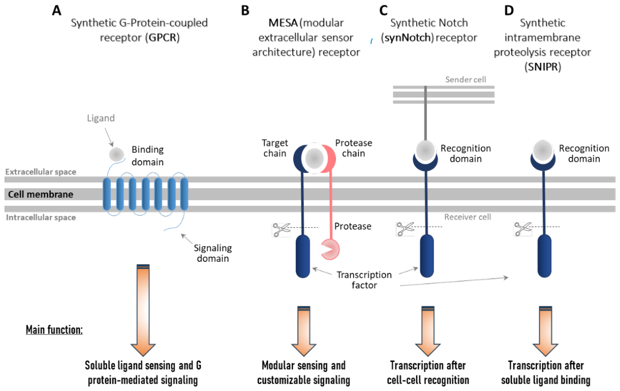
Figure 2.
Structure of chimeric antigen receptors (CARs) over five generations. Left: The components of the basic CAR-structure (found in the 1st generation of CARs) are derived from the native T-cell receptor (TCR, left bottom) and of antibodies (left top). CARs consist of a single-chain variable fragment (scFv) in the extracellular side, which is derived from the antigen-binding site of antibodies linked to transmembrane domain over a hinge region and connected to CD3ζ signaling motif, wich is derived from the TCR. 2nd generation CAR have an additional intracellular co-stimulatory domain (CM) and 3rd to 5th generation CARs have two costimulatory signaling domains, which can also induce the expression of cytokines or activate further signaling pathways [27,32].
Figure 2.
Structure of chimeric antigen receptors (CARs) over five generations. Left: The components of the basic CAR-structure (found in the 1st generation of CARs) are derived from the native T-cell receptor (TCR, left bottom) and of antibodies (left top). CARs consist of a single-chain variable fragment (scFv) in the extracellular side, which is derived from the antigen-binding site of antibodies linked to transmembrane domain over a hinge region and connected to CD3ζ signaling motif, wich is derived from the TCR. 2nd generation CAR have an additional intracellular co-stimulatory domain (CM) and 3rd to 5th generation CARs have two costimulatory signaling domains, which can also induce the expression of cytokines or activate further signaling pathways [27,32].
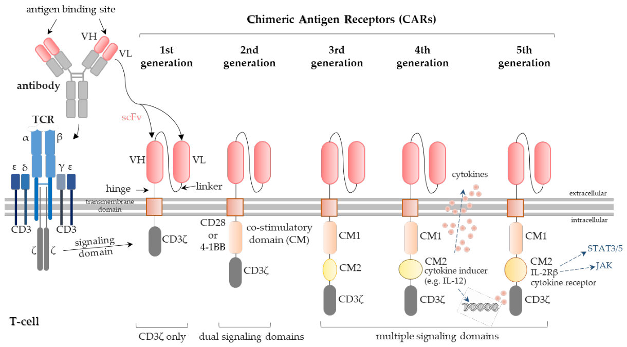
Figure 3.
Structure and functionality of synthetic cargo internalization receptors (CIR). (A) Three synthetic cargo internalization receptor types composed of an N-terminal myc-tag, followed by a green fluorescent protein (GFP)-specific nanobody (GFP-VHH) and connected via a small spacer to the different transmembrane (TMD) and cytoplasmic domains (CTD). CIR-1 has TMD and CTD of the human IL-6 receptor α (IL-6Rα, cytokine receptor); CIR-2 contains the TMD of IL-6Rα and the CTD of Endo180 with tyrosine-based and dihydrophobic motifs for internalization (yellow dots). CIR-3 has the TMD and CTD of the phagocytic FcγRIIA receptor that contains tyrosin based activation motifs (blue dots). VHH = nanobody sequence, derived from heavy-chain antibodies in camelids; Myc-tag = small polypetide-tag of ten amino acids derived from the human c-Myc gene. (B) Flow cytometric analysis of the cell surface binding of GFP to CIR-1-3 expressing cells. Grey histograms display control cells that were not incubated with GFP. Figures reproduced with permission from Temme et al. 2018 [1].
Figure 3.
Structure and functionality of synthetic cargo internalization receptors (CIR). (A) Three synthetic cargo internalization receptor types composed of an N-terminal myc-tag, followed by a green fluorescent protein (GFP)-specific nanobody (GFP-VHH) and connected via a small spacer to the different transmembrane (TMD) and cytoplasmic domains (CTD). CIR-1 has TMD and CTD of the human IL-6 receptor α (IL-6Rα, cytokine receptor); CIR-2 contains the TMD of IL-6Rα and the CTD of Endo180 with tyrosine-based and dihydrophobic motifs for internalization (yellow dots). CIR-3 has the TMD and CTD of the phagocytic FcγRIIA receptor that contains tyrosin based activation motifs (blue dots). VHH = nanobody sequence, derived from heavy-chain antibodies in camelids; Myc-tag = small polypetide-tag of ten amino acids derived from the human c-Myc gene. (B) Flow cytometric analysis of the cell surface binding of GFP to CIR-1-3 expressing cells. Grey histograms display control cells that were not incubated with GFP. Figures reproduced with permission from Temme et al. 2018 [1].
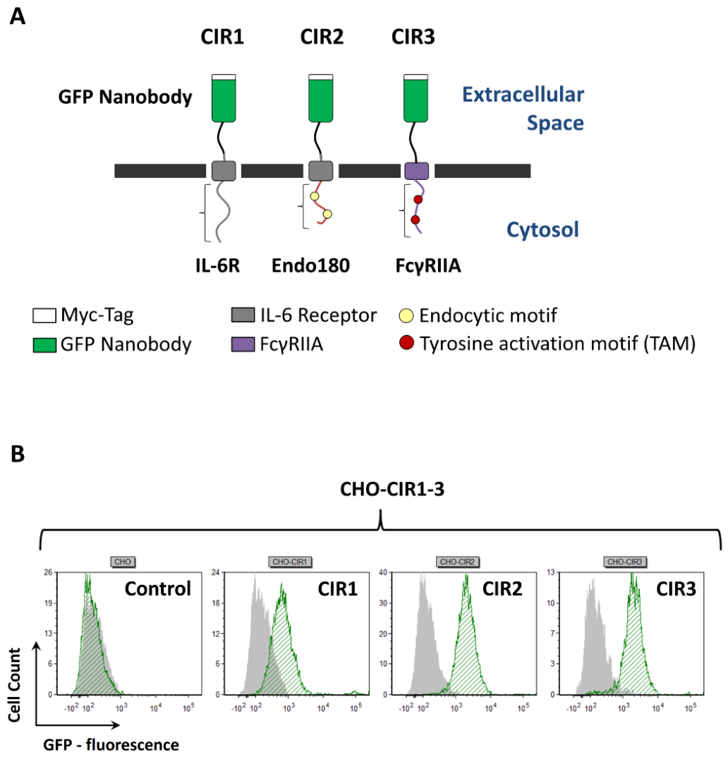
Figure 4.
Targeting CIR-expressing cells with GFP-PFCs. (A) CHO (Chinese hamster ovarian) or CIR-expressing CHO-cells were incubated with GFP-PFCs, separated on a density gradient and analyzed by 1H/19F MRI. Note that the cells are visible as bright structure on top of the ficoll layer in the photograph, but appears as a darker line in the T2-weighted 1H MRI images. Left = image of the 15 ml tube, middle = corresponding T2-weighted 1H MRI measurement, right = merging of 1H and 19F MRI measurement. (B) Quantification of the 19F signal in CHO cells that are stably transfected with CIRs (left), transiently transfected CHO-CIR cells (middle) or CIR-3 expressing Ba/F3-gp130 cells (right). The figures is reproduced with permission from Temme et al. 2018 [1].
Figure 4.
Targeting CIR-expressing cells with GFP-PFCs. (A) CHO (Chinese hamster ovarian) or CIR-expressing CHO-cells were incubated with GFP-PFCs, separated on a density gradient and analyzed by 1H/19F MRI. Note that the cells are visible as bright structure on top of the ficoll layer in the photograph, but appears as a darker line in the T2-weighted 1H MRI images. Left = image of the 15 ml tube, middle = corresponding T2-weighted 1H MRI measurement, right = merging of 1H and 19F MRI measurement. (B) Quantification of the 19F signal in CHO cells that are stably transfected with CIRs (left), transiently transfected CHO-CIR cells (middle) or CIR-3 expressing Ba/F3-gp130 cells (right). The figures is reproduced with permission from Temme et al. 2018 [1].
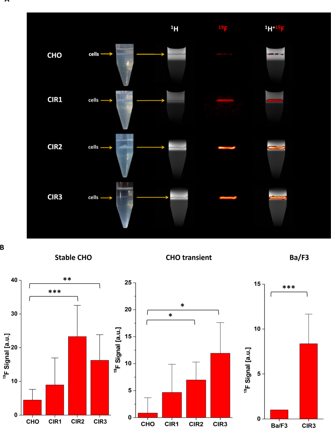
Figure 5.
GFP and mCherry based synthetic cytokine receptors (SyCyRs). (A) Left: Schematic illustration of SyCyR for IL-23 [SyCyR(IL-23/2A)] to simulate IL-23-induced signal transduction after GFP-mCherry fusion protein binding. This SyCyR cntains GFP and mCherry nanobodies (VHH) on the extracellular side and IL-12Rβ1 and IL-23R domains on the intracellular side to induce signaling (GVHH-IL-12Rβ1 and CVHH-IL-23R). Right: Proliferation of SyCyR-expressing Ba/F3-gp130 cells incubated with synthetic ligands (6.25 ng/ml GFP/mCherry) or HIL-6 (10 ng/ml) [2]. (B) Left: Illustrative scheme of a GFPVHH-gp130-SyCyR with GFP-VHHs on the extracellular side and a gp130 domain on the intracellular side. This receptor activates intracellular IL-6 signaling after binding of GFP-Fc fusion proteins. Middle: Proliferation of Ba/F3-gp130 and Ba/F3-gp130 (GFPVHH-gp130) cells without cytokine (-), with 10 ng/ml Hyper-IL-6 or with 100 ng/ml GFP-Fc. Right: Western blot analyses with specific antibodies detecting phospho-STAT3/ERK1/2 and STAT3/ERK1/2 after STAT3 and ERK1/2 activation in Ba/F3-gp130 (GFPVHH-gp130) cells treated without cytokine (-), with 10 ng/ml Hyper-IL-6 or 100 ng/ml GFP-Fc for 15 min. ***p≤0.001; ****p≤0.0001 [74]. (C) Simplified illustration of synthetic IFNAR consisting of IFNAR1 and IFNAR2 units, in which the extracellular domains have been replaced by nanobodies directed against GFP and mCherry (left). These receptors can be activated by multimeric GFP/mCherry ligands (GC, GG, CC, GCCG). Middle: HEK293 cells transiently expressing human VGIFNAR1 and VCIFNAR2 that were treated with 1,000 U/ml IFNα4, 10 ng/ml Hyper-IL-6 or 100 ng/ml of the synthetic cytokine ligands (GCCG, GG, CC, GC) for 30 min and were analyzed with specific antibodies detecting phospho-STAT1, phospho-STAT2, STAT1, STAT2, IFNAR1 (myc-tagged) and IFNAR2 (HA-tagged). Right: Synthetic IFNARs have antiviral activity in MC57 cells. Therefore, they were transduced with murine VGIFNAR1 and VCIFNAR2 (MC57-VGmIFNAR1-VCmIFNAR2) and were treated with medium, IFNα4 (150U/ml) or synthetic GC ligand (100ng/ml), followed by infection with vesicular stomatitis virus (VSV, multiplicity of infection [MOI]). 12h after infection viral titers were measured in the supernatants. ns = not significant; ***p≤0.001 [76]. The experimental results of figures A, B and C (right part) were adapted with permission under the Creative Commons Attribution 4.0 International License (CC BY 4.0) from Engelowski et al. [2](A), Mossner et al. [74] (B) and Zoellner et al. [76] (C). (No data were reanalyzed or modified).
Figure 5.
GFP and mCherry based synthetic cytokine receptors (SyCyRs). (A) Left: Schematic illustration of SyCyR for IL-23 [SyCyR(IL-23/2A)] to simulate IL-23-induced signal transduction after GFP-mCherry fusion protein binding. This SyCyR cntains GFP and mCherry nanobodies (VHH) on the extracellular side and IL-12Rβ1 and IL-23R domains on the intracellular side to induce signaling (GVHH-IL-12Rβ1 and CVHH-IL-23R). Right: Proliferation of SyCyR-expressing Ba/F3-gp130 cells incubated with synthetic ligands (6.25 ng/ml GFP/mCherry) or HIL-6 (10 ng/ml) [2]. (B) Left: Illustrative scheme of a GFPVHH-gp130-SyCyR with GFP-VHHs on the extracellular side and a gp130 domain on the intracellular side. This receptor activates intracellular IL-6 signaling after binding of GFP-Fc fusion proteins. Middle: Proliferation of Ba/F3-gp130 and Ba/F3-gp130 (GFPVHH-gp130) cells without cytokine (-), with 10 ng/ml Hyper-IL-6 or with 100 ng/ml GFP-Fc. Right: Western blot analyses with specific antibodies detecting phospho-STAT3/ERK1/2 and STAT3/ERK1/2 after STAT3 and ERK1/2 activation in Ba/F3-gp130 (GFPVHH-gp130) cells treated without cytokine (-), with 10 ng/ml Hyper-IL-6 or 100 ng/ml GFP-Fc for 15 min. ***p≤0.001; ****p≤0.0001 [74]. (C) Simplified illustration of synthetic IFNAR consisting of IFNAR1 and IFNAR2 units, in which the extracellular domains have been replaced by nanobodies directed against GFP and mCherry (left). These receptors can be activated by multimeric GFP/mCherry ligands (GC, GG, CC, GCCG). Middle: HEK293 cells transiently expressing human VGIFNAR1 and VCIFNAR2 that were treated with 1,000 U/ml IFNα4, 10 ng/ml Hyper-IL-6 or 100 ng/ml of the synthetic cytokine ligands (GCCG, GG, CC, GC) for 30 min and were analyzed with specific antibodies detecting phospho-STAT1, phospho-STAT2, STAT1, STAT2, IFNAR1 (myc-tagged) and IFNAR2 (HA-tagged). Right: Synthetic IFNARs have antiviral activity in MC57 cells. Therefore, they were transduced with murine VGIFNAR1 and VCIFNAR2 (MC57-VGmIFNAR1-VCmIFNAR2) and were treated with medium, IFNα4 (150U/ml) or synthetic GC ligand (100ng/ml), followed by infection with vesicular stomatitis virus (VSV, multiplicity of infection [MOI]). 12h after infection viral titers were measured in the supernatants. ns = not significant; ***p≤0.001 [76]. The experimental results of figures A, B and C (right part) were adapted with permission under the Creative Commons Attribution 4.0 International License (CC BY 4.0) from Engelowski et al. [2](A), Mossner et al. [74] (B) and Zoellner et al. [76] (C). (No data were reanalyzed or modified).
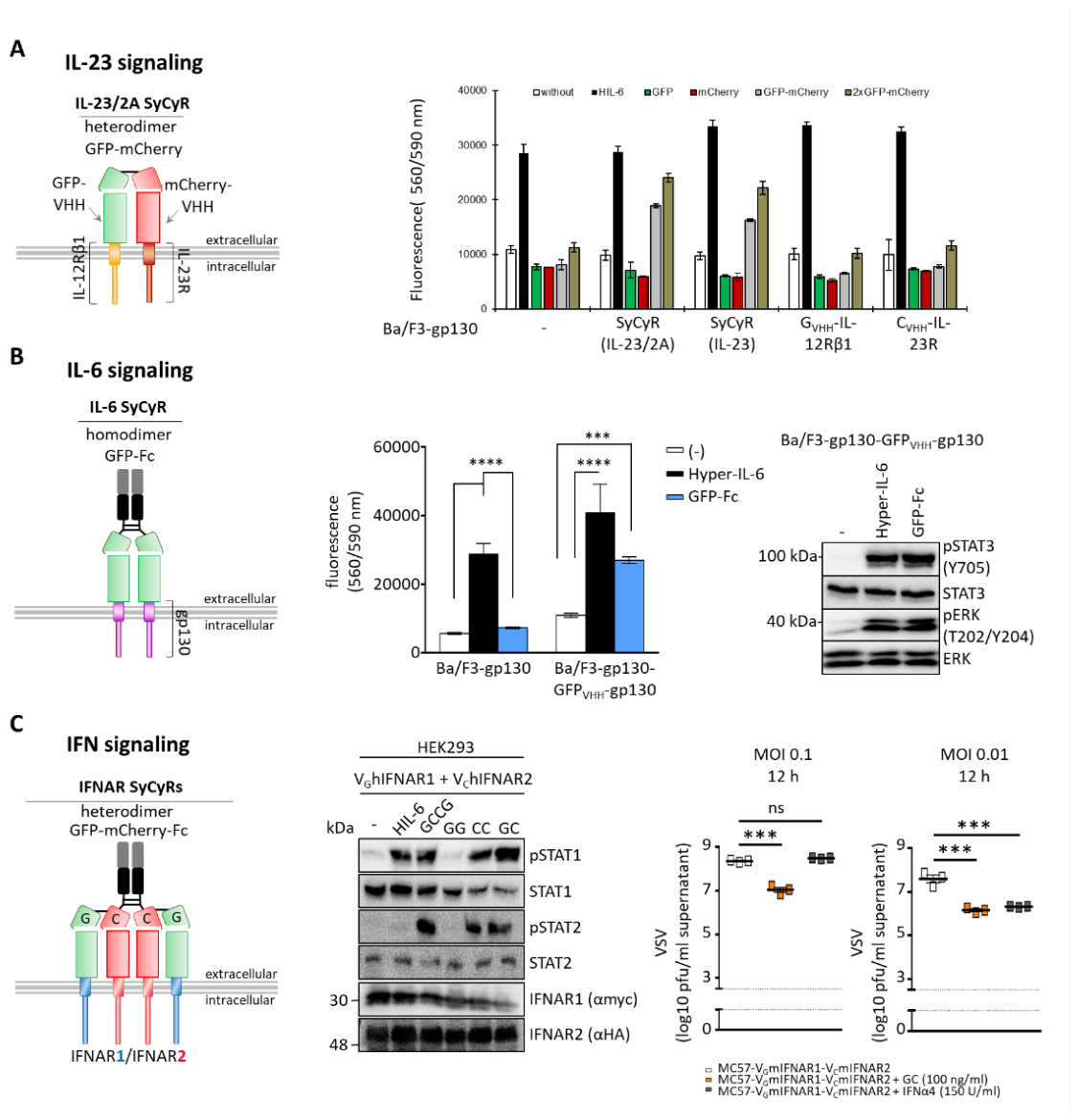
Figure 6.
Optimization of synthetic cytokine receptors for enhanced signaling. (A) Surface plasmon resonance analyses of Palivizumab coated on a Protein A chip and soluble AIP1VHH injected for 120 s and the dissociation rate was recorded for 500 s. The sensograms in response units (RU) over time are shown as colored lines, and the global fit is shown as black lines. (B) Small-angle X-ray scattering structural characterization of AIP1VHH (anti-idiotypic nanobody) binding to Palivizumab. The red dotted line shows the 146 Å distance between the paratopes. The squares at the top indicate hotspot amino acids and the distances are marked by blue dotted lines. (C) Schematic illustration of Palivizumab (left) and modified versions (middle, right) to enhance signal transduction. To induce stronger signaling, Palivizumab was cross-linked by a Palivizumab Fc–binding antibody (middle). To further increase signaling Palivizumab was reformatted into single-chain Fv fragments where the variable domains of the light and the heavy chain were fused by a flexible peptide linker (PscFvLHFc, right). (D) Proliferation of Ba/F3-gp130 cells expressing AIP1-4VHHgp130 at increasing concentrations of PscFvLHFc (0.00053–93.5 nM) normalized to HIL-6 (10 ng/ml) induced proliferation of each cell line (left). STAT3 phosphorylation in Ba/F3-gp130 cells expressing AIP1-4VHHgp130 were either treated with 10 nM PscFvLHFc for 90 minutes or left untreated. Western blot analyses were then performed using specific antibodies to detect phospho-STAT3 and STAT3 (right). Figures A, B and D were reproduced from Ettich et al. [3]. No data were reanalyzed or modified. Licensed under CC BY 4.0.
Figure 6.
Optimization of synthetic cytokine receptors for enhanced signaling. (A) Surface plasmon resonance analyses of Palivizumab coated on a Protein A chip and soluble AIP1VHH injected for 120 s and the dissociation rate was recorded for 500 s. The sensograms in response units (RU) over time are shown as colored lines, and the global fit is shown as black lines. (B) Small-angle X-ray scattering structural characterization of AIP1VHH (anti-idiotypic nanobody) binding to Palivizumab. The red dotted line shows the 146 Å distance between the paratopes. The squares at the top indicate hotspot amino acids and the distances are marked by blue dotted lines. (C) Schematic illustration of Palivizumab (left) and modified versions (middle, right) to enhance signal transduction. To induce stronger signaling, Palivizumab was cross-linked by a Palivizumab Fc–binding antibody (middle). To further increase signaling Palivizumab was reformatted into single-chain Fv fragments where the variable domains of the light and the heavy chain were fused by a flexible peptide linker (PscFvLHFc, right). (D) Proliferation of Ba/F3-gp130 cells expressing AIP1-4VHHgp130 at increasing concentrations of PscFvLHFc (0.00053–93.5 nM) normalized to HIL-6 (10 ng/ml) induced proliferation of each cell line (left). STAT3 phosphorylation in Ba/F3-gp130 cells expressing AIP1-4VHHgp130 were either treated with 10 nM PscFvLHFc for 90 minutes or left untreated. Western blot analyses were then performed using specific antibodies to detect phospho-STAT3 and STAT3 (right). Figures A, B and D were reproduced from Ettich et al. [3]. No data were reanalyzed or modified. Licensed under CC BY 4.0.
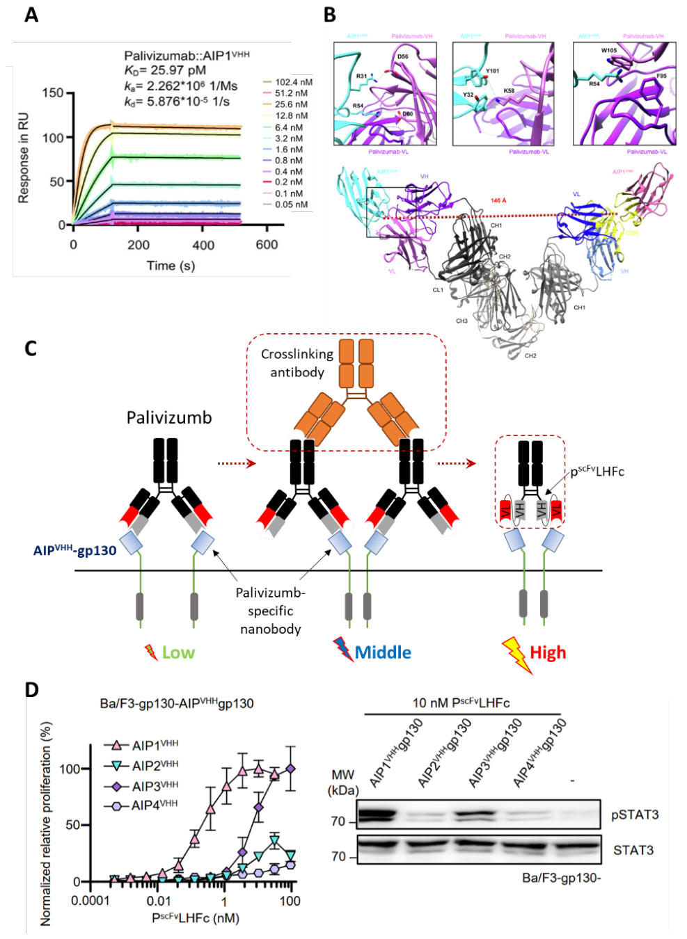

Table 1.
Synthetic receptor/ligand systems.
| Synthetic receptors | Description | Examples for applications | Ligands | References |
|---|---|---|---|---|
| Synthetic G-protein-coupled receptors (GPCRs) | ⦁ Engineered versions of GPCRs designed to recognize novel ligands and produce customized signaling outputs after binding ⦁ DREADDs (designer receptors exclusively activated by designer drugs) can activate or silence neuronal activity ⦁ PAGERs (programmable antigen-gated G-protein-coupled engineered receptors) with a GFP-nanobody and a peptide antagonist on the N-terminus of the GPCR inhibit the ligand binding until GFP binds to the GFP-nanobody, thereby releasing the ligand-binding site ⦁ Function: Used to program (therapeutic) cells for sensing and response functions such as gene expression, G-protein activation, but also macrophage activation or T-cell killing. |
Synthetic adhesion GPCRs (ADGR), called ADGRG6 (GPR126), which are activated by enterokinase, induce Gαs, Gαq and Gα12 G-protein pathways | Enterokinase | [8,10,109] |
| Synthetic Notch receptors (synNotch) | ⦁ Modular cell-surface receptors ⦁ Binding of a specific ligand on the cell surface (sender) to a ligand-specific receptor on another cell (receiver) triggers proteolytic cleavage to release an intracellular transcription factor ⦁ Function: Inducible expression of target genes such as CARs or cytokines in a programmable manner. |
Reporter gene expression and activation after CD19-binding (sender) to anti-CD19-synNotch receptor (receiver) | CD19 | [15,110] |
| Synthetic intramembrane proteolysis receptors (SNIPRs) | ⦁ An evolved version of synNotch with improved specificity and reduced background activation ⦁ Function: Orthogonal gene control and reduced immunogenicity. |
Development of SNIPRs for cell-based therapies (e.g. CAR T-cell therapy) | CD19 and ALPPL2 | [111] |
| MESA (modular extracellular sensor architecture) receptors | ⦁ MESA receptors respond to soluble or surface-bound biomarkers ⦁ Function: Control the expression of imaging genes, that can be used to monitor therapeutic cells in which these synthetic systems are integrated. |
MESA based on CD4 and dTomato ectodomains leads - after ligand binding - to cleavage and release of the transcription factor on the intracellular side of the membrane. | CD4, dTomato, mCherry, Rapamycin | [14] |
| Chimeric antigen receptors (CARs) | ⦁ Expressed on the cell surface of immune cells (T-cells, NK cells, or macrophages) ⦁ Recognize specific tumor-associated antigens via an extracellular antigen-binding domain ⦁ CAR T cells can also be modified to target fibroblast activation protein (FAP), which is expressed on cancer-associated fibroblasts (CAFs), in order to specifically target them ⦁ Function: Intracellular signaling and antitumor activities. |
⦁ Targeting CD19 for B-cell malignancies | CD19 (on tumor cells) | [19,20] |
| Cargo internalization Receptors (CIRs) | ⦁ Synthetic cell surface receptors ⦁ Bind specific extracellular ligands that can be linked to cargo molecules or nanoparticles ⦁ Function: Internalization of the cargo into the cell for cell tracking. |
⦁ Nanoparticle (PFC) uptake for cell-tracking by 1H/19F magnetic resonance imaging | GFP, GFP-PFCs | [1] |
| Synthetic cytokine receptors (SyCyRs) | ⦁ Cell surface receptors engineered with extracellular nanobody domains recognizing synthetic ligands ⦁ Intracellular signaling domains derived from natural cytokine receptors ⦁ Function: Signaling pathway can be switched on or off with specific synthetic ligands to regulate immune cell proliferation, activation, or apoptosis in a controllable and orthogonal manner. |
SyCyRs mimicking interleukin (IL)-23 and IL-6-signaling that consist of a changed extracellular domain with GFP or mCherry nanobodies and can activate JAK (Janus kinases)/STAT (Signal Transducers and Activators of Transcription) pathway through GFP/mCherry binding | GFP, mCherry | [2] |
Disclaimer/Publisher’s Note: The statements, opinions and data contained in all publications are solely those of the individual author(s) and contributor(s) and not of MDPI and/or the editor(s). MDPI and/or the editor(s) disclaim responsibility for any injury to people or property resulting from any ideas, methods, instructions or products referred to in the content. |
© 2026 by the authors. Licensee MDPI, Basel, Switzerland. This article is an open access article distributed under the terms and conditions of the Creative Commons Attribution (CC BY) license (http://creativecommons.org/licenses/by/4.0/).
Copyright: This open access article is published under a Creative Commons CC BY 4.0 license, which permit the free download, distribution, and reuse, provided that the author and preprint are cited in any reuse.