

Submitted:
16 September 2026
Posted:
18 September 2026
You are already at the latest version
Abstract
Background/Objectives: Streptococcus pyogenes (Group A Streptococcus, GAS) remains a major human pathogen. This study aimed to determine the phenotypic profiles of macrolide resistance in S. pyogenes isolates from southeastern Turkey and to investigate underlying genotypic mechanisms using Oxford Nanopore sequencing. Methods: A total of 83 clinical isolates of S. pyogenes were collected between January 2024 and June 2025. Antimicrobial susceptibility was assessed by disk diffusion following EUCAST guidelines. Macrolide resistance phenotypes (iMLSB, cMLSB, M) were determined by the D‑zone test. Whole‑genome sequencing of erythromycin‑resistant isolates was performed using Oxford Nanopore technology, and resistance determinants were analyzed with the CARD/RGI pipeline.Results: All isolates were susceptible to penicillin. Six isolates (7.2%) exhibited erythromycin resistance: three iMLSB, two cMLSB, and one M phenotype. Adult isolates were exclusively iMLSB, whereas pediatric isolates included one M and two cMLSB phenotypes. Genotypic analysis revealed erm(A) in all iMLSB isolates, mef(A)/msr(D) in the M phenotype, and absence of major resistance genes in cMLSB isolates, which instead carried chromosomal lmrP and mef(E)‑like sequences. Co‑occurrence of tetracycline resistance genes (tetM/O) was observed in several isolates.Conclusions: Penicillin remains the cornerstone of GAS therapy; however, macrolide resistance persists through diverse mechanisms. The coexistence of erm(A)‑mediated iMLSB, mef(A)/msr(D)‑associated M phenotype, and unexplained cMLSB resistance highlights the need for routine D‑testing and genomic surveillance. These findings emphasize the importance of integrating phenotypic and genotypic approaches to monitor resistance evolution in GAS.
Keywords:
Streptococcus pyogenes
; macrolide resistance
; iMLSB
; cMLSB
; Oxford Nanopore sequencing
; erm(A)
; mef(A)
; tetracycline resistance
1. Introduction
Streptococcus pyogenes (Group A Streptococcus, GAS) is a major human pathogen responsible for a wide clinical spectrum, ranging from superficial infections such as pharyngitis and skin/soft tissue infections to life-threatening invasive diseases including streptococcal toxic shock syndrome and necrotizing fasciitis [1]. β-lactam antibiotics, particularly penicillin, remain the cornerstone of therapy for GAS infections, as they have largely retained their efficacy. In patients with a history of penicillin allergy, macrolides and clindamycin may serve as alternative agents [2,3]. Moreover, protein synthesis–inhibiting antibiotics are of particular clinical importance in severe invasive GAS infections, as they can suppress streptococcal toxin production. However, treatment failure has been reported in strains exhibiting inducible clindamycin resistance [4].
Recent data reported from diverse geographical regions indicate that macrolide resistance among GAS isolates may increase in association with antibiotic usage patterns and the dissemination of specific clones [3,5]. In GAS strains, macrolide resistance is genetically categorized into three principal mechanisms. The first involves modification of the ribosomal target site mediated by erm genes. The second major mechanism is active efflux, most commonly associated with mef(A/E)–msr(D) [6,7]. The third group encompasses rarer mechanisms, including mutations in the 23S rRNA gene or in genes encoding ribosomal proteins (e.g., L4, L22), whereby phenotypic resistance is observed despite the absence of classical resistance determinants, representing mechanisms that remain incompletely elucidated [8,9].
The distribution of macrolide resistance mechanisms, their associated phenotypic patterns, and the predominant circulating clones may vary substantially across regions [10,11]. Consequently, it is essential for each center to monitor its own local resistance data and circulating genotypic characteristics, both to guide empirical therapeutic strategies and to strengthen epidemiological surveillance.
Long-read sequencing technologies, particularly Oxford Nanopore sequencing, provide a comprehensive means of assessing the presence of resistance genes, their genetic context, and their association with mobile genetic elements, thereby contributing to a more detailed characterization of phenotype–genotype correlations [12]. The objective of this study is to determine the phenotypic profiles of macrolide resistance in S. pyogenes isolates obtained from clinical samples in Diyarbakır—a city in southeastern Turkey—and to investigate, in a comprehensive manner, both the established and rare genotypic mechanisms underlying this resistance using Oxford Nanopore sequencing technology.
2. Results
2.1. Demographic Data and Distribution of Clinical Samples
The study included 83 Streptococcus pyogenes isolates obtained from patients with a mean age of 24.5 ± 22.0 years. Of these, 48 (57.8%) were children and 35 (42.2%) were adults. The gender distribution was balanced, with 42 females (50.6%) and 41 males (49.4%).
Analysis of the clinical specimens revealed that respiratory tract samples constituted the largest group, accounting for 45 isolates (54.2%), including 42 throat swabs and 3 sputum samples. These were followed by 14 aspirates (16.9%), 9 wound specimens (10.8%), 3 blood cultures (3.6%), and 12 other clinical samples (14.5%) such as urine, cerebrospinal fluid, and ear drainage.
2.2. Antimicrobial Susceptibility and Resistance Phenotypes
According to disk diffusion testing, all 83 isolates (100%) were susceptible to penicillin. A total of six isolates (7.2%) exhibited resistance to erythromycin. Phenotypic characterization of these erythromycin-resistant isolates revealed three with an inducible MLSB phenotype (iMLSB, D-zone positive), two with a constitutive MLSB phenotype (cMLSB), and one with an M phenotype.
Macrolide-resistant isolates were further screened for tetracycline resistance. In four isolates, the presence of corresponding resistance genes was confirmed, and phenotypic resistance to tetracycline was observed. In contrast, two isolates lacking detectable resistance genes remained phenotypically susceptible to tetracycline. The remaining 77 isolates (92.8%) did not display macrolide resistance.
Among the six erythromycin-resistant isolates, three were recovered from adult patients and three from children. All adult isolates exhibited the iMLSB phenotype, whereas the pediatric isolates comprised one M phenotype and two cMLSB phenotypes. With respect to specimen type, resistant isolates from adults were obtained from two aspirates and one sputum sample, while those from children were derived exclusively from throat swabs.
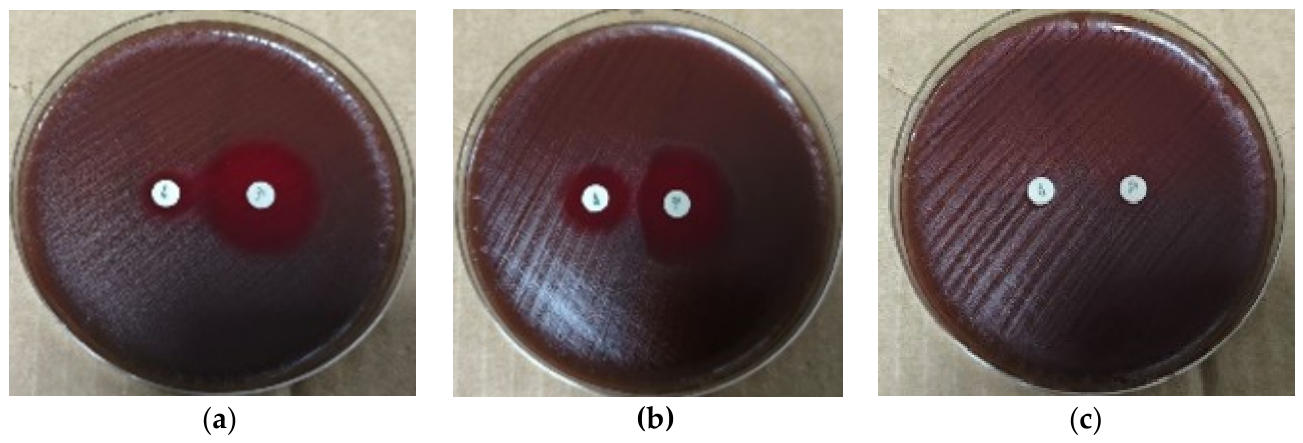
Figure 1.
D-zone test results for Streptococcus pyogenes isolates: (a) M phenotype—resistant to erythromycin but susceptible to clindamycin (D-zone negative); (b) iMLSB phenotype—resistant to both antibiotics with inducible resistance (D-zone positive); (c) cMLSB phenotype—constitutive resistance to erythromycin and clindamycin.
Figure 1.
D-zone test results for Streptococcus pyogenes isolates: (a) M phenotype—resistant to erythromycin but susceptible to clindamycin (D-zone negative); (b) iMLSB phenotype—resistant to both antibiotics with inducible resistance (D-zone positive); (c) cMLSB phenotype—constitutive resistance to erythromycin and clindamycin.
2.3. Genotypic Characteristics of Erythromycin-Resistant Isolates
Oxford Nanopore sequencing was performed on all six erythromycin-resistant S. pyogenes isolates. Among the three isolates exhibiting the iMLSB phenotype, two belonged to the ST46/emm22 clonal profile and one to ST63/emm77. All three carried the erm(A) gene. The two ST46/emm22 isolates harbored tet(M), while the ST63/emm77 isolate carried tet(O); in each case, tetracycline resistance was phenotypically confirmed.
The isolate with the M phenotype was identified as ST25/emm44 and carried mef(A), msr(D), and tet(M) genes, with tetracycline resistance also confirmed phenotypically.
The two isolates displaying the cMLSB phenotype shared the ST49/emm75 clonal profile and were obtained from throat swabs of pediatric patients. Sequencing yielded single-contig circular chromosome assemblies of approximately 1.79 Mb. CARD/RGI analysis did not reveal major macrolide resistance genes; however, chromosomal lmrP and low-similarity mef(E)-like sequences were detected. The demographic, clinical, phenotypic, and genotypic characteristics of the macrolide-resistant isolates are summarized in Table 1.
3. Discussion
GAS is an important human pathogen responsible for diseases ranging from pharyngitis to invasive infections. Penicillin and other β-lactam antibiotics remain uniformly effective and continue to form the basis of current treatment strategies [1,2]. In our study, all 83 isolates were susceptible to penicillin, consistent with national and international reports . By contrast, resistance to macrolides—key alternatives for patients with penicillin allergy—persists as a clinically relevant concern [5,9,13,14].
In this study, the erythromycin resistance rate was determined as 7.2%. This finding corresponds to a low-to-moderate resistance pattern, consistent with data previously reported from Turkey. For instance, Akata et al. documented a resistance rate of 2.7% in Edirne, located in northwestern Turkey [14], while Açıkgöz et al. reported a rate of 2.6% in Ankara, in Central Anatolia [13]. In two studies conducted in western Turkey, Telli et al. reported erythromycin resistance rates as high as 15% in Aydın [15], while Dündar et al. documented a rate of 9% in Kocaeli [16]. In Eastern Anatolia, Uyanık and Yazgı reported an erythromycin resistance rate of 8% in Erzurum [17]. Taken together, these data indicate that macrolide resistance in GAS varies among centers in Turkey but generally remains within low-to-moderate levels. The 7.2% resistance rate observed in our study, conducted in southeastern Turkey, is consistent with this overall pattern. This variability underscores the need to examine phenotypic distributions across different regions and time periods.
When phenotypic distributions of macrolide resistance reported from Turkey are considered, early studies particularly highlighted the predominance of the M phenotype. Akata et al. reported that resistant isolates were mainly of the M phenotype and carried the mef(A) gene [14]. Similarly, studies by Açıkgöz et al. and Çolakoğlu et al. also identified the M phenotype as the most frequent among resistant isolates, with mef(A) detected in these strains [13,18]. In contrast, Telli et al. and Dündar et al. documented a notable frequency of the erm(TR)-mediated inducible Macrolide–Lincosamide–Streptogramin B (iMLSB) phenotype [15,16]. These findings suggest that macrolide resistance in Turkey cannot be explained by a single phenotype or genetic mechanism, but rather shows temporal and geographic variability .
In this study, the phenotypic distribution of six erythromycin-resistant isolates included three iMLSB, two constitutive Macrolide–Lincosamide–Streptogramin B (cMLSB), and one M phenotype. Although the number of resistant isolates was limited, this distribution indicates that macrolide resistance in southeastern Turkey also exhibits heterogeneity. The detection of the erm(A) gene in all three iMLSB isolates supports the association of this phenotype with ribosomal target site modification [6,7,8,9]. In this study, the phenotypic distribution of six erythromycin-resistant isolates included three iMLSB, two cMLSB, and one M phenotype. Although the number of resistant isolates was limited, this heterogeneity reflects the diverse mechanisms of macrolide resistance in southeastern Turkey. The detection of the erm(A) gene in all three iMLSB isolates supports the association of this phenotype with ribosomal target site modification. Two isolates belonged to the ST46/emm22 clonal profile and one to ST63/emm77, suggesting that specific clonal lineages may co-circulate with defined resistance mechanisms. Notably, the ST63/emm77 isolate carried both erm(A) and tet(O), resembling macrolide-resistant clones reported in Europe [3,7,10]. Similarly, the presence of erm(A) together with tet(M) in ST46/emm22 isolates indicates that macrolide and tetracycline resistance determinants can be co-transferred [7,16].
In the pediatric age group, one isolate belonging to the ST25/emm44 clonal profile exhibited the M phenotype and carried both mef(A) and msr(D), indicating the presence of active efflux mechanisms of macrolide resistance in southeastern Turkey [6,7,8,9]. This finding demonstrates that classical resistance determinants can be reliably confirmed by sequencing in isolates with clear phenotype–genotype concordance. The additional detection of tet(M) in the same isolate is noteworthy, as it highlights the potential for co-occurrence of macrolide and tetracycline resistance genes [7,16].
One of the most notable findings of this study was the absence of major macrolide resistance genes in two ST49/emm75 isolates exhibiting the cMLSB phenotype. Both isolates were obtained independently from throat swabs of different three-year-old male pediatric patients and showed phenotypic resistance to erythromycin and clindamycin. The detection of the same clonal profile in two separate patients suggests that this strain is circulating in the community, which is epidemiologically significant. Oxford Nanopore sequencing yielded single-contig circular chromosome assemblies of approximately 1.79 Mb for both isolates. Comprehensive Antibiotic Resistance Database/ Resistance Gene Identifier (CARD/RGI ) analysis identified chromosomal lmrP and low-similarity mef(E)-like sequences; however, these findings do not sufficiently explain the phenotypic cMLSB resistance observed. In the literature, such phenotype–genotype discrepancies have been associated with mutations in the 23S rRNA gene or alterations in ribosomal protein-encoding genes such as L4 and L22 [8,9]. Therefore, similar mechanisms may also play a role in the ST49/emm75 isolates identified in this study. In the literature from Spain and Portugal, emm75 lineages are typically characterized by the M phenotype and mef(A) carriage; however, the fact that our isolates exhibited the cMLSB phenotype while testing negative for major resistance genes suggests that this clone may be following a distinct evolutionary pathway in southeastern Turkey [19,20]. Since confirmatory mutation analysis of the relevant target regions was not performed in this study, this possibility should be regarded as an explanatory hypothesis rather than a definitive mechanism.
The distribution of resistant isolates according to age group and clinical specimen type was also noteworthy. All three erythromycin-resistant isolates obtained from adult patients exhibited the iMLSB phenotype and carried erm(A), whereas among pediatric patients, one isolate showed the M phenotype and two displayed the cMLSB phenotype. Moreover, resistant isolates from adults were recovered from aspirate and sputum specimens, while those from children were exclusively obtained from throat swabs. These findings suggest potential differences in circulating clones and resistance mechanisms between age groups. However, given the limited number of resistant isolates, this observation warrants confirmation in larger series.
When international data on macrolide resistance are considered, marked geographic differences become evident. While resistance rates remain low in several European countries [3,5,10], much higher levels—particularly with erm(B) predominance—have been reported from regions such as China [8]. These variations are thought to be associated with differences in antibiotic usage practices, the circulation of predominant clones, and the dissemination of resistance determinants [3,5,6,9,10,11]. The diverse clonal profiles identified in this study (ST46/emm22, ST63/emm77, ST25/emm44, and ST49/emm75) further indicate that macrolide resistance in southeastern Turkey cannot be explained by a single clone or mechanism.
This study has several limitations. First, it was conducted in a single center and the total number of resistant isolates remained limited. Second, whole-genome sequencing was performed only for the six erythromycin-resistant isolates rather than for all strains. In addition, targeted analyses to confirm potential ribosomal mutations in isolates with the cMLSB phenotype were not undertaken. Nevertheless, a major strength of the study is the integration of phenotypic resistance patterns with Oxford Nanopore–based whole-genome sequencing, enabling comprehensive genotypic evaluation [12].
4. Materials and Methods
4.1. Collection and Identification of Clinical Isolates
Between January 2024 and June 2025, 83 Streptococcus pyogenes isolates were obtained from diverse clinical specimens submitted to the Central Laboratory of Dicle University Hospitals. Cultures were performed on appropriate media (e.g., 5% sheep blood agar, chocolate agar) under standard incubation conditions. Preliminary identification of β-hemolytic colonies was based on bacitracin and trimethoprim–sulfamethoxazole susceptibility together with PYR testing. Definitive species identification was achieved using the BD Phoenix™ M50 automated system. Isolates were stored in Tryptic Soy Broth (TSB) with 20% glycerol at −20 °C until further analysis.
4.2. Antimicrobial Susceptibility Testing and Macrolide Resistance Phenotypes
Antimicrobial susceptibility was assessed by the Kirby–Bauer disk diffusion method following EUCAST guidelines [21]. Isolates were adjusted to a 0.5 McFarland standard and inoculated onto MH-F agar (Mueller–Hinton agar with 5% defibrinated horse blood and 20 mg/L β-NAD). Penicillin, erythromycin, clindamycin, and tetracycline disks were used.
For erythromycin-resistant isolates, macrolide resistance phenotypes (cMLSB, iMLSB, M) were determined by the D-zone test . Plates were incubated at 35 ± 1 °C in 5% CO₂ for 16–20 h, and inhibition zones were interpreted according to EUCAST criteria [21]. Streptococcus pneumoniae ATCC 49619 served as the quality control strain.
4.3. Genomic DNA Isolation and Library Preparation
Genomic DNA was extracted using a commercial silica-membrane column kit (Vazyme Biotech, China) according to the manufacturer’s instructions. Due to the thick cell wall of Gram-positive cocci, the lysis step was optimized with lysozyme treatment and extended incubation. For Oxford Nanopore sequencing, libraries were prepared using the Ligation Sequencing gDNA—Native Barcoding Kit 24 V14 (SQK-NBD114.24) protocol (Oxford Nanopore Technologies, UK) [22]. End-prep, barcode ligation, and adapter ligation were performed sequentially, with purification at each step using AMPure XP magnetic bead [22]. DNA quantity and library concentrations were measured throughout the workflow using a Qubit fluorometer (Invitrogen, USA).
4.4. Oxford Nanopore Sequencing and Bioinformatic Analysis
Prepared libraries were loaded onto R10.4.1 Spot-On flow cells (FLO-MIN114) and sequenced on the Mk1C device (Oxford Nanopore Technologies, UK) using MinKNOW software. Raw sequence data were processed with Dorado for base-calling and de-multiplexing, generating FASTQ files. Where necessary, potential host-derived reads were filtered using minimap2 [23] and samtools [24]. De novo genome assemblies were generated from bacterial reads with Flye [25], and contigs were taxonomically validated by BLAST analysis against the NCBI “nt” database [26]. Consensus genomes were aligned to appropriate references in Geneious Prime (version 2023.2.2) for polymorphism analysis [27]. Antibiotic resistance gene profiles were assessed using the Resistance Gene Identifier (RGI) algorithm based on the Comprehensive Antibiotic Resistance Database (CARD) [28]. DNA extraction, library preparation, sequencing, and bioinformatic analyses were performed as outsourced services.
This study was approved by the Non-Interventional Clinical Research Ethics Committee of Dicle University Faculty of Medicine (Decision No. 35, dated December 18, 2024).
Generative artificial intelligence (Microsoft Copilot) was used to assist in the preparation of the manuscript by improving the clarity, fluency, and academic phrasing of English text. The AI tool was employed for language editing and translation purposes only. No AI was used in study design, data collection, analysis, or interpretation.
5. Conclusions
Penicillin continues to remain the first-line therapy for S. pyogenes infections. However, routine implementation of the D-test is critical to prevent potential treatment failures with clindamycin. The tendency of resistant strains to disseminate via mobile genetic elements (e.g., co-expression of tetM/O) underscores the necessity of sustained molecular surveillance. In this study, all isolates retained susceptibility to penicillin, whereas macrolide resistance was mediated through diverse phenotypic and genotypic mechanisms. The coexistence of the erm(A)-associated iMLSB phenotype, the mef(A)–msr(D)-associated M phenotype, and the cMLSB phenotype not explained by major resistance genes within the same series is particularly noteworthy. These findings highlight that local surveillance efforts should not rely solely on phenotypic testing, but must also incorporate genomic approaches to ensure comprehensive monitoring of resistance evolution. Future multicenter genomic surveillance studies will be essential to track emerging clones and resistance determinants across different regions.
Supplementary Materials
The following supporting information can be downloaded at the website of this paper posted on Preprints.org, Table S1: Characteristics of erythromycin-resistant Streptococcus pyogenes isolates; Figure S1: D zone test results for Streptococcus pyogenes isolates.
Author Contributions
Conceptualization, N.Ö. , S.K. and A.A; methodology, N.Ö. and A.B.K,; software, A.B.K; validation, S.K. and A.B. K.; formal analysis, S.K.; investigation, S.K and A.A.; resources, N.Ö.; data curation, S.K.; writing—original draft preparation, S.K.; writing—review and editing, N.Ö.; visualization, S.K.; supervision, N.Ö.; project administration, N.Ö. All authors have read and agreed to the published version of the manuscript.
Funding
This research was funded by the Dicle University Scientific Research Projects Coordination Unit (Project No. TIP.25.008, 2025).
Institutional Review Board Statement
The study was conducted in accordance with the Declaration of Helsinki, and approved by the Non-Interventional Clinical Research Ethics Committee of Dicle University Faculty of Medicine (Decision No. 35, December 18, 2024).
Informed Consent Statement
Written informed consent was obtained from all adult participants and from the parents or legal guardians of child participants; assent was also obtained from children, as appropriate.
Data Availability Statement
The data presented in this study are available on request from the corresponding author. The data are not publicly available due to privacy and ethical restrictions. All relevant data supporting the findings of this study are included within the article and its supplementary materials.
Acknowledgments
Generative artificial intelligence (Microsoft Copilot) was used to assist in the preparation of the manuscript by improving the clarity, fluency, and academic phrasing of English text. The AI tool was employed for language editing and translation purposes only. No AI was used in study design, data collection, analysis, or interpretation. The authors have reviewed and edited the output and take full responsibility for the content of this publication.
Conflicts of Interest
The authors declare no conflicts of interest. The funders had no role in the design of the study; in the collection, analyses, or interpretation of data; in the writing of the manuscript; or in the decision to publish the results.
Abbreviations
The following abbreviations are used in this manuscript:
| GAS | Group A Streptococcus, Streptococcus pyogenes |
| MLSB | Macrolide–Lincosamide–Streptogramin B |
| iMLSB | Inducible Macrolide–Lincosamide–Streptogramin B |
| cMLSB | Constitutive Macrolide–Lincosamide–Streptogramin B |
| CARD | Comprehensive Antibiotic Resistance Database |
| RGI | Resistance Gene Identifier |
| EUCAST | European Committee on Antimicrobial Susceptibility Testing |
| MH-F | MH-F (Mueller–Hinton agar with 5% defibrinated horse blood |
| TSB | Tryptic Soy Broth |
References
- Walker, M.J.; Barnett, T.C.; McArthur, J.D.; Cole, J.N.; Gillen, C.M.; Henningham, A.; Sriprakash, K.S.; Sanderson-Smith, M.L.; Nizet, V. Disease Manifestations and Pathogenic Mechanisms of Group A Streptococcus. Clin. Microbiol. Rev. 2014, 27, 264–301. [CrossRef]
- Shulman, S.T.; Bisno, A.L.; Clegg, H.W.; Gerber, M.A.; Kaplan, E.L.; Lee, G.; Martin, J.M.; Van Beneden, C.; Infectious Diseases Society of America Clinical Practice Guideline for the Diagnosis and Management of Group A Streptococcal Pharyngitis: 2012 Update by the Infectious Diseases Society of America. Clin. Infect. Dis. Off. Publ. Infect. Dis. Soc. Am. 2012, 55, e86-102. [CrossRef]
- Friães, A.; Mamede, R.; Santos, B.; Marrão, G.; Melo-Cristino, J.; Ramirez, M. Increase of Macrolide Resistance among Streptococcus Pyogenes Pharyngitis Driven by a Mef(A)-Msr(D)/Emm2-ST55 Lineage in Portugal (2014-2019). Antimicrob. Agents Chemother. 2025, 69, e0096825. [CrossRef]
- Lewis, J.S.; Lepak, A.J.; Thompson, G.R.; Craig, W.A.; Andes, D.R.; Sabol-Dzintars, K.E.; Jorgensen, J.H. Failure of Clindamycin to Eradicate Infection with Beta-Hemolytic Streptococci Inducibly Resistant to Clindamycin in an Animal Model and in Human Infections. Antimicrob. Agents Chemother. 2014, 58, 1327–1331. [CrossRef]
- Van Heirstraeten, L.; Coenen, S.; Lammens, C.; Hens, N.; Goossens, H.; Malhotra-Kumar, S. Antimicrobial Drug Use and Macrolide-Resistant Streptococcus Pyogenes, Belgium. Emerg. Infect. Dis. 2012, 18, 1515–1518. [CrossRef]
- Silva-Costa, C.; Friães, A.; Ramirez, M.; Melo-Cristino, J. Macrolide-Resistant Streptococcus Pyogenes: Prevalence and Treatment Strategies. Expert Rev. Anti Infect. Ther. 2015, 13, 615–628. [CrossRef]
- Rubio-López, V.; Valdezate, S.; Alvarez, D.; Villalón, P.; Medina, M.J.; Salcedo, C.; Sáez-Nieto, J.-A. Molecular Epidemiology, Antimicrobial Susceptibilities and Resistance Mechanisms of Streptococcus Pyogenes Isolates Resistant to Erythromycin and Tetracycline in Spain (1994-2006). BMC Microbiol. 2012, 12, 215. [CrossRef]
- Lu, B.; Fang, Y.; Fan, Y.; Chen, X.; Wang, J.; Zeng, J.; Li, Y.; Zhang, Z.; Huang, L.; Li, H.; et al. High Prevalence of Macrolide-Resistance and Molecular Characterization of Streptococcus Pyogenes Isolates Circulating in China from 2009 to 2016. Front. Microbiol. 2017, 8, 1052. [CrossRef]
- Berbel, D.; González-Díaz, A.; López de Egea, G.; Càmara, J.; Ardanuy, C. An Overview of Macrolide Resistance in Streptococci: Prevalence, Mobile Elements and Dynamics. Microorganisms 2022, 10, 2316. [CrossRef]
- Silva-Costa, C.; Pinto, F.R.; Ramirez, M.; Melo-Cristino, J.; Portuguese Surveillance Group for the Study of Respiratory Pathogens Decrease in Macrolide Resistance and Clonal Instability among Streptococcus Pyogenes in Portugal. Clin. Microbiol. Infect. Off. Publ. Eur. Soc. Clin. Microbiol. Infect. Dis. 2008, 14, 1152–1159. [CrossRef]
- Silva-Costa, C.; Friães, A.; Ramirez, M.; Melo-Cristino, J.; Portuguese Group for the Study of Streptococcal Infections Differences between Macrolide-Resistant and -Susceptible Streptococcus Pyogenes: Importance of Clonal Properties in Addition to Antibiotic Consumption. Antimicrob. Agents Chemother. 2012, 56, 5661–5666. [CrossRef]
- Jain, M.; Olsen, H.E.; Paten, B.; Akeson, M. The Oxford Nanopore MinION: Delivery of Nanopore Sequencing to the Genomics Community. Genome Biol. 2016, 17, 239. [CrossRef]
- Acikgoz, Z.C.; Gocer, S.; Tuncer, S. Macrolide Resistance Determinants of Group A Streptococci in Ankara, Turkey. J. Antimicrob. Chemother. 2003, 52, 110–112. [CrossRef]
- Akata, F.; Oztürk, D.; Tansel, O.; Tatman-Otkun, M.; Otkun, M.; Fitoussi, F.; Bingen, E.; Tugrul, M. Resistance to Macrolides in Group A Streptococci from the European Section of Turkey: Genetic and Phenotypic Characterization. Int. J. Antimicrob. Agents 2002, 20, 461–463. [CrossRef]
- Telli, M.; Eyigör, M.; Tiryaki, Y.; Aydın, N. Streptococcus Pyogenes Suşlarında Makrolid Direnç Mekanizmaları ve Oranları. ANKEM Derg. 2012, 26, 74–79.
- Dundar, D.; Sayan, M.; Tamer, G.S. Macrolide and Tetracycline Resistance and Emm Type Distribution of Streptococcus Pyogenes Isolates Recovered from Turkish Patients. Microb. Drug Resist. 2010, 16, 279–284. [CrossRef]
- Uyanık, M.H.; Yazgı, Halil A Grubu Beta-Hemolitik Streptokoklarda Eritromisin Direnci. ANKEM Derg. 2008, 22, 85–88.
- Colakoglu, S.; Alacam, R.; Hascelik, G. Prevalence and Mechanisms of Macrolide Resistance in Streptococcus Pyogenes in Ankara, Turkey. Scand. J. Infect. Dis. 2006, 38, 456–459. [CrossRef]
- Friães, A.; Melo-Cristino, J.; Ramirez, M.; Portuguese Group for the Study of Streptococcal Infections Changes in Emm Types and Superantigen Gene Content of Streptococcus Pyogenes Causing Invasive Infections in Portugal. Sci. Rep. 2019, 9, 18051. [CrossRef]
- Bellés-Bellés, A.; Prim, N.; Mormeneo-Bayo, S.; Villalón-Panzano, P.; Valiente-Novillo, M.; Jover-Sáenz, A.; Aixalà, N.; Bernet, A.; López-González, É.; Prats, I.; et al. Changes in Group A Streptococcus Emm Types Associated with Invasive Infections in Adults, Spain, 2023. Emerg. Infect. Dis. 2023, 29, 2390–2392. [CrossRef]
- European Committee on Antimicrobial Susceptibility Testing. Breakpoint Tables for Interpretation of MICs and Zone Diameters. Version 15.0. 2025.
- Available online: https://nanoporetech.com/document/ligation-sequencing-gdna-native-barcoding-v14-sqk-nbd114-24 (accessed on 30 August 2026).
- Li, H. Minimap2: Pairwise Alignment for Nucleotide Sequences. Bioinformatics 2018, 34, 3094–3100. [CrossRef]
- Danecek, P.; Bonfield, J.K.; Liddle, J.; Marshall, J.; Ohan, V.; Pollard, M.O.; Whitwham, A.; Keane, T.; McCarthy, S.A.; Davies, R.M.; et al. Twelve Years of SAMtools and BCFtools. GigaScience 2021, 10, giab008. [CrossRef]
- Kolmogorov, M.; Yuan, J.; Lin, Y.; Pevzner, P.A. Assembly of Long, Error-Prone Reads Using Repeat Graphs. Nat. Biotechnol. 2019, 37, 540–546. [CrossRef]
- Camacho, C.; Coulouris, G.; Avagyan, V.; Ma, N.; Papadopoulos, J.; Bealer, K.; Madden, T.L. BLAST+: Architecture and Applications. BMC Bioinformatics 2009, 10, 421. [CrossRef]
- Kearse, M.; Moir, R.; Wilson, A.; Stones-Havas, S.; Cheung, M.; Sturrock, S.; Buxton, S.; Cooper, A.; Markowitz, S.; Duran, C.; et al. Geneious Basic: An Integrated and Extendable Desktop Software Platform for the Organization and Analysis of Sequence Data. Bioinformatics 2012, 28, 1647–1649. [CrossRef]
- Alcock, B.P.; Raphenya, A.R.; Lau, T.T.Y.; Tsang, K.K.; Bouchard, M.; Edalatmand, A.; Huynh, W.; Nguyen, A.-L.V.; Cheng, A.A.; Liu, S.; et al. CARD 2020: Antibiotic Resistome Surveillance with the Comprehensive Antibiotic Resistance Database. Nucleic Acids Res. 2020, 48, D517–D525. [CrossRef]

Table 1.
Characteristics of erythromycin-resistant Streptococcus pyogenes isolates.
| Isolate Number | Age / Sex |
Sample Type |
Clonal Profile (ST/emm) |
Resistance Phenotype |
Susceptibility (E/DA)* | Detected Resistance Genes |
| 1 | 75 / F | Aspirate | ST46 / emm22 | iMLSB (D phenotype) |
R/S | erm(A), tet(M) |
| 2 | 30 / M | Sputum | ST46 / emm22 | iMLSB (D phenotype) |
R/S | erm(A), tet(M) |
| 3 | 38 / M | Aspirate | ST63 / emm77 | iMLSB (D phenotype) |
R/S | erm(A), tet(O) |
| 4 | 3 / M | Throat swab | ST49 / emm75 | cMLSB (Constitutive) |
R/R | Not detected 1 |
| 5 | 3 / M | Throat swab | ST49 / emm75 | cMLSB (Constitutive) |
R/R | Not detected 1 |
| 6 | 9 /F | Throat swab | ST25 / emm44 | M Phenotype (Eflux) | R / S |
mef(A), msr(D), tet(M) 2 |
F: Female, M: Male, E: Erythromycin, DA: Clindamycin, R: Resistant, S: Susceptible, 1: phenotypically tetracycline- susceptible, **: Tetracycline resistance was phenotypically confirmed.
Disclaimer/Publisher’s Note: The statements, opinions and data contained in all publications are solely those of the individual author(s) and contributor(s) and not of MDPI and/or the editor(s). MDPI and/or the editor(s) disclaim responsibility for any injury to people or property resulting from any ideas, methods, instructions or products referred to in the content. |
© 2026 by the authors. Licensee MDPI, Basel, Switzerland. This article is an open access article distributed under the terms and conditions of the Creative Commons Attribution (CC BY) license (http://creativecommons.org/licenses/by/4.0/).
Copyright: This open access article is published under a Creative Commons CC BY 4.0 license, which permit the free download, distribution, and reuse, provided that the author and preprint are cited in any reuse.