

Submitted:
16 September 2026
Posted:
17 September 2026
You are already at the latest version
Abstract
In 100 and plus years of stem cell research, scientists are trying to define all the types of stem cells, their markers, differentiation abilities, and possibilities that those cells hold [1]. Induced pluripotent stem cells (iPSCS) are "human-made", induced stem cells that can be produced from different donors and cell types. They have shown to own multiple advantages compared to "natural" stem cells, such as embryonic stem cells (ESCs), adult stem cells, and fetal stem cells. iPSCs were induced by reprogramming of somatic cells, mouse embryonic fibroblasts (MEFs) in 2006, by Takahasi and Yamanaka [2]. The new era started after that, and it is still ongoing. Defining usage of different cells, the best donors, then states of cells during iPSCs reprogramming, and the markers that define them may pave the way for easier reprogramming, thus the focus of research may be on the possibilities that iPSCs hold in regenerative medicine [3], preservation of vulnerable and endangered species [4], drug discovery and testing, disease modeling [3] and cell therapy. One of the ways that can make the path easier, is tools that would collect data, analyze data and give the approximation of the solution. Artificial intelligence which applies machine learning, deep learning and other techniques, may be a valuable factor in producing models and comparing in effectiveness during the reprogramming. Human factor provides data, while artificial intelligence-based methods can bring improvement in accuracy, costs and speed of data analysis [5].
Keywords:
stem cells
; iPSCs
; artificial intelligence
1. Stem Cells
The root of embryonic development are pluripotent stem cells. Pluripotent stem cells are able to generate all three germ layers: mesoderm, endoderm, and ectoderm, thus they can produce any type of embryonic and adult cells [6]. According to their differentiation ability, besides pluripotent cells other types of stem cells exist: totipotent, multipotent, oligopotent, unipotent. Totipotent cells are present in zygotes and can differentiate into any type of cell and develop an entire organism. As totipotent stem cells seem to have the greatest abilities, their self-renewal capacity is limited in vivo, as they rapidly begin to differentiate during embryogenesis, and stable long-term cultures of totipotent stem cells have not been established in vitro [7]. Multipotent cells can differentiate into a limited number of cell types, within a specific tissue or organ lineage. Oligopotent stem cells can differentiated into only a few closely related cell types within a specific tissue, while unipotent cells can only differentiate into cells of the same type [8], for example Corneal Epithelial Stem Cells, that maintain the outer surface of the eye [9]. Stem cells can be, as well, classified based on their origin to embryonic stem cells (ESC), adult stem cells and fetal stem cells. ESCs are considered as pluripotent stem cells and are derived from the inner cell mass of a blastocyst, which is a preimplantation embryo 5-6 days after fertilization [10]. The first human embryonic stem cells (hESCs) are isolated in 1998 by James Thomson in the USA [11]. To develop the ESCs, cells from the inner cell mass are separated from the trophoblast, the outer mass of cells that will form the placenta, and transferred to culture dish under specific conditions [10]. ESCs are able to differentiate into all 3 germ layers: ectoderm, mesoderm and endoderm, and they hold a great promise for regenerative medicine, considering they can replace damaged or diseased tissues. On the other hand, adult stem cells are known as tissue-specific stem cells and are present in many adult tissues, while fetal stem cells are present in the fetus and are obtained from fetal tissues or extraembryonic tissues such as umbilical cord, amniotic fluid, placenta. Adult stem cells are considered as multipotent, they are also very rare and difficult to isolate from adult tissues, and usually restricted to only become cells of their tissue of origin [12]. On the other hand, fetal stem cells are pluripotent, and some may consider fetal cells as controversial especially if obtained from legally aborted fetuses [13].
There is also an ongoing debate if destroying a human blastocyst to obtain ESCs is a destruction of a human life. As mentioned above blastocyst is a preimplantation embryo 5-6 days after fertilization, while around 6-7 days post-fertilization the blastocyst attaches to the endometrial lining of the uterus, thus marking the beginning of pregnancy. On top of that, ESCs are not genetically identical to the patient, thus ESCs derived tissues can be rejected by the immune system of the host [14]. In 2006, Takahashi and Yamanaka reprogrammed mouse embryonic fibroblasts (MEFs) using retroviral transduction method. Ectopic expression of OCT4, SOX2, c-Myc, and KLF4 pluripotency genes, produced cells that are named induced pluripotent stem cells (iPSCs) and are like ESCs in morphology, expression of genes, in vitro differentiation potential, and teratoma formation in vivo. Also, iPSCS can differentiate into primary germ layers both in vivo and in vitro [2]. The one important trade is that iPSCs are derived from the patient itself, thus there should be no immune system rejection.
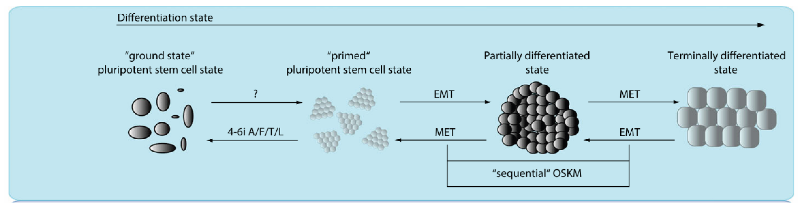
Pluripotent stem cells have a spectrum of different morphological, functional, transcriptional and epigenetic states, dependent on external developmental cues and potential final destiny of the cells. Figure 1 [6] shows the transition between 'ground state' pluripotent, 'primed' pluripotent state, partially differentiated state and terminally differentiated state and the ways cells may come back. The ground state ESCs are more undifferentiated than primed state cells. Morphology of both ESCs and iPSCs 'ground' state pluripotent stem cells resembles dome-shaped structures that are consistent of small and round cells. Cell aggregates are with irregular cell shape and invisible cell borders. This stage is decorated with high self-renewal capacity and high degree of clonogenicity. Progressing, 'primed' state ESCs cultures are distinguishable by flattened epithelial monolayer colonies that have reduced self-renewal capacity and lack of clonogenicity in individualized cells. They can be considered as ESCs from post-implantation embryo. The next transition is partially differentiated cell state that corresponds to ESCs differentiation into three germ layers, and corresponds to gastrulation event in vivo. In the embryo this process is dependent on an epithelial-mesenchymal transition (EMT). Finally, somatic cell differentiation, named here as terminally differentiated state, includes multiple events of EMT and mesenchymal-epithelial transition (MET) and corresponds to the development of cells in different organs [6,15].
2. Cellular Plasticity
During different biological processes, cell transit from one state to another. Two fundamental processes underline this cellular plasticity and are named EMT and MET. Multiple approaches can describe these cell transitions. For example, EMT can be described functionally as the loss of barrier-forming ability in the epithelial sheet and gaining of migratory and invasive properties of mesenchymal cells. Organizationally, EMT, can be described as a change in the apicobasal polarity of epithelial cells and gaining of front-rear polarity of a migrating mesenchymal cells and this includes changes in cell adhesion and cell-cell junctions. Moreover, changes in molecular profile are made of protein and transcription factor expression alterations [16]. The concept of EMT was initially proposed as the epithelial mesenchymal transformation by Elizabeth Hay in 1968 as a way to describe the important changes in embryogenesis, and later was renamed to EMT so it can be distinguished from the neoplastic transformation [17,18,19].
EMT can be summarized in down-regulation of epithelial markers (E-cadherin, TJP/ZO-1 and Occludin), rearrangement of the cytoskeleton, loss of cell-cell adhesion and apical-basal polarity and the acquisition of mesenchymal phenotype markers (such as Vimentin, N-Cadherin and Fibronectin) that allow increased cell protrusions and migratory potential of the transitioned cells [20]. It is recognized that embryonic EMT process is orchestrated and maintained through the collaboration of extracellular signals and intracellular transcription factors [21]. There are three types of EMT process. EMT Type 1 is related to embryo formation, EMT Type 2 is related to tissue regeneration and EMT Type 3 clarifies cancer progression [22].
3. EMT, MET, Stemness and Pluripotency
During induction of iPSCs and early phase reprogramming of fibroblasts it seems that MET is a crucial step which is shown by the up-regulation of epithelial genes such as E-cadherin and Epcam and down-regulation of mesenchymal genes such as Snail and N-cadherin in generating iPSCs [23]. It was shown that Sox2, Oct4 and c-Myc suppress TGF-beta signaling while Klf4 activates multiple epithelial genes. During iPSCs generation, the cells aggregate into distinguished epithelial-like colonies with well defined intracellular junctions [22]. Based on the observed gene expression dynamics, Samavarchi-Tehrani and colleagues identified three phases of reprogramming: initiation, maturation and stabilization. MET-associated alterations are evident during the initiation phase of reprogramming, when cells are still dependent on exogenous factor (OSKM) expression [24,25,26].
The process starts by the exogenous factors influence which initiates MET program to power fibroblasts through an epigenetic barrier made during development. Thus, the exogenous factors must suppress an intrinsic barrier presented by fibroblast-enriched transcription factors such as Snail that preserve mesenchymal phenotype, and TGF-beta1 [27]. It was shown that the expression of genes associated with MET induction is reverted and cells can return to parental fibroblastic profile after SKOM removal, showing that initiation phase is unstable and reversible. On the other hand, maturation phase lead to irreversible commitment to reprogramming and it is associated with the expression of ESCs markers including Nanog, which is described as a mediator of embryonic and induced pluripotency, thus Nanog expression is linked with irreversibility and pluripotency during maturation phase of the reprogramming and finally commitment during stabilization phase [15].
Controversially, during MEFs reprogramming an early EMT and factors that push EMT actually enhance reprogramming. Considering that MEFs are heterogenous population of cells that is not fully mesenchymal, treatment with TGF-beta pushes this population to mesenchymal homogeneous population, and by that gives the possibility of OSKM exogenous factors to initiate MET [6]. If we take this into account, does this mean that reprogramming of epithelial cells is easier?
For example, ciliary body epithelial cells have higher reprogramming efficiency to iPSCs than fibroblasts [28]. In mice, hepatocytes and gastric epithelial cells are reprogrammed with less difficulty than fibroblasts [23]. Additionally, mammary gland epithelial cells iPSCs generation is also with higher reprogramming efficiency. Since this population express abundant endogenous Klf4 it does not require exogenous expression of Klf4 to convert to iPSCs. Klf4 activates E-cadherin. Moreover, c-Myc expression is sufficient to downregulate TGF-betaR1 and TGF-betaR2 receptors, and in the absence of c-Myc, Alk51 that is TGF-beta receptor inhibitor, increases reprogramming of these cells, thus additionally proving the importance of TGF-beta to oppose the reprogramming [24].
Somatic cells that are most available and widely used for iPSCs derivation and include skin fibroblasts, hair keratinocytes, mononuclear cells from peripheral or umbilical cord blood (including B and T lymphocytes, and CD34+ cells), and urine cells containing tubular epithelial cells and fibroblast-like or urothelial cells. Since the number of cells that can be used is growing, this implies that cells of almost all tissues can be used for the generation of iPSCs. On the other hand, reprogramming process is highly inefficient with only a minority of donor cells are being reprogrammed to pluripotency. Successful and efficient generation of iPSCs depends of somatic cells age and the origin of somatic cells that are used for iPSCs reprogramming. As explained, cells have different states throughout development, thus cell differentiation stage also having an impact on reprogramming efficiency. It has been shown that hematopoietic stem and progenitor cells are programmed to iPSCS more efficiently than terminally differentiated B and T lymphocytes [23]. Moreover, Lapasset et al, showed that reprogramming senescent cells and cells from the elderly requires six-transcription factor cocktail with LIN28 and NANOG in addition to OSKM. Lo Sardo et al, also showed that age of the donor of the somatic cells is associated with the probability of genetic alterations, thus may decrease the efficiency or reprogramming. As a whole, successful reprogramming of healthy somatic cells into iPSCs depends on correct reprogramming of the cell's epigenetic landscape which means shutting down somatic gene expression in order to activate pluripotency related transcriptional program [29,30,31].
ESCs that are isolated from the inner cell mass, are considered to be typical epithelial cells, and they are also in undifferentiated state. Markers such as E-cadherin, SSEA1, alkaline phosphatase, Oct4 and Nanog are present. It seems that EMT may prohibit stemness in iPSCSs and ESCs, since promotes mesenchymal phenotype [28]. Interestingly, EMT has been shown to induce stem cell characteristics in mammary epithelial cells, which is in contrast with the notion that MET is the step for acquiring stem cell properties. What is important to mention that pluripotency and stemness are not the same [25]. ESCs marker Nanog is described as a mediator of embryonic and induced pluripotency. During cancer progression, the combination of EMT and stemness is pertinent, since cancer EMT, EMT type 3, allows cancer cell migration and dissemination to different tissues and organs and is like the EMT process during development, the steps are less ordered and lack coordination [32].
The migratory cancer cells combine the mesenchymal phenotype that is crucial for the efficient dissemination with, stem cell properties, since circulating cancer stem cells (CSCs) have the ability to form new tumors, to self-renew and to produce non-stem differentiated tumor cells. The important difference that CSCs are not pluripotent, as during metastasis formation, cancer cells that go though MET do not generate any other cell type and they revert to the phenotype of the primary tumor. If Nanog expression describes the irreversible state to full reprogramming and pluripotency, then the migrating CSCs should be Nanog negative or low, and their progeny in the metastasis should be a result of a MET that produces epithelial cells resembling the primary tumor [15,33]. There is a paradox in the dynamics of the EMT/MET processes during reprogramming and cancer progression [15]. The differences in steps and final destination still has to be defined on the molecular level, and compared with the EMT during development, or even wound healing, thus allowing us to use that approach to make the reprogramming more successful.
4. AI, a Tool to Collect, Analyze and Produce
In the middle of challenges associated with long-lasting culture time and difficult cell characterization, there is a demand for quicker and precise methods for proof of cell identity and function at different stages of iPSCs reprogramming. The regular practice is observation of changes in morphology and/or lineage marker expression, that may be considered as subjectively-based and laborious [34]. Approach that allows observation and recording changes in markers throughout the process of iPSCs reprogramming, while comparing and calculating the possible success, is the goal. Developments in digital pathology and computational image analysis provide a way for morphology description and classification [34,35]. For cell image classification method, artificial intelligence (AI) can make predictions based on machine learning algorithms that are able to learn from large datasets. They can evaluate multiple parameters without a priori knowledge. Machine learning methods are being developed in the last fifty years [34,36]. Moreover Deep learning uses multilayered neural network that mimics human neural circuit structure and can automatically extract features from an image, while traditional machine learning methods require human intervention. For example, an open source utility wnd-charm (weighted neighbor distance using a compound hierarchy of algorithms representing morphology) developed by Image Informatics and Computational Biology Unit at the National Institute on Ageing in Baltimore, allows users to define classes by providing images for each class: completely reprogrammed cells or partially reprogrammed cells or any other opposing classes. Additionally, Danter et al developed an unsupervised deep machine learning technology-DeepNEU to stimulate artificial iPSC system by using a defined group of reprogramming transcription factors [34,37,38,39].
The DeepNEU database (Version 3.2) contains 3589 gene/proteins (around 10% of human genome) and 27 566 nonzero relationships. On average, each node in the network has more than 7 inputs and 7 outputs. This system uses a literature validated set of reprogramming factors and allows simulations of relationships of a specific gene/protein to another gene/protein, or gene/protein to multiple genes/proteins or multiple genes/proteins to one gene/protein and finally multiple genes/proteins to different multiple/genes proteins, to explain it simply [40].
The possibilities are vast and all include an interaction with data obtained from the past research, current research data and different artificial intelligence-based methods that may provide outputs that are more accurate and effective, including powerful story telling tools such as graphs and tables. Laboratory work provides information that will be used for the final interpretation by artificial intelligence-based methods, providing aid to human factor that produces the data used. From the start of stem cell research until now the progress is unmissable. iPSCs led to the new era of research, that these days should implement all fields of STEM.
References
- Lendahl, U. 100 plus years of stem cell research-20 years of ISSCR. Stem Cell Rep. 2022, 17(6), 1248–1267. [Google Scholar] [CrossRef] [PubMed] [PubMed Central]
- Takahashi, K.; Yamanaka, S. Induction of pluripotent stem cells from mouse embryonic and adult fibroblast cultures by defined factors. Cell 2006, 126(4), 663–76. [Google Scholar] [CrossRef] [PubMed]
- Singh, V.K.; Kalsan, M.; Kumar, N.; Saini, A.; Chandra, R. Induced pluripotent stem cells: applications in regenerative medicine, disease modeling, and drug discovery. Front Cell Dev. Biol. 2015, 3, 2. [Google Scholar] [CrossRef] [PubMed] [PubMed Central]
- Ziętek, M.M.; Bihorac, A.; Wenta-Muchalska, E.; Duszewska, A.M.; Olech, W.; Sampino, S.; Bernat, A. Wisent Somatic Cells Resist Reprogramming by the PiggyBac Transposon System: A Case Study Highlighting Methodological and Conservation Hurdles. Int. J. Mol. Sci. 2025, 26, 4327. [Google Scholar] [CrossRef]
- Coronnello, C.; Francipane, M.G. Moving Towards Induced Pluripotent Stem Cell-based Therapies with Artificial Intelligence and Machine Learning. Stem Cell Rev. Rep. 2022, 18(2), 559–569. [Google Scholar] [CrossRef] [PubMed] [PubMed Central]
- Kovacic, Boris; et al. Clinical impact of studying epithelial–mesenchymal plasticity in pluripotent stem cells. Eur. J. Clin. Investig. 2015, 45.4, 415–422. [Google Scholar] [CrossRef]
- Malik, V.; Wang, J. Pursuing totipotency: authentic totipotent stem cells in culture. Trends Genet. 2022, 38(7), 632–636. [Google Scholar] [CrossRef] [PubMed] [PubMed Central]
- Poliwoda, S.; Noor, N.; Downs, E.; Schaaf, A.; Cantwell, A.; Ganti, L.; Kaye, A.D.; Mosel, L.I.; Carroll, C.B.; Viswanath, O.; Urits, I. Stem cells: a comprehensive review of origins and emerging clinical roles in medical practice. Orthop. Rev. 2022, 14(3), 37498. [Google Scholar] [CrossRef] [PubMed] [PubMed Central]
- Kruse, F. Stem cells and corneal epithelial regeneration. Eye 1994, 8, 170–183. [Google Scholar] [CrossRef]
- Thanaskody, K.; Jusop, A.S.; Tye, G.J.; Wan Kamarul Zaman, W.S.; Dass, S.A.; Nordin, F. MSCs vs. iPSCs: Potential in therapeutic applications. Front Cell Dev. Biol. 2022, 10, 1005926. [Google Scholar] [CrossRef] [PubMed] [PubMed Central]
- Thomson, J.A.; Itskovitz-Eldor, J.; Shapiro, S.S.; Waknitz, M.A.; Swiergiel, J.J.; Marshall, V.S.; Jones, J.M. Embryonic stem cell lines derived from human blastocysts. Science Erratum in: Science 1998 Dec 4;282(5395):1827. PMID: 9804556. 1998, 282(5391), 1145–7. [Google Scholar] [CrossRef]
- Passier, R.; Mummery, C. Origin and use of embryonic and adult stem cells in differentiation and tissue repair. Cardiovasc Res. 2003, 58(2), 324–35. [Google Scholar] [CrossRef] [PubMed]
- Robertson, J.A. Ethics and policy in embryonic stem cell research. Kennedy Inst. Ethics J. 1999, 9(2), 109–36. [Google Scholar] [CrossRef] [PubMed]
- Gerecht-Nir, S.; Itskovitz-Eldor, J. Human embryonic stem cells: a potential source for cellular therapy. Am. J. Transplant. 2004, 4 Suppl 6, 51–7. [Google Scholar] [CrossRef] [PubMed]
- Ocaña, O.H.; Nieto, M.A. Epithelial plasticity, stemness and pluripotency. Cell Res. Epub. 2010, 20(10), 1086–8. [Google Scholar] [CrossRef] [PubMed]
- Hookway, C.; Borensztejn, A.; Harris, L.K.; et al. A human induced pluripotent stem cell model for the holistic study of epithelial-to-mesenchymal transitions. Nat. Methods 2026, 23, 1411–1423. [Google Scholar] [CrossRef]
- Hay, E.D. Organization and fine structure of epithelium and mesenchyme in the developing chick embryo. In Epithelial-Mesenchymal Interactions; Fleischmajer, R., Billingham, R.E., Eds.; Williams and Wilkins: Baltimore, 1968; pp. 31–55. [Google Scholar]
- Akhurst, R.J. From shape-shifting embryonic cells to oncology: The fascinating history of epithelial mesenchymal transition. Semin Cancer Biol. 2023, 96, 100–114. [Google Scholar] [CrossRef] [PubMed] [PubMed Central]
- Lai, X.; Li, Q.; Wu, F.; Lin, J.; Chen, J.; Zheng, H.; Guo, L. Epithelial-Mesenchymal Transition and Metabolic Switching in Cancer: Lessons From Somatic Cell Reprogramming. Front Cell Dev. Biol. 2020, 8, 760. [Google Scholar] [CrossRef] [PubMed] [PubMed Central]
- Lamouille, S.; Xu, J.; Derynck, R. Molecular mechanisms of epithelial-mesenchymal transition. Nat. Rev. Mol. Cell Biol. 2014. [Google Scholar] [CrossRef] [PubMed]
- Chen, J.; Han, Q.; Pei, D. EMT and MET as paradigms for cell fate switching. J. Mol. Cell Biol. 2012, 4(2), 66–9. [Google Scholar] [CrossRef] [PubMed]
- Chen, T.; You, Y.; Jiang, H.; Wang, Z.Z. Epithelial-mesenchymal transition (EMT): A biological process in the development, stem cell differentiation, and tumorigenesis. J. Cell Physiol. 2017, 232(12), 3261–3272. [Google Scholar] [CrossRef] [PubMed] [PubMed Central]
- Yee, J. Turning Somatic Cells into Pluripotent Stem Cells. Nat. Educ. 2010, 3(9), 25. [Google Scholar]
- Polo, J.M.; Hochedlinger, K. When fibroblasts MET iPSCs. Cell Stem Cell Epub. 2010, 7(1), 5–6. [Google Scholar] [CrossRef] [PubMed]
- Li, R.; Liang, J.; Ni, S.; Zhou, T.; Qing, X.; Li, H.; He, W.; Chen, J.; Li, F.; Zhuang, Q.; Qin, B.; Xu, J.; Li, W.; Yang, J.; Gan, Y.; Qin, D.; Feng, S.; Song, H.; Yang, D.; Zhang, B.; Zeng, L.; Lai, L.; Esteban, M.A.; Pei, D. A mesenchymal-to-epithelial transition initiates and is required for the nuclear reprogramming of mouse fibroblasts. Cell Stem Cell 2010, 7(1), 51–63. [Google Scholar] [CrossRef]
- Samavarchi-Tehrani, P.; Golipour, A.; David, L.; Sung, H.-k.; Beyer, T.A.; Datti, A.; Woltjen, K.; Nagy, A.; Wrana, J.L. Cell Stem Cell7, this issue. 2010, 64–77. [Google Scholar]
- Li, R.; Liang, J.; Ni, S.; Zhou, T.; Qing, X.; Li, H.; He, W.; Chen, J.; Li, F.; Zhuang, Q.; Qin, B.; Xu, J.; Li, W.; Yang, J.; Gan, Y.; Qin, D.; Feng, S.; Song, H.; Yang, D.; Zhang, B.; Zeng, L.; Lai, L.; Esteban, M.A.; Pei, D. A mesenchymal-to-epithelial transition initiates and is required for the nuclear reprogramming of mouse fibroblasts. Cell Stem Cell Epub. 2010, 7(1), 51–63. [Google Scholar] [CrossRef] [PubMed]
- Li, X.; Pei, D.; Zheng, H. Transitions between epithelial and mesenchymal states during cell fate conversions. Protein Cell 2014, 5(8), 580–91. [Google Scholar] [CrossRef] [PubMed] [PubMed Central]
- Poetsch, M.S.; Strano, A.; Guan, K. Human Induced Pluripotent Stem Cells: From Cell Origin, Genomic Stability, and Epigenetic Memory to Translational Medicine. Stem Cells 2022, 40(6), 546–555. [Google Scholar] [CrossRef] [PubMed] [PubMed Central]
- Lapasset, L.; Milhavet, O.; Prieur, A.; et al. Rejuvenating senescent and centenarian human cells by reprogramming through the pluripotent state. Genes Dev. 2011, 25(21), 2248–2253. [Google Scholar] [CrossRef]
- Lo Sardo, V.; Ferguson, W.; Erikson, G.A.; et al. Influence of donor age on induced pluripotent stem cells. Nat. Biotechnol. 2017, 35(1), 69–74. [Google Scholar] [CrossRef]
- Bihorac, A. Role of Transglutaminase 2 in Migration of Tumor Cells and How Mouse Models Fit. Med. Sci. 2018, 6(3), 70. [Google Scholar] [CrossRef] [PubMed] [PubMed Central]
- Brabletz, T.; Jung, A.; Spaderna, S.; Hlubek, F.; Kirchner, T. Opinion: migrating can cer stem cells - an integrated concept of malignant tumour progression. Nat. Rev. Cancer 2005, 5, 744–749. [Google Scholar]
- Coronnello, C.; Francipane, M.G. Moving Towards Induced Pluripotent Stem Cell-based Therapies with Artificial Intelligence and Machine Learning. Stem Cell Rev. Rep. 2022, 18(2), 559–569. [Google Scholar] [CrossRef] [PubMed] [PubMed Central]
- Barisoni, L.; Lafata, K. J.; Hewitt, S. M.; Madabhushi, A.; Balis, U. G. J. Digital pathology and computational image analysis in nephropathology. In Nature Reviews Nephrology; Nature Research, 2020; Vol. 16, Issue 11, pp. 669–685. [Google Scholar] [CrossRef]
- Sommer, C.; Gerlich, D.W. Machine learning in cell biology - teaching computers to recognize phenotypes. J. Cell Sci. 2013, 126 Pt 24, 5529–39. [Google Scholar] [CrossRef] [PubMed]
- Zhang, H.; Shao, X.; Peng, Y.; Teng, Y.; Saravanan, K.M.; Zhang, H.; Li, H.; Wei, Y. A novel machine learning based approach for iPS progenitor cell identification. PLoS Comput Biol. 2019, 15(12), e1007351. [Google Scholar] [CrossRef] [PubMed] [PubMed Central]
- Shamir, L.; Orlov, N.; Eckley, D.M.; Macura, T.; Johnston, J.; Goldberg, I.G. Wndchrm - an open source utility for biological image analysis. Source Code Biol. Med. 2008, 3, 13. [Google Scholar] [CrossRef] [PubMed] [PubMed Central]
- Joutsijoki, H.; Haponen, M.; Rasku, J.; Aalto-Setälä, K.; Juhola, M. Machine Learning Approach to Automated Quality Identification of Human Induced Pluripotent Stem Cell Colony Images. Comput Math. Methods Med. 2016, 2016, 3091039. [Google Scholar] [CrossRef] [PubMed] [PubMed Central]
- Danter, W.R. DeepNEU: cellular reprogramming comes of age - a machine learning platform with application to rare diseases research. Orphanet J. Rare Dis. 2019, 14(1), 13. [Google Scholar] [CrossRef] [PubMed] [PubMed Central]
- Author 1, A.; Author 2, B. Title of the chapter. In Book Title, 2nd ed.; Editor 1, A., Editor 2, B., Eds.; Publisher: Publisher Location, Country, 2007; Volume 3, pp. 154–196. [Google Scholar]
- Author 1, A.; Author 2, B. Book Title, 3rd ed.; Publisher Location, Country: Publisher, 2008; pp. 154–196. [Google Scholar]
- Author 1, A.B.; Author 2, C. Title of Unpublished Work. Abbreviated Journal Name year. Phrase Indic. Stage Publ. submitted; accepted. in press. [Google Scholar]
- Author 1, A.B.; Author 2, C. Personal communication, 2012.
- Author 1, A.B.; Author 2, C.D.; Author 3, E.F. Title of Presentation. In Proceedings of the Name of the Conference, Location of Conference, Country, Date of Conference (Day Month Year).
- Author 1, A.B. Title of Thesis. Level of Thesis, Degree-Granting University, Location of University, Date of Completion.
- Title of Site. Available online: URL (accessed on. (accessed on Day Month Year).
Figure 1.
Differentiation state, modified from Kovacic et al, 2015 [6].
Figure 1.
Differentiation state, modified from Kovacic et al, 2015 [6].
Disclaimer/Publisher’s Note: The statements, opinions and data contained in all publications are solely those of the individual author(s) and contributor(s) and not of MDPI and/or the editor(s). MDPI and/or the editor(s) disclaim responsibility for any injury to people or property resulting from any ideas, methods, instructions or products referred to in the content. |
© 2026 by the authors. Licensee MDPI, Basel, Switzerland. This article is an open access article distributed under the terms and conditions of the Creative Commons Attribution (CC BY) license (http://creativecommons.org/licenses/by/4.0/).
Copyright: This open access article is published under a Creative Commons CC BY 4.0 license, which permit the free download, distribution, and reuse, provided that the author and preprint are cited in any reuse.