

Submitted:
16 September 2026
Posted:
17 September 2026
You are already at the latest version
Abstract
Major histocompatibility complex class II antigen presentation enables professional antigen-presenting cells to display endogenous and exogenous antigens to CD4+ T cells, linking antigen acquisition to the induction and regulation of adaptive immunity. While MHC II-mediated antigen presentation is defined as MHC II-peptide display at the cell surface, its outcome is determined by a coordinated series of regulatory checkpoints that control antigen entry, MHC II expression, intracellular routing, peptide loading, surface stability, and costimulatory signals that provide context to the presented antigen. The regulation of antigen presentation on MHC II is governed by multiple mechanisms — some universal, and some utilized selectively by dendritic cells, macrophages, or B cells. Herein we review the structural and cellular basis of MHC II function, antigen uptake mechanisms, MHC II transcriptional regulation, MHC II intracellular trafficking, and the processes regulating peptide editing, loading, and surface presentation. Together, these regulatory layers determine not only how much MHC II is displayed, but also which peptides are presented, where antigen loading occurs, and how long peptide–MHC II complexes remain available for CD4+ T cell recognition. Critically, disruption of these checkpoints contributes to immunodeficiency, autoimmunity, cancer immune evasion, and pathogen-mediated immune evasion. Across these areas, important gaps remain, including how pAPCs select between indirect and direct MHC II trafficking routes, how cargo-sorting mediators distinguish innocuous from infected materials, and how anti-inflammatory regulators directly regulate MHC II expression, peptide loading, or peptide-MHC II surface stability. Addressing these gaps would further clarify how pAPCs calibrate antigen presentation at the intersection of immunity, tolerance, and inflammatory resolution.
Keywords:
MHC II
; dendritic cells
; macrophages
; antigen presentation
; vesicular trafficking
; MHC II loading compartment
; CIITA
; HLA
1. Introduction of MHC II and Antigen Presenting Cells
Major histocompatibility complex class II (MHC II) molecules are cell-surface glycoproteins that allow immune cells to display peptide antigens to CD4+ T cells. In contrast to MHC class I molecules, which primarily present peptides derived from intracellular proteins, MHC II molecules are specialized for presenting peptides generated from extracellular or endocytosed material [1]. Through this pathway, the immune system surveys antigens derived from pathogens, allergens, apoptotic cells (ACs), and other extracellular sources.
MHC II-mediated antigen presentation is primarily performed by a subset of specialized immune cells collectively called professional antigen-presenting cells (pAPCs). These cells are defined by their capacity to acquire extracellular antigen, process it within intracellular compartments, load peptide-derived antigens onto MHC II, and provide the additional costimulatory signals required to drive CD4+ T cell responses. The primary pAPC populations are dendritic cells (DCs), macrophages, and B cells [2,3,4,5]. pAPCs are highly efficient at internalizing particulate and soluble exogenous antigens. Once internalized, these exogenous antigens are processed into smaller peptides and loaded onto MHC II molecules within an intracellular loading compartment termed the MHC II loading compartment (MIIC), after which peptide–MHC II (p-MHC II) complexes traffic to the cell surface. At this stage, p-MHC II complexes are recognized by cognate CD4+ T cells. In this way, pAPCs translate extracellular antigen encounters into the regulation of adaptive immune responses. In addition, some nonprofessional APCs can also present antigens on MHC II following certain inflammatory stimuli, including endothelial cells, fibroblasts, and lymph node stromal cells.
2. Factors Dictating the Fate of MHC II-Mediated Antigen Presentation
The outcome of MHC II antigen presentation depends on the mode of antigen capture, the peptide displayed, the activation state of the presenting cell, and the signals delivered alongside antigen recognition. Although pAPCs share the core machinery required for MHC II antigen presentation, different pAPCs capture antigens for loading onto MHC II through distinct mechanisms. These differences are shaped in part by the functional specialization of myeloid and lymphoid immune cell lineages. Myeloid and lymphoid are the two primary lineages derived from the hematopoietic stem cell (HSC) in the bone marrow, which gives rise to all blood and immune cells [6].
Myeloid pAPCs, including macrophages and DCs, acquire antigens largely through engulfment and sampling pathways such as phagocytosis, efferocytosis, macropinocytosis, trogocytosis, and receptor-mediated endocytosis. In contrast, B cells—the major lymphoid pAPC population—primarily internalize antigens via B-cell receptor (BCR)-dependent uptake of cognate antigen; although emerging evidence indicates that some B cell subsets retain some phagocytic capacity and can present the resulting antigens on MHC II [7]. Following antigen capture through these pathways, internalized antigens are processed and presented to T cells.
Presentation of a cognate antigen alone is insufficient to induce the activation of naïve CD4+ T cells, with additional co-stimulatory signals (CD80/CD86) and pro-inflammatory cytokines (e.g., IL-12, TNF-α, IL-1β) required to generate an immunogenic response. This activation induces the differentiation of naïve CD4+ T cells into effector T cell subsets [8,9]. In contrast, when antigens are presented on MHC II in the absence of strong co-stimulation and in the presence of regulatory cytokines (e.g., TGF-β, IL-10), a tolerogenic response is induced instead. This process, often referred to as steady-state antigen presentation, functions to display self-antigens and thereby tolerize autoreactive T cells in the periphery, promoting their anergy or deletion [10,11]. In parallel, steady-state presentation can drive the differentiation of regulatory T cell subsets, which suppress effector T cell responses and contribute to the maintenance of peripheral immune tolerance [12,13]. These regulatory processes are necessary as deletion of self-reactive T cells in the thymus via central tolerance pathways is incomplete and allows 25 - 40% of the self-reactive T cell repertoire to escape clonal deletion [14]. Thus, the removal of autoreactive T cells from the peripheral pool is essential for preventing autoimmunity [11,14,15].
3. MHC II Structure, Genetics and Cellular Distribution
MHC II molecules are as heterodimeric transmembrane glycoproteins composed of one α and one β chain. Both the α and β chains are membrane-anchored and contain an extracellular domain involved in peptide binding and T cell receptor (TCR) interactions, a transmembrane domain that anchors the molecule within the membrane, and a cytoplasmic tail involved in trafficking and signalling [16,17]. The extracellular portion of the α chain contains α1 and α2 domains, whereas the β chain contains β1 and β2 domains. Together, the α1 and β1 domains form the peptide-binding groove (Figure 1). Unlike the closed-ended peptide-binding groove of MHC I molecules, the MHC II groove is open-ended, allowing it to accommodate peptides that are 12–25 amino acids in length [16,18]. During antigen presentation, peptides loaded within this groove are recognized by the TCR, which simultaneously engages both the peptide and the MHC II molecule. The CD4 co-receptor interacts with the β2 domain, thus providing CD4 specificity to the interaction between pAPCs and T cells [19].
MHC II molecules are encoded within the human leukocyte antigen (HLA) gene cluster, a dense 3.6 megabase region located on chromosome 6 (6p21.3) [20]. In humans, there are three classical MHC II isotypes: HLA-DP, HLA-DQ, and HLA-DR [21,22,23]. HLA-DR is composed of the gene products DRA (α-chain) and DRB1 (β-chain), with the α chain showing significant evolutionary conservation while the β chain is highly polymorphic. Polymorphisms within DRB1 strongly influence the repertoire of peptides that HLA-DR can bind and are also associated with variations in disease susceptibility [24,25,26]. These polymorphisms alter peptide-binding preferences and are strongly associated with susceptibility to several autoimmune diseases, including rheumatoid arthritis, systemic lupus erythematosus, and multiple sclerosis [24,25,27,28,29]. In comparison to HLA-DR chains, polymorphism extends to both the α- and β-chain genes of HLA-DQ and HLA-DP, although it is more extensive at the HLA-DQ loci [26].
3.1. MHC II in Nonprofessional APCs
While this review focuses on pAPCs, other, non-classical cell types express MHC-II, including epithelial cells in the thymus, airways, and intestines, endothelial cells, astrocytes, microglia, neutrophils, mast cells, and some innate lymphoid cells [30,31,32,33,34,35,36]. These cells are not considered pAPCs as they do not fulfill one or more of the following criteria: (1) constitutively express MHC II, (2) specialize in extracellular antigen capture, or (3) retain costimulatory capacities [35,36].
Among these non-classical cell types, thymic epithelial cells (TECs) express MHC II constitutively to present self-antigens to developing T cells in the thymus in the absence of costimulation [37]. In this manner, TECs mediate central tolerance and eliminate autoreactive CD4+ T cells. In fact, medullary TECs and thymic DCs cooperate to mediate negative T cell selection via MHC II-mediated self-antigen presentation [38,39]. Other nonprofessional APCs are enriched in tissues where the immune system interfaces with the environment, or in the brain which lacks endogenous secondary lymphoid tissues, with these cells upregulating MHC II and costimulatory molecules in response to damage and inflammatory signals. This allows these cells to directly mediate T cell responses to locally derived inflammatory signals [40,41].
4. MHC II and Endocytosis
Antigen acquisition for MHC II presentation occurs largely via endocytosis, with three primary endocytic pathways: (1) engulfment pathways, including phagocytosis, efferocytosis, and macropinocytosis; (2) contact-dependent uptake pathways, such as trogocytosis; and (3) receptor-mediated endocytosis [42,43,44,45]. Macrophages and DCs utilize phagocytosis and efferocytosis to non-specifically acquire antigens, whereas B cells acquire their cognate antigen via B cell receptor-mediated endocytosis [2,3,42,46]
4.1. Phagocytosis
Phagocytosis is a key component of the innate immune response, in which intracellular or extracellular pathogens, particulate materials, or infected ACs are internalized into the pAPC. Following engulfment these are degraded via the endo-lysosomal pathway, and the resulting antigens loaded onto MHC II for antigen presentation [3,47]. In phagocytosis, a pathogen is engulfed into a membrane-bound pocket within the pAPC termed the phagosome [48]. This phagosome then sequentially fuses with early endosomes, late endosomes, and finally lysosomes, to form a mature degradative organelle termed the phagolysosome [49]. The environment of the phagolysosome is highly acidic and contains degradative enzymes, including proteases, that break down pathogen-derived proteins into peptides [47,48]. During this process, MHC II is trafficked to the phagolysosome, where they are loaded with these peptides. The MHC II-antigen complexes are then trafficked to the cell surface, where, alongside the costimulatory molecules CD80/CD86, they present antigens to CD4+ T cells, generating an immune response [48].
4.2. Efferocytosis
Efferocytosis is a phagocytosis-like process wherein phagocytes clear and recycle apoptotic debris. In canonical efferocytosis, healthy apoptotic cargo is engulfed into phagosome-like vacuoles called efferosomes. These efferosomes then undergo the same endo-lysosomal maturation pathway as phagosomes, thereby degrading the apoptotic cargo, with the resulting materials recycled or exported into the extracellular space [50]. While efferocytosis primarily contributes immune tolerance by presenting AC-derived antigens under tolerogenic conditions, many pathogens are intracellular and apoptosis functions as a host strategy to limit pathogen dissemination. Consequentially, many pathogens are first encountered via the efferocytic pathway, rather than phagocytosis [50,51]. Incredibly, by utilizing efferosome-derived Toll-Like Receptor (TLR) signaling, pAPCs can selectively present antigens from efferosomes containing infected ACs without presenting antigens from non-infected ACs that are simultaneously efferocytosed by the same pAPC [52]. Thus, immune homeostasis is maintained by selectively mounting a response against non-self antigens encountered through efferocytosis while remaining tolerant towards self-materials.
Because both self- and pathogen-derived antigens can be acquired by efferocytosis, the post-efferocytosis regulation of co-stimulatory molecule and cytokine expression has important implications for the regulation of the adaptive immune response. For example, following efferocytosis DCs remain immature and both DCs and macrophages adopt a non-activated, tolerogenic or non-inflammatory state characterized by minimal to no co-stimulation and regulatory cytokine production [11,53,54,55]. Consequently, when antigens from healthy ACs are loaded onto MHC II and presented to T cells in the absence of co-stimulation, a tolerogenic environment is generated that supports peripheral tolerance through the differentiation of T regulatory cells [11,56,57,58]. On the other hand, TLR-mediated intraefferosomal signaling induces antigen presentation alongside CD80/86 and inflammatory cytokine expression, resulting in the differentiation of T effector cells [59,60].
There is limited direct evidence indicating that B cells can engage in efferocytosis, or present antigens from efferocytosed materials. However, there is evidence that B1-cell-derived natural antibodies contribute to immune homeostasis by enhancing AC clearance and suppressing inflammation [61,62]. More specifically, Ogden et al. demonstrate how this AC clearance mainly occurs through a complement-dependent mechanism wherein bound IgM Abs may recruit C1q, subsequently activating the classical complement pathway and promoting the phagocytosis of opsonized ACs [62].
4.3. Macropinocytosis
In macropinocytosis, macrophages and DCs form large plasma membrane ruffles which can engulf large volumes of extracellular fluid, thereby non-specifically ingesting soluble molecules, including antigens [63]. This is an actin-dependent mechanism that forms large endocytic vesicles called macropinosomes, which range in size from 0.5 to 5 µm in diameter [63,64]. Both macrophages and DCs perform macropinocytosis under steady-state conditions. Studies by Burgdorf et al. and Sallusto et al., have shown that fluorescently-tagged fluidic tracers are engulfed via macropinocytosis and are subsequently trafficked to MHC II+ late endosomes and lysosomal compartments [44,65].
Resting B cells do not engage in macropinocytosis, but there is some evidence that activation of protein kinase C by the plant-derived toxin PMA, or infection with Salmonella typhimurium, Mycobacterium tuberculosis, or Mycobacterium smegmatis may induce macropinocytosis in B cells. However, these experiments rely on the human Raji B cell lymphoblast cell line, and it remains unclear if this behaviour occurs in primary B cells, nor is it known whether this micropinocytosis contributes to antigen presentation on MHC II [66].
4.4. Trogocytosis
Trogocytosis is a mode of intercellular transfer of membrane-associated proteins between immune cells [67]. During trogocytosis, a contacting cell acquires portions of the plasma membrane and associated proteins from another cell through a poorly defined endocytic mechanism [68]. These transferred membrane proteins are then trafficked in a manner which transports the proteins to the recipient cell surface in their native orientation [68]. While most commonly used by T cells to acquire MHC molecules from other lymphocytes and pAPCs, DCs and B cells can also acquire p-MHC II complexes from other cells through this pathway [45,69,70,71]. This can occur in the presence or absence of antigen receptor stimulation.
4.5. B Cell Receptor-Mediated Endocytosis
Upon cognate antigen binding to the BCR, the BCR-antigen complexes undergo receptor-mediated endocytosis and traffic through the endosomal system for processing and MHC II-mediated presentation. Early studies yielded differing evidence regarding the requirement for BCR signaling in this process. In 1995, Salamero and colleagues found that pharmacological inhibition of tyrosine kinase or phosphatase activity involved in BCR signaling impaired cross-linking-induced BCR internalization [72]. In contrast, Song et al. reported that BCR cross-linking was not required for antigen delivery to the MIIC, although cross-linking of the BCR and subsequent signalling markedly accelerated the trafficking of BCR-antigen complexes to lysosomes and the MIIC [73].
The most well-studied mechanism of BCR internalization is clathrin-mediated endocytosis [74,75]. During this process, clathrin gets recruited onto BCR-antigen complexes through adaptor protein 2 (AP-2), leading to the formation of clathrin-coated pits [76]. Dynamin then constricts the neck of these pits, resulting in a vesicle that buds from the plasma membrane [77]. The clathrin coat is subsequently removed, and the internalized complexes enter the endolysosomal pathway. Throughout the process, actin remodelling facilitates initial BCR internalization and trafficking to late endosomes and lysosomes [78]. Vav promotes Rac1/2 activation, linking BCR stimulation to the actin-dependent machinery required for efficient internalization, while dynamin mediates vesicle scission [77,79]. Although these proteins do not directly affect MHC II or CD80/86 expression, their disruption reduces BCR endocytosis, which in turn impairs MHC II-mediated antigen presentation to T cells. Using the DO.11.10 T cell line as a reporter for surface OVA p-MHC II complexes, the authors found reduced presentation of BCR-internalized PC-OVA by B cells expressing dominant-negative Vav or dynamin, while presentation was restored when preprocessed OVA peptide was supplied [77].
Although clathrin-mediated endocytosis accounts for a substantial proportion of BCR uptake, additional mechanisms are required to explain the diversity of antigen-induced BCR internalization. BCR cluster radii can range from 60 nm to 1 μm, while clathrin-coated pits average between 56-126 nm in radius [80,81]. The larger BCR clusters, therefore, likely use an alternative endocytic mechanism for their internalization, such as the phagocytosis pathway which allows some B cell subsets to internalize larger particles (>0.5 μm) [7,82,83].
In addition, BCRs can undergo fast endophilin-mediated endocytosis, an antigen-induced, clathrin-independent pathway mediated in B cells by Endophilin A2. Endophilin A2 is recruited following antigen stimulation, while its BAR domain promotes membrane curvature required for endocytic carrier formation [84,85]. Like clathrin-mediated endocytosis, fast-endophilin-mediated endocytosis requires actin rearrangement and dynamin [86,87]. Malinova et al. discovered that Endophilin A2 loss reduced BCR-mediated antigen uptake but had little effect on total MHC II-mediated antigen presentation. In fact, there was a marked increase in surface MHC II expression in Endophilin A2-deficient B cells. The authors proposed that preserved antigen presentation may reflect compensatory antigen delivery through clathrin-mediated endocytosis and/or the increased surface MHC II expression observed in Endophilin A2-deficient B cells [88].
Altogether, these acquisition pathways demonstrate that MHC II-mediated antigen presentation is shaped, in part, by the route and context through which antigen is acquired. Phagocytosis and macropinocytosis allow myeloid pAPCs to sample particulate and soluble material from the extracellular environment, whereas BCR-mediated endocytosis enables B cells to concentrate their cognate antigen. In contrast, canonical efferocytosis generally supports tolerance by coupling AC clearance to the steady state antigen presentation pathway, while infected apoptotic cells redirect this pathway toward immunogenic antigen presentation via TLR-dependent signaling triggered by microbial products. Trogocytosis further expands the ways in which p-MHC-II complexes can be transferred or acquired between immune cells, although how trogocytosed materials intersect with the antigen loading compartment remains unclear. Thus, antigen uptake pathways simultaneously deliver cargo to MHC II-loading compartments and help determine whether MHC II presentation reinforces homeostasis, supports peripheral tolerance, or initiates adaptive immune activation.
5. Cell-Type Specific Trafficking and Regulation of MHC II
The regulatory mechanisms described above provide a general framework for controlling MHC II expression, trafficking, peptide loading, and surface stability. Adding further complexity, there are distinct inflammatory, trafficking, and endocytic programs employed by some pAPC subsets to further regulate MHC II-mediated antigen presentation (Figure 2). These pathways are tuned to each cell type’s dominant immunological role: DCs are specialized for initiating CD4+ T cell responses within peripheral lymphoid tissues, macrophages modulate antigen responsiveness in inflamed tissues, and B cells present cognate antigens to helper T cells during the induction of humoral immunity [2,5,89]. Across these subsets, MHC II trafficking is tightly balanced to promote tolerogenic presentation of self-antigens under steady-state conditions and immunogenic presentation of non-self-antigens in inflammatory environments [11,52,90].
5.1. DCs
Among pAPCs, DCs are the principal initiators of naïve T-cell responses. DCs are a heterogeneous population composed of multiple subsets with distinct functional specializations [91,92]. There are important subset-specific differences in MHC II regulation and trafficking, which shape the type and quality of T-cell responses induced by each DC population. In mammals, four major DC subsets have been described: conventional dendritic cells (cDCs), plasmacytoid dendritic cells (pDCs), monocyte-derived dendritic cells (moDCs), and Langerhans cells (LCs) [93]. cDCs and moDCs arise predominantly through myeloid developmental pathways, while LCs derive from embryonic myeloid progenitors; in contrast, pDC development shows a strong association with lymphoid progenitors [94,95,96,97,98,99]. Conventional DCs can be further subdivided into cDC1 and cDC2 subsets, each with specialized roles in antigen presentation [91,100].
Among these, cDCs represent the principal pAPCs responsible for the activation of naïve T cells. cDC1s are specialized in priming CD8+ T cells via cross-presentation on MHC I, whereas cDC2s direct MHC II-dependent CD4+ T-cell responses [101,102]. In contrast, moDCs arise predominantly under inflammatory conditions and contribute to antigen presentation in these contexts, while LCs function as tissue-resident APCs within the epidermis [95,96,103,104]. pDCs exhibit comparatively inefficient phagocytic capacities, and are primarily specialized for the production of type I interferons during antiviral responses [105,106]. Despite their reduced intrinsic antigen-presenting function, activated pDCs can modulate the activity of other DC subsets. In particular, type I interferons (IFN-α/β) produced by pDCs enhance MHC II expression, co-stimulatory molecule upregulation, and antigen presentation capacity in cDCs and moDCs, thereby providing a helper function that promotes effective CD4+ T cell activation and initiation of adaptive immune responses [107,108].
cDC2s are particularly specialized for MHC II-dependent CD4+ T-cell priming, while moDCs contribute to CD4+ T-cell responses during inflammatory conditions [93,95,102]. cDC2 subsets preferentially promote Th2- and Th17-associated responses, whereas inflammatory moDCs more commonly support Th1- and Th17-polarized effector responses [95,102,109,110]. In cDC2 and moDCs, MHC II trafficking is dynamically regulated during their maturation. Immature DCs display very few p-MHC II complexes on their surface. Instead, MHC-II is retained largely in the intracellular antigen-processing compartments [90,111,112]. This intracellular retention allows immature DCs to efficiently acquire material in peripheral tissues through efferocytosis and macropinocytosis [44,55]. When these cells enter sites of infection or inflammation—e.g., where exogenous antigens may be encountered—they begin undergoing maturation while simultaneously migrating to secondary lymphoid organs [47,113].
DC maturation and antigen presentation are not functionally synonymous. Historically, it was thought that DC maturation was the prerequisite ‘switch’ that enabled immunogenic antigen presentation. However, Jiang et al. have demonstrated that DCs can acquire phenotypic features of maturation under steady-state conditions, increased MHC II and co-stimulatory molecule expression, without acquiring a corresponding inflammatory cytokine program [114]. These DCs instead promoted the development of regulatory T cells rather than effector T-cells [114]. Complementing this distinction, Blander and Medzhitov showed that DC maturation alone is insufficient to determine which phagocytosed antigens are selected for MHC II presentation. Instead, only cargos containing TLR ligands are trafficked to the MIIC to generate p–MHC II, while antigens from cargos lacking these ligands are not presented [115]. Jointly, these findings demonstrate that phenotypic DC maturation, immunogenic activation, and antigen selection for MHC II presentation are related but independently regulated processes. The increased surface expression of p-MHC-II during DC activation is driven by coordinated changes in MHC II synthesis, intracellular trafficking, and antigen processing [90,111,112,116]. Alterations in endosomal and lysosomal protease activity further influence peptide generation and loading, collectively enhancing the formation and stability of p-MHC-II complexes [117]. Thus, in DCs, MHC II trafficking is organized around a developmental transition from antigen acquisition in peripheral tissues to sustained p–MHC II display in lymphoid organs.
DC maturation is a larger cellular program involving several components: (1) the increased expression of surface MHC II and CD80/CD86, (2) the upregulation of inflammatory cytokine production including TNF-α, IL-6, IL-1β and IL-12, (3) the migration to lymph nodes, (4) a redistribution of MHC II from intracellular compartments to the plasma membrane, and (5) the enhanced capacity to stimulate T cells [111,112,113,114,116]. In addition, there is an increase in the expression of adhesion molecules and downregulation of MHC II endocytic activity. Wilson et al. demonstrated that immature DCs constitutively present self-antigens in vivo but rapidly internalize and degrade p-MHC II complexes under steady-state conditions. During maturation, reduced MHC II endocytosis and increased surface retention of p-MHC II complexes enable prolonged antigen presentation and enhance T-cell stimulatory capacity [90]. Altogether, these changes create optimal conditions conducive to enhanced p-MHC-II formation [90,111,112,116]. In comparison, activation refers to the transition from tolerogenic to immunogenic antigen presentation, in which p-MHC II complexes are presented alongside inflammatory cytokines and co-stimulatory signals that promote effector T-cell activation. During DC activation, reduced re-endocytosis and degradation of p–MHC II complexes extend their surface persistence [118].
5.2. Macrophages
Whereas DCs are specialized for initiating naïve T-cell responses, macrophages function as highly plastic pAPCs that integrate MHC II-mediated antigen presentation with phagocytosis, efferocytosis, tissue homeostasis, inflammatory cytokine production, and the reactivation of previously primed effector CD4+ T cells within peripheral tissues [3,89,119,120]. Macrophages employ MHC II trafficking and antigen-presentation pathways that overlap with those of DCs, but these processes are integrated with a highly phagocytic cellular program characterized by abundant lysosomal proteases, shaped by the local tissue environment, and promoting the rapid processing of internalized material [121,122,123]. Moreover, macrophages tend to engage in antigen presentation within peripheral tissues, rather than in lymphoid tissues, and therefore tend to modulate the activity of already activated CD4+ T cells, rather than induce the activation of naive T cells [124].
Macrophages arise from both circulating monocytes and tissue-resident embryonic precursors [125,126]. Bone marrow derived macrophages (BMDMs) and monocyte-derived macrophages have also been widely used as experimental models to investigate the mechanisms regulating macrophage MHC II expression [127,128,129,130].
In macrophages, MHC II-mediated antigen presentation is closely linked to activation state. Historically, macrophages have been broadly categorized into classically activated macrophages (M1-like) and alternatively activated anti-inflammatory macrophages (M2-like). Although the M1/M2 framework oversimplifies macrophage diversity, it remains useful for distinguishing inflammatory antigen-presenting macrophages from more regulatory or tissue-repair-associated states [131,132]. Classically activated (M1-like) macrophages exhibit an inflammatory antigen-presenting phenotype characterized by increased expression of MHC II and co-stimulatory molecules in many experimental systems, together with production of inflammatory cytokines such as IL-1β, IL-6, IL-12, and TNF-α [132,133,134,135]. However, unlike DCs, macrophages retain substantial phagocytic capacity during activation. Macrophage phagosomes can themselves function as MHC II antigen-processing compartments, acquiring MHC II and HLA-DM and supporting the formation of p–MHC II complexes that subsequently traffic to the cell surface [136]. Phagosome maturation is also influenced by macrophage activation state. In human M1 macrophages, phagosomes exhibit delayed acidification and fusion with late endosomes and lysosomes relative to M2 macrophages, while prioritizing NADPH oxidase-dependent antimicrobial activity [137]. This delayed acidification may promote the retention of antigenic peptides by limiting the extent to which they are degraded fully into amino acids. M1-like macrophages tend to drive Th1 and Th17 responses due to their expression of p-MHC II, co-stimulatory molecules, and the inflammatory cytokines IL-12, IL-23, IL-1β, and TNF-α [133,138]
In contrast to inflammatory M1-like macrophages, alternatively activated and pro-resolving M2 macrophages arise in response to tissue-repair and anti-inflammatory cues. M2 macrophages typically exhibit reduced immunostimulatory capacity, and constitutively produce IL-10 which acts in an autocrine and paracrine fashion to reduce surface p-MHC II complexes and promote regulatory T-cell responses [139,140]. Although M2 macrophages can still process and present antigens on MHC II, the absence of co-stimulatory molecules on these cells and their production of IL-10 and TGF-β are associated with reduced T cell activation, induction of T cell anergy, and the promotion of regulatory T cell responses. In line with the pro vs anti-inflammatory phenotype of M1 and M2 macrophages, antigen-presenting M1-like macrophages downregulate macropinocytosis during the shift from antigen capture to antigen presentation, thereby biasing antigen presentation towards phagocytosed targets [141]. In comparison, M2 macrophages engage in substantial micropinocytosis and thereby present antigens derived from the local environment [141].
5.3. B Cells
Unlike DCs and macrophages, MHC II trafficking in B cells has less to do with broad environmental sampling and instead is largely restricted to antigen-specific BCR-mediated uptake [2]. Resting B cells constitutively express MHC II but have relatively limited co-stimulatory capacity [142]. Following cognate antigen binding, B cells transition from an antigen-surveillance state to an activated pAPC state specialized for interactions with CD4+ T cells, particularly T follicular helper (Tfh) cells [2,143,144]. BCR engagement with a cognate antigen induces extensive intracellular reprogramming that promotes BCR-mediated endocytosis, increased peptide processing, cytoskeletal remodeling, redistribution of MIICs, and increased surface display of p-MHC II complexes [145,146]. Activated B cells upregulate co-stimulatory molecules including CD80 and CD86 and migrate toward T cell zones within secondary lymphoid tissues, where they efficiently engage Tfh cells [143]. Through these interactions, activated B cells initiate germinal center responses, antibody class switching, affinity maturation, and plasma cell differentiation, with p–MHC II density contributing to Tfh-mediated selection of germinal center B cells [144]. These processes ultimately result in the generation of an antigen-specific humoral immune response.
Although B cells were classically considered non-phagocytic, emerging evidence suggests that B cells from various species can perform phagocytosis. In mammals, innate-like B cells, including peritoneal cavity B1a and B1b cells, can internalize bacteria and particulate antigens through an actin-dependent, BCR independent phagocytosis-like mechanism, however it remains unclear whether antigens from these targets are presented on MHC II [147]. In contrast, follicular and marginal zone B cells are more restricted, with phagocytic uptake limited to engulfment of particulates bearing the antigen recognized by a B cells BCR [7]. The phagocytic uptake of non-cognate antigens by B1 cells is also less efficient than phagocytic uptake of cognate antigens by Fo and MZ B cells. Thus, B cells use MHC II trafficking to convert antigen-specific BCR recognition into productive Tfh-cell help, linking cognate antigen capture to germinal center formation, class switching, affinity maturation, and plasma cell differentiation.
6. MHC II Expression and Movement as Regulatory Checkpoints
Endocytic pathways determine how extracellular antigens enter pAPCs and are delivered into antigen-processing compartments. However, antigen capture alone does not define the efficiency or outcome of the MHC II-mediated antigen presentation. For an antigen to be displayed to CD4+ T cells, MHC II molecules must first be synthesized, correctly assembled, transported to appropriate intracellular compartments, loaded with antigen-derived peptides, and maintained at the cell surface long enough to support T cell recognition — with each of these steps used to regulate aspects of MHC II-mediated antigen display. By controlling these steps, pAPCs fine-tune antigen presentation according to cell identity, activation state, inflammatory context, and antigen source.
6.1. Induction of MHC II Expression
6.1.1. CIITA
MHC II transcription is tightly controlled, with the Class II Major Histocompatibility Complex Transactivator (CIITA) acting as a master regulator of MHC II transcription. CIITA both acetylates histones at the MHC II locus – thereby opening DNA to other transcription factors – and recruits the transcriptional machinery that then transcribes MHC II [148,149,150] (Figure 3). Thus, the regulation of CIITA expression generally dictates the transcription of MHC II. CIITA is expressed constitutively in pAPCs, although its expression levels can be modulated by cytokines and other immunomodulatory signals [127,148]. Clinically, CIITA was first discovered by analyzing patients who had hereditary MHC II deficiency, also known as bare lymphocyte syndrome, where these patients were found to bear autosomal recessive mutations that inactivate CIITA or its downstream RFX effector complex [151]. Beyond regulating the MHC II genes, CIITA also regulates the expression of several components of the antigen presentation machinery, including HLA-DM, HLA-DO, and CD74. These non-classical components play critical roles in antigen processing, peptide editing, and MHC II trafficking [152,153].
CIITA expression is controlled in a cell type-specific manner by three separate CIITA promoters (pI, pIII, and pIV, Table 1 and Figure 4) [154,155]. The specific signals pathways and transcription factors that regulate each promoter differ, and consequently, most cell types express CIITA from a single promoter. Each promoter generates a CIITA transcript containing a promoter-specific first exon, resulting in distinct CIITA mRNAs and proteins [156]. CIITA produced from pI contains an N-terminal caspase recruitment domain (CARD) that is absent from the pIII- and pIV-derived isoforms, while the pIII and pIV produce transcripts with different 5′ UTRs and thus produce identical proteins that lack the CARD domain [157]. Thus, differential regulation of CIITA promoters is an important determinant of the cellular and context-dependent patterns of MHC II expression. While some cell types constitutively express CIITA, interferon γ (IFN-γ) can drive de novo or increased CIITA expression in many cell types, allowing for upregulation of MHC II across many pAPCs and nonprofessional APCs, with promoter pIV serving as the principal IFN-γ-responsive promoter [156,160]. Binding of IFN-γ to its receptor induces activation of the receptor-associated kinases Jak-1 and Jak-2 [164,165]. These kinases phosphorylate STAT1, which subsequently homodimerizes and translocates to the nucleus where it activates transcription of IFN-γ-responsive genes, including IRF-1 [159,160,164]. IRF-1 then binds to regulatory elements within the CIITA pIV promoter, thereby promoting CIITA transcription. Additionally, STAT1 works cooperatively with the ubiquitously expressed upstream stimulatory factor 1 (USF-1) to bind cis-acting elements on pIV, further enabling CIITA transcription [160]. Once translated, the generated CIITA protein contains an N-terminal transcriptional activation region with intrinsic acetyltransferase activity, intrinsic serine/threonine kinase activity, a proline/serine/threonine-rich (P/S/T) region, a central GTP-binding domain, and four C-terminal leucine-rich repeats [149,166,167]. To promote MHC II transcription, CIITA does not directly bind DNA. Rather, it is recruited to MHC II promoters through numerous protein-protein interactions with DNA-bound components of the MHC II enhanceosome. CIITA’s C-terminal leucine-rich repeat region enables efficient promoter recruitment [166,168]. Meanwhile, its N-terminal acidic and P/S/T-rich regions contribute to transcriptional activation by interacting with components of the general transcriptional machinery [169,170,171]. With these elements, CIITA transcriptionally regulates MHC II expression in pAPCs.
CIITA also regulates MHC II expression on an epigenetic level. Specifically, it regulates MHC II through chromatin modification and assembly of the transcriptional machinery. CIITA contains an N-terminal region with intrinsic acetyltransferase activity that is activated upon GTP binding to its central GTP-binding domain [149]. Once activated, CIITA promotes histone acetylation at MHC II regulatory regions, including the HLA-DRA promoter and distal locus control region, contributing to chromatin remodeling of the MHC II locus [150,172,173]. Once the MHC II promoter is accessible, CIITA then mediates the formation of a transcriptional complex containing CIITA, RFX5, CREB/ATF1, and NF-Y (Figure 3) [168,174]. Through its serine/threonine kinase activity, CIITA can autophosphorylate, enhancing its acetyltransferase activity, and phosphorylate RAP74, the large subunit of the general transcription factor TFIIF [167,175,176]. Through these activities, CIITA can modulate both its own transcriptional activity and indue activity the transcriptional machinery, thereby promoting transcription of MHC II.
cDCs express CIITA primarily through promoter I (pI). When cDCs are in their immature, antigen-sampling state, the constitutive expression of CIITA from pI enables constitutive biosynthesis of MHC II. Luciferase assays comparing the transactivator activity of CIITA in DCs (pI) versus B cells (pIII) have shown that pI is a more potent activator of CIITA expression than pIII [157]. Following exposure to inflammatory stimuli such as TLR ligands and TNFα, cDCs can undergo transient upregulation of MHC II synthesis, with IFN-γ/JAK–STAT1 signaling inducing this upregulation of CIITA [111,159,160]. As a result, DCs increase MHC II biosynthesis during the early stages of DC maturation.
As DCs mature, pI activity declines, reducing CIITA expression and consequently limiting the synthesis of new MHC II molecules [17,158,177]. In splenic DCs, MHC II synthesis increases modestly within the first 2 hours of maturation, then subsequently declines to 25% of the baseline within 6 hours, and is fully suppressed by 18 hours [90]. Transcription factors such as the positive regulatory domain I (PRDM1, or BLIMP1) and IRF8/PU.1 have been implicated in negatively regulating CIITA expression during this maturation process [158]. Although MHC II synthesis is reduced following CIITA silencing in mature DCs, surface MHC II expression is maintained through reduced turnover of pre-existing peptide–MHC II complexes, thereby prolonging their display at the cell surface. Indeed, mature cDCs have higher levels of p-MHC II on their cell surface than do immature cDCs due to the extended half-life of p-MHC II molecules [111,178,179,180]. Consequently, mature cDCs undergo a functional transition from antigen acquisition toward optimized T cell priming and antigen presentation.
Due to their lymphoid origin, pDCs mediate CIITA expression via the pIII promoter, consistent with these cells emerging from lymphopoiesis rather than myelopoiesis [181,182].
In macrophages, MHC II expression is strongly induced by IFN-γ, with CIITA transcription arising predominantly from pI and pIV. These promoters display distinct kinetics following IFN-γ stimulation: pIV-derived CIITA is induced rapidly but subsequently declines, whereas pI-derived CIITA appears several hours after stimulation but is sustained and therefore contributes to prolonged MHC II expression [127]. Consistent with this, macrophages lacking pIV retain IFN-γ-inducible MHC II expression through compensatory activity of the pI promoter, demonstrating that pIV is not essential for MHC II induction in the macrophage lineage [156].
Buxadé et al. further identified a macrophage-specific NFAT5-dependent enhancer located upstream of the Ciita locus that interacts with pI and is required to sustain CIITA and MHC II expression under both steady-state and IFN-γ-stimulated conditions [130]. Acute IFN-γ stimulation induces transcription from both promoters I and IV and consequently MHC II expression; however, NFAT5 is specifically required to sustain CIITA expression in macrophages following transient IFN-γ stimulation [130]. Taken together, these findings indicate that macrophage MHC II expression is regulated through coordinated pI and pIV activity, with NFAT5-pI activity providing an important macrophage-specific mechanism for sustained CIITA expression.
6.1.2. Regulation of MHC II Translation
Compared with transcriptional regulation, translational regulation of MHC II expression is less well-characterized. However, evidence from both cytokine stimulation and DC maturation studies indicates that translation provides an additional level of control over MHC II protein production. Goñalons et al. demonstrated that IFN-γ can regulate MHC II expression at the translational level in murine B cells and macrophages. In a murine B-cell line, IFN-γ increased surface MHC II I-A expression without detectable changes in I-A mRNA levels, whereas in bone marrow-derived macrophages, IFN-γ increased both I-A mRNA and surface MHC II expression. In both cell types, IFN-γ increased synthesis of I-Aα and I-Aβ proteins and enhanced ribosome loading onto both mRNAs, indicating regulation at the level of translation initiation [183].
Similarly, translational regulation appears to contribute to the maintenance of MHC II expression during lipopolysaccharide (LPS)-induced DC maturation. Although LPS suppresses IFN-γ induced CIITA expression, Malanga et al. identified additional post-transcriptional mechanisms in human monocyte-derived DCs undergoing LPS-induced maturation. Using qRT-PCR and polysome profiling, they found that CIITA transcript abundance decreased during maturation, while HLA-DRA and HLA-DQA1 transcripts remained strongly associated with polysome-rich fractions across the first 24 hours of LPS stimulation. This continued association with actively translating polysomes enabled the continued synthesis of MHC II despite reduced MHC II transcription, allowing for newly synthesized MHC II proteins to contribute to contribute the pool of p-MHC II complexes accumulating at the cell surface of maturing DCs [184]. Jointly, these findings indicate that translation acts as a secondary but relevant checkpoint in MHC II antigen- presentation regulation.
6.1.3. CIITA/MHC II Regulation in Nonprofessional APCs
MHC II expression in many nonprofessional APCs is generally dependent on CIITA pIV. Waldburger et al. demonstrated this directly using mice carrying a targeted deletion of pIV, which abolished IFN-γ-induced MHC II expression in a broad range of extrahematopoietic cells, including fibroblasts, astrocytes, vascular endothelial cells, epithelial cells, and hepatocytes. In contrast, constitutive and IFN-γ-inducible MHC II expression was retained in B cells, DCs, and macrophage-lineage cells through alternative CIITA promoter usage. Thus, while pIV is dispensable in pAPCs due to compensatory activity from alternative CIITA promoters, it is required for MHC II synthesis in extrahematopoietic cells [156]. pIV is not, however, the only CIITA promoter capable of responding to IFN-γ. Piskurich et al. demonstrated IFN-γ-inducible activity of pIII in fibroblasts and endothelial cells, although its induction is weaker than that of pIV [159]. Nevertheless, the complete loss of IFN-γ-induced MHC II expression in many extrahematopoietic tissues of pIV-deficient mice indicates that this additional promoter activity is insufficient to compensate for loss of pIV in vivo [156]. As such, while CIITA promoter usage can overlap following IFN-γ stimulation, the relative contribution of each promoter remains strongly cell-type dependent [156,159].
Cortical thymic epithelial cells (cTECs) represent an important exception to this predominantly inducible pattern. cTECs constitutively express MHC II to mediate positive selection of developing CD4+ T cells [185]. Notably, this constitutive MHC II expression remains dependent on CIITA pIV such that deletion of pIV eliminates MHC II expression on cTECs and severely impairs CD4+ T cell positive selection [156,185]. In contrast to IFN-γ-induced pIV activity in other extrahematopoietic cells, constitutive MHC II expression on cTECs is maintained independently of IFN-γ signaling. Thus, while pIV primarily mediates inducible MHC II expression in extrahematopoietic cells, cTECs demonstrate that the same promoter can support constitutive MHC II expression through an IFN-γ-independent, tissue-specific regulatory program [185].
6.2. Negative Regulation of MHC II Expression
6.2.1. ERK and MAPK Signaling
Yao et al. demonstrated that ERK and MAPK signals negatively govern constitutive and IFN-γ inducible CIITA expression in DCs and macrophages [129]. Using bone marrow-derived DCs (BMDCs), the authors showed that LPS stimulation strongly reduced constitutive CIITA and MHC II transcription through a MyD88-dependent pathway. Pharmacologic inhibition of MEK/ERK or p38 MAPK partially prevented this suppression, whereas inhibition of JNK signaling had little effect, indicating that ERK and p38 MAPK were the dominant MAPK pathways involved. Using a range of macrophage cell lines, bone marrow derived macrophages, and peritoneal macrophages, it was found that simultaneous IFN-γ and LPS treatment markedly reduced CIITA and MHC II expression despite preserved STAT1 phosphorylation and IRF-1 induction, demonstrating that LPS-mediated suppression occurred independently of IFN-γ/JAK-STAT signaling [129]. As with DCs, inhibition of ERK or p38 MAPK signaling augmented IFN-γ-induced CIITA expression in macrophages—even in the absence of LPS—suggesting that MAPK signaling also functions as a negative feedback mechanism in macrophages to limit CIITA expression. Overexpression of MAPK phosphatase-1, which antagonizes ERK and p38 MAPK activation, induced CIITA expression, whereas MAPK phosphatase-1 knockouts enhanced LPS-mediated suppression of CIITA. Although combined ERK and p38 MAPK inhibition completely rescued CIITA expression from the pIII and pIV promoters following LPS treatment, CIITA expression from the pI promoter was only partially restored, highlighting the importance of these promoters in generating cell-type CIITA expression patterns [129].
In pre-B and B cells the pIII promoter enables constitutive CIITA expression, and thus, efficient antigen presentation to CD4+ T cells. This expression is mediated by epigenetic mechanisms that regulate the accessibility of pIII during B-cell differentiation [186]. Recent evidence identifies the transcriptional activator ZBTB48 as a priming factor that binds activating elements within CIITA pIII, thereby promoting the opening of chromatin at the CIITA locus [163]. During plasma cell differentiation, this antigen-presenting program is actively silenced by the repressors HIC1 and ZBTB32 which repress the expression of CIITA, and thereby downregulate MHC II as B cells transition towards antibody secretion [163,186,187].
This downregulation of CIITA in plasma cells by HIC1 and ZBTB32 is driven by the plasma cell master regulator BLIMP1. BLIMP1 expression strongly suppresses pIII activity, whereas mutation of the BLIMP1-binding site in pIII alleviates this repression [161]. This effect of BLIMP1 is specific to the pIII CIITA promoter and serves to downregulate MHC II and multiple other genes within the HLA region [161]. Ectopic BLIMP1 expression in splenic B cells and B cell lines reduces endogenous CIITA mRNA levels, with this accompanied by decreased expression of genes downstream of CIITA including MHC II, CD74, and H2-DM (the mouse equivalent of human HLA-DM). Importantly, BLIMP1 induction during plasma cell differentiation inversely correlated with CIITA expression, supporting the conclusion that BLIMP1 directly shuts off CIITA-dependent MHC II antigen presentation during terminal plasma cell differentiation [161].
In addition to promoter-level regulation, CIITA is also controlled post-translationally through ubiquitin-dependent protein turnover. Kasuga and colleagues identified the F-box protein FBXO11 as a negative regulator of MHC II expression by promoting CIITA ubiquitination and proteasomal degradation. Using FLAG-tagged CIITA-, NLRC5-, and NOD2-expressing HeLa cells followed by immunoprecipitation and mass spectrometry, the authors identified FBXO11 as a CIITA-associated protein. This interaction was validated in HEK293T cells by immunoprecipitation, where FBXO11 coprecipitated with CIITA but not the MHC-I transactivator NLRC5. Functionally, FBXO11 reduced CIITA-dependent HLA-DRA and HLA-DPA reporter activity, while an F-box deletion mutant failed to suppress CIITA activity, indicating that the E3 ligase-associated F-box domain is required. In HEK293T cells, FBXO11 increased CIITA ubiquitination in assays using FLAG-CIITA and His-ubiquitin, and cycloheximide chase experiments showed that FBXO11 accelerated CIITA protein degradation. Importantly, this mechanism was also supported in an immune-cell model: CRISPR/Cas9-mediated deletion of Fbxo11 in RAW264.7 macrophages increased MHC II mRNA and surface MHC II expression, whereas re-expression of wild-type FBXO11, but not the F-box mutant, restored MHC II expression toward wild-type levels. Thus, FBXO11 represses MHC II transcription indirectly by limiting CIITA protein stability [188].
6.2.2. IL-27p28
The cytokine IL-27p28 has recently been identified as a negative regulator of MHC II expression in macrophages, that function by suppressing CIITA-dependent transcription. Han et al. demonstrated that macrophage-specific deletion of the IL-27p28 gene increased CIITA and MHC II expression in BMDMs, thereby enhancing antigen presentation and adaptive immune responses during Pseudomonas aeruginosa reinfection [189]. At the promoter level, these increases in CIITA expression were mainly driven by pI, although all activity from all three promoters was observed. Interestingly, this effect appeared selective for macrophages and was not observed in DCs, further emphasizing the cell-type specificity of CIITA regulation across pAPC subsets [189]. However, more extensive research is needed to uncover the detailed molecular signalling mechanism(s) behind these events.
6.2.3. Specialized Pro-Resolving Lipid Mediators
An emerging area of interest are specialized pro-resolving lipid mediators (SPMs)—endogenous lipid mediators that coordinate the resolution of inflammation. SPMs promote tissue homeostasis by limiting leukocyte recruitment, enhancing clearance of ACs, supporting microbial clearance, and promoting tissue repair [190,191]. In addition, there is growing evidence that some SPMs may modulate MHC II directly or influence the recruitment of MHC II expressing cells into inflamed sites. The major SPM families include lipoxins, resolvins, protectins, and maresins. Lipoxins are derived from arachidonic acid, whereas E-series resolvins are derived from eicosapentaenoic acid (EPA), and D-series resolvins, protectins, and maresins are derived from docosahexaenoic acid (DHA) [160,161]. SPMs are known to enhance efferocytosis and phagocytosis, reduce inflammatory cytokine production, and promote inflammation resolution and tissue repair [192,193,194,195]. These activities may also affect MHC II antigen presentation as in macrophages and DCs, as MHC II expression is closely tied to an activated pro-inflammatory state [196,197]. However, the direct impact of SPMs on MHC II expression remains incompletely defined.
6.2.3.1. Lipoxin A4
Among the SPMs, lipoxin A4 (LXA4) has one of the more direct links to the regulation of MHC II. Zhang et al. reported that LXA4 negatively regulated LPS-induced differentiation of RAW264.7 murine macrophages into dendritic-like cells and inhibited expression of MHC II [198]. This suggests that LXA4 can interfere with the acquisition of antigen-presenting features in a macrophage-lineage cell model.
6.2.3.2. Maresin 1
The relationship between maresin 1 (MaR1) and MHC II expression is incompletely understood. There is emerging evidence that MaR1 can reduce MHC II-associated inflammatory phenotypes in disease models. For example, in a murine model of multiple sclerosis, MaR1 treatment decreased expression of pro-inflammatory markers including MHC II and CD38 while increasing expression of markers associated with anti-inflammatory or reparative responses [199]. This suggests a possible link between MaR1 and reduced antigen presentation on MHC II, although further work is needed to determine whether this reflects decreased MHC II expression versus reduced accumulation of MHC II-expressing cell types.
6.2.3.3. Resolvin D1
There is currently limited direct evidence that resolvin D1 (RvD1) regulates MHC II expression. Hua et al. reported that RvD1a, an analogue of RvD1, caused a decrease in the expression of MHC II, CD40, and IL-12 in BMDCs following LPS stimulation, impairing maturation [200]. RvD1 has also been shown to enhance macrophage efferocytosis; Lee et al. reported that RvD1 restored macrophage clearance of ACs through p50/p50 NF-κB-mediated suppression of TNF-α expression [201]. Collectively, these studies suggest that RvD1 promotes macrophages to adopt an anti-inflammatory phenotype, but they do not establish a direct effect on macrophage MHC II expression.
6.2.3.4. Resolvin E1 and Protectin D1
Among the SPMs, both Resolvin E1 (RvE1) and Protectin D1 have the least direct evidence supporting a role in the regulation of MHC II. In a polymicrobial sepsis model, Chen et al. reported that RvE1 increased the proportion of a peritoneal MHC II-negative macrophage subset [202]. This suggests that RvE1 may influence macrophage population composition or activation states in a manner associated with reduced antigen-presenting capacity. However, it remains unclear whether RvE1 directly represses MHC II expression, versus selectively promoting the recruitment, survival, or expansion of MHC II- negative macrophages.
Meanwhile, most studies on Protectin D1 focus on macrophage differentiation, inflammatory activity, and disease-associated macrophage responses rather than antigen presentation. Navarini et al. examined Protectin D1 in the context of adult-onset Still’s disease (AOSD) and COVID-19, two hyperinflammatory diseases with overlapping transcriptomic features. Monocyte-derived macrophages isolated from AOSD and COVID-19 patients treated with Protectin D1 significantly increased M2 macrophage polarization compared to controls and exhibited greater production of anti-inflammatory cytokines such as IL-10. This is consistent with Protectin D1 driving a M2-like phenotype in macrophages within disease settings [203]. However, this study does not establish Protectin D1 as a direct regulator of macrophage MHC II expression. Therefore, Protectin D1 is positioned as an SPM with potential macrophage-modulating and pro-resolving activity, warranting further investigation into whether it directly regulates macrophage MHC II antigen presentation.
6.3. Intracellular Trafficking and Endosomal Routing of MHC II
Following transcription and synthesis, there is an additional layer of MHC II regulation wherein newly synthesized MHC II molecules undergo a complex series of intracellular trafficking and endosomal routing events that control MHC II loading and trafficking to the cell surface. These pathways are not as well understood as those mediating the transcriptional and translational control of MHC II production, with some significant aspects of MHC II trafficking remaining unelucidated [17,204].
Like most transmembrane proteins, MHC II molecules are transcribed into the ER, where the α and β subunits associate with each other and with CD74 (invariant chain). CD74 stabilizes the newly synthesized MHC II dimer and blocks the peptide binding groove to prevent loading of self-derived antigens. CD74 itself forms a homotrimer, resulting in a trimeric CD74-MHC II complex that subsequently trafficked through the Golgi and then onto a phagolysosome containing digested cargo, thus forming MIIC [17,205,206,207]. In the MIIC, cathepsin S cleaves CD74, leaving the small Class II-associated invariant chain peptide (CLIP) fragment in the MHC II peptide binding grove. This CLIP peptide is then exchanged for a phagolysosome-derived peptide, with the non-classical MHC II molecule HLA-DM acting as a chaperone that mediates this exchange [208].
After antigen loading, MHC II-antigen complexes are transported to the cell surface for presentation to T cells [209]. Although it is well-established that MHC II translocates from the Golgi to the MIIC, there is debate over the specific pathway MHC II takes to do this, with two potential pathways identified: the indirect endo-lysosomal pathway in which MHC II is routed through the plasma membrane, and the direct Golgi-MIIC pathway, where MHC II is trafficked directly to the antigen-containing phagolysosome. Both the indirect and direct pathways are implicated in APC and pAPC function based on previous studies in intracellular protein trafficking (Figure 5).
6.3.1. Indirect vs Direct Pathway of MHC II Trafficking from Golgi to MIIC
6.3.1.1 The Endo-lysosomal Pathway: Trafficking MHC II Indirectly Through the Plasma Membrane
In the indirect pathway, it is postulated that newly synthesized MHC II/CD74 complexes are initially trafficked to the cell surface via exocytic vesicles (Figure 5, blue arrows) [210]. The complexes are then endocytosed and trafficked via the endo-lysosomal pathway to the phagolysosome, thus delivering MHC II to this compartment and forming an MIIC. As CD74 remains intact throughout this pathway, it is not until the MHC II/CD74 complexes enter the hydrolytic environment of the phagolysosome that CD74 is unloaded from MHC II and replaced by HLA-DM [205,211]. Given the essential role of CD74 in the assembly and subcellular trafficking of MHC II molecules, it has been commonly used as a marker for tracking the movement of newly synthesized MHC II.
In the 1990s, Castellino and Germain used a cell fractionation approach on mouse spleen-derived B cells to examine the trafficking of newly synthesized MHC II, including how it is transported to the cell surface for antigen presentation. They found that most of the newly formed MHC II-CD74 complexes appeared in fractions corresponding to the endocytic pathway, which they proposed indicated that these complexes pass through early and late endosomes on route to the MIIC [212]. Furthermore, a recent study by McCormick et al. used RNA interference (RNAi), fluorescence microscopy, and flow cytometry to investigate CD74 trafficking in HeLa cells transfected with inducible CIITA (HeLa-CIITA) and in the melanoma cell line Mel JuSo, which naturally expresses MHC II. They demonstrated that silencing clathrin or adaptor protein AP-2, two key components that help form endocytic vesicles during endocytosis, significantly increased the amount of CD74 detected at the cell surface and decreased the number of peptide-loaded MHC II complexes inside the cell [213,214]. Together, these findings suggest that AP-2 mediated endocytosis is required for MHC II loading. Reinforcing this idea, Walseng et al. demonstrated that in both HeLa-CIITA and human pAPCs, the majority of MHC II-CD74 complexes are trafficked via AP-2- and clathrin-mediated pathways, which are distinct from the pathways responsible for recycling antigen-loaded MHC II molecules following antigen presentation [215]. Together, these results favor the indirect pathway where MHC II must pass through the exocytic and endocytic routes to be delivered to the MIIC.
Until recently, most evidence supporting the indirect pathway has assumed that CD74 is always associated with newly-synthesized MHC II before encountering antigen. Contrary to this, evidence from Henne et al., using T1 and T2 T-B lymphoblast hybrid cell lines, has shown that CD74 can travel to the plasma membrane independently of MHC II, and utilizes an exocytic route that is separate from the route utilized by MHC II [216]. Consistent with this, fluorescence microscopy data utilizing BMDCs or splenic DCs has also demonstrated that the localization patterns of CD74 and MHC II appear distinct from each other throughout mid to late DC maturation after LPS treatment, even though these two molecules tend to localize together during early maturation [217]. These findings indicate that while CD74 travels to the cell surface, it may not be doing so while in complex with MHC II, suggesting that CD74 plays a role beyond just stabilizing unloaded MHC II inside the cell. The presence of parallel but separate trafficking pathways for MHC II, CD74, and MHC II-CD74 complexes raises questions of whether cell fractionation and whole-cell molecular approaches can differentiate between pathways trafficking MHC II-CD74 complexes versus those trafficking only one of these components.
6.3.1.2. The Golgi-MIIC Pathway: Direct Trafficking MHC II from Trans-Golgi Network to MIIC
The second potential pathway is the direct Golgi-to-MIIC pathway. In this model, newly synthesized MHC II-CD74 complexes are transported directly from the trans-Golgi network to the MIIC without passing through the plasma membrane (Figure 5, Red Arrow). Bénaroch et al. demonstrated that treatment of human B cells with concanamycin B, an inhibitor of endocytic trafficking, did not interfere with MHC II-CD74 delivery to the MIIC, indicating that the majority of MHC II-CD74 complexes are delivered to the MIIC via an intracellular pathway without passing through the endocytic system [218]. In addition, microscopy and subcellular data from Glickman et al. demonstrate that newly synthesized MHC II may be transported intracellularly via a pathway from the Golgi, independent of clathrin-coated secretory vesicles and mannose 6-phosphate (Man-6-P) receptor signalling [219]. As clathrin and Man-6-P are two essential markers of the endosome compartmented proposed to mediate the indirect MHC II trafficking route, these findings challenge earlier studies which suggest that clathrin and endocytosis are required for MHC II-CD74 trafficking to the MIIC [213,219,220].
To date, the specific trafficking regulators that are required to deliver MHC II to the MIIC remain elusive. In 2016, we identified that Rab6, a protein involved in the transport of proteins from the Golgi to other intracellular organelles, was enriched alongside MHC II on the phagosomes of human macrophages [46,221]. Being a member of the family of Rab small GTPases, Rab6 is highly expressed at the Golgi where it regulates the export of materials from the trans-Golgi network to other membrane-bound organelles [221,222]. Although direct evidence linking Rab6 to MIIC formation is lacking, Rab6 has been shown to be required for the biosynthesis of other lysosome-derived organelles. In 2017, Patwardhan et al. demonstrated that Rab6 and one of its docking mediators, ERC1 (also known as ELKS), are required for the transport of protein cargo from the Golgi to lysosome-derived melanosomes [223]. Since the MIIC is itself a lysosome-derived organelle, it is possible that a similar Rab6/ERC1 pathway may function to transport of newly-synthesized MHC II-CD74 complexes from the Golgi apparatus to the MIIC [223].
Overall, there is mixed evidence supporting both the indirect and direct pathways of MHC II delivery to the MIIC, but there is a paucity of identified trafficking regulators mediating this trafficking. Unraveling these pathways is further complicated by degradatory and recycling pathways that regulate total cellular MHC II levels that simultaneously operate in pAPCs and which often utilize components of the endocytic pathway. Unraveling this process will likely require linking MHC II-CD74 delivery to the MIIC to specific trafficking regulators that function within known trafficking pathways.
6.3.2. Mediators of MHC II Trafficking and Routing
The direct and indirect trafficking models raise an important mechanistic question: which molecular regulators control the routing of MHC II between the trans-Golgi network, plasma membrane, endosomal compartments, and MIICs? MHC II movement depends on trafficking mediators that regulate vesicle formation, cargo sorting, compartment maturation, tethering, and fusion. These proteins and signaling pathways help determine where newly synthesized or recycled MHC II molecules are delivered, how they intersect with antigen-containing compartments, and whether they ultimately contribute to peptide loading, surface display, or degradation. The following section focuses on key mediators implicated in these processes, including Rab GTPases, CD9, and mTOR.
6.3.2.1. Rab GTPases
Rab GTPases direct the movement of membrane-bound organelles through the cell, with specific Rab GTPases mediating the movement of cargos between specific organelles and specific subcellular locations [224]. Therefore, it is likely that Rabs are required for the movement of newly synthesized MHC II to the MIIC, and for the trafficking of p-MHC II to the cell surface (Table 2).
6.3.2.1.1. Rab GTPases Involved in the Indirect Endolysosomal Pathways
The indirect pathway is expected to occur via two distinct stages – exocytosis then endocytosis – each of which are regulated by distinct Rab GTPases. The intracellular transport of newly synthesized proteins from the trans-Golgi network to the plasma membrane occurs via tightly regulated secretory pathway mediated by the Rab3 subfamily comprised of Rab3A, Rab3B, Rab3C, and Rab3D. The primary function of these Rab3 isoforms is to regulate the secretion of hormones, neurotransmitters, and other protein cargoes from the ER-Golgi to the plasma membrane, and have been best characterized in neurons where they mediate secretion within the neuronal synapse [225,226,227,228]. While Rab3A and Rab3C have mainly been reported to regulate exocytosis in neurons, Rab3B and Rab3D are expressed in a wider variety of cells [225,229]. Rab3B has been shown to regulate apical and junctional protein trafficking from trans-Golgi networks in epithelial cells, while Rab3D is expressed in several non-neural peripheral tissues, including heart, lung, and liver tissues, and is present in mast cells [226,228].
In addition to Rab3, the Rab8 subfamily, comprised of Rab8A and Rab8B, is also involved in Golgi-to-plasma membrane exocytosis. The Rab8 subfamily functions to maintain cell morphology and polarization through actin reorganization and mediates the trafficking of newly synthesized proteins from the Golgi towards the basolateral membrane in both neurons and epithelial cells [230]. In contrast to Rab3 which transports proteins directly to the plasma membrane, Rab8 uses an indirect pathway in which newly synthesized proteins first travel to the recycling endosome. Once in the recycling endosomes, these cargos are sorted into exocytic vesicles, followed by Rab 8-independent transportation to the basolateral membrane [231,232]. Evidence from Finetti et al. shows that Rab8 functions to mediate trafficking and recycling surface proteins like TCR and CXCR4 in human T cells to immune synapses [232]. This suggests that Rab8 could serve two roles in MHC II trafficking – it could potentially mediate the exocytosis of newly synthesized MHC II-CD74 complexes from the Golgi, or could mediate the recycling and potentially re-loading of previously loaded MHC II. Consistent with these models, Pérez-Montesinos et al. demonstrated that Rab11—a marker of recycling endosomes—co-localizes with p-MHC II in mature DCs with a localization pattern that is distinct from the localization pattern of CD74 [217]. Whether this indicates that antigen-loaded MHC II passes through the recycling endosome on its way to the cell surface from the MIIC, or that DCs use the Rab17 recycling pathway to reinternalized antigen-loaded MHC II from the cell surface, remains unclear.
In the indirect model, once MHC II:CD74 complexes are exocytosed, they must then be endocytosed to be delivered to the MIIC. Endocytosis internalizes surface proteins and extracellular cargo from the plasma membrane into intracellular vesicles that develop into early endosomes. These endosomes will eventually develop into endolysosomes for lysosome-mediated degradation of the protein cargo, maturing through the sequential fusion with late endosomes and lysosomes [213,233,234]. This may include fusion with the MIIC, thereby delivering internalized MHCII-CD74 complexes. The Rabs mediating this endolysosomal trafficking are well established. Rab5 is recruited to the early endosome where it mediates the maturation of the endosome by membrane remodelling and effector recruitment [235]. On the early endosome, Rab5 binds to the early endosome antigen 1 (EEA1) and recruits the Phosphatidylinositol 3-Kinase (PI3K) Vps34 [236]. Vps34 phosphorylates phosphatidylinositol (PI) at the 3′ position, forming PI3P [235,236]. This PI3P then mediates the replacement of Rab5 with Rab7, which then drives the maturation of the vacuole into a late endosome [235,237,238,239].
Once formed, Rab7+ late endosomes fuse with lysosomes to form endolysosomes. These endolysosomes are highly acidic organelles, thus driving proteolysis and other degradative processes. As the MIIC is a lysosome-derived organelle, it is possible the MHC II-CD74 complexes that have trafficked through the early and late endosome stages may fuse with the MIIC, thereby delivering MHC II-CD74 complexes to the MIIC [209,240]. Indeed, Rab7 plays an essential role in the formation of the MIIC in B cells [241]. Specifically, Bertram et al. showed that overexpression of Rab7 in primary mouse B cells upregulates MHC II antigen presentation [242]. Moreover, both Rab5 and Rab7 have previously been used as markers for the trafficking of MHC II and show colocalization with cytosolic MHC II in both early pre-DCs and immature DCs. However, it is not known if this association is with MHC II or MHC II-CD74 complexes. Moreover, inflammatory stimuli from pathogen-derived factors such as LPS results in exclusion of both Rab5 and Rab7 from MHC II-positive vesicles, which may indicate that the endolysosomal system is used to prevent premature MHC II loading rather than facilitating MHC II trafficking to the MIIC [217].
Other Rab GTPases that have been shown to play a key role in regulating endocytic trafficking and immune function includes Rab4, Rab22, and Rab27, with the former two being identified as early endosome markers, and the latter as a marker of late endosomes [243,244,245]. Rab22 shares structural similarities with Rab5 and has been shown to regulate cargo sorting, including the trafficking of transferrin to recycling endosomes in CHO cells. Rab22 also promotes the recycling of protein cargoes such as MHC I from the cell surface via early endosomes. In addition, Rab22 interacts with the Rab5 effector Rabex-5 to regulate early endosome dynamics and maturation, promoting endosomal fusion and the subsequent recruitment of Rab7 required [243]. Whether Rab22 is essential for MHC II trafficking, however, remains unclear. Similar to Rab5 and Rab22, Rab4 is also an early endosome marker, with recent reports showing that it plays an important role in mediating early endosome morphology and intraendosomal signalling [246]. Rab4 also interacts with Rab5 via the effector protein rabip4’, allowing it to regulate membrane dynamics and the recycling of membrane-bound proteins [244,247]. By expressing a dominant negative mutant of Rab4 in murine B cells, Lazzarino et al. demonstrated that Rab4 is essential for antigen processing before presenting them on the cell surface [248]. This suggests that Rab4 might be mediating the delivery of antigens, or endocytosed MHC II-CD74 complexes. to the MIIC of B cells.
Rab27 has been reported to mediate the trafficking of multivesicular endosomes inside cells, and to progress their maturation into secretory exosomes [249]. Furthermore, Rab27 has been reported to regulate the acidification and NADPH production of late endolysosomes and MIIC-like compartments in DCs, preventing the degradation of antigens and enhancing their cross-presentation [245]. This idea is supported by other studies which show that Rab27 is essential to promoting the mobility of late endosomes and their fusion with lysosomes to mediate the breakdown of endocytosed cargoes in neuronal cells. With that said, whether there is a role for Rab27 in recruiting MHC II to the MIIC remains to be elucidated.
6.3.2.1.2. Rab GTPases Involved in the Direct Golgi-MIIC pathway
Several Rab GTPases are found within the ER-Golgi complex that have the potential to mediate the direct pathway, including Rab1, Rab2, Rab6, Rab8, Rab11, and Rab33 [250,251,252]. While these Rab GTPases function with the ER-Golgi complex, they have differing functions, with Rab6 being the predominant GTPase regulating the export of newly-synthesized proteins. Rab6 belongs to a subfamily comprised of Rab6A, Rab6A’, Rab6B, Rab6C, and the more distantly related Rab6D (Rab41) [250,252]. Among these, Rab6A and Rab6A’, two isoforms resulting from differential splicing of the Rab6A gene, are the most well-studied in Golgi-related trafficking. Due to their high amino acid sequence similarity, most studies refer to them collectively as Rab6 [250]. Rab6A tends to regulate the trafficking of protein cargos between the trans-Golgi network and other membrane-bound compartments such as lysosomes, while Rab6A’ is mainly involved in the recycling of materials from endosomes back to the Golgi [253]. Cargo delivery by Rab6 depends on the recruitment of ERC1 to the membrane of the target compartment, where ERC1 functions as a docking factor that mediates the tethering and fusion of Rab6+ vesicles [222,254,255].
In contrast to Rab6A and Rab6A’, which are found across various cell types including pAPCs and epithelial cells, Rab6B is mainly expressed in neural cells. However, some recent reports indicate the presence of Rab6B on the surface of phagosomes along with MHC II in mouse macrophages [46,256,257]. Rab6C is another isoform that is less abundant and mainly found in the brain, prostate, and spinal cord, with reported roles in regulating centrosome formation and cell cycle, and no reported role in the export of proteins from the Golgi [258]. The most recently characterized Rab6 subfamily member, Rab41/Rab6D, regulates ER-to-Golgi trafficking and the maintenance of Golgi structure independent of the other Rab6 isoforms [259]. However, a study by Nozawa et al. demonstrated that, in response to bacterial infection, Rab41 was recruited to autolysosome-related xenophagolysosomes where it plays an important role in ensuring the acidification of the xenophagolysosome by regulating the activity of the endosomal sorting complexes required for transport (ESCRT) machinery [260]. This indicates a possibility that Rab41/6d might also play a role in regulating MHC II transportation to phagosome-like compartments, or in the acidification and proteolytic activity of this compartment. Besides Rab6, two other members of the Rab GTPase family that play key roles in the ER-Golgi complex are Rab1 and Rab2, which share very similar protein sequences [261]. They are canonically involved in the intra-Golgi and ER-to-Golgi trafficking of newly-made proteins via the formation of intermediate compartments, and therefore are likely involved in the movement of MHC II as it is synthesized and matured in the ER-Golgi complex. However, recent studies have shown that Rab1 and Rab2 may have a broader role, and localize to stress-induced autophagosomes derived from late endosomes to help with their formation and maturation [262,263]. Whether these two GTPases could function to carry protein cargoes from the trans-Golgi network to other endosome-like organelles, such as the MIIC, remains to be determined.
6.3.2.1.3. Other Rab Proteins Involved in the Endocytic Pathways
In addition to Rab GTPases that regulate intracellular vesicles involved in the direct and indirect MHC II trafficking pathways, other members of this protein family may function independently of either pathway. One Rab GTPase previously studied by our group is Rab17, which has been shown to localize to recycling endosomes and to mediate transcytosis of materials from the apical to basolateral membrane in polarized cells [264]. Similar to Rab11, Rab17 mediates the recycling of membrane components from Rab5-positive early endosomes [265]. Mass spectrometry and microscopy data generated by our lab show that following efferocytosis, Rab17 recovers degraded AC materials from phagolysosomes and directs this material into recycling endosomes. In doing so, this removes these materials from the phagolysosome before it matures into a MIIC. These self-antigens are either recycled or secreted as waste, preventing their loading onto MHC II [46,266]. When Rab17 expression is knocked out, the AC-containing phagolysosome recruits MHC II, although whether this forms a functional MIIC or results in the presentation of AC-derived antigens, remains unclear.
6.3.2.2. CD9
Another critical regulator of endosomal trafficking is the tetraspanin CD9 [267,268,269,270]. A study by Rocha-Perugini et al., using microscopy and a CD9-/- mouse model, shows that CD9 is functional in a MARCH1-independent pathway that is required for the export of antigen-loaded MHC II from the MIIC to the cell surface in both immature and mature monocyte-derived DCs [268]. They also found that CD9 regulates this MHC II pathway without affecting the surface expression of other co-stimulatory molecules or overall levels of MHC II synthesis. In addition, they found that in immature DCs, CD9 is essential for MHC II internalization at the cell surface, where MHC II then travels to a lysosome-related compartment for degradation [268]. Similarly, in mature DCs, CD9 has been shown to be important for the recycling of MHC II after antigen presentation. These results indicate that CD9 acts to downregulate MHC II in immature DCs by mediating its endocytosis into degradative compartments and is also involved in the recycling of antigen-loaded MHC II following antigen presentation [268]. Given the potential roles of recycling endosomes in the indirect pathway, CD9 might also be involved in the trafficking of MHC II-CD74 complexes to the MIIC.
6.4. Peptide Loading and Stability
Once newly synthesized MHC II reaches the MIIC, peptide loading and stability become important regulatory checkpoints. These processes determine which antigen-derived peptides are generated, loaded into the MHC II groove, edited for stability, and ultimately displayed at the cell surface (Figure 6). After peptide loading, additional post-translational mechanisms regulate the duration and magnitude of antigen presentation by controlling the balance between surface expression and turnover of p-MHC II complexes. Thus, peptide loading, editing, and p-MHC II stability serve as critical checkpoints for maintaining immune homeostasis and adapting antigen presentation to changing environmental and inflammatory cues.
6.4.1. CD74
During synthesis, MHC II forms a complex with CD74 to prevent premature loading or improper trafficking of MHC II [205,211]. CD74, also known as HLA-DR-associated invariant chain, is a glycoprotein encoded by the CD74 gene linked to the genes encoded for the α and β chains of MHC II [271]. There are two CD74 isoforms in mice: p31 and p41, generated by alternative splicing. Humans have four different isoforms (p33, p35, p41, and p43) which arise by the alternative splicing of two distinct transcripts which are created by the presence of two start codons in the resulting mRNAs [272,273,274]. CD74 forms homotrimers, which then associate with MHC II to form a large complex of three CD74 molecules stabilizing three MH II dimers. Once formed, this complex is trafficked to the Golgi, with the trimer required for the formation of specialized large vesicles that traffic the newly synthesized MHC II complex [275,276].
CD74 functions as a chaperone that ensures proper MHC II folding and dimerization [277]. Evidence by Bikoff et al. shows that mice bearing a null mutation in CD74, MHC II surface expression and post-Golgi transportation are significantly decreased in splenic B cells [278]. In addition, since MHC II also functions to regulate CD4+ T cell selection and expansion in the thymus, they observed a decrease in mature CD4+ CD8- T cell population in these mice [278].
After trafficking to antigen-containing phagosomes that serve as MIIC, CD74 is cleaved by cathepsin S, leaving a small CLIP peptide within the MHC II antigen binding groove [17]. To facilitate the generation and loading of antigenic peptides, the p41 isoform of CD74 can modulate cysteine protease activity within antigen-processing compartments, including through inhibition of Cathepsin L (CatL), thereby protecting a subset of antigenic epitopes from complete proteolytic degradation [279]. Similar regulatory effects have also been observed for Cathepsin V, K and F [280]. In addition, the p41 isoform acts as a chaperone for cathepsins, stabilizing their mature forms and protecting them from degradation in the acidic endosomal environment [281,282]. CD74 has also been reported to delay early endosomal maturation and lysosomal fusion, thereby preserving peptide and MHC II stability [283,284]. Collectively, these functions allow CD74 to regulate the maturation of MHC II-containing endosomes and the timing of antigen processing to help establish an appropriate environment for the generation and loading of peptides onto MHC II.
CD74 has also been reported to regulate the recycling and turnover of p-MHC II at the plasma membrane. Using mutagenesis, Pieters et al. revealed two independent endocytic targeting signalling sequences (LI and IL/ML) in the cytoplasmic tail of CD74 [285]. MHC II and other proteins can bind to these motifs, and are then internalized from the cell surfaceinto the endocytic compartment.
6.4.2. CLIP
The class II-associated Ii chain peptide (CLIP) portion of CD74 is the critical region that mediates CD74-MHC II interactions, with the 24-amino-acid CLIP sequence (human residues L81–M104) binding in, and blocking, the peptide-loading cleft of MHC II [286]. This both stabilizes the MHC II/CD74 trimeric complex, and prevents loading of peptides encountered during the trafficking of MHC II to the MIIC [287]. In late endosomal and MIIC compartments, CD74 is progressively degraded by cathepsins, leaving the CLIP within the MHC II peptide binding cleft, thus forming a mature MHC II–CLIP complex [17,282]. CLIP remains in the MHC II binding cleft until a suitable antigenic peptide is generated and exchanged with the CLIP [17,282]. The degradation of CD74 is strictly regulated by cysteine proteases such as cathepsin S (CatS), which have been shown to ensure the formation of MHCII-CLIP complexes in mature DC and MHC II-expressing epithelial cells [288,289,290], while cathepsin F (CatF) plays a similar role in macrophages [291]. Studies by Driessen et al. demonstrated that in DCs derived from CatS-deficient mice, as well as those treated with CatS inhibitor N-morpholinurea-homophenylalanyl-leucyl-vinylsulfonemethyl, degradation and processing of CD74 are impaired, which results in the accumulation of newly synthesized MHC II inside LAMP-1+ late endocytic compartments instead of being transported to the cell surface for antigen presentation as in wild type cells [288]. Later studies conducted by Shi et al. further demonstrated that in cells from CatS and F -deficient mice, impaired proteolytic degradation of CD74 resulted in accumulation of an approximately 10 kDa peptide fragment containing the CLIP region, with further processing of this fragment into mature CLIP, and the subsequent loading of MHC II with peptide, markedly delayed compared to wild type mice [291].
6.4.3. pH and Acidification
Endosomal and lysosomal acidification is a central environmental regulator of MHC II peptide loading. MHC II loading depends on progressive maturation of endosomal and phagosomal compartments into acidic organelles. This acidic environment is established largely by vacuolar H+-ATPases (V-ATPases), which pump protons into endolysosomal compartments. Acidification coordinates several steps required for antigen presentation, including antigen unfolding, CD74 degradation, generation of antigen-derived peptides, and peptide exchange on MHC II. Therefore, pH acts as a compartment-level checkpoint that determines whether antigen processing and peptide loading occur efficiently [116,292].
Direct evidence for the importance of acidification comes from studies using lysosomotropic agents and V-ATPase inhibitors. Chloroquine, which raises endosomal/lysosomal pH, inhibits MHC II-restricted antigen presentation by interfering with the acid-dependent processing pathway [293]. Similarly, bafilomycin A1, a V-ATPase inhibitor, prevents normal acidification of endosomal compartments and has been used to demonstrate that peptide loading and antigen presentation depends on proton pump activity [292,294]. These studies demonstrate that low pH is not simply a background feature of MIICs, but a functional requirement for efficient generation and loading of antigenic peptides.
Acidification is dynamically regulated during DC maturation. Trombetta and colleagues showed that maturation drives lysosomal activity in DCs by inducing activation of the vacuolar proton pump V-ATPase, thereby enhancing lysosomal acidification and antigen proteolysis to efficiently generate p–MHC II complexes [116]. This maturation-associated increase in lysosomal activity helps explain how DCs transition from antigen capture to efficient peptide loading and antigen display. Later work further connected this process to V-ATPase regulation: DC maturation increases V-ATPase assembly and proton transport in lysosomal compartments, with inhibition of PI3K or mTORC1 signaling reduceing this maturation-associated V-ATPase assembly [294]. Thus, inflammatory maturation does not merely move MHC II to the surface; it also remodels the acidity and degradative capacity of intracellular compartments where peptide loading occurs.
The degree of acidification must also be limited as excessive or premature proteolysis destroys antigenic epitopes before they are loaded onto MHC II. Delamarre and colleagues showed that pAPC subsets differ in lysosomal proteolytic strength: macrophages contain high levels of lysosomal proteases that rapidly degrade internalized proteins, whereas DC and B cells have more limited lysosomal proteolysis [121]. This suggests that MHC II presentation requires a balance between sufficient acid-dependent proteolysis to generate peptides and restrained activity to preserve epitopes. Indeed, Canton et al. demonstrated that M1- versus M2-polarized macrophages engage in different rates of lysosomal acidification, with M1 cells exhibiting reduced acidification of bacteria-containing phagosomes compared to a more rapid acidification of apoptotic cell containing phagosomes by M2 cells, thereby preserving antigenic epitopes from bacteria but not from ACs [137].
pH also constrains where peptide exchange occurs within the endosomal pathway. HLA-DM activity is favored in acidic late endosomal and MIIC compartments, meaning that efficient CLIP exchange is spatially linked to compartment maturation and pH [295]. HLA-DO adds an additional layer of compartmental control by limiting HLA-DM activity in a pH-dependent manner [296]. Rather than acting as a separate peptide editor, HLA-DO helps restrict the conditions under which HLA-DM-mediated exchange proceeds. In this way, acidification helps coordinate peptide generation with peptide exchange, ensuring that MHC II molecules encounter antigen-derived peptides in compartments where both proteolysis and editing are appropriately regulated.
Overall, endosomal acidification provides the biochemical context that links antigen processing to peptide loading. By regulating protease activity, antigen preservation, V-ATPase-dependent compartment maturation, and HLA-DM/DO-mediated peptide exchange, pH determines both the efficiency and selectivity of MHC II antigen presentation.
6.4.4. Cathepsins
Cathepsins are lysosomal proteases that hydrolyse and breakdown proteins in acidic compartments (pH 5.0 – 6.5) [297,298]. This pH dependence restricts cathepsin activity to lysosome-like environments with reducing and acidic pH conditions, such as in mature phagosomes [299,300,301]. Different cathepsins are classified according to their catalytic mechanism, including cysteine, serine, and aspartyl proteases. Among these, cysteine cathepsins are particularly important in MHC II antigen presentation because they regulate both antigen degradation and CD74 processing. In this way, cathepsins influence peptide loading at two levels: they generate antigen-derived peptide fragments available for MHC II binding, and they remove invariant chain intermediates to permit formation of MHC II–CLIP complexes
One of the most important cathepsins regulating MHC II and peptide loading inside the MIIC is CatS. CatS mediates the cleavage of the membrane-bound domain of CD74 to produce MHC II-CLIP complexes in both B cells and DCs, and in non-professional APCs such as epithelial cells and CD4+ HLA-DR+ T cells [288,289,290,302]. In addition, alongside other cathepsins such as CatL, CatS also mediates the processing proteins into smaller peptides suitable for loading into MHC II [303].
CatL performs similar functions as CatS in both DCs and macrophages. Using TECs from CatL deficient mice, Nakagawa et al., demonstrated that CatL is essential for CD74 cleavage and peptide loading [304]. CatL is also involved in the breakdown and processing of proteins into antigenic peptides suitable for loading into MHC II [281]. Nakagawa et al. also found that the positive selection of CD4+ T cell is downregulated in CatL deficient mice, indicating that CatL might also regulate MHC II maturation and trafficking to the cell surface for antigen presentation. Yet, in macrophages from mice with double knockdown of CatL and CatS, peptide loading seems to be minimally impacted due to the presence of cathepsin F (CatF) [291]. Differential roles for CatL and CatS are observed in several immune cells. Magister et al. found that both CatL and CatS are downregulated by a cysteine proteases regulator cystatin F in human DCs, with CatL been observed to be interact with cystatin F specifically after DC maturation [305]. One of the other members in the cathepsin family that being shown to mediate DC maturation is cathepsin X (CatX), with evidence by Obermajer et al., showing that DC maturation is also dependent on cathepsin-mediated cleavage of the entire β2-integrin cytoplasmic domain by four amino acids to enable the recruitment of macrophage antigen-1 (MC-1 or CD11b/CD18) to the cell surface [306]. Overall, different types of cathepsins play important role in mediating peptide processing and CD74 degradation for the formation of mature MHC II-CLIP complex, with potential roles in progressing immune cell maturation upon the encounter of antigens.
6.4.5. GILT
Gamma-interferon-inducible lysosomal thiol reductase (GILT; IFI30) regulates MHC II antigen processing by controlling the redox state of protein antigens within endosomal and lysosomal compartments [307,308]. Unlike HLA-DM, which directly edits peptide occupancy within the MHC II groove, GILT acts upstream by reducing disulfide bonds in antigenic proteins [307]. This reduction disrupts tertiary structures and increases accessibility to proteolytic cleavage by lysosomal proteases [307,308]. As a result, GILT influences which antigen-derived peptides are generated and made available for loading onto MHC II molecules [308,309]. GILT can therefore regulate peptide availability, and consequently, contribute to the MHC II-restricted peptidome composition [309]. Importantly, GILT expression is not controlled by CIITA, distinguishing it from classical MHC II pathway genes such as MHC II, CD74, and HLA-DM [310].
In GILT-deficient mice, Maric and colleagues showed defective processing and presentation of protein antigens containing disulfide bonds, including hen egg lysozyme, a compact model antigen with four disulfide bonds. This work demonstrated that disulfide reduction is not merely a biochemical accessory event but can be limiting for antigen processing and CD4+ T cell recognition [308]. Subsequent studies refined this model by showing that GILT dependence is not determined solely by the presence of a disulfide bond within or near a given epitope. Rather, GILT dependence appears to be influenced by the structural accessibility of an epitope, with GILT being particularly important when reduction is required to disrupt protein structure and expose otherwise buried regions for proteases [311,312].
GILT also shapes the hierarchy of epitopes displayed by MHC II. Haque and colleagues showed this in melanoma cells, where the absence of GILT disrupted CD4+ T cell recognition of selected immunodominant epitopes. In this study, GILT expression affected recognition of both exogenous and endogenous antigens, with mass spectrometry showing that GILT reduces cysteinylated epitopes. These findings are important because they show that GILT can alter which peptides dominate the MHC II repertoire, thereby changing the immunodominance hierarchy recognized by CD4+ T cells [313].
In macrophages, GILT may also influence MHC II peptide loading indirectly by maintaining phagosomal proteolytic capacity. Balce and colleagues showed that GILT supports cysteine cathepsin proteolytic efficiency in phagosomes, particularly in IL-4-activated macrophages. Through modulating cathepsin activity, GILT regulates antigen processing via two mechanisms: exposure of internal epitopes for cathepsin-mediated cleavage, and modulation of the cathepsins that generate MHC II-binding fragments. Therefore, in macrophages, GILT can be viewed as part of the larger redox–protease axis that determines whether internalized antigens are processed into peptides suitable for MHC II loading [314].
6.4.6. HLA-DM
HLA-DM is the central peptide editor of the MHC II pathway. Unlike classical MHC II molecules, HLA-DM does not present peptides at the cell surface; instead, it functions primarily within acidic endosomal and MIIC compartments where it catalyzes the exchange of CLIP for antigenic peptides within the MHC II antigen binding groove, thereby forming mature p–MHC II complexes. Direct biochemical evidence for this role came from Denzin and Cresswell, who showed that HLA-DM catalyzes CLIP dissociation from MHC II–CLIP complexes in vitro and promotes the binding of antigenic peptides [295]. This reaction required an acidic pH consistent with the localization of HLA-DM activity in lysosome-like antigen-processing compartments. Antibody-blocking experiments further suggested that HLA-DM must transiently interact with MHC II–CLIP complexes to facilitate peptide exchange [295].
Genetic evidence confirmed that HLA-DM is required for efficient peptide loading in vivo. In H2-M (murine ortholog of HLA-DM) deficient mice, MHC II molecules reached the cell surface but carried a highly restricted peptide repertoire dominated by the CLIP peptide rather than the diverse peptides normally displayed by MHC II [315,316]. Cells from these mice presented protein antigens poorly, demonstrating that HLA-DM is not simply an accessory factor for CLIP removal but is required for productive generation of antigenic p–MHC II complexes [315,316]. Similarly, Lightstone et al. showed that HLA-DM-negative, HLA-DR/Ii-expressing cells accumulated MHC II–CLIP complexes at the cell surface, whereas co-expression of HLA-DM markedly reduced surface CLIP without substantially altering total surface HLA-DR expression [317]. These findings establish that HLA-DM controls the quality of peptide loading.
HLA-DM also functions as a peptide editor – e.g., exchange of low-affinity peptides for peptides which more stably bind MHC II – via catalyzing the dissociation of unstable peptides from MHC II, thereby favoring the accumulation of more kinetically stable p–MHC II complexes. Kropshofer and colleagues showed that HLA-DM edits the HLA-DR peptide repertoire by promoting release of DR-associated non-CLIP peptides, indicating that HLA-DM continues to act after the initial release of CLIP [318]. Lazarski and colleagues further demonstrated that susceptibility to HLA-DM editing is strongly related to the kinetic stability of the p–MHC II complex: stable complexes tend to resist HLA-DM-mediated removal, whereas unstable complexes are preferentially edited out [319]. In this way, HLA-DM enriches for p–MHC II complexes that are more likely to survive intracellular transport and persist at the pAPC surface long enough for CD4+ T cell recognition.
Mechanistically, HLA-DM edits the MHC II peptide repertoire by preferentially acting on MHC II molecules that are empty, CLIP-bound, or occupied by unstable peptides. These forms of MHC II adopt a more flexible, peptide-receptive conformation that allows HLA-DM to bind and promote peptide exchange. Mutagenesis studies identified that a lateral HLA-DR surface is required for HLA-DM-dependent peptide exchange, supporting the idea that direct HLA-DM–MHC II contact is necessary for editing [320]. Once bound, HLA-DM destabilizes interactions between weak peptides and the MHC II groove, especially when the peptide does not anchor well in key binding sites such as the P1 pocket. In contrast, peptides that bind stably induce a more closed and stable MHC II conformation and are relatively resistant to HLA-DM-mediated removal [319,321]. Thus, HLA-DM acts as a quality-control factor: it removes CLIP and unstable peptides while favoring the persistence of stable p–MHC II complexes. By catalyzing CLIP release, promoting peptide exchange, and editing unstable ligands, HLA-DM shapes both the efficiency and specificity of MHC II peptide presentation.
6.4.7. HLA-DO
HLA-DO is a non-classical MHC II molecule that regulates peptide loading indirectly by modulating HLA-DM activity. In contrast to HLA-DM, which is broadly expressed in pAPCs, HLA-DO has a more restricted expression pattern, with strongest expression in B cells and certain thymic epithelial cell populations. Biochemical and cellular studies show that HLA-DO binds HLA-DM and limits HLA-DM-mediated peptide exchange, thereby altering the peptide repertoire displayed by MHC II [322,323].
Early functional evidence came from van Ham and colleagues, who expressed HLA-DO in HLA-DR3+ Mel JuSo cells. HLA-DO expression markedly increased surface CLIP–HLA-DR complexes, converted class II molecules toward an SDS-unstable phenotype, reduced antigen presentation to T-cell clones, and caused CLIP to become the dominant peptide eluted from HLA-DR3 [323]. In vitro peptide-exchange assays further showed that HLA-DO directly inhibited the catalytic activity of HLA-DM [323]. These results established HLA-DO as a negative modulator of HLA-DM-mediated peptide loading.
Mouse genetic studies supported this model in a physiological B-cell context. Liljedahl and colleagues analyzed B cells from H2-O (murine ortholog of HLA-DO)-deficient mice and combined these experiments with biochemical analysis of purified HLA-DO and HLA-DM [322]. They found that H2-O influenced antigen presentation by B cells and proposed that DO limits the pH range over which HLA-DM remains active [322]. Functionally, this mechanism may reduce presentation of antigens internalized non-specifically by fluid-phase endocytosis while favoring antigen presentation following BCR-mediated uptake. This is particularly relevant for B cells, where antigen presentation is normally coupled to antigen-specific BCR recognition rather than broad environmental sampling [322].
Later studies refined the molecular mechanism of HLA-DO inhibition. Structural and biochemical analyses showed that HLA-DO acts as a substrate mimic for MHC II, binding HLA-DM in a way that prevents HLA-DM from engaging classical MHC II substrates [324]. Guce and colleagues solved the HLA-DO–HLA-DM crystal structure and showed that HLA-DO occupies the HLA-DM interaction surface used for MHC II, thereby competitively inhibiting HLA-DM-mediated peptide exchange [324]. This substrate-mimic model explains why HLA-DO does not need to bind peptides itself to alter the peptide-loading pathway: by sequestering HLA-DM, HLA-DO modulates CLIP removal and peptide editing.
DO tunes the MHC II peptide repertoire by controlling the amount, timing, and compartmental context of free HLA-DM activity. Studies of H2-O-deficient B cells showed that loss of HLA-DO can enhance, reduce, or leave unchanged the presentation of different epitopes from the same antigen, depending on how the antigen is internalized [325]. This indicates that HLA-DO modifies peptide selection in an epitope- and uptake-route-dependent manner. More recent work using HLA-DM/DO stoichiometry measurements and super-resolution microscopy further supports this model, showing that the balance between HLA-DM and HLA-DO governs the availability of free HLA-DM for CLIP exchange and high-affinity peptide loading in B-cell antigen-processing compartments [326].
Overall, HLA-DM and HLA-DO form a paired regulatory module controlling MHC II peptide loading. This HLA-DM–DO axis is especially important in B cells, where peptide loading must be coordinated with BCR-mediated antigen uptake and cognate CD4+ T cell help.
6.5. Post-Translational Regulation and Surface Stability
Following the transport of peptide-loaded MHC II complexes to the plasma membrane, a variety of post-translational regulatory mechanisms determine the abundance, longevity, and functional activity of MHC II at the cell surface. These dynamic processes govern both the duration and magnitude of antigen presentation by controlling the balance between surface expression and turnover of MHC II molecules. Thus, post-translational regulation serves as an important checkpoint for maintaining immune homeostasis and adapting antigen presentation to changing environmental and inflammatory cues.
6.5.1. MARCH1 and CD83
MARCH1 (Membrane-Associated RING-CH1) is an E3 ubiquitin ligase that plays a major role the negative regulation of MHC II surface expression. In immature DCs and resting B cells, MARCH1 ubiquitinates the cytoplasmic tail of the MHC II β-chain on lysine K225, promoting MHC II endocytosis, intracellular retention, and lysosomal degradation [118,327,328]. This limits the steady-state levels of p–MHC II complexes at the plasma membrane in these cells. During DC activation, this inhibitory pathway is attenuated: MARCH1 expression decreases, allowing p–MHC II complexes to accumulate at the cell surface, thereby enabling prolonged antigen display to CD4+ T cells [329]. Multiple studies have collectively framed this as a key mechanism by which mature DCs increase surface MHC II [330,331,332]. CD83 acts as a counter-regulator of this MARCH1-dependent pathway. In a 2011 study, Tze and colleagues used an ENU-induced CD83 mutant lacking the CD83 transmembrane region, retroviral rescue constructs, and CD83 chimeras to show that the transmembrane domain of CD83 is both necessary and sufficient to promote surface MHC II expression. Mechanistically, CD83 did not act through its cytoplasmic tail; instead, its transmembrane domain opposed MARCH1-mediated downregulation of MHC II. In transduced B cells and BMDCs, CD83 constructs containing the CD83 transmembrane domain antagonized MARCH1-dependent loss of surface MHC II, whereas constructs lacking this region failed to rescue MHC II expression [332].
This pathway is particularly important in the context of IL-10-mediated immunosuppression. Tze et al. showed that IL-10 increased MARCH1 mRNA approximately sixfold in BMDCs, thereby reducing surface MHC II expression [332]. This effect was lost in MARCH1-deficient BMDCs, and the IL-10-dependent downregulation of MHC II was also abolished when the MARCH1 ubiquitination site in the MHC II β-chain was mutated. Using MHC II immunoprecipitation followed by ubiquitin immunoblotting, the authors further showed that expression of a CD83 construct reduced the relative ubiquitination of MHC II in IL-10-treated BMDCs. Thus, CD83 promotes MHC II surface display by preventing MARCH1-dependent ubiquitination and degradation [332].
More recent work by Kaul and colleagues strengthened this model by examining endogenous MARCH1 protein rather than relying on overexpression systems [333]. Because endogenous MARCH1 had been difficult to detect, the authors generated a mouse in which a V5 epitope tag was knocked into the endogenous MARCH1 locus, allowing MARCH1 protein to be measured with anti-V5 antibodies. Quantitative analysis showed that resting splenic DCs and B cells express very low amounts of MARCH1 protein, approximately 500 molecules per DC and 125 molecules per B cell. Despite this low abundance, endogenous MARCH1 was sufficient to drive MHC II ubiquitination and turnover in resting pAPCs. Following pAPC activation, MARCH1 mRNA and protein were rapidly downregulated, coinciding with reduced MHC II ubiquitination and stabilization of surface p-MHC-II complexes [333].
Importantly, this study showed that CD83 suppresses MARCH1 binding activity, not MARCH1 expression. By crossing MARCH-I-V5 mice with CD83-deficient mice, the authors found that CD83-deficient DCs had increased MARCH-I-dependent MHC II ubiquitination, accelerated MHC II endocytosis, and increased MHC II degradation, despite no increase in MARCH-I expression [333]. Indeed, less than 30% of surface-tagged p–MHC II was degraded after a 4-hour chase in wild-type DCs, whereas approximately 50% was degraded in CD83-deficient DCs. Proximity ligation and co-immunoprecipitation approaches further supported the conclusion that CD83 limits the association of endogenous MARCH1 with MHC II in DCs [333].
These studies support a model in which MARCH1 functions as the degradative arm of MHC II regulation in DCs and B cells, while CD83 acts as a maturation-associated brake on MARCH1 activity. In immature or IL-10-conditioned pAPCs, MARCH1-dependent ubiquitination promotes MHC II internalization and degradation, limiting the persistence of p–MHC II complexes at the cell surface. During pAPC activation, MARCH1 expression declines and CD83 expression increases, jointly reducing MHC II ubiquitination and stabilizing p–MHC II complexes at the plasma membrane. This coordinated shift supports the transition from antigen capture and turnover toward sustained antigen display and productive CD4+ T cell stimulation.
One important caveat is that this CD83–MARCH relationship is cell-type specific. Liu et al. showed that in TECs, MHC II surface turnover is controlled mainly by MARCH8, not MARCH1. In this study, MARCH8-deficient mice had increased MHC II on cortical TECs and AIRE− medullary TECs, and deletion of MARCH8 rescued MHC II levels and CD4+ T cell selection defects in CD83 mutant mice. Thus, CD83 appears to restrain MARCH-family control of MHC II surface turnover more broadly, but the relevant ubiquitin ligase differs by cell type: MARCH1 predominates in DCs and B cells, whereas MARCH8 regulates MHC II turnover in APCs within the thymic epithelium [334].
6.5.2. Galectin 9
Galectins are soluble β-galactoside-binding proteins that can regulate immune cell adhesion, membrane organization, and receptor function [335,336]. Rodgers-Furones and colleagues recently identified one member of this family, galectin-9 (Gal9), as an essential regulator of immune synapse formation between moDCs and CD4+ T cells. Confocal microscopy experiments demonstrated that intracellular Gal9 contributes to the functional positioning and recruitment of HLA-DR at the DC surface. Using co-immunoprecipitation, mass spectrometry, and NMR-based analyses, the authors showed that Gal9 interacts with the cytoplasmic domains of both the HLA-DR α- and β-chains. In Gal9-deficient DCs, HLA-DR membrane lateral mobility and recruitment to the immune synapse were impaired. Consequently, Gal9-depleted DCs failed to establish stable immune synapses with CD4+ T cells, leading to reduced T cell activation and proliferation. Thus, Gal9 does not regulate the total amount of MHC II displayed at the plasma membrane and rather structures the surface organization and retention of MHC II within the immune synapse. This identifies intracellular galectins as regulators of MHC II surface organization and functional stability [337].
6.5.3. CD37
The tetraspanin CD37 has also been implicated in the post-translational regulation of MHC II antigen presentation in B cells through its role in membrane organization and BCR trafficking. Using proximity-based biotinylation and mass spectrometry, Hoeger and colleagues identified MHC II and CD37 as molecular neighbors in the murine IgM BCR. This places CD37 within the BCR-proximal membrane environment, alongside other proteins involved in membrane microdomain organization. CRISPR-mediated deletion of CD37 in a B-cell line increased BCR signaling but slowed BCR endocytosis and reduced the formation of antigen-derived peptide–MHC II complexes following BCR-mediated antigen uptake [338]. Evidently, CD37 regulates the membrane-proximal events that link antigen-specific BCR engagement to antigen internalization, processing, and productive p–MHC II display. In this way, CD37 provides a mechanism in B cells by which tetraspanin-organized membrane domains can tune MHC II-dependent antigen presentation without directly modifying MHC II itself [338].
Together, these regulatory pathways demonstrate that MHC II antigen presentation is controlled by a series of coordinated checkpoints rather than by a single rate-limiting step. CIITA-dependent transcription determines the overall capacity of a pAPC to produce MHC II and associated loading machinery, while post-translational control of CIITA further limits the duration and magnitude of this transcriptional program. Intracellular trafficking pathways then determine whether newly synthesized or internalized MHC II molecules reach antigen-processing compartments, recycle through endosomal routes, or traffic to the cell surface. Within these compartments, acidification, proteolysis, redox regulation, CLIP removal, and HLA-DM/DO-mediated peptide editing determine which peptides are generated, loaded, and stabilized within the MHC II groove. Finally, surface regulators such as MARCH1, CD83, Gal9, and CD37 determine whether p–MHC II complexes are degraded, retained, spatially organized, or positioned for productive T-cell engagement.
This layered regulation explains how the same MHC II pathway can support different immunological outcomes across pAPC subsets. In immature DCs, macrophages, and resting B cells, MHC II trafficking and turnover are tuned toward antigen sampling, controlled degradation, or cognate antigen surveillance. Following inflammatory stimulation or antigen receptor engagement, these checkpoints are reconfigured to increase peptide loading, prolong peptide–MHC II surface display, and promote CD4+ T cell activation. Conversely, tolerogenic or pro-resolving states limit MHC II expression, peptide generation, co-stimulation, or surface persistence to restrain inappropriate T cell activation. Thus, positive and negative regulation of MHC II does not simply alter antigen presentation quantitatively; it determines the peptide repertoire, cellular location, duration, and inflammatory context of antigen display.
This checkpoint-based framework also provides the mechanistic basis for understanding how MHC II biology is disrupted in infection and disease. Because productive antigen presentation depends on transcriptional induction, endosomal delivery, peptide loading, editing, and surface stabilization, pathogens can evade CD4+ T cell immunity by targeting any one of these steps. Similarly, genetic or inflammatory defects in these regulatory layers can shift the balance between tolerance and immunity, contributing to immunodeficiency, autoimmunity, chronic inflammation, or cancer immune escape. The following sections therefore build on this regulatory framework by examining how pathogens manipulate MHC II pathways and how altered MHC II expression, trafficking, and peptide presentation contribute to disease.
7. MHC II and Disease
Although many MHC II-associated diseases are classically linked to HLA polymorphisms that alter peptide binding, increasing evidence suggests that disease may also be influenced by variation in MHC II expression, antigen processing, peptide editing, and turnover [339]. These genetic associations are particularly prominent in autoimmune disease. In type 1 diabetes, HLA class II genes make a major contribution to genetic susceptibility, with specific DR3- and DR4-containing DR-DQ haplotypes conferring particularly strong disease risk [340,341]. Similarly, systemic lupus erythematosus (SLE) is associated with several HLA class II alleles, including HLA-DRB1*03:01 and HLA-DRB1*15:01, while shared-epitope-containing HLA-DRB1 alleles are strongly associated with anti-citrullinated protein antibody -positive rheumatoid arthritis (RA), and HLA-DRB1*15:01 represents the predominant MHC II susceptibility allele for multiple sclerosis [27,28,29]. These disease-associated polymorphisms can alter the peptide-binding properties of MHC II molecules, thereby influencing the repertoire of self-peptides available for recognition by CD4+ T cells and susceptibility to loss of self-tolerance.
Beyond variation within the MHC II genes themselves, disease can also arise from defects in the regulatory machinery controlling MHC II expression and function. The most direct example is MHC class II deficiency, or bare lymphocyte syndrome type II, in which mutations in MHC II regulatory genes such as CIITA cause loss of HLA-DR, HLA-DQ, and HLA-DP expression on pAPCs and result in severe immunodeficiency [342].
In cancer, CIITA rearrangements or genomic lesions can reduce MHC II expression in B-cell lymphomas, providing a mechanism of immune escape [343,344]. At the level of intracellular processing, cathepsin S links MHC II maturation to autoimmunity, as cathepsin S inhibition reduces invariant-chain processing, MHC II expression, autoantigen presentation, and autoimmune pathology in lupus and Sjögren’s-related models [345,346,347].
Peptide-editing regulators also influence disease susceptibility: loss of H2-O alters the MHC II self-peptidome and increases susceptibility to EAE and collagen-induced arthritis [348]. Finally, post-translational regulators such as MARCH1 and CD83 control p-MHC-II surface turnover and thereby influence tolerance, Treg selection, and alloimmune responses [332,349,350,351]. Collectively, these studies show that disease risk is shaped not only by which peptides an MHC II allele can bind, but also by how MHC II expression, endosomal processing, peptide editing, and surface persistence are regulated. The following section will explore how MHC II dysregulation and trafficking intersects with specific autoimmune and infectious diseases.
7.1. Dysregulated Efferocytosis, MHC II Presentation, and Autoimmunity
7.1.1. Systemic Lupus Erythematosus
Defective efferocytosis has strong implications for MHC II-mediated autoimmunity in conditions like SLE. SLE is a chronic systemic autoimmune disease characterized by loss of self-tolerance, autoantibody and immune-complex formation, complement activation, and inflammatory damage across multiple organs, including the kidneys, skin, joints, and central nervous system [352]. Normally, the uptake of ACs by DCs is paired with steady-state antigen presentation, allowing AC-derived self-antigens to be processed under conditions that limit effector T-cell activation. In contrast, Berkun et al. found that DCs derived from patients with SLE exhibited reduced uptake of iC3b-opsonized ACs alongside increased MHC II and CD86 expression and enhanced autologous T cell activation [353]. Similarly, Fehr et al. discovered that exposure of DCs to AC-derived cell fragments reduced surface MHC II on healthy donor DCs, whereas this MHC II downregulation was impaired in SLE-derived DCs and correlated with disease activity [354]. Thus, SLE can involve both impaired clearance of apoptotic material and defective suppression of MHC II antigen presentation following its uptake, increasing the potential for AC-derived self-antigens to be presented in an immunogenic context.
7.1.2. Rheumatoid Arthritis
Defective AC clearance pathways and dysregulated antigen presentation also influence RA pathology. RA is a chronic systemic autoimmune disease centered on persistent synovial joint inflammation. Of particular relevance is anti-citrullinated protein antibody-positive RA, which is characterized by an autoreactive response against citrullinated self-proteins [355]. Importantly, this response is directly linked to MHC II, as citrullinated self-peptides including those derived from vimentin and aggrecan can be presented by disease-associated HLA-DRB1 molecules to autoreactive CD4+ T cells [356,357]. While we know that efferocytosis failure can contribute to autoimmunity when uncleared ACs undergo secondary necrosis and release intracellular autoantigens alongside danger-associated molecular patterns, emerging work has also linked efferocytic uptake of infected ACs to MHC II-mediated self-antigen presentation and autoimmunity [358,359]. Campisi and colleagues demonstrated that efferocytosis of infected ACs enabled the generation of autoreactive Th17 cells and autoantibodies upon presentation of self antigens on MHC II in the presence of microbial inflammatory signals [358]. Although this mechanism has not been directly demonstrated in RA, several features of RA make such a pathway plausible. First, disruption of MERTK-dependent efferocytosis exacerbated experimental arthritis. This increased production of inflammatory mediators, including IL-16c and TNF-α, worsened arthritis [360]. Conversely, enhancing MerTK activation through administration of its ligand PROS1 improved disease. These findings support a protective role for MERTK-dependent efferocytosis in experimental arthritis [360]. Second, a 2024 study found that IL-17A impaired efferocytosis in arthritic synovial macrophages through STAT3/ADAM17-mediated MerTK shedding [361]. Third, the periodontal pathogen Aggregatibacter actinomycetemcomitans can induce hypercitrullination of host neutrophil proteins through its pore-forming leukotoxin A, generating a citrullination pattern resembling that observed in rheumatoid joints. König et al. reported that exposure to leukotoxic A. actinomycetemcomitans was associated with ACPAs and rheumatoid factor, and importantly, the association between HLA-DRB1 shared-epitope alleles and autoantibody positivity was concentrated among pathogen-exposed patients [362]. Taken together, these lines of evidence raise the possibility that infection can intersect with efferocytosis dysregulation: immunogenic presentation of self-antigens in the presence of pathogens can contribute to the differentiation of autoreactive CD4+ T cells into Th17 cells. In turn, Th17-derived IL-17A can further impair MERTK-dependent efferocytosis through STAT3/ADAM17-mediated MERTK shedding, potentially generating a feed-forward cycle of impaired AC clearance and inflammation. Overall, the combination of impaired AC clearance and inflammatory processing of infected ACs could promote the persistence and immunogenic exposure of self-antigens that subsequently become available for MHC II presentation in RA. Whether this cycle directly contributes to the presentation of citrullinated AC-derived antigens in RA remains to be established.
Jointly, these findings position MHC II as more than a passive genetic susceptibility factor in autoimmunity. The route through which self-antigens are acquired and processed, and the inflammatory context in which they intersect with MHC II, can determine whether presentation preserves tolerance or activates autoreactive CD4+ T cells. Defects in AC clearance and antigen-processing pathways may therefore convert normally tolerated self-material into immunogenic cargo, linking dysregulated cellular clearance to MHC II-mediated autoimmune pathology.
7.2. Pathogens and MHC II: Pathogen Strategies to Manipulate MHC II Trafficking and Expression
Since MHC II is key to triggering adaptive immunity and CD4+ T cell activation, numerous pathogens have evolved strategies to counteract MHC II-activated immunity. These immune escape or immune evasion strategies include several common mechanisms including the targeting of immune checkpoints or specific molecules like MHC II to avoid recognition by the immune system [363,364].
7.2.1. HIV
One well-studied pathogen that has evolved to persist in the host immune system is Human Immunodeficiency Virus (HIV), which is classified into two major types: HIV-1 and HIV-2 [365]. HIV mainly targets and attacks CD4+ T cells, which in turn results in their decline in the host immune system, suppressing host immunity and enabling further viral replication [366]. HIV is transmitted primarily through blood and sexual contact [367]. To persist in the body long-term, HIV reverse transcribes the viral RNA into DNA and integrates it into the host genome, generating a latent infection [368]. This latency period can last up to ten years [368]. If the host is left untreated, the integrated viral genome will periodically reactivate, resulting in the production of high levels of virus and eventually causing host cell lysis to release the newly produced viral particles [369]. Once released from the host cell, the virus then infects CD4+ T cells in the body, which will subsequently result in CD4+ T cell depletion and the loss of adaptive immunity [368].
One strategy used by HIV-1 to evade the immune system is the manipulation of surface proteins, including MHC I and MHC II, by altering their trafficking through the endosomal pathway. Studies by Dikeakos et al. identified the viral protein Nef as a component of immune evasion, as it manipulates T cell activation and apoptosis in the infected cells [370,371]. Nef downregulates expression of MHC I on the cell surface by binding to regulatory phosphofurin acidic cluster sorting protein 1 and 2 (PACS-1 and PACS-2), thus altering intracellular trafficking of early and late endosomes in both HeLa and peripheral blood mononuclear cells [370]. Similarly, Nef has also been reported to downregulate MHC II expression by increasing expression of immature CD74-loaded MHC II and decreasing mature antigen-loaded MHC II expression on the cell surface, allowing HIV-1 to downregulate T cell activation without impacting cellular protein synthesis [372]. A 2002 study by Raposo et al. found that HIV-1 particles produced by infected human macrophages accumulated within MHC II-enriched intracellular compartments identified as MIICs. The authors also provided evidence that viral particles assemble and bud at the limiting membrane of these MIIC and are subsequently released into the extracellular space through fusion of the MIIC with the plasma membrane, providing a potential pathway for viral dissemination [373]. Lastly, they also showed that the majority of newly synthesized viral particles acquire host-derived MHC II molecules on their surfaces. Previous studies have demonstrated that incorporation of host-derived MHC II can enhance HIV-1 infectivity, with Cantin et al. showing that HLA-DR1-bearing virions exhibited a 1.6- to 2.3-fold increase in infectivity and faster infection kinetics compared with HLA-DR1-free virions [374]. This suggests that acquisition of host-derived MHC II during viral budding may enhance HIV-1 attachment to susceptible target cells and thereby promote viral infectivity. Along with this, a more recent study by Chaudhry et al. used confocal microscopy and functional tracking assays in human and mouse monocytic cells to demonstrate that the HIV accessory protein Nef reduces cell-surface MHC II by accelerating its endocytosis and delivery to lysosomal compartments. Nef-mediated MHC II internalization required Rab5 and lyst activity, with internalized MHC II subsequently trafficking through Rab7-positive late endosomes before reaching LAMP-1-positive lysosomes for degradation [375]. While most of the recent research on Nef focuses on its role in manipulating MHC I, future studies will be needed to fully characterize the detailed mechanism by which Nef affects MHC II trafficking.
The HIV-1 accessory protein Vpu is also known to alter MHC. Vpu generally mediates two primary functions: First, it assists with the release of viral particles from infected cells [376]. Second, it induces the ubiquitination and degradation of CD4 in the ER after infection to prevent CD4 from binding and retaining the Env precursor gp160 – a process which would otherwise antagonize the incorporation of Env into forming virions [377,378]. Hussain et al. demonstrated that Vpu interacts with CD74 associated with immature MHC II, likely within the ER-Golgi complex [379]. This interaction was linked with a reduction in cell surface MHC II and impaired antigen presentation, suggesting that Vpu interferes with MHC II maturation and thereby attenuates CD4+ T cell responses.
Besides accessory proteins, there are also regulatory proteins encoded in the HIV genome that play a role in immune evasion and the breakdown of host immunity. The HIV transactivator of transcription (Tat) protein is actively secreted from infected T cells and circulates in the bloodstream and cerebrospinal fluid, particularly in chronically-infected HIV patients [380,381,382]. RNA sequencing experiments by Shmakova et al. found that in B cells HIV-1 Tat downregulated MHC II-related genes, including HLA-DRB1 and HLA-DRB5. This was associated with reduced total and surface HLA-DR expression, which the authors demonstrated was sufficient to impair the presentation of EBV-derived antigens and to limit anti-EBV CD4+ T cell responses [383].
7.2.2. Herpesvirus
The herpesviridae family is divided into three subfamilies: Alphaherpesviridae, Betaherpesviridae, and Gammaherpesviridae [384]. Within these subfamilies, there are nine herpesviruses that routinely infect humans, including herpes simplex virus 1 (HSV-1), herpes simplex virus 2 (HSV-2), Epstein-Barr virus (EBV), varicella-zoster virus (VSV), and human cytomegalovirus (HCMV). These viruses are prevalent worldwide and manifest clinically in numerous ways, ranging from cold sores and keratitis, to cancers, encephalitis, and neurological disorders [384].
The gammaherpesvirus EBV, which is estimated to have infected over 90% of the world’s population, utilizes several methods to manipulate MHC II. Using transcriptome analysis, Tagawa et al. found that EBV miRNAs inhibited expression of genes encoding the lysosomal enzymes IFI30, LGMN, and CTSB which are actively involved in MHC II peptide processing. Critically, knockdown of these three genes impaired antigen presentation of exogenously loaded protein on MHC II [385]. Furthermore, when WT EBV-infected B cells were co-cultured with CD4+ T cells, the T cells produced significantly less IFN-γ than when co-cultured with mutant EBV-infected B cells lacking the viral miRNAs, indicating that EBV miRNAs attenuate CD4+ T cell responses to infected B cells [385]. While viral miRNAs play a critical role in evading the host immune response, several proteins encoded by EBV serve a similar function. The viral transcription and replication factor Zta represses CIITA promoter activity in EBV-positive Raji cells [386], consistent with previous findings that Zta-mediated suppression of CIITA reduces surface MHC II expression in B cells undergoing the EBV lytic cycle [387]. The viral protein BGLF5 functions as a host shutoff factor that promotes global degradation of cellular mRNAs, including transcripts encoding components of the MHC II antigen-presentation pathway [388]. More recently, EBV infection was shown to downregulate MHC II and CD74 expression in primary B cells, with the viral latent membrane protein LMP2A identified as a key mediator of this effect [389]. Though the mechanism by which LMP2A does this has not yet been elucidated, collectively, these findings show that EBV utilizes several miRNAs and viral proteins to inhibit exogenous antigen presentation.
Another herpesvirus that manipulates MHC II is HCMV. HCMV is a ubiquitous virus with an estimated prevalence of approximately 100% in Africa and Asia, and 80% in Europe and North America [390]. While infection is typically asymptomatic, in some immunosuppressed patients, HCMV can develop into severe disease [390]. Part of what makes HCMV so widespread is its ability to establish latent, life-long infections, which it accomplishes by disrupting MHC II and exogenous antigen loading. Cebulla et al. showed that HCMV infection reduced cell surface MHC II expression in U373-CIITA cells without significantly affecting class II RNA or steady-state levels of peptide-loaded MHC II. Instead, MHC II-positive vesicles accumulated in an abnormal perinuclear distribution, suggesting that HCMV immune evasion is partly due to altered protein trafficking [391]. Studies by Tomazin et al. demonstrated that HCMV-mediated downregulation of MHC II in U373-MG human glioblastoma/astroglioma cells was associated with the viral US2/US3 genomic region, with US2 promoting degradation of HLA-DRα and HLA-DMα by forcing these proteins out of the ER and into the proteosome degradative pathway [392]. Interestingly, the regulatory mechanisms utilized by HCMV may vary between cell types. In Kasumi-3 human myeloid progenitor cells, HCMV-mediated MHC II downregulation was independent of US2 and US3, and was not associated with an increased rate of MHC II internalization [393]. Rather, this downregulation was associated with decreased HLA-DR and CIITA transcript levels, suggesting that in this cell type, HCMV primarily downregulates MHC II at the transcriptional level [393].
Other herpesviruses also regulate MHC II expression. Abendroth et al. utilized in situ hybridization to examine the expression of MHC II DR-α transcripts in human fibroblasts infected with VZV and subsequently stimulated with IFN-γ [394]. Whereas MHC II DR-α transcripts accumulated in uninfected fibroblasts following IFN-γ stimulation, they were undetectable in VSV-infected cells. This inhibition was associated with reduced expression of STAT1α and JAK2 and accompanied by impaired transcription of IRF-1 and CIITA, indicating that VSV suppresses IFN- γ-induced MHC II expression by disrupting the JAK/STAT signalling pathway [394]. VZV infection of DCs also downregulates expression of costimulatory molecules CD80, CD83, and CD86, in addition to surface MHC I and II [395]. Another neurotropic alphaherpesvirus is HSV-1, with HSV-1 infected glioblastoma cells displaying a twofold reduction in surface MHC II, despite no significant change in total MHC II protein levels [396,397]. This effect was mediated in part by the virion host shutoff protein which promotes nonspecific mRNA degradation and limits de novo MHC II synthesis, and by ICP34.5, which impairs the exocytosis of MHC II from the MIIC to the cell surface. Another study demonstrated that the HSV-1 γ134.5 gene, which encodes ICP34.5, is required to suppress DC maturation, as γ134.5-null mutants induced increased surface expression of MHC II and CD86, as well as production of cytokines including IFN-α/β, in immature DCs [398]. Critically, γ134.5-medidated suppression of DC maturation was associated with attenuated CD4+ T cell activation, as DCs infected with the γ134.5-null mutant more effectively stimulated CD4+ T-cell compared to DCs infected with wild-type HSV-1 [398]. In some cell types, ICP34.5 is known to interact with the host protein Beclin-1 through its N-terminal Beclin-1-binding domain to inhibit autophagy, thereby reducing autophagic processing of viral components and limiting autophagy-mediated MHC II antigen presentation to decrease CD4+ T cell stimulation [399]. Furthermore, HSV-1’s glycoprotein B has been shown to bind MHC II HLA–DR and HLA–DM heterodimers and to compete with CD74 for binding to HLA-DR, thereby disrupting MHC II peptide loading and limiting cell surface expression of HLA-DR in HSV-1-infected human lymphoblastoid cells [400]. Collectively, these findings demonstrate that herpesviruses employ diverse, cell-type dependent mechanisms to disrupt MHC II antigen presentation at multiple levels, including transcription, mRNA stability, intracellular trafficking, protein degradation, and antigen processing, thereby limiting CD4+ T cell recognition of infected cells.
7.2.3. COVID-19
Severe acute respiratory syndrome coronavirus 2 (SARS-CoV-2), part of the coronaviridae family, was the causative agent of the COVID-19 pandemic. Having infected an estimated 230 million people worldwide, this virus has evolved several mechanisms that allow it to evade the immune system and alter host immune responses [401,402]. SARS-CoV-2 infection is associated with impaired antigen-presentation in professional APCs, including reduced MHC II expression in monocytes, macrophages, and DCs from infected patients, and in cell-based models [403,404,405,406]. During severe COVID-19 infection, circulating monocytes display reduced HLA-DR expression, consistent with impaired antigen-presenting capacity [403,404]. Single-cell analyses of blood APCs further show that COVID-19 infection is associated with defects in DC and monocyte antiviral immunity. Specifically, DCs from patients with severe COVID-19 had a global decrease in MHC II gene expression (mainly HLA-DQA2 and HLA-DRB5), while both DCs and monocytes experienced downregulated anti-inflammatory cytokine production (IL10, CCL5 and TGF-β). This negatively impacted monocyte and DC interactions with T cells, dampening antigen presentation [406]. In vitro infection models also support the idea that SARS-CoV-2 can alter macrophage immune function and antigen-presentation pathways, linking viral infection to impaired MHC II-dependent immune activation [405].
Taefehshokr et al. demonstrated that SARS-CoV-2, through the viral protein NSP5, inhibits CIITA expression. NSP5 is expressed early in infection, where it serves to proteolytically cleave the early polypeptide into 11 functional proteins. Outside of this proteolytic function, NSP5 also interacts with several host proteins to modulate cellular function. In macrophages and dendritic cells, NSP5 recruits HDAC2 to IRF3 at an IRF-binding site within the CIITA promoter, where HDAC2 deacetylates histones to suppresses CIITA promoter activity, thereby reducing CIITA transcription and MHC II expression [407]. Knockdown of IRF3, or deletion of the IRF3 binding site within the CIITA promoter, abrogates NSP5-mediated suppression of CIITA, supporting a role for IRF3 in recruiting HDAC2 to the promoter [407]. Together, these findings support a model in which SARS-CoV-2 may exploit host epigenetic regulators, such as HDAC2, to interfere with CIITA-dependent MHC II expression. By reducing MHC II expression, SARS-CoV-2 may weaken CD4+ T cell priming, reduce helper T cell support for antiviral immunity, and contribute to immune evasion during COVID-19 [403,404,406].
While SARS-CoV-2 utilizes several mechanisms to downregulate MHC II, infection can also induce MHC II upregulation in some cell types. For example, Bagato et al. demonstrated that SARS-CoV-2 infection rapidly upregulated MHC II expression in infected lung epithelial cells in a hamster model [408]. This aligns with previous studies demonstrating that respiratory infections upregulate MHC II on alveolar epithelial cells, which uniformly express MHC II during homeostasis [409,410,411]. The upregulation of MHC II in these alveolar epithelial cells is likely a result of the canonical JAK/STAT signaling pathway and the pIV CIITA promoter, though this may occur independently of IFN-γ [412]. Unlike the pI promoter utilized by macrophages and DCs, the pIV promoter appears to lack an IRF3 binding site, thereby making it refractory to the IRF3-NSP5-HDAC2 complex that downregulates CIITA expression in infected myeloid cells [407,413,414].
8. Concluding Remarks
The precise regulation of MHC II expression is fundamental to the initiation and maintenance of adaptive immunity by enabling the presentation of exogenous antigens to CD4+ T cells. This is achieved through a multilayered and highly coordinated network of regulatory elements that oversee the entire MHC II life cycle from transcription and intracellular trafficking to peptide loading, surface stability, and its eventual turnover. Collectively, these processes ensure that antigen presentation is dynamically adapted to the activation state, function, and specialized roles of pAPCs.
Despite significant advances in our general understanding of MHC II biology, there are still fundamental questions that remain unresolved. In particular, the relative contributions and significance of the direct and indirect pathways of MHC II intracellular trafficking continue to be debated, which highlights a need for improved experimental models capable of resolving MHC II trafficking dynamics both in vitro and in vivo. Additionally, the trafficking mediators that coordinate MHC II routing, recycling, MIIC formation, and surface export remain incompletely defined. Rab GTPases such as Rab27, Rab11, Rab4, and Rab22, as well as non-Rab regulators such as CD9 and mTOR, may influence distinct stages of MHC II movement, including endosomal maturation, MIIC acidification, antigen-loaded MHC II export, and lysosomal tubulation. However, how these pathways are coordinated across pAPC subsets, activation states, and antigen-capture routes remains an important unresolved question. MHC II biology is fundamental to human heath, serving as the foundation upon which the activity of the adaptive immune system is built. By better understanding the regulation and function of the MHC II antigen presentation system, we will further our understanding of how productive immunity is generated, of how autoimmune disorders are formed, and of how pathogens evade immune detection.
References
- Babbitt, B.P.; Allen, P.M.; Matsueda, G.; Haber, E.; Unanue, E.R. Binding of immunogenic peptides to Ia histocompatibility molecules. Nature 1985, 317(6035), 359–361. [Google Scholar] [CrossRef] [PubMed]
- Lanzavecchia, A. Antigen-specific interaction between T and B cells. Nature 1985, 314(6011), 537–539. [Google Scholar] [CrossRef] [PubMed]
- Ziegler, K.; Unanue, E.R. Identification of a macrophage antigen-processing event required for I-region-restricted antigen presentation to T lymphocytes. J. Immunol. 1981, 127(5), 1869–1875. [Google Scholar] [CrossRef]
- Van Noesel, C.J.; Brouns, G.S.; Van Schijndel, G.M.; et al. Comparison of human B cell antigen receptor complexes: membrane-expressed forms of immunoglobulin (Ig)M, IgD, and IgG are associated with structurally related heterodimers. J. Exp. Med. 1992, 175(6), 1511–1519. [Google Scholar] [CrossRef] [PubMed]
- Banchereau, J.; Steinman, R.M. Dendritic cells and the control of immunity. Nature 1998, 392(6673), 245–252. [Google Scholar] [CrossRef] [PubMed]
- Akashi, K.; Traver, D.; Miyamoto, T.; Weissman, I.L. A clonogenic common myeloid progenitor that gives rise to all myeloid lineages. Nature 2000, 404(6774), 193–197. [Google Scholar] [CrossRef] [PubMed]
- Morelli, D.M.; Langille, M.; Zhang, R.; et al. B-cell subsets have different capacities for phagocytosis and subsequent presentation of antigen to cognate T cells. J. Immunol. 2026, 215(2), vkaf282. [Google Scholar] [CrossRef] [PubMed]
- Bhatia, S.; Edidin, M.; Almo, S.C.; Nathenson, S.G. B7-1 and B7-2: similar costimulatory ligands with different biochemical, oligomeric and signaling properties. Immunol. Lett. 2006, 104(1-2), 70–75. [Google Scholar] [CrossRef] [PubMed]
- Greene, J.L.; Leytze, G.M.; Emswiler, J.; et al. Covalent Dimerization of CD28/CTLA-4 and Oligomerization of CD80/CD86 Regulate T Cell Costimulatory Interactions*. J. Biol. Chem. 1996, 271(43), 26762–26771. [Google Scholar] [CrossRef] [PubMed]
- Gimmi, C.D.; Freeman, G.J.; Gribben, J.G.; Gray, G.; Nadler, L.M. Human T-cell clonal anergy is induced by antigen presentation in the absence of B7 costimulation. Proc. Natl. Acad. Sci. U S A 1993, 90(14), 6586–6590. [Google Scholar] [CrossRef] [PubMed]
- Hawiger, D.; Inaba, K.; Dorsett, Y.; et al. Dendritic Cells Induce Peripheral T Cell Unresponsiveness under Steady State Conditions in Vivo. J. Exp. Med. 2001, 194(6), 769–780. [Google Scholar] [CrossRef] [PubMed]
- Steinman, R.M.; Hawiger, D.; Liu, K.; et al. Dendritic cell function in vivo during the steady state: a role in peripheral tolerance. Ann. N Y Acad. Sci. 2003, 987, 15–25. [Google Scholar] [CrossRef] [PubMed]
- Liu, K.; Iyoda, T.; Saternus, M.; Kimura, Y.; Inaba, K.; Steinman, R.M. Immune tolerance after delivery of dying cells to dendritic cells in situ. J. Exp. Med. 2002, 196(8), 1091–1097. [Google Scholar] [CrossRef] [PubMed]
- Bouneaud, C.; Kourilsky, P.; Bousso, P. Impact of Negative Selection on the T Cell Repertoire Reactive to a Self-Peptide: A Large Fraction of T Cell Clones Escapes Clonal Deletion. Immunity 2000, 13(6), 829–840. [Google Scholar] [CrossRef] [PubMed]
- Kretschmer, K.; Apostolou, I.; Hawiger, D.; Khazaie, K.; Nussenzweig, M.C.; von Boehmer, H. Inducing and expanding regulatory T cell populations by foreign antigen. Nat. Immunol. 2005, 6(12), 1219–1227. [Google Scholar] [CrossRef] [PubMed]
- Brown, J.H.; Jardetzky, T.S.; Gorga, J.C.; et al. Three-dimensional structure of the human class II histocompatibility antigen HLA-DR1. Nature 1993, 364(6432), 33–39. [Google Scholar] [CrossRef] [PubMed]
- Roche, P.A.; Furuta, K. The ins and outs of MHC class II-mediated antigen processing and presentation. Nat. Rev. Immunol. 2015, 15(4), 203–216. [Google Scholar] [CrossRef] [PubMed]
- Stern, L.J.; Brown, J.H.; Jardetzky, T.S.; et al. Crystal structure of the human class II MHC protein HLA-DR1 complexed with an influenza virus peptide. Nature 1994, 368(6468), 215–221. [Google Scholar] [CrossRef] [PubMed]
- Wang, J.; huai; Meijers, R.; Xiong, Y.; et al. Crystal structure of the human CD4 N-terminal two-domain fragment complexed to a class II MHC molecule. Proc. Natl. Acad. Sci. U S A 2001, 98(19), 10799–10804. [Google Scholar] [CrossRef]
- Norman, P.J.; Norberg, S.J.; Guethlein, L.A.; et al. Sequences of 95 human MHC haplotypes reveal extreme coding variation in genes other than highly polymorphic HLA class I and II. Genome Res. 2017, 27(5), 813–823. [Google Scholar] [CrossRef] [PubMed]
- Taylor, M.; Hussain, A.; Urayama, K.; et al. The human major histocompatibility complex and childhood leukemia: An etiological hypothesis based on molecular mimicry. Blood Cells Mol. Dis. 2009, 42(2), 129–135. [Google Scholar] [CrossRef] [PubMed]
- van Lith, M.; McEwen-Smith, R.M.; Benham, A.M. HLA-DP, HLA-DQ, and HLA-DR Have Different Requirements for Invariant Chain and HLA-DM. J. Biol. Chem. 2010, 285(52), 40800–40808. [Google Scholar] [CrossRef] [PubMed]
- The MHC sequencing consortium. Complete sequence and gene map of a human major histocompatibility complex. Nature 1999, 401(6756), 921–923. [CrossRef] [PubMed]
- Trowsdale, J.; Knight, J.C. Major Histocompatibility Complex Genomics and Human Disease. Annu Rev. Genom. Hum. Genet. 2013, 14, 301–323. [Google Scholar] [CrossRef] [PubMed]
- Ting, Y.T.; Petersen, J.; Ramarathinam, S.H.; et al. The interplay between citrullination and HLA-DRB1 polymorphism in shaping peptide binding hierarchies in rheumatoid arthritis. J. Biol. Chem. 2018, 293(9), 3236–3251. [Google Scholar] [CrossRef] [PubMed]
- Trowsdale, J.; Young, J.A.T.; Kelly, A.R.; et al. Structure, Sequence and Polymorphism in the HLA-D Region. Immunol. Rev. 1985, 85(1), 5–44. [Google Scholar] [CrossRef] [PubMed]
- Morris, D.L.; Taylor, K.E.; Fernando, M.M.A.; et al. Unraveling Multiple MHC Gene Associations with Systemic Lupus Erythematosus: Model Choice Indicates a Role for HLA Alleles and Non-HLA Genes in Europeans. Am. J. Hum. Genet. 2012, 91(5), 778–793. [Google Scholar] [CrossRef] [PubMed]
- Wysocki, T.; Olesińska, M.; Paradowska-Gorycka, A. Current Understanding of an Emerging Role of HLA-DRB1 Gene in Rheumatoid Arthritis–From Research to Clinical Practice. Cells 2020, 9(5), 1127. [Google Scholar] [CrossRef] [PubMed]
- Schmidt, H.; Williamson, D.; Ashley-Koch, A. HLA-DR15 Haplotype and Multiple Sclerosis: A HuGE Review. American Journal of Epidemiology. Published online. 2007. [CrossRef]
- Hepworth, M.R.; Monticelli, L.A.; Fung, T.C.; et al. Innate lymphoid cells regulate CD4+ T cell responses to intestinal commensal bacteria. Nature 2013, 498(7452), 113–117. [Google Scholar] [CrossRef] [PubMed]
- Kambayashi, T.; Allenspach, E.J.; Chang, J.T.; et al. Inducible MHC class II expression by mast cells supports effector and regulatory T cell activation. J. Immunol. 2009, 182(8), 4686–4695. [Google Scholar] [CrossRef] [PubMed]
- Vono, M.; Lin, A.; Norrby-Teglund, A.; Koup, R.A.; Liang, F.; Loré, K. Neutrophils acquire the capacity for antigen presentation to memory CD4+ T cells in vitro and ex vivo. Blood 2017, 129(14), 1991. [Google Scholar] [CrossRef] [PubMed]
- Heuberger, C.E.; Janney, A.; Ilott, N.; et al. MHC class II antigen presentation by intestinal epithelial cells fine-tunes bacteria-reactive CD4 T-cell responses. Mucosal Immunol. 2024, 17(3), 416–430. [Google Scholar] [CrossRef] [PubMed]
- Shenoy, A.T.; Lyon De Ana, C.; Arafa, E.I.; et al. Antigen presentation by lung epithelial cells directs CD4+ TRM cell function and regulates barrier immunity. Nat. Commun. 2021, 12(1), 5834. [Google Scholar] [CrossRef] [PubMed]
- Cunningham, A.C.; Zhang, J.G.; Moy, J.V.; Ali, S.; Kirby, J.A. A comparison of the antigen-presenting capabilities of class II MHC-expressing human lung epithelial and endothelial cells. Immunology 1997, 91(3), 458–463. [Google Scholar] [CrossRef] [PubMed]
- Wosen, J.E.; Mukhopadhyay, D.; Macaubas, C.; Mellins, E.D. Epithelial MHC Class II Expression and Its Role in Antigen Presentation in the Gastrointestinal and Respiratory Tracts. Front Immunol. 2018, 9, 2144. [Google Scholar] [CrossRef] [PubMed]
- Jenkinson, E.J.; Anderson, G.; Moore, N.C.; Smith, C.A.; Owen, J.J.T. Positive Selection by Purified MHC Class II / Thymic Epithelial Cells In Vitro: Costimulatory Signals Mediated by B7 Are Not Involved. Dev. Immunol. 1994, 3(4), 265–271. [Google Scholar] [CrossRef] [PubMed]
- Gallegos, A.M.; Bevan, M.J. Central Tolerance to Tissue-specific Antigens Mediated by Direct and Indirect Antigen Presentation. J. Exp. Med. 2004, 200(8), 1039–1049. [Google Scholar] [CrossRef] [PubMed]
- Perry, J.S.A.; Lio, C.W.J.; Kau, A.L.; et al. Distinct contributions of Aire and antigen presenting cell subsets to the generation of self-tolerance in the thymus. Immunity 2014, 41(3), 414–426. [Google Scholar] [CrossRef] [PubMed]
- Girvin, A.M.; Gordon, K.B.; Welsh, C.J.; Clipstone, N.A.; Miller, S.D. Differential abilities of central nervous system resident endothelial cells and astrocytes to serve as inducible antigen-presenting cells. Blood 2002, 99(10), 3692–3701. [Google Scholar] [CrossRef] [PubMed]
- Harryvan, T.J.; de Lange, S.; Hawinkels, L.J.A.C.; Verdegaal, E.M.E. The ABCs of Antigen Presentation by Stromal Non-Professional Antigen-Presenting Cells. Int. J. Mol. Sci. 2021, 23(1), 137. [Google Scholar] [CrossRef] [PubMed]
- Inaba, K.; Turley, S.; Yamaide, F.; et al. Efficient Presentation of Phagocytosed Cellular Fragments on the Major Histocompatibility Complex Class II Products of Dendritic Cells. J. Exp. Med. 1998, 188(11), 2163–2173. [Google Scholar] [CrossRef] [PubMed]
- Miyake, K.; Shiozawa, N.; Nagao, T.; Yoshikawa, S.; Yamanishi, Y.; Karasuyama, H. Trogocytosis of peptide–MHC class II complexes from dendritic cells confers antigen-presenting ability on basophils. Proc. Natl. Acad. Sci. 2017, 114(5), 1111–1116. [Google Scholar] [CrossRef] [PubMed]
- Sallusto, F.; Cella, M.; Danieli, C.; Lanzavecchia, A. Dendritic cells use macropinocytosis and the mannose receptor to concentrate macromolecules in the major histocompatibility complex class II compartment: downregulation by cytokines and bacterial products. J. Exp. Med. 1995, 182(2), 389–400. [Google Scholar] [CrossRef] [PubMed]
- Schriek, P.; Ching, A.C.; Moily, N.S.; et al. Marginal zone B cells acquire dendritic cell functions by trogocytosis. Science 2022, 375(6581), eabf7470. [Google Scholar] [CrossRef] [PubMed]
- Yin, C.; Kim, Y.; Argintaru, D.; Heit, B. Rab17 mediates differential antigen sorting following efferocytosis and phagocytosis. Cell Death Dis. 2016, 7(12), e2529–e2529. [Google Scholar] [CrossRef] [PubMed]
- Blander, J.M.; Medzhitov, R. On regulation of phagosome maturation and antigen presentation. Nat. Immunol. 2006, 7(10), 1029–1035. [Google Scholar] [CrossRef] [PubMed]
- Uribe-Querol, E.; Rosales, C. Phagocytosis: Our Current Understanding of a Universal Biological Process. Front Immunol. 2020, 11, 1066. [Google Scholar] [CrossRef] [PubMed]
- Levin, R.; Grinstein, S.; Canton, J. The life cycle of phagosomes: formation, maturation, and resolution. Immunol. Rev. 2016, 273(1), 156–179. [Google Scholar] [CrossRef] [PubMed]
- Elliott, M.; Ravichandran, K. Clearance of apoptotic cells: implications in health and disease. J. Cell Biol. 2010, 189, 1059–1070. [Google Scholar] [CrossRef] [PubMed]
- Morioka, S.; Maueröder, C.; Ravichandran, K.S. Living on the edge: Efferocytosis at the interface of homeostasis and pathology. Immunity 2019, 50(5), 1149. [Google Scholar] [CrossRef] [PubMed]
- Blander, J.M.; Medzhitov, R. Toll-dependent selection of microbial antigens for presentation by dendritic cells. Nature 2006, 440(7085), 808–812. [Google Scholar] [CrossRef] [PubMed]
- Steinman, R.M.; Turley, S.; Mellman, I.; Inaba, K. The Induction of Tolerance by Dendritic Cells That Have Captured Apoptotic Cells. J. Exp. Med. 2000, 191(3), 411–416. [Google Scholar] [CrossRef] [PubMed]
- Fadok, V.A.; Bratton, D.L.; Konowal, A.; Freed, P.W.; Westcott, J.Y.; Henson, P.M. Macrophages that have ingested apoptotic cells in vitro inhibit proinflammatory cytokine production through autocrine/paracrine mechanisms involving TGF-beta, PGE2, and PAF. J. Clin. Invest. 1998, 101(4), 890–898. [Google Scholar] [CrossRef] [PubMed]
- Sauter, B.; Albert, M.L.; Francisco, L.; Larsson, M.; Somersan, S.; Bhardwaj, N. Consequences of Cell Death. J. Exp. Med. 2000, 191(3), 423–434. [Google Scholar] [CrossRef] [PubMed]
- Cummings, R.J.; Barbet, G.; Bongers, G.; et al. Different tissue phagocytes sample apoptotic cells to direct distinct homeostasis programs. Nature 2016, 539(7630), 565–569. [Google Scholar] [CrossRef] [PubMed]
- Huang, F.P.; Platt, N.; Wykes, M.; et al. A Discrete Subpopulation of Dendritic Cells Transports Apoptotic Intestinal Epithelial Cells to T Cell Areas of Mesenteric Lymph Nodes. J. Exp. Med. 2000, 191(3), 435–444. [Google Scholar] [CrossRef] [PubMed]
- Devi, K. S.P.; Anandasabapathy, N. The origin of DCs and capacity for immunologic tolerance in central and peripheral tissues. Semin Immunopathol. 2017, 39(2), 137–152. [Google Scholar] [CrossRef] [PubMed]
- Ge, Y.; Huang, M.; Yao, Y.M. Efferocytosis and Its Role in Inflammatory Disorders. Front Cell Dev. Biol. 2022, 10. [Google Scholar] [CrossRef] [PubMed]
- Torchinsky, M.B.; Garaude, J.; Martin, A.P.; Blander, J.M. Innate immune recognition of infected apoptotic cells directs T(H)17 cell differentiation. Nature 2009, 458(7234), 78–82. [Google Scholar] [CrossRef] [PubMed]
- Miles, K.; Simpson, J.; Brown, S.; Cowan, G.; Gray, D.; Gray, M. Immune Tolerance to Apoptotic Self Is Mediated Primarily by Regulatory B1a Cells. Front Immunol. 2018, 8, 1952. [Google Scholar] [CrossRef] [PubMed]
- Ogden, C.A.; Kowalewski, R.; Peng, Y.; Montenegro, V.; Elkon, K.B. IGM is required for efficient complement mediated phagocytosis of apoptotic cells in vivo. Autoimmunity 2005, 38(4), 259–264. [Google Scholar] [CrossRef] [PubMed]
- Swanson, J.A.; Watts, C. Macropinocytosis. Trends Cell Biol. 1995, 5(11), 424–428. [Google Scholar] [CrossRef] [PubMed]
- Hewlett, L.; Prescott, A.; Watts, C. The coated pit and macropinocytic pathways serve distinct endosome populations. J. Cell Biol. 1994, 124(5), 689–703. [Google Scholar] [CrossRef] [PubMed]
- Burgdorf, S.; Kautz, A.; Böhnert, V.; Knolle, P.A.; Kurts, C. Distinct Pathways of Antigen Uptake and Intracellular Routing in CD4 and CD8 T Cell Activation. Science 2007, 316(5824), 612–616. [Google Scholar] [CrossRef] [PubMed]
- García-Pérez, B.E.; De la Cruz-López, J.J.; Castañeda-Sánchez, J.I.; et al. Macropinocytosis is responsible for the uptake of pathogenic and non-pathogenic mycobacteria by B lymphocytes (Raji cells). BMC Microbiol. 2012, 12, 246. [Google Scholar] [CrossRef] [PubMed]
- Joly, E.; Hudrisier, D. What is trogocytosis and what is its purpose? Nat. Immunol. 2003, 4(9), 815. [Google Scholar] [CrossRef] [PubMed]
- Zhao, S.; Zhang, L.; Xiang, S.; Hu, Y.; Wu, Z.; Shen, J. Gnawing Between Cells and Cells in the Immune System: Friend or Foe? A Review of Trogocytosis. Front Immunol. 2022, 13. [Google Scholar] [CrossRef] [PubMed]
- Russo, V.; Zhou, D.; Sartirana, C.; et al. Acquisition of intact allogeneic human leukocyte antigen molecules by human dendritic cells. Blood 2000, 95(11), 3473–3477. [Google Scholar] [CrossRef]
- Dolan, B.P.; Gibbs, K.D., Jr.; Ostrand-Rosenberg, S. Tumor-Specific CD4+ T Cells Are Activated by “Cross-Dressed” Dendritic Cells Presenting Peptide-MHC Class II Complexes Acquired from Cell-Based Cancer Vaccines. J. Immunol. 2006, 176(3), 1447–1455. [Google Scholar] [CrossRef] [PubMed]
- Herrera, O.B.; Golshayan, D.; Tibbott, R.; et al. A Novel Pathway of Alloantigen Presentation by Dendritic Cells. J. Immunol. 2004, 173(8), 4828–4837. [Google Scholar] [CrossRef] [PubMed]
- Salamero, J.; Fougereau, M.; Seckinger, P. Internalization of B cell and pre-B cell receptors is regulated by tyrosine kinase and phosphatase activities. Eur. J. Immunol. 1995, 25(10), 2757–2764. [Google Scholar] [CrossRef] [PubMed]
- Song, W.; Cho, H.; Cheng, P.; Pierce, S.K. Entry of B cell antigen receptor and antigen into class II peptide-loading compartment is independent of receptor cross-linking. J. Immunol. 1995, 155(9), 4255–4263. [Google Scholar] [CrossRef]
- Salisbury, J.L.; Condeelis, J.S.; Satir, P. Role of coated vesicles, microfilaments, and calmodulin in receptor-mediated endocytosis by cultured B lymphoblastoid cells. The Journal of cell biology. Published online. 1980. [CrossRef]
- Stoddart, A.; Dykstra, M.L.; Brown, B.K.; Song, W.; Pierce, S.K.; Brodsky, F.M. Lipid Rafts Unite Signaling Cascades with Clathrin to Regulate BCR Internalization. Immunity 2002, 17(4), 451–462. [Google Scholar] [CrossRef] [PubMed]
- Busman-Sahay, K.; Drake, L.; Sitaram, A.; Marks, M.; Drake, J.R. Cis and Trans Regulatory Mechanisms Control AP2-Mediated B Cell Receptor Endocytosis via Select Tyrosine-Based Motifs. PLoS ONE 2013, 8(1), e54938. [Google Scholar] [CrossRef] [PubMed]
- Malhotra, S.; Kovats, S.; Zhang, W.; Coggeshall, K.M. B Cell Antigen Receptor Endocytosis and Antigen Presentation to T Cells Require Vav and Dynamin. J. Biol. Chem. 2009, 284(36), 24088–24097. [Google Scholar] [CrossRef] [PubMed]
- Brown, B.K.; Song, W. The Actin Cytoskeleton is Required for the Trafficking of the B Cell Antigen Receptor to the Late Endosomes. Traffic. Published online. 2001. [CrossRef]
- Fischer, K.D.; Kong, Y.Y.; Nishina, H.; et al. Vav is a regulator of cytoskeletal reorganization mediated by the T-cell receptor. Curr. Biol. 1998, 8(10), 554–S3. [Google Scholar] [CrossRef] [PubMed]
- Dambournet, D.; Sochacki, K.A.; Cheng, A.T.; et al. Genome-edited human stem cells expressing fluorescently labeled endocytic markers allow quantitative analysis of clathrin-mediated endocytosis during differentiation. J. Cell Biol. 2018, 217(9), 3301–3311. [Google Scholar] [CrossRef] [PubMed]
- Lee, J.; Sengupta, P.; Brzostowski, J.; Lippincott-Schwartz, J.; Pierce, S.K. The nanoscale spatial organization of B-cell receptors on immunoglobulin M– and G–expressing human B-cells. Mol. Biol. Cell 2017, 28(4), 511–523. [Google Scholar] [CrossRef] [PubMed]
- Zhu, Q.; Zhang, M.; Shi, M.; et al. Human B cells have an active phagocytic capability and undergo immune activation upon phagocytosis of Mycobacterium tuberculosis. Immunobiology 2016, 221(4), 558–567. [Google Scholar] [CrossRef] [PubMed]
- Souwer, Y.; Griekspoor, A.; Jorritsma, T.; et al. B Cell Receptor-Mediated Internalization of Salmonella: A Novel Pathway for Autonomous B Cell Activation and Antibody Production. J. Immunol. 2009, 182, 7473–7481. [Google Scholar] [CrossRef] [PubMed]
- Mim, C.; Cui, H.; Gawronski-Salerno, J.A.; et al. Structural Basis of Membrane Bending by the N-BAR Protein Endophilin. Cell 2012, 149(1), 137–145. [Google Scholar] [CrossRef] [PubMed]
- Bai, J.; Hu, Z.; Dittman, J.S.; Pym, E.C.G.; Kaplan, J.M. Endophilin functions as a membrane-bending molecule provided by exocytosis. Cell 2010, 143(3), 430–441. [Google Scholar] [CrossRef] [PubMed]
- Boucrot, E.; Ferreira, A.P.A.; Almeida-Souza, L.; et al. Endophilin marks and controls a clathrin-independent endocytic pathway. Nature 2015, 517(7535), 460–465. [Google Scholar] [CrossRef] [PubMed]
- Watanabe, S.; Boucrot, E. Fast and ultrafast endocytosis. Curr. Opin. Cell Biol. 2017, 47, 64–71. [Google Scholar] [CrossRef] [PubMed]
- Malinova, D.; Wasim, L.; Newman, R.; Martínez-Riaño, A.; Engels, N.; Tolar, P. Endophilin A2 regulates B-cell endocytosis and is required for germinal center and humoral responses. EMBO Rep. 2021, 22(9), e51328. [Google Scholar] [CrossRef] [PubMed]
- Morris, D.L.; Cho, K.W.; DelProposto, J.L.; et al. Adipose Tissue Macrophages Function As Antigen-Presenting Cells and Regulate Adipose Tissue CD4+ T Cells in Mice. Diabetes 2013, 62(8), 2762–2772. [Google Scholar] [CrossRef] [PubMed]
- Wilson, N.S.; El-Sukkari, D.; Villadangos, J.A. Dendritic cells constitutively present self antigens in their immature state in vivo and regulate antigen presentation by controlling the rates of MHC class II synthesis and endocytosis. Blood 2004, 103(6), 2187–2195. [Google Scholar] [CrossRef] [PubMed]
- Guilliams, M.; Ginhoux, F.; Jakubzick, C.; et al. Dendritic cells, monocytes and macrophages: a unified nomenclature based on ontogeny. Nat. Rev. Immunol. 2014, 14(8), 571–578. [Google Scholar] [CrossRef] [PubMed]
- Guilliams, M.; Dutertre, C.A.; Scott, C.L.; et al. Unsupervised High-Dimensional Analysis Aligns Dendritic Cells across Tissues and Species. Immunity 2016, 45(3), 669–684. [Google Scholar] [CrossRef] [PubMed]
- Abbaszadeh, M.; Naseri, B.; Taghizadeh-Teymorloei, M.; et al. Overview of dendritic cells subsets and their involvement in immune-related pathological disease. Bioimpacts 2025, 15, 30671. [Google Scholar] [CrossRef] [PubMed]
- Manz, M.G.; Traver, D.; Akashi, K.; et al. Dendritic Cell Development from Common Myeloid Progenitors. Ann. N. Y. Acad. Sci. 2001, 938(1), 167–174. [Google Scholar] [CrossRef] [PubMed]
- León, B.; López-Bravo, M.; Ardavín, C. Monocyte-Derived Dendritic Cells Formed at the Infection Site Control the Induction of Protective T Helper 1 Responses against Leishmania. Immunity 2007, 26(4), 519–531. [Google Scholar] [CrossRef] [PubMed]
- Hoeffel, G.; Wang, Y.; Greter, M.; et al. Adult Langerhans cells derive predominantly from embryonic fetal liver monocytes with a minor contribution of yolk sac–derived macrophages. J. Exp. Med. 2012, 209(6), 1167–1181. [Google Scholar] [CrossRef] [PubMed]
- Rodrigues, P.F.; Alberti-Servera, L.; Eremin, A.; Grajales-Reyes, G.E.; Ivanek, R.; Tussiwand, R. Distinct progenitor lineages contribute to the heterogeneity of plasmacytoid dendritic cells. Nat. Immunol. 2018, 19(7), 711–722. [Google Scholar] [CrossRef] [PubMed]
- Dress, R.J.; Dutertre, C.A.; Giladi, A.; et al. Plasmacytoid dendritic cells develop from Ly6D+ lymphoid progenitors distinct from the myeloid lineage. Nat. Immunol. 2019, 20(7), 852–864. [Google Scholar] [CrossRef] [PubMed]
- Chistiakov, D.A.; Sobenin, I.A.; Orekhov, A.N.; Bobryshev, Y.V. Myeloid dendritic cells: Development, functions, and role in atherosclerotic inflammation. Immunobiology 2015, 220(6), 833–844. [Google Scholar] [CrossRef] [PubMed]
- Schlitzer, A.; Sivakamasundari, V.; Chen, J.; et al. Identification of cDC1- and cDC2-committed DC progenitors reveals early lineage priming at the common DC progenitor stage in the bone marrow. Nat. Immunol. 2015, 16(7), 718–728. [Google Scholar] [CrossRef] [PubMed]
- Hildner, K.; Edelson, B.T.; Purtha, W.E.; et al. Batf3 Deficiency Reveals a Critical Role for CD8α+ Dendritic Cells in Cytotoxic T Cell Immunity. Science 2008, 322(5904), 1097–1100. [Google Scholar] [CrossRef] [PubMed]
- Tatsumi, N.; Tjitropranoto, A.; Davila-Pagan, A.; Kumamoto, Y. Functional Specialization and Collaboration of cDC2 Subsets in CD4+ T Cell Priming and Differentiation. Immunol. Rev. 2025, 336(1), e70069. [Google Scholar] [CrossRef] [PubMed]
- Tang-Huau, T.L.; Gueguen, P.; Goudot, C.; et al. Human in vivo-generated monocyte-derived dendritic cells and macrophages cross-present antigens through a vacuolar pathway. Nat. Commun. 2018, 9(1), 2570. [Google Scholar] [CrossRef] [PubMed]
- Merad, M.; Manz, M.G.; Karsunky, H.; et al. Langerhans cells renew in the skin throughout life under steady-state conditions. Nat. Immunol. 2002, 3(12), 1135–1141. [Google Scholar] [CrossRef] [PubMed]
- Bonaccorsi, I.; Morandi, B.; Antsiferova, O.; et al. Membrane Transfer from Tumor Cells Overcomes Deficient Phagocytic Ability of Plasmacytoid Dendritic Cells for the Acquisition and Presentation of Tumor Antigens. J. Immunol. 2014, 192(2), 824–832. [Google Scholar] [CrossRef] [PubMed]
- Siegal, F.P.; Kadowaki, N.; Shodell, M.; et al. The Nature of the Principal Type 1 Interferon-Producing Cells in Human Blood. Science 1999, 284(5421), 1835–1837. [Google Scholar] [CrossRef] [PubMed]
- Beignon, A.S.; McKenna, K.; Skoberne, M.; et al. Endocytosis of HIV-1 activates plasmacytoid dendritic cells via Toll-like receptor– viral RNA interactions. J. Clin. Invest. 2005, 115(11), 3265–3275. [Google Scholar] [CrossRef] [PubMed]
- Fonteneau, J.F.; Larsson, M.; Beignon, A.S.; et al. Human Immunodeficiency Virus Type 1 Activates Plasmacytoid Dendritic Cells and Concomitantly Induces the Bystander Maturation of Myeloid Dendritic Cells. J. Virol. 2004, 78(10), 5223–5232. [Google Scholar] [CrossRef] [PubMed]
- Tanaka, H.; Demeure, C.E.; Rubio, M.; Delespesse, G.; Sarfati, M. Human Monocyte–Derived Dendritic Cells Induce Naive T Cell Differentiation into T Helper Cell Type 2 (Th2) or Th1/Th2 Effectors. J. Exp. Med. 2000, 192(3), 405–412. [Google Scholar] [CrossRef] [PubMed]
- Segura, E.; Touzot, M.; Bohineust, A.; et al. Human Inflammatory Dendritic Cells Induce Th17 Cell Differentiation. Immunity 2013, 38(2), 336–348. [Google Scholar] [CrossRef] [PubMed]
- Cella, M.; Engering, A.; Pinet, V.; Pieters, J.; Lanzavecchia, A. Inflammatory stimuli induce accumulation of MHC class II complexes on dendritic cells. Nature 1997, 388(6644), 782–787. [Google Scholar] [CrossRef] [PubMed]
- Pierre, P.; Turley, S.J.; Gatti, E.; et al. Developmental regulation of MHC class II transport in mouse dendritic cells. Nature 1997, 388(6644), 787–792. [Google Scholar] [CrossRef] [PubMed]
- Förster, R.; Schubel, A.; Breitfeld, D.; et al. CCR7 Coordinates the Primary Immune Response by Establishing Functional Microenvironments in Secondary Lymphoid Organs. Cell 1999, 99(1), 23–33. [Google Scholar] [CrossRef] [PubMed]
- Jiang, A.; Bloom, O.; Ono, S.; et al. Disruption of E-cadherin-mediated adhesion induces a functionally distinct pathway of dendritic cell maturation. Immunity 2007, 27(4), 610–624. [Google Scholar] [CrossRef] [PubMed]
- Blander, J.M.; Medzhitov, R. Toll-dependent selection of microbial antigens for presentation by dendritic cells. Nature 2006, 440(7085), 808–812. [Google Scholar] [CrossRef] [PubMed]
- Trombetta, E.S.; Ebersold, M.; Garrett, W.; Pypaert, M.; Mellman, I. Activation of Lysosomal Function During Dendritic Cell Maturation. Science 2003, 299(5611), 1400–1403. [Google Scholar] [CrossRef] [PubMed]
- Fiebiger, E.; Meraner, P.; Weber, E.; et al. Cytokines Regulate Proteolysis in Major Histocompatibility Complex Class II–Dependent Antigen Presentation by Dendritic Cells. J. Exp. Med. 2001, 193(8), 881–892. [Google Scholar] [CrossRef] [PubMed]
- Villadangos, J.A.; Cardoso, M.; Steptoe, R.J.; et al. MHC Class II Expression Is Regulated in Dendritic Cells Independently of Invariant Chain Degradation. Immunity 2001, 14(6), 739–749. [Google Scholar] [CrossRef] [PubMed]
- Guillaume, J.; Leufgen, A.; Hager, F.T.; Pabst, O.; Cerovic, V. MHCII expression on gut macrophages supports T cell homeostasis and is regulated by microbiota and ontogeny. Sci. Rep. 2023, 13(1), 1509. [Google Scholar] [CrossRef] [PubMed]
- Cho, K.W.; Morris, D.L.; DelProposto, J.L.; et al. An MHC Class II Dependent Activation Loop Between Adipose Tissue Macrophages and CD4+ T cells Controls Obesity-Induced Inflammation. Cell Rep. 2014, 9(2), 605–617. [Google Scholar] [CrossRef] [PubMed]
- Delamarre, L.; Pack, M.; Chang, H.; Mellman, I.; Trombetta, E.S. Differential Lysosomal Proteolysis in Antigen-Presenting Cells Determines Antigen Fate. Science 2005, 307(5715), 1630–1634. [Google Scholar] [CrossRef] [PubMed]
- Mantegazza, A.R.; Magalhaes, J.G.; Amigorena, S.; Marks, M.S. Presentation of phagocytosed antigens by MHC class I and II. Traffic 2013, 14(2), 135–152. [Google Scholar] [CrossRef] [PubMed]
- Mass, E.; Nimmerjahn, F.; Kierdorf, K.; Schlitzer, A. Tissue-specific macrophages: how they develop and choreograph tissue biology. Nat. Rev. Immunol. Published online. 2023, 1–17. [CrossRef] [PubMed]
- Itano, A.A.; Jenkins, M.K. Antigen presentation to naive CD4 T cells in the lymph node. Nat. Immunol. 2003, 4(8), 733–739. [Google Scholar] [CrossRef] [PubMed]
- Yona, S.; Kim, K.W.; Wolf, Y.; et al. Fate mapping reveals origins and dynamics of monocytes and tissue macrophages under homeostasis. Immunity 2013, 38(1), 79–91. [Google Scholar] [CrossRef] [PubMed]
- Gomez Perdiguero, E.; Klapproth, K.; Schulz, C.; et al. Tissue-resident macrophages originate from yolk sac-derived erythro-myeloid progenitors. Nature 2015, 518(7540), 547–551. [Google Scholar] [CrossRef] [PubMed]
- Pai, R.K.; Askew, D.; Boom, W.H.; Harding, C.V. Regulation of Class II MHC Expression in APCs: Roles of Types I, III, and IV Class II Transactivator. J. Immunol. 2002, 169(3), 1326–1333. [Google Scholar] [CrossRef] [PubMed]
- Pennini, M.E.; Liu, Y.; Yang, J.; Croniger, C.M.; Boom, W.H.; Harding, C.V. CCAAT/Enhancer-Binding Protein β and δ Binding to CIITA Promoters Is Associated with the Inhibition of CIITA Expression in Response to Mycobacterium tuberculosis 19-kDa Lipoprotein. J. Immunol. 2007, 179(10), 6910–6918. [Google Scholar] [CrossRef] [PubMed]
- Yao, Y.; Xu, Q.; Kwon, M.J.; et al. ERK and p38 MAPK Signaling Pathways Negatively Regulate CIITA Gene Expression in Dendritic Cells and Macrophages. J. Immunol. 2006, 177(1), 70–76. [Google Scholar] [CrossRef] [PubMed]
- Buxadé, M.; Huerga Encabo, H.; Riera-Borrull, M.; et al. Macrophage-specific MHCII expression is regulated by a remote Ciita enhancer controlled by NFAT5. J. Exp. Med. 2018, 215(11), 2901–2918. [Google Scholar] [CrossRef] [PubMed]
- Murray, P.J.; Allen, J.E.; Biswas, S.K.; et al. Macrophage activation and polarization: nomenclature and experimental guidelines. Immunity 2014, 41(1), 14–20. [Google Scholar] [CrossRef] [PubMed]
- Jablonski, K.A.; Amici, S.A.; Webb, L.M.; et al. Novel Markers to Delineate Murine M1 and M2 Macrophages. PLoS ONE 2015, 10(12), e0145342. [Google Scholar] [CrossRef] [PubMed]
- Krausgruber, T.; Blazek, K.; Smallie, T.; et al. IRF5 promotes inflammatory macrophage polarization and TH1-TH17 responses. Nat. Immunol. 2011, 12(3), 231–238. [Google Scholar] [CrossRef] [PubMed]
- Edwards, J.P.; Zhang, X.; Frauwirth, K.A.; Mosser, D.M. Biochemical and functional characterization of three activated macrophage populations. J. Leukoc. Biol. 2006, 80(6), 1298–1307. [Google Scholar] [CrossRef] [PubMed]
- Hsieh, C.S.; Macatonia, S.E.; Tripp, C.S.; Wolf, S.F.; O’Garra, A.; Murphy, K.M. Development of TH 1 CD4+ T Cells Through IL-12 Produced by Listeria -Induced Macrophages. Science 1993, 260(5107), 547–549. [Google Scholar] [CrossRef] [PubMed]
- Ramachandra, L.; Song, R.; Harding, C.V. Phagosomes are fully competent antigen-processing organelles that mediate the formation of peptide:class II MHC complexes. J. Immunol. 1999, 162(6), 3263–3272. [Google Scholar] [CrossRef]
- Canton, J.; Khezri, R.; Glogauer, M.; Grinstein, S. Contrasting phagosome pH regulation and maturation in human M1 and M2 macrophages. Mol. Biol. Cell 2014, 25(21), 3330–3341. [Google Scholar] [CrossRef] [PubMed]
- Arnold, C.E.; Gordon, P.; Barker, R.N.; Wilson, H.M. The activation status of human macrophages presenting antigen determines the efficiency of Th17 responses. Immunobiology 2015, 220(1), 10–19. [Google Scholar] [CrossRef] [PubMed]
- Koppelman, B.; Neefjes, J.J.; de Vries, J.E.; de Waal Malefyt, R. Interleukin-10 Down-Regulates MHC Class II αβ Peptide Complexes at the Plasma Membrane of Monocytes by Affecting Arrival and Recycling. Immunity 1997, 7(6), 861–871. [Google Scholar] [CrossRef] [PubMed]
- Cao, Q.; Wang, Y.; Zheng, D.; et al. IL-10/TGF-β–Modified Macrophages Induce Regulatory T Cells and Protect against Adriamycin Nephrosis. J. Am. Soc. Nephrol. 2010, 21(6), 933–942. [Google Scholar] [CrossRef] [PubMed]
- Redka, D.S.; G ütschow, M.; Grinstein, S.; Canton, J.; Gruenberg, J.E. Differential ability of proinflammatory and anti-inflammatory macrophages to perform macropinocytosis. Mol. Biol. Cell 2018, 29(1), 53–65. [Google Scholar] [CrossRef] [PubMed]
- Evans, D.E.; Munks, M.W.; Purkerson, J.M.; Parker, D.C. Resting B Lymphocytes as APC for Naive T Lymphocytes: Dependence on CD40 Ligand/CD40. J. Immunol. 2000, 164(2), 688–697. [Google Scholar] [CrossRef] [PubMed]
- Reif, K.; Ekland, E.H.; Ohl, L.; et al. Balanced responsiveness to chemoattractants from adjacent zones determines B-cell position. Nature 2002, 416(6876), 94–99. [Google Scholar] [CrossRef] [PubMed]
- Shulman, Z.; Gitlin, A.D.; Weinstein, J.S.; et al. Dynamic signaling by T follicular helper cells during germinal center B cell selection. Science 2014, 345(6200), 1058–1062. [Google Scholar] [CrossRef] [PubMed]
- Lankar, D.; Vincent-Schneider, H.; Briken, V.; Yokozeki, T.; Raposo, G.; Bonnerot, C. Dynamics of Major Histocompatibility Complex Class II Compartments during B Cell Receptor–mediated Cell Activation. J. Exp. Med. 2002, 195(4), 461–472. [Google Scholar] [CrossRef] [PubMed]
- Vascotto, F.; Lankar, D.; Faure-André, G.; et al. The actin-based motor protein myosin II regulates MHC class II trafficking and BCR-driven antigen presentation. J. Cell Biol. 2007, 176(7), 1007–1019. [Google Scholar] [CrossRef] [PubMed]
- Parra, D.; Rieger, A.M.; Li, J.; et al. Pivotal Advance: Peritoneal cavity B-1 B cells have phagocytic and microbicidal capacities and present phagocytosed antigen to CD4+ T cells. J. Leukoc. Biol. 2012, 91(4), 525–536. [Google Scholar] [CrossRef] [PubMed]
- Reith, W.; Steimle, V.; Mach, B. Molecular defects in the bare lymphocyte syndrome and regulation of MHC class II genes. Immunol. Today 1995, 16(11), 539–546. [Google Scholar] [CrossRef] [PubMed]
- Raval, A.; Howcroft, T.K.; Weissman, J.D.; et al. Transcriptional Coactivator, CIITA, Is an Acetyltransferase that Bypasses a Promoter Requirement for TAFII250. Mol. Cell 2001, 7(1), 105–115. [Google Scholar] [CrossRef] [PubMed]
- Beresford, G.W.; Boss, J.M. CIITA coordinates multiple histone acetylation modifications at the HLA-DRA promoter. Nat. Immunol. 2001, 2(7), 652–657. [Google Scholar] [CrossRef] [PubMed]
- Steimle, V.; Otten, L.A.; Zufferey, M.; Mach, B. Complementation cloning of an MHC class II transactivator mutated in hereditary MHC class II deficiency (or bare lymphocyte syndrome). Cell 1993, 75(1), 135–146. [Google Scholar] [CrossRef]
- Westerheide, S.D.; Louis-Plence, P.; Ping, D.; He, X.F.; Boss, J.M. HLA-DMA and HLA-DMB gene expression functions through the conserved S-X-Y region. J. Immunol. 1997, 158(10), 4812–4821. [Google Scholar] [CrossRef]
- Anderson, D.A.; Grajales-Reyes, G.E.; Satpathy, A.T.; Vasquez Hueichucura, C.E.; Murphy, T.L.; Murphy, K.M. Revisiting the specificity of the MHC class II transactivator CIITA in classical murine dendritic cells in vivo. Eur. J. Immunol. 2017, 47(8), 1317–1323. [Google Scholar] [CrossRef] [PubMed]
- Muhlethaler-Mottet, A.; Otten, L.A.; Steimle, V.; Mach, B. Expression of MHC class II molecules in different cellular and functional compartments is controlled by differential usage of multiple promoters of the transactivator CIITA. EMBO J. 1997, 16(10), 2851–2860. [Google Scholar] [CrossRef] [PubMed]
- Reith, W.; LeibundGut-Landmann, S.; Waldburger, J.M. Regulation of MHC class II gene expression by the class II transactivator. Nat. Rev. Immunol. 2005, 5(10), 793–806. [Google Scholar] [CrossRef] [PubMed]
- Waldburger, J.M.; Suter, T.; Fontana, A.; Acha-Orbea, H.; Reith, W. Selective Abrogation of Major Histocompatibility Complex Class II Expression on Extrahematopoietic Cells in Mice Lacking Promoter IV of the Class II Transactivator Gene. J. Exp. Med. 2001, 194(4), 393–406. [Google Scholar] [CrossRef] [PubMed]
- Nickerson, K.; Sisk, T.J.; Inohara, N.; et al. Dendritic Cell-specific MHC Class II Transactivator Contains a Caspase Recruitment Domain That Confers Potent Transactivation Activity*. J. Biol. Chem. 2001, 276(22), 19089–19093. [Google Scholar] [CrossRef] [PubMed]
- Smith, M.A.; Wright, G.; Wu, J.; et al. Positive Regulatory Domain I (PRDM1) and IRF8/PU.1 Counter-regulate MHC Class II Transactivator (CIITA) Expression during Dendritic Cell Maturation. J. Biol. Chem. 2011, 286(10), 7893–7904. [Google Scholar] [CrossRef] [PubMed]
- Piskurich, J.F.; Linhoff, M.W.; Wang, Y.; Ting, J.P.Y. Two Distinct Gamma Interferon-Inducible Promoters of the Major Histocompatibility Complex Class II Transactivator Gene Are Differentially Regulated by STAT1, Interferon Regulatory Factor 1, and Transforming Growth Factor β. Mol. Cell Biol. 1999, 19(1), 431–440. [Google Scholar] [CrossRef] [PubMed]
- Muhlethaler-Mottet, A.; Berardino, W.D.; Otten, L.A.; Mach, B. Activation of the MHC Class II Transactivator CIITA by Interferon-γ Requires Cooperative Interaction between Stat1 and USF-1. Immunity 1998, 8(2), 157–166. [Google Scholar] [CrossRef] [PubMed]
- Piskurich, J.F.; Lin, K.I.; Lin, Y.; Wang, Y.; Ting, J.P.Y.; Calame, K. BLIMP-1 mediates extinction of major histocompatibility class II transactivator expression in plasma cells. Nat. Immunol. 2000, 1(6), 526–532. [Google Scholar] [CrossRef] [PubMed]
- van der Stoep, N.; Quinten, E.; Rezende, M.M.; van den Elsen, P.J. E47, IRF-4, and PU.1 synergize to induce B-cell-specific activation of the class II transactivator promoter III (CIITA-PIII). Blood 2004, 104(9), 2849–2857. [Google Scholar] [CrossRef] [PubMed]
- Rane, G.; Kuan, V.L.S.; Wang, S.; et al. ZBTB48 is a priming factor regulating B-cell-specific CIITA expression. EMBO J. 2024, 43(24), 2. [Google Scholar] [CrossRef] [PubMed]
- Sakatsume, M.; ichi, Igarashi K; Winestock, K.D.; Garotta, G.; Larner, A.C.; Finbloom, D.S. The Jak Kinases Differentially Associate with the α and β (Accessory Factor) Chains of the Interferon γ Receptor to Form a Functional Receptor Unit Capable of Activating STAT Transcription Factors *. J. Biol. Chem. 1995, 270(29), 17528–17534. [Google Scholar] [CrossRef] [PubMed]
- Igarashi, K.; Garotta, G.; Ozmen, L.; et al. Interferon-gamma induces tyrosine phosphorylation of interferon-gamma receptor and regulated association of protein tyrosine kinases, Jak1 and Jak2, with its receptor. J. Biol. Chem. 1994, 269(20), 14333–14336. [Google Scholar] [CrossRef]
- Hake, S.B.; Masternak, K.; Kammerbauer, C.; Janzen, C.; Reith, W.; Steimle, V. CIITA Leucine-Rich Repeats Control Nuclear Localization, In Vivo Recruitment to the Major Histocompatibility Complex (MHC) Class II Enhanceosome, and MHC Class II Gene Transactivation. Mol. Cell Biol. 2000, 20(20), 7716–7725. [Google Scholar] [CrossRef] [PubMed]
- Soe, K.C.; Devaiah, B.N.; Singer, D.S. Transcriptional Coactivator CIITA, a functional homolog of TAF1, has kinase activity. Biochim Biophys. Acta 2013, 1829(11). [Google Scholar] [CrossRef] [PubMed]
- Masternak, K.; Muhlethaler-Mottet, A.; Villard, J.; Zufferey, M.; Steimle, V.; Reith, W. CIITA is a transcriptional coactivator that is recruited to MHC class II promoters by multiple synergistic interactions with an enhanceosome complex. Genes Dev. 2000, 14(9), 1156–1166. [Google Scholar] [CrossRef]
- Fontes, J.D.; Jiang, B.; Peterlin, B.M. The class II trans-activator CIITA interacts with the TBP-associated factor TAFII32. Nucleic Acids Res. 1997, 25(12), 2522–2528. [Google Scholar] [CrossRef] [PubMed]
- Mahanta, S.K.; Scholl, T.; Yang, F.C.; Strominger, J.L. Transactivation by CIITA, the type II bare lymphocyte syndrome-associated factor, requires participation of multiple regions of the TATA box binding protein. Proc. Natl. Acad. Sci. U S A 1997, 94(12), 6324–6329. [Google Scholar] [CrossRef] [PubMed]
- Chin, K.C.; Li, G.G.X.; Ting, J.P.Y. Importance of acidic, proline/serine/threonine-rich, and GTPbinding regions in the major histocompatibility complex class II transactivator: Generation of transdominant-negative mutants. Proc. Natl. Acad. Sci. U S A 1997, 94(6), 2501–2506. [Google Scholar] [CrossRef] [PubMed]
- Rybtsova, N.; Leimgruber, E.; Seguin-Estévez, Q.; Dunand-Sauthier, I.; Krawczyk, M.; Reith, W. Transcription-coupled deposition of histone modifications during MHC class II gene activation. Nucleic Acids Res. 2007, 35(10), 3431–3441. [Google Scholar] [CrossRef] [PubMed]
- Masternak, K.; Peyraud, N.; Krawczyk, M.; Barras, E.; Reith, W. Chromatin remodeling and extragenic transcription at the MHC class II locus control region. Nat. Immunol. 2003, 4(2), 132–137. [Google Scholar] [CrossRef] [PubMed]
- Zhu, X.S.; Linhoff, M.W.; Li, G.; Chin, K.C.; Maity, S.N.; Ting, J.P.Y. Transcriptional Scaffold: CIITA Interacts with NF-Y, RFX, and CREB To Cause Stereospecific Regulation of the Class II Major Histocompatibility Complex Promoter. Mol. Cell Biol. 2000, 20(16), 6051–6061. [Google Scholar] [CrossRef] [PubMed]
- Sisk, T.J.; Nickerson, K.; Kwok, R.P.S.; Chang, C. Phosphorylation of class II transactivator regulates its interaction ability and transactivation function. Int. Immunol. 2003, 15(10), 1195–1205. [Google Scholar] [CrossRef] [PubMed]
- Dikstein, R.; Ruppert, S.; Tjian, R. TAFII250 Is a Bipartite Protein Kinase That Phosphorylates the Basal Transcription Factor RAP74. Cell 1996, 84(5), 781–790. [Google Scholar] [CrossRef] [PubMed]
- Landmann, S.; Mühlethaler-Mottet, A.; Bernasconi, L.; et al. Maturation of Dendritic Cells Is Accompanied by Rapid Transcriptional Silencing of Class II Transactivator (Ciita) Expression. J. Exp. Med. 2001, 194(4), 379–392. [Google Scholar] [CrossRef] [PubMed]
- Askew, D.; Chu, R.S.; Krieg, A.M.; Harding, C.V. CpG DNA Induces Maturation of Dendritic Cells with Distinct Effects on Nascent and Recycling MHC-II Antigen-Processing Mechanisms. J. Immunol. 2000, 165(12), 6889–6895. [Google Scholar] [CrossRef] [PubMed]
- Rescigno, M.; Granucci, F.; Citterio, S.; Foti, M.; Ricciardi-Castagnoli, P. Coordinated events during bacteria-induced DC maturation. Immunol. Today 1999, 20(5), 200–203. [Google Scholar] [CrossRef] [PubMed]
- Kämpgen, E.; Koch, N.; Koch, F.; et al. Class II major histocompatibility complex molecules of murine dendritic cells: synthesis, sialylation of invariant chain, and antigen processing capacity are down-regulated upon culture. Proc. Natl. Acad. Sci. U S A 1991, 88(8), 3014–3018. [Google Scholar] [CrossRef] [PubMed]
- LeibundGut-Landmann, S.; Waldburger, J.M.; e Sousa, C.R.; Acha-Orbea, H.; Reith, W. MHC class II expression is differentially regulated in plasmacytoid and conventional dendritic cells. Nat. Immunol. 2004, 5(9), 899–908. [Google Scholar] [CrossRef] [PubMed]
- Spits, H.; Couwenberg, F.; Bakker, A.Q.; Weijer, K.; Uittenbogaart, C.H. Id2 and Id3 Inhibit Development of Cd34+ Stem Cells into Predendritic Cell (Pre-Dc)2 but Not into Pre-Dc1: Evidence for a Lymphoid Origin of Pre-Dc2. J. Exp. Med. 2000, 192(12), 1775–1784. [Google Scholar] [CrossRef] [PubMed]
- Goñalons, E.; Barrachina, M.; García-Sanz, J.A.; Celada, A. Translational control of MHC class II I-A molecules by IFN-gamma. J. Immunol. 1998, 161(4), 1837–1843. [Google Scholar] [CrossRef]
- Malanga, D.; Barba, P.; Harris, P.E.; Maffei, A.; Del Pozzo, G. The active translation of MHCII mRNA during dendritic cells maturation supplies new molecules to the cell surface pool. Cell. Immunol. 2007, 246(2), 75–80. [Google Scholar] [CrossRef] [PubMed]
- Waldburger, J.M.; Rossi, S.; Hollander, G.A.; Rodewald, H.R.; Reith, W.; Acha-Orbea, H. Promoter IV of the class II transactivator gene is essential for positive selection of CD4+ T cells. Blood 2003, 101(9), 3550–3559. [Google Scholar] [CrossRef] [PubMed]
- Zeng, S.; Yang, Y.; Cheng, X.; et al. HIC1 epigenetically represses CIITA transcription in B lymphocytes. Biochim. Et. Biophys. Acta (BBA) -Gene Regul. Mech. 2016, 1859(12), 1481–1489. [Google Scholar] [CrossRef] [PubMed]
- Yoon, H.S.; Scharer, C.D.; Majumder, P.; et al. ZBTB32 Is an Early Repressor of the CIITA and MHC Class II Gene Expression during B Cell Differentiation to Plasma Cells. J. Immunol. 2012, 189(5), 2393–2403. [Google Scholar] [CrossRef] [PubMed]
- Kasuga, Y.; Ouda, R.; Watanabe, M.; et al. FBXO11 constitutes a major negative regulator of MHC class II through ubiquitin-dependent proteasomal degradation of CIITA. Proc. Natl. Acad. Sci. 2023, 120(24), e2218955120. [Google Scholar] [CrossRef] [PubMed]
- Han, Y.; Zhang, X.; Wang, Q.; et al. IL-27p28 specifically regulates MHC II expression in macrophages through CIITA. Immunobiology 2023, 228(6), 152757. [Google Scholar] [CrossRef] [PubMed]
- Basil, M.C.; Levy, B.D. Specialized pro-resolving mediators: endogenous regulators of infection and inflammation. Nat. Rev. Immunol. 2016, 16(1), 51–67. [Google Scholar] [CrossRef] [PubMed]
- Serhan, C.N.; Petasis, N.A. Resolvins and Protectins in Inflammation Resolution. Chem. Rev. 2011, 111(10), 5922–5943. [Google Scholar] [CrossRef] [PubMed]
- Serhan, C.N.; Yang, R.; Martinod, K.; et al. Maresins: novel macrophage mediators with potent antiinflammatory and proresolving actions. J. Exp. Med. 2009, 206(1), 15–23. [Google Scholar] [CrossRef] [PubMed]
- Godson, C.; Mitchell, S.; Harvey, K.; Petasis, N.A.; Hogg, N.; Brady, H.R. Cutting Edge: Lipoxins Rapidly Stimulate Nonphlogistic Phagocytosis of Apoptotic Neutrophils by Monocyte-Derived Macrophages. J. Immunol. 2000, 164(4), 1663–1667. [Google Scholar] [CrossRef] [PubMed]
- Schwab, J.M.; Chiang, N.; Arita, M.; Serhan, C.N. Resolvin E1 and protectin D1 activate inflammation-resolution programmes. Nature 2007, 447(7146), 869–874. [Google Scholar] [CrossRef] [PubMed]
- Serhan, C.N.; Dalli, J.; Karamnov, S.; et al. Macrophage proresolving mediator maresin 1 stimulates tissue regeneration and controls pain. FASEB Journal. 2012, 26(4), 1755–1765. [Google Scholar] [CrossRef] [PubMed]
- Steimle, V.; Siegrist, C.A.; Mottet, A.; Lisowska-Grospierre, B.; Mach, B. Regulation of MHC Class II Expression by Interferon-γ Mediated by the Transactivator Gene CIITA. Science 1994, 265(5168), 106–109. [Google Scholar] [CrossRef] [PubMed]
- Kiritsy, M.C.; Ankley, L.M.; Trombley, J.; et al. A genetic screen in macrophages identifies new regulators of IFNγ-inducible MHCII that contribute to T cell activation. eLife 2021, 10, e65110. [Google Scholar] [CrossRef] [PubMed]
- Zhang, L.; Wu, P.; wei, Jin S; et al. Lipoxin A4 negatively regulates lipopolysaccharide-induced differentiation of RAW264.7 murine macrophages into dendritic-like cells. Chin. Med. J. (Engl) 2007, 120(11), 981–987. [Google Scholar] [CrossRef]
- Zahoor, I.; Nematullah, M.; Ahmed, M.E.; et al. Maresin-1 promotes neuroprotection and modulates metabolic and inflammatory responses in disease-associated cell types in preclinical models of multiple sclerosis. J. Biol. Chem. 2025, 301(3), 108226. [Google Scholar] [CrossRef] [PubMed]
- Hua, J.; Jin, Y.; Chen, Y.; et al. The Resolvin D1 Analogue Controls Maturation of Dendritic Cells and Suppresses Alloimmunity in Corneal Transplantation. Invest Ophthalmol. Vis. Sci. 2014, 55(9), 5944. [Google Scholar] [CrossRef] [PubMed]
- Hellmann, J.; Tang, Y.; Kosuri, M.; Bhatnagar, A.; Spite, M. Resolvin D1 decreases adipose tissue macrophage accumulation and improves insulin sensitivity in obese-diabetic mice. FASEB J. 2011, 25(7), 2399–2407. [Google Scholar] [CrossRef] [PubMed]
- Chen, J.; Purvis, G.S.D.; Collotta, D.; et al. RvE1 Attenuates Polymicrobial Sepsis-Induced Cardiac Dysfunction and Enhances Bacterial Clearance. Front Immunol. 2020, 11, 2080. [Google Scholar] [CrossRef] [PubMed]
- Navarini, L.; Vomero, M.; Currado, D.; et al. The specialized pro-resolving lipid mediator Protectin D1 affects macrophages differentiation and activity in Adult-onset Still’s disease and COVID-19, two hyperinflammatory diseases sharing similar transcriptomic profiles. Front Immunol. 2023, 14, 1148268. [Google Scholar] [CrossRef] [PubMed]
- Bell, E. MHC class II trafficking. Nat. Rev. Immunol. 2002, 2(9), 628–628. [Google Scholar] [CrossRef]
- Busch, R.; Rinderknecht, C.H.; Roh, S.; et al. Achieving stability through editing and chaperoning: regulation of MHC class II peptide binding and expression. Immunol. Rev. 2005, 207(1), 242–260. [Google Scholar] [CrossRef] [PubMed]
- Wolf, P.R.; Ploegh, H.L. How MHC Class II Molecules Acquire Peptide Cargo: Biosynthesis and Trafficking through the Endocytic Pathway. Annu. Rev. Cell Dev. Biol. 1995, 11((Volume 11), 267–306. [Google Scholar] [CrossRef] [PubMed]
- Wang, N.; Waghray, D.; Caveney, N.A.; Jude, K.M.; Garcia, K.C. Structural insights into human MHC-II association with invariant chain. Proc. Natl. Acad. Sci. 2024, 121(19), e2403031121. [Google Scholar] [CrossRef] [PubMed]
- Schulze, M.S.E.D.; Wucherpfennig, K.W. The Mechanism of HLA-DM Induced Peptide Exchange in the MHC class II Antigen Presentation Pathway. Curr. Opin. Immunol. 2012, 24(1), 105–111. [Google Scholar] [CrossRef] [PubMed]
- Neefjes, J.; Jongsma, M.L.M.; Paul, P.; Bakke, O. Towards a systems understanding of MHC class I and MHC class II antigen presentation. Nat. Rev. Immunol. 2011, 11(12), 823–836. [Google Scholar] [CrossRef] [PubMed]
- Neefjes, J.J.; Stollorz, V.; Peters, P.J.; Geuze, H.J.; Ploegh, H.L. The biosynthetic pathway of MHC class II but not class I molecules intersects the endocytic route. Cell 1990, 61(1), 171–183. [Google Scholar] [CrossRef] [PubMed]
- Germain, R.N.; Margulies, D.H. The biochemistry and cell biology of antigen processing and presentation. Annu Rev. Immunol. 1993, 11, 403–450. [Google Scholar] [CrossRef] [PubMed]
- Castellino, F.; Germain, R.N. Extensive trafficking of MHC class II-invariant chain complexes in the endocytic pathway and appearance of peptide-loaded class II in multiple compartments. Immunity 1995, 2(1), 73–88. [Google Scholar] [CrossRef] [PubMed]
- McCormick, P.J.; Martina, J.A.; Bonifacino, J.S. Involvement of clathrin and AP-2 in the trafficking of MHC class II molecules to antigen-processing compartments. Proc. Natl. Acad. Sci. USA 2005, 102(22), 7910–7915. [Google Scholar] [CrossRef] [PubMed]
- Hofmann, M.W.; Höning, S.; Rodionov, D.; Dobberstein, B.; von Figura, K.; Bakke, O. The Leucine-based Sorting Motifs in the Cytoplasmic Domain of the Invariant Chain Are Recognized by the Clathrin Adaptors AP1 and AP2 and their Medium Chains*. J. Biol. Chem. 1999, 274(51), 36153–36158. [Google Scholar] [CrossRef] [PubMed]
- Walseng, E.; Bakke, O.; Roche, P.A. Major Histocompatibility Complex Class II-Peptide Complexes Internalize Using a Clathrin- and Dynamin-independent Endocytosis Pathway. J. Biol. Chem. 2008, 283(21), 14717–14727. [Google Scholar] [CrossRef] [PubMed]
- Henne, C.; Schwenk, F.; Koch, N.; Möller, P. Surface expression of the invariant chain (CD74) is independent of concomitant expression of major histocompatibility complex class II antigens. Immunology 1995, 84(2), 177–182. [Google Scholar] [PubMed]
- Pérez-Montesinos, G.; López-Ortega, O.; Piedra-Reyes, J.; Bonifaz, L.C.; Moreno, J. Dynamic Changes in the Intracellular Association of Selected Rab Small GTPases with MHC Class II and DM during Dendritic Cell Maturation. Front Immunol. 2017, 8, 340. [Google Scholar] [CrossRef] [PubMed]
- Bénaroch, P.; Yilla, M.; Raposo, G.; et al. How MHC class II molecules reach the endocytic pathway. EMBO J. 1995, 14(1), 37–49. [Google Scholar] [CrossRef] [PubMed]
- Glickman, J.N.; Morton, P.A.; Slot, J.W.; Kornfeld, S.; Geuze, H.J. The biogenesis of the MHC class II compartment in human I-cell disease B lymphoblasts. J. Cell Biol. 1996, 132(5), 769–785. [Google Scholar] [CrossRef] [PubMed]
- Bourguignon, L.Y.; Balazovich, K.; Suchard, S.J.; Hindsgaul, O.; Pierce, M. Endocytosis of mannose-6-phosphate binding sites by mouse T-lymphoma cells. J. Cell Physiol. 1986, 127(1), 146–161. [Google Scholar] [CrossRef] [PubMed]
- Gilchrist, A.; Au, C.E.; Hiding, J.; et al. Quantitative Proteomics Analysis of the Secretory Pathway. Cell 2006, 127(6), 1265–1281. [Google Scholar] [CrossRef] [PubMed]
- Iwanami, N.; Nakamura, Y.; Satoh, T.; Liu, Z.; Satoh, A.K. Rab6 Is Required for Multiple Apical Transport Pathways but Not the Basolateral Transport Pathway in Drosophila Photoreceptors. St Johnston D, ed. PLoS Genet. 2016, 12(2), e1005828. [Google Scholar] [CrossRef] [PubMed]
- Patwardhan, A.; Bardin, S.; Miserey-Lenkei, S.; et al. Routing of the RAB6 secretory pathway towards the lysosome related organelle of melanocytes. Nat. Commun. 2017, 8(1), 15835. [Google Scholar] [CrossRef] [PubMed]
- Stenmark, H. Rab GTPases as coordinators of vesicle traffic. Nat. Rev. Mol. Cell Biol. 2009, 10(8), 513–525. [Google Scholar] [CrossRef] [PubMed]
- van Weering, J.R.T.; Toonen, R.F.; Verhage, M. The Role of Rab3a in Secretory Vesicle Docking Requires Association/Dissociation of Guanidine Phosphates and Munc18-1. Lu B, ed. PLoS ONE 2007, 2(7), e616. [Google Scholar] [CrossRef] [PubMed]
- Weber, E.; Berta, G.; Tousson, A.; et al. Expression and polarized targeting of a rab3 isoform in epithelial cells. J. Cell Biol. 1994, 125(3), 583–594. [Google Scholar] [CrossRef] [PubMed]
- von Mollard, G.F.; Südhof, T.C.; Jahn, R. A small GTP-binding protein dissociates from synaptic vesicles during exocytosis. Nature 1991, 349(6304), 79–81. [Google Scholar] [CrossRef] [PubMed]
- Adachi, R.; Nigam, R.; Tuvim, M.J.; DeMayo, F.; Dickey, B.F. Genomic Organization, Chromosomal Localization, and Expression of the Murine RAB3D Gene. Biochem. Biophys. Res. Commun. 2000, 273(3), 877–883. [Google Scholar] [CrossRef] [PubMed]
- Mollard, G.F.; von; Stahl, B.; Khokhlatchev, A.; Südhof, T.C.; Jahn, R. Rab3C is a synaptic vesicle protein that dissociates from synaptic vesicles after stimulation of exocytosis. J. Biol. Chem. 1994, 269(15), 10971–10974. [Google Scholar] [CrossRef]
- Peränen, J.; Auvinen, P.; Virta, H.; Wepf, R.; Simons, K. Rab8 promotes polarized membrane transport through reorganization of actin and microtubules in fibroblasts. J. Cell Biol. 1996, 135(1), 153–167. [Google Scholar] [CrossRef] [PubMed]
- Henry, L.; Sheff, D.R. Rab8 Regulates Basolateral Secretory, But Not Recycling, Traffic at the Recycling Endosome. Lippincott-Schwartz J, ed. MBoC 2008, 19(5), 2059–2068. [Google Scholar] [CrossRef] [PubMed]
- Finetti, F.; Patrussi, L.; Galgano, D.; et al. The small GTPase Rab8 interacts with VAMP-3 to regulate the delivery of recycling T-cell receptors to the immune synapse. J. Cell Sci. 2015, 128(14), 2541–2552. [Google Scholar] [CrossRef] [PubMed]
- Kaksonen, M.; Roux, A. Mechanisms of clathrin-mediated endocytosis. Nat. Rev. Mol. Cell Biol. 2018, 19(5), 313–326. [Google Scholar] [CrossRef] [PubMed]
- Bitsikas, V.; Corrêa, I.R., Jr.; Nichols, B.J. Clathrin-independent pathways do not contribute significantly to endocytic flux. Pfeffer SR, ed. eLife 2014, 3, e03970. [Google Scholar] [CrossRef] [PubMed]
- Vieira, O.V.; Bucci, C.; Harrison, R.E.; et al. Modulation of Rab5 and Rab7 Recruitment to Phagosomes by Phosphatidylinositol 3-Kinase. Mol. Cell Biol. 2003, 23(7), 2501–2514. [Google Scholar] [CrossRef] [PubMed]
- Tremel, S.; Ohashi, Y.; Morado, D.R.; et al. Structural basis for VPS34 kinase activation by Rab1 and Rab5 on membranes. Nat. Commun. 2021, 12, 1564. [Google Scholar] [CrossRef] [PubMed]
- Roberts, E.A.; Chua, J.; Kyei, G.B.; Deretic, V. Higher order Rab programming in phagolysosome biogenesis. J. Cell Biol. 2006, 174(7), 923–929. [Google Scholar] [CrossRef] [PubMed]
- Rink, J.; Ghigo, E.; Kalaidzidis, Y.; Zerial, M. Rab Conversion as a Mechanism of Progression from Early to Late Endosomes. Cell 2005, 122(5), 735–749. [Google Scholar] [CrossRef] [PubMed]
- Jaber, N.; Mohd-Naim, N.; Wang, Z.; et al. Vps34 regulates Rab7 and late endocytic trafficking through recruitment of the GTPase-activating protein Armus. J. Cell Sci. 2016, 129(23), 4424–4435. [Google Scholar] [CrossRef] [PubMed]
- Harrison, R.E.; Bucci, C.; Vieira, O.V.; Schroer, T.A.; Grinstein, S. Phagosomes Fuse with Late Endosomes and/or Lysosomes by Extension of Membrane Protrusions along Microtubules: Role of Rab7 and RILP. Mol. Cell Biol. 2003, 23(18), 6494–6506. [Google Scholar] [CrossRef] [PubMed]
- Runsala, M.; Kuokkanen, E.; Uski, E.; et al. The Small GTPase Rab7 Regulates Antigen Processing in B Cells in a Possible Interplay with Autophagy Machinery. Cells 2023, 12(21), 2566. [Google Scholar] [CrossRef] [PubMed]
- Bertram, E.M.; Hawley, R.G.; Watts, T.H. Overexpression of rab7 enhances the kinetics of antigen processing and presentation with MHC class II molecules in B cells. Int. Immunol. 2002, 14(3), 309–318. [Google Scholar] [CrossRef] [PubMed]
- Zhu, H.; Liang, Z.; Li, G.; Linstedt, A. Rabex-5 Is a Rab22 Effector and Mediates a Rab22-Rab5 Signaling Cascade in Endocytosis. Mol. Biol. Cell 2009, 20(22), 4720–4729. [Google Scholar] [CrossRef] [PubMed]
- Fouraux, M.A.; Deneka, M.; Ivan, V.; et al. rabip4′ Is an Effector of rab5 and rab4 and Regulates Transport through Early Endosomes. Mol. Biol. Cell 2004, 15(2), 611–624. [Google Scholar] [CrossRef] [PubMed]
- Jancic, C.; Savina, A.; Wasmeier, C.; et al. Rab27a regulates phagosomal pH and NADPH oxidase recruitment to dendritic cell phagosomes. Nat. Cell Biol. 2007, 9(4), 367–378. [Google Scholar] [CrossRef] [PubMed]
- Tubbesing, K.; Ward, J.; Abini-Agbomson, R.; et al. Complex Rab4-Mediated Regulation of Endosomal Size and EGFR Activation. Mol. Cancer Res. 2020, 18(5), 757–773. [Google Scholar] [CrossRef] [PubMed]
- Mohrmann, K.; Gerez, L.; Oorschot, V.; Klumperman, J.; Sluijs, P.; van der. rab4 Function in Membrane Recycling from Early Endosomes Depends on a Membrane to Cytoplasm Cycle *. J. Biol. Chem. 2002, 277(35), 32029–32035. [Google Scholar] [CrossRef] [PubMed]
- Lazzarino, D.A.; Blier, P.; Mellman, I. The Monomeric Guanosine Triphosphatase rab4 Controls an Essential Step on the Pathway of Receptor-mediated Antigen Processing in B Cells. J. Exp. Med. 1998, 188(10), 1769–1774. [Google Scholar] [CrossRef] [PubMed]
- Ostrowski, M.; Carmo, N.B.; Krumeich, S.; et al. Rab27a and Rab27b control different steps of the exosome secretion pathway. Nat. Cell Biol. 2010, 12(1), 19–30. [Google Scholar] [CrossRef] [PubMed]
- Dornan, L.G.; Simpson, J.C. Rab6-mediated retrograde trafficking from the Golgi: the trouble with tubules. Small GTPases 2023, 14(1), 26–44. [Google Scholar] [CrossRef] [PubMed]
- Taefehshokr, N.; Yin, C.; Heit, B. Rab GTPases in the differential processing of phagocytosed pathogens versus efferocytosed apoptotic cells. Histol. Histopathol. 2021, 36(2), 123–135. [Google Scholar] [CrossRef] [PubMed]
- Liu, S.; Storrie, B. How Do Rab Proteins Determine Golgi Structure? Int. Rev. Cell Mol. Biol. 2015, 315, 1–22. [Google Scholar] [CrossRef] [PubMed]
- Del Nery, E.; Miserey-Lenkei, S.; Falguières, T.; et al. Rab6A and Rab6A′ GTPases Play Non-overlapping Roles in Membrane Trafficking. Traffic 2006, 7(4), 394–407. [Google Scholar] [CrossRef] [PubMed]
- Sigala, J.L.D.; Bottero, V.; Young, D.B.; Shevchenko, A.; Mercurio, F.; Verma, I.M. Activation of Transcription Factor NF-κB Requires ELKS, an IκB Kinase Regulatory Subunit. Science 2004, 304(5679), 1963–1967. [Google Scholar] [CrossRef] [PubMed]
- Grigoriev, I.; Splinter, D.; Keijzer, N.; et al. Rab6 Regulates Transport and Targeting of Exocytotic Carriers. Dev. Cell 2007, 13(2), 305–314. [Google Scholar] [CrossRef] [PubMed]
- Garcia-Saez, I.; Tcherniuk, S.; Kozielski, F. The structure of human neuronal Rab6B in the active and inactive form. Acta Crystallogr. D. Biol. Crystallogr. 2006, 62(7), 725–733. [Google Scholar] [CrossRef] [PubMed]
- Domínguez Cadena, L.C.; Schultz, T.E.; Zamoshnikova, A.; et al. Rab6b localizes to the Golgi complex in murine macrophages and promotes tumor necrosis factor release in response to mycobacterial infection. Immunol. Cell Biol. 2021, 99(10), 1067–1076. [Google Scholar] [CrossRef] [PubMed]
- Young, J.; Ménétrey, J.; Goud, B. RAB6C is a retrogene that encodes a centrosomal protein involved in cell cycle progression. J. Mol. Biol. 2010, 397(1), 69–88. [Google Scholar] [CrossRef] [PubMed]
- Liu, S.; Hunt, L.; Storrie, B. Rab41 Is a Novel Regulator of Golgi Apparatus Organization That Is Needed for ER-To-Golgi Trafficking and Cell Growth. Bassham D, ed. PLoS ONE 2013, 8(8), e71886. [Google Scholar] [CrossRef] [PubMed]
- Nozawa, T.; Toh, H.; Iibushi, J.; et al. Rab41-mediated ESCRT machinery repairs membrane rupture by a bacterial toxin in xenophagy. Nat. Commun. 2023, 14(1), 6230. [Google Scholar] [CrossRef] [PubMed]
- Saraste, J. Spatial and Functional Aspects of ER-Golgi Rabs and Tethers. Front Cell Dev. Biol. 2016, 4, 28. [Google Scholar] [CrossRef] [PubMed]
- Ding, X.; Jiang, X.; Tian, R.; et al. RAB2 regulates the formation of autophagosome and autolysosome in mammalian cells. Autophagy 2019, 15(10), 1774–1786. [Google Scholar] [CrossRef] [PubMed]
- Gyurkovska, V.; Murtazina, R.; Zhao, S.F.; Shikano, S.; Okamoto, Y.; Segev, N. Dual function of Rab1A in secretion and autophagy: hypervariable domain dependence. Life Sci. Alliance 2023, 6(5), e202201810. [Google Scholar] [CrossRef] [PubMed]
- Hunziker, W.; Peters, P.J. Rab17 Localizes to Recycling Endosomes and Regulates Receptor-mediated Transcytosis in Epithelial Cells*. J. Biol. Chem. 1998, 273(25), 15734–15741. [Google Scholar] [CrossRef] [PubMed]
- Ono, S.; Otomo, A.; Murakoshi, S.; et al. ALS2, the small GTPase Rab17-interacting protein, regulates maturation and sorting of Rab17-associated endosomes. Biochem. Biophys. Res. Commun. 2020, 523(4), 908–915. [Google Scholar] [CrossRef] [PubMed]
- Yin, C.; Argintaru, D.; Heit, B. Rab17 mediates intermixing of phagocytosed apoptotic cells with recycling endosomes. Small GTPases 2019, 10(3), 218–226. [Google Scholar] [CrossRef] [PubMed]
- Susa, K.J.; Kruse, A.C.; Blacklow, S.C. Tetraspanins: structure, dynamics, and principles of partner-protein recognition. Trends Cell Biol. 2024, 34(6), 509–522. [Google Scholar] [CrossRef] [PubMed]
- Rocha-Perugini, V.; Martínez del Hoyo, G.; González-Granado, J.M.; et al. CD9 Regulates Major Histocompatibility Complex Class II Trafficking in Monocyte-Derived Dendritic Cells. Mol. Cell Biol. 2017, 37(15), e00202-17. [Google Scholar] [CrossRef] [PubMed]
- Reyes, R.; Cardeñes, B.; Machado-Pineda, Y.; Cabañas, C. Tetraspanin CD9: A Key Regulator of Cell Adhesion in the Immune System. Front Immunol. 2018, 9, 863. [Google Scholar] [CrossRef] [PubMed]
- Unternaehrer, J.J.; Chow, A.; Pypaert, M.; Inaba, K.; Mellman, I. The tetraspanin CD9 mediates lateral association of MHC class II molecules on the dendritic cell surface. Proc. Natl. Acad. Sci. 2007, 104(1), 234–239. [Google Scholar] [CrossRef] [PubMed]
- Kudo, J.; Chao, L.Y.; Narni, F.; Saunders, G.F. Structure of the human gene encoding the invariant gamma-chain of class II histocompatibility antigens. Nucleic Acids Res. 1985, 13(24), 8827–8841. [Google Scholar] [CrossRef] [PubMed]
- Strubin, M.; Long, E.O.; Mach, B. Two forms of the la antigen-associated invariant chain result from alternative initiations at two in-phase AUGs. Cell 1986, 47(4), 619–625. [Google Scholar] [CrossRef] [PubMed]
- O’Sullivan, D.M.; Noonan, D.; Quaranta, V. Four Ia invariant chain forms derive from a single gene by alternate splicing and alternate initiation of transcription/translation. J. Exp. Med. 1987, 166(2), 444–460. [Google Scholar] [CrossRef] [PubMed]
- Shachar, I.; Elliott, E.A.; Chasnoff, B.; Grewal, I.S.; Flavell, R.A. Reconstitution of invariant chain function in transgenic mice in vivo by individual p31 and p41 isoforms. Immunity 1995, 3(3), 373–383. [Google Scholar] [CrossRef] [PubMed]
- Gedde-Dahl, M.; Freisewinkel, I.; Staschewski, M.; Schenck, K.; Koch, N.; Bakke, O. Exon 6 Is Essential for Invariant Chain Trimerization and Induction of Large Endosomal Structures*. J. Biol. Chem. 1997, 272(13), 8281–8287. [Google Scholar] [CrossRef] [PubMed]
- Marks, M.S.; Blum, J.S.; Cresswell, P. Invariant chain trimers are sequestered in the rough endoplasmic reticulum in the absence of association with HLA class II antigens. J. Cell Biol. 1990, 111(3), 839–855. [Google Scholar] [CrossRef] [PubMed]
- Anderson, M.S.; Miller, J. Invariant chain can function as a chaperone protein for class II major histocompatibility complex molecules. Proc. Natl. Acad. Sci. U S A 1992, 89(6), 2282–2286. [Google Scholar] [CrossRef] [PubMed]
- Bikoff, E.K.; Huang, L.Y.; Episkopou, V.; van Meerwijk, J.; Germain, R.N.; Robertson, E.J. Defective major histocompatibility complex class II assembly, transport, peptide acquisition, and CD4+ T cell selection in mice lacking invariant chain expression. J. Exp. Med. 1993, 177(6), 1699–1712. [Google Scholar] [CrossRef] [PubMed]
- Fineschi, B.; Sakaguchi, K.; Appella, E.; Miller, J. The proteolytic environment involved in MHC class II-restricted antigen presentation can be modulated by the p41 form of invariant chain. J. Immunol. 1996, 157(8), 3211–3215. [Google Scholar] [CrossRef]
- Miheliĕ, M.; Doberšek, A.; Gunĕar, G.; Turk, D. Inhibitory Fragment from the p41 Form of Invariant Chain Can Regulate Activity of Cysteine Cathepsins in Antigen Presentation. J. Biol. Chem. 2008, 283(21), 14453–14460. [Google Scholar] [CrossRef] [PubMed]
- Lennon-Duménil, A.; Roberts, R.A.; Valentijn, K.; et al. The p41 isoform of invariant chain is a chaperone for cathepsin L. EMBO J. 2001, 20(15), 4055–4064. [Google Scholar] [CrossRef] [PubMed]
- Schröder, B. The multifaceted roles of the invariant chain CD74 — More than just a chaperone. Biochim. Et. Biophys. Acta (BBA) -Mol. Cell Res. 2016, 1863(6, Part A), 1269–1281. [Google Scholar] [CrossRef] [PubMed]
- Landsverk, O.J.B.; Barois, N.; Gregers, T.F.; Bakke, O. Invariant chain increases the half-life of MHC II by delaying endosomal maturation. Immunol. Cell Biol. 2011, 89(5), 619–629. [Google Scholar] [CrossRef] [PubMed]
- Gorvel, J.P.; Escola, J.M.; Stang, E.; Bakke, O. Invariant Chain Induces a Delayed Transport from Early to Late Endosomes (∗). J. Biol. Chem. 1995, 270(6), 2741–2746. [Google Scholar] [CrossRef] [PubMed]
- Pieters, J.; Bakke, O.; Dobberstein, B. The MHC class II-associated invariant chain contains two endosomal targeting signals within its cytoplasmic tail. J. Cell Sci. 1993, 106(3), 831–846. [Google Scholar] [CrossRef] [PubMed]
- Bijlmakers, M.J.; Benaroch, P.; Ploegh, H.L. Mapping functional regions in the lumenal domain of the class II-associated invariant chain. J. Exp. Med. 1994, 180(2), 623–629. [Google Scholar] [CrossRef] [PubMed]
- Koch, N.; McLellan, A.D.; Neumann, J. A revised model for invariant chain-mediated assembly of MHC class II peptide receptors. Trends Biochem. Sci. 2007, 32(12), 532–537. [Google Scholar] [CrossRef] [PubMed]
- Driessen, C.; Bryant, R.A.R.; Lennon-Duménil, A.M.; et al. Cathepsin S Controls the Trafficking and Maturation of Mhc Class II Molecules in Dendritic Cells. J. Cell Biol. 1999, 147(4), 775–790. [Google Scholar] [CrossRef] [PubMed]
- Beers, C.; Burich, A.; Kleijmeer, M.J.; Griffith, J.M.; Wong, P.; Rudensky, A.Y. Cathepsin S Controls MHC Class II-Mediated Antigen Presentation by Epithelial Cells In Vivo. J. Immunol. 2005, 174(3), 1205–1212. [Google Scholar] [CrossRef] [PubMed]
- Riese, R.J.; Wolf, P.R.; Brömme, D.; et al. Essential Role for Cathepsin S in MHC Class II–Associated Invariant Chain Processing and Peptide Loading. Immunity 1996, 4(4), 357–366. [Google Scholar] [CrossRef] [PubMed]
- Shi, G.P.; Bryant, R.A.R.; Riese, R.; et al. Role for Cathepsin F in Invariant Chain Processing and Major Histocompatibility Complex Class II Peptide Loading by Macrophages. J. Exp. Med. 2000, 191(7), 1177–1186. [Google Scholar] [CrossRef] [PubMed]
- Collins, M.P.; Forgac, M. Regulation of V-ATPase Assembly in Nutrient Sensing and Function of V-ATPases in Breast Cancer Metastasis. Front Physiol. 2018, 9. [Google Scholar] [CrossRef] [PubMed]
- Lombard-Platlet, S.; Bertolino, P.; Deng, H.; Gerlier, D.; Rabourdin-Combe, C. Inhibition by chloroquine of the class II major histocompatibility complex-restricted presentation of endogenous antigens varies according to the cellular origin of the antigen-presenting cells, the nature of the T-cell epitope, and the responding T cell. Immunology 1993, 80(4), 566–573. [Google Scholar] [PubMed]
- Liberman, R.; Bond, S.; Shainheit, M.G.; Stadecker, M.J.; Forgac, M. Regulated Assembly of Vacuolar ATPase Is Increased during Cluster Disruption-induced Maturation of Dendritic Cells through a Phosphatidylinositol 3-Kinase/mTOR-dependent Pathway. J. Biol. Chem. 2014, 289(3), 1355–1363. [Google Scholar] [CrossRef] [PubMed]
- Denzin, L.K.; Cresswell, P. HLA-DM induces clip dissociation from MHC class II αβ dimers and facilitates peptide loading. Cell 1995, 82(1), 155–165. [Google Scholar] [CrossRef] [PubMed]
- van Ham, M.; van Lith, M.; Lillemeier, B.; et al. Modulation of the Major Histocompatibility Complex Class II–Associated Peptide Repertoire by Human Histocompatibility Leukocyte Antigen (Hla)-Do. J. Exp. Med. 2000, 191(7), 1127–1136. [Google Scholar] [CrossRef] [PubMed]
- Turk, V.; Stoka, V.; Vasiljeva, O.; et al. Cysteine cathepsins: From structure, function and regulation to new frontiers. Biochim Biophys. Acta Proteins Proteom. 2012, 1824(1), 68–88. [Google Scholar] [CrossRef] [PubMed]
- Turk, B.; Turk, V.; Turk, D. Structural and functional aspects of papain-like cysteine proteinases and their protein inhibitors. Biol. Chem. 1997, 378(3-4), 141–150. [Google Scholar] [PubMed]
- Turk, B.; Turk, D.; Turk, V. Lysosomal cysteine proteases: more than scavengers. Biochim. Et. Biophys. Acta (BBA) -Protein Struct. Mol. Enzymol. 2000, 1477(1), 98–111. [Google Scholar] [CrossRef] [PubMed]
- Szulc-Dąbrowska, L.; Bossowska-Nowicka, M.; Struzik, J.; Toka, F.N. Cathepsins in Bacteria-Macrophage Interaction: Defenders or Victims of Circumstance? Front Cell Infect. Microbiol. 2020, 10, 601072. [Google Scholar] [CrossRef] [PubMed]
- Qi, R.; Singh, D.; Kao, C.C. Proteolytic Processing Regulates Toll-like Receptor 3 Stability and Endosomal Localization. J. Biol. Chem. 2012, 287(39), 32617–32629. [Google Scholar] [CrossRef] [PubMed]
- Costantino, C.M.; Ploegh, H.L.; Hafler, D.A. Cathepsin S regulates class II MHC processing in human CD4+ HLA-DR+ T cells. J. Immunol. 2009, 183(2), 945–952. [Google Scholar] [CrossRef] [PubMed]
- Hsieh, C.S.; deRoos, P.; Honey, K.; Beers, C.; Rudensky, A.Y. A Role for Cathepsin L and Cathepsin S in Peptide Generation for MHC Class II Presentation. J. Immunol. 2002, 168(6), 2618–2625. [Google Scholar] [CrossRef] [PubMed]
- Nakagawa, T.; Roth, W.; Wong, P.; et al. Cathepsin L: critical role in Ii degradation and CD4 T cell selection in the thymus. Science 1998, 280(5362), 450–453. [Google Scholar] [CrossRef] [PubMed]
- Magister, Š.; Obermajer, N.; Mirković, B.; et al. Regulation of cathepsins S and L by cystatin F during maturation of dendritic cells. Eur. J. Cell Biol. 2012, 91(5), 391–401. [Google Scholar] [CrossRef] [PubMed]
- Obermajer, N.; Švajger, U.; Bogyo, M.; Jeras, M.; Kos, J. Maturation of dendritic cells depends on proteolytic cleavage by cathepsin X. J. Leukoc. Biol. 2008, 84(5), 1306–1315. [Google Scholar] [CrossRef] [PubMed]
- Phan, U.T.; Arunachalam, B.; Cresswell, P. Gamma-Interferon-inducibleLysosomal Thiol Reductase (GILT): MATURATION, ACTIVITY, AND MECHANISM OF ACTION *. J. Biol. Chem. 2000, 275(34), 25907–25914. [Google Scholar] [CrossRef] [PubMed]
- Maric, M.; Arunachalam, B.; Phan, U.T.; et al. Defective Antigen Processing in GILT-Free Mice. Science 2001, 294(5545), 1361–1365. [Google Scholar] [CrossRef] [PubMed]
- Bogunovic, B.; Srinivasan, P.; Ueda, Y.; Tomita, Y.; Maric, M. Comparative Quantitative Mass Spectrometry Analysis of MHC Class II-Associated Peptides Reveals a Role of GILT in Formation of Self-Peptide Repertoire. PLoS ONE 2010, 5(5), e10599. [Google Scholar] [CrossRef] [PubMed]
- O’Donnell, P.; Haque, A.; Klemsz, M.; Kaplan, M.; Blum, J. Cutting Edge: Induction of the Antigen-Processing Enzyme IFN-γ-Inducible Lysosomal Thiol Reductase in Melanoma Cells Is STAT1-Dependent but CIITA-Independent. J. Immunol. 2004, 173, 731–735. [Google Scholar] [CrossRef] [PubMed]
- Sinnathamby, G.; Maric, M.; Cresswell, P.; Eisenlohr, L.C. Differential requirements for endosomal reduction in the presentation of two H2-E(d)-restricted epitopes from influenza hemagglutinin. J. Immunol. 2004, 172(11), 6607–6614. [Google Scholar] [CrossRef] [PubMed]
- Hastings, K.T. GILT: Shaping the MHC Class II-Restricted Peptidome and CD4+ T Cell-Mediated Immunity. Front Immunol. 2013, 4. [Google Scholar] [CrossRef] [PubMed]
- Haque, M.A.; Li, P.; Jackson, S.K.; et al. Absence of γ-Interferon–inducible Lysosomal Thiol Reductase in Melanomas Disrupts T Cell Recognition of Select Immunodominant Epitopes. J. Exp. Med. 2002, 195(10), 1267–1277. [Google Scholar] [CrossRef] [PubMed]
- Balce, D.R.; Allan, E.R.O.; McKenna, N.; Yates, R.M. γ-Interferon-inducible Lysosomal Thiol Reductase (GILT) Maintains Phagosomal Proteolysis in Alternatively Activated Macrophages*. J. Biol. Chem. 2014, 289(46), 31891–31904. [Google Scholar] [CrossRef] [PubMed]
- Miyazaki, T.; Wolf, P.; Tourne, S.; et al. Mice Lacking H2-M Complexes, Enigmatic Elements of the MHC Class II Peptide-Loading Pathway. Cell 1996, 84(4), 531–541. [Google Scholar] [CrossRef] [PubMed]
- Martin, W.D.; Hicks, G.G.; Mendiratta, S.K.; Leva, H.I.; Ruley, H.E.; Van Kaer, L. H2-M Mutant Mice Are Defective in the Peptide Loading of Class II Molecules, Antigen Presentation, and T Cell Repertoire Selection. Cell 1996, 84(4), 543–550. [Google Scholar] [CrossRef] [PubMed]
- Lightstone, L.; Hargreaves, R.; Bobek, G.; et al. In the absence of the invariant chain, HLA-DR molecules display a distinct array of peptides which is influenced by the presence or absence of HLA-DM. Proc. Natl. Acad. Sci. U S A 1997, 94(11), 5772–5777. [Google Scholar] [CrossRef] [PubMed]
- Kropshofer, H.; Vogt, A.B.; Moldenhauer, G.; Hammer, J.; Blum, J.S.; Hämmerling, G.J. Editing of the HLA-DR-peptide repertoire by HLA-DM. EMBO J. 1996, 15(22), 6144–6154. [Google Scholar] [CrossRef]
- Lazarski, C.A.; Chaves, F.A.; Jenks, S.A.; et al. The Kinetic Stability of MHC Class II:Peptide Complexes Is a Key Parameter that Dictates Immunodominance. Immunity 2005, 23(1), 29–40. [Google Scholar] [CrossRef] [PubMed]
- Doebele, R.C.; Busch, R.; Scott, H.M.; Pashine, A.; Mellins, E.D. Determination of the HLA-DM Interaction Site on HLA-DR Molecules. Immunity 2000, 13(4), 517–527. [Google Scholar] [CrossRef] [PubMed]
- Yin, L.; Trenh, P.; Guce, A.; et al. Susceptibility to HLA-DM Protein Is Determined by a Dynamic Conformation of Major Histocompatibility Complex Class II Molecule Bound with Peptide*. J. Biol. Chem. 2014, 289(34), 23449–23464. [Google Scholar] [CrossRef] [PubMed]
- Liljedahl, M.; Winqvist, O.; Surh, C.D.; et al. Altered Antigen Presentation in Mice Lacking H2-O. Immunity 1998, 8(2), 233–243. [Google Scholar] [CrossRef] [PubMed]
- van Ham, S.M.; Tjin, E.P.; Lillemeier, B.F.; et al. HLA-DO is a negative modulator of HLA-DM-mediated MHC class II peptide loading. Curr. Biol. 1997, 7(12), 950–957. [Google Scholar] [CrossRef] [PubMed]
- Guce, A.I.; Mortimer, S.E.; Yoon, T.; et al. HLA-DO acts as a substrate mimic to inhibit HLA-DM by a competitive mechanism. Nat. Struct. Mol. Biol. 2013, 20(1), 90–98. [Google Scholar] [CrossRef] [PubMed]
- Alfonso, C.; Williams, G.S.; Han, J.O.; Westberg, J.A.; Winqvist, O.; Karlsson, L. Analysis of H2-O Influence on Antigen Presentation by B Cells. J. Immunol. 2003, 171(5), 2331–2337. [Google Scholar] [CrossRef] [PubMed]
- Jiang, W.; Adler, L.N.; Macmillan, H.; Mellins, E.D. Synergy between B cell receptor/antigen uptake and MHCII peptide editing relies on HLA-DO tuning. Sci. Rep. 2019, 9, 13877. [Google Scholar] [CrossRef] [PubMed]
- van, Niel G; Wubbolts, R.; ten, Broeke T; et al. Dendritic Cells Regulate Exposure of MHC Class II at Their Plasma Membrane by Oligoubiquitination. Immunity 2006, 25(6), 885–894. [Google Scholar] [CrossRef] [PubMed]
- Shin, J.S.; Ebersold, M.; Pypaert, M.; Delamarre, L.; Hartley, A.; Mellman, I. Surface expression of MHC class II in dendritic cells is controlled by regulated ubiquitination. Nature 2006, 444(7115), 115–118. [Google Scholar] [CrossRef] [PubMed]
- Moffat, J.M.; Mintern, J.D.; Villadangos, J.A. Control of MHC II antigen presentation by ubiquitination. Curr. Opin. Immunol. 2013, 25(1), 109–114. [Google Scholar] [CrossRef] [PubMed]
- De Gassart, A.; Camosseto, V.; Thibodeau, J.; et al. MHC class II stabilization at the surface of human dendritic cells is the result of maturation-dependent MARCH I down-regulation. Proc. Natl. Acad. Sci. 2008, 105(9), 3491–3496. [Google Scholar] [CrossRef] [PubMed]
- Walseng, E.; Furuta, K.; Goldszmid, R.S.; Weih, K.A.; Sher, A.; Roche, P.A. Dendritic Cell Activation Prevents MHC Class II Ubiquitination and Promotes MHC Class II Survival Regardless of the Activation Stimulus*. J. Biol. Chem. 2010, 285(53), 41749–41754. [Google Scholar] [CrossRef] [PubMed]
- Tze, L.E.; Horikawa, K.; Domaschenz, H.; et al. CD83 increases MHC II and CD86 on dendritic cells by opposing IL-10–driven MARCH1-mediated ubiquitination and degradation. J. Exp. Med. 2011, 208(1), 149–165. [Google Scholar] [CrossRef] [PubMed]
- Kaul, S.; Cole, N.B.; Bandola-Simon, J.; et al. CD83 suppresses endogenous March-I-dependent MHC class II ubiquitination, endocytosis, and degradation. Proc. Natl. Acad. Sci. 2025, 122(21), e2504077122. [Google Scholar] [CrossRef] [PubMed]
- Liu, H.; Jain, R.; Guan, J.; et al. Ubiquitin ligase MARCH 8 cooperates with CD83 to control surface MHC II expression in thymic epithelium and CD4 T cell selection. J. Exp. Med. 2016, 213(9), 1695–1703. [Google Scholar] [CrossRef] [PubMed]
- Querol Cano, L.; Dunlock, V.M.E.; Schwerdtfeger, F.; van Spriel, A.B. Membrane organization by tetraspanins and galectins shapes lymphocyte function. Nat. Rev. Immunol. 2024, 24(3), 193–212. [Google Scholar] [CrossRef] [PubMed]
- Johannes, L.; Jacob, R.; Leffler, H. Galectins at a glance. J. Cell Sci. 2018, 131(9), jcs208884. [Google Scholar] [CrossRef] [PubMed]
- Rodgers-Furones, A.; Brands, T.; van Gameren, G.; et al. Galectin-9 binding to HLA-DR in dendritic cells controls immune synapse formation and T cell proliferation. Proc. Natl. Acad. Sci. 2025, 122(50), e2501381122. [Google Scholar] [CrossRef] [PubMed]
- Hoeger, S.; Drake, L.A.; Drake, J.R. Proximity-Based Labeling Identifies MHC Class II and CD37 as B Cell Receptor–Proximal Proteins with Immunological Functions. Immunohorizons 2024, 8(4), 326–338. [Google Scholar] [CrossRef] [PubMed]
- Unanue, E.R.; Turk, V.; Neefjes, J. Variations in MHC Class II Antigen Processing and Presentation in Health and Disease. Annu. Rev. Immunol. 2016, 34, 265–297. [Google Scholar] [CrossRef] [PubMed]
- Noble, J.A.; Erlich, H.A. Genetics of Type 1 Diabetes. Cold Spring Harb. Perspect. Med. 2012, 2(1), a007732. [Google Scholar] [CrossRef] [PubMed]
- Erlich, H.; Valdes, A.M.; Noble, J.; et al. HLA DR-DQ Haplotypes and Genotypes and Type 1 Diabetes Risk. Diabetes 2008, 57(4), 1084–1092. [Google Scholar] [CrossRef] [PubMed]
- Reith, W.; Mach, B. The Bare Lymphocyte Syndrome and the Regulation of MHC Expression. Annu. Rev. Immunol. 2001, 19((Volume 19), 2001):331–373. [Google Scholar] [CrossRef] [PubMed]
- Steidl, C.; Shah, S.P.; Woolcock, B.W.; et al. MHC class II transactivator CIITA is a recurrent gene fusion partner in lymphoid cancers. Nature 2011, 471(7338), 377–381. [Google Scholar] [CrossRef] [PubMed]
- Mottok, A.; Woolcock, B.; Chan, F.C.; et al. Genomic Alterations in CIITA Are Frequent in Primary Mediastinal Large B Cell Lymphoma and Are Associated with Diminished MHC Class II Expression. Cell Rep. 2015, 13(7), 1418–1431. [Google Scholar] [CrossRef] [PubMed]
- Thanei, S.; Theron, M.; Silva, A.P.; et al. Cathepsin S inhibition suppresses autoimmune-triggered inflammatory responses in macrophages. Biochem. Pharmacol. 2017, 146, 151–164. [Google Scholar] [CrossRef] [PubMed]
- Rupanagudi, K.V.; Kulkarni, O.P.; Lichtnekert, J.; et al. Cathepsin S inhibition suppresses systemic lupus erythematosus and lupus nephritis because cathepsin S is essential for MHC class II-mediated CD4 T cell and B cell priming. Ann. Rheum. Dis. 2015, 74(2), 452–463. [Google Scholar] [CrossRef] [PubMed]
- Saegusa, K.; Ishimaru, N.; Yanagi, K.; et al. Cathepsin S inhibitor prevents autoantigen presentation and autoimmunity. J. Clin. Invest. 2002, 110(3), 361–369. [Google Scholar] [CrossRef] [PubMed]
- Welsh, R.A.; Song, N.; Foss, C.A.; Boronina, T.; Cole, R.N.; Sadegh-Nasseri, S. Lack of the MHC class II chaperone H2-O causes susceptibility to autoimmune diseases. PLoS Biol. 2020, 18(2), e3000590. [Google Scholar] [CrossRef] [PubMed]
- Oh, J.; Wu, N.; Baravalle, G.; et al. MARCH1-mediated MHCII ubiquitination promotes dendritic cell selection of natural regulatory T cells. J. Exp. Med. 2013, 210(6), 1069–1077. [Google Scholar] [CrossRef] [PubMed]
- Borges, T.J.; Murakami, N.; Machado, F.D.; et al. March1-dependent modulation of donor MHC II on CD103+ dendritic cells mitigates alloimmunity. Nat. Commun. 2018, 9(1), 3482. [Google Scholar] [CrossRef] [PubMed]
- Wild, A.B.; Krzyzak, L.; Peckert, K.; et al. CD83 orchestrates immunity toward self and non-self in dendritic cells. JCI Insight 2019, 4(20). [Google Scholar] [CrossRef] [PubMed]
- Tang, X.; Peng, Y.; Jiang, Z.; et al. Efferocytosis and Its Role in Rheumatic Diseases. Arthritis Rheumatol. 2025, 77(12), 1635–1650. [Google Scholar] [CrossRef] [PubMed]
- Berkun, Y.; Verbovetski, I.; Ben-Ami, A.; et al. Altered dendritic cells with tolerizing phenotype in patients with systemic lupus erythematosus. Eur. J. Immunol. 2008, 38(10), 2896–2904. [Google Scholar] [CrossRef] [PubMed]
- Fehr, E.M.; Spoerl, S.; Heyder, P.; et al. Apoptotic-cell-derived membrane vesicles induce an alternative maturation of human dendritic cells which is disturbed in SLE. J. Autoimmun. 2013, 40, 86–95. [Google Scholar] [CrossRef] [PubMed]
- England, B.R.; Thiele, G.M.; Mikuls, T.R. Anti-Citrullinated Protein Antibodies: Origin and Role in the Pathogenesis of Rheumatoid Arthritis. Curr. Opin. Rheumatol. 2017, 29(1), 57–64. [Google Scholar] [CrossRef] [PubMed]
- Feitsma, A.L.; van der Voort, E.I.H.; Franken, K.L.M.C.; et al. Identification of citrullinated vimentin peptides as T cell epitopes in HLA–DR4–positive patients with rheumatoid arthritis. Arthritis Rheum. 2010, 62(1), 117–125. [Google Scholar] [CrossRef] [PubMed]
- Scally, S.W.; Petersen, J.; Law, S.C.; et al. A molecular basis for the association of the HLA-DRB1 locus, citrullination, and rheumatoid arthritis. J. Exp. Med. 2013, 210(12), 2569–2582. [Google Scholar] [CrossRef] [PubMed]
- Campisi, L.; Barbet, G.; Ding, Y.; Esplugues, E.; Flavell, R.A.; Blander, J.M. Apoptosis in response to microbial infection induces autoreactive TH17 cells. Nat. Immunol. 2016, 17(9), 1084–1092. [Google Scholar] [CrossRef] [PubMed]
- Kawano, M.; Nagata, S. Efferocytosis and autoimmune disease. Int. Immunol. 2018, 30(12), 551–558. [Google Scholar] [CrossRef] [PubMed]
- Waterborg, C.E.J.; Beermann, S.; Broeren, M.G.A.; et al. Protective Role of the MER Tyrosine Kinase via Efferocytosis in Rheumatoid Arthritis Models. Front Immunol. 2018, 9. [Google Scholar] [CrossRef] [PubMed]
- Samarpita, S.; Srivastava, S.; Srikanth, M.; Miriam Jose, A.; Rithvik, A.; Rasool, M. IL-17A/IL-17RA interaction blockade sensitizes synovial macrophages to efferocytosis and PD-L1 signaling via rewiring STAT-3/ADAM17/MERTK axis in rheumatoid arthritis animal model. Int. Immunopharmacol. 2024, 136, 112343. [Google Scholar] [CrossRef] [PubMed]
- Konig, M.F.; Abusleme, L.; Reinholdt, J.; et al. Aggregatibacter actinomycetemcomitans-induced hypercitrullination links periodontal infection to autoimmunity in rheumatoid arthritis. Sci. Transl. Med. 2016, 8(369), 369ra176. [Google Scholar] [CrossRef] [PubMed]
- Finlay, B.B.; McFadden, G. Anti-Immunology: Evasion of the Host Immune System by Bacterial and Viral Pathogens. Cell 2006, 124(4), 767–782. [Google Scholar] [CrossRef] [PubMed]
- Sorci, G.; Cornet, S.; Faivre, B. Immune Evasion, Immunopathology and the Regulation of the Immune System. Pathogens 2013, 2(1), 71–91. [Google Scholar] [CrossRef] [PubMed]
- Nastri, B.M.; Pagliano, P.; Zannella, C.; et al. HIV and Drug-Resistant Subtypes. Microorganisms 2023, 11(1), 221. [Google Scholar] [CrossRef] [PubMed]
- Gantner, P.; Buranapraditkun, S.; Pagliuzza, A.; et al. HIV rapidly targets a diverse pool of CD4+ T cells to establish productive and latent infections. Immunity 2023, 56(3), 653–668.e5. [Google Scholar] [CrossRef] [PubMed]
- Shaw, G.M.; Hunter, E. HIV Transmission. Cold Spring Harb. Perspect. Med. 2012, 2(11), a006965. [Google Scholar] [CrossRef] [PubMed]
- Chou, T.C.; Maggirwar, N.S.; Marsden, M.D. HIV Persistence, Latency, and Cure Approaches: Where Are We Now? Viruses 2024, 16(7), 1163. [Google Scholar] [CrossRef] [PubMed]
- Chen, J.; Zhou, T.; Zhang, Y.; et al. The reservoir of latent HIV. Front Cell Infect. Microbiol. 2022, 12. [Google Scholar] [CrossRef] [PubMed]
- Dikeakos, J.D.; Thomas, L.; Kwon, G.; et al. An interdomain binding site on HIV-1 Nef interacts with PACS-1 and PACS-2 on endosomes to down-regulate MHC-I. Mol. Biol. Cell 2012, 23(11), 2184–2197. [Google Scholar] [CrossRef] [PubMed]
- Dikeakos, J.D.; Atkins, K.M.; Thomas, L.; et al. Small Molecule Inhibition of HIV-1–Induced MHC-I Down-Regulation Identifies a Temporally Regulated Switch in Nef Action. Mol. Biol. Cell 2010, 21(19), 3279–3292. [Google Scholar] [CrossRef] [PubMed]
- Stumptner-Cuvelette, P.; Morchoisne, S.; Dugast, M.; et al. HIV-1 Nef impairs MHC class II antigen presentation and surface expression. Proc. Natl. Acad. Sci. U S A 2001, 98(21), 12144–12149. [Google Scholar] [CrossRef] [PubMed]
- Raposo, G.; Moore, M.; Innes, D.; et al. Human Macrophages Accumulate HIV-1 Particles in MHC II Compartments. Traffic 2002, 3(10), 718–729. [Google Scholar] [CrossRef] [PubMed]
- Cantin, R.; Fortin, J.F.; Lamontagne, G.; Tremblay, M. The Acquisition of Host-Derived Major Histocompatibility Complex Class II Glycoproteins by Human Immunodeficiency Virus Type 1 Accelerates the Process of Virus Entry and Infection in Human T-Lymphoid Cells. Blood 1997, 90(3), 1091–1100. [Google Scholar] [CrossRef]
- Chaudhry, A.; Verghese, D.A.; Das, S.R.; et al. HIV-1 Nef Promotes Endocytosis of Cell Surface MHC Class II Molecules via a Constitutive Pathway. J. Immunol. 2009, 183(4), 2415–2424. [Google Scholar] [CrossRef] [PubMed]
- Terwilliger, E.F.; Cohen, E.A.; Lu, Y.C.; Sodroski, J.G.; Haseltine, W.A. Functional role of human immunodeficiency virus type 1 vpu. Proc. Natl. Acad. Sci. U S A 1989, 86(13), 5163–5167. [Google Scholar] [CrossRef] [PubMed]
- Willey, R.L.; Maldarelli, F.; Martin, M.A.; Strebel, K. Human immunodeficiency virus type 1 Vpu protein induces rapid degradation of CD4. J. Virol. 1992, 66(12), 7193–7200. [Google Scholar] [CrossRef] [PubMed]
- Lama, J.; Mangasarian, A.; Trono, D. Cell-surface expression of CD4 reduces HIV-1 infectivity by blocking Env incorporation in a Nef- and Vpu-inhibitable manner. Curr. Biol. 1999, 9(12), 622–631. [Google Scholar] [CrossRef] [PubMed]
- Hussain, A.; Wesley, C.; Khalid, M.; Chaudhry, A.; Jameel, S. Human Immunodeficiency Virus Type 1 Vpu Protein Interacts with CD74 and Modulates Major Histocompatibility Complex Class II Presentation. J. Virol. 2008, 82(2), 893–902. [Google Scholar] [CrossRef] [PubMed]
- Rayne, F.; Debaisieux, S.; Yezid, H.; et al. Phosphatidylinositol-(4,5)-bisphosphate enables efficient secretion of HIV-1 Tat by infected T-cells. EMBO J. 2010, 29(8), 1348–1362. [Google Scholar] [CrossRef] [PubMed]
- HENDERSON, L.J.; JOHNSON, T.P.; SMITH, B.R.; et al. Presence of Tat and Trans-activation response (TAR) element in spinal fluid despite antiretroviral therapy. AIDS 2019, 33 (Suppl 2), S145–S157. [Google Scholar] [CrossRef] [PubMed]
- Xiao, H.; Neuveut, C.; Tiffany, H.L.; et al. Selective CXCR4 antagonism by Tat: Implications for in vivo expansion of coreceptor use by HIV-1. Proc. Natl. Acad. Sci. 2000, 97(21), 11466–11471. [Google Scholar] [CrossRef] [PubMed]
- Shmakova, A.; Hugot, C.; Kozhevnikova, Y.; et al. Chronic HIV-1 Tat action induces HLA-DR downregulation in B cells: A mechanism for lymphoma immune escape in people living with HIV. J. Med. Virol. 2024, 96(2), e29423. [Google Scholar] [CrossRef] [PubMed]
- Carneiro, V.C.; de, S.; Pereira, J.G.; de Paula, V.S. Family Herpesviridae and neuroinfections: current status and research in progress. Mem. Inst. Oswaldo Cruz 2022, 117, e220200. [Google Scholar] [CrossRef] [PubMed]
- Tagawa, T.; Albanese, M.; Bouvet, M.; et al. Epstein-Barr viral miRNAs inhibit antiviral CD4+ T cell responses targeting IL-12 and peptide processing. J. Exp. Med. 2016, 213(10), 2065–2080. [Google Scholar] [CrossRef] [PubMed]
- Balan, N.; Osborn, K.; Sinclair, A.J. Repression of CIITA by the Epstein–Barr virus transcription factor Zta is independent of its dimerization and DNA binding. J. Gen. Virol. 2016, 97 Pt 3, 725–732. [Google Scholar] [CrossRef] [PubMed]
- Li, D.; Qian, L.; Chen, C.; et al. Down-regulation of MHC class II expression through inhibition of CIITA transcription by lytic transactivator Zta during Epstein-Barr virus reactivation. J. Immunol. 2009, 182(4), 1799–1809. [Google Scholar] [CrossRef] [PubMed]
- Rowe, M.; Glaunsinger, B.; van Leeuwen, D.; et al. Host shutoff during productive Epstein-Barr virus infection is mediated by BGLF5 and may contribute to immune evasion. Proc. Natl. Acad. Sci. U S A 2007, 104(9), 3366–3371. [Google Scholar] [CrossRef] [PubMed]
- Lin, J.H.; Lin, J.Y.; Chou, Y.C.; et al. Epstein-Barr virus LMP2A suppresses MHC class II expression by regulating the B-cell transcription factors E47 and PU.1. Blood 2015, 125(14), 2228–2238. [Google Scholar] [CrossRef] [PubMed]
- Al Mana, H.; Yassine, H.M.; Younes, N.N.; et al. The Current Status of Cytomegalovirus (CMV) Prevalence in the MENA Region: A Systematic Review. Pathogens 2019, 8(4), 213. [Google Scholar] [CrossRef] [PubMed]
- Cebulla, C.M.; Miller, D.M.; Zhang, Y.; et al. Human Cytomegalovirus Disrupts Constitutive MHC Class II Expression. J. Immunol. 2002, 169(1), 167–176. [Google Scholar] [CrossRef] [PubMed]
- Tomazin, R.; Boname, J.; Hegde, N.R.; et al. Cytomegalovirus US2 destroys two components of the MHC class II pathway, preventing recognition by CD4+ T cells. Nat. Med. 1999, 5(9), 1039–1043. [Google Scholar] [CrossRef] [PubMed]
- Sandhu, P.K.; Buchkovich, N.J. Human Cytomegalovirus Decreases Major Histocompatibility Complex Class II by Regulating Class II Transactivator Transcript Levels in a Myeloid Cell Line. J. Virol. 2020, 94(7), e01901-19. [Google Scholar] [CrossRef] [PubMed]
- Abendroth, A.; Slobedman, B.; Lee, E.; Mellins, E.; Wallace, M.; Arvin, A.M. Modulation of Major Histocompatibility Class II Protein Expression by Varicella-Zoster Virus. J. Virol. 2000, 74(4), 1900–1907. [Google Scholar] [CrossRef] [PubMed]
- Laing, K.J.; Ouwendijk, W.J.D.; Koelle, D.M.; Verjans, G.M.G.M. Immunobiology of Varicella-Zoster Virus Infection. J. Infect. Dis. 2018, 218 (Suppl 2), S68–S74. [Google Scholar] [CrossRef] [PubMed]
- Kennedy, P.G.E.; Rovnak, J.; Badani, H.; Cohrs, R.J. A comparison of herpes simplex virus type 1 and varicella-zoster virus latency and reactivation. J. Gen. Virol. 2015, 96 Pt 7, 1581–1602. [Google Scholar] [CrossRef] [PubMed]
- Trgovcich, J.; Johnson, D.; Roizman, B. Cell Surface Major Histocompatibility Complex Class II Proteins Are Regulated by the Products of the γ134.5 and UL41 Genes of Herpes Simplex Virus 1. J. Virol. 2002, 76(14), 6974–6986. [Google Scholar] [CrossRef] [PubMed]
- Jin, H.; Ma, Y.; Prabhakar, B.S.; et al. The γ134.5 Protein of Herpes Simplex Virus 1 Is Required To Interfere with Dendritic Cell Maturation during Productive Infection. J. Virol. 2009, 83(10), 4984–4994. [Google Scholar] [CrossRef] [PubMed]
- Ripa, I.; Andreu, S.; López-Guerrero, J.A.; Bello-Morales, R. Interplay between Autophagy and Herpes Simplex Virus Type 1: ICP34.5, One of the Main Actors. Int. J. Mol. Sci. 2022, 23(21), 13643. [Google Scholar] [CrossRef] [PubMed]
- Neumann, J.; Eis-Hübinger, A.M.; Koch, N. Herpes Simplex Virus Type 1 Targets the MHC Class II Processing Pathway for Immune Evasion. J. Immunol. 2003, 171(6), 3075–3083. [Google Scholar] [CrossRef] [PubMed]
- Pishesha, N.; Harmand, T.J.; Rothlauf, P.W.; et al. A class II MHC-targeted vaccine elicits immunity against SARS-CoV-2 and its variants. Proc. Natl. Acad. Sci. 2021, 118(44), e2116147118. [Google Scholar] [CrossRef] [PubMed]
- Chen, Q.; Zhang, J.; Wang, P.; Zhang, Z. The mechanisms of immune response and evasion by the main SARS-CoV-2 variants. iScience 2022, 25(10), 105044. [Google Scholar] [CrossRef] [PubMed]
- Giamarellos-Bourboulis, E.J.; Netea, M.G.; Rovina, N.; et al. Complex Immune Dysregulation in COVID-19 Patients with Severe Respiratory Failure. Cell Host Microbe 2020, 27(6), 992–1000.e3. [Google Scholar] [CrossRef] [PubMed]
- Wilk, A.J.; Rustagi, A.; Zhao, N.Q.; et al. A single-cell atlas of the peripheral immune response in patients with severe COVID-19. Nat. Med. 2020, 26(7), 1070–1076. [Google Scholar] [CrossRef] [PubMed]
- Munnur, D.; Teo, Q.; Eggermont, D.; et al. Altered ISGylation drives aberrant macrophage-dependent immune responses during SARS-CoV-2 infection. Nat. Immunol. 2021, 22(11), 1416–1427. [Google Scholar] [CrossRef] [PubMed]
- Saichi, M.; Ladjemi, M.Z.; Korniotis, S.; et al. Single-cell RNA sequencing of blood antigen-presenting cells in severe COVID-19 reveals multi-process defects in antiviral immunity. Nat. Cell Biol. 2021, 23(5), 538–551. [Google Scholar] [CrossRef] [PubMed]
- Taefehshokr, N.; Lac, A.; Vrieze, A.M.; et al. SARS-CoV-2 NSP5 antagonizes MHC II expression by subverting histone deacetylase 2. J. Cell Sci. 2024, 137(10), jcs262172. [Google Scholar] [CrossRef] [PubMed]
- Bagato, O.; Balkema-Buschmann, A.; Todt, D.; et al. Spatiotemporal analysis of SARS-CoV-2 infection reveals an expansive wave of monocyte-derived macrophages associated with vascular damage and virus clearance in hamster lungs. Arumugaswami V, ed. Microbiol Spectr. 2024, 12(1), e02469-23. [Google Scholar] [CrossRef] [PubMed]
- Toulmin, S.A.; Bhadiadra, C.; Paris, A.J.; et al. Type II alveolar cell MHCII improves respiratory viral disease outcomes while exhibiting limited antigen presentation. Nat. Commun. 2021, 12(1), 3993. [Google Scholar] [CrossRef] [PubMed]
- Gao, J.; De, B.P.; Banerjee, A.K. Human Parainfluenza Virus Type 3 Up-Regulates Major Histocompatibility Complex Class I and II Expression on Respiratory Epithelial Cells: Involvement of a STAT1- and CIITA-Independent Pathway. J. Virol. 1999, 73(2), 1411–1418. [Google Scholar] [CrossRef] [PubMed]
- Janas, P.P.; T’Jonck, W.; Burgess, M.O.; et al. Epithelial Memory After Respiratory Viral Infection in Mice Results in Prolonged Enhancement of Antigen Presentation. Allergy 2025, 80(9), 2501–2518. [Google Scholar] [CrossRef] [PubMed]
- Toulmin, S.A.; Bhadiadra, C.; Paris, A.J.; et al. Type II alveolar cell MHCII improves respiratory viral disease outcomes while exhibiting limited antigen presentation. Nat. Commun. 2021, 12, 3993. [Google Scholar] [CrossRef] [PubMed]
- Zinzow-Kramer, W.M.; Long, A.B.; Youngblood, B.A.; et al. CIITA promoter I CARD-deficient mice express functional MHC class II genes in myeloid and lymphoid compartments. Genes Immun. 2012, 13(4), 299–310. [Google Scholar] [CrossRef] [PubMed]
- Piskurich, J.F.; Gilbert, C.A.; Ashley, B.D.; et al. Expression of the MHC Class II Transactivator (CIITA) type IV promoter in B lymphocytes and regulation by IFN-γ. Mol. Immunol. 2006, 43(6), 519–528. [Google Scholar] [CrossRef] [PubMed]
Figure 1.
Heterodimeric MHC II Molecule Presents Antigenic Peptide on the Surface of Antigen Presenting Cell (APC) to a T Cell Receptor (TCR) on a CD4+ T Cell with the Assistance of Co-Stimulatory Molecules. Heterodimeric MHC II molecules encoded with in the human leukocyte antigen (HLA) gene cluster on chromosome 6. MHC II contains a α chain and a β chain that each contains two domains (α1 and α2, β1 and β2) that forms the extracellular portion of this molecule. In addition, each chain also contains a transmembrane domain and a cytoplasmic tail for membrane-anchoring and signalling or signalling respectively. The α1 and β1 domain interact to form the peptide-binding groove that contain an antigenic peptide, with a length of 12 to 25 amino acids (aa), to a T cell receptor (TCR) stabilized by CD3 molecules on the surface of a CD4+ T cells to trigger downstream immune response. The β2 domain interacts with CD4 co-receptor to provide specificity of APC-T cell interaction. Interaction of CD28 and CD80/86 functions as co-stimulatory signal to enhance T cell activation signalling.
Figure 1.
Heterodimeric MHC II Molecule Presents Antigenic Peptide on the Surface of Antigen Presenting Cell (APC) to a T Cell Receptor (TCR) on a CD4+ T Cell with the Assistance of Co-Stimulatory Molecules. Heterodimeric MHC II molecules encoded with in the human leukocyte antigen (HLA) gene cluster on chromosome 6. MHC II contains a α chain and a β chain that each contains two domains (α1 and α2, β1 and β2) that forms the extracellular portion of this molecule. In addition, each chain also contains a transmembrane domain and a cytoplasmic tail for membrane-anchoring and signalling or signalling respectively. The α1 and β1 domain interact to form the peptide-binding groove that contain an antigenic peptide, with a length of 12 to 25 amino acids (aa), to a T cell receptor (TCR) stabilized by CD3 molecules on the surface of a CD4+ T cells to trigger downstream immune response. The β2 domain interacts with CD4 co-receptor to provide specificity of APC-T cell interaction. Interaction of CD28 and CD80/86 functions as co-stimulatory signal to enhance T cell activation signalling.
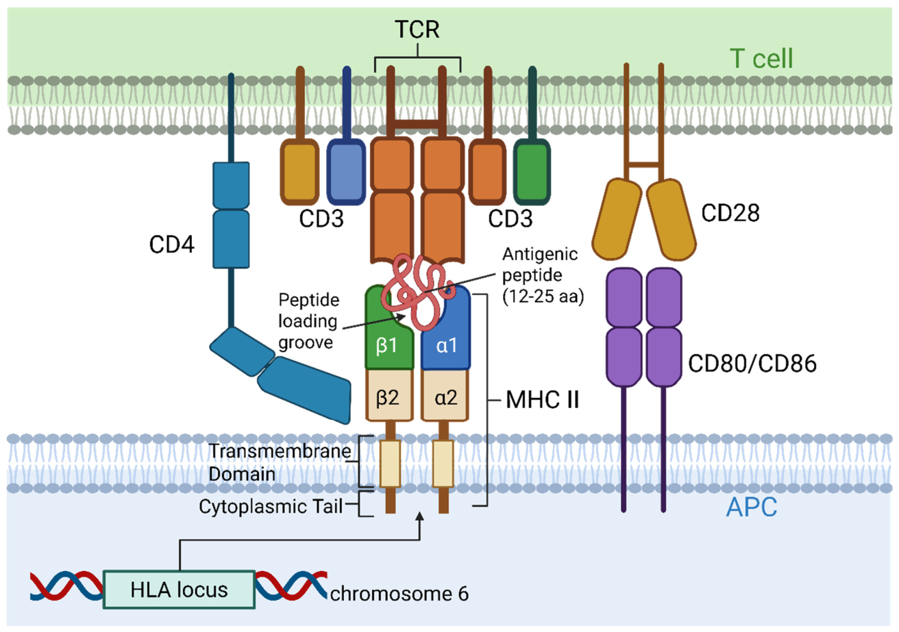
Figure 2.
Cell Type Specific Trafficking Regulation of MHC II in pAPCs. Left: In immature dendritic cells (DCs), most MHC II molecules are internalized and retained intracellularly, eventually undergoing degradation via MARCH1-mediated ubiquitination. Once DCs encounter antigens, they begin to mature with the upregulation of antigen processing mechanisms inside the cells, resulting in increased levels of peptide-loaded MHC II (p-MHCII) and co-stimulatory molecules (CD80/CD86) on the cell surface. Middle: In M1 macrophages, the cell possesses high phagocytic capacity for antigen uptake. This is marked by increased expression of p-MHC II and co-stimulatory molecules (CD80/CD86/CD40) on the cell surface, resulting in increased activation of Th1 and Th17 cells and upregulated inflammatory cytokine secretion (IL-12, IL-23, IL1β). In M2 macrophages, antigen acquisition occurs mainly through efferocytosis and micropinocytosis, which results in the downregulation of surface p-MHC II and co-stimulatory molecules. Anti-inflammatory cytokines (IL-10, TGF-β) are secreted, driving Treg cell responses. Right: In resting B cells with no antigen binding to the B cell receptor (BCR), surface MHC II and costimulatory molecule levels are downregulated by MARCH1-mediated internalization. After the BCR binds to specific antigens, the BCR-antigen complex is internalized, allowing antigens to be loaded onto MHC II via CD37. This leads to upregulation of co-stimulatory molecules and p-MHC II on the cell surface and increases the antigen presentation capacity of the cells.
Figure 2.
Cell Type Specific Trafficking Regulation of MHC II in pAPCs. Left: In immature dendritic cells (DCs), most MHC II molecules are internalized and retained intracellularly, eventually undergoing degradation via MARCH1-mediated ubiquitination. Once DCs encounter antigens, they begin to mature with the upregulation of antigen processing mechanisms inside the cells, resulting in increased levels of peptide-loaded MHC II (p-MHCII) and co-stimulatory molecules (CD80/CD86) on the cell surface. Middle: In M1 macrophages, the cell possesses high phagocytic capacity for antigen uptake. This is marked by increased expression of p-MHC II and co-stimulatory molecules (CD80/CD86/CD40) on the cell surface, resulting in increased activation of Th1 and Th17 cells and upregulated inflammatory cytokine secretion (IL-12, IL-23, IL1β). In M2 macrophages, antigen acquisition occurs mainly through efferocytosis and micropinocytosis, which results in the downregulation of surface p-MHC II and co-stimulatory molecules. Anti-inflammatory cytokines (IL-10, TGF-β) are secreted, driving Treg cell responses. Right: In resting B cells with no antigen binding to the B cell receptor (BCR), surface MHC II and costimulatory molecule levels are downregulated by MARCH1-mediated internalization. After the BCR binds to specific antigens, the BCR-antigen complex is internalized, allowing antigens to be loaded onto MHC II via CD37. This leads to upregulation of co-stimulatory molecules and p-MHC II on the cell surface and increases the antigen presentation capacity of the cells.
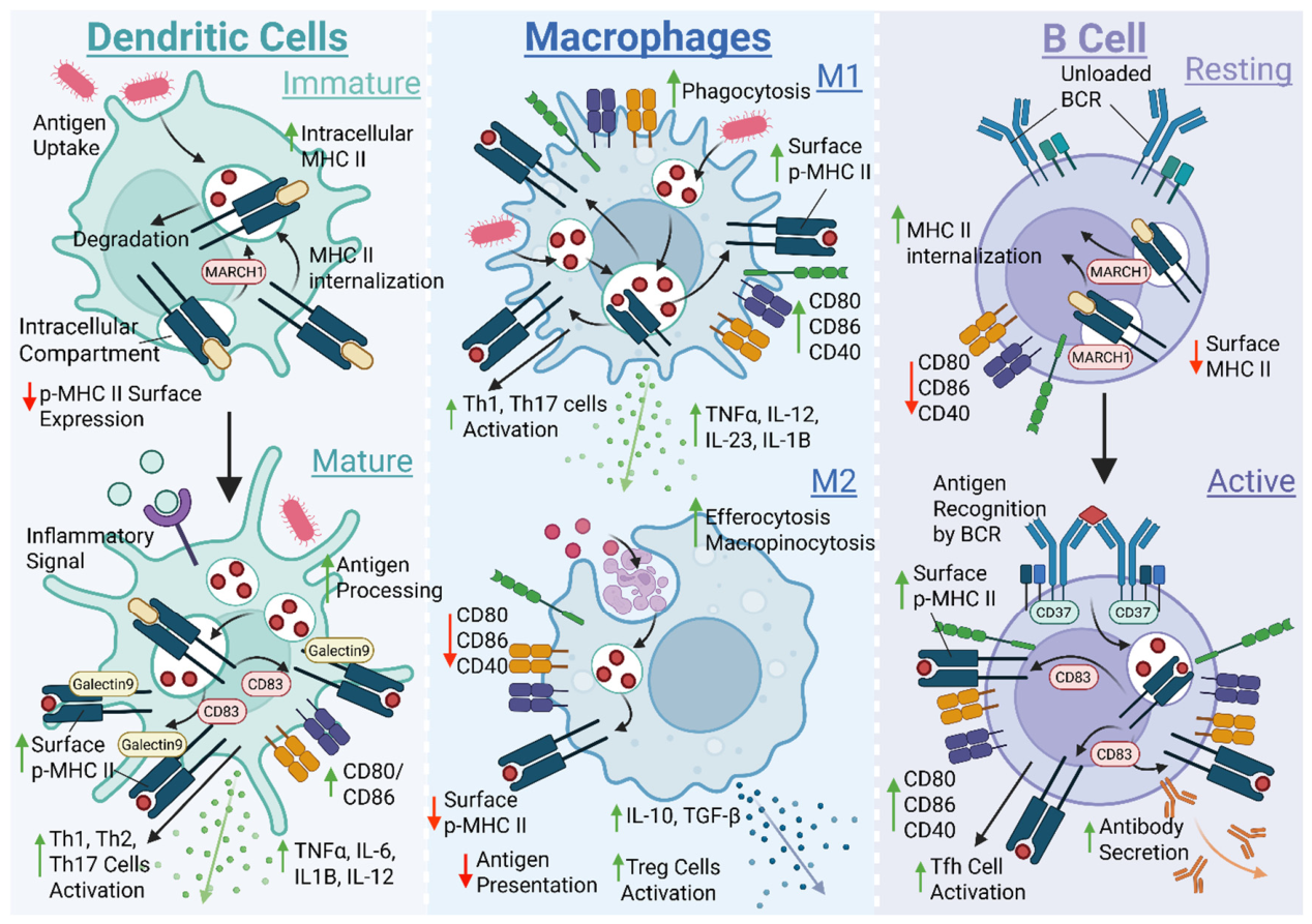
Figure 3.
Class II Major Histocompatibility Transactivator (CIITA) Works with Three Different Tissue-Specific Promoters to Regulate Expression of MHC II and Linked Proteins. CIITA functions as a transactivator to regulate MHC II expression and the expression of genes encoding linked MHC II regulators in the nucleus, including HLA-DM, HLA-DO, and CD74. CIITA binds to the upstream regulatory region (S/W, X1, X2, and Y) through the help of adaptor proteins RFX5, CREB/ATF1, and NF-Y. Three different CIITA mRNAs can be expressed according to the cell type and the promoter used (either pI, pIII, or pIV) with pIV being specifically targeted by the interferon regulatory factor 1 (IRF-1) downstream of the phosphorylation of homodimerized STAT1 in the cytoplasm triggered by the signal of interferon gamma (IFN-ƴ) and JAK-1/2. The resulting CIITA contains a C-terminal Leucine-rich repeats, a GTP binding domain with serine/threonine kinase activity, a P/S/T domain, and an acetyltransferase domain at the N-terminus. The regulation of the CIITA promoter is what determines the cell- and tissue-specific regulation of MHC II expression.
Figure 3.
Class II Major Histocompatibility Transactivator (CIITA) Works with Three Different Tissue-Specific Promoters to Regulate Expression of MHC II and Linked Proteins. CIITA functions as a transactivator to regulate MHC II expression and the expression of genes encoding linked MHC II regulators in the nucleus, including HLA-DM, HLA-DO, and CD74. CIITA binds to the upstream regulatory region (S/W, X1, X2, and Y) through the help of adaptor proteins RFX5, CREB/ATF1, and NF-Y. Three different CIITA mRNAs can be expressed according to the cell type and the promoter used (either pI, pIII, or pIV) with pIV being specifically targeted by the interferon regulatory factor 1 (IRF-1) downstream of the phosphorylation of homodimerized STAT1 in the cytoplasm triggered by the signal of interferon gamma (IFN-ƴ) and JAK-1/2. The resulting CIITA contains a C-terminal Leucine-rich repeats, a GTP binding domain with serine/threonine kinase activity, a P/S/T domain, and an acetyltransferase domain at the N-terminus. The regulation of the CIITA promoter is what determines the cell- and tissue-specific regulation of MHC II expression.
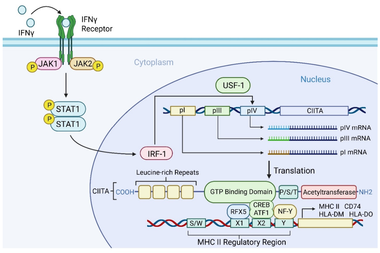
Figure 4.
Cell Type Specific Transcriptional Regulation of MHC II in pAPCs. Left: Upon exposure to TNFα and IFN-γ signals through the JAK1/2-STAT1 signalling pathway, immature conventional dendritic cells (cDCs) tend to express CIITA through the pI promoter to constitutively synthesize MHC II. As cDCs mature, further stimulation by LPS tends to downregulate CIITA activity through MEK/ERK and MAPK/p38 pathways, while pre-existing peptide-loaded MHC II (p-MHC II) complexes are stabilized at the cell surface for presentation to CD4+ T cells. Middle: Inactive macrophages express CIITA (pI) and (pIV) upon IFNƴ induction for MHC II biosynthesis. The function of CIITA (pI and pIV) is maintained by the remote enhancer, NFAT5. When macrophages receive inflammatory signals and become activated, CIITA expression is negatively regulated by MEK/ERK and p38 signalling without affecting the IFNƴ-induced STAT1 pathway, which functions as a negative feedback loop for p38 to downregulate CIITA expression. In the meantime, IL27p28 also downregulates CIITA activity, lowering the level of new MHC II synthesis. Right: Pre-B cells and resting B cells tend to express CIITA (pIII) upon IFNƴ induction. ZBTB48 works as a priming factor to help with MHC II upregulation by promoting the opening of chromatin. Once B cells undergo plasma cell differentiation or become more mature upon antigen recognition by B cell receptors (BCR), the repressors BLIMP1, HIC1 and ZBTB32 downregulate CIITA pIII activity and MHC II biosynthesis. This enables the B cells to further differentiate into plasma cells and secrete antibodies.
Figure 4.
Cell Type Specific Transcriptional Regulation of MHC II in pAPCs. Left: Upon exposure to TNFα and IFN-γ signals through the JAK1/2-STAT1 signalling pathway, immature conventional dendritic cells (cDCs) tend to express CIITA through the pI promoter to constitutively synthesize MHC II. As cDCs mature, further stimulation by LPS tends to downregulate CIITA activity through MEK/ERK and MAPK/p38 pathways, while pre-existing peptide-loaded MHC II (p-MHC II) complexes are stabilized at the cell surface for presentation to CD4+ T cells. Middle: Inactive macrophages express CIITA (pI) and (pIV) upon IFNƴ induction for MHC II biosynthesis. The function of CIITA (pI and pIV) is maintained by the remote enhancer, NFAT5. When macrophages receive inflammatory signals and become activated, CIITA expression is negatively regulated by MEK/ERK and p38 signalling without affecting the IFNƴ-induced STAT1 pathway, which functions as a negative feedback loop for p38 to downregulate CIITA expression. In the meantime, IL27p28 also downregulates CIITA activity, lowering the level of new MHC II synthesis. Right: Pre-B cells and resting B cells tend to express CIITA (pIII) upon IFNƴ induction. ZBTB48 works as a priming factor to help with MHC II upregulation by promoting the opening of chromatin. Once B cells undergo plasma cell differentiation or become more mature upon antigen recognition by B cell receptors (BCR), the repressors BLIMP1, HIC1 and ZBTB32 downregulate CIITA pIII activity and MHC II biosynthesis. This enables the B cells to further differentiate into plasma cells and secrete antibodies.
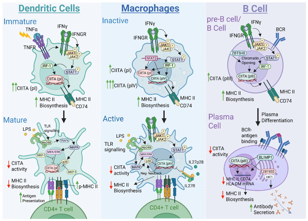
Figure 5.
Indirect versus Direct Pathway of MHC II Trafficking from the Golgi Apparatus to the MHC II loading compartment (MIIC). Two proposed intracellular pathways that transport newly synthesized MHC II molecules from the Golgi apparatus to the MHC II loading compartment (MIIC) for antigen loading upon the engulfment of extracellular pathogens through phagocytosis. The endo-lysosomal pathway (blue arrows): newly synthesized MHC II-CD74 complex will first be transported to the plasma membrane of a professional antigen-presenting cells (pAPC) followed by endocytosis, mediated by clathrin and AP2, to be brought back into the cells to form MIIC by fusing with the late endosome containing extracellular antigenic peptides. The Golgi-MIIC pathway (red arrows): newly synthesized MHC II-CD74 complex will be transported from the Golgi to form MIIC directly. CD74 will then be dissociated into class II-associated Ii chain peptide (CLIP) by cathepsin, followed by the loading of antigenic peptide mediated by HLA-DM for the presentation of the antigen to CD4+ T cells on the surface of pAPCs.
Figure 5.
Indirect versus Direct Pathway of MHC II Trafficking from the Golgi Apparatus to the MHC II loading compartment (MIIC). Two proposed intracellular pathways that transport newly synthesized MHC II molecules from the Golgi apparatus to the MHC II loading compartment (MIIC) for antigen loading upon the engulfment of extracellular pathogens through phagocytosis. The endo-lysosomal pathway (blue arrows): newly synthesized MHC II-CD74 complex will first be transported to the plasma membrane of a professional antigen-presenting cells (pAPC) followed by endocytosis, mediated by clathrin and AP2, to be brought back into the cells to form MIIC by fusing with the late endosome containing extracellular antigenic peptides. The Golgi-MIIC pathway (red arrows): newly synthesized MHC II-CD74 complex will be transported from the Golgi to form MIIC directly. CD74 will then be dissociated into class II-associated Ii chain peptide (CLIP) by cathepsin, followed by the loading of antigenic peptide mediated by HLA-DM for the presentation of the antigen to CD4+ T cells on the surface of pAPCs.
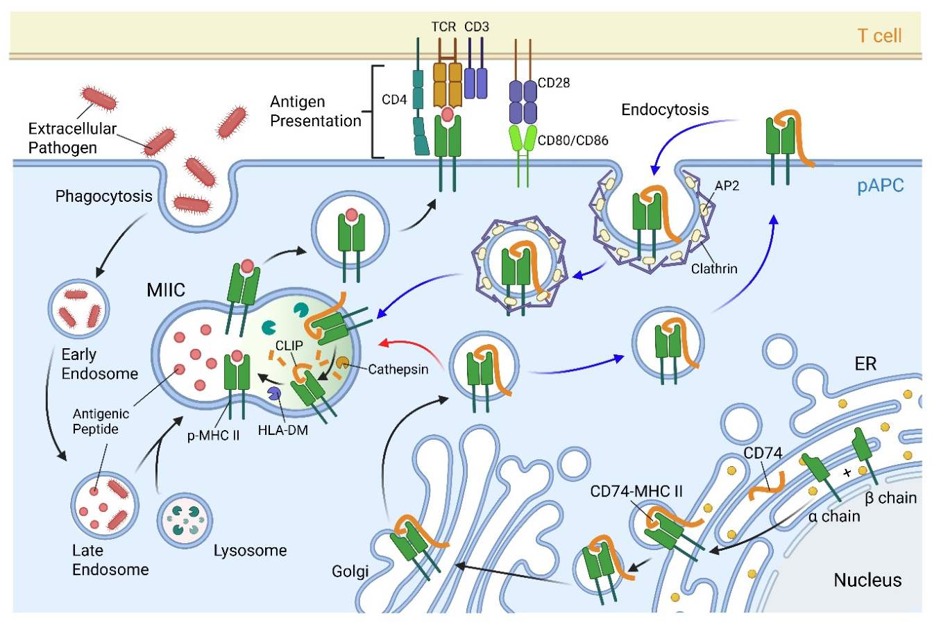
Figure 6.
Antigenic Peptide Loading and Editing onto MHC II. Newly synthesized MHC II is loaded with trimerized CD74 via the class II-associated Ii chain peptide (CLIP) inside the endoplasmic reticulum (ER) immediately after its assembly. MHC II-CD74 is then transported to the MHC II loading compartment (MIIC), where CD74 is degraded by acids and proteases like cathepsin L and S (CatL/CatS), forming MHC II-CLIP. In the meantime, the engulfed extracellular pathogen is broken down into small antigenic fragments in the endolysosome, which then fuses with the vesicle containing MHC II-CD74 to form MIIC. Antigenic fragments will then be further digested by CatL, CatS and Gamma-interferon-inducible lysosomal thiol reductase (GILT) to generate smaller antigenic peptides of a suitable size for MHC II to be loaded. With suitable-sized peptides generated, the dissociation of CLIP is mediated by HLA-DM and the exchange of CLIP with a generated peptide. In the case where unstable p-MHC II loading occurs, HLA-DM functions to dissociate the unstable peptide and replace it with a peptide that could lead to stable loading onto MHC II. In specific cell types, the function of HLA-DM is regulated by HLA-DO to limit the capacity of MHC II-peptide loading and antigen presentation on the cell surface.
Figure 6.
Antigenic Peptide Loading and Editing onto MHC II. Newly synthesized MHC II is loaded with trimerized CD74 via the class II-associated Ii chain peptide (CLIP) inside the endoplasmic reticulum (ER) immediately after its assembly. MHC II-CD74 is then transported to the MHC II loading compartment (MIIC), where CD74 is degraded by acids and proteases like cathepsin L and S (CatL/CatS), forming MHC II-CLIP. In the meantime, the engulfed extracellular pathogen is broken down into small antigenic fragments in the endolysosome, which then fuses with the vesicle containing MHC II-CD74 to form MIIC. Antigenic fragments will then be further digested by CatL, CatS and Gamma-interferon-inducible lysosomal thiol reductase (GILT) to generate smaller antigenic peptides of a suitable size for MHC II to be loaded. With suitable-sized peptides generated, the dissociation of CLIP is mediated by HLA-DM and the exchange of CLIP with a generated peptide. In the case where unstable p-MHC II loading occurs, HLA-DM functions to dissociate the unstable peptide and replace it with a peptide that could lead to stable loading onto MHC II. In specific cell types, the function of HLA-DM is regulated by HLA-DO to limit the capacity of MHC II-peptide loading and antigen presentation on the cell surface.
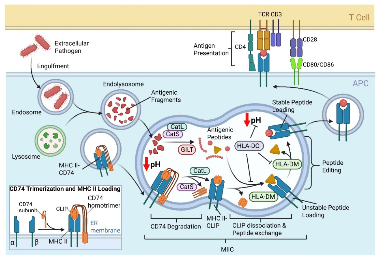

Table 1.
Summary of CIITA Promoter Usage in pAPC Subsets.
| Cell Type | Dominant CIITA Promoter | Inducing Signals | Major Transcriptional Regulators |
|---|---|---|---|
| DCs | Promoter I (pI) | Lineage-dependent constitutive expression; pI is subsequently repressed during maturation [154,158] | TLR ligand [111], TNF [111], STAT1/IRF-1/USF-1 [159,160], PRDM1/BLIMP1 [161] |
| IFN-γ-induced macrophages | Promoter I (pI) & IV (pIV) | IFN-γ | STAT1/IRF1/USF-1 [160], NFAT5 [130], |
| B cells | Promoter III (pIII) | Primarily constitutive B-cell lineage program; pIII can also respond to IFN-γ [154,159] | STAT1, E47 [162], PU.1 [162], IRF4 [162], CREB [155], and ZBTB48 [163] |
Table 2.
Summary of the regular functions of Rab GTPases and the MHC II trafficking pathway in which they are predicted to be involved.
Table 2.
Summary of the regular functions of Rab GTPases and the MHC II trafficking pathway in which they are predicted to be involved.
| Rab GTPase | Function in Intracellular Trafficking | MHC II Trafficking Pathway Involved |
|---|---|---|
| Rab1 |
|
Both direct and indirect pathway |
| Rab2 |
|
Both direct and indirect pathway |
| Rab3 |
|
Indirect pathway |
| Rab4 |
|
Unknown |
| Rab5 |
|
Indirect pathway |
| Rab6 |
|
Direct pathway |
| Rab7 |
|
Indirect pathway |
| Rab8 |
|
Indirect pathway |
| Rab11 |
|
Indirect pathway |
| Rab17 |
|
Unknown |
| Rab22 |
|
Indirect pathway |
| Rab27 |
|
Unknown |
| Rab41 |
|
Unknown |
Disclaimer/Publisher’s Note: The statements, opinions and data contained in all publications are solely those of the individual author(s) and contributor(s) and not of MDPI and/or the editor(s). MDPI and/or the editor(s) disclaim responsibility for any injury to people or property resulting from any ideas, methods, instructions or products referred to in the content. |
© 2026 by the authors. Licensee MDPI, Basel, Switzerland. This article is an open access article distributed under the terms and conditions of the Creative Commons Attribution (CC BY) license (http://creativecommons.org/licenses/by/4.0/).
Copyright: This open access article is published under a Creative Commons CC BY 4.0 license, which permit the free download, distribution, and reuse, provided that the author and preprint are cited in any reuse.