

Submitted:
16 September 2026
Posted:
17 September 2026
You are already at the latest version
Abstract
Fusion-positive rhabdomyosarcoma (FP-RMS) is driven by fusion transcription factors but retains an incomplete myogenic program. The roles of the two mammalian ISWI ATPases, SMARCA1/SNF2L and SMARCA5/SNF2H, remain poorly defined. This article proposes that they support FP-RMS through distinct but connected mechanisms. In RH30 cells, SMARCA1 loss markedly suppressed PAX3::FOXO1 and fusion-associated outputs, disrupted EGR1/Wnt- and TGF-β-linked chromatin programs, impaired differentiation and three-dimensional organization, and increased MEK-inhibitor sensitivity. SMARCA1 loss was also accompanied by marked depletion of MYCN and reductions in FUS and EP300, factors with established or emerging links to intrinsically disordered regions, transcriptional hubs, or biomolecular condensates. Together with recent evidence for PAX3::FOXO1/N-Myc transcriptional condensates, these observations motivate the hypothesis that SMARCA1-dependent chromatin competence may indirectly support the molecular environment required for fusion-associated hub organization, without implying that SMARCA1 itself nucleates condensates. By contrast, SMARCA5 was more closely associated with PI3K/mTOR signaling, cell-cycle progression, proliferation, and survival. In RH28 cells, strong SMARCA5 depletion with retained SMARCA1 reduced viability while preserving Wnt-responsive differentiation competence. Independent proximity labeling also detected SMARCA5 near selected FP-RMS fusion proteins, raising a context-specific possibility that SMARCA5 participates in selected fusion-associated molecular environments without establishing a condensate-scaffolding role. Single-cell analysis of 70,600 malignant cells from 16 RMS tumors further showed fusion-context-dependent associations of both ATPases with a high-risk transcriptional program. Together, these observations support a division-of-labor hypothesis in which SMARCA1 maintains chromatin competence for fusion-dependent and plastic cell states and may indirectly regulate fusion-associated transcriptional hubs, whereas SMARCA5 primarily supports proliferative fitness and may contribute to selected fusion-associated molecular environments. Matched perturbation, rescue, chromatin, protein, hub-imaging, single-cell, and three-dimensional studies are required to test this model.
Keywords:
SMARCA1
; SMARCA5
; ISWI
; PAX3::FOXO1
; rhabdomyosarcoma
; chromatin remodeling
; cell-state plasticity
; biomolecular condensates
; intrinsically disordered regions
; transcriptional hubs
; myogenic differentiation
; MEK inhibition
; TGF-β signaling
1. Rationale and Scope
Fusion-positive rhabdomyosarcoma is a developmental cancer in which tumor cells retain elements of the skeletal-muscle regulatory machinery yet fail to complete terminal myogenic differentiation [1,2]. PAX3::FOXO1 is the major fusion oncoprotein in this group, while additional PAX3- or PAX7-containing fusions define related molecular contexts [1,3]. Fusion transcription factors do not act independently of chromatin: their ability to establish enhancer programs and sustain tumor-specific transcription depends on the regulatory landscape in which they operate [4].
The ISWI family offers a useful way to ask whether closely related chromatin-remodeling ATPases can sustain the same malignant state through different mechanisms. Mammalian cells express two principal ISWI ATPases, SMARCA1/SNF2L and SMARCA5/SNF2H. Although they share substantial sequence homology and nucleosome-remodeling activity, they participate in different complexes and have distinct developmental and transcriptional functions [5,6]. My dissertation identified different relationships between these ATPases and differentiation or proliferation in RMS [7]. Later conference reports linked SMARCA1 loss to changes in PAX3::FOXO1-associated chromatin and transcriptional output [8,9,10,11,12]. Independently, a proximity-labeling study detected SMARCA5, but not SMARCA1, near selected FP-RMS oncofusions [13]. I briefly connected these observations in the sarcoma review preprint [14]. Here, I develop these observations into a focused SMARCA1-SMARCA5 division-of-labor hypothesis centered on nonredundant ATPase outputs in FP-RMS: SMARCA1-linked fusion/chromatin-state competence and plasticity-related consequences versus SMARCA5-linked proliferative and survival support. The immediate mechanistic priorities are PAX3::FOXO1/FGFR4 output, EGR1/Wnt-linked differentiation competence, SMARCA1-regulated canonical and non-canonical TGF-β signaling, 3D organization, SMARCA5-associated mTOR/proliferative support, and the MEK-inhibitor vulnerability associated with SMARCA1 loss. Vasculogenic mimicry/SOX2 plasticity, JAK-STAT-linked immune-state remodeling, an indirect SMARCA1 chromatin-competence/transcriptional-hub hypothesis, antigen-state/CAR T-cell vulnerability, and cross-sarcoma generalization remain staged, longer-term extensions and are not presented as established mechanisms. A parallel Withaferin A/NF-κB-associated RH30 response is retained as a comparator for distinguishing state-dependent from state-independent therapeutic vulnerabilities.
A complementary objective is to make this division-of-labor model testable across public and experimental functional-genomic resources without prespecifying an outcome. Genome-scale dependency data, perturbational transcriptomics, chromatin-accessibility and chromatin-occupancy profiles, bulk and single-cell RMS transcriptomics, pharmacogenomic resources, and selected fusion-driven cancer systems can be used to ask whether the proposed ATPase-specific relationships are reproducible across biological contexts. A parallel hub-focused layer should test whether ATPase perturbation changes IDR-rich coactivator composition, transcriptional-hub dynamics, or local chromatin output, while explicitly separating RNA abundance from protein recruitment and condensate biophysics. Cross-context findings would provide mechanistic precedent rather than validation of the RMS-specific mechanism.
1.1. Conceptual Origin and Computational Development
The doctoral research underlying this article originated from antecedent work by Bo Zhu, a member of Dr. Judy Davie’s laboratory at the time. An unpublished mass-spectrometry analysis of MyoG-interacting proteins identified SMARCA1 as a candidate MyoG-associated protein and raised the question of its role in skeletal-muscle differentiation and RMS. Bo Zhu had also cloned the pre-existing pcDNA3.1-SMARCA1 expression construct used in the antecedent project [7]. The present division-of-labor hypothesis originated from the SMARCA1/SMARCA5 differentiation and computational studies initiated during the doctoral project in 2018 and was later extended by the RH30 PAX3::FOXO1-associated transcriptomic and chromatin findings. The experimental SMARCA1/SMARCA5 work began in 2018 during the doctoral project and was expanded that year through volunteer training in Dr. Matt Geisler’s laboratory in bioinformatics and cluster analysis. Because directly relevant literature on SMARCA1/SMARCA5 in RMS was limited, I used exploratory data mining, correlation analysis, clustering, and machine-learning approaches to ask which transcriptional programs tracked with these remodelers. This work identified an early association between SMARCA1 and the SNAI1/SNAI2 (SNAIL/SLUG) regulatory program, providing the first computational bridge between the ISWI project, EMT-like plasticity, and cell-state regulation [7,12].
The computational analysis was then extended to patient-level RMS transcriptomic data. In GSE108022, the clinical dataset used in this research comprised 101 RMS patient samples together with five healthy skeletal-muscle samples. SMARCA1 expression was evaluated across RMS subtypes and normal muscle, and the same dataset was used for unsupervised hierarchical and k-means clustering to identify genes co-expressed with SMARCA1. Functional enrichment of the resulting SMARCA1-associated gene set implicated RNA and protein processing, metabolic and cancer pathways, and signaling networks including AMPK, PI3K-Akt, Wnt, JAK-STAT, and Ras [7]. The patient-level analysis defined a broader SMARCA1-associated transcriptional network beyond the subtype expression comparison.
Within that cluster-derived network, Hippo-pathway transcriptional regulators, particularly TEAD2 and TEAD3, emerged as additional SMARCA1-associated candidates. Because YAP1/TAZ (WWTR1) abundance, TEAD expression, YAP/TAZ nuclear localization, and YAP/TAZ-TEAD target activity are distinct measurements, this association is presented only as a co-expression-based rationale for testing whether SMARCA1-linked chromatin competence intersects Hippo transcriptional circuitry; it does not establish direct SMARCA1 occupancy, YAP/TAZ activation, or a causal Hippo mechanism.
Parallel mining of SNAIL-related datasets further strengthened this direction. Across multiple datasets, SMARCA1, HDAC2, and SNAI1 repeatedly appeared together, and SMARCA1 was reduced following SNAI1 knockdown in GSE38236. These observations motivated later experimental testing. Subsequent work confirmed a SMARCA1-HDAC2 association and demonstrated that SMARCA1 loss reduces SNAI1/SNAI2 expression, while RNA-seq, ATAC-seq, and CUT&RUN progressively connected the earlier computational hypothesis to chromatin-level regulation and functional cell-state phenotypes [7,12].
The project developed in stages: SMARCA1/SMARCA5 experiments and exploratory analyses began in 2018; the RH28 differentiation phenotype was documented in dated presentation materials during 2020-2021; the established RH30 SMARCA1-knockout model was expanded through transcriptomic, chromatin-accessibility, and chromatin-occupancy studies after the author’s entry into the NIH Intramural Research Program in May 2022; and the ISWI findings were incorporated into the 2025 doctoral dissertation [7]. The dissertation also documented the RH30 EYA2 decrease after SMARCA1 knockout and, in the separate SMS-CTR/CTR-557 resistance comparison, EYA2 upregulation together with TGF-β pathway enrichment in MEK-inhibitor-resistant CTR-557. The dissertation’s EYA2 observations therefore predate the independent 2026 EYA coactivator preprint [15]. This chronology does not establish a causal relationship between EYA2 and TGF-β signaling. The RH28 model was not subjected to the RH30 multi-omic workflow. This sequence records a research direction initiated by the author during the doctoral work and subsequently refined through fusion-state, chromatin, and therapeutic analyses.
The chronology of the RH28 terminal-differentiation observation is also important. The phenotype was documented in dated 2020-2021 presentation materials before the first 2024 AACR report of CDK8-associated differentiation in RH28 [16], and it was incorporated into the 2025 dissertation before the later peer-reviewed CDK8 study [17]. The later CDK8 work is treated here as independent phenotypic convergence through a distinct Mediator-SAGA-SIX4 mechanism, not as the origin or validation of the earlier ISWI observation.
A master’s thesis subsequently awarded in December 2025 used the pre-existing RH30 SMARCA1-knockout model and selected experimental targets informed by earlier RNA-seq observations attributed in that thesis to a prior laboratory member. Those antecedent models, RNA-seq analyses, and associated mechanistic hypotheses originated in the doctoral work described here and were documented in the author’s May 2025 dissertation. The later master’s thesis is therefore cited as a same-laboratory validation and extension of selected findings, rather than as their origin or as fully independent replication [18].
1.2. Research Provenance and Author Role
The mechanistic hypotheses advanced from the doctoral SMARCA1/SMARCA5 project and documented in the 2025 dissertation [7] were developed by A.K.A. through iterative integration of experimental observations, computational analyses, and functional validation. These include hypotheses concerning non-equivalent SMARCA1 and SMARCA5 functions; SMARCA1-associated differentiation and EMT/plasticity programs; EGR1/Wnt regulation; canonical and non-canonical TGF-β signaling; SMARCA5-associated mTOR and proliferative regulation; three-dimensional organization; therapeutic-response states; and subsequent integration with PAX3::FOXO1-associated biology. A.K.A. performed the doctoral wet-laboratory experiments described as author-derived findings in this article and conducted the computational and integrative analyses of the resulting datasets, including transcriptomic, chromatin-accessibility, chromatin-occupancy, pathway, network, and patient-level transcriptomic analyses. The later RH30 multi-omic phase incorporated RNA-seq, ATAC-seq, and CUT&RUN analyses. The RH28 differentiation experiments were based on experimental and protein-level observations and were not subjected to the same RH30 multi-omic workflow. Antecedent observations, reagents, technical training, scientific guidance, institutional resources, sequencing and core-facility services, and supervision are acknowledged separately and are distinguished from the author-developed hypotheses and analyses described here.
In January 2026, A.K.A. also documented a multi-level PAX3::FOXO1 validation plan while exploring a proposed collaboration with the Khan laboratory. Section 10.1 records the plan only as dated evidence of author-initiated hypothesis development and prospective experimental planning, not as a contribution or endorsement by the proposed collaborators.
2. Evidence for Distinct ISWI Programs in Myogenic Differentiation
2.1. Differential Behavior of SMARCA1 and SMARCA5 During Differentiation
In the dissertation, SMARCA1 transcript abundance increased after initiation of differentiation in human skeletal-muscle myoblasts and C2C12 cells. In C2C12 cells, SMARCA1 mRNA increased progressively and reached approximately sixfold above the undifferentiated state by day 2. SMARCA5 behaved differently: its transcript abundance remained relatively stable during human myoblast differentiation but decreased to approximately 40-45% of baseline during C2C12 differentiation [7]. These divergent patterns suggested that the two ATPases are not simply interchangeable components of a single ISWI output.
SMARCA1 protein levels did not follow a single pattern across experimental settings, making a simple expression-based explanation unlikely. The perturbation studies are more informative: SMARCA1 effects varied with dosage and signaling context, consistent with a requirement for an appropriately balanced chromatin state during differentiation [7].
2.2. SMARCA1 Supports Differentiation Competence
SMARCA1 directly occupied the EGR1 promoter, and SMARCA1 loss was associated with reduced chromatin accessibility and reduced EGR1 expression. The dissertation further linked SMARCA1 to Wnt-associated regulators, including NOTUM and TCF7L1, connecting ISWI remodeling to signaling programs that influence myogenic differentiation [7].
The differentiation experiments showed that this relationship is not linear. C2C12 cells overexpressing SMARCA1 differentiated poorly under baseline conditions, but LiCl-mediated Wnt activation restored robust differentiation. RH30 SMARCA1-knockout cells were severely impaired in differentiation and responded only weakly to LiCl. In contrast, RH28 cells with strong SMARCA5 depletion but retained/detectable SMARCA1 remained differentiation competent and differentiated robustly after Wnt activation [7]. These findings are consistent with a requirement for SMARCA1-dependent chromatin competence during myogenic differentiation, while also indicating that excess or imbalanced SMARCA1 activity can be inhibitory.
In these experiments, LiCl was used as a pharmacologic activator of canonical Wnt/β-catenin signaling. Its relevant proximal action is inhibition of glycogen synthase kinase 3 (GSK3α/GSK3β), which reduces GSK3-dependent phosphorylation and degradation of β-catenin and thereby permits β-catenin accumulation and transcriptional activity. LiCl is nevertheless pleiotropic and does not establish that the differentiation phenotype is mediated exclusively through GSK3/β-catenin signaling. It should be described as the experimental pathway probe used in the dissertation, not as the proposed translational therapy.
3. SMARCA5 Defines a Predominantly Proliferative and Survival-Associated Axis
The dissertation identified a different functional emphasis for SMARCA5. SMARCA5 knockout in RH28 cells markedly reduced mTOR protein, whereas SMARCA1 knockout in RH30 cells had little effect on mTOR protein abundance. CUT&RUN mapping identified SMARCA5 occupancy at regulatory regions of several mTOR/PI3K-related genes, including GSK3B, PIK3R2, LAMTOR1, and RPTOR. SMARCA5 also occupied regulatory regions associated with Wnt pathway genes [7].
Experiments conducted in 2018 showed that SMARCA5 depletion in RH30 FP-RMS cells was followed by marked loss of recoverable cells and failure to maintain the depleted population. This observation is consistent with a requirement for SMARCA5 in proliferative fitness or survival in this model. Because PAX3::FOXO1 expression and fusion-associated targets were not measured in that experiment, the phenotype cannot presently be attributed to disruption of the fusion-driven transcriptional program.
SMARCA5 overexpression during C2C12 differentiation prevented the normal decline in CCND1 at day 1, consistent with continued cell-cycle activity when myoblasts would normally begin to exit the cycle. In RH28 cells, the population initially described as SMARCA1/SMARCA5-edited but later shown at the protein level to be primarily SMARCA5 deficient had reduced viability. In the FP-RMS systems studied, the clearest SMARCA5 phenotype therefore lies in growth, survival, and proliferative signaling [7]. This does not exclude a role in differentiation, but it separates the dominant effects observed for SMARCA5 from those observed for SMARCA1.
3.1. MSH2 as a Candidate Shared ISWI-Linked Genome-Maintenance Output
MSH2 adds a potential genome-maintenance dimension to the model. In the dissertation datasets, MSH2 was downregulated in the RH28 simultaneous SMARCA1/SMARCA5-editing condition and after SMARCA1 knockout in RH30 [7]. Because the RH28 population showed strong SMARCA5 depletion while retaining detectable SMARCA1 protein, that observation is compatible with a SMARCA5 contribution; the RH30 result shows that MSH2 reduction is not specific to SMARCA5 loss. MSH2 is therefore treated as a candidate convergent output of ISWI perturbation whose ATPase dependence may vary by cellular and fusion context, not as an established SMARCA5-specific target.
MSH2 is an essential component of DNA mismatch repair, but reduced MSH2 expression alone does not establish functional mismatch-repair deficiency, microsatellite instability, increased mutation burden, or immune-checkpoint sensitivity [19]. Cross-context studies have linked SMARCA5/SNF2H to DNA-damage signaling and repair through RNF168-dependent chromatin responses and XRCC1-associated repair [20,21], providing biological precedent for a genome-maintenance role without demonstrating direct SMARCA5 control of MSH2. The hypothesis predicts that SMARCA1 and SMARCA5 perturbation may converge on MSH2/MMR competence through distinct chromatin or transcriptional routes. Direct testing must distinguish MSH2 RNA and protein abundance from functional MMR activity and determine whether SMARCA5 occupies or regulates the MSH2 locus.
Reduced MSH2 following SMARCA1 loss raises the possibility that the resulting state has impaired mismatch-repair capacity and an associated DNA-damage-response vulnerability. However, reduced MSH2 expression alone does not establish functional mismatch-repair deficiency or microsatellite instability. If deficient mismatch repair and MSI are confirmed, WRN dependence would become a testable synthetic-lethal hypothesis, because WRN is selectively required for the survival of MSI cancer models. This possibility should be evaluated through MSH2/MSH6 protein measurements, functional mismatch-repair and MSI assays, and genetic or pharmacologic WRN perturbation [22].
4. The RH28 Editing Experiment Defines an Informative Intermediate State
The RH28 simultaneous-editing experiment requires careful interpretation. Sequencing showed editing at both SMARCA1 and SMARCA5 loci, but protein validation demonstrated complete or strong SMARCA5 depletion while SMARCA1 remained detectable. The dissertation discusses incomplete or mixed SMARCA1 editing, alternative transcript usage, and population mosaicism as possible explanations [7]. These cells should not be described as a confirmed complete SMARCA1/SMARCA5 double-protein knockout.
Although technically incomplete as a double knockout, this experiment created an informative intermediate state: strong SMARCA5 loss with retained SMARCA1. These RH28 cells remained able to differentiate, and LiCl-mediated Wnt activation produced robust differentiation over days 2-6 [7]. This finding suggests that lowering SMARCA5-associated proliferative support while retaining SMARCA1 may preserve responsiveness to differentiation signals. It does not support the stronger claim that complete loss of both ISWI ATPases directly releases the differentiation block.
4.1. Independent CDK8 Evidence Defines a Mechanistically Distinct Route out of the RH28 Differentiation Block
An independent study provides a useful same-model comparison. In RH28 and RH30 FP-RMS cells, CDK8 knockdown reduced growth, and inducible CDK8 depletion suppressed RH28 xenograft growth. The first AACR report, published in 2024, already documented increased myogenin staining in vitro and myofibril formation with increased myoglobin in vivo after CDK8 inhibition [16]. The later peer-reviewed study showed that pharmacologic CDK8 inhibition activates a terminal myogenic program through the Mediator kinase module, the SAGA complex, and SIX4, with increased transcription or enhancer activity at genes including VGLL2, RUNX1, SEMA3D, and MYL1 [17].
This comparison supports phenotypic convergence with mechanistic divergence. The dissertation-derived RH28 result indicates that strong SMARCA5 depletion with retained SMARCA1 preserves differentiation competence and permits robust Wnt-responsive differentiation, whereas CDK8 inhibition engages a defined Mediator-SAGA-SIX4 differentiation response. The CDK8 study did not examine SMARCA1, SMARCA5, or another ISWI component. In the RH30 SMARCA1-knockout transcriptomic data, downregulation of multiple CDK8-Mediator/SAGA-associated genes may reflect collapse of fusion-associated transcription, a later adapted state, or an alternative state trajectory; transcript abundance alone does not establish reduced complex activity or direct SMARCA1 regulation. Exact genes, effect sizes, adjusted P values, and SMARCA1 occupancy or accessibility relationships should be verified before assigning a direct ISWI-Mediator/SAGA connection.
A direct experimental test would compare CDK8 inhibition across protein-validated wild-type, SMARCA1-loss, SMARCA5-loss, and true double-loss states in matched FP-RMS backgrounds. Expression and chromatin analyses should include CDK8, CCNC, MED12, MED13, SIX4, KAT2A, KAT2B, TADA2B, and TAF5L together with VGLL2, RUNX1, SEMA3D, and MYL1. Time-resolved RNA-seq, ATAC-seq, and, where feasible, SMARCA1/SMARCA5 CUT&RUN overlap with CDK8-, SIX4-, or SAGA-associated regulatory regions could distinguish acute differentiation induction from stable knockout-associated adaptation and determine whether the perturbations are additive, redundant, or antagonistic.
5. SMARCA1 as an Upstream Regulator of the PAX3::FOXO1 State
Later loss-of-function studies in PAX3::FOXO1-positive RH30 cells linked SMARCA1 more directly to fusion-oncogene biology. Conference-reported and manuscript-level analyses showed marked suppression of the PAX3::FOXO1 transcript after SMARCA1 depletion [10], together with broad loss of fusion-associated transcriptional programs, including MYOD1, MYOG, SNAI2, FGFR4, ALK, MYCN, IGF2, and CXCR4 [9,10]. FGFR4 is particularly useful in this model because it provides a cell-surface, fusion-responsive readout of the SMARCA1-supported state. These mechanistic observations have not yet been peer reviewed and require independent validation.
An exploratory, descriptive reanalysis of the RH30 wild-type versus SMARCA1-knockout RNA-seq dataset identified a non-uniform shift among genes encoding proteins with established or plausible IDR, coactivator, or transcriptional-hub relevance. MYCN was nearly extinguished (approximately 99.995% lower mean FPKM) and NCOA1 decreased by approximately 83%; EP300 decreased by approximately 29%, FUS by 21%, CREBBP by 18%, and BRD4 by 12%. In the opposite direction, MED15 increased by approximately 71%, INO80D by 45%, and POLR2A by 25%. These values are descriptive FPKM-based comparisons rather than formal differential-expression statistics. They do not establish corresponding protein changes, IDR activity, phase separation, hub number, or condensate material properties. Their value is as a hypothesis-generating pattern: SMARCA1 loss appears to reconfigure rather than uniformly suppress the fusion-associated coactivator/transcriptional environment.
Among these downstream outputs, FGFR4 offers a concrete test of pathway order. Conference-reported chromatin analyses linked SMARCA1 loss to reduced PAX3::FOXO1-associated occupancy and transcriptional output at FGFR4 and MYCN regulatory loci [9,10]. I place FGFR4 downstream of the SMARCA1-to-PAX3::FOXO1 axis rather than treating FGFR4 as an established direct SMARCA1 target. A parallel direct-chromatin branch remains testable: SMARCA1 occupancy and accessibility at FGFR4 regulatory elements should be measured explicitly before assigning direct regulation.
These findings do not require SMARCA1 to be a stable fusion-recruited cofactor. A simpler explanation is that SMARCA1 acts upstream by maintaining the chromatin environment needed for fusion-transcript expression and for accessibility at fusion-responsive regulatory loci, with FGFR4 serving as a representative locus at which fusion occupancy and output can be followed. I first stated this interpretation explicitly in the sarcoma review preprint [14].
This view also helps reconcile the apparently different roles of SMARCA1 in fusion maintenance and differentiation. A chromatin remodeler can support more than one transcriptional outcome because the result depends on the transcription factors and signaling inputs present in a given state. In a PAX3::FOXO1-dominated state, SMARCA1-dependent chromatin may sustain the malignant fusion program. Under differentiation-promoting conditions, the same remodeling capacity may be needed for EGR1-, Wnt-, MYOD1-, and MYOG-associated programs to proceed.
6. SMARCA5 Proximity to Selected Fusion-Protein Molecular Environments
Zimmerman and colleagues used proximity labeling to map the molecular environments of seven FP-RMS oncofusions. SMARCA5 was detected in proximity to PAX3::NCOA1 and PAX3::INO80D, and the SMARCA5-associated factor BAZ1A/ACF1 was detected with PAX3::INO80D. SMARCA5 and BAZ1A also showed small but significant negative proliferation effects in the interactome-linked CRISPR/Cas9 dependency screen [13]. SMARCA1 was not detected in proximity to any of the seven oncofusions examined [13,14].
Proximity labeling does not establish direct SMARCA5-fusion binding or a regulatory role; it identifies proteins within the molecular neighborhood of the bait under the conditions tested. Likewise, failure to detect SMARCA1 does not exclude a transient, indirect, chromatin-mediated, or context-dependent association. These proximity-labeling data support only a narrow conclusion: SMARCA5 was detected near PAX3::NCOA1 and PAX3::INO80D, whereas SMARCA1 was not detected in the datasets for the seven oncofusions examined. Whether this proximity affects fusion-associated transcription, chromatin regulation, or local protein composition requires direct testing. The proximity data do not establish condensate involvement or implicate SMARCA5 in EMT/MMT, vasculogenic mimicry, SOX2-associated plasticity, or immune-state remodeling in RMS.
The 2018 RH30 depletion phenotype raises the additional possibility that SMARCA5 supports more than general proliferative fitness in some FP-RMS settings. However, the RH30 experiment did not measure PAX3::FOXO1 or its downstream targets and should not be interpreted as validating the later proximity findings, which involved PAX3::NCOA1 and PAX3::INO80D. Whether SMARCA5 contributes to fusion-associated transcription therefore remains a separate, context-specific hypothesis requiring direct testing.
The condensate-level comparison between the ATPases must therefore remain asymmetric. RH30 SMARCA1 loss can be linked descriptively to a broad change in hub-associated and IDR-containing transcriptional components, whereas an equivalent protein-validated SMARCA5-loss transcriptomic and live-cell hub dataset is not yet available. For SMARCA5, the testable extension is narrower: in PAX3::NCOA1- or PAX3::INO80D-relevant settings, SMARCA5 may function as a local chromatin-remodeling client or effector within a fusion-proximal molecular assembly rather than as the scaffold that drives phase separation. Acute perturbation before loss of cellular fitness is required to distinguish a local fusion-environment function from the established PI3K/mTOR-proliferation/survival branch.
7. 3D Tumor Architecture and Functional Plasticity as Outputs of the SMARCA1-Supported State
The SMARCA1 phenotype extends beyond transcriptional regulation. In three-dimensional culture, wild-type RH30 cells formed compact, organized spheroids with clear boundaries, whereas SMARCA1-knockout cells formed larger, irregular, loosely connected structures with poorly defined borders. Integrated RNA-seq and ATAC-seq analyses showed coordinated changes in cell-adhesion, tight-junction, and adherens-junction programs, including alterations in cadherins, claudins, integrins, cytoskeletal components, SNAI1, TGFB1, and TGFBR1 [7].
The 3D phenotype provides a functional readout that is independent of transcript abundance alone. SMARCA1 appears to contribute to multicellular cohesion and tissue-like organization as well as to fusion-associated transcription. It also exposes a mismatch between transcriptional and behavioral state: SMARCA1-deficient cells showed enrichment of an EMT-associated expression signature while migration and invasion were strongly impaired and spheroid organization was lost [7,12]. Chromatin perturbation can therefore uncouple an EMT-like transcriptional signature from the behavior normally associated with it.
Patient-derived RMS tumoroids provide a useful precedent for extending this work beyond conventional cell lines. Meister and colleagues established 19 pediatric RMS tumoroid models representing major molecular and clinical subtypes and showed that they retained key features of the originating tumors [23]. The system also supported CRISPR/Cas9 editing. TP53 disruption in the TP53-wild-type RMS012 fusion-negative embryonal RMS tumoroid produced loss of P53 protein and increased sensitivity to prexasertib compared with the matched TP53-wild-type cells (P = 0.008) [23].
8. Central Hypothesis: An ISWI Division of Labor
I propose that SMARCA1 and SMARCA5 occupy distinct but connected regulatory positions in FP-RMS.
In this model, SMARCA1 acts mainly as a chromatin-competence regulator. It maintains a regulatory landscape that supports PAX3::FOXO1 transcript expression and downstream transcription, including FGFR4 expression, while also preserving the ability to engage differentiation, adhesion, 3D organizational, and TGF-β-responsive programs. The hub-level extension places SMARCA1 upstream of transcriptional-hub competence: by shaping accessible chromatin and the coactivator environment, SMARCA1 may influence PAX3::FOXO1 residence, cofactor recruitment, or hub productivity without being a required structural scaffold. The TGF-β branch remains an experimentally supported chromatin-signaling interface spanning canonical SMAD-dependent and non-canonical SMAD-independent programs. Which output predominates depends on the transcription factors, cofactors, and signaling inputs acting on that chromatin state.
SMARCA5 contributes more strongly to a second layer centered on mTOR/PI3K-associated growth signaling, cell-cycle progression, proliferation, and survival. Separately, proximity-labeling data place SMARCA5 within the molecular neighborhood of selected fusion proteins. This motivates a restricted fusion-assembly hypothesis in which SMARCA5 could act as a local remodeler or functional client in selected molecular environments, but it does not establish that SMARCA5 nucleates, stabilizes, or is required to form condensates in RMS. This selected-fusion question remains distinct from the supported SMARCA5 mTOR/proliferation branch.
FP-RMS may therefore depend on a balance among fusion circuitry, SMARCA1-dependent chromatin and hub competence, and SMARCA5-associated proliferative support. Reducing SMARCA5 while retaining SMARCA1 could lower proliferative pressure without removing differentiation competence, allowing a strong differentiation signal to redirect cell state. Complete SMARCA1 loss may have a different outcome: the PAX3::FOXO1 program can weaken, the IDR-rich/coactivator transcriptional environment can be selectively reconfigured, and terminal myogenic differentiation can remain impaired. Loss of tumor identity, reorganization of transcriptional hubs, and acquisition of terminal differentiation are therefore separable biological events. Plasticity and surface-antigen outputs, including vasculogenic mimicry and FGFR4 abundance, may also change as cells move between these states.
The core hypothesis is that the two ATPases have nonredundant outputs in FP-RMS. Near-term tests focus on PAX3::FOXO1/FGFR4 output, EGR1/Wnt-linked differentiation, SMARCA1-regulated canonical and non-canonical TGF-β signaling, SMARCA5-mTOR/proliferative support, 3D organization, and MEK-inhibitor sensitivity after SMARCA1 loss. The emerging IDR/hub-associated transcript pattern adds a secondary mechanistic layer that can now be tested directly at the protein and live-cell levels. Single-cell and 3D models provide integrated state readouts. Direct condensate biophysics, VM/SOX2 plasticity, JAK-STAT and immune-state remodeling, antigen-state effects on CAR T-cell response, and cross-sarcoma extension remain staged questions. The selected-fusion SMARCA5 proximity observation remains a separate fusion-context hypothesis pending functional evidence.
8.1. TGF-β Signaling as a Chromatin-Regulated Plasticity Interface
TGF-β signaling provides an experimentally supported interface between SMARCA1-dependent chromatin regulation and RMS cell-state plasticity. In RH30 cells, doctoral multi-omic analyses showed SMARCA1 occupancy at regulatory regions of TGF-β pathway genes, reduced accessibility at TGF-β-associated chromatin after SMARCA1 loss, and altered expression of pathway components including TGFBR1 and SMAD-family regulators. These findings support a SMARCA1-linked canonical TGF-β branch in which receptor and SMAD-dependent signaling competence is sensitive to chromatin state [7].
The same doctoral analyses also identified coordinated disruption of major non-canonical, SMAD-independent TGF-β-associated networks after SMARCA1 loss, including MAPK/ERK, PI3K/AKT, RAS, mTOR, and NF-κB signaling. I treat TGF-β as a chromatin-regulated plasticity interface that may connect differentiation, EMT/MMT-associated transcription, migration and invasion, multicellular organization, survival signaling, and therapeutic adaptation. The separate SMS-CTR/CTR-557 ATAC-seq comparison also identified TGF-β pathway enrichment in the MEK-inhibitor-resistant CTR-557 state, where EYA2 was upregulated. This places EYA2 and TGF-β-associated chromatin among parallel features of that resistant state, but it does not establish a direct relationship between them or show that either feature is SMARCA1-dependent. This integration does not imply that every downstream pathway is explained solely by TGF-β: the SMARCA5-mTOR axis remains a distinct ATPase-specific observation, the trametinib phenotype remains a prioritized SMARCA1-loss-associated MEK vulnerability, and the Withaferin A response remains a parallel SMARCA1-independent drug-response phenotype [7].
The doctoral drug-response data also demonstrate state dependence, but the two model comparisons must remain separate. In isogenic RH30 cells, SB505124 reduced viability more strongly in wild-type than in SMARCA1-knockout cells, whereas Dactolisib produced moderate, concentration-dependent effects. In the separate SMS-CTR/CTR-557 comparison, MEK-inhibitor-resistant CTR-557 was more sensitive to SB505124 than parental SMS-CTR, while Dactolisib affected both states with partial resistance in CTR-557 at intermediate concentrations. The opposing SB505124 response patterns indicate that TGF-β-receptor inhibitor sensitivity depends on the specific genetic or acquired-resistance state [7].
8.2. IDR- and Condensate-Level Extension of the Model
An additional layer of the hypothesis concerns transcriptional hubs and biomolecular condensates. This is relevant in FP-RMS because core-regulatory transcription is organized around super-enhancer-associated assemblies, and recent work has visualized PAX3::FOXO1/N-Myc condensates as well as endogenous PAX3::FOXO1 transcriptional hubs whose low-complexity regions influence hub formation, chromatin residence, target-gene transcription, and p300 recruitment [24,25,26]. A separate muscle-lineage study provides a direct precedent for SMARCA1 entering transcriptionally active condensates: SMARCA1 formed nuclear puncta that substantially colocalized with MYOCD-containing condensates during smooth-muscle gene activation [27]. This establishes a muscle-lineage precedent, not an RMS mechanism. In RMS, neither SMARCA1 nor SMARCA5 has been demonstrated as a structural component of a PAX3::FOXO1 condensate or transcriptional hub.
The RH30 transcriptomic pattern adds a new, but explicitly indirect, layer to this extension. Among genes examined descriptively, MYCN and NCOA1 fell most strongly after SMARCA1 loss, with additional decreases in EP300, CREBBP, FUS, and BRD4, whereas MED15, POLR2A, and INO80D increased. Several of these proteins contain substantial IDRs or participate in enhancer, Mediator, RNA polymerase II, or other multivalent transcriptional assemblies. The bidirectional pattern is therefore more compatible with selective reconfiguration of hub/coactivator composition than with a simple model of global condensate loss. RNA abundance alone cannot establish whether any protein enters a condensate, whether its IDR is functionally engaged, or whether hub material properties change.
I propose that SMARCA1 may indirectly shape the chromatin substrate and coactivator environment on which PAX3::FOXO1 transcriptional hubs assemble. By maintaining an accessible and transcriptionally competent substrate, SMARCA1 could influence hub formation, fusion residence, cofactor concentration, or transcriptional productivity without being a stable hub constituent or direct fusion-binding partner. The observed loss of MYCN/NCOA1 and more modest reduction of EP300/CREBBP, BRD4, and FUS, together with increased MED15/POLR2A/INO80D, provides a concrete set of candidate readouts for this reconfiguration model. Here, the term ‘SMARCA1-centered hub hypothesis’ refers only to this chromatin-competence and composition model and does not imply demonstrated recruitment of SMARCA1 into an RMS condensate. FGFR4 expression and surface abundance provide a practical downstream readout of whether those changes propagate into a canonical fusion-responsive effector program.
SMARCA5 is not assigned a condensate role in RMS. In NUP98::NSD1 leukemia, SMARCA5 was detected in fusion-associated condensates and supported transformation, but depletion or inhibition did not abolish condensate formation. That cross-context result and the RMS proximity observation must remain separate: proximity to PAX3::NCOA1 and PAX3::INO80D does not establish condensate membership or function in RMS [13,28]. A negative result in those selected fusion contexts would not affect the supported SMARCA5 mTOR/PI3K-proliferation/survival branch.
The resulting ATPase comparison is deliberately mechanistic rather than symmetrical. SMARCA1 is hypothesized to influence a fusion-associated hub primarily through the chromatin substrate and the availability or recruitment of hub-associated cofactors. SMARCA5, if it has a condensate-related role in RMS, is hypothesized instead to function as a local remodeler or client within selected fusion-proximal assemblies, analogous only in principle to its cross-context behavior in NUP98::NSD1 leukemia. Neither ATPase is proposed here as the IDR scaffold that nucleates PAX3::FOXO1 phase separation. This distinction can be tested using endogenous fusion tagging and live-cell imaging, hub number/size and single-particle residence measurements, FRAP where appropriate, proximity labeling or PhaseID-type interactome mapping, co-immunoprecipitation/mass spectrometry, and matched CUT&RUN/CUT&Tag or accessibility measurements before and after acute ATPase perturbation.
8.3. Vasculogenic Mimicry as a State-Dependent Plasticity Output
Vasculogenic mimicry (VM) provides a clinically relevant extension of the cell-state model because it represents a tumor-cell plasticity program rather than conventional endothelial angiogenesis. VM has been reported in alveolar rhabdomyosarcoma and has been associated with adverse outcome, and more recent RMS work showed that SNAI2 promotes tumor-cell VM, proliferation, and metastasis through a USP7/TRIM21-regulated protein-stability axis [29,30]. These observations are particularly relevant to the present model because SMARCA1 loss in RH30 suppresses SNAI2 together with migration and invasion.
An important counterpoint is the selective increase in SOX2 observed after SMARCA1 loss in RH30. SOX2 is functionally linked to stemness in RMS, including rhabdosphere formation in embryonal RMS, and has been shown in other tumor systems to support vasculogenic mimicry [31,32]. I treat SOX2 here only as a candidate compensatory plasticity signal that could modify VM competence, not as evidence that SOX2 drives VM in fusion-positive RMS. SOX2 abundance alone does not establish functional stemness; this extension requires orthogonal testing of self-renewal, persistence, or tumor-initiating capacity in the relevant FP-RMS state.
I propose a two-axis interpretation of VM after SMARCA1 loss. Suppression of SNAI2 together with reduced migration and invasion argues that the canonical SNAI2-linked, invasive VM program should weaken. At the same time, SOX2 upregulation raises the possibility that a stem-like compensatory state could preserve or redirect some forms of vascular-mimetic plasticity. The net VM phenotype is therefore a testable outcome rather than a predetermined consequence of SMARCA1 loss. This framing does not imply that SMARCA1 directly controls endothelial genes or that every PAX3::FOXO1-high or SOX2-high cell forms VM.
This extension also creates a useful therapeutic distinction. A VM-competent, FGFR4-high fusion state may remain aggressive but surface-targetable, whereas a SMARCA1-loss state is predicted to lose much of its SNAI2-linked invasive program and become more MEK-inhibitor-sensitive while simultaneously expressing less FGFR4. Whether SOX2-high compensatory cells retain residual VM competence is left open. The hypothesis predicts state-dependent tradeoffs rather than assuming that VM, invasion, stemness, antigen abundance, and drug sensitivity move in the same direction.
8.4. Single-Cell Mapping of Therapeutically Relevant States
Single-cell RNA sequencing is especially important in the expanded model because the proposed VM, FGFR4/CAR T-cell, and MEK-inhibitor branches are all expected to vary by cell state. Bulk RNA-seq can show an average fall in PAX3::FOXO1, FGFR4, SNAI2, or a differentiation program, but it cannot determine whether all cells move together or whether one subpopulation disappears while another persists or expands. Published RMS single-cell profiling has already identified tumor-acquired and therapy-resistant cell states, providing a direct precedent for using single-cell analysis to resolve state heterogeneity in this disease [33].
To evaluate ISWI expression in a recently reported high-risk state, I performed an exploratory reanalysis of the Whitfield et al. dataset, restricted to 70,600 malignant cells from 16 RMS tumors [34]. As an internal check, I reconstructed the published 89-gene high-risk program. The score was higher in fusion-negative high-risk/lethal tumors than in surviving fusion-negative tumors (P = 0.015) and was also higher in fusion-positive tumors than in surviving fusion-negative tumors (P ≈ 4 × 10⁻⁶). These comparisons reproduced the reported high-risk biology.
At the tumor level, neither SMARCA1 nor SMARCA5 was significantly overexpressed in high-risk fusion-negative RMS. Within individual tumors, however, the relationship between ATPase expression and the high-risk program depended on fusion context. After removing the effects of S-phase and G2/M activity from both variables, SMARCA1 remained positively associated with the high-risk program in FP-RMS (mean patient-level Spearman ρ ≈ +0.17) and negatively associated with it in surviving fusion-negative tumors (mean ρ ≈ −0.27; P = 0.004; Benjamini-Hochberg FDR ≈ 0.02). The neural component showed the clearest separation: mean adjusted ρ ≈ +0.24 in FP tumors and −0.22 in surviving fusion-negative tumors (P = 0.004; FDR ≈ 0.02). All four FP tumors had a positive adjusted SMARCA1-neural correlation, whereas all eight surviving fusion-negative tumors had a negative correlation.
SMARCA5 showed a similar fusion-context-dependent association with the adjusted high-risk program (mean ρ ≈ +0.21 in FP-RMS and −0.15 in surviving fusion-negative RMS; P = 0.004; FDR ≈ 0.02). Its expression, however, showed little correlation with S-phase or G2/M scores. The single-cell data therefore support context-dependent coupling of both ISWI ATPases to the aggressive transcriptional state rather than uniform ATPase overexpression. The persistent SMARCA1-neural/high-risk relationship is consistent with the proposed chromatin-competence arm. This analysis does not independently support the proposed SMARCA5 proliferative/survival arm, which remains grounded in perturbational and functional evidence. The cohort included only four FP tumors and four fusion-negative high-risk tumors, and some samples contained relatively few malignant cells; these results are exploratory and require independent replication.
The most informative state coordinates would include PAX3::FOXO1 target activity; FGFR4 and CD276 expression; MYOD1/MYOG-associated differentiation; EGR1/Wnt signaling; canonical TGF-β/SMAD activity; non-canonical TGF-β-associated MAPK/ERK, PI3K/AKT, RAS, mTOR, and NF-κB programs; SNAI2, SOX2, and VM-linked plasticity; JAK-STAT/interferon-associated immune states; YAP/TAZ-associated adaptation; cell-cycle programs; and adhesion/ECM features. Comparing matched WT, SMARCA1-loss, SMARCA5-loss, and validated double-loss states, with and without trametinib, could determine whether SMARCA1 loss contracts a fusion-high/FGFR4-high/SNAI2-high population while enriching a SOX2-high compensatory state that is selectively MEK-sensitive, while SMARCA5 loss preferentially reduces proliferative support without eliminating differentiation competence. EYA2 can be evaluated separately as a secondary resistant-state marker alongside TGF-β pathway status, without prespecifying a direct relationship.
Single-cell transcriptomics should not be treated as proof of every functional phenotype. A VM-like transcriptional state would still require 3D morphologic or functional validation, and RNA-level FGFR4 or CD276 expression should be paired with surface-protein measurements, such as flow cytometry or a joint RNA/protein approach, before inferring CAR T-cell target availability. Single-cell mapping can then relate chromatin perturbation to cell-state redistribution, antigen heterogeneity, VM competence, and therapeutic response.
8.5. JAK-STAT Signaling and SOX2 as a Candidate Chromatin-Immune State Interface
Preliminary transcriptomic observations in SMARCA1-deficient RH30 cells indicate changes in the expression of STAT-family components. This observation is potentially important because the earlier SMARCA1-associated computational network also implicated JAK-STAT signaling. However, changes in STAT transcript abundance do not by themselves establish pathway activation. The direction and functional meaning of the response should be resolved separately for individual STAT programs and validated at the level of phosphorylation, nuclear localization, and downstream target activity.
SMARCA1-dependent chromatin state may also influence cytokine responsiveness and immune visibility in RMS. STAT1/STAT2-associated interferon signaling can be tested for effects on antigen processing and MHC-I competence. STAT3 may define an adaptive survival or immunomodulatory branch that intersects with MEK-inhibitor response, and STAT6 may connect cytokine signaling with differentiation blockade and plasticity. Each STAT program requires separate pathway and functional validation.
The SOX2-high state observed after SMARCA1 loss provides an additional candidate interface between plasticity and immunity. Rather than assuming that SOX2 directly causes immune evasion in RMS, the hypothesis is that SOX2-high cells may occupy a distinct immune-modulatory state characterized by altered interferon/JAK-STAT responsiveness, antigen-presentation competence, cytokine or chemokine output, or interaction with immune-cell populations. This possibility also links the SOX2 branch to the VM model: a compensatory SOX2-high population could, in principle, differ simultaneously in stem-like plasticity, vascular-mimetic competence, immune visibility, FGFR4/CD276 surface state, and MEK sensitivity. Single-cell profiling is therefore well suited to determine whether these features coexist in one population or segregate into distinct adaptive states.
8.6. NF-κB-Associated Drug Sensitivity as a Parallel, SMARCA1-Independent Axis
The doctoral Withaferin A result is distinct from the SMARCA1-loss-associated MEK phenotype. In RH30 cells, Withaferin A produced a strong viability response in both wild-type and SMARCA1-knockout states, with closely overlapping dose-response behavior. Withaferin A also produced similarly strong responses in the separate SMS-CTR and MEK-inhibitor-resistant CTR-557 comparison [7]. The isogenic RH30 result supports independence from SMARCA1 status; the SMS-CTR/CTR-557 comparison supports activity across parental and resistant states but is not an isogenic test of SMARCA1 dependence. The result defines a parallel therapeutic response; current evidence does not place NF-κB sensitivity downstream of SMARCA1 loss.
This distinction is important for the expanded immune-state model. NF-κB can be evaluated as an inflammatory, survival, and cytokine-regulatory pathway that may intersect with immune communication, but the existing drug-response results do not show that SMARCA1 loss or acquired MEK-inhibitor resistance creates the Withaferin A response. Accordingly, MEK inhibition remains the prioritized SMARCA1-state-dependent vulnerability, whereas Withaferin A is treated as a genotype comparator in RH30 and a separate resistance-state comparator in SMS-CTR/CTR-557. Because Withaferin A is pleiotropic, the experiments are not proof of a selective NF-κB dependency; orthogonal NF-κB/IKK perturbation is required before assigning the response specifically to that pathway.
The proposed SMARCA1-SMARCA5 division-of-labor model and resulting FP-RMS cell-state framework are summarized in Figure 1.
9. Extension to Multi-Level Control of PAX3::FOXO1
Current evidence strongly supports an effect at the RNA and transcriptional-program level, but it does not define the full hierarchy of SMARCA1 control over PAX3::FOXO1. Four levels can be tested separately: fusion-transcript abundance, fusion-protein abundance or stability, fusion transcriptional activity, and a representative downstream output such as FGFR4 expression and surface abundance.
The first level is supported by the reduction in fusion RNA after SMARCA1 depletion [9,10]. The second remains unresolved. A fall in fusion protein could simply reflect lower RNA, although an additional effect on the stability of residual PAX3::FOXO1 protein is possible. That possibility should remain explicitly hypothetical until tested. The third level can be examined with a validated PAX3::FOXO1-responsive reporter to determine whether functional fusion output declines independently of any single downstream target gene.
These levels should be resolved independently so that a change in fusion RNA is not mistaken for a direct effect on fusion-protein stability or transcriptional activity. FGFR4 provides a useful downstream sentinel because its RNA, protein, and cell-surface output can be considered alongside the fusion state. Any additional effect of SMARCA1 on residual PAX3::FOXO1 protein stability should remain a separate hypothesis until directly demonstrated.
10. MEK-Inhibitor Vulnerability and Candidate Compensation
MEK inhibition is a central therapeutic arm of this hypothesis. In FP-RMS RH30 cells, doctoral experiments showed that trametinib suppressed pERK more strongly after SMARCA1 loss than in matched wild-type cells, and subsequent author-derived analyses showed preferential loss of viability in the SMARCA1-deficient state [7,8,12]. These observations support the idea that disruption of SMARCA1 creates a state with heightened dependence on residual MEK/ERK signaling. The effect should be interpreted as a selective vulnerability that requires validation across additional models, rather than as proof of a universal synthetic-lethal interaction.
One secondary dissertation observation concerns EYA2 across two distinct experimental contexts. In RH30, EYA2 fell from approximately 37 FPKM in wild-type cells to near zero after SMARCA1 knockout (p < 0.0001), and CUT&RUN identified a nearby distal SMARCA1 peak. In the separate SMS-CTR/CTR-557 resistance comparison, EYA2 was upregulated and ATAC-seq pathway analysis showed TGF-β pathway enrichment in MEK-inhibitor-resistant CTR-557, alongside other resistance-associated changes including SNAI1, SNAI2, MYCN, and NF-κB pathway components [7]. The dissertation therefore documented both the RH30 and CTR-557 EYA2 observations before the independent 2026 preprint, which placed EYA2 within a pan-RMS core-regulatory circuit and reported suppression of the RMS transcriptional program and growth after combined EYA1/2 and EP300/CBP inhibition [15]. The later study is cited only as independent external support, not as the source of the dissertation findings. Neither dataset establishes direct SMARCA1 regulation of EYA2, a causal relationship between EYA2 and TGF-β signaling, or preferential sensitivity of SMARCA1-deficient or CTR-557 cells to EYA inhibition.
This MEK-inhibitor phenotype provides a functional test of pathway order. If trametinib sensitivity is coupled to collapse of the SMARCA1-supported fusion state, increased drug response should occur alongside reduced PAX3::FOXO1 activity and diminished FGFR4 output. FGFR4 is therefore useful as a state marker in this comparison, but the model does not assume that FGFR4 loss itself is the sole cause of MEK-inhibitor sensitivity. A dissociation between fusion/FGFR4 collapse and trametinib response would indicate that the therapeutic vulnerability arises through a parallel adaptive or signaling mechanism.
Loss of a dominant oncogenic program may also expose alternative survival routes rather than simply silencing transcription. YAP/TAZ signaling is one candidate adaptive branch for prospective testing and may help explain how a SMARCA1-deficient state remains viable until an additional stress such as MEK inhibition is imposed, but the model does not depend on this pathway. Evidence of a reproducible compensatory program would refine the state-transition and MEK-vulnerability model; failure to detect YAP/TAZ activation would simply remove that branch.
The dissertation identified DDX21 as an indirect SMARCA1-associated candidate through co-expression mapping onto the BioGRID interactome, together with TEAD2, without validating a physical SMARCA1-DDX21 interaction [7]. Preliminary RH30 RNA-seq observations indicate that DDX21 increases after SMARCA1 knockout. Because DDX21 activated YAP in colorectal-cancer models [35], this direction of change motivates testing a compensatory DDX21-YAP branch after SMARCA1 loss. The cross-context study does not establish this mechanism in RMS, and increased DDX21 is not evidence of YAP activation without protein-level and functional validation.
10.1. Dated Prospective Validation Plan
Several validation ideas in the present model were documented before preparation of this article. On January 30, 2026, I prepared a document titled “SMARCA1-PAX3::FOXO1 Validation Study: Experimental Plan for Collaboration with Khan Lab” while exploring a possible validation collaboration. The author-developed plan distinguished three separable outcomes of SMARCA1 loss: PAX3::FOXO1 transcript abundance, residual fusion-protein stability, and transcriptional activity. It defined my proposed contribution as the SMARCA1-knockout RH30 model, RNA-seq and ATAC-seq datasets, and the MEK-inhibitor vulnerability observation.
The planned tests included a PAX3::FOXO1-responsive reporter, fusion-junction qRT-PCR, fusion-protein immunoblotting, CETSA for residual fusion-protein stability, trametinib dose-response linked to reporter output, and YAP/TAZ nuclear localization with CTGF, CYR61, and ANKRD1 as candidate adaptive readouts. Later phases proposed global proteomics and correlation with 3D models or patient datasets. These were prospective experiments, not completed findings.
This dated work plan is included only to document the chronology of the author’s hypothesis development and experimental planning. It does not imply participation, endorsement, or scientific contribution by Dr. Khan or the Khan laboratory.
11. Testable Predictions and Falsification Criteria
Prediction 1. SMARCA1 and SMARCA5 perturbation will generate partially non-overlapping chromatin and transcriptional states. SMARCA1 loss should preferentially affect PAX3::FOXO1 output, differentiation competence, adhesion, and 3D architecture, whereas SMARCA5 loss should exert stronger effects on mTOR, cell-cycle progression, proliferation, and survival; selective SMARCA5 depletion with retained SMARCA1 should preserve greater responsiveness to differentiation-promoting signals.
Prediction 2. Complete SMARCA1 loss will not necessarily produce terminal differentiation; fusion-program collapse and differentiation must therefore be measured independently.
Prediction 3. Matched occupancy and rescue experiments will reveal shared and ATPase-specific functions. CUT&RUN or CUT&Tag should identify non-identical regulatory landscapes; reciprocal rescue should be incomplete; and ATPase-defective constructs should fail to restore at least a subset of phenotypes if catalytic remodeling is required.
Prediction 4. Based on the 2018 observation that SMARCA5 depletion in RH30 cells was followed by marked cell loss and failure to maintain the depleted population, acute or partial SMARCA5 depletion should be evaluated before substantial cell loss occurs. If PAX3::FOXO1 expression and fusion-associated outputs, including FGFR4, decline before proliferative or survival fitness is impaired, this would support an additional role for SMARCA5 in maintaining fusion-associated transcription. If these outputs remain stable until cellular fitness deteriorates, the results would favor the narrower model in which SMARCA5 primarily supports PI3K/mTOR signaling, proliferation, and survival.
Prediction 5. If the reported proximity of SMARCA5 to PAX3::NCOA1 and PAX3::INO80D is biologically meaningful, selective SMARCA5 perturbation should produce a reproducible change in fusion-associated transcription, local chromatin output, or the protein environment in the relevant fusion contexts. A condensate-related SMARCA5 function, if present, is predicted to resemble a functional client/remodeler role rather than an obligate phase-separation scaffold: hub-like structures could persist while local transcriptional productivity or chromatin state changes. A negative fusion-output and local-environment result would narrow the SMARCA5 proximity extension without weakening the independently supported PI3K/mTOR, proliferation, and survival branch. The separate SMARCA1 hub hypothesis predicts that SMARCA1 loss may indirectly alter PAX3::FOXO1 hub properties, cofactor composition, or output without assigning SMARCA1 stable membership within the hub.
Prediction 6. Acute SMARCA1 depletion will alter fusion-associated chromatin before or concurrently with loss of PAX3::FOXO1 RNA and reporter output and the downstream FGFR4 RNA, protein, and surface state. Fusion-protein stability must be measured separately. Unchanged stability despite reduced RNA and activity would favor chromatin/transcriptional regulation; a reproducible stability change would expand the model. Stable FGFR4 despite verified fusion collapse would falsify the FGFR4 branch without invalidating the broader division-of-labor model.
Prediction 7. Re-expression of functional SMARCA1 should restore compact 3D architecture and adhesion-associated programs more effectively than ATPase-defective SMARCA1 if the phenotype depends on SMARCA1 remodeling.
Prediction 8. If YAP/TAZ compensation is present, SMARCA1 loss will increase nuclear YAP/TAZ and canonical target activity. DDX21 elevation should reproduce at RNA and protein levels, and DDX21 depletion should attenuate YAP/TEAD output if it is functionally upstream. Failure to reproduce either relationship would remove the DDX21 branch without invalidating the broader compensation question or the core model.
Prediction 9. SMARCA1 loss will reproducibly increase MEK-inhibitor sensitivity in FP-RMS and strengthen suppression of ERK output. If this relationship does not reproduce across independent, protein-validated models, the vulnerability should be considered context dependent rather than a general consequence of SMARCA1 loss.
Prediction 10. Vasculogenic mimicry will behave as a state-dependent output rather than a surrogate for migration or EMT. SMARCA1 loss is predicted to weaken the SNAI2-linked invasive branch, whereas increased SOX2 may mark a compensatory state. Direct VM assays will determine the net outcome. If SOX2 elevation does not associate with functional self-renewal or persistence, the stem-like extension should be rejected without altering the core ISWI model.
Prediction 11. FGFR4-directed CAR T-cell susceptibility should be evaluated against measured surface FGFR4 rather than inferred from RNA alone. Published preclinical evidence supports dual FGFR4/CD276 targeting as a strategy to reduce antigen-heterogeneity-associated escape in RMS [36]. Whether dual targeting buffers a SMARCA1-associated reduction in FGFR4 surface abundance remains untested, and no direct SMARCA1-CD276 relationship is proposed.
Prediction 12. Single-cell profiling will reveal whether SMARCA1 loss redistributes fusion-high/FGFR4-high/SNAI2-high cells toward SOX2-high, immune-adaptive, or MEK-sensitive states and whether SMARCA5 loss preferentially contracts proliferative/mTOR-high states. An IDR/hub-associated expression score can be mapped across these states, but RNA-defined coactivator or IDR signatures will not be interpreted as direct evidence of condensate formation. RNA-defined antigen, STAT, stem-like, VM, or hub-associated states require corresponding surface-protein, pathway-activation, self-renewal, immune-interaction, 3D functional, or live-cell/protein-level validation. Failure to identify reproducible redistribution would weaken the state-space extension without invalidating the core molecular effects.
Prediction 13. Withaferin A sensitivity will remain substantially similar between SMARCA1-proficient and SMARCA1-deficient RH30 states and between parental SMS-CTR and MEK-inhibitor-resistant CTR-557 states on repeat testing. The first comparison tests SMARCA1 status; the second tests acquired-resistance state. Orthogonal NF-κB/IKK perturbations are required before generalizing either observation to an NF-κB dependency.
Prediction 14. SMARCA1 loss will produce a reproducible shift in canonical and noncanonical TGF-β-associated signaling. Protein-level and chromatin measurements should distinguish receptor and SMAD competence from MAPK/ERK, PI3K/AKT, RAS, mTOR, and NF-κB adaptation. On repeat testing, RH30 SMARCA1-knockout cells are expected to remain relatively less sensitive to SB505124 than wild-type cells, whereas CTR-557 is expected to remain more sensitive to SB505124 than SMS-CTR. Dactolisib is expected to produce moderate, concentration-dependent effects rather than a binary state-selective response [7]. These opposing cross-context results do not establish a universal TGF-β drug-response rule. Failure to reproduce either directional response would restrict the finding to its original experimental context without invalidating the broader chromatin-signaling hypothesis.
Prediction 15. MSH2 reduction will reproduce after SMARCA1 loss in RH30 and after newly generated, selective, protein-validated SMARCA5 loss in RH28 or another matched RMS model. MSH2 RNA and protein must be evaluated together with MSH6, other mismatch-repair components, functional repair capacity, and genome-instability readouts. Reduced expression alone will not be interpreted as mismatch-repair deficiency, microsatellite instability, or immunotherapy sensitivity.
Prediction 16. In independent FP-RMS cohorts, SMARCA1 expression will remain positively coupled to the neural/high-risk transcriptional program after cell-cycle adjustment. Failure to reproduce the direction at the patient level would remove this single-cell extension without invalidating the perturbation-based SMARCA1 model. SMARCA5 transcript abundance should not be used as a proxy for proliferative activity. Its proposed fitness function should be evaluated through direct perturbation and corresponding cell-cycle, proliferation, and survival measurements.
Prediction 17. SMARCA1-deficient FP-RMS cells will show greater sensitivity than matched SMARCA1-intact cells to genetic or pharmacologic disruption of EPHA2 or DDX21, with stronger effects when both pathways are perturbed. Failure to observe selective single-agent or combined sensitivity would indicate that increased EPHA2 and DDX21 expression marks the altered state without creating a functional dependency.
Prediction 18. EYA2 depletion or inhibition will preferentially resensitize MEK-inhibitor-resistant CTR-557 cells to trametinib relative to matched parental SMS-CTR cells. Rescue with wild-type and phosphatase-defective EYA2 should determine whether any response requires EYA2 itself and its phosphatase activity. Combined EYA2 and TGFBR1 perturbation should be considered only after determining whether the two interventions affect separable or incompletely overlapping resistance-associated outputs. Failure of EYA2 perturbation to alter trametinib or SB505124 response would indicate that increased EYA2 marks the resistant state without creating a functional dependency.
Prediction 19. ATPase-specific protein-complex environments. SMARCA1 and SMARCA5 are predicted to occupy partially non-overlapping protein-interaction environments in FP-RMS. SMARCA1-associated complexes are expected to show greater representation of myogenic transcriptional and chromatin-state regulators, with candidate recovery of MyoG- and HDAC2-associated environments varying with differentiation and fusion state. SMARCA5-associated complexes are predicted to show greater representation of canonical ISWI complex components and proteins associated with proliferation, cell-cycle control, and survival. Fusion-proximal SMARCA5 proteins may be recovered selectively in the relevant PAX3::NCOA1 and PAX3::INO80D contexts. These predictions should be tested using reciprocal immunoprecipitation followed by mass spectrometry, affinity-purification mass spectrometry, and proximity labeling where appropriate. Protein abundance, nonspecific background, and DNA- or RNA-mediated associations must be controlled. Detection by mass spectrometry or proximity labeling alone will not establish direct binding or functional dependence. Failure to reproduce an individual interaction would remove that branch without invalidating the broader division-of-labor model. Conversely, highly similar and reproducible SMARCA1 and SMARCA5 protein environments, together with functional cross-rescue, would weaken the proposed protein-complex component of the model.
Prediction 20. SMARCA1 and SMARCA5 will have distinguishable effects on fusion-associated transcriptional hubs. After acute SMARCA1 perturbation in a PAX3::FOXO1 model, at least a subset of hub-associated protein abundance, recruitment, residence time, hub size/number, or target-locus transcriptional output is predicted to change before or alongside fusion-program collapse; the MYCN/NCOA1, EP300/CREBBP, BRD4/FUS, and MED15/POLR2A/INO80D directions provide candidate molecular readouts rather than required universal markers. In selected PAX3::NCOA1 or PAX3::INO80D contexts, acute SMARCA5 perturbation should be tested for effects on local chromatin and fusion output independently of whether visible hubs persist. If SMARCA5 loss changes transcription or chromatin without abolishing hub formation, this would support a client/remodeler model; if it changes neither the local environment nor output, the condensate/fusion-neighborhood extension should be rejected. Expression changes alone will not satisfy this prediction.
12. Experimental Strategy
12.1. Comparative Perturbation Framework
The central comparison is among protein-validated wild-type, SMARCA1-loss, SMARCA5-loss, and true double-loss states in matched RMS backgrounds. The RH28 dissertation experiment illustrates why protein-level validation is essential: genomic editing alone did not establish complete loss of both ATPases [7]. The aim is to compare defined biological states rather than prescribe a single perturbation platform or sequence of experiments.
For the MSH2 extension, the minimum comparison should include MSH2 transcript and protein abundance, MSH6, MLH1, and PMS2, together with a functional MMR assay. Chromatin accessibility and SMARCA1/SMARCA5 occupancy at the MSH2 locus can test direct versus indirect regulation. Microsatellite instability, mutation burden, DNA-damage accumulation, or altered therapeutic response should be assessed only after a reproducible functional MMR defect is established.
12.2. Molecular-State and Pathway-Order Analysis
The model should be tested across complementary layers of fusion output, chromatin state, protein composition, and transcriptional-hub behavior. PAX3::FOXO1 and FGFR4 should be evaluated in the same perturbation series so that FGFR4 can be tested as a downstream sentinel of the fusion state. A direct SMARCA1 contribution at FGFR4 regulatory chromatin remains a separate branch of the hypothesis and should be distinguished from an indirect effect mediated through PAX3::FOXO1. In parallel, MYCN, NCOA1, EP300, CREBBP, BRD4, FUS, MED15, POLR2A, and INO80D should be assessed at RNA and protein levels to determine whether the descriptive RH30 signature is reproduced acutely. SMARCA5-mTOR relationships should continue to be interpreted from chromatin and signaling evidence rather than inferred from protein-interaction measurements.
For the hub/condensate extension, the most informative experiments are endogenous or near-endogenous imaging combined with temporal perturbation. PAX3::FOXO1 hub number, size, mobility, chromatin residence, and p300/CBP or BRD4 recruitment should be measured after acute SMARCA1 loss or rescue. Parallel SMARCA5 experiments should focus first on PAX3::NCOA1 and PAX3::INO80D contexts and ask whether SMARCA5 changes local protein composition, accessibility, or transcription without assuming that it controls phase separation. FRAP, single-particle tracking, proximity labeling/PhaseID-type mapping, and orthogonal biochemical assays should be interpreted together; sensitivity to a single condensate-disrupting reagent is not sufficient evidence by itself.
The DDX21 extension should be tested by confirming its differential expression and adjusted statistical significance in independent RH30 perturbations, followed by DDX21 protein measurement, YAP/TAZ nuclear localization, and canonical TEAD-target readouts. SMARCA1 occupancy and chromatin accessibility at the DDX21 locus can assess direct regulation, while reciprocal co-immunoprecipitation or proximity-based assays would be required before claiming physical association. DDX21 perturbation should then determine whether the helicase is necessary for the candidate compensatory state.
The Wnt-differentiation branch should be translated through a staged validation strategy. First, the LiCl phenotype should be reproduced with chemically distinct and more selective GSK3 inhibitors and confirmed with orthogonal pathway controls, including WNT3A or stabilized β-catenin, β-catenin loss or blockade, nuclear β-catenin, and canonical target readouts such as AXIN2. CHIR99021 is useful as a selective laboratory GSK3 control but is not itself a clinical candidate. For translational testing, elraglusib (9-ING-41), a clinical-stage GSK3β inhibitor, and LY2090314, a GSK3α/β inhibitor previously evaluated in oncology trials, provide more relevant pharmacologic comparators. Neither agent is established therapy for RMS, and their antitumor effects may involve β-catenin-independent GSK3 functions. Advancement should require pathway confirmation, a therapeutic window in normal myogenic cells, and replication in RH28 together with additional fusion-positive and fusion-negative RMS models, 3D tumoroids, and in vivo systems.
12.3. Single-Cell Mapping of Therapeutically Relevant States
Single-cell state mapping is a central integrative analysis. The goal is to determine whether SMARCA1 perturbation changes the proportions and trajectories of fusion-high/FGFR4-high, differentiation-responsive, SNAI2/VM-associated, SOX2-high compensatory, immune/JAK-STAT-associated, adaptive, and drug-responsive populations, while evaluating SMARCA5 perturbation separately through mTOR/PI3K, cell-cycle, proliferative, and survival-associated populations. State scoring should connect PAX3::FOXO1 output, FGFR4/CD276 antigen expression, myogenic programs, SNAI2/VM plasticity, SOX2-associated stemness, EGR1/Wnt differentiation signaling, canonical TGF-β/SMAD and non-canonical TGF-β-associated pathway activity, JAK-STAT activity, interferon/antigen-presentation programs, NF-κB-associated inflammatory signaling, MAPK/ERK and YAP/TAZ adaptation, mTOR/cell-cycle activity, and adhesion/ECM programs. Treatment-linked profiling, particularly around trametinib exposure, can test whether MEK inhibition selectively contracts the SMARCA1-loss state or instead selects a distinct resistant population. Because transcript abundance does not directly measure surface-antigen density or VM function, relevant RNA-defined states should be paired with orthogonal surface-protein and 3D functional readouts [33].
Independent single-cell cohorts should test the observed fusion-context-dependent associations of SMARCA1 and SMARCA5 with the high-risk program at the patient level. Replication should retain cell-cycle adjustment, separate the neural component from the full 89-gene score, and avoid treating cells from the same tumor as independent biological replicates.
12.4. Three-Dimensional and VM Validation
Fusion collapse, differentiation, VM competence, and multicellular architecture should be measured as separable 3D outcomes. Spheroids and patient-derived RMS tumoroids can test whether state changes persist in a tissue-like context. Genetically edited RMS tumoroids provide a methodological precedent for linking defined genotypes with 3D drug-response phenotypes [23]. SNAI2 and SOX2 measurements should be paired with direct 3D or histologic VM assays; neither factor alone establishes VM.
12.5. Context, Protein-Complex Environment, and Therapeutic Response
Comparative studies across relevant fusion backgrounds can determine whether the proposed division of labor is general or fusion-context dependent. Protein-complex and proteomic analyses may define how each ATPase is embedded in the broader regulatory environment. Therapeutically, MEK inhibition should remain the pre-specified, high-priority stress test because existing RH30 observations point to enhanced trametinib sensitivity after SMARCA1 loss [7,8,12]. In parallel, surface-antigen state should be evaluated because FGFR4-directed CAR T cells have shown preclinical activity in RMS, while dual FGFR4/CD276 CAR T cells have shown stronger activity in models with heterogeneous antigen expression [36,37]. These experiments should determine whether different SMARCA1/fusion-defined states expose different vulnerabilities: a SMARCA1-low/FGFR4-low state may favor MEK-directed therapy, whereas an FGFR4-high fusion-supported state may remain more accessible to FGFR4-directed cellular therapy. The analysis is intended to resolve pathway order and biological state; it does not prescribe a treatment sequence. Orthogonal protein-interaction mapping, including affinity purification or immunoprecipitation followed by mass spectrometry and proximity-labeling approaches where appropriate, should define the SMARCA1- and SMARCA5-associated protein environments across matched fusion contexts. These analyses should distinguish stable protein complexes from proximity-based molecular neighborhoods and should not be interpreted as evidence of functional dependence without perturbation and rescue.
12.6. STAT and Immune-State Validation
The STAT/immune extension should be tested as a state-validation problem rather than inferred from RNA abundance alone. Candidate readouts include STAT-family expression together with pathway-appropriate phospho-STAT and nuclear-localization measurements, interferon-response scores, NLRC5 and antigen-processing machinery, surface MHC-I, and selected cytokine or chemokine outputs. These measurements can be integrated with SOX2, SNAI2/VM, PAX3::FOXO1, FGFR4/CD276, ERK-pathway activity, and differentiation state to determine whether SMARCA1 loss produces a coherent immune-adaptive state or several separable subpopulations. Where feasible, functional immune-cell interaction assays can then test whether molecular changes translate into altered immune recognition or suppression. Because FGFR4/CD276 CAR T-cell recognition is MHC-I independent, antigen-presentation changes should be interpreted separately from CAR target density while still being mapped onto the same SMARCA1-centered state landscape.
12.7. Parallel NF-κB/Withaferin A Validation
The parallel Withaferin A response should be retested in two deliberately separated comparisons: matched RH30 wild-type and SMARCA1-loss cells to evaluate SMARCA1 status, and parental SMS-CTR versus MEK-inhibitor-resistant CTR-557 to evaluate resistance state. Dose-response and cell-death measurements should be paired with orthogonal NF-κB/IKK perturbation and pathway readouts such as RELA/p65 nuclear localization and IκB-family regulation. Cytokine or chemokine outputs can determine whether NF-κB state contributes to immune communication, but none of these measurements should be interpreted as downstream of SMARCA1 loss or MEK resistance unless a state-dependent relationship is demonstrated. This design uses the Withaferin A response as a parallel control for distinguishing state-dependent from state-independent therapeutic activity.
12.8. Canonical and Non-Canonical TGF-β Validation
TGF-β should be validated as a bifurcated signaling interface rather than as a single pathway score. Canonical testing should pair TGFBR1 and SMAD-family expression with pathway-appropriate protein-level activation and nuclear readouts, while chromatin measurements can determine whether SMARCA1 loss reproducibly reduces accessibility at TGF-β-responsive regulatory regions. Matched SB505124 dose-response experiments should test the reported relative resistance of RH30 SMARCA1-knockout cells and the opposite, greater sensitivity of MEK-inhibitor-resistant CTR-557 compared with SMS-CTR. Dactolisib should be evaluated in parallel as a PI3K/mTOR comparator with explicit concentration dependence. Non-canonical branches should then be assessed separately across MAPK/ERK, PI3K/AKT, RAS, mTOR, and NF-κB signaling. EYA2 status can be measured alongside TGF-β pathway activity in CTR-557, but no EYA2-TGF-β regulatory axis is assumed. The purpose is to determine which branches are coupled to SMARCA1-dependent chromatin competence and which reflect broader state adaptation. These analyses should preserve the existing distinctions: SMARCA5-associated mTOR regulation is not assumed to be the same phenomenon as SMARCA1-linked non-canonical TGF-β rewiring, and Withaferin A sensitivity is not interpreted as a downstream consequence of SMARCA1 loss [7].
The integrated strategy for testing the proposed ATPase-specific states and their broader context-dependent implications is summarized in Figure 2.
13. Alternative Explanations
- SMARCA1 and SMARCA5 may be more redundant than the current evidence suggests. Nearly identical chromatin occupancy, transcriptional signatures, and full reciprocal rescue would argue against a strong division of labor.
- SMARCA5 proximity to selected fusion proteins may be incidental rather than functionally important. If selective SMARCA5 perturbation does not alter fusion activity in the relevant fusion backgrounds, proximity should not be equated with dependency.
- SMARCA1-associated suppression of PAX3::FOXO1 may result from generalized cellular stress rather than a specific upstream chromatin mechanism. Acute time-resolved perturbation is required to resolve this possibility.
- The RH28 differentiation phenotype may be driven primarily by LiCl/Wnt activation rather than by SMARCA5 status. Direct matched comparison of WT and selectively SMARCA5-depleted RH28 cells under identical conditions is required.
- Because LiCl inhibits both GSK3 isoforms and has additional molecular effects, concordant differentiation after selective pharmacologic, ligand-based, and genetic activation of β-catenin is required before assigning the RH28 response specifically to canonical Wnt signaling. Failure of those orthogonal approaches to reproduce the phenotype would indicate a LiCl-specific or β-catenin-independent mechanism.
- A rigorously validated complete SMARCA1/SMARCA5 double knockout may behave differently from the partially edited RH28 population. Robust differentiation after confirmed double loss would favor a cooperative differentiation-blockade model; failure to differentiate when SMARCA1 is absent would support the chromatin-competence model.
- YAP/TAZ activation may not be the relevant compensatory mechanism. Negative validation would narrow the adaptive branch without weakening the central distinction between SMARCA1 and SMARCA5.
- The condensate extension may also prove incorrect or may reduce to a secondary consequence of cell-state change. SMARCA1 loss could alter MYCN, NCOA1, EP300/CREBBP, BRD4, FUS, MED15, POLR2A, and INO80D expression while leaving PAX3::FOXO1 hub number, material properties, or cofactor recruitment largely unchanged; that result would favor a primarily chromatin/transcriptional-state mechanism. Conversely, hub changes could occur without reproducing the full RNA signature. No general condensate mechanism is assumed for SMARCA5 in RMS. In fusion contexts where SMARCA5 is proximity-detected, failure of acute SMARCA5 perturbation to alter fusion-associated transcription, chromatin output, local protein composition, or hub behavior would indicate little or no functional consequence of the proximity signal and would not weaken the supported SMARCA5 mTOR/PI3K-proliferation/survival branch.
- The FGFR4 branch may also be context dependent. If SMARCA1 loss consistently disrupts PAX3::FOXO1 but does not reduce FGFR4 in matched FP-RMS models, FGFR4 should be treated as a context-specific output rather than a universal sentinel of the SMARCA1-supported fusion state.
- The MEK-inhibitor vulnerability may also be context dependent. If SMARCA1 loss is validated but trametinib sensitivity is not reproduced across independent FP-RMS models, MEK sensitivity should be retained as a model-specific therapeutic consequence rather than generalized to all SMARCA1-deficient FP-RMS.
- The VM branch may be SNAI2-dependent but largely SMARCA1-independent. If SMARCA1 loss suppresses SNAI2 without reducing VM, then VM should be treated as a parallel plasticity program rather than an output of the SMARCA1-supported state.
- FGFR4 surface abundance may not predict FGFR4 CAR T-cell response in a simple linear manner. CAR design, antigen threshold, trafficking, stromal barriers, and T-cell persistence can dominate efficacy even when FGFR4 is present [37].
- Dual FGFR4/CD276 targeting may reduce antigen escape without being mechanistically linked to ISWI biology. Its role in this hypothesis is to test whether state-dependent FGFR4 heterogeneity can be therapeutically buffered, not to imply that SMARCA1 directly regulates CD276.
- The TGF-β branch may partly reflect broad cell-state collapse rather than a single direct SMARCA1-to-TGF-β mechanism. Acute time-resolved perturbation, pathway-specific rescue, and separation of canonical from non-canonical outputs are required to determine whether TGF-β rewiring is causal, secondary, or context dependent.
- Withaferin A activity may not be explained solely by NF-κB inhibition. The dissertation establishes similar drug sensitivity in RH30 WT and SMARCA1-KO cells, but pathway-specific causality requires orthogonal validation. Failure of independent NF-κB/IKK perturbations to reproduce the response would narrow the conclusion to a SMARCA1-independent Withaferin A sensitivity rather than an NF-κB dependency.
- The single-cell correlations may reflect fusion-associated lineage composition, RNA detection differences, or residual state confounding rather than direct ATPase activity. Patient-level replication, protein measurements, chromatin assays, and perturbation are required before assigning a causal mechanism.
14. Relationship to a Chromatin-Constrained State-Space Model
The proposed division of labor is a specific molecular version of the chromatin-constrained state-space framework described for sarcoma plasticity [14]. In that model, chromatin architecture limits which transcriptional and behavioral states are accessible to a tumor cell. Accordingly, SMARCA1 perturbation is predicted to alter accessible FP-RMS state routes, whereas SMARCA5 perturbation is expected primarily to change proliferative/survival support and the abundance of mTOR/cell-cycle-high states and is not assigned a general role in plasticity.
One accessible state may combine functional SMARCA1, strong SMARCA5-associated proliferative support, active fusion circuitry, and a fusion-compatible hub/coactivator environment, thereby stabilizing the proliferative FP-RMS identity. Within that composite state, high SNAI2 and invasive competence are assigned to the SMARCA1/fusion-supported arm and may permit entry into a VM-like phenotype under permissive microenvironmental conditions, while high FGFR4 surface abundance creates a therapeutically visible target. A second state may retain SMARCA1 but have reduced SMARCA5 activity, lowering proliferative drive while preserving differentiation competence; selected fusion-local chromatin effects of SMARCA5, if present, would be measured separately. A third state may emerge after SMARCA1 loss, with reduced PAX3::FOXO1, FGFR4, SNAI2, MYCN, and NCOA1, reconfigured coactivator/hub-associated machinery, reduced migration/invasion and potentially VM competence, together with increased MEK-inhibitor vulnerability. These states need not be terminal; they define experimentally distinguishable regions of a chromatin-constrained landscape.
Single-cell profiling provides the most direct way to operationalize this state-space model at the RNA-state level. Rather than assigning a whole tumor to one category, it can quantify the proportions of cells occupying fusion-high/FGFR4-high, differentiation-responsive, VM-associated, SOX2-high compensatory, adaptive, and therapy-responsive regions of the landscape and determine how those proportions change after SMARCA1 perturbation. Hub-associated RNA signatures can be overlaid on these states, but phase separation or condensate membership must be established by protein-level and imaging approaches. Parallel SMARCA5 profiling should focus on proliferative/mTOR-high and survival-associated compartments while separately testing selected fusion contexts; it should not be interpreted as evidence of direct VM, SOX2, or general plasticity control.
15. Therapeutic Implications
These distinctions argue against treating all ISWI activity as a single therapeutic target. The most immediate therapeutic implication of the SMARCA1 arm is the possibility that loss or inhibition of SMARCA1 creates a druggable MEK/ERK dependency. In RH30, trametinib produced greater pERK suppression after SMARCA1 loss, and subsequent analyses showed preferential viability loss in the SMARCA1-deficient state [7,8,12]. This makes MEK inhibition the highest-priority pharmacologic vulnerability in the present hypothesis, while still requiring validation across additional FP-RMS models and clinically relevant 3D systems.
Patient-derived tumoroids could bridge mechanistic cell-line work and translational testing. In RMS012, TP53 knockout demonstrated that genetically edited tumoroids can retain a measurable genotype-specific drug response, including increased prexasertib sensitivity [23]. An analogous design could test whether SMARCA1-defined states retain differential trametinib sensitivity in a patient-derived 3D context.
The therapeutic consequence is likely to depend on fusion context, degree and timing of ATPase perturbation, and the state that emerges afterward. A central prediction is that collapse of the SMARCA1-supported PAX3::FOXO1 state creates enhanced sensitivity to MEK inhibition rather than simply reducing oncogenic transcription. MEK-inhibitor response should be interpreted together with fusion activity, FGFR4 abundance, ERK-pathway output, SNAI2/SOX2/VM state, differentiation, and adaptive signaling. Because FGFR4 is a cell-surface fusion-associated effector, loss of the SMARCA1-supported state may also reduce a therapeutically addressable antigen. Thus, lowering the oncogenic state could simultaneously increase MEK vulnerability and decrease suitability for FGFR4-only cellular targeting. This is a testable therapeutic tradeoff, not a proposed clinical sequence.
15.1. Parallel SMARCA1-Independent Withaferin A/NF-κB-Associated Vulnerability
The therapeutic framework should distinguish vulnerabilities that emerge specifically after SMARCA1 loss from responses retained across states. Doctoral experiments showed closely similar Withaferin A dose-response behavior in RH30 wild-type and SMARCA1-knockout cells and in the separate SMS-CTR/CTR-557 comparison [7]. In contrast to trametinib, this response should not be placed downstream of the SMARCA1-loss state. The two comparisons remain analytically distinct because only the RH30 experiment is an isogenic SMARCA1-status test.
The most conservative interpretation is a parallel, SMARCA1-status-independent Withaferin A response in RH30, with similar activity also observed across the separate parental and MEK-resistant states. This may be biologically relevant to the immune-state extension because NF-κB regulates inflammatory and survival programs, but Withaferin A is pleiotropic and the result does not establish pathway-selective NF-κB dependence. Future selective pathway inhibition or genetic NF-κB/IKK perturbation should determine which mechanism accounts for the shared response.
15.2. State-Dependent Implications for FGFR4-Directed CAR T-Cell Therapy
FGFR4 is particularly useful in this model because it is both a downstream readout of PAX3::FOXO1 activity and a cell-surface therapeutic target. Independent preclinical studies have shown that FGFR4-directed CAR T cells can recognize and eliminate FGFR4-expressing RMS models [37]. The hypothesis predicts that SMARCA1-linked state transitions may change not only signaling dependency but also immunotherapeutic target density. A SMARCA1-intact, fusion-high state may retain stronger FGFR4 surface expression and therefore greater opportunity for FGFR4-directed recognition, whereas a SMARCA1-loss state may become relatively less visible to an FGFR4-only CAR as it becomes more vulnerable to MEK inhibition.
This potential antigen-state tradeoff argues for measuring rather than assuming surface target stability. It also provides a rationale for considering dual-antigen approaches when FGFR4 heterogeneity is substantial. Bicistronic CAR T cells targeting FGFR4 and CD276 have shown superior and more durable preclinical activity than single-target strategies in RMS models, including settings with heterogeneous antigen expression [36]. The published study supports dual targeting as an antigen-heterogeneity strategy; whether CD276 targeting can compensate for an FGFR4 reduction specifically arising from an SMARCA1-associated state transition remains untested.
VM adds a separate but potentially important layer. VM marks a plastic, aggressive tumor-cell state and may coexist with extracellular-matrix remodeling and altered tissue organization [29,30]. Whether a VM-high state changes CAR T-cell access, persistence, or killing in RMS is not established and should remain exploratory. The more defensible prediction is that VM, FGFR4 abundance, and MEK sensitivity may mark different facets of the same underlying state transition and can therefore be measured together when evaluating therapeutic response.
15.3. Candidate Immune-State Consequences of SMARCA1 Perturbation
The proposed JAK-STAT/SOX2 branch adds a therapeutic dimension because chromatin-state transitions may alter signaling dependence, surface-antigen abundance, and immune recognition. A STAT1/STAT2-dominant interferon-responsive state could differ in antigen-presentation competence from a STAT3- or SOX2-associated adaptive state, while STAT6-linked cytokine responses may intersect with differentiation and plasticity. The hypothesis predicts that SMARCA1 perturbation redistributes RMS cells among immune-distinct states; it does not assign SMARCA1 loss an intrinsically immune-activating or immune-suppressive effect. MHC-I and antigen-processing changes apply most directly to conventional cytotoxic T-cell or TCR-based recognition. FGFR4/CD276 CAR T-cell recognition depends primarily on surface target abundance and can remain MHC-I independent.
15.4. Candidate EPHA2–DDX21 Adaptive Vulnerability
The RH30 SMARCA1-knockout transcriptomic data showed increased EPHA2, and preliminary reanalysis of the same dataset indicated increased DDX21, raising the possibility of a candidate combinatorial vulnerability involving distinct adaptive compartments [7]. EPHA2 may contribute to membrane-proximal signaling, adhesion remodeling, and plasticity, with cross-tumor evidence linking EphA2 to vasculogenic-mimicry-associated network formation [38]. DDX21 may support RNA-dependent transcriptional adaptation and can be investigated using KI-DX-014, an experimental chemical probe of the DDX21-RNA interaction [39]. We hypothesize that combined disruption of EPHA2-dependent signaling and DDX21-dependent RNA regulation may preferentially impair SMARCA1-deficient FP-RMS cells. Because neither dependency has been functionally demonstrated in this setting, genetic perturbation, rescue, target-engagement measurements, and formal combination analyses are required before interpreting the response as an adaptive synthetic vulnerability.
15.5 Candidate EYA2-Associated MEK-Resistance Vulnerability
The reciprocal EYA2 patterns suggest a context-specific resistance hypothesis rather than a general SMARCA1 mechanism. EYA2 was reduced in trametinib-sensitive RH30 SMARCA1-knockout cells but increased in MEK-inhibitor-resistant CTR-557, where TGF-β pathway enrichment was also observed [7]. The primary therapeutic test is whether genetic or pharmacologic EYA2 perturbation resensitizes CTR-557 to trametinib, with SMS-CTR and RH30 wild-type and SMARCA1-knockout cells serving as context controls. EYA2 re-expression and phosphatase-defective rescue should distinguish target dependence from a requirement for EYA2 phosphatase activity. Combined EYA2 and TGFBR1 perturbation is a lower-priority extension and should be pursued only if EYA2 loss leaves residual TGF-β signaling. Failure of EYA2 perturbation to alter either MEK-inhibitor or SB505124 response would indicate that EYA2 marks the resistant state without creating a functional dependency.
16. A Generalizable Chromatin–Transcription Factor Dependency Model Across Sarcomas
The RMS-centered ISWI model can be extended cautiously to other sarcomas without assuming that the same remodeler or mechanism operates in every subtype. The relevant question for each sarcoma is which context-specific chromatin remodeler preserves its dominant oncogenic transcriptional circuitry and associated cell state.
Within fusion-driven sarcomas, this framework predicts that lineage-restricted or context-permissive ATP-dependent chromatin remodelers may cooperate with, stabilize, or maintain fusion-oncoprotein programs and their associated condensate-like regulatory assemblies. Testable examples include EWSR1::FLI1 in Ewing sarcoma, SS18::SSX in synovial sarcoma, FUS::DDIT3 in myxoid liposarcoma, EWSR1::WT1 in desmoplastic small round-cell tumor, CIC::DUX4 in CIC-rearranged sarcoma, and ASPSCR1::TFE3 in alveolar soft-part sarcoma. The exact remodeler dependency may differ by subtype, but the shared principle is that remodeler–transcription-factor cooperation may help maintain an undifferentiated or plastic tumor state while exposing context-dependent signaling, surface-antigen, and epigenetic vulnerabilities.
More broadly, sarcoma cell-state plasticity may reflect recurrent dependencies between ATP-dependent chromatin-remodeling complexes and dominant oncogenic transcription-factor networks, whether the initiating driver is a fusion oncoprotein, an aberrant lineage factor, or altered signaling circuitry. These dependencies may converge on differentiation blockade, adaptive state transitions, tumor-associated vascular mimicry, and therapy resistance, even when the upstream oncogenic lesion differs.
The RMS findings provide the mechanistic starting point for this broader analysis. Extension to other sarcomas asks which chromatin remodeler maintains the dominant transcriptional circuitry and cell state of each subtype, and which vulnerability appears when that dependency is disrupted. This formulation preserves the specificity of the RMS evidence while defining a testable chromatin-transcription factor dependency model.
Figure 3 summarizes this generalizable framework as a deliberately hypothesis-generating sequence: sarcoma subtype → dominant oncogenic transcriptional driver → context-specific candidate chromatin-remodeler dependency → cell-state consequence → candidate therapeutic vulnerability. RMS is shown as the mechanistically developed anchor, whereas remodeler assignments in the other sarcoma subtypes remain open questions to be tested rather than inferred from the RMS model.
17. Independent Functional-Genomic Evaluation
Independent public and experimental resources can challenge the model across genetic dependency, perturbational transcriptomics, chromatin accessibility and occupancy, bulk and single-cell RMS transcriptomics, pharmacogenomics, and selected fusion-driven systems. The Whitfield reanalysis provides an initial single-cell test; further analyses should distinguish fusion status, cellular context, and evidence type without prespecifying the direction of an ATPase effect.
Support would require non-equivalent relationships: SMARCA1 should track more strongly with fusion/chromatin-state competence, differentiation, adhesion, and plasticity, whereas SMARCA5 should track more strongly with mTOR/PI3K, cell-cycle, proliferation, and survival. Expression, chromatin occupancy, pathway activity, protein interaction, and genetic dependency must be evaluated as distinct measurements.
The cluster-derived SMARCA1-TEAD2/TEAD3 relationship provides a specific secondary test. Reproducibility should be assessed at the levels of TEAD expression, chromatin accessibility or occupancy, YAP/TAZ-TEAD activity, and phenotype; failure would remove the Hippo branch without invalidating the core division-of-labor model.
The proposed sequence links genetic or fusion context to chromatin constraints, differential ISWI requirements, non-genetic heterogeneity, and state-dependent vulnerability. SMARCA1 is assigned primarily to cell-state competence and SMARCA5 to proliferative and survival fitness. This is a falsifiable organizing model, not an established causal pathway.
18. Discussion
18.1. Evidence Supporting the Proposed Division of Labor
The evidence assembled here supports a model in which SMARCA1 and SMARCA5 make distinct contributions to FP-RMS biology. The central distinction is between chromatin and transcriptional-hub competence versus proliferative fitness. SMARCA1 appears to maintain chromatin states that permit fusion-dependent transcription, differentiation responses, cellular organization, and adaptation to signaling perturbation, and the RH30 RNA-seq reanalysis suggests that loss of this state is accompanied by selective reconfiguration of IDR-rich/coactivator transcriptional machinery. SMARCA5 is more strongly associated with PI3K/mTOR signaling, cell-cycle progression, proliferation, and survival, with a separate selected-fusion proximity question that could reflect local assembly function rather than a general condensate role. These activities may cooperate within the same tumor cells, but the available findings do not support treating the two ATPases as interchangeable components of a single ISWI output.
The SMARCA1 arm is supported most directly by the RH30 perturbation studies. SMARCA1 loss markedly reduced PAX3::FOXO1 expression and fusion-associated transcription, including FGFR4, while disrupting EGR1/Wnt- and TGF-β-associated chromatin programs. The same perturbation impaired myogenic differentiation, migration, invasion, adhesion, and organized three-dimensional growth. These changes support a requirement for SMARCA1 in maintaining several features of the fusion-supported cell state. Importantly, suppression of the fusion program did not restore terminal differentiation. Loss of malignant identity and acquisition of a differentiated state are therefore separate biological outcomes, consistent with a model in which SMARCA1 supports both fusion-associated transcription and the chromatin competence needed to respond to differentiation signals [7].
The hub-related RH30 expression pattern further sharpens the SMARCA1 arm without proving a condensate mechanism. Near-complete MYCN loss and strong NCOA1 reduction occur together with more modest decreases in EP300/CREBBP, BRD4, and FUS, while MED15, POLR2A, and INO80D increase. The opposite directions are important: they argue against interpreting SMARCA1 loss as nonspecific shutdown of all condensate-associated genes. Instead, they motivate a model in which the fusion-supported coactivator environment is replaced by a different transcriptional configuration. Whether that reconfiguration is reflected in PAX3::FOXO1 hub number, residence time, cofactor recruitment, or material properties remains an experimental question.
The RH28 experiment provides a complementary, but more limited, view of the SMARCA5 arm. The simultaneously edited population showed strong SMARCA5 depletion while retaining detectable SMARCA1 protein, reduced viability, and robust Wnt-responsive differentiation. Because this population was not a validated complete SMARCA1/SMARCA5 double-protein knockout, it should be interpreted as an informative intermediate state rather than as definitive evidence from selective SMARCA5 loss. The phenotype is nevertheless consistent with reduced proliferative support without elimination of differentiation competence when SMARCA1 remains available. Together with SMARCA5 occupancy at PI3K/mTOR-related regulatory regions and perturbation-associated changes in mTOR, CCND1, viability, and cell-cycle behavior, these observations support a predominantly proliferative and survival-associated role that requires selective perturbation and reciprocal rescue for confirmation [7].
18.2. Context-Dependent and Patient-Level Evidence
The external proximity-labeling and public single-cell results provide complementary evidence without defining the ATPase-specific mechanisms. SMARCA5 was detected near PAX3::NCOA1 and PAX3::INO80D, but proximity alone does not establish direct binding, transcriptional regulation, or condensate function; this observation remains separate from the experimentally supported SMARCA5-mTOR/proliferation arm [13]. In the public single-cell dataset, both ATPases showed positive FP-specific associations with the high-risk program after cell-cycle adjustment, and the clearest state-specific relationship linked SMARCA1 to the neural component of that program. These findings support fusion-context-dependent ISWI-state coupling but do not determine pathway order or replace perturbational evidence. In particular, the SMARCA5 association should not be interpreted as evidence that transcript abundance itself drives proliferation [34].
18.3. Secondary Mechanistic and Therapeutic Extensions
Several observations may refine the model without defining its core. Reduced MSH2 after SMARCA1 loss in RH30 and in the RH28 editing population raises the possibility of a convergent ISWI-linked genome-maintenance output, although functional mismatch-repair deficiency has not been demonstrated. Increased DDX21 may identify an RNA-dependent adaptive program with a candidate relationship to YAP/TEAD signaling, whereas increased SOX2 and EPHA2 may mark a plasticity-associated state with potential relevance to stem-like behavior and vasculogenic mimicry. JAK-STAT changes remain a separate candidate immune-state branch, and EYA2 and fusion-associated transcriptional hubs represent additional context-specific extensions. Within the hub extension, the bidirectional IDR/coactivator transcript pattern is treated as a candidate state marker and source of testable proteins, not as a direct condensate assay. The concurrent EPHA2 and DDX21 changes provide a rationale for testing combined perturbation in SMARCA1-deficient FP-RMS, but neither protein is currently established as an RMS dependency. Each secondary branch should be narrowed or removed if protein-level and functional validation does not reproduce the predicted phenotype.
The reciprocal EYA2 patterns define a separate resistance-state hypothesis. EYA2 was reduced in the MEK-sensitive RH30 SMARCA1-knockout state but increased in MEK-inhibitor-resistant CTR-557. This motivates testing whether EYA2 depletion or inhibition resensitizes CTR-557 to trametinib. TGFBR1 cotargeting should be considered only if EYA2 perturbation leaves a residual TGF-β program, because the available data do not establish an EYA2-TGF-β regulatory axis. A negative result would classify EYA2 as a state marker rather than a functional resistance dependency.
The most immediate therapeutic prediction remains the MEK/ERK vulnerability associated with SMARCA1 loss. Greater trametinib-associated ERK suppression and preferential viability loss in RH30 suggest that collapse of the SMARCA1-supported fusion state may expose dependence on residual MEK/ERK signaling. At the same time, reduced FGFR4 expression could decrease the availability of a fusion-associated surface target, creating a testable tradeoff between MEK sensitivity and FGFR4-directed cellular therapy. Withaferin A and SB505124 provide useful context controls: Withaferin A produced similar responses across SMARCA1 states, whereas the opposing SB505124 patterns in RH30 SMARCA1-knockout cells and CTR-557 demonstrate that TGF-β-pathway sensitivity depends on the specific genetic or acquired-resistance state [7].
18.4. State-Space Interpretation and Decisive Tests
In a chromatin-constrained state-space model, SMARCA1 perturbation is predicted to change which transcriptional, hub-associated, and behavioral states are accessible, whereas SMARCA5 perturbation is predicted primarily to change the proliferative and survival fitness of those states, with a possible additional local role in selected fusion molecular environments. A fusion-high, FGFR4-high state may require SMARCA1-dependent chromatin/hub competence together with SMARCA5-associated proliferative support. A state with reduced SMARCA5 activity but retained SMARCA1 may remain responsive to differentiation signals despite reduced fitness. SMARCA1 loss may produce a third state characterized by weakened fusion output, reconfigured IDR-rich/coactivator machinery, impaired differentiation competence, disrupted three-dimensional organization, and increased MEK vulnerability. Single-cell profiling, matched chromatin and protein measurements, live-cell hub analysis, and three-dimensional functional assays will be needed to determine whether these states coexist within tumors and how their proportions change after ATPase perturbation.
The most decisive test will compare protein-validated wild-type, SMARCA1-loss, SMARCA5-loss, and complete double-loss states in matched RMS backgrounds. Reciprocal rescue, ATPase-defective constructs, time-resolved chromatin and transcriptional measurements, protein-interaction mapping, and hub-imaging assays should determine whether the ATPases have nonredundant activities or whether the apparent division of labor reflects cellular context, incomplete perturbation, or adaptation. A strong SMARCA1-specific effect on fusion-hub composition or productivity together with a predominantly fitness-local or selected-fusion SMARCA5 effect would support the expanded model. Equivalent hub, chromatin, transcriptional, and functional consequences after selective SMARCA1 and SMARCA5 perturbation, or full cross-rescue by either ATPase, would substantially weaken it. This falsifiable framework preserves the strongest experimental conclusions while allowing the condensate, plasticity, immune, and therapeutic branches to be supported, refined, or rejected independently.
19. Conclusions
The available evidence supports a testable division-of-labor model in which SMARCA1 primarily maintains chromatin competence for fusion-associated transcription, differentiation responses, cellular organization, and state-dependent therapeutic adaptation, whereas SMARCA5 primarily supports PI3K/mTOR-linked proliferation and survival. The expanded model adds a transcriptional-hub layer without converting correlation into mechanism. In RH30, SMARCA1 loss is accompanied by a bidirectional reconfiguration of IDR-rich/coactivator machinery, including marked loss of MYCN and NCOA1, decreases in EP300/CREBBP, BRD4, and FUS, and increases in MED15, POLR2A, and INO80D. This pattern supports testing selective hub/coactivator reorganization rather than global condensate collapse. The RH30 perturbation and multi-omic findings provide the strongest evidence for the SMARCA1 arm. The RH28 intermediate state, SMARCA5-associated chromatin and signaling observations, proximity-labeling findings involving selected PAX3 fusion proteins, and public single-cell analysis provide complementary evidence while retaining their experimental limitations.
The condensate comparison is therefore a mechanistic prediction rather than an established equivalence between the ATPases. SMARCA1 is proposed to influence fusion-associated hubs indirectly through chromatin accessibility and coactivator-state competence; SMARCA5, if relevant to condensates in RMS, is proposed to act more locally as a functional remodeler/client in selected fusion environments rather than as a general phase-separation scaffold. Matched ATPase-specific perturbation and rescue experiments across independent RMS models, combined with endogenous hub imaging, protein-interaction mapping, chromatin assays, single-cell profiling, three-dimensional models, and in vivo validation, are required to determine whether these functions are genuinely nonredundant. If validated, this framework would connect chromatin state, transcriptional-hub organization, proliferative fitness, and therapeutic vulnerability. Failure to reproduce the predicted ATPase-specific hub and functional phenotypes, or evidence that either ATPase can fully rescue both phenotype classes, would require revision of the model.
The evidence underlying the hypothesis, including its source, evidence status, and role in the model, is summarized in Table 1.
Evidence Status and Scope
This article distinguishes completed dissertation experiments, conference-reported findings, peer-reviewed external evidence, preprints, and proposed tests. Dissertation and conference findings support the core SMARCA1/SMARCA5 observations but require independent validation. The 2025 dissertation [7] is the source of the RH30 EYA2 decrease and the separate CTR-557 observations of EYA2 upregulation and TGF-β pathway enrichment. The dissertation’s EYA2 observations predate the independent 2026 EYA coactivator preprint [15]; the later study is cited only as external support and remains non-peer-reviewed. These chronology statements do not establish an EYA2-TGF-β or direct SMARCA1-EYA2 mechanism. Cross-context studies are used as mechanistic precedent only. Relationships involving condensates, VM/SOX2, JAK-STAT/immune remodeling, DDX21-YAP, MSH2/MMR function, antigen-directed therapy, and extension to other sarcomas remain hypothesis-generating unless directly demonstrated in RMS. Expression, chromatin occupancy, pathway activation, protein interaction, functional phenotype, and therapeutic sensitivity are not treated as interchangeable evidence. The project chronology and author role are summarized in Section 1.1 and Section 1.2 and are separated from evidence supporting mechanistic claims. The January 2026 validation plan in Section 10.1 is included only as a provenance record and is not treated as evidence for any mechanistic claim. The Whitfield dataset reanalysis is an author-performed exploratory secondary analysis. It supports a fusion-context-dependent association but does not establish direct regulation, ATPase activity, or causal dependence.
Author Contributions
Conceptualization of the doctoral SMARCA1/SMARCA5 hypotheses and the present division-of-labor hypothesis, A.K.A.; methodology, investigation, and wet-laboratory experimentation underlying the author-derived doctoral findings, A.K.A.; computational and formal analysis and integrative interpretation of transcriptomic, ATAC-seq, CUT&RUN, pathway, network, and patient-level bulk and public single-cell transcriptomic data, A.K.A.; data curation and visualization, A.K.A.; writing—original draft preparation, A.K.A.; writing—review and editing, A.K.A. The author has read and approved the final manuscript.
Funding
The author’s doctoral training at Southern Illinois University was supported by a scholarship administered by the Saudi Arabian Cultural Mission. Portions of this work were also supported by the Intramural Research Program of the National Institutes of Health, National Cancer Institute, under projects 1ZIABC012148-01, 1ZIABC012148-02, and 1ZIABC012148-03.
Data Availability Statement
No new experimental datasets were generated specifically for this hypothesis article. The exploratory single-cell analysis used the publicly available dataset reported by Whitfield et al. [34]. Other evidence discussed here derives from the cited dissertation, conference reports, preprints, and peer-reviewed literature.
Acknowledgments
The author acknowledges Bo Zhu, then a member of Dr. Judy Davie’s laboratory, for an unpublished mass spectrometry observation from the MyoG interactome analysis that identified SMARCA1 as a candidate MyoG-associated protein and helped motivate the original doctoral research question, as well as for cloning the pre-existing pcDNA3.1-SMARCA1 expression construct described in the dissertation. The author also acknowledges Dr. Matt Geisler for guidance in bioinformatics and cluster analysis and Dr. Marielle Yohe for scientific guidance on MEK inhibitor resistance and supervision of the later sequencing phase of the doctoral work. These contributions did not include participation in the RH28 differentiation experiments and do not imply authorship of or endorsement of the present hypothesis article. Portions of the experimental and multi-omic work were conducted while the author was a predoctoral visiting fellow in the Intramural Research Program of the National Institutes of Health, National Cancer Institute. The author gratefully acknowledges the Saudi Arabian Cultural Mission for scholarship support during her doctoral training at Southern Illinois University.
Conflicts of Interest
The author declares no financial competing interests.
References
- Gustafson, A.L.; Durbin, A.D.; Artinger, K.B.; Ford, H.L. Myogenesis gone awry: the role of developmental pathways in rhabdomyosarcoma. Front Cell Dev. Biol. 2024, 12, 1521523. [Google Scholar] [CrossRef] [PubMed]
- Keller, C.; Guttridge, D.C. Mechanisms of impaired differentiation in rhabdomyosarcoma. FEBS J. 2013, 280(17), 4323–34. [Google Scholar] [CrossRef] [PubMed]
- Arnold, M.A.; Barr, F.G. Molecular diagnostics in the management of rhabdomyosarcoma. Expert Rev. Mol. Diagn. 2017, 17(2), 189–94. [Google Scholar] [CrossRef] [PubMed]
- Gryder, B.E.; Yohe, M.E.; Chou, H.C.; Zhang, X.; Marques, J.; Wachtel, M.; et al. PAX3-FOXO1 Establishes Myogenic Super Enhancers and Confers BET Bromodomain Vulnerability. Cancer Discov. 2017, 7(8), 884–99. [Google Scholar] [CrossRef] [PubMed]
- Clapier, C.R.; Iwasa, J.; Cairns, B.R.; Peterson, C.L. Mechanisms of action and regulation of ATP-dependent chromatin-remodelling complexes. Nat. Rev. Mol. Cell Biol. 2017, 18(7), 407–22. [Google Scholar] [CrossRef] [PubMed]
- Goodwin, L.R.; Picketts, D.J. The role of ISWI chromatin remodeling complexes in brain development and neurodevelopmental disorders. Mol. Cell Neurosci. 2018, 87, 55–64. [Google Scholar] [CrossRef] [PubMed]
- Aljabri, A. SMARCA1: A Master Epigenetic Regulator of Differentiation, Epithelial-to-mesenchymal Transition (EMT), and Therapeutic Resistance in Rhabdomyosarcoma. 2025. [Google Scholar] [PubMed]
- Aljabri, A.; Hebron, K.; Kriga, Y.; Caravaca, J.M.; Althobaiti, A.; Stauffer, S.; et al. Targeting SMARCA1 in rhabdomyosarcoma: Potential therapeutic implications. Cancer Res. 2024, 84((6_) Supplement, 1723. [Google Scholar] [CrossRef]
- Aljabri, A.K. Abstract LB439: ISWI double-knockout signatures stratify PAX3/7-FOXO1 fusion-positive vs fusion-negative rhabdomyosarcoma. Cancer Res. 2026, 86((8_) Supplement. [Google Scholar] [CrossRef]
- Aljabri, A.K.; Geisler, M.; Yohe, M.E.; Davie, J.K. Smarca1 maintains PAX3-FOXO1 fusion oncogene and coordinates EMT networks through chromatin remodeling. Cancer Res. 2026, 86((7_) Supplement, 4843. [Google Scholar] [CrossRef]
- Aljabri, A.K.; Hebron, K.E.; Kriga, Y.; Caravaca, J.M.; Althobaiti, A.; Emmons, A.; et al. Abstract LB_A23: The role of SMARCA1 in Rhabdomyosarcoma and skeletal muscle differentiation. Mol. Cancer Ther. 2023, 22((12_) Supplement. [Google Scholar] [CrossRef]
- Aljabri, A.K.; Kriga, Y.; Caravaca, J.M.; Shetty, J.; Tran, B.; Geisler, M.; et al. Unraveling SMARCA1’s role in cancer progression, drug resistance, and muscle differentiation through EMT-related signaling, TGF-β pathway, and key transcription factors SNAI1 and EGR1. Cancer Res. 2025, 85((8_) Supplement_1, 5136. [Google Scholar] [CrossRef]
- Zimmerman, S.P.; Delaney, C.D.; Lau, B.K.; DeGraw, L.B.; Rupprecht, G.H.; Groot Koerkamp, M.J.A.; et al. Fusion-positive rhabdomyosarcoma oncofusions share a common interactome. Nat. Commun. 2026. [Google Scholar] [CrossRef] [PubMed]
- Aljabri, A.K. Chromatin Remodeling and Transcription Factor Networks Drive Sarcoma Cell-State Plasticity and Therapeutic Vulnerabilities. 2026. [Google Scholar]
- Gustafson, A.L.; Nance, S.; Gryder, B.; Shendy, N.A.; Wick, L.; McKay-Corkum, G.; et al. Synergistic targeting of EP300/CBP and EYA co-activators collapses the rhabdomyosarcoma core regulatory circuit. bioRxiv 2026, 2026.09. 02.748955. [Google Scholar] [CrossRef]
- Zhang, S.; Malone, C.; Guedes, B. Abstract 5960: Targeting CDK8: A translatable therapeutic approach for fusion-positive aRMS. Cancer Res. (6_Supplement). 2024, 84. [Google Scholar] [CrossRef]
- Zhang, S.; Engel, K.L.; Fahs, A. CDK8 Inhibition Releases the Muscle Differentiation Block in Fusion-Driven Alveolar Rhabdomyosarcoma. Cancer Discov. 2026. [Google Scholar] [CrossRef] [PubMed]
- Alnakhli, J. Characterizing the Molecular Function of ISWI Chromatin Remodeling Complex Factors in Normal Skeletal Muscle and Rhabdomyosarcoma; Southern Illinois University at Carbondale, 2025. [Google Scholar]
- Jiricny, J. The multifaceted mismatch-repair system. Nat. Rev. Mol. Cell Biol. 2006, 7(5), 335–46. [Google Scholar] [CrossRef] [PubMed]
- Kubota, Y.; Shimizu, S.; Yasuhira, S.; Horiuchi, S. SNF2H interacts with XRCC1 and is involved in repair of H2O2-induced DNA damage. DNA Repair 2016, 43, 69–77. [Google Scholar] [CrossRef] [PubMed]
- Smeenk, G.; Wiegant, W.W.; Marteijn, J.A.; Luijsterburg, M.S.; Sroczynski, N.; Costelloe, T.; et al. Poly(ADP-ribosyl)ation links the chromatin remodeler SMARCA5/SNF2H to RNF168-dependent DNA damage signaling. J. Cell Sci. 2013, 126(4), 889–903. [Google Scholar] [CrossRef] [PubMed]
- Chan, E.M.; Shibue, T.; McFarland, J.M.; Gaeta, B.; Ghandi, M.; Dumont, N.; et al. WRN helicase is a synthetic lethal target in microsatellite unstable cancers. Nature 2019, 568(7753), 551–6. [Google Scholar] [CrossRef] [PubMed]
- Meister, M.T.; Groot Koerkamp, M.J.A.; de Souza, T.; Breunis, W.B.; Frazer-Mendelewska, E.; Brok, M.; et al. Mesenchymal tumor organoid models recapitulate rhabdomyosarcoma subtypes. EMBO Mol. Med. 2022, 14(10), e16001. [Google Scholar] [CrossRef] [PubMed]
- Gryder, B.E.; Wu, L.; Woldemichael, G.M.; Pomella, S.; Quinn, T.R.; Park, P.M.; et al. Chemical genomics reveals histone deacetylases are required for core regulatory transcription. Nat. Commun. 2019, 10(1), 3004. [Google Scholar] [CrossRef] [PubMed]
- Ying, W.; Yu, J.; Wang, X.; Liu, J.; Deng, B.; Shao, X.; et al. Therapeutic targeting of YOD1 disrupts the PAX-FOXO1/N-Myc feedback loop in rhabdomyosarcoma. JCI Insight 2026, 11(3). [Google Scholar] [CrossRef] [PubMed]
- Zhong, Y.; Di Martino, M.; Chong, S. Abstract 5950: Dynamic low-complexity domain interactions of PAX3::FOXO1 mediate endogenous pathological transcriptional hub formation in alveolar rhabdomyosarcoma. Cancer Res. 2026, 86((7_) Supplement. [Google Scholar] [CrossRef]
- Gan, P.; Zhou, X.; Shah, A.M.; Bezprozvannaya, S.; Chen, K.; Ding, Q.; et al. Engineering a serum response factor superactivator of smooth muscle gene expression with a condensate-forming domain. Proc. Natl. Acad. Sci. 2025, 122(38), e2514363122. [Google Scholar] [CrossRef] [PubMed]
- Jevtic, Z.; Matafora, V.; Casagrande, F.; Santoro, F.; Minucci, S.; Garre’, M.; et al. SMARCA5 interacts with NUP98-NSD1 oncofusion protein and sustains hematopoietic cells transformation. J. Exp. Clin. Cancer Res. 2022, 41(1), 34. [Google Scholar] [CrossRef] [PubMed]
- Sun, B.; Zhang, S.; Zhao, X.; Zhang, W.; Hao, X. Vasculogenic mimicry is associated with poor survival in patients with mesothelial sarcomas and alveolar rhabdomyosarcomas. Int. J. Oncol. 2004, 25(6), 1609–14. [Google Scholar] [CrossRef]
- Zhang, R.; Zhang, D.; Han, F.; Song, X.; Zhang, Y.; Zhang, J.; et al. The deubiquitinase USP7 and E3 ligase TRIM21 regulate vasculogenic mimicry and malignant progression of RMS by balancing SNAI2 homeostasis. J. Exp. Clin. Cancer Res. 2024, 43(1), 135. [Google Scholar] [CrossRef] [PubMed]
- Chen, J.; Chen, S.; Zhuo, L.; Zhu, Y.; Zheng, H. Regulation of cancer stem cell properties, angiogenesis, and vasculogenic mimicry by miR-450a-5p/SOX2 axis in colorectal cancer. Cell Death Dis. 2020, 11(3), 173. [Google Scholar] [CrossRef] [PubMed]
- Slemmons, K.K.; Crose, L.E.S.; Riedel, S.; Sushnitha, M.; Belyea, B.; Linardic, C.M. A Novel Notch–YAP Circuit Drives Stemness and Tumorigenesis in Embryonal Rhabdomyosarcoma. Mol. Cancer Res. 2017, 15(12), 1777–91. [Google Scholar] [CrossRef] [PubMed]
- Danielli, S.G.; Wei, Y.; Dyer, M.A.; Stewart, E.; Sheppard, H.; Wachtel, M.; et al. Single cell transcriptomic profiling identifies tumor-acquired and therapy-resistant cell states in pediatric rhabdomyosarcoma. Nat. Commun. 2024, 15(1), 6307. [Google Scholar] [CrossRef] [PubMed]
- Whitfield, H.J.; Anderson, N.D.; Burke, C.; Groot Koerkamp, M.J.; Parks, C.; Ogbonnah, T.; et al. High-Risk Rhabdomyosarcomas Feature a Convergent Cell State. Cancer Res. 2026, OF1–OF19. [Google Scholar] [CrossRef] [PubMed]
- Tang, W.; Yang, Y.; Fu, Z.; Xu, W.; Ou, W.; Liu, F.; et al. The RNA helicase DDX21 activates YAP to promote tumorigenesis and is transcriptionally upregulated by β-catenin in colorectal cancer. Oncogene 2024, 43(44), 3227–39. [Google Scholar] [CrossRef] [PubMed]
- Tian, M.; Wei, J.S.; Cheuk, A.T.-C.; Milewski, D.; Zhang, Z.; Kim, Y.Y.; et al. CAR T-cells targeting FGFR4 and CD276 simultaneously show potent antitumor effect against childhood rhabdomyosarcoma. Nat. Commun. 2024, 15(1), 6222. [Google Scholar] [CrossRef] [PubMed]
- Tian, M.; Wei, J.S.; Shivaprasad, N.; Highfill, S.L.; Gryder, B.E.; Milewski, D.; et al. Preclinical development of a chimeric antigen receptor T cell therapy targeting FGFR4 in rhabdomyosarcoma. Cell Rep. Med. 2023, 4(10). [Google Scholar] [CrossRef] [PubMed]
- Hess, A.R.; Seftor, E.A.; Gardner, L.M.G.; Carles-Kinch, K.; Schneider, G.B.; Seftor, R.E.B.; et al. Molecular regulation of tumor cell vasculogenic mimicry by tyrosine phosphorylation: role of epithelial cell kinase (Eck/EphA2). 2001, 61(8), 3250–5. [Google Scholar] [PubMed]
- Aiba, T.; Calo, E.; Koehler, A.N. Chemical Probe Discovery for DEAD-Box RNA-Binding Protein DDX21 Using Small-Molecule Microarrays. 2025, 20(8), 1928–38. [Google Scholar] [CrossRef] [PubMed]
Figure 1.
Proposed ISWI division-of-labor model and cell-state framework in fusion-positive rhabdomyosarcoma. SMARCA1/SNF2L is proposed to function primarily as a chromatin-competence and cell-state regulator, supporting PAX3::FOXO1-associated transcription, FGFR4 output, EGR1/Wnt-linked differentiation competence, canonical and non-canonical TGF-β signaling, myogenic differentiation, adhesion, and three-dimensional organization. The transcriptional-hub/condensate branch is explicitly indirect: SMARCA1-dependent chromatin accessibility may influence hub composition or productivity, but SMARCA1 has not been established as a condensate scaffold in RMS. MYCN, FUS, and EP300 are shown as candidate hub-associated readouts after SMARCA1 loss. SMARCA5/SNF2H is assigned a predominant role in PI3K/AKT/mTOR signaling, cell-cycle progression, proliferation, and survival. Its proximity to selected PAX3 fusion proteins motivates a context-specific fusion-environment hypothesis but does not establish direct binding or condensate regulation. The right panel illustrates predicted cell states arising from different ISWI balances, including the observed RH28 state with strong SMARCA5 depletion and retained SMARCA1. DDX21, MSH2, EYA2, SOX2/VM, and immune-state branches are shown as testable extensions rather than established mechanisms.
Figure 1.
Proposed ISWI division-of-labor model and cell-state framework in fusion-positive rhabdomyosarcoma. SMARCA1/SNF2L is proposed to function primarily as a chromatin-competence and cell-state regulator, supporting PAX3::FOXO1-associated transcription, FGFR4 output, EGR1/Wnt-linked differentiation competence, canonical and non-canonical TGF-β signaling, myogenic differentiation, adhesion, and three-dimensional organization. The transcriptional-hub/condensate branch is explicitly indirect: SMARCA1-dependent chromatin accessibility may influence hub composition or productivity, but SMARCA1 has not been established as a condensate scaffold in RMS. MYCN, FUS, and EP300 are shown as candidate hub-associated readouts after SMARCA1 loss. SMARCA5/SNF2H is assigned a predominant role in PI3K/AKT/mTOR signaling, cell-cycle progression, proliferation, and survival. Its proximity to selected PAX3 fusion proteins motivates a context-specific fusion-environment hypothesis but does not establish direct binding or condensate regulation. The right panel illustrates predicted cell states arising from different ISWI balances, including the observed RH28 state with strong SMARCA5 depletion and retained SMARCA1. DDX21, MSH2, EYA2, SOX2/VM, and immune-state branches are shown as testable extensions rather than established mechanisms.
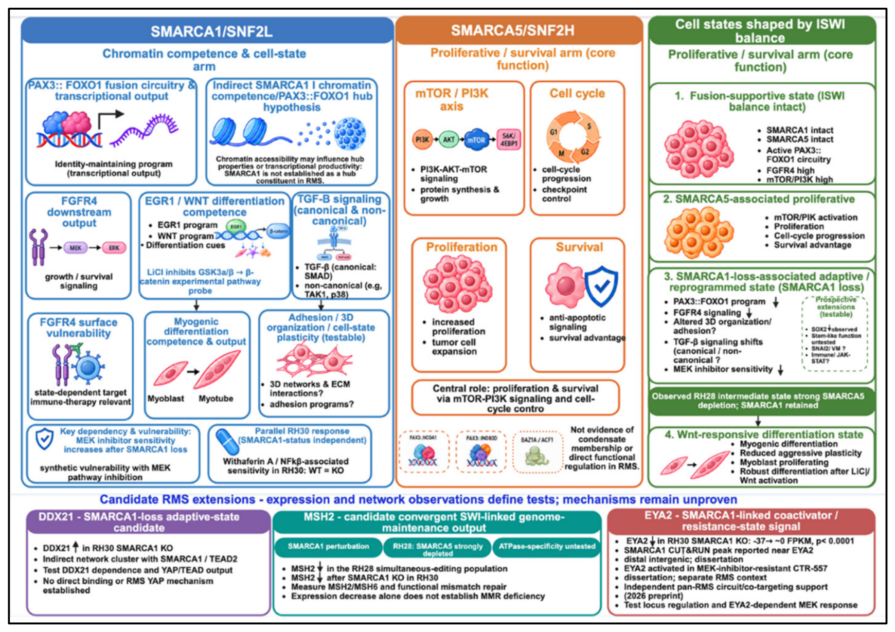
Figure 2.
Integrated experimental framework for testing the ISWI division-of-labor hypothesis and its context-dependent extension. (A) Protein-validated wild-type, SMARCA1-loss, SMARCA5-loss, and double-loss FP-RMS states are compared across fusion-program output, chromatin/hub competence, cell-state regulation, mTOR-linked cellular fitness, therapeutic response, and surface-antigen state. Integrated readouts include fusion RNA/protein/activity, chromatin accessibility and occupancy, protein-interaction state, pathway activity, three-dimensional phenotypes, and surface-antigen abundance. Hub and condensate functions remain to be demonstrated directly. (B) Complementary single-cell analysis evaluates fusion-context-dependent coupling of SMARCA1 and SMARCA5 to RMS transcriptional states while accounting for cell-cycle activity. These associations support state coupling but do not establish causal plasticity or pathway order. (C) The same dependency logic is extended conceptually to other fusion-driven sarcomas by identifying the dominant fusion/transcription factor, integrating multi-omic and protein-interaction evidence, nominating a context-specific chromatin remodeler, and testing its function by perturbation and rescue. No universal SMARCA1 or SMARCA5 assignment is assumed across sarcoma types.
Figure 2.
Integrated experimental framework for testing the ISWI division-of-labor hypothesis and its context-dependent extension. (A) Protein-validated wild-type, SMARCA1-loss, SMARCA5-loss, and double-loss FP-RMS states are compared across fusion-program output, chromatin/hub competence, cell-state regulation, mTOR-linked cellular fitness, therapeutic response, and surface-antigen state. Integrated readouts include fusion RNA/protein/activity, chromatin accessibility and occupancy, protein-interaction state, pathway activity, three-dimensional phenotypes, and surface-antigen abundance. Hub and condensate functions remain to be demonstrated directly. (B) Complementary single-cell analysis evaluates fusion-context-dependent coupling of SMARCA1 and SMARCA5 to RMS transcriptional states while accounting for cell-cycle activity. These associations support state coupling but do not establish causal plasticity or pathway order. (C) The same dependency logic is extended conceptually to other fusion-driven sarcomas by identifying the dominant fusion/transcription factor, integrating multi-omic and protein-interaction evidence, nominating a context-specific chromatin remodeler, and testing its function by perturbation and rescue. No universal SMARCA1 or SMARCA5 assignment is assumed across sarcoma types.
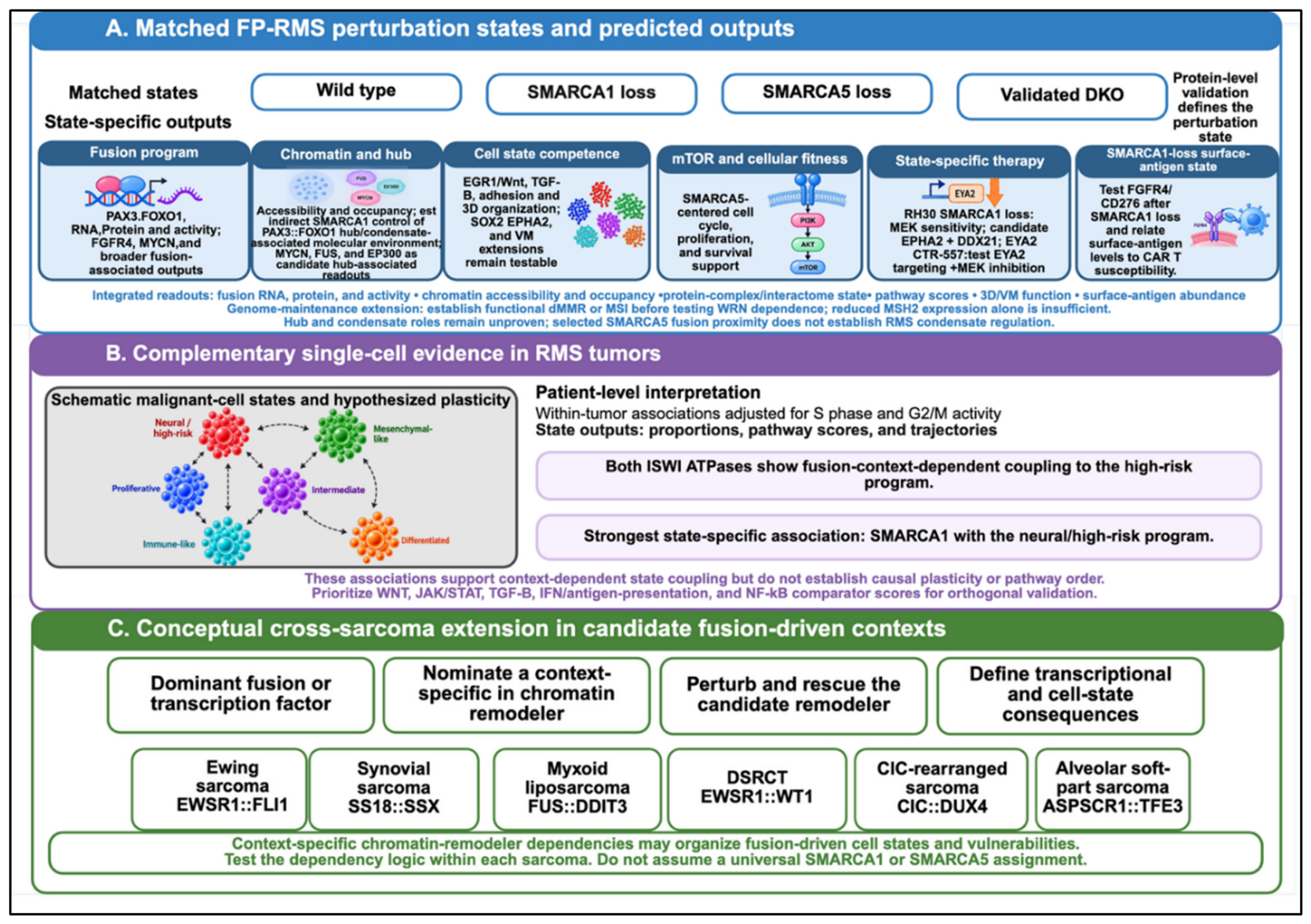

Table 1.
Evidence Underlying the Hypothesis.
| Observation | Primary source | Evidence status | Role in hypothesis |
| SMARCA1 is required for effective myogenic differentiation and Wnt-responsive differentiation; SMARCA5 is more strongly associated with mTOR/proliferation. | Dissertation [7] | Completed doctoral experiments | Foundation for ATPase-specific functions. |
| RH28 cells with SMARCA5 depletion but retained SMARCA1 remain differentiation competent and respond robustly to LiCl/Wnt activation. | Dissertation [7] | Completed experiment; not a confirmed complete DKO | Supports a differentiation-responsive intermediate state. |
| SMARCA1 loss disrupts migration, invasion, adhesion programs, and organized 3D spheroid architecture. | Dissertation [7] | Completed experiments | Extends SMARCA1 function to multicellular architecture and behavioral plasticity. |
| SMARCA1 loss markedly suppresses PAX3::FOXO1 transcript and fusion-associated transcriptional programs, including FGFR4; conference-reported chromatin analyses also linked SMARCA1 loss to reduced PAX3::FOXO1-associated occupancy/output at FGFR4 and MYCN loci. | Conference-reported work [10] | Emerging, non-peer-reviewed mechanistic evidence | Supports an upstream SMARCA1-dependent fusion state and identifies FGFR4 as a measurable downstream sentinel without establishing direct SMARCA1 regulation of FGFR4. |
| SMARCA5 is proximity-detected with PAX3::NCOA1 and PAX3::INO80D; SMARCA1 is not proximity-detected with the seven oncofusions tested. | Zimmerman et al. [13] | Peer-reviewed proximity-labeling evidence | Supports a selected-fusion SMARCA5 proximity observation distinct from SMARCA1; functional relevance remains untested. |
| Exploratory RH30 wild-type versus SMARCA1-knockout RNA-seq reanalysis shows bidirectional changes among IDR/coactivator/hub-associated genes: MYCN and NCOA1 decrease strongly; EP300, CREBBP, FUS, and BRD4 decrease more modestly; MED15, POLR2A, and INO80D increase. | Existing RH30 author-derived RNA-seq dataset; exploratory reanalysis | Descriptive FPKM-based secondary analysis; protein abundance, formal differential-expression statistics, and condensate behavior remain unvalidated | Supports a selective hub/coactivator-reconfiguration hypothesis after SMARCA1 loss and provides candidate readouts for direct protein, chromatin, interactome, and live-cell testing; does not establish global condensate loss. |
| Patient-derived RMS tumoroids retain tumor-relevant features, are CRISPR-editable, and reveal genotype-dependent drug sensitivity; TP53 loss in RMS012 FN-eRMS increased sensitivity to prexasertib. | Meister et al. [23] | Peer-reviewed patient-derived tumoroid evidence | Provides methodological precedent for testing ISWI-defined 3D states and therapeutic vulnerabilities in RMS tumoroids; does not directly establish an FP-RMS ISWI mechanism. |
| SMARCA1 loss increases vulnerability to MEK inhibition in RH30, including greater trametinib-associated pERK suppression and preferential viability loss in subsequent analyses. | Dissertation [7]; author-derived/conference-reported analyses [8,12] | Completed author-derived experiments; requires independent validation | Establishes MEK inhibition as the prioritized therapeutic vulnerability and a functional readout of the SMARCA1-loss state. |
| Withaferin A produces strong, closely overlapping viability responses in RH30 wild-type and SMARCA1-knockout cells and in the separate SMS-CTR/CTR-557 comparison. | Dissertation [7] | Completed doctoral experiments; the isogenic RH30 comparison tests SMARCA1 status, whereas SMS-CTR/CTR-557 compares parental and MEK-resistant states | Defines a parallel response distinct from the SMARCA1-loss-associated MEK phenotype; Withaferin A pleiotropy requires orthogonal NF-κB/IKK validation before assigning pathway-selective dependence. |
| SB505124 responses differ by context: RH30 SMARCA1-knockout cells are relatively less sensitive than wild-type cells, whereas MEK-inhibitor-resistant CTR-557 is more sensitive than SMS-CTR; Dactolisib produces moderate, concentration-dependent responses. | Dissertation [7] | Completed doctoral drug-response experiments in two distinct model comparisons; directional effects require matched replication and pathway readouts | Supports state-dependent TGF-β-pathway vulnerability and context-dependent PI3K/mTOR response; does not establish a universal SMARCA1-, EYA2-, or TGF-β-dependent drug-response rule. |
| Fusion protein stability, reporter collapse, and YAP/TAZ compensation after SMARCA1 loss. | Prospective validation strategy | Hypotheses to be tested | Distinguishes transcript, protein, activity, and adaptive-state mechanisms. |
| RMS can form vasculogenic mimicry; SNAI2 promotes VM, proliferation, and metastasis, and VM has been associated with poor outcome in alveolar RMS. | Zhang et al.; Sun et al. [29,30] | Peer-reviewed RMS VM evidence | Supports VM as a plausible SNAI2-linked plasticity output that can be integrated with the SMARCA1 state model; does not prove SMARCA1 directly controls VM. |
| FGFR4-directed CAR T cells show preclinical activity against RMS, and dual FGFR4/CD276 bicistronic CAR T cells show enhanced activity in RMS models with heterogeneous antigen expression. | Tian et al. [36,37] | Peer-reviewed preclinical immunotherapy evidence | Supports an antigen-heterogeneity-mitigation strategy; whether dual targeting buffers SMARCA1-associated changes in FGFR4 surface abundance remains untested. |
| Single-cell RMS profiling identifies tumor-acquired and therapy-resistant states and demonstrates that RMS cell-state heterogeneity can be resolved beyond bulk averages. | Danielli et al.[33] | Peer-reviewed single-cell transcriptomic evidence | Supports single-cell state mapping as the central integrating layer for testing fusion state, differentiation, VM-associated plasticity, antigen heterogeneity, and treatment-responsive subpopulations. |
| Exploratory reanalysis of 70,600 malignant cells from 16 tumors found no tumor-level SMARCA1/5 overexpression in high-risk FN-RMS. After cell-cycle adjustment, both ATPases showed fusion-context-dependent coupling to the high-risk program; SMARCA1 had the strongest association with the neural component. SMARCA5 did not track S/G2M scores. | Whitfield et al. [34]; author reanalysis | Exploratory secondary analysis; 4 FP and 4 high-risk FN tumors; requires patient-level replication | Supports context-dependent ISWI-state coupling and the SMARCA1 cell-state arm; does not independently support SMARCA5 proliferation. |
| The SMARCA1-associated computational network implicated JAK-STAT signaling, and preliminary RH30 transcriptomic observations show changes in STAT-family expression after SMARCA1 loss. | Dissertation [7]; preliminary author-observed transcriptomic analysis | Historical computational association plus preliminary transcriptomic observation; STAT pathway activation and immune function not established. | Supports the prospective JAK-STAT/SOX2 immune-state branch and defines pathway-level validation requirements. |
| SMARCA1 loss alters TGF-β pathway chromatin and expression, including canonical receptor/SMAD-associated components, and disrupts non-canonical MAPK/ERK, PI3K/AKT, RAS, mTOR, and NF-κB-associated programs. | Dissertation [7] | Completed doctoral multi-omic analyses; pathway-specific functional hierarchy requires further validation | Establishes TGF-β as a supported SMARCA1-linked chromatin/signaling interface while keeping individual downstream branches mechanistically separable. |
| The 2025 dissertation reported marked EYA2 downregulation after RH30 SMARCA1 knockout, with a distal SMARCA1 CUT&RUN peak nearby. In the separate SMS-CTR/CTR-557 resistance comparison, EYA2 was upregulated and TGF-β pathway enrichment was detected in MEK-inhibitor-resistant CTR-557. The independent 2026 pan-RMS preprint appeared later and identified EYA2 as a regulatory-circuit cofactor and EYA1/2 plus EP300/CBP cotargeting as a vulnerability. | Dissertation [7]; Gustafson et al. [15] | Dissertation observations (2025) predate the independent EYA preprint (2026); direct SMARCA1-EYA2 regulation, EYA2-TGF-β coupling, and functional drug-response roles remain untested. | Records the chronology and motivates testing EYA2 and TGF-β-associated features as separate resistant-state markers without elevating them into a causal branch of the core model. |
| MSH2 is downregulated in the RH28 simultaneous SMARCA1/SMARCA5-editing condition and after SMARCA1 knockout in RH30. | Dissertation [7]; genome-maintenance context [19,20,21] | Completed expression observations in different RMS backgrounds; functional MMR deficiency and direct ATPase-to-MSH2 regulation not established. | Supports MSH2 as a candidate convergent ISWI-linked genome-maintenance output, with SMARCA1-versus-SMARCA5 dependence requiring matched validation. |
| DDX21 was indirectly connected to SMARCA1/TEAD2 by dissertation co-expression-BioGRID analysis; preliminary RH30 RNA-seq observation indicates increased DDX21 after SMARCA1 knockout. | Dissertation [7]; Tang et al. [35] | Computational/network association plus preliminary expression observation; no validated SMARCA1-DDX21 binding or RMS DDX21-YAP mechanism. | Supports a testable DDX21-YAP compensatory extension after SMARCA1 loss, not a core SMARCA1 effector. |
| EPHA2 and DDX21 increase in the RH30 SMARCA1-knockout transcriptomic state; cross-tumor work links EphA2 to vasculogenic mimicry, and KI-DX-014 provides an experimental DDX21-RNA interaction probe. | RH30 author-derived analysis/dissertation [7]; Hess et al. [38]; Aiba et al. [39] | Preliminary RMS expression observation plus cross-tumor functional precedent and chemical-probe availability; no demonstrated RMS dependency or combination effect | Supports a testable EPHA2-DDX21 adaptive-vulnerability branch that remains secondary to the core division-of-labor model. |
Note: Evidence is distinguished as completed experimental work, conference-reported findings, peer-reviewed external evidence, exploratory secondary analysis, or prospective hypothesis. Association, expression, chromatin occupancy, protein interaction, and functional dependence are not treated as equivalent levels of evidence.
Disclaimer/Publisher’s Note: The statements, opinions and data contained in all publications are solely those of the individual author(s) and contributor(s) and not of MDPI and/or the editor(s). MDPI and/or the editor(s) disclaim responsibility for any injury to people or property resulting from any ideas, methods, instructions or products referred to in the content. |
© 2026 by the author. Licensee MDPI, Basel, Switzerland. This article is an open access article distributed under the terms and conditions of the Creative Commons Attribution (CC BY) license.
Copyright: This open access article is published under a Creative Commons CC BY 4.0 license, which permit the free download, distribution, and reuse, provided that the author and preprint are cited in any reuse.