
Submitted:
12 September 2026
Posted:
17 September 2026
You are already at the latest version
Abstract
Rheumatoid arthritis (RA) is a chronic autoimmune disorder characterized by persistent joint inflammation, bone erosion, and systemic involvement with genetic factors contributing significantly to disease susceptibility. Interleukin-17 (IL-17) signaling has been strongly implicated in autoimmune inflammation, making IL-17A and its receptor IL-17RC key candidates for genetic association studies. This case control study investigated IL-17A (rs2275913 G/A) and IL-17RC (rs708567 G/A) polymorphisms in 120 RA patients and 120 controls from Pakistan. Genotyping was performed using the PCR-RFLP method, and statistical analyses included allele and genotype frequency comparisons, odds ratio calculations, multiple inheritance model testing, and Hardy–Weinberg equilibrium assessment. The IL-17RC rs708567 variant showed a robust association with RA, with the A allele significantly more frequent in patients (43.3%) than controls (22.1%) and conferring an almost threefold increased risk (OR = 2.88, p = 1.42 × 10⁻⁷). Genotypic, allelic, and dominant model analyses confirmed this strong association. By contrast, the IL-17A rs2275913 variant showed no significant association, although a suggestive trend was observed under the dominant model. Hardy–Weinberg equilibrium analysis revealed deviations in the overall and control groups but equilibrium in patients, suggesting possible stratification or underlying genetic association. These findings highlight IL-17RC rs708567 as a potential genetic marker for RA susceptibility in the Pakistani population, reinforcing the importance of IL-17 signaling in RA pathogenesis and warranting further studies in larger cohorts. In conclusion, this study provides novel evidence for the role of IL-17RC in RA in Pakistan and supports its relevance as a therapeutic target. Further large-scale investigations are essential to validate these findings and advance personalized treatment strategies.
Keywords:
RA
; IL-17A(rs2275913)
; IL-17RC (rs708567)
; gene polymorphism
; PCR-RFLP
Introduction
Rheumatoid arthritis (RA) is a chronic autoimmune disorder marked by persistent joint inflammation, bone loss, and symmetric polyarthritis with systemic involvement [1]. Globally, its prevalence ranges from 0.24% to 2% [2], with women disproportionately affected compared to men [3]. In Pakistan, studies report a higher disease burden among women, including 0.142% prevalence in Karachi [4]. RA pathogenesis arises from genetic predisposition, immune dysregulation, and environmental triggers such as smoking, diet, obesity, and infections, with cytokines serving as central mediators of chronic inflammation [5,6,7]. Among these, interleukins (ILs) are particularly important, as they regulate immune cell activation, differentiation, and migration, exerting both pro- and anti-inflammatory effects [8]. Notably, IL-17, a pro-inflammatory cytokine secreted by T helper cells (Th17), has emerged as a key driver of autoimmune inflammation and is strongly implicated in RA progression [9]. Th17 cells play a central role in host defense by releasing effector cytokines IL-17A and IL-17F [10].
The IL-17A gene (chromosome 6p12.2) encodes a proinflammatory cytokine secreted by Th17 cells that amplifies immune cell recruitment through induction of mediators such as IL-6, COX-2, and nitric oxide [11]. Elevated IL-17A levels are strongly linked to rheumatoid arthritis (RA), where IL-17A synergizes with TNF-α to stimulate pro-inflammatory cytokines and chemokines, promoting chronic joint inflammation and tissue destruction characteristic of RA [12]. The IL-17RC gene (chromosome 3p25.3) encodes a receptor subunit that pairs with IL-17RA to mediate IL-17A and IL-17F signaling via NF-κB and MAPK activation [13]. Overexpression of IL-17RA/RC in RA synovium enhances responsiveness to IL-17A, and functional studies confirm that IL-17A-induced IL-6 and CCL-20 secretion requires both receptor subunits [14]. IL-17A forms homodimers or heterodimers with IL-17F, with the homodimer being more potent. These cytokines signal through the IL-17RA/IL-17RC dimeric receptor, underscore the pathogenic role of IL-17A/IL-17RC signaling in RA [15].
Polymorphisms in IL-17A (ligand) and IL-17RC (receptor) may alter cytokine signaling efficiency, influencing susceptibility to RA and shaping disease progression [10]. Specifically, IL-17A -152 G/A (rs2275913), IL-17F 7488 A/G (rs763780), and IL-17F 7383 A/G (rs2397084) polymorphisms correlate positively with RA, with the −197 G/A promoter variant increasing IL-17A secretion [16]. The IL-17 gene has therefore been implicated in RA susceptibility across different populations. This study aimed to investigate the association of IL-17A (rs2275913 G/A) and IL-17RC (rs708567 G/A) polymorphisms with RA onset in the Pakistani population, and to explore the relationship between IL-17RC variants and clinical parameters.
Materials and Methods
Sample Collection and DNA Extraction
This study included 240 participants: 120 rheumatoid arthritis (RA) patients diagnosed at P.O.F Hospital WAH Cantt using 2010 RA criteria and 120 age, gender and ethnicity-matched healthy controls from Pakistan with no personal or family history of autoimmune disease. Clinical and demographic data were recorded, and written informed consent was obtained prior to sample collection. Peripheral blood was drawn in EDTA vials for DNA extraction and in gel vacutainers for biochemical analysis. Genomic DNA was isolated from EDTA blood using the salting-out method: 1 ml blood was lysed with TKM1-Triton X, centrifuged, and the nuclear pellet washed, dissolved in TKM2, treated with SDS, incubated, and precipitated with NaCl and ethanol. The DNA pellet was centrifuged, dried, and resuspended in TE buffer for storage at 4°C. DNA quality was checked by 1% agarose gel electrophoresis, and concentration/purity assessed with a NanoDrop ND-1000 spectrophotometer at 260/280 nm. Biochemical parameters (triglycerides, LDL, and total cholesterol) were measured using a fully automated chemistry analyzer (Figure 1).
Genotyping
Genotyping of IL-17A (rs2275913, A/G) and IL-17RC (rs708567, G/A) polymorphisms was performed using the PCR-RFLP method. Specific primers were used to amplify target fragments of 102 bp for IL-17A and 233 bp for IL-17RC. The PCR products were then digested with restriction enzymes EcoNI (for IL-17A) and HinfI (for IL-17RC) to distinguish allelic variants. Primer details are given in Table 1.
After making the reaction mixture for gene amplification, the following conditions were applied: First denaturation at 95℃ for 5 minutes, final denaturation at 94℃ for 1min, primer annealing at 56℃ for 1 min, primer initial extension at 72℃ for 1 min and final extension at 72℃ for 10 min. PCR was carried out for 40 cycles. And for the enzyme digestion, the reaction mixture with the amplified product was incubated at 37℃ for 24 hours. Then a 5% agarose gel was used to separate digested products and then visualized in gel documentation system under UV light. The band size was confirmed with the help of 1kb DNA ladder.
Statistical Analysis
Statistical analyses were performed to evaluate the genetic association of the studied SNP with disease susceptibility. Hardy–Weinberg equilibrium (HWE) was assessed separately in cases, controls and the overall study population to ensure genotype distributions conformed to expected proportions. Allele and genotype frequencies between affected and unaffected groups were compared using chi-square (χ²) tests, and odds ratios (ORs) with corresponding p-values were calculated to estimate the strength of association. In addition, association was further examined under different inheritance models, including genotypic, allelic, trend, dominant, and recessive models, to identify the most likely mode of genetic effect. All statistical analyses were performed at a significant level of p < 0.05.
Results
A total of 120 patients with rheumatoid arthritis and 120 healthy control subjects were included in the study. The mean age of the studied groups was 52.12 ± 7.77 years in RA patients and 51.43 ± 10.25 in the control group (Figure 2), with a sex ratio (male/female) of 0.12(14/116) in the disease group and a sex ratio (male/female) of 0.10 (11/109) in the control group (Figure 3). About 10% of RA cases were reported with Hypertension, and 18.3% of RA patients were suffering from Diabetes mellitus, while no associated diseases were found in healthy patients. Only 22.5% of RA patients showed a positive family history of rheumatoid arthritis (Figure 4) but in the control group, not a single individual had a positive family history.
The unpaired t-test was used to see the difference in the biochemical parameters among healthy control individuals and rheumatoid arthritis patients. RA patients exhibit significantly higher total cholesterol levels, triglycerides levels, and LDL levels compared to the control group with a p-value of <0.01. RA had a well-known association with an increased risk of cardiovascular disease due to chronic inflammation. Chronic inflammation in RA, driven by cytokines like TNF-α, IL-6, and IL-1, disrupt lipid metabolism. With increased levels of LDL, total cholesterol, and triglycerides, there is a risk of developing atherosclerosis and metabolic syndrome. Table 2 compared key clinical and biochemical parameters between rheumatoid arthritis (RA) patients and a control group. The results were expressed as mean and standard deviation (±), and the statistic al significance in terms of p-values using an unpaired t-test. The body mass index (Kg/m2) and diastolic blood pressure (mmHg) showed no significant difference between RA patients and the control group. However, RA patients have a higher mean systolic BP (mmHg) than the control group with a p-value of <0.01, indicating a statistically significant result.
In the present study, comparative analysis of cardiovascular and metabolic parameters between rheumatoid arthritis (RA) patients and healthy controls revealed notable differences. As illustrated in Figure 5 and Figure 6, systolic blood pressure was significantly higher among RA patients compared to the control group (p < 0.01), whereas no significant difference was observed in diastolic blood pressure (p = 0.97) or body mass index (BMI) (p = 0.61).
Furthermore, lipid profile analysis demonstrated that cholesterol, triglyceride, and LDL levels were significantly lower in RA patients relative to controls (all p < 0.01). These results indicate that while RA patients exhibited elevated systolic blood pressure, their lipid parameters were comparatively reduced, suggesting a possible alteration in lipid metabolism associated with rheumatoid arthritis.
Genotype and Allele Frequency Analysis
A total of 240 Samples (120 cases and 120 controls) were analyzed for IL-17RC and IL-17RA polymorphism rs708567G/A, rs2275913G/A respectively. Genotyping was performed using the PCR-RFLP method and the restriction enzyme Hinf1 (IL-17RC) and EcoNI (IL-17RA). The size of PCR products was 210bp for IL-17RC (Figure 7) and 103bp for IL-17RA (Figure 8). The IL-17RC G allele was cut to 161 and 49 bp while the A allele remained uncut using Hinf1 restriction enzyme (Figure 7). On the other hand, the IL-17RA G allele was cut into 70 and 33 bp while A allele remained uncut using EcoNI restriction enzyme (Figure 8).
The distribution of IL-17RC genotypes showed that GA and AA variants were more frequent in rheumatoid arthritis (RA) patients compared to controls, whereas the GG genotype was predominant among controls. The A allele of IL-17RC was also more common in patients (43.3%) than in controls (22.1%). For IL-17RA, the GA and AA genotypes appeared at slightly higher frequencies in patients than in controls, with the A allele observed more often in patients (38.8%) relative to controls (29.2%). These patterns indicate a potential role of IL-17RC and IL-17RA polymorphisms in increasing susceptibility to RA. (Table 3)
Statistical Analysis
- I.
- Allele and Genotype Association Analysis
Association analysis of SNP 52186235 located on chromosome 6 revealed that the risk allele (A1 = 2) was more frequent in cases (36.7%) compared to controls (28.3%). The odds ratio (OR = 1.46) indicated that carriers of this allele may have an elevated risk of developing the disease. Although the association approached significance (χ² = 3.799, p = 0.051), it did not cross the conventional threshold, suggesting a possible genetic contribution that warrants validation in larger cohorts (Table 4).
Allelic association analysis of SNP rs708567 (Chr 3) revealed that the risk allele (A1 = 2) occurred at a markedly higher frequency in rheumatoid arthritis cases (43.8%) compared to controls (21.3%). The association was highly significant (χ² = 27.69, p = 1.42 × 10⁻⁷), with an odds ratio of 2.88, indicating that carriers of the risk allele have nearly a threefold increased likelihood of developing the disease (Table 5)
- II.
- Genetic Model–Based Association Tests
The association analysis of SNP rs2275913 under different genetic models shows that the dominant model demonstrated a statistically significant association with the disease (χ² = 4.82, p = 0.028), indicating that carriers of at least one copy of the risk allele may have increased susceptibility. The allelic (p = 0.051) and trend (p = 0.069) models showed borderline associations, suggesting a possible additive effect. In contrast, the genotypic (p = 0.085) and recessive (p = 0.574) models did not reveal significant associations. These findings suggest that the SNP may act primarily through a dominant inheritance pattern (Table 6).
The IL-17RC SNP rs708567 (chromosome 3) showed a strong genetic association with rheumatoid arthritis across multiple inheritance models. Allelic analysis revealed a markedly higher frequency of the risk allele in cases compared to controls (χ² = 27.69, p = 1.42 × 10⁻⁷), representing the most significant finding. The genotypic (p = 5.58 × 10⁻⁶) and trend tests (p = 1.08 × 10⁻⁶) also demonstrated robust associations. Both dominant and recessive models were statistically significant, with the dominant model (p = 3.35 × 10⁻⁶) indicating that carriers of at least one risk allele had a considerably higher likelihood of developing RA. Collectively, these results underscore rs708567 in IL-17RC as a potential susceptibility marker for rheumatoid arthritis in the studied cohort (Table 7).
- III.
- Hardy–Weinberg Equilibrium Analysis
Hardy–Weinberg Equilibrium Analysis (HWE) analysis of the SNP located at chromosome 6 (position 52186235) revealed a significant deviation in the overall cohort (p=0.027) and in the unaffected subgroup (p=0.023), while the affected group remained in equilibrium (p=0.439). The excess deviation observed in the unaffected group compared to cases may indicate potential population stratification or underlying genetic association with disease susceptibility. These findings suggest that this SNP may play a role in the genetic predisposition to the studied condition, with the distortion in equilibrium highlighting its possible biological relevance (Table 8).
HWE analysis of SNP rs708567 (Chr 3) showed a significant deviation in the overall cohort (p = 0.0122), whereas both affected (p = 0.2685) and unaffected groups (p = 0.1717) were in equilibrium. The discrepancy observed at the overall level may reflect genetic stratification or a potential association with disease susceptibility (Table 9).
Discussion
Rheumatoid arthritis (RA) is a common systemic autoimmune illness that causes bone loss and persistent joint inflammation and affects approximately 0.24% to 2% of people around the world, especially females [8]. The influence of genetic predisposition on the pathogenesis of RA has been extensively studied, particularly focusing on cytokine gene polymorphisms, such as those in interleukin-17 (IL-17). The current study investigated the association between IL-17A rs2275913 and IL-17RC rs708567 polymorphisms and susceptibility to RA. This study also explored some clinical parameters in RA patients with normal healthy individuals.
In the current study, most of the RA patients were females (88.3%) and these results were in line with Ashiq et al. [3] who reported women are generally more likely to be affected than men. About 10% of RA cases were reported with hypertension, consistent with Liang et al. [17], who showed a significant association of hypertension with RA. In addition, 18.3% of RA patients suffered from diabetes mellitus, a finding supported by Tian et al. [18]. Abnormal activation of the immune system is the main pathological process of RA; for example, monocytes and other immune cells activate the production of pro-inflammatory and anti-inflammatory cytokines such as TNF-α, IL-1, IL-10, and TGF-α by polarization of macrophages [19]. Recently, several studies confirmed that effector cytokines such as IL-17 and IL-23 produced from Th17 cells play a significant role in RA pathogenesis [20,21]. The current research is the first attempt in Pakistan to assess the association of IL-17A and IL-17RC polymorphisms (rs2275913G/A, rs708567G/A) with RA, as immunogenetic data of RA patients remain scarce in this population.
Our findings demonstrate a significant association between the IL-17RC rs708567 polymorphism and rheumatoid arthritis, with the risk allele conferring nearly a threefold increase in disease susceptibility. In contrast, the IL-17A rs2275913 variant showed no significant association, suggesting that receptor-mediated IL-17 signaling may play a more critical role in RA pathogenesis within the studied population. These results emphasize IL-17RC as a potential genetic marker and therapeutic target for RA. These results were consistent with Diab et al. [21], Tarak Dhaouadi et al. [10], and Marwa et al. [22], who all reported no significant correlation. However, contrary results were found in Brazil by Isabelle et al. [23] (OR = 3.18, 95% CI = 1.13–9.95) and in another study by Agonia et al. [24]. A meta-analysis by Chen et al. further showed a significant association of IL-17A rs2275913 with RA susceptibility in the European population. Elevated IL-17A levels in synovial fluid and tissue explants have been reported to contribute to joint deterioration through synergy with TNF receptors, ultimately promoting pro-inflammatory cytokine production [25].
Conclusion
In conclusion, this study provides novel evidence from the Pakistani population that IL-17RC rs708567 polymorphism is strongly associated with increased susceptibility to rheumatoid arthritis, conferring nearly a threefold higher risk, whereas the IL-17A rs2275913 variant showed no significant correlation. These findings suggest that receptor-mediated IL-17 signaling, rather than ligand variation, may play a more decisive role in disease predisposition. The results not only strengthen the role of IL-17RC as a potential genetic marker but also highlight it as a promising therapeutic target in RA management. Future studies with larger sample sizes and functional assays are warranted to validate these associations and to further elucidate the mechanistic contribution of IL-17RC in RA pathogenesis.
References
- Zhao, R.; Zhang, Y-w; Yao, J-y; Qiao, J.; Song, S.; Zhang, S-x; et al. Genetic association between interleukin-17 and susceptibility to rheumatoid arthritis. BMC Med. Genom. 2023, 16(1), 277. [Google Scholar] [CrossRef] [PubMed]
- Uke, P.; Maharaj, A.; Adebajo, A. A review on the epidemiology of rheumatoid arthritis: An update and trends from current literature. Best Pract. Res. Clin. Rheumatol. 2025, 102036. [Google Scholar] [CrossRef] [PubMed]
- Ashiq, K.; Ashiq, S.; Mobashar, A.; Abid, F.; Yasmeen, A.; Shehzadi, N.; et al. An updated review on rheumatoid arthritis (RA): epidemiology, pathophysiology, diagnosis, and the current approaches for its treatment. Sudan J. Med. Sci. 2023, 18(4), 539–551. [Google Scholar] [CrossRef]
- Riaz, N.; Arshad, M.; Zubair, F. Polymorphism in CD40 gene associated with the prevalence of rheumatoid arthritis in Pakistan. Pak. J. Zool. 2024, 1–4. [Google Scholar] [CrossRef]
- Hussain, M.Z.; Mahjabeen, I.; Khan, M.S.; Mumtaz, N.; Maqsood, S.U.; Ikram, F.; et al. Genetic and expression deregulation of immunoregulatory genes in rheumatoid arthritis. Mol. Biol. Rep. 2021, 48(6), 5171–5180. [Google Scholar] [CrossRef] [PubMed]
- Romão, V.C.; Fonseca, J.E. Etiology and risk factors for rheumatoid arthritis: a state-of-the-art review. Front. Med. 2021, 8, 689698. [Google Scholar] [CrossRef] [PubMed]
- Akhter, S.; Irfan, H.M.; Alamgeer; Jahan, S.; Shahzad, M.; Latif, M.B. Nerolidol: a potential approach in rheumatoid arthritis through reduction of TNF-α, IL-1β, IL-6, NF-kB, COX-2 and antioxidant effect in CFA-induced arthritic model. Inflammopharmacology 2022, 30(2), 537–548. [Google Scholar] [CrossRef] [PubMed]
- Justiz Vaillant, A.A.; Interleukin, Q.A. StatPearls. Treasure Island (FL) with ineligible companies. Disclosure: Ahmad Qurie declares no relevant financial relationships with ineligible companies; StatPearls Publishing Copyright © 2025, StatPearls Publishing LLC, 2025. [Google Scholar]
- Al-Lami, R.S.S.; Al-Hilfy, J.H.Y. Role of Interleukins-8,-17 and-22 in Iraqi postmenopausal women with Osteoporosis. Cytokine 2025, 187, 156853. [Google Scholar] [CrossRef] [PubMed]
- Dhaouadi, T.; Chahbi, M.; Haouami, Y.; Sfar, I.; Abdelmoula, L.; Ben Abdallah, T.; et al. IL-17A, IL-17RC polymorphisms and IL17 plasma levels in Tunisian patients with rheumatoid arthritis. PLoS ONE 2018, 13(3), e0194883. [Google Scholar] [CrossRef] [PubMed]
- McGeachy, M.J.; Cua, D.J.; Gaffen, S.L. The IL-17 Family of Cytokines in Health and Disease. Immunity 2019, 50(4), 892–906. [Google Scholar] [CrossRef] [PubMed]
- Gaffen, S.L. The role of interleukin-17 in the pathogenesis of rheumatoid arthritis. Curr. Rheumatol. Rep. 2009, 11(5), 365–370. [Google Scholar] [CrossRef] [PubMed]
- Ho, A.W.; Gaffen, S.L. IL-17RC: a partner in IL-17 signaling and beyond. Semin. Immunopathol. 2010, 32(1), 33–42. [Google Scholar] [CrossRef] [PubMed]
- Zrioual, S.; Toh, M.L.; Tournadre, A.; Zhou, Y.; Cazalis, M.A.; Pachot, A.; et al. IL-17RA and IL-17RC receptors are essential for IL-17A-induced ELR+ CXC chemokine expression in synoviocytes and are overexpressed in rheumatoid blood. J. Immunol. 2008, 180(1), 655–663. [Google Scholar] [CrossRef] [PubMed]
- Brevi, A.; Cogrossi, L.L.; Grazia, G.; Masciovecchio, D.; Impellizzieri, D.; Lacanfora, L.; et al. Much More Than IL-17A: Cytokines of the IL-17 Family Between Microbiota and Cancer. Front Immunol. 2020, 11, 565470. [Google Scholar] [CrossRef] [PubMed]
- Bogunia-Kubik, K.; Świerkot, J.; Malak, A.; Wysoczańska, B.; Nowak, B.; Białowąs, K.; et al. IL-17A, IL-17F and IL-23R Gene Polymorphisms in Polish Patients with Rheumatoid Arthritis. Arch. Immunol. Ther. Exp. 2015, 63(3), 215–221. [Google Scholar] [CrossRef] [PubMed]
- Liang, X.; Chou, O.H.I.; Cheung, C.L.; Cheung, B.M. Is hypertension associated with arthritis? The United States national health and nutrition examination survey 1999–2018. Ann. Med. 2022, 54(1), 1767–1775. [Google Scholar] [CrossRef] [PubMed]
- Tian, Z.; McLaughlin, J.; Verma, A.; Chinoy, H.; Heald, A.H. The relationship between rheumatoid arthritis and diabetes mellitus: a systematic review and meta-analysis. Cardiovasc. Endocrinol. Metab. 2021, 10(2), 125–131. [Google Scholar] [CrossRef] [PubMed]
- Fessler, J.; Raicht, A.; Husic, R.; Ficjan, A.; Schwarz, C.; Duftner, C.; et al. Novel Senescent Regulatory T-Cell Subset with Impaired Suppressive Function in Rheumatoid Arthritis. Front Immunol. 2017, 8, 300. [Google Scholar] [CrossRef] [PubMed]
- Schinocca, C.; Rizzo, C.; Fasano, S.; Grasso, G.; La Barbera, L.; Ciccia, F.; et al. Role of the IL-23/IL-17 Pathway in Rheumatic Diseases: An Overview. Front Immunol. 2021, 12, 637829. [Google Scholar] [CrossRef] [PubMed]
- Gan, Y.; Zhao, X.; He, J.; Liu, X.; Li, Y.; Sun, X.; et al. Increased Interleukin-17F is Associated with Elevated Autoantibody Levels and More Clinically Relevant Than Interleukin-17A in Primary Sjögren’s Syndrome. J. Immunol. Res. 2017, 2017, 4768408. [Google Scholar] [CrossRef] [PubMed]
- Marwa, O.S.; Kalthoum, T.; Wajih, K.; Kamel, H. Association of IL17A and IL17F genes with rheumatoid arthritis disease and the impact of genetic polymorphisms on response to treatment. Immunol. Lett. 2017, 183, 24–36. [Google Scholar] [CrossRef] [PubMed]
- Gomes da Silva, I.I.F.; Angelo, H.D.; Rushansky, E.; Mariano, M.H.; Maia, M.M.D.; de Souza, P.R.E. Interleukin (IL)-23 Receptor, IL-17A and IL-17F Gene Polymorphisms in Brazilian Patients with Rheumatoid Arthritis. Arch. Immunol. Ther. Exp. 2017, 65(6), 537–543. [Google Scholar] [CrossRef] [PubMed]
- Agonia, I.; Couras, J.; Cunha, A.; Andrade, A.J.; Macedo, J.; Sousa-Pinto, B. IL-17, IL-21 and IL-22 polymorphisms in rheumatoid arthritis: A systematic review and meta-analysis. Cytokine 2020, 125, 154813. [Google Scholar] [CrossRef] [PubMed]
- Chen, P.; Li, Y.; Li, L.; Zhang, G.; Zhang, F.; Tang, Y.; et al. Association between the interleukin (IL)-17A rs2275913 polymorphism and rheumatoid arthritis susceptibility: a meta-analysis and trial sequential analysis. J. Int. Med. Res. 2021, 49(10), 3000605211053233. [Google Scholar] [CrossRef] [PubMed]
Figure 1.
Schematic Diagram of Methodology of the Study.
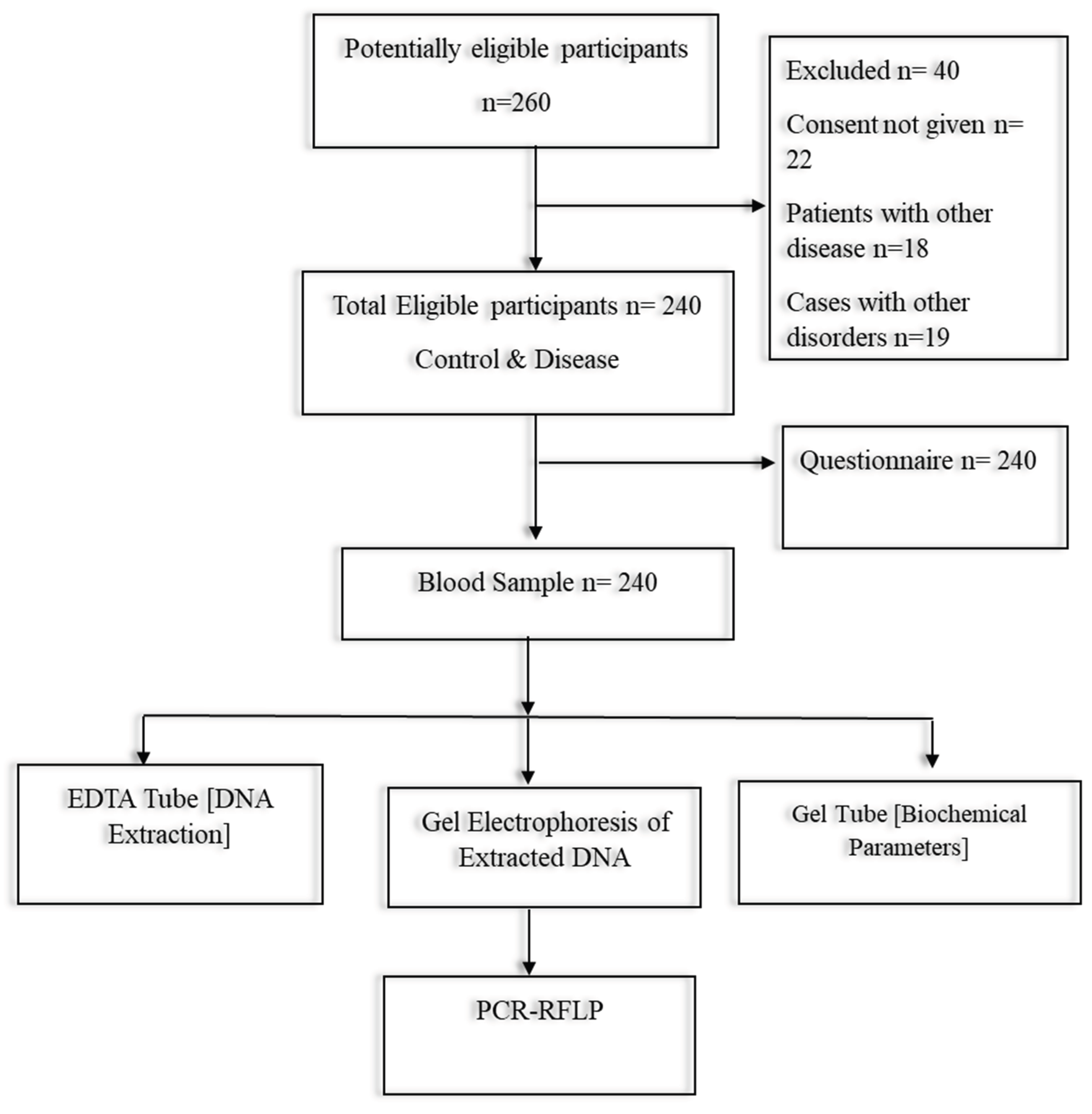
Figure 2.
Age of Studied RA Patients and Healthy Individuals (Control).
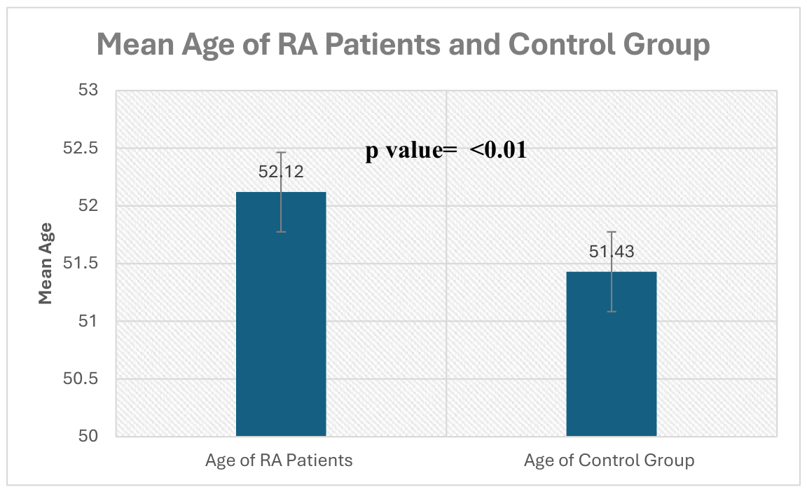
Figure 3.
Gender of Studied Participants among Both Groups.
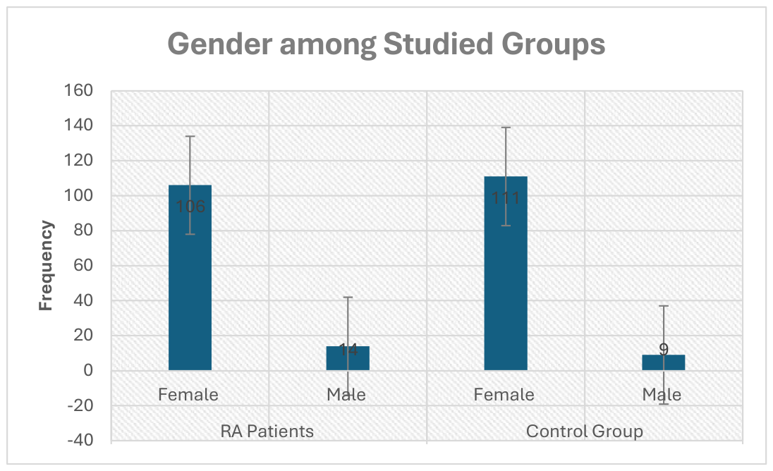
Figure 4.
Family History of Rheumatoid Arthritis among RA Patients.
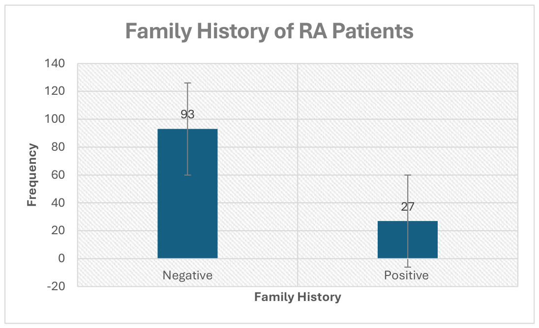
Figure 5.
Comparative Analysis of Systolic BP between the Control and Disease Groups (A) Comparative Analysis of Diastolic BP between the Control and Disease Groups (B) Comparative Analysis of BMI between the Control and Disease Groups (C) The graph displays the mean and standard deviation for each group. The p-value of Systolic BP, which is less than 0.01, indicates statistically significant differences between patients with RA and healthy individuals. Meanwhile, the results for diastolic BP and BMI showed no statistical significance, with p-values of 0.97 and 0.61, respectively.
Figure 5.
Comparative Analysis of Systolic BP between the Control and Disease Groups (A) Comparative Analysis of Diastolic BP between the Control and Disease Groups (B) Comparative Analysis of BMI between the Control and Disease Groups (C) The graph displays the mean and standard deviation for each group. The p-value of Systolic BP, which is less than 0.01, indicates statistically significant differences between patients with RA and healthy individuals. Meanwhile, the results for diastolic BP and BMI showed no statistical significance, with p-values of 0.97 and 0.61, respectively.
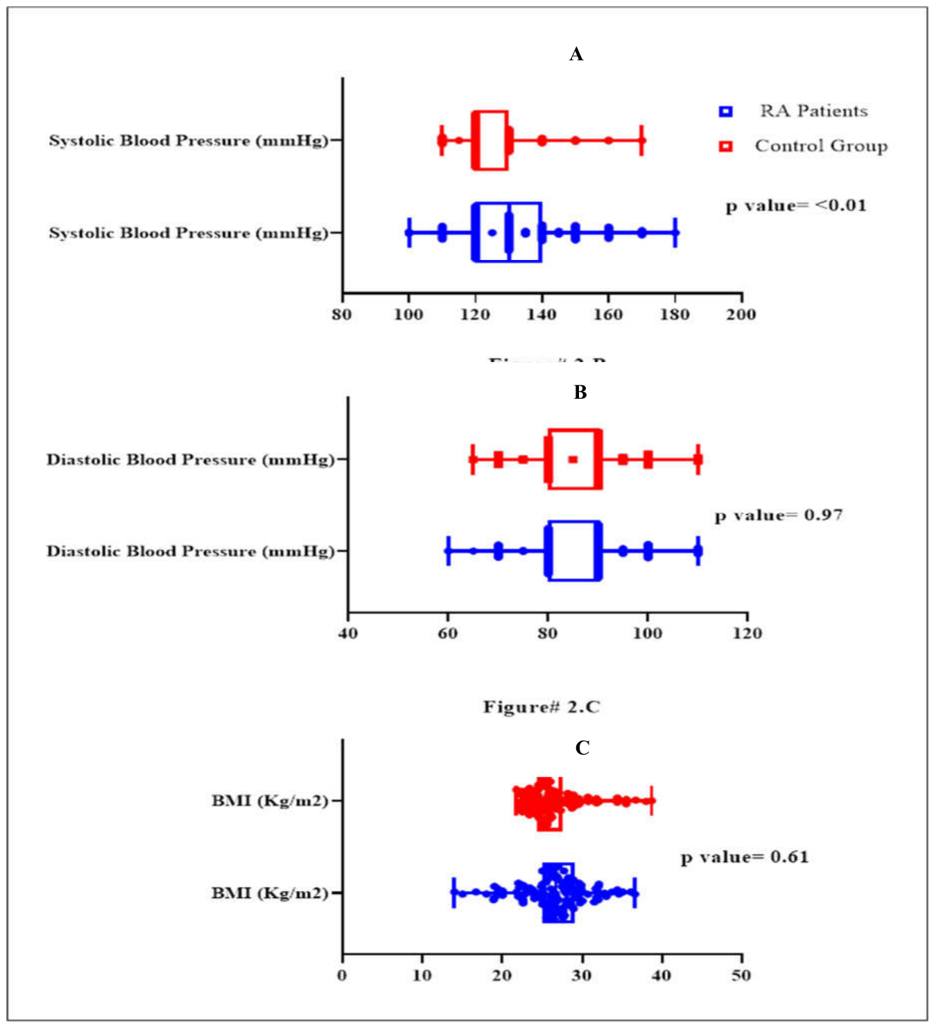
Figure 6.
Comparative Analysis of Total Cholesterol Levels among Control and Disease Group (A). Comparative Analysis of Triglycerides Levels among Control and Disease Group (B). Comparative Analysis of LDL Levels among Control and Disease Group (C). This graph showed the mean and standard deviation among groups, and a p-value <0.01 suggests statistically significant results of all parameters between RA Arthritis Patients and healthy Individuals.
Figure 6.
Comparative Analysis of Total Cholesterol Levels among Control and Disease Group (A). Comparative Analysis of Triglycerides Levels among Control and Disease Group (B). Comparative Analysis of LDL Levels among Control and Disease Group (C). This graph showed the mean and standard deviation among groups, and a p-value <0.01 suggests statistically significant results of all parameters between RA Arthritis Patients and healthy Individuals.
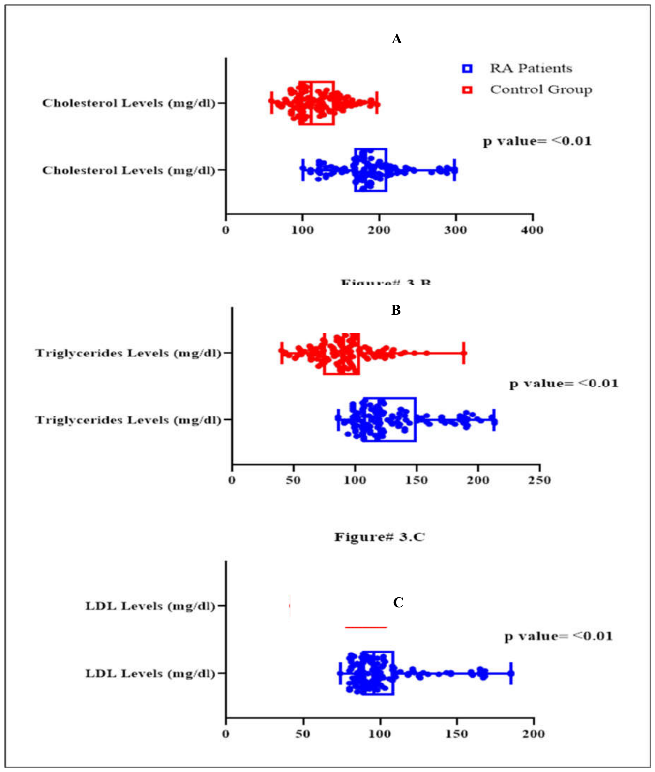
Figure 7.
Agarose 2% gel electrophoresis of PCR product IL-17RC for SNP rs708567 G/A. Tai1 digest at rs708567G/A. L is the ladder. In between the two ladders are restricted products.
Figure 7.
Agarose 2% gel electrophoresis of PCR product IL-17RC for SNP rs708567 G/A. Tai1 digest at rs708567G/A. L is the ladder. In between the two ladders are restricted products.
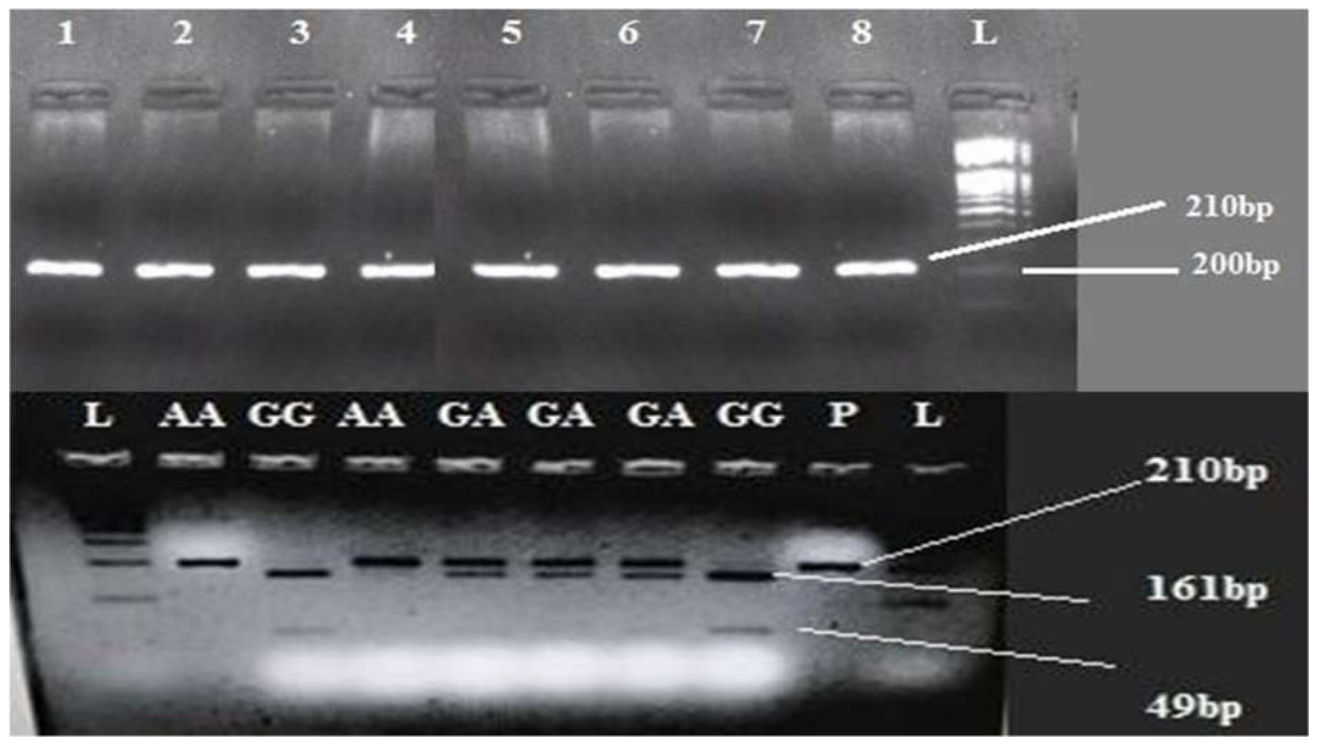
Figure 8.
Agarose 2% gel electrophoresis of PCR product IL-17RA for SNP rs2275913G/A. Tai1 digest at rs2275913G/A. L is the ladder. In between the two ladders are restricted products.
Figure 8.
Agarose 2% gel electrophoresis of PCR product IL-17RA for SNP rs2275913G/A. Tai1 digest at rs2275913G/A. L is the ladder. In between the two ladders are restricted products.
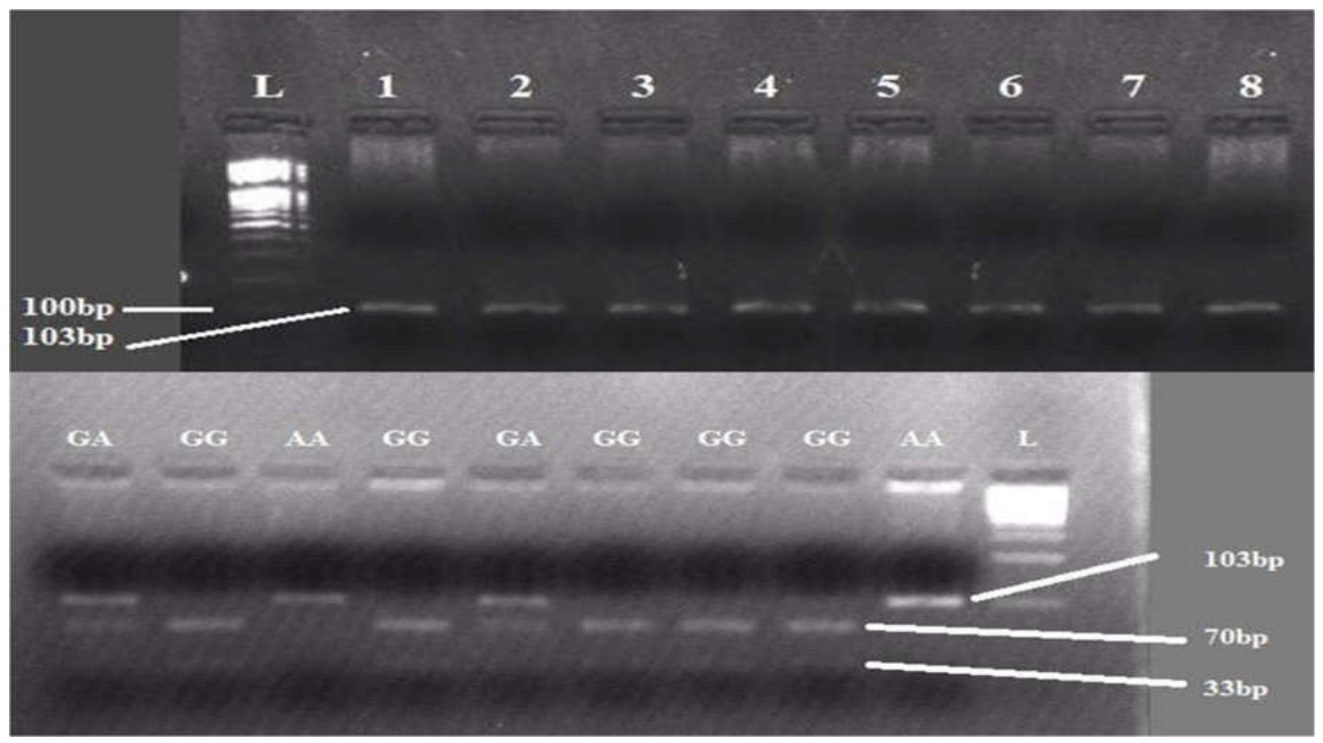

Table 1.
Primer details, PCR conditions, and restriction enzymes used for IL-17A and IL-17RC SNP genotyping by PCR-RFLP.
Table 1.
Primer details, PCR conditions, and restriction enzymes used for IL-17A and IL-17RC SNP genotyping by PCR-RFLP.
| Gene / SNP | Primer Sequence (5′→3′) | Product Size | Genotyping Method | Annealing Temperature (oC) | Restriction Enzyme | Fragment size |
| IL-17A (rs2275913G/A) |
F.P: 5’AACAAGTAAGAATGAAAAGAGGACATGGT3’ R.P: 5’CCCCCAATGAGGTCATAGAAGAATC3’ |
102 bp | PCR-RFLP | 60 | EcoNI | 161bp 49bp |
| IL-17RC (rs708567 G/A) |
F.P: 5’AGTAGGGTAGGCCTGGAAGG 3’ R.P: 5’CACTGGGAAGAGCCTGAAGA3’ |
233 bp | PCR-RFLP | 60 | HinfI | 70bp 33bp |
Table 2.
Descriptive and Comparative Analysis of Quantitative Continuous Variables.
| Variable Name | RA Patients | Control Group | p-value |
| BMI (Kg/m2) | 26.71 ± 4.14 | 26.45 ± 3.43 | 0.61 |
| Systolic BP (mmHg) | 131.96 ± 16.53 | 124.54 ± 10.31 | <0.01 |
| Diastolic BP (mmHg) | 86.50 ± 10.22 | 86.54 ± 9.25 | 0.97 |
| Total Cholesterol (mg/dL) | 187.61 ± 45.74 | 118.09 ± 31.02 | <0.01 |
| Triglycerides (mg/dL) | 130.93 ± 33.36 | 90.81 ± 24.99 | <0.01 |
| LDL (mg/dL) | 103.88 ± 25.11 | 89.98 ± 17.64 | <0.01 |
± = Means & Standard Deviation, RA= Rheumatoid Arthritis, BMI= Body Mass Index, LDL= Low Density Lipoprotein.
Table 3.
Distribution of IL-17RC and IL-17RA genotypes and allele frequencies among rheumatoid arthritis (RA) patients and healthy controls.
Table 3.
Distribution of IL-17RC and IL-17RA genotypes and allele frequencies among rheumatoid arthritis (RA) patients and healthy controls.
| Genotype | RA Patients (n=120) | Control Group (n=120) |
|---|---|---|
| IL-17 RC Genotype | ||
| GG | 41 (34.2%) | 75 (62.5%) |
| GA | 54 (45.0%) | 35 (29.2%) |
| AA | 25 (20.8%) | 10 (8.3%) |
| IL-17 RC Allele frequency | ||
| G Allele | 136 (56.7%) | 187 (77.9%) |
| A Allele | 104 (43.3%) | 53 (22.1%) |
| IL-17 RA Genotype | ||
| GG | 48 (40.0%) | 66 (55.0%) |
| GA | 51 (42.5%) | 38 (31.7%) |
| AA | 21 (17.5%) | 16 (13.3%) |
| IL-17 RA Allele frequency | ||
| G Allele | 147 (61.25%) | 170 (70.8%) |
| A Allele | 93 (38.75%) | 70 (29.2%) |
Table 4.
Allelic Association Analysis of SNP rs2275913.
| Chr | rs ID | Normal Allele | Risk Allele | Frequency in Cases |
Frequency in Controls |
χ² | P-value | (OR) |
|---|---|---|---|---|---|---|---|---|
| 6 | rs2275913 | G | A | 0.3667 | 0.2833 | 3.799 | 0.051 | 1.464 |
Table 5.
Allelic Association Analysis of SNP rs708567.
| Chr | rsID | Risk Allele |
Normal Allele |
Frequency in Cases |
Frequency in Controls |
χ² | p-value | (OR) | |
|---|---|---|---|---|---|---|---|---|---|
| 3 | rs708567 | A | G | 0.4375 | 0.2125 | 27.69 | 1.42 × 10⁻⁷ | 2.882 |
Table 6.
Association Analysis of SNP rs2275913 with Rheumatoid Arthritis under Different Genetic Models.
Table 6.
Association Analysis of SNP rs2275913 with Rheumatoid Arthritis under Different Genetic Models.
| Genetic Model | Case Group | Control Group | χ² | p-value |
|---|---|---|---|---|
| Genotypic (AA/GA/GG) | 18 / 52 / 50 | 15 / 38 / 67 | 4.921 | 0.085 |
| Trend (A vs. G) | 88 / 152 | 68 / 172 | 3.317 | 0.069 |
| Allelic (A vs. G) | 88 / 152 | 68 / 172 | 3.799 | 0.051 |
| Dominant (AA+GA vs. GG) | 70 / 50 | 53 / 67 | 4.820 | 0.028 |
| Recessive (AA vs. GA+GG) | 18 / 102 | 15 / 105 | 0.316 | 0.574 |
Table 7.
Association Analysis of IL-17RC rs708567 with Rheumatoid Arthritis under Different Genetic Models.
Table 7.
Association Analysis of IL-17RC rs708567 with Rheumatoid Arthritis under Different Genetic Models.
| Genetic Model | Case Group | Control Group | χ² | p-value |
|---|---|---|---|---|
| Genotypic (AA/GA/GG) | 26 / 53 / 41 | 8 / 35 / 77 | 24.19 | 5.58 × 10⁻⁶ |
| Trend (A vs. G) | 105 / 135 | 51 / 189 | 23.78 | 1.08 × 10⁻⁶ |
| Allelic (A vs. G) | 105 / 135 | 51 / 189 | 27.69 | 1.42 × 10⁻⁷ |
| Dominant (AA+GA vs. GG) | 79 / 41 | 43 / 77 | 21.61 | 3.35 × 10⁻⁶ |
| Recessive (AA vs. GA+GG) | 26 / 94 | 8 / 112 | 11.10 | 8.62 × 10⁻⁴ |
Table 8.
Hardy–Weinberg equilibrium (HWE) analysis for SNP rs2275913.
| Group | Genotype Counts (AA/GA/GG) |
Observed Heterozygosity |
Expected Heterozygosity |
HWE p-value |
|---|---|---|---|---|
| Overall | 33 / 90 / 117 | 0.3750 | 0.4388 | 0.02712 |
| Affected | 18 / 52 / 50 | 0.4333 | 0.4644 | 0.43910 |
| Unaffected | 15 / 38 / 67 | 0.3167 | 0.4061 | 0.02280 |
Table 9.
Hardy–Weinberg Equilibrium (HWE) Analysis of SNP rs708567.
| Group | Genotype Counts (AA/GA/GG) | Observed Heterozygosity | Expected Heterozygosity | HWE p-value |
|---|---|---|---|---|
| Overall | 34 / 88 / 118 | 0.3667 | 0.4388 | 0.0122 |
| Affected | 26 / 53 / 41 | 0.4417 | 0.4922 | 0.2685 |
| Unaffected | 8 / 35 / 77 | 0.2917 | 0.3347 | 0.1717 |
Disclaimer/Publisher’s Note: The statements, opinions and data contained in all publications are solely those of the individual author(s) and contributor(s) and not of MDPI and/or the editor(s). MDPI and/or the editor(s) disclaim responsibility for any injury to people or property resulting from any ideas, methods, instructions or products referred to in the content. |
© 2026 by the authors. Licensee MDPI, Basel, Switzerland. This article is an open access article distributed under the terms and conditions of the Creative Commons Attribution (CC BY) license.
Copyright: This open access article is published under a Creative Commons CC BY 4.0 license, which permit the free download, distribution, and reuse, provided that the author and preprint are cited in any reuse.