

Submitted:
15 September 2026
Posted:
17 September 2026
You are already at the latest version
Abstract
Background: The maternal microbiome represents the first microbial environment for the developing infant, with emerging evidence linking maternal dysbiosis to adverse pregnancy outcomes and lifelong offspring health. Aims: This review synthesizes evidence on the maternal-infant microbial continuum, examining microbial dynamics during pregnancy, vertical transmission pathways, infant microbiome trajectories, and intervention strategies. Key Findings: Dynamic changes in maternal gut, vaginal, and oral microbiomes during pregnancy influence fetal development through mechanisms including hormonal modulation (e.g., Clostridium innocuum-mediated 17β-estradiol degradation), immune programming via short-chain fatty acids, and barrier integrity maintenance. Delivery mode exerts persistent effects, with Cesarean-section associated with higher allergy risk (OR=1.5-2.2), while probiotic supplementation may mitigate this risk (RR=0.6-0.8). Conclusion: The maternal-infant microbiome continuum represents a critical therapeutic target. Standardized longitudinal research and personalized intervention strategies are urgently needed to translate mechanistic insights into clinical practice.
Keywords:
1. Introduction
1.1. The Maternal Microbiome as the First Environment:
1.2. Historical Underrepresentation of Women and Infants in Microbiome Research:
2. Rationale for Studying Maternal–Infant Microbial Crosstalk:
- a)
- Maternal and Obstetric Complications: Alterations in the maternal microbiota are critically linked to major adverse pregnancy outcomes, including Preterm Birth (PTB), Preeclampsia (PE), and Gestational Diabetes Mellitus (GDM). For instance, certain microbial features, such as the enrichment of the species Clostridium innocuum in the maternal gut during early pregnancy, have been identified as predictive biomarkers for a higher risk of PTB [13].
- b)
- Long-Term Offspring Disease Programming: The maternal microbial status influences the child’s susceptibility to chronic Non-Communicable Diseases (NCDs), including obesity, metabolic syndrome, diabetes, and allergies. Maternal dysbiosis is also recognized as a potential triggering factor in fetal neurodevelopment, being associated with neurodevelopmental disorders such as Autism Spectrum Disorder (ASD) and Attention-Deficit/Hyperactivity Disorder (ADHD) [16,18].
- c)
3. Scope of the Review: Highlighting the Maternal–Infant Microbial Continuum:
- a)
- Vertical Transmission and Delivery Mode: The microbial composition acquired by the infant is heavily influenced by the mode of delivery. Vaginal birth facilitates colonization by beneficial maternal microbes, while Caesarean section (CS) results in aberrant colonization, which is associated with increased disease risk [24].
- b)
- Maternal Exposome and Interventions: The maternal microbiota is shaped by external influences, referred to as the exposome. This includes diet (e.g., Mediterranean diet, high-fat diets), antibiotic use, and maternal stress. Interventions such as probiotic supplementation or vaginal seeding are explored for their potential to modulate microbial health in both mother and infant [7,23].
- c)
4. Pregnancy Microbiome Changes
4.1. Gut Microbiome Shifts: Metabolic Adaptations to Pregnancy:
4.2. Vaginal Microbiome: Dominance of Lactobacillus and Protective Role:
4.2.1. Dominance and Mechanism:
- Lactobacillus Dominance: The most notable change in the VMB is the shift toward a less diverse, more stable state dominated by Lactobacillus species. In nonpregnant women, the VMB is typically classified into Community State Types (CSTs), four of which are dominated by Lactobacillus spp. Such as L. crispatus, L. gasseri, L. iners and L. jensenii [39,44].
- Hormonal Driver: Elevated estrogen and progesterone levels during pregnancy stimulate vaginal epithelial maturation, leading to the accumulation of glycogen. Lactobacillus utilizes this glycogen via fermentation to produce lactic acid [45].
- Protective Function: Lactic acid maintains a low vaginal pH (typically acidic), which is essential for preserving reproductive health. This acidic environment reduces microbial diversity and inhibits the growth of pathogenic microorganisms. Furthermore, Lactobacillus spp. stimulate the production of anti-inflammatory cytokines, enhancing maternal–fetal protection [46,47].
4.2.2. Species Differences and Adverse Outcomes:
- Protective Species: The species Lactobacillus crispatus is highly represented during pregnancy and its dominance is associated with low infection risk and favourable pregnancy outcomes. A higher relative abundance of Lactobacillus in early pregnancy is associated with a greater likelihood of the pregnancy continuing beyond 38 weeks [48].
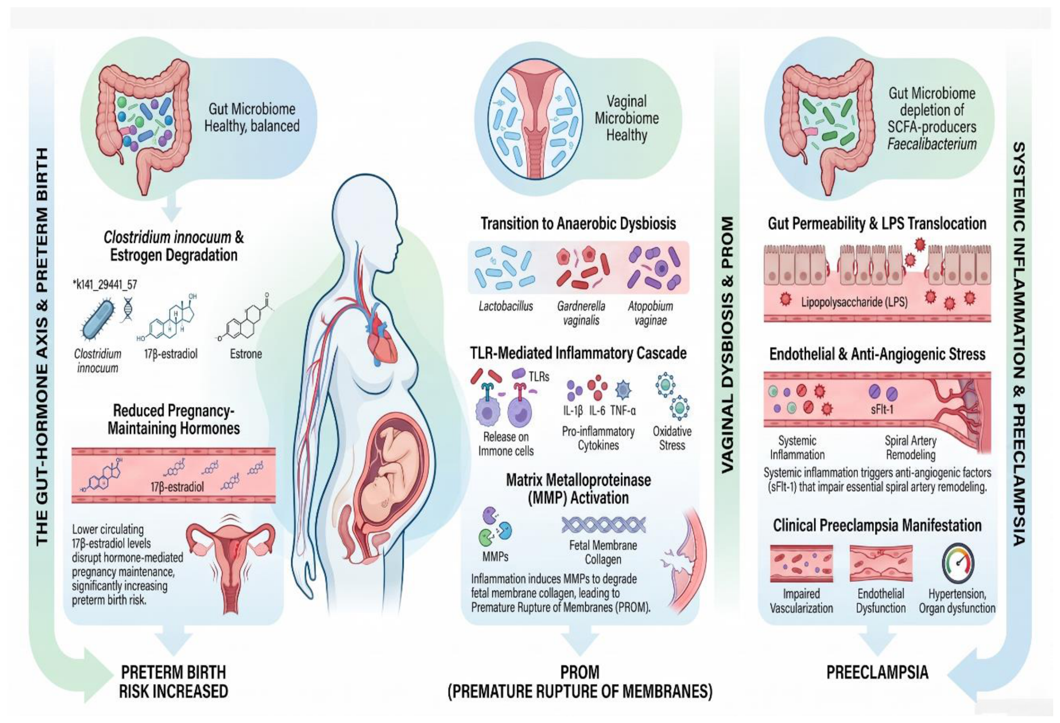
- Dysbiosis and Pathogenesis: When dysbiosis occurs, Lactobacillus abundance decreases, and the environment is taken over by facultative anaerobes such as Gardnerella vaginalis, Prevotella, Megasphaera, Sneathia, and Atopobium vaginae. This dysbiosis, known as Bacterial Vaginosis (BV), is linked to an increase in local innate immune system activation and inflammation [49], which may contribute to complications such as Preterm Birth (PTB) (Figure 2) and Premature Pre-Labor Rupture of Membranes (PPROM) [13,50].
4.3. Oral Microbiome Changes and Links to Adverse Pregnancy Outcomes
5. Transmission Pathways:
5.1. The Placental Microbiome: Resolving the Controversy
5.2. Vaginal Delivery vs. Caesarean Section: Microbial Seeding of the Neonate:
5.2.1. Vaginal Delivery (VB):
- Colonization Pattern: VB facilitates colonization by beneficial species such as Bifidobacterium, Lactobacillus, Escherichia coli, and Bacteroides. These Commensal bacteria play pivotal roles in fermenting Human Milk Oligosaccharides (HMOs) [42].
- Immune Impact: VB induces immediate activation of intestinal epithelial cells by bacterial lipopolysaccharides (LPS), which promotes immune tolerance, intestinal barrier maturation, and a more balanced Th1/Th2 immune response. Vaginally delivered infants have been shown to have a healthier and more diverse bifidogenic microbiota compared to Caesarean Section (CS)-delivered infants [58].
- Source Contribution: While it is often assumed that vaginal flora is the critical source, increasing evidence suggests that taxa from the mother’s rectum and fecally-contaminated perineal surface are a more important source of engrafting intestinal bacteria during vaginal birth than the vagina itself. In vaginally-born neonates, the gut colonization is predominantly contributed by the maternal rectal microbiota rather than the vaginal microbiota at one month of age. Shared ASVs (Amplicon Sequence Variants) between the neonate and maternal rectal swab often come from the genera Bacteroides and Bifidobacterium [2].
5.2.2. Caesarean Section (CS):
- Microbial Exposure: CS delivery bypasses exposure to the maternal vaginal and fecal microbiota, leading to the acquisition of a less diverse intestinal microbiome. Infants born via CS acquire microbiota dominated by environmental bacteria and nosocomial pathogens found in the hospital, such as Staphylococcus and Corynebacterium [24].
- Colonization Pattern: CS-born infants show delayed and altered colonization patterns. They have reduced colonization by Bifidobacterium and Bacteroides and are often dominated by opportunistic pathogenic species like Enterococcus, Staphylococcus, Streptococcus, Klebsiella, and Clostridium perfringens. The gut microbiota profile of CS-born neonates may be more similar to their mother’s skin communities [24,59].
- Immune Consequences: This altered colonization is associated with a reduced innate immune response, fewer regulatory T cells (Tregs), and a less diverse and more unstable microbiota. The limited microbial diversity in CS-born infants delays immune maturation.
- Persistent Effects: These changes in gut microbiota have been observed to persist for up to 1–2 years after birth. CS delivery is strongly associated with a significantly lower microbial diversity (Chao1 alpha diversity) on the neonatal skin compared to VB neonates. CS-delivered neonates also had a significantly higher relative abundance of the potential opportunistic pathogen Ureaplasma in their oral microbiome [60].
5.2.3. Intervention Strategies:
- Vaginal Seeding: This involves applying maternal vaginal fluids to the newborn immediately after CS. While this technique has shown promise in some small, controlled studies, sometimes improving gut microbiota, metabolome, and neurodevelopmental scores in actively treated infants, results have been inconsistent. Some research found that oral administration of maternal vaginal organisms did not modify the infants’ intestinal microbiome. Furthermore, vaginal seeding raises ethical and safety concerns regarding the potential transmission of pathogenic microorganisms [23].
- Fecal Microbiota Transplantation (FMT): Sincematter is a source of initial colonization in VB infants, FMT using the mother as a donor has been trailed for CS-born infants, showing that this approach can rapidly restore normal gut microbial development [59].
5.3. The Placental Microbiome: Evaluating the Evidence for in Utero Colonization
5.4. Human Milk Oligosaccharides (HMOs) as a Microbial and Immunological Bridge:
5.4.1. Microbial Source
- Diverse Community: Breast milk contains a diverse microbiota, with between 590 and 600 bacterial genera identified. The main bacterial groups include Firmicutes (Staphylococcus, Streptococcus, Lactobacillus, Bifidobacterium), Proteobacteria (Escherichia, Enterobacter), and Actinobacteria [68].
-
Origin Hypotheses: Four main hypotheses regarding the origin of breast milk bacteria exist [68]:
- Enteromammary Route: Bacteria from the maternal gut are transported by immune cells (macrophages, dendritic cells) to the mammary gland, supporting the presence of anaerobic bacteria like Bifidobacterium.
- Infant’s Oral Cavity: Retrograde flow due to the sucking motion carries bacteria from the infant’s mouth into the breast tissue, supported by the abundance of Streptococcus in both infant saliva and breast milk.
- Maternal Skin: Contamination from the skin, supported by the presence of skin-associated bacteria (Staphylococcus, Corynebacterium).
- Intrinsic Mammary Microbiota: The mammary gland may host its own stable microbiota [68].
- Impact on Infant Gut: It is estimated that between 10% and 27% of the infant’s gut microbiota originates from breast milk. Breastfed infants tend to have microbiota dominated by Bifidobacterium, promoting immune system maturation and potentially reducing the risk of chronic diseases like allergies and obesity later in life [68].
5.4.2. Immunological and Prebiotic Components
-
Human Milk Oligosaccharides (HMOs): human milk contains essential oligosaccharides (HMOs) that function as selective substrates (prebiotics) for beneficial bacteria, particularly Bifidobacterium species [68,69,70].
- HMOs promote the growth of specific bacteria (B. longum subsp. Infantis, B. bifidum) which produce Short-Chain Fatty Acids (SCFAs) like acetate and lactate, lowering intestinal pH and inhibiting pathogens [70].
- HMOs also act as decoy receptors for pathogens (e.g., Campylobacter jejuni, E. coli), preventing their adhesion to the gut epithelium, and modulate the expression of inflammatory genes, supporting immune defense [70].
- Maternal Factors: The composition of the human milk oligosaccharides (HMOs) microbiota is influenced by maternal factors, including gestational age (higher Bifidobacterium in term mothers), mode of delivery (VB linked to higher Bifidobacterium, Lactobacillus), maternal BMI, and antibiotic exposure (which reduces Bifidobacterium and Lactobacillus) [70].
5.5. Skin and Environmental Exposure Shaping Early Colonization:
- Skin-to-Skin Contact (Kangaroo Care): This practice promotes maternal and neonatal bonding and exerts a beneficial effect on the development of the gut microbiota. Kangaroo care increases vertical microbial transmission from mother to infant [71]. Skin-to-skin contact has been shown to improve the oral microbial composition, promoting the growth of Streptococcus and reducing pathogenic bacteria like E. coli.
- Environmental Exposure (Hospital/NICU): In CS-born infants, the initial microbial environment is often acquired from the general environment of the hospital [58]. In Neonatal Intensive Care Units (NICUs), the neonatal unit environment, including equipment and surfaces, influences the acquisition of bacteria by premature infants. Each NICU room has been shown to possess a unique microbial signature [58].
- Non-Maternal Human Contact: Horizontal transmission of microbial species from family members (e.g., fathers and siblings) is another significant source of colonization (Table 1), influencing the neonatal microbiome after the initial period. One study suggested that fathers introduced most new shared strains [54].
- Home/Farm Exposure: Direct exposure to microbes via hygienic factors or unique environments (e.g., soil, animals) contributes to the evolution of the child’s microbiome and long-term health. Children raised on farms, exposed to animals, showed reduced frequency of later allergies and inflammatory bowel disease [72].
6. Infant Microbiome Trajectories
6.1. Dynamics During the First 1,000 Days of Life:
6.1.1. Key Dynamics and Shifts:
-
Initial Colonization (Birth to Day 4): The initial microbial exposure occurs during birth, and the neonatal gut microbiome undergoes rapid changes immediately after birth [58].
- Alpha diversity (richness) may initially decline before increasing again. In one cohort, alpha diversity was significantly lower on Day 4 compared to Day 0 and Day 1.
- The bacterial composition changes significantly shortly after birth (Days 1, 4, and 1Month). Taxa such as Staphylococcus, Rothia, Streptococcus, and Haemophilus were observed to be higher around Day 4 than Day 1 [58].
-
Maturation Phase (Post-Weaning): The introduction of solid foods (weaning) at approximately four to six months of age alters the gut microbiota, moving it toward a more mature profile [74].
- This change is driven by exposure to dietary carbohydrates and glycans.
- The intestinal microbiome development in the first 2–3 years of life is considered the most critical phase [25].
6.1.2. Intervention Opportunities:
6.2. Factors Shaping Colonization: Feeding Mode, Antibiotic Exposure, Maternal Health
6.2.1. Mode of Delivery
-
Vaginal Birth (VB): Vaginally born neonates are exposed to maternal vaginal and rectal microbiota, facilitating colonization by beneficial, bifidogenic microbiota [69].
- VB infants show higher levels of Bifidobacterium, Lactobacillus, and Bacteroides.
- This physiological process promotes early immune tolerance and barrier maturation.
- In vaginally delivered neonates, the gut microbiome becomes significantly more similar to the maternal gut microbiome over time compared to the vaginal microbiome [69].
-
Caesarean Section (CS): CS delivery results in an aberrant intestinal microbial colonization and a less diverse intestinal microbiome [69].
- CS infants acquire microbiota dominated by environmental bacteria and opportunistic pathogens from the hospital setting, such as Staphylococcus, Enterococcus, Streptococcus, Klebsiella, and Clostridium perfringens [58].
- This altered colonization pattern, which may persist for up to 1–2 years after birth, is linked to long-term health problems such as allergies, asthma, obesity, and Type 1 diabetes [72].
-
Breastfeeding: Breastfed infants typically exhibit a healthier, diverse microbial composition [68].
- They have microbiota dominated by Bifidobacterium, Lactobacillus, and Bacteroides.
- Breast milk contains essential Human Milk Oligosaccharides (HMOs), which act as selective substrates (prebiotics) for beneficial bacteria, particularly Bifidobacterium species [70].
- Breastfed infants are 30–40% less likely to develop allergies compared with those fed formula [72].
-
Formula Feeding: Formula-fed infants develop a more diverse, adult-like microbiota earlier [69].
- They often show a higher prevalence of potentially pathogenic taxa, including Clostridium, Enterobacteriaceae, Streptococcus, and C. difficile, and low levels of Bifidobacterium [69].
- Supplementation of infant formulas with prebiotics like Galactooligosaccharides (GOS) and Fructooligosaccharides (FOS) can improve the infant gut microbiome to be more like breastfed infants [76].
6.2.3. Antibiotic Exposure
- Maternal/Perinatal Exposure: Antibiotics given during pregnancy significantly alter the gut and vaginal microbiome of the mother and, more importantly, lead to a sustained drop in abundance of Bifidobacterium and Bacteroidetes in the newborn [78].
-
Neonatal Exposure: Antibiotic administration in neonates induces dysbiosis, characterized by reduced bacterial diversity (reduced Shannon index/alpha diversity), decreased or absent Bifidobacteria species, and a higher abundance of pathogenic bacteria like Enterobacteriaceae [78].
- The damage to the intestinal microbiome from perinatal antibiotics can persist for at least one year of age, a critical duration for immune system development, and can result in future allergic and metabolic consequences.
- Each additional day of antibiotic exposure in newborn infants is associated with a reduction in obligate anaerobic bacterial load in stools [78].
- Increased Risk of Allergies: Maternal antibiotic use during pregnancy was strongly associated with an increased risk of allergies in the offspring (OR = 1.8–2.6) [12].
- Maternal Stress: Maternal psychosocial stress during pregnancy is associated with changes in gut microbiota diversity that may undermine neonatal immunity (). This stress has been associated with an increased chance of immune problems in babies (OR = 1.9–2.4) [12].
- Maternal Obesity: Maternal obesity is associated with alterations in infant gut microbiota and metabolism. Neonates born to obese mothers showed a higher relative abundance of opportunistic genera like Peptoniphilus on their skin and Bacteroides and Ruminococcus in their oral cavities, potentially increasing infection risk [79].
- Environmental Exposure: Factors like skin-to-skin contact (which promotes vertical microbial transmission) and exposure to the Neonatal Intensive Care Unit (NICU) environment (which can introduce unique microbial signatures from equipment and surfaces) significantly influence colonization, especially in premature infants [58,71].
- Protection Against Pathogens: The indigenous microbiota provides colonization resistance against enteric pathogens like Cryptosporidium parvum. The absence of a healthy diverse microbiota (dysbiosis) can lead to alteration of the immune system and the development of atopic disorders and food allergies [27].
- Metabolite Mediation: Maternal gut microbiota-derived metabolites, such as Short-Chain Fatty Acids (SCFAs), play an essential role in shaping the fetal immune system and neurological development. A reduction in SCFA availability due to dysbiosis is linked to enhanced immune reactivity and chronic inflammation [27].
- Vaccine Response: A healthy microbiome is important for the immune response to pathogens and to vaccines, which results in protective immunity. Higher abundance of Bifidobacterium infantis in the early months is associated with stronger CD4+ T-cell responses and more robust immune memory after vaccination [27].
-
Interventions to Support Immunity:
- Probiotic supplementation during pregnancy or early life is associated with a 20–40% reduction in allergy risk (RR = 0.6–0.8), suggesting that modulating the maternal gut microbiota confers long-term benefits to the infant immune system [20].
- Breastfeeding provides immune components (e.g., IgA) and beneficial bacteria that help build the baby’s immune system and prevent allergic reactions caused by inflammation [68].
7. Microbial Dysbiosis and Disease Links in Mother & Infabts
7.1. Maternal Dysbiosis: Preeclampsia, Gestational Diabetes, Preterm Birth:
7.2. Preeclampsia (PE)
-
Gut Dysbiosis and PE: Women with PE show gut dysbiosis, characterized by a reduction in beneficial bacteria, such as Faecalibacterium and Akkermansia, and an increase in opportunistic pathogens, such as Fusobacterium and Veillonella [51].
- This dysbiosis reduces the production of Short-Chain Fatty Acids (SCFAs), which typically suppress inflammation and support gut barrier integrity.
- Dysbiosis also contributes to elevated circulating levels of lipopolysaccharides (LPS) from Gram-negative bacteria and Trimethylamine-N-oxide (TMAO). TMAO exacerbates endothelial dysfunction and intensifies gestational hypertension [28].
- Vaginal and Oral Links to PE: Vaginal dysbiosis (loss of Lactobacillus) promotes local inflammation, increasing the risk of placental dysfunction [49]. Furthermore, oral dysbiosis and associated periodontal disease have been plausibly linked to PE pathogenesis, potentially through the translocation of oral bacteria like Prevotella, Porphyromonas, and Dialister to the placenta [52,53].
7.3. Gestational Diabetes Mellitus (GDM) and Insulin Resistance
-
Gut Dysbiosis in GDM: Women with GDM exhibit a significant reduction in microbial diversity compared to healthy women, displaying a pro-inflammatory bacterial profile [15]. There is often an increase in taxa associated with inflammation and insulin resistance, such as Firmicutes and Proteobacteria, and a decrease in beneficial taxa like Bacteroidetes.
7.4. Preterm Birth (PTB)
- Vaginal Dysbiosis and PTB: Alterations in the vaginal microbiota (VMB), particularly a loss of Lactobacillus species and an increase in opportunistic pathogens (e.g., Gardnerella vaginalis, Prevotella, Sneathia, and Atopobium vaginae), are strongly associated with PTB and PPROM [13].
- a. A significant decrease in the abundance of Lactobacillus crispatus in the VMB during the last trimester may be linked to preterm delivery [13]. Conversely, a higher relative abundance of Lactobacillus in early pregnancy is associated with a greater likelihood of pregnancy continuation beyond 38 weeks [48].
- Gut Dysbiosis and PTB: Maternal gut dysbiosis in early pregnancy can predict PTB. Notably, the genus Clostridium innocuum was identified as a predictive marker for PTB across independent cohorts. C. innocuum is known to degrade 17β-estradiol in vitro and in vivo, suggesting that early-pregnancy microbial dysbiosis may initiate hormonal dysbiosiss that trigger preterm labor [39].
7.5. Infant Dysbiosis as Disorders:
8. Mechanistic Pathways Linking Dysbiosis to Pathology
- Hormonal Metabolism: Certain pathogenic bacteria can directly interfere with host endocrinology. For example, Clostridium innocuum degrades 17β-estradiol, potentially disrupting the hormonal balance necessary for maintaining pregnancy and increasing PTB risk [4].
- Immunomodulation & Barrier Integrity: Commensal bacteria and their metabolites are essential for immune education and barrier function. Short-chain fatty acids (SCFAs) promote regulatory T cell differentiation and anti-inflammatory cytokine production [27]. Conversely, reduced SCFA availability impairs gut barrier integrity, leading to inflammation. Vaginal Lactobacillus spp. maintain a low pH and produce lactic acid, protecting against ascending infections [13].
- Microbial Metabolite Signaling: The gut microbiota modulates host metabolism via bile acid transformation. Dysbiosis-induced changes in bile acid pools can affect host metabolism and pathogen susceptibility. For instance, Veillonella parvula suppresses the bile acid transporter ASBT, leading to bile acid accumulation and C. difficile spore germination [81].
- Pathogen Exclusion & Competition: A healthy, diverse microbiota provides colonization resistance against pathogens. Bifidobacterium species, promoted by human milk oligosaccharides (HMOs), outcompete pathogens for resources and produce inhibitory metabolites [70].
8.1. Clinical Implications and Its Gaps
8.1.1. Perspectives for Stakeholders for Clinicians and Healthcare Providers:
- Postnatal Support: Actively promote and support exclusive breastfeeding for at least 6 months, given its proven benefits for microbiome and immune development.
8.1.2. For Researchers:
8.1.3. For Policy Makers and Public Health:
- Guideline Development: Support the development of evidence-based guidelines for probiotic use in pregnancy and infancy [10].
- Education Initiatives: Fund public health campaigns about the importance of early-life microbiome health [32].
- Research Investment: Prioritize funding for longitudinal mother-infant cohort studies across diverse populations [10].
8.2. Gaps and Challenges in Translating Interventions to Clinical Practice:
- Causality vs. Association: Most studies are observational, limiting the ability to establish definitive causal relationships between dysbiosis and pregnancy-related diseases [32].
- Heterogeneity and Standardization: Current studies show significant heterogeneity in design, methodologies, sample size, and outcomes, making it difficult to draw definitive conclusions. There is a critical need for standardization in sampling, analysis, and data reporting methodologies [32].
- Intervention Optimization: Key gaps remain in identifying the optimal probiotic strains, dosages, and timing for interventions. Probiotic efficacy is highly variable, and the unregulated market poses safety concerns [82].
- Underrepresentation and Generalizability: The underrepresentation of diverse populations in current research limits the generalizability of findings regarding microbial composition and disease risk [11].
- Lack of Longitudinal RCTs: There is a lack of large-scale, well-designed Randomized Controlled Trials (RCTs) to validate the safety and efficacy of microbiota-based interventions across diverse populations.
- Complex Interactions: Future research must address the intricate interplay between microbiota, genetics, psychosocial stress, immune profile, and placental epigenetics [30].
9. Interventions and Preventive Strategies
9.1. Maternal Diet and Nutritional Interventions
-
Mediterranean Dietary Pattern (MD): The MD, characterized by high consumption of fiber, fruits, vegetables, and unsaturated fats, is associated with greater bacterial diversity and a protective effect against disease risk [7].
- An MD-based counselling intervention during pregnancy significantly influenced the overall structure of the maternal gut microbiota.
- MD adherence was associated with an increase in gut microbial diversity and the enrichment of beneficial genera like Blautia, Faecalibacterium, and Christensenella[7].
- MD promotes SCFA production, enhances intestinal barrier integrity, and reduces systemic inflammation, mitigating the release of proinflammatory cytokines [29].
- Stress Reduction (SR): A structured Stress Reduction program during pregnancy showed a significant link with the maternal gut microbiota composition, modulating the relative abundance of some key health-associated genera. High maternal stress was associated with lower relative abundance of SCFA producers like Faecalibacterium [53].
9.2. Probiotics, Prebiotics, and Synbiotics in Pregnancy and Infancy:
-
Probiotics in Pregnancy: Probiotic supplementation during pregnancy may be associated with a reduced risk of GDM and other complications.
- Probiotic administration has been shown to improve glucose and lipid metabolism in pregnant women and may reduce allergic symptoms in newborns [20].
- Probiotics can improve diversity of the intestinal or vaginal microbiome and insulin sensitivity, and reduce inflammation, as seen in studies using multi-strain products [20].
- Some studies, however, report mixed results regarding their impact on GDM prevention in overweight/obese women [31].
- Prebiotics in Pregnancy and Infancy: Prebiotics, such as fructans (FOS and inulin) and Galactooligosaccharides (GOS), are soluble fibers that selectively promote the growth of beneficial bacteria, such as those that produce SCFAs. Prebiotic supplementation during pregnancy modified the gut microbiota and increased metabolites in amniotic fluid, driving a tolerogenic environment in utero [76].
- Vaginal Modulation: Therapeutic strategies utilizing oral or local probiotics (e.g., Lactobacillus crispatus and L. rhamnosus) are being explored to modulate vaginal microbiota, particularly when dysbiosis is present (Table 3). This dual approach may reduce proinflammatory markers and restore microbial balance, diminishing the risk of reinfection and inflammatory flare-ups [40].
9.3. Microbiome Restoration Strategies:
- Vaginal Seeding: This involves exposing Caesarean Section (CS)-born neonates to maternal vaginal microbiota to partially restore physiological colonization [23]. While studies are small and results mixed, some have shown that vaginal microbiota transfer may improve the neurodevelopmental scores and microbiome composition in CS-born infants [67]. However, oral administration of maternal vaginal organisms has also been shown not to modify the infants’ intestinal microbiome in some trials [23].
- Fecal Microbiota Transplantation (FMT): FMT involves harnessing fecal-derived microbiota to reverse dysbiosis. FMT has been successfully used to treat recurrent C. difficile infection (CDI) in pregnant women, resulting in engraftment of the donor microbiota which was later transferred to the infant during vaginal delivery. This technique is typically limited to research centers as part of IRB-approved clinical trials [58].
- Exclusive Breastfeeding: Breast milk remains the gold standard for promoting healthy gut microbiota development. It contains prebiotics (HMOs) and live probiotic bacteria that shape the infant gut microbiota and support immune development. This influence may reduce the risk of chronic diseases such as allergies, obesity, and diabetes later in life [68].
- Donor Milk (DM) Modulation: DM, often pasteurized, loses some probiotic benefits. Future strategies should consider personalization of DM by adding components of mother’s own milk or specific human milk components to improve its quality [68]. Research indicates that the use of prebiotics, probiotic strains, and synbiotics in formula milk holds the potential to influence the establishment and development of infant gut microbiota when human milk is unavailable [66].
Author Contributions
Funding
Institutional Review Board Statement
Informed Consent Statement
Data Availability Statement
Acknowledgments
Conflicts of Interest
References
- Fasano, A.; Chassaing, B.; Haller, D.; Flores Ventura, E.; Carmen-Collado, M.; Pastor, N.; ...; Berni Canani, R. Microbiota during pregnancy and early life: role in maternal−neonatal outcomes based on human evidence. Gut Microbes 2024, 16(1), 2392009. [Google Scholar] [CrossRef] [PubMed]
- Xiao, L.; Zhao, F. Microbial transmission, colonisation and succession: from pregnancy to infancy. Gut 2023, 72(4), 772–786. [Google Scholar] [CrossRef] [PubMed]
- Koren, O.; Konnikova, L.; Brodin, P.; Mysorekar, I. U.; Collado, M. C. The maternal gut microbiome in pregnancy: implications for the developing immune system. Nat. Rev. Gastroenterol. Hepatol. 2024, 21(1), 35–45. [Google Scholar] [CrossRef] [PubMed]
- Biagioli, V.; Matera, M.; Ramenghi, L. A.; Falsaperla, R.; Striano, P. Microbiome and Pregnancy Dysbiosis: A Narrative Review on Offspring Health. Nutrients 2025, 17(6), 1033. [Google Scholar] [CrossRef] [PubMed]
- Ibarra, A. A. M.; Hernández, L. F. B.; Gregorio, E. V.; Aguirre, F. J. O.; Vargas, J. T. D. J. Q. Microbiota during pregnancy, childbirth, and postpartum, and its relationship with health and disease states. Med. Microecol. 2025, 100141. [Google Scholar] [CrossRef]
- Heidrich, V.; Valles-Colomer, M.; Segata, N. Human microbiome acquisition and transmission. Nat. Rev. Microbiol. 2025, 1–17. [Google Scholar] [CrossRef]
- Flores Ventura, E.; Lane, J. A.; Turjeman, S.; Vidra, N.; Weiss, G. A.; Gross, G.; Chang, C.Y.; Koren, O. ILSI Europe perspective review: site-specific microbiota changes during pregnancy associated with biological consequences and clinical outcomes: opportunities for probiotic interventions. Gut Microbes 2025, 17(1), 2501186. [Google Scholar] [CrossRef]
- Banchi, P.; Colitti, B.; Opsomer, G.; Rota, A.; Van Soom, A. The dogma of the sterile uterus revisited: does microbial seeding occur during fetal life in humans and animals? Reproduction 2024, 167(1). [Google Scholar] [CrossRef] [PubMed]
- de Goffau, M. C.; Lager, S.; Sovio, U.; Gaccioli, F.; Cook, E.; Peacock, S. J.; ...; Charnock-Jones, D. S. Human placenta has no microbiome but can contain potential pathogens. Nature 2019, 572(7769), 329–334. [Google Scholar] [CrossRef] [PubMed]
- Metwaly, A.; Kriaa, A.; Hassani, Z.; Carraturo, F.; Druart, C.; Arnauts, K.; ...; Haller, D. A Consensus Statement on establishing causality, therapeutic applications and the use of preclinical models in microbiome research. Nat. Rev. Gastroenterol. Hepatol. 2025, 22(5), 343–356. [Google Scholar] [CrossRef] [PubMed]
- Stupak, A.; Kwaśniewski, W. Evaluating current molecular techniques and evidence in assessing microbiome in placenta-related health and disorders in pregnancy. Biomolecules 2023, 13(6), 911. [Google Scholar] [CrossRef] [PubMed]
- Al Jehani, A.N.; Shuaib, M.; Alsharif, A.; Alsubaie, K.A.; Khraisat, A.; Alsharif, A.; Altaf, M.; Almasry, R.H.; Kayali, A.M.; Abdallah, S.A. Impact of Maternal Microbiota Composition on Neonatal Immunity and Early Childhood Allergies: A Systematic Review. Pediatr. Rep. 2025, 17(3), 67. [Google Scholar] [CrossRef] [PubMed]
- Li, H.; Han, M.; Xu, J.; Li, N.; Cui, H. The vaginal microbial signatures of preterm birth woman. BMC Pregnancy Childbirth 2024, 24(1), 428. [Google Scholar] [CrossRef] [PubMed]
- Jordan, M. M.; Amabebe, E.; Khanipov, K.; Taylor, B. D. Scoping review of microbiota dysbiosis and risk of preeclampsia. Am. J. Reprod. Immunol. 2024, 92(4), e70003. [Google Scholar] [CrossRef] [PubMed]
- Li, J.; Wang, M.; Ma, S.; Jin, Z.; Yin, H.; Yang, S. Association of gastrointestinal microbiome and obesity with gestational diabetes mellitus-an updated globally based review of the high-quality literatures. Nutr. Diabetes 2024, 14(1), 31. [Google Scholar] [CrossRef] [PubMed]
- Borrego-Ruiz, A.; Borrego, J. J. Neurodevelopmental disorders associated with gut microbiome dysbiosis in children. Children 2024, 11(7), 796. [Google Scholar] [CrossRef]
- Pattaroni, C.; Marsland, B.J.; Harris, N.L. Early-Life Host–Microbial Interactions and Asthma Development: A Lifelong Impact? Immunol. Rev. 2025, 330(1), e70019. [Google Scholar] [CrossRef] [PubMed]
- Chandra, M. Developmental Origins of Non-Communicable Chronic Diseases: Role of Fetal Undernutrition and Gut Dysbiosis in Infancy. Children 2024, 11(11), 1387. [Google Scholar] [CrossRef] [PubMed]
- Shukla, V.; Singh, S.; Verma, S.; Verma, S.; Rizvi, A. A.; Abbas, M. Targeting the microbiome to improve human health with the approach of personalized medicine: Latest aspects and current updates. Clin. Nutr. ESPEN 2024, 63, 813–820. [Google Scholar] [CrossRef] [PubMed]
- Jiang, L.; Zhang, L.; Xia, J.; Cheng, L.; Chen, G.; Wang, J.; Raghavan, V. Probiotics supplementation during pregnancy or infancy on multiple food allergies and gut microbiota: a systematic review and meta-analysis. Nutr. Rev. 2025, 83(2), e25–e41. [Google Scholar] [CrossRef] [PubMed]
- Eslami, M.; Naderian, R.; Ahmadpour, A.; Shushtari, A.; Maleki, S.; Mohammadian, P.; ...; Yousefi, B. Microbiome structure in healthy and pregnant women and importance of vaginal dysbiosis in spontaneous abortion. Front. Cell. Infect. Microbiol. 2025, 14, 1401610. [Google Scholar] [CrossRef] [PubMed]
- Rus, M. J.; Sauco Carballo, C.; Faria, F. D.; Simon-Soro, A. Hormonal Environment Shapes the Oral Microbiome. Oral Microbiome: Symbiosis, Dysbiosis and Microbiome Interventions for Maintaining Oral and Systemic Health 2025, 225–242. [Google Scholar] [CrossRef] [PubMed]
- LaPoint, P.; Banks, K.; Bacorn, M.; Prasad, R.; Romero-Soto, H.N.; Namasivayam, S.; Chen, Q.; Patel, A.; Levy, S.; Hourigan, S.K. Can Vaginal Seeding at Birth Improve Health Outcomes of Cesarean Section-Delivered Infants? A Scoping Review. Microorganisms 2025, 13(6), 1236. [Google Scholar] [CrossRef] [PubMed]
- Inchingolo, F.; Inchingolo, A.D.; Palumbo, I.; Trilli, I.; Guglielmo, M.; Mancini, A.; Palermo, A.; Inchingolo, A.M.; Dipalma, G. The impact of cesarean section delivery on intestinal microbiota: mechanisms, consequences, and perspectives a systematic review. Int. J. Mol. Sci. 2024, 25(2), 1055. [Google Scholar] [CrossRef] [PubMed]
- Pantazi, A.C.; Balasa, A.L.; Mihai, C.M.; Chisnoiu, T.; Lupu, V.V.; Kassim, M.A.K.; Mihai, L.; Frecus, C.E.; Chirila, S.I.; Lupu, A.; Andrusca, A. Development of gut microbiota in the first 1000 days after birth and potential interventions. Nutrients 2023, 15(16), 3647. [Google Scholar] [CrossRef] [PubMed]
- Zhu, J.; He, M.; Li, S.; Lei, Y.; Xiang, X.; Guo, Z.; Wang, Q. Shaping oral and intestinal microbiota and the immune system during the first 1,000 days of life. Front. Pediatr. 2025, 13, 1471743. [Google Scholar] [CrossRef] [PubMed]
- Dera, N.; Kosińska-Kaczyńska, K.; Żeber-Lubecka, N.; Brawura-Biskupski-Samaha, R.; Massalska, D.; Szymusik, I.; Dera, K.; Ciebiera, M. Impact of early-life microbiota on immune system development and allergic disorders. Biomolecules 2025, 13(1), 121. [Google Scholar] [CrossRef] [PubMed]
- Mohsenzadeh, A.; Pourasgar, S.; Mohammadi, A.; Nazari, M.; Nematollahi, S.; Karimi, Y.; Firoozbakhsh, P.; Mohsenzadeh, H.; Kamali, K.; Elahi, R. The gut microbiota and cardiovascular disease: Exploring the role of microbial dysbiosis and metabolites in pathogenesis and therapeutics. Life Sci. 2025, 123981. [Google Scholar] [CrossRef] [PubMed]
- Di Vincenzo, F.; Del Gaudio, A.; Petito, V.; Lopetuso, L.R.; Scaldaferri, F. Gut microbiota, intestinal permeability, and systemic inflammation: a narrative review. Intern. Emerg. Med. 2024, 19(2), 275–293. [Google Scholar] [CrossRef] [PubMed]
- Mostafavi Abdolmaleky, H.; Zhou, J.R. Gut microbiota dysbiosis, oxidative stress, inflammation, and epigenetic alterations in metabolic diseases. Antioxidants 2024, 13(8), 985. [Google Scholar] [CrossRef] [PubMed]
- Mora-Janiszewska, O.; Bialic, K.; Faryniak, A.; Darmochwał-Kolarz, D. A Review of Modulation of Gut Microbiota to Mitigate Gestational Diabetes: Implications for Maternal and Child Health. Med. Sci. Monit. 2025, 31, e948897. [Google Scholar] [CrossRef] [PubMed]
- Schoultz, I.; Claesson, M.J.; Dominguez-Bello, M.G.; Fåk Hållenius, F.; Konturek, P.; Korpela, K.; Laursen, M.F.; Penders, J.; Roager, H.; Vatanen, T.; Öhman, L. Gut microbiota development across the lifespan: Disease links and health-promoting interventions. J. Intern. Med. 2025, 297(6), 560–583. [Google Scholar] [CrossRef] [PubMed]
- Olate, P.; Martínez, A.; Sans-Serramitjana, E.; Cortés, M.; Díaz, R.; Hernández, G.; ...; Quiñones, J. The Infant Oral Microbiome: Developmental Dynamics, Modulating Factors, and Implications for Oral and Systemic Health. Int. J. Mol. Sci. 2025, 26(16), 7983. [Google Scholar] [CrossRef] [PubMed]
- Escorcia Mora, P.; Valbuena, D.; Diez-Juan, A. The Role of the Gut Microbiota in Female Reproductive and Gynecological Health: Insights into Endometrial Signaling Pathways. Life 2025, 15(5), 762. [Google Scholar] [CrossRef] [PubMed]
- Adamczak, A.M.; Werblińska, A.; Jamka, M.; Walkowiak, J. Maternal-Foetal/Infant Interactions Gut Microbiota and Immune Health. Biomedicines 2024, 12(3), 490. [Google Scholar] [CrossRef] [PubMed]
- Hong, J.Y.; Medzhitov, R. On developmental programming of the immune system. Trends Immunol. 2023, 44(11), 877–889. [Google Scholar] [CrossRef] [PubMed]
- Law, S. R.; Mathes, F.; Paten, A. M.; Alexandre, P. A.; Regmi, R.; Reid, C.; ...; Gupta, V. V. Life at the borderlands: microbiomes of interfaces critical to One Health. FEMS Microbiol. Rev. 2024, 48(2), fuae008. [Google Scholar] [CrossRef] [PubMed]
- Xie, Z.; Chen, Z.; Chai, Y.; Yao, W.; Ma, G. Unveiling the placental bacterial microbiota: Implications for maternal and infant health. Front. Physiol. 2025, 16, 1544216. [Google Scholar] [CrossRef] [PubMed]
- Romero, R.; Theis, K.R.; Gomez-Lopez, N.; Winters, A.D.; Panzer, J.J.; Lin, H.; Galaz, J.; Greenberg, J.M.; Shaffer, Z.; Kracht, D.J.; Chaiworapongsa, T. The vaginal microbiota of pregnant women varies with gestational age, maternal age, and parity. Microbiol. Spectr. 2023, 11(4), e03429–22. [Google Scholar] [CrossRef] [PubMed]
- Corbett, G.A.; Corcoran, S.; Feehily, C.; Soldati, B.; Rafferty, A.; Macintyre, D.A.; Cotter, P.D.; Mcauliffe, F.M. Preterm-birth-prevention with Lactobacillus crispatus oral probiotics: Protocol for a double blinded randomised placebo-controlled trial (the PrePOP study). Contemp. Clin. Trials 2025, 149, 107776. [Google Scholar] [CrossRef] [PubMed]
- Samarra, A.; Alcañiz, A. J.; Martínez-Costa, C.; Marina, A.; Comas, I.; Segata, N.; ...; Collado, M. C. Breastfeeding and early Bifidobacterium-driven microbial colonization shape the infant gut resistome. Nat. Commun. 2025, 16(1), 6099. [Google Scholar] [CrossRef] [PubMed]
- Lordan, C.; Roche, A.K.; Delsing, D.; Nauta, A.; Groeneveld, A.; MacSharry, J.; Cotter, P.D.; van Sinderen, D. Linking human milk oligosaccharide metabolism and early life gut microbiota: bifidobacteria and beyond. Microbiol. Mol. Biol. Rev. 2024, 88(1), e00094–23. [Google Scholar] [CrossRef] [PubMed]
- Enache, R.M.; Roşu, O.A.; Profir, M.; Pavelescu, L.A.; Creţoiu, S.M.; Gaspar, B.S. Correlations Between Gut Microbiota Composition, Medical Nutrition Therapy, and Insulin Resistance in Pregnancy A Narrative Review. Int. J. Mol. Sci. 2025, 26(3), 1372. [Google Scholar] [CrossRef] [PubMed]
- Alizhan, D.; Ukybassova, T.; Bapayeva, G.; Aimagambetova, G.; Kongrtay, K.; Kamzayeva, N.; Terzic, M. Cervicovaginal microbiome: Physiology, age-related changes, and protective role against human papillomavirus infection. J. Clin. Med. 2025, 14(5), 1521. [Google Scholar] [CrossRef] [PubMed]
- Xu, H.; Zhang, S.; Zhang, B.; Jiang, N.; Xu, Y.; Chen, X.; Han, L. Vaginal colonization of Lactobacilli: Mechanism and function. Microb. Pathog. 2025, 198, 107141. [Google Scholar] [CrossRef] [PubMed]
- Avitabile, E.; Menotti, L.; Croatti, V.; Giordani, B.; Parolin, C.; Vitali, B. Protective mechanisms of vaginal lactobacilli against sexually transmitted viral infections. Int. J. Mol. Sci. 2024, 25(17), 9168. [Google Scholar] [CrossRef] [PubMed]
- Das, S.; Bhattacharjee, M.J.; Mukherjee, A.K.; Khan, M.R. Recent advances in understanding of multifaceted changes in the vaginal microenvironment: implications in vaginal health and therapeutics. Crit. Rev. Microbiol. 2023, 49(2), 256–282. [Google Scholar] [CrossRef] [PubMed]
- Oguri, N.; Kobayashi, C.; Ozawa, Y.; Kimura, T.; Nishinarita, Y.; Wada, H.; ...; Hisamatsu, T. Vaginal Lactobacillus crispatus in early pregnancy associates with favorable gestational outcomes in a Japanese maternal-neonatal microbiome cohort. Nat. Commun. 2025, 16(1), 8032. [Google Scholar] [CrossRef] [PubMed]
- Sethi, N.; Narayanan, V.; Saaid, R.; Ahmad Adlan, A.S.; Ngoi, S.T.; Teh, C.S.J.; Hamidi, M.; WHOW research group. Prevalence, risk factors, and adverse outcomes of bacterial vaginosis among pregnant women: a systematic review. BMC Pregnancy Childbirth 2025, 25(1), 40. [Google Scholar] [CrossRef] [PubMed]
- Borrego-Ruiz, A.; Borrego, J. J. Microbial Pathogens Linked to Vaginal Microbiome Dysbiosis and Therapeutic Tools for Their Treatment. Acta Microbiol. Hell. 2025, 70(2), 19. [Google Scholar] [CrossRef]
- Gupte, S.; Purandare, C.N.; Nazareth, A.K.; Chatla, D.; Deulkar, M.G.; Kirdat, K.; Arora, P.; Shah, S. The role of gut and vaginal microbiomes in reproductive health: Implications for PCOS, endometriosis, pre-eclampsia, and beyond. Int. J. Gynecol. Obstet. 2025. [Google Scholar] [CrossRef] [PubMed]
- Marcickiewicz, J.; Jamka, M.; Walkowiak, J. A Potential Link Between Oral Microbiota and Female Reproductive Health. Microorganisms 2025, 13(3), 619. [Google Scholar] [CrossRef] [PubMed]
- Alex, A.M.; Levendosky, A.A.; Bogat, G.A.; Muzik, M.; Nuttall, A.K.; Knickmeyer, R.C.; Lonstein, J.S. Stress and mental health symptoms in early pregnancy are associated with the oral microbiome. BMJ Ment. Health 2024, 27(1). [Google Scholar] [CrossRef] [PubMed]
- Bernabeu, M.; Cabello-Yeves, E.; Flores, E.; Samarra, A.; Summers, J.K.; Marina, A.; Collado, M.C. Role of vertical and horizontal microbial transmission of antimicrobial resistance genes in early life: insights from maternal-infant dyads. Curr. Opin. Microbiol. 2024, 77, 102424. [Google Scholar] [CrossRef] [PubMed]
- Zhong, L.; Yan, Y.; Chen, L.; Sun, N.; Li, H.; Wang, Y.; Liu, H.; Jia, Y.; Lu, Y.; Liu, X.; Zhang, Y. Nanopore-based metagenomics analysis reveals microbial presence in amniotic fluid: A prospective study. Heliyon 2024, 10(6). [Google Scholar] [CrossRef] [PubMed]
- Fonseca, M.V.B. Placental microbiome and preeclampsia: from basic research to clinical practice. Master’s thesis, Universidade do Porto (Portugal)), 2022. [Google Scholar]
- Steinberg, A.; Goebel, S.; Eckert, T.; Sturmfels, M.; Meixner, L.; Schülke, S.; ...; Ahrens, B. Premastication Review of an Infant Feeding Practice and Its Potential Impact on Allergy and Microbiome Development. Allergy 2025. [Google Scholar] [CrossRef] [PubMed]
- Ma, G.; Shi, Y.; Meng, L.; Fan, H.; Tang, X.; Luo, H.; Wang, D.; Zhou, J.; Xiao, X. Factors affecting the early establishment of neonatal intestinal flora and its intervention measures. Front. Cell. Infect. Microbiol. 2023, 13, 1295111. [Google Scholar] [CrossRef] [PubMed]
- Ma, G.; Yan, H.; Tye, K. D.; Tang, X.; Luo, H.; Li, Z.; Xiao, X. Effect of probiotic administration during pregnancy on the functional diversity of the gut microbiota in healthy pregnant women. Microbiol. Spectr. 2024, 12(6), e00413-24. [Google Scholar] [CrossRef] [PubMed]
- Seifert, A.; Ingram, K.; Eko, E.N.; Nunziato, J.; Ahrens, M.; Howell, B.R. Impact of maternal obesity and mode of delivery on the newborn skin and oral microbiomes. J. Med. Microbiol. 2025, 74(4), 002000. [Google Scholar] [CrossRef] [PubMed]
- Zhang, W.; Jia, J.; Yang, Y.; Ye, D.; Li, Y.; Li, D.; Wang, J. Estradiol metabolism by gut microbiota in women’s depression pathogenesis: inspiration from nature. Front. Psychiatry 2025, 16, 1505991. [Google Scholar] [CrossRef] [PubMed]
- Zgutka, K.; Tkacz, M.; Tomasiak, P.; Piotrowska, K.; Ustianowski, P.; Pawlik, A.; Tarnowski, M. Gestational diabetes mellitus-induced inflammation in the placenta via IL-1β and toll-like receptor pathways. Int. J. Mol. Sci. 2024, 25(21), 11409. [Google Scholar] [CrossRef] [PubMed]
- Kumari, N.; Kumari, R.; Dua, A.; Singh, M.; Kumar, R.; Singh, P.; ...; Kumar, R. From gut to hormones: unraveling the role of gut microbiota in (phyto) estrogen modulation in health and disease. Mol. Nutr. Food Res. 2024, 68(6), 2300688. [Google Scholar] [CrossRef] [PubMed]
- Forcina, G.; Di Filippo, P.; De Biasio, D.; Cesaro, F. G.; Frattolillo, V.; Massa, A.; ...; Di Sessa, A. Targeting the Gut Microbiota in Pediatric Obesity: A Paradigm Shift in Prevention and Treatment? A Comprehensive Review. Nutrients 2025, 17(18), 2942. [Google Scholar] [CrossRef] [PubMed]
- Mititelu, M.; Lupuliasa, D.; Neacșu, S. M.; Olteanu, G.; Busnatu, Ș. S.; Mihai, A.; ...; Ioniță-Mîndrican, C. B. Polyunsaturated fatty acids and human health: a key to modern nutritional balance in association with polyphenolic compounds from food sources. Foods 2024, 14(1), 46. [Google Scholar] [CrossRef] [PubMed]
- Ambrogi, V.; Bottacini, F.; Cao, L.; Kuipers, B.; Schoterman, M.; van Sinderen, D. Galacto-oligosaccharides as infant prebiotics: production, application, bioactive activities and future perspectives. Crit. Rev. Food Sci. Nutr. 2023, 63(6), 753–766. [Google Scholar] [CrossRef] [PubMed]
- Wang, X.; Cui, H.; Li, N.; Liu, B.; Zhang, X.; Yang, J.; ...; Wen, D. Impact of vaginal seeding on the gut microbiome of infants born via cesarean section: A systematic review. J. Infect. 2024, 89(6), 106348. [Google Scholar] [CrossRef] [PubMed]
- Sakarya, E.; Sanlier, N.T.; Sanlier, N. The relationship between human milk, a functional nutrient, and microbiota. Crit. Rev. Food Sci. Nutr. 2023, 63(21), 4842–4854. [Google Scholar] [CrossRef] [PubMed]
- Morais, J. P. Impact of Infant Feeding on the Development of Preterm Gut Microbiota. Master’s thesis, Universidade NOVA de Lisboa (Portugal)), 2019. [Google Scholar]
- Wong, C.B.; Huang, H.; Ning, Y.; Xiao, J. Probiotics in the new era of Human Milk Oligosaccharides (HMOs): HMO utilization and beneficial effects of Bifidobacterium longum subsp. infantis M-63 on infant health. Microorganisms 2024, 12(5), 1014. [Google Scholar] [CrossRef] [PubMed]
- Laleh, S.S.; Mirghafourvand, M.; İnal, S.; Karaahmet, A.Y. More Than a Sense: The Sense of Smell and Its Reflection in Mother and Baby: A Comprehensive Review. J. Eval. Clin. Pract. 2025, 31(1), e14284. [Google Scholar] [CrossRef] [PubMed]
- Tuniyazi, M.; Li, S.; Hu, X.; Fu, Y.; Zhang, N. The role of early life microbiota composition in the development of allergic diseases. Microorganisms 2022, 10(6), 1190. [Google Scholar] [CrossRef] [PubMed]
- Wolska, M.; Wypych, T.P.; Rodríguez-Viso, P. The Influence of Premature Birth on the Development of Pulmonary Diseases: Focus on the Microbiome. Metabolites 2024, 14(7), 382. [Google Scholar] [CrossRef] [PubMed]
- Beaumont, M.; Mussard, E.; Barilly, C.; Lencina, C.; Gress, L.; Painteaux, L.; Gabinaud, B.; Cauquil, L.; Aymard, P.; Canlet, C.; Paës, C. Developmental stage, solid food introduction, and suckling cessation differentially influence the comaturation of the gut microbiota and intestinal epithelium in rabbits. J. Nutr. 2022, 152(3), 723–736. [Google Scholar] [CrossRef] [PubMed]
- Meale, S.J.; Li, S.C.; Azevedo, P.; Derakhshani, H.; DeVries, T.J.; Plaizier, J.C.; Steele, M.A.; Khafipour, E. Weaning age influences the severity of gastrointestinal microbiome shifts in dairy calves. Sci. Rep. 2017, 7(1), 198. [Google Scholar] [CrossRef] [PubMed]
- Bunyatratchata, A. Prebiotics in Early Life Nutrition: A Qualitative and Quantitative Investigation of Gut Microbiome-Promoting Compounds in Liquid Formulations for Infants and Calves; University of California: Davis, 2020. [Google Scholar]
- Hodoșan, V.; Daina, C.M.; Zaha, D.C.; Cotrău, P.; Vladu, A.; Pantiș, C.; Dorobanțu, F.R.; Negrău, M.; Maghiar, A.; Daina, L.G. Pattern of antibiotic use in the perinatal period in a public university hospital in Romania. Medicina 2022, 58(6), 772. [Google Scholar] [CrossRef] [PubMed]
- Morreale, C.; Giaroni, C.; Baj, A.; Folgori, L.; Barcellini, L.; Dhami, A.; Agosti, M.; Bresesti, I. Effects of perinatal antibiotic exposure and neonatal gut microbiota. Antibiotics 2023, 12(2), 258. [Google Scholar] [CrossRef] [PubMed]
- Magalhães, M.I.; Azevedo, M.J.; Castro, F.; Oliveira, M.J.; Costa, Â.M.; Sampaio Maia, B. The link between obesity and the gut microbiota and immune system in early-life. Crit. Rev. Microbiol. 2025, 51(2), 264–284. [Google Scholar] [CrossRef] [PubMed]
- Li, H.; Shan, C.; Zhu, Y.; Yao, X.; Lin, L.; Zhang, X.; Qian, Y.; Wang, Y.; Xu, J.; Zhang, Y.; Li, H. Helminth-induced immune modulation in colorectal cancer: exploring therapeutic applications. Front. Immunol. 2025, 16, 1484686. [Google Scholar] [CrossRef] [PubMed]
- Yang, Z.; Wang, J.; Chen, Y.; Chen, T.; Shen, Z.; Wang, Y.; Jian, Y.; Xiang, G.; Ma, X.; Zhao, N.; Song, Y. Veillonella intestinal colonization promotes C. difficile infection in Crohn’s disease. Cell Host Microbe 2025, 33(9), 1518–1534. [Google Scholar] [CrossRef] [PubMed]
- Misra, S.; Raghuwanshi, S. Safety concerns, regulatory guidelines, current market trends, and future directions toward the use of probiotics in gut-brain-skin axis. In Probiotic Research in Therapeutics; 2021; pp. 245–268. [Google Scholar]
- Srinivasjois, R.; Rao, S.; Pereira, G. Management of Neonates in the Special Care Nursery and Its Impact on the Developing Gut Microbiota: A Comprehensive Clinical Review. Microorganisms 2025, 13(8), 1772. [Google Scholar] [CrossRef]
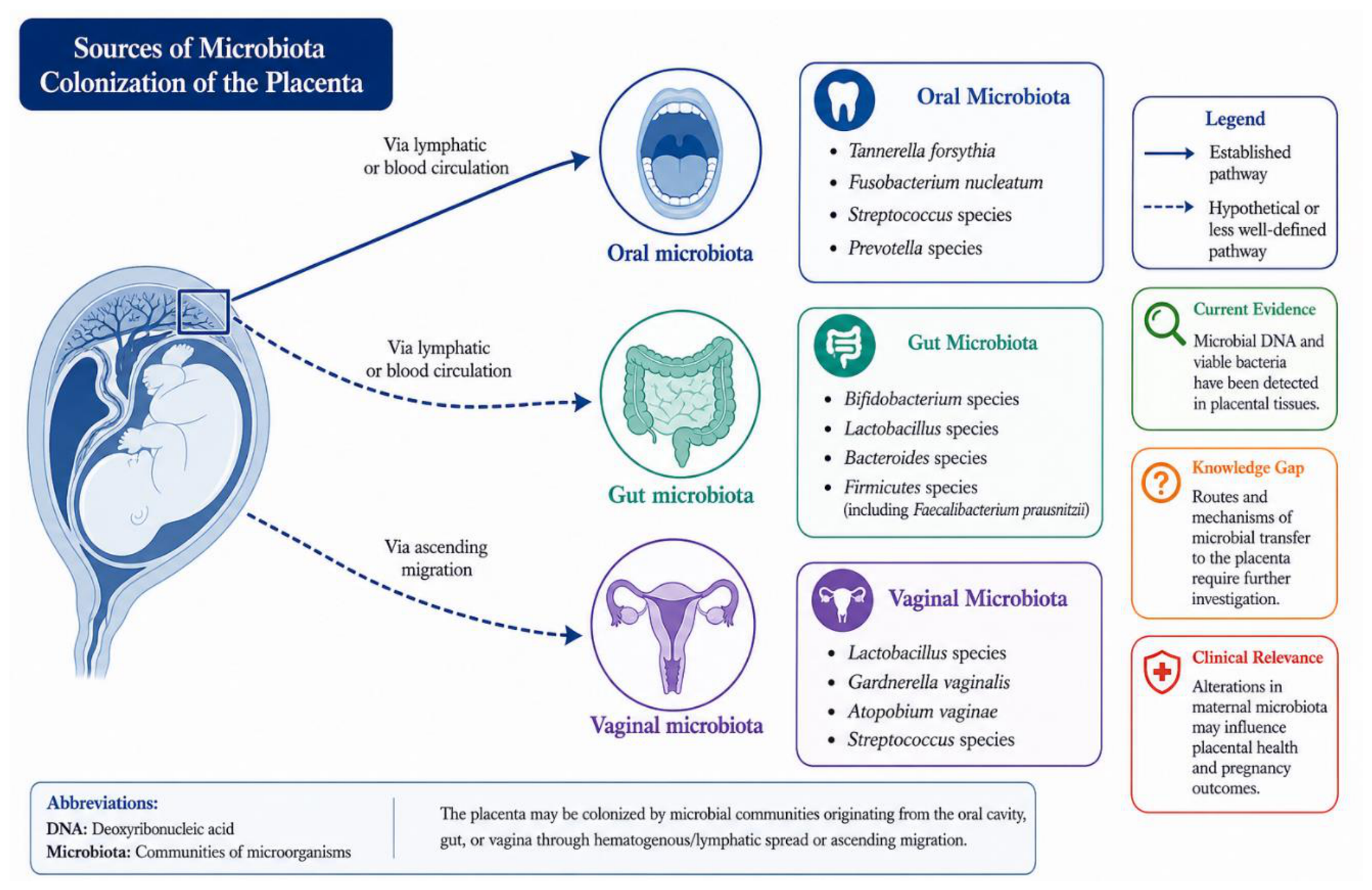

| Pathway | Timing | Microbial Impact & Physiological Consequences | Ref. |
| In Utero | Prenatal | Potential low-level microbial translocation; maternal short-chain fatty acid (SCFA) metabolites cross the placental barrier to initiate fetal immune programming. | [3,9,11] |
|
Vaginal Birth |
Birth | Physiological seeding standard; direct exposure to maternal vaginal and fecal microbes initializes colonization by beneficial Bifidobacterium, Lactobacillus, and Bacteroides. | [2,41,54] |
| C-section | Birth | Disruption of physiological seeding; primary colonization by skin and environmental/nosocomial bacteria (e.g., Staphylococcus, Corynebacterium) resulting in severely reduced initial microbial diversity. | [23,24] |
|
Breast feeding |
Postnatal | Selective enrichment of Bifidobacterium species via Human Milk Oligosaccharides (HMOs); supplies passive immune protection (e.g., secretory IgA) and crucial prebiotics. | [41,42] |
|
Skin Contact |
Postnatal | Facilitates direct vertical transmission of maternal skin commensals (e.g., during Kangaroo Care); promotes early colonization resistance and limits pathogenic overgrowth. | [1,4] |
| Environ-ment | Postnatal | Horizontal transmission via family members, hospital/NICU exposures, or protective early-life farm/animal environmental contact. | [2,6,54] |
| Body Site | Eubiosis (Physiological Changes) | Dysbiosis (Condition: Signature) | Ref. |
| Gut | ↑Firmicutes, Proteobacteria, Actinobacteria; ↓alpha diversity (late pregnancy); metabolic adaptation for energy storage |
PE: ↓Faecalibacterium, Akkermansia; ↑Fusobacterium, Veillonella GDM: ↓diversity, SCFA producers; ↑Proteobacteria, Collinsella PTB: ↑Clostridium innocuum (early pregnancy) Obesity/High-Fat Diet: ↑Firmicutes, Staphylococcus; ↓Bifidobacterium, Bacteroides Systemic Inflammation (stress): ↓Faecalibacterium, Ruminococcaceae |
Eubiosis: [1,3] Dysbiosis: [4,13,15,28,43] |
| Vaginal | Stable, less diverse, Lactobacillus-dominated; ↑glycogen→lactic acid; low pH; protective |
PTB/PE: ↓Lactobacillus (esp. L. crispatus); ↑Gardnerella vaginalis, Prevotella, Sneathia, Atopobium vaginae |
Eubiosis: [39,44,45] Dysbiosis: [40,48,49] |
| Place-ntal | Low-biomass community (if any); resembles oral microbiota; controversial (may be contamination) |
PTB/Infection: ↑Burkholderia, Actinomycetales, Mycoplasma, Gardnerella, Fusobacterium GDM: ↓Pseudomonadales, Acinetobacter; ↑Ruminococcus, Coprococcus, Paraprevotella FGR: ↑Actinopolyspora, Listeria, E. coli, Bacteroidetes, Clostridiales |
Eubiosis: [9,38] Dysbiosis: [11,18,38,56,61,62,63] |
| Oral | Hormonal changes alter pH and bacterial composition; ↑pathogenic bacteria (e.g., Porphyromonas gingivalis) |
PTB/LBW/PE: ↑Porphyromonas, Prevotella, Fusobacterium nucleatum, Bergeyella (translocation via bloodstream) |
Eubiosis: [52] Dysbiosis: [38,53,64,65,66,67] |
| Condition | Host | Site(s) | Microbial Changes | Key Mechanisms | Ref. |
|
Preecl- ampsia |
Maternal | Gut, Vagina, Oral |
↓ Faecalibacterium, Akkermansia, Lactobacillus ↑ Fusobacterium, Veillonella, Blautia |
SCFA depletion → barrier dysfunction → LPS translocation → inflammation→ endothelial dysfunction (↑TMAO, Th17) |
[3,9,13,25,33] |
| GDM | Maternal | Gut, Placenta |
↓ Diversity, SCFA producers, Bacteroidetes ↑ Proteobacteria, Collinsella, Blautia |
Metabolic endotoxemia → insulin resistance; ↓SCFA → impaired barrier; ↑BCAAs → insulin interference |
[5,13,22,35,43 |
|
Preterm Birth |
Maternal | Vagina, Gut, Oral |
↓ L. crispatus ↑ Gardnerella, Sneathia, Prevotella, C. innocuum |
Vaginal inflammation; C. innocuum degrades estradiol → hormonal disruption |
[10,33,41] |
|
Fetal Growth Restriction |
Fetal | Gut, Placenta |
↑ Actinopolyspora, Listeria, E. coli, Bacteroidetes |
Placental bacterial invasion → pathology |
[16,20,35] |
|
Allergy/ Asthma |
Offspring | Gut | CS/antibiotic- induced dysbiosis |
Immune imprinting failure → tolerance impairment; ↑allergy risk 1.8–2.6× |
[11,45] |
|
Obesity/ Metabolic Syndrome |
Offspring | Gut, Skin |
↓ Diversity ↑ Proinflammatory bacteria |
Maternal dysbiosis → fetal metabolic programming → obesity risk |
[8,11] |
| Neurodevelopmental Disorders | Offspring | Gut, Brain |
Maternal stress/ antibiotic dysbiosis |
SCFA dysregulation → microglia dysfunction → impaired neurodevelopment |
[19,22,57] |
| C. difficile Infection | Adult | Gut |
↑ Veillonella parvula |
Bile acid inhibition → C. difficile spore germination |
[68] |
| PCOS | Women | Gut, Vagina |
↓ SCFA producers, vaginal Lactobacillus |
↓SCFA → inflammation → metabolic/endocrine dysfunction |
[22,57,69] |
| Systemic Inflammation | Maternal/ Offspring |
Gut |
↓ Faecalibacterium, Ruminococcaceae (stress) |
Stress → ↓SCFA producers → inflammation → immune impairment |
[8,9,49] |
| Mechanism Category |
Example Microbe/ Metabolite |
Mode of Action | Health Impact | Ref. |
| Hormonal Modulation |
Clostridium innocuum |
Degrades 17β-estradiol, potentially disrupting hormonal balance | Increased risk of preterm birth |
[10,51,52] |
| Immune Programming | Short-chain fatty acids (SCFAs) |
Promote regulatory T cell differentiation and anti-inflammatory cytokine production |
Enhanced immune tolerance; dysbiosis leads to allergy and inflammation |
[8,39] |
|
Barrier Integrity |
Lactobacillus spp. in vagina |
Produce lactic acid, maintain low pH, and inhibit pathogens |
Protection against ascending infections and preterm birth |
22,23,27 |
| Metabolic Signaling | Bile acids | Modulation by gut microbiota affects host metabolism and C. difficile germination |
Dysbiosis contributes to metabolic syndrome and C. difficile infection |
[6,54] |
|
Neuro- endocrine Axis |
Bifidobacter- ium breve (in animal models) |
Modifies fetal brain metabolism and microglial polarization |
Impacts neurodevelopment and cognitive function |
[1,15] |
Disclaimer/Publisher’s Note: The statements, opinions and data contained in all publications are solely those of the individual author(s) and contributor(s) and not of MDPI and/or the editor(s). MDPI and/or the editor(s) disclaim responsibility for any injury to people or property resulting from any ideas, methods, instructions or products referred to in the content. |
© 2026 by the authors. Licensee MDPI, Basel, Switzerland. This article is an open access article distributed under the terms and conditions of the Creative Commons Attribution (CC BY) license.