
Submitted:
16 September 2026
Posted:
16 September 2026
You are already at the latest version
Abstract
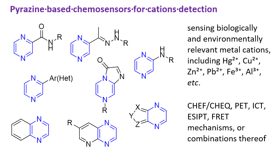
Pyrazine (1,4-diazine) and its annelated derivatives have emerged as promising platforms for developing selective and sensitive colorimetric and/or fluorescent chemosensors for biologically and environmentally relevant metal cations, including Hg²⁺, Cu²⁺, Zn²⁺, Pb²⁺, Fe³⁺, Al³⁺, etc. Their sensing potential arises from the strongly electron-deficient character of the pyrazine ring, low-lying unoccupied molecular orbitals, and considerable synthetic versatility. Reported sensor architectures include pyrazinamide, pyrazinehydrazide, 2-acetylpyrazine, aminopyrazine, hydrazinopyrazine, and aryl(hetaryl) substituted pyrazine derivatives, as well as pyrazines fused with five-membered carbocyclic or heterocyclic rings, quinoxaline systems, and pyrido[2,3-b]pyrazines. Rational molecular design enables the incorporation of multiple coordination sites with suitable properties and geometries for the specific recognition of metal cations. Sensors operate through CHEF/CHEQ, PET, ICT, ESIPT, FRET mechanisms, or combinations thereof, producing responses that can be observed visually or recorded instrumentally as changes in the intensity and/or position of absorption and emission bands. This review summarizes the structural diversity, metal-ion recognition strategies, photophysical response mechanisms, and practical applications of pyrazine-derived chemosensors for metal-cation detection, with particular emphasis on studies published between 2015 and 2026. It also highlights current limitations, remaining gaps in the literature, and promising directions for the development of more selective, biologically compatible, portable, sensing platforms.
Keywords:
pyrazines
; chemosensors
; Naked-eye sensors
; absorption
; fluorescence
; metal ion detection
; limit of detection
1. Introduction
Pyrazine (1,4-diazine) and its annelated derivatives represent an important class of electron-deficient nitrogen heterocycles that possess a strong electron-accepting character, making them valuable building blocks for the construction of π-conjugated push-pull systems in combination with electron-donating groups [1,2]. The parent compound, pyrazine, has been extensively studied; its derivatives exhibit diverse biological activities and are widely employed in pharmaceutical chemistry, while also finding broad application in materials science as components of organic light-emitting diodes (OLEDs), dye-sensitized solar cells, and nonlinear optical materials [2,3,4]. Although the basicity of the pyrazine ring (pKa ~ 0.65) is significantly lower than that of pyridine (pKa ~ 5.2) [3], the presence of two nitrogen atoms in the 1,4-diazine ring enables protonation, participation in hydrogen bonding, and chelation with metal ions. These structural and electronic features, coupled with their favorable photophysical properties and convenient synthetic tunability, have positioned pyrazine derivatives as promising platforms for the development of chromogenic and fluorogenic chemosensors for metal cation detection [2,5,6]. In this context, luminescent materials incorporating pyrazine fragments have been the subject of recent comprehensive reviews [2,7], and the application of pyrazine-based sensors for the detection of metal ions in environmental and biological systems has attracted considerable research attention due to the growing demand for sensitive, selective, and cost-effective analytical tools for monitoring toxic metal contamination [3,8].
It is important to note that interest in pyrazine-containing compounds is not limited to classical small-molecule chemosensors. In recent years, a field related to the development of aggregation-induced emission (AIE) systems based on pyrazine has been actively advancing, which opens up new opportunities for the development of highly efficient sensor materials [9]. In parallel with this, extensive research has been devoted to the design of functionalized 1,4-diazenes for optoelectronic applications, including fluorescence sensing [3]. In particular, quinoxaline derivatives, which are benzo-annulated analogues of pyrazine, also demonstrate outstanding photophysical properties, making them promising platforms for the development of chromogenic and fluorogenic chemosensors capable of detecting a wide range of analytes [10]; a broad range of benzo-annulated azaheterocycles were analyzed as diverse fluorescent probes [11].
A complementary and rapidly expanding area of pyrazine-based sensing involves their incorporation into metal-organic frameworks (MOFs), where the pyrazine moiety often serves as an integral linker within the extended porous architecture. 2,3,5,6-Tetra(4-carboxyphenyl)pyrazine [12,13,14] tetraphenylpyrazine [15,16], 2,3,5,6-tetrakis[2-(4-carboxyphenyl)vinyl]pyrazine [17], 2,3,5,6-tetrakis[4-[(1H-imidazole-1-yl)-methyl]phenyl]pyrazine [18] and tetrakis(4-(pyridin-4-yl)phenyl)pyrazine [19] are applied for design of MOF sensors. These hybrid materials leverage the intrinsic porosity, high surface area, and structural tunability of MOFs to achieve exceptional performance in fluorescence-based detection of various analytes, including metal ions [12,14] antibiotics [17], explosives [13,15,18], and even carcinoid biomarkers [19], often with remarkable sensitivity and selectivity. However, while such MOF-based sensors represent a powerful paradigm in sensing technology, the present review is focused on a different class of pyrazine derivatives: discrete small molecules that function as bi- or tridentate ligands capable of directly coordinating to metal cations, generating a distinct chromogenic or fluorogenic signal through well-defined molecular interactions.
Comprehensive surveys dedicated to the detection of various biologically and environmentally relevant metal ions, including copper, zinc, aluminum, mercury, nickel, iron, cadmium, and palladium, have demonstrated that Schiff bases, hydrazones, and related functionalized derivatives represent some of the most common and effective ligand scaffolds for the construction of efficient chemosensors for cations [20,21]. These structural motifs, particularly those containing imine (C=N) and amide functionalities, provide multiple coordination sites that enable strong and selective binding to metal cations. Metal coordination may generate an optical response in the form of spectral and/or color changes, fluorescence enhancement/quenching, or an emission-wavelength shift. The nature of this response is determined by the photophysical process operating in the sensor, with reported examples operating through chelation-enhanced fluorescence (CHEF) or quenching (CHEQ), photoinduced electron transfer (PET), intramolecular charge transfer (ICT), excited-state intramolecular proton transfer (ESIPT), Förster resonance energy transfer (FRET), and aggregation-induced emission (AIE) [20,22,23,24,25]. The prevalence of such ligands in literature underscores their significance as privileged structures in the field of cation chemosensing, offering the advantages of straightforward synthetic accessibility, tunable photophysical properties, and versatile coordination modes [21].
Heavy metals are ubiquitous environmental pollutants characterized by high atomic mass and density (typically > 5 g/cm3). Common metal ions of concern include mercury, nickel, cadmium, lead, chromium, cobalt, iron, copper, zinc, silver and manganese [26]. Unlike organic contaminants, these elements are essentially non-biodegradable and therefore persistently accumulate in soils, waters and the atmosphere as a result of industrial, agricultural and pharmaceutical activities [27]. While some heavy metals occur naturally in the environment, anthropogenic emissions have substantially increased their concentrations, resulting in toxic effects on human health, fauna and flora. Consequently, it is essential to monitor metal concentrations in industrial emissions and in environment to prevent harmful impacts on living organisms and human life.
Mercury (Hg) is a hazardous element of both natural and anthropogenic origin, present in air, water and soil. It has been recognized as a global pollutant with a toxic, mobile, and persistent nature [28]. All forms of mercury are harmful, including inorganic species such as metallic mercury, mercury vapor (Hg0), mercurous (Hg2+) and mercuric (Hg2+) salts, as well as organic compounds in which mercury is bonded to methyl, ethyl, phenyl, or related groups [29]. Toxic effects of Hg are associated with neurological disorders, cognitive impairment, developmental delays, cardiovascular damage, and high-level exposure may be fatal; overall toxicity depends on the chemical form, dose and rate of exposure [27,29]. The mechanisms of mercury toxicity are complex and multifactorial; however, direct binding to protein thiol groups plays a key role in microtubule disruption, protein deactivation and consequent metabolic and redox imbalance in cells [30].
Lead (Pb) stands out among heavy metal ions as particularly dangerous for both human health and marine life. Soluble or sparingly water-soluble Pb²⁺ salts can readily enter biological systems through contaminated water and soil, exerting significant negative effects on the health of organisms. Lead has no physiological role in the body, and even low levels can cause toxicity. Lead toxicity disrupts the functions of the digestive, nervous, respiratory, and reproductive systems, among others, and interferes with enzymatic activity by preventing enzymes from performing their normal functions. Of all organs, the nervous system is the most affected target in lead toxicity, both in children and adults; excessive accumulation of Pb²⁺ not only seriously damages the central nervous system but has also been linked to neurodegenerative disorders such as Alzheimer’s disease [31]. Moreover, Pb²⁺ disrupts normal DNA transcription processes and causes abnormalities in bone development [32].
Copper (Сu) is an essential trace element with critical roles in physical and mental health, primarily as a cofactor in endogenous enzymes. Although metallic copper is generally not highly toxic, several copper salts (e.g., CuSO4, CuCO3 and copper sub-acetate) can cause acute and chronic poisoning through inhalation, ingestion or dermal exposure. Excess Cu2+ accumulates mainly in the liver, ultimately affecting the nervous, reproductive and endocrine systems and connective tissues, and high-dose exposure may lead to severe gastrointestinal symptoms, neurological complications and even death [33]. Human exposure to Cu²⁺ may increase through contaminated drinking water and environmental sources; therefore, monitoring copper levels in water and other environmental sources is necessary [34]. Real-life applications of organic sensors for Cu²⁺ detection, including environmental monitoring of water quality, point-of-care diagnostics, and quality control in industrial processes, have been extensively reviewed [35].
Nickel (Ni) is another essential trace element, and Ni2+ is the most common oxidation state in biological systems. Its use has increased considerably with the expansion of modern technologies, leading to greater human exposure to nickel compounds, which can cause a variety of adverse, threshold-dependent health effects [36]. The most important and frequent effects include nickel allergy in the form of contact dermatitis, lung fibrosis, cardiovascular and kidney diseases, and lung and nasal cancers [37,38].
Zinc chloride (ZnCl₂) is a versatile reagent widely used in industrial organic synthesis as a catalyst, dehydrating agent, and precursor for functional materials. Beyond process control, monitoring ZnCl₂ is important for environmental protection because of its toxicity, and for biological studies, since excessive zinc in its chloride form may exert corrosive and damaging effects despite the essential role of Zn²⁺ ions in living systems [39]. Although numerous fluorescent probes for Zn²⁺ ions have been developed, most are not selective for the chloride salt form, highlighting the need for systems capable of detecting ZnCl₂ specifically [40].
Silver (Ag) is an important industrial and biomedical metal, and its ionic form is widely used in photography, agriculture, electronic imaging, medical devices, and the pharmaceutical industry, making its detection and monitoring highly significant. Ag⁺ can contaminate soil, water, air and food and may accumulate in the human body through the food chain. By coordinating with thiol, imidazole, amino and carboxyl groups, Ag⁺ can cause developmental delays, skin damage, and toxicity to the kidney, liver, and central nervous system [41].
Iron (Fe) is an essential metal in humans and plays a wide range of biological functions. It serves as a cofactor for several enzymes involved in redox reactions because it can switch between ferrous Fe2+ and ferric Fe3+ states [42]. Maintaining appropriate iron levels is vital for human health, as both deficiency and overload can lead to disorders such as anemia and hemochromatosis and may damage the liver, heart, and endocrine glands. Iron enters environments through mining, industrial effluents, pipe corrosion, and manufacturing processes. Disruptions in iron levels can adversely affect ecosystems, and excessive human exposure may cause serious health effects [43].
Chromium (Cr) mainly exists in the +3 and +6 oxidation states. Cr3+ is less toxic and less mobile, and it is considered an essential trace element in biological systems; however, prolonged exposure may still have adverse health effects. Cr3+ can be easily oxidized to Cr6+, which is genotoxic, hemotoxic, and carcinogenic. Owing to its smaller size and strong oxidizing properties, Cr6+ can readily penetrate biological cell membranes, leading to various disorders [44].
Cobalt (Co) is an essential trace element whose biological significance in humans is primarily associated with its incorporation into vitamin B₁₂ (cobalamin). Through cobalamin-dependent enzymes, it indirectly contributes to erythropoiesis and DNA synthesis and supports normal neurological function and the metabolism of lipids and carbohydrates. Excessive cobalt exposure, however, may result in cardiotoxicity and dysfunction of the thyroid, gastrointestinal, and nervous systems [45]. Systematic monitoring of cobalt, particularly in soils, is essential for assessing environmental pollution and protecting the quality of feed, food, and human and animal health [46].
Aluminum (Al) is one of the most abundant metals in the Earth’s crust and is widely used in industry and consumer products [47,48]. Due to its extensive use, it can enter food, water, and air, accumulate in the human body, and impair biological functions, especially under prolonged exposure [48]. Because excessive Al3+ exposure has been linked to neurotoxicity and several neurological disorders, the development of sensitive and selective probes for Al3+ detection in both environmental and biological systems is important [49,50].
Beyond their application in cation sensing, pyrazine-based small-molecule chemosensors have also been developed for the detection of a variety of other important analytes. For instance, a simple pyrazine-2-carboxamide derivative was reported as a highly selective colorimetric sensor for the detection of phenylalanine in blood plasma, exhibiting a distinct color change from colorless to bright green upon analyte recognition [51]. Furthermore, annulated pyrazine derivatives, particularly those incorporating aggregation-induced emission (AIE)-active tetraphenylethylene moieties, have been demonstrated to serve as effective fluorescent probes for the selective detection of nitroaromatic compounds (NACs), including explosives such as TNT, with high sensitivity and fast response times in aqueous media [52]. Other studies have explored pyrazine-derived chemosensors for the detection of anions, such as cyanide and fluoride, through distinct mechanisms including nucleophilic addition and deprotonation [53] Related quinoxaline compounds (benzo[b]pyrazines) were analyzed as chromogenic and fluorogenic chemosensors for fluoride, cyanide, acetate, and phosphate anions, in biomolecular applications [54]. However, as the present review is dedicated to pyrazine-based small-molecule sensors for metal cations, these examples involving alternative analytes are beyond the scope of this work and will not be discussed herein.
The molecular structures of sensors discussed herein are systematically presented in Figure 1.
This communication provides a comprehensive overview of the pyrazine-based optical sensors to metal cation developed mostly from 2015 to 2026. Readers can quickly grasp both the recent advances and the existing gaps in the field of specific material recognition, facilitating them to create more effective pyrazine sensors.
2. Substituted Pyrazines
2.1. Pyrazinamide and Pyrazinehydrazide Derivatives
Pyrazine carboxamides represent an important class of pyrazine-based chemosensors because the combination of the ring nitrogens and the amide group enables diverse coordination chemistry. N-substituted pyrazine-2-carboxamides can form a pincer-like cavity that chelates metal ions, often giving stable, planar complexes.
A chemosensor 1, based on pyrazine-2-carboxamide covalently linked to a quinoline fragment (Figure 2) showed “turn-on” fluorescent response to Zn²⁺ in acetonitrile among 13 other tested metal ions, including Na+, K+, Mg2+, Ca2+, Al3+, Co2+, Cd2+, Cr3+, Fe2+, Hg2+, Ni2+, Mn2+, as well as Cu2+ [55]. Compound 1 also demonstrated selective colorimetric (naked-eye) detection of Cu²⁺, with a color change from colorless to yellow and a bathochromic shift of the long-wavelength absorption maximum from 328 nm to 378 nm, attributed to an intense charge-transfer transition. The color change remains stable even in the presence of competing metal ions, indicating high selectivity for Cu2+.
The fluorescence enhancement of pyrazine-2-carboxamide 1 at 476 nm (λₑₓ = 360 nm) upon addition of Zn(ClO4)2×6H2O was explained by the CHEF mechanism: coordination of Zn²⁺ induces deprotonation of the ligand and formation of a more rigid, extended π-conjugated system, that suppresses non-radiative decay pathways. Fluorescence and UV–vis titrations, a Job’s plot, ESI-MS (m/z 396.74), and density functional theory (DFT) calculations support a 1:1 binding stoichiometry, assigned as [Zn1(NCCH₃)₂]⁺, with N,N,N-coordination of the ligand 1 (Figure 2). The reported association constants and limit of detections (LODs) for Zn2+ were 3.837 × 10⁴ M⁻¹ and 1.11 µM, respectively; for Cu2+, the reported values were 7.352 × 10⁷ M⁻¹ and 14.8 µM.
DFT and time-dependent DFT (TD-DFT) calculations (B3LYP/6-31G(2d,p)/LANL2DZ) of 1 and its Zn2+ and Cu2+ complexes in acetonitrile are consistent with the experimental observations: the calculated emission for the 1-Zn²⁺ complex (482 nm) closely matches the experimental emission (476 nm), and the calculated absorption for the 1-Cu²⁺ complex shifts from 326 nm (free ligand) to 386 nm upon complexation, in agreement with the experimental shift to 378 nm. These results are consistent with a binding mode in which the deprotonated ligand (L‒) coordinates the metal center while acetonitrile molecules complete the coordination sphere [55].
The reversible change in emission upon sequential addition of Zn2+ and SCN− was exploited to construct an INHIBIT logic gate.
Ravandi et al. [56] reported N-(pyridine-2-ylmethyl)pyrazine-2-carboxamide 2 as a “turn-off” fluorescent probe for selective detection of Cu²⁺ and Pb²⁺ in aqueous media (Figure 3). The authors systematically evaluated key analytical parameters, including pH, response time, and reversibility. Upon addition of Cu²⁺ and Pb²⁺, sensor 2 exhibited fluorescence quenching at 340 nm (λₑₓ = 281 nm). Stern–Volmer of the titration data yielded quenching constants Ksv = 2.30 × 10⁴ M⁻¹ for Cu²⁺ and 1.10 × 10⁴ M⁻¹ for Pb²⁺. Limits of detection and quantification (LOQ) were determined as 5.73/9.62 µM for Cu²⁺ and 17.4/29.2 µM for Pb²⁺. Job’s plot, corroborated by ESI-MS and FT-IR spectroscopy, indicated a 1:1 binding stoichiometry for both metal complexes. The observed fluorescence quenching was attributed to chelation-enhanced quenching (CHEQ). For Cu2+, paramagnetic quenching (d9 configuration) via enhanced intersystem crossing (ISC) is likely dominant; for Pb2+, a heavy-atom effect contributes to efficient non-radiative decay. Sensor 2 displayed a rapid response (< 1 min), high selectivity over 18 competing metal ions, and greater sensitivity toward Cu²⁺ than toward Pd2+. Practical applicability was demonstrated in drinking water, human blood plasma, and on paper test strips. In addition, carboxamide 2 showed reversible “Write-Read-Erase-Read” memory behavior using ethylenediaminetetraacetic acid (EDTA) and Cu²⁺ as chemical inputs, with the fluorescence intensity at 354 nm as the output signal, highlighting its potential for molecular memory applications.
Extending this series, Ravandi et al. [57] described two N-(2-halogenophenyl)pyrazine-2-carboxamides (X = I, Br) 3a,b as highly selective “turn-off” fluorescent and colorimetric sensors for Fe³⁺ in DMSO (Figure 4). The free iodo and bromo ligands absorb at 270 nm (X = I) and 264 nm (X = Br) and emit at 349 nm (λex = 268 nm) and 346 nm (λex = 284 nm), respectively. Addition of Fe³⁺ induces pronounced fluorescence quenching and an increase in absorption intensity. This behavior was attributed to CHEQ, driven by the paramagnetic nature of Fe³⁺ (d⁵ configuration) that facilitates non-radiative deactivation of the excited state. Selectivity was confirmed in the presence of a broad range of competing cations, including monovalent Na⁺, K⁺, Ag⁺, bivalent Mg²⁺, Ca²⁺, Ba²⁺, Mn²⁺, Sn²⁺, Co²⁺, Cd²⁺, Pb²⁺, Ni²⁺, Cu²⁺, Hg²⁺, Zn²⁺, as well as trivalent Al³⁺, Cr³⁺, with minimal interference.
Job’s continuous variation method and ESI-MS (m/z 503.19, assigned to [3a·Fe(H₂O)Cl₃]) supported a 1:1 stoichiometry for the Fe3+ complex. From fluorescence titration, LODs were determined as 11.7 μM (iodo) and 11.6 μM (bromo). Association constants obtained by Benesi–Hildebrand analysis were Ka = 4.90 × 10⁴ M⁻¹ and 1.75 × 10³ M⁻¹ for 3a and 3b, respectively. Stern–Volmer constants were 4.24 × 10⁵ M⁻¹ and 7.59 × 10⁴ M⁻¹, respectively, consistent with a predominantly static quenching mechanism. The binding was reversible upon addition of EDTA over at least eight cycles. The iodine-containing sensor 3a was successfully applied for Fe³⁺ determination in real water samples (drinking, tap, and river water) and on test strips, where complexation with Fe3+ produced a visible color change from colorless to bright yellow, demonstrating its potential for rapid water-quality monitoring.
Heydari et al. developed N-(2-alkoxyphenyl)- and N-(2-phenoxyphenyl)pyrazine-2-carboxamides 4a-c (Figure 5) as chemosensors for Cu²⁺ [58,59,60]. Upon the addition of Cu²⁺, initially colorless solutions of 4a-c turned pale yellow under visible light and dark yellow under UV illumination (fluorescence studied for 4a and 4c derivatives), with no observable interference from 18 competing metal ions. Spectral studies revealed an enhancement of the absorption band and the appearance of a new low-intensity band at ~454–458 nm, attributed to d–d transitions and partial ligand-to-metal charge transfer (LMCT), consistent with complex formation. The fluorescence quenching observed around 514 nm for 4a (λₑₓ = 283 nm) and 359 nm for 4c (λₑₓ = 280 nm) was triggered by non-radiative relaxation due to the paramagnetism of Cu2+ (d9 configuration). This quenching mechanism is consistent with CHEQ.
Variation of the ortho substituent on the phenyl ring (from methoxy to ethoxy and phenoxy) enabled tuning of the analytical performance, with reported limits of detection spanning from the millimolar to the micromolar range across the series (Figure 5).
The coordination behavior of ligands 4a-c toward Cu2+ depends on the copper salt, stoichiometry, and solvent, leading to different structural motifs [58,59,60]. For N-(2-methoxyphenyl)pyrazine-2-carboxamide 4a, reaction with CuCl₂×2H2O (1 equiv.) in MeOH in the presence of Et3N afforded a one-dimensional polymeric complex {[Cu(4aCl]·H₂O}ₙ (Scheme 1). In this structure, the deprotonated ligand 4a coordinates in a tridentate N,N,O fashion and bridges adjacent Cu2+ centers via the pyrazine nitrogen, generating ladder-like chains with distorted square-pyramidal geometry. In contrast, using 2 equiv. of Cu(NO₃)₂×3H2O in methanol or acetonitrile yielded mononuclear spacers [Cu(4a)₂(η¹-NO₃)₂(CH₃OH)₂] or [Cu(4a)2(η2-NO3)2(H2O)2] [Cu(4a)2(η2-NO3)(η1-NO3)(H2O)] (Scheme 1), respectively, in which two ligands 4a binds in a monodentate mode through the pyrazine nitrogen, while nitrate anions and solvent molecules complete distorted octahedral or pentagonal-bipyramidal coordination spheres. These results highlight the conformational flexibility of 4a and the strong influence of reaction conditions on the final coordination mode [58]. For the ethoxy derivative 4b, addition of Cu(NO₃)₂×3H2O to 4b (2:1 equiv.) in acetonitrile led to formation of a mononuclear complex described as [Cu(4b)2(η2-NO3)2(H2O)] with spectroscopic and crystallographic data supporting this formulation [59].
In case of phenoxy derivative 4c, reaction with CuCl2×2H2O (4 equiv.) in acetonitrile afforded [Cu(4c)₄Cl₂] (Scheme 2), as confirmed by single-crystal X-ray diffraction. XRD revealed a six-coordinate distorted octahedral geometry of 4c with the Cu²⁺ ion at an inversion center, four monodentate 4c ligands coordinated through pyrazine nitrogen atoms, and two chloride ligands. A pronounced Jahn–Teller distortion was observed, with elongated axial Cu–N bonds (2.543(3) Å) and shorter equatorial Cu–N bonds (2.056(3) Å).
Job’s plot analyses based on fluorescence titrations indicated L:Cu2+ stoichiometries of 2:1 for 4b and 4:1 for 4c, in agreement with the crystallographically characterized complexes [59,60]. For 4a, the stoichiometry depended on the copper salt and solvent, as described above; in some titration experiments the exact Cu2+ salt and conditions were not specified in the original reports [58].
Across the series 4a-c, the sensors exhibited a rapid response (less than 1 min), good reversibility with EDTA (three to five cycles), stability for at least one week, and effective performance in a broad pH range (including the pH of environmental water sources and biological fluids, pH ≈ 6). Together, these properties establish N-(2-alkoxyphenyl)- and N-(2-phenoxyphenyl)pyrazine-2-carboxamides 4a-c as selective, reversible, and practically useful Cu2+ probes for both solution-phase and solid-state (test-strip) applications.
Hydrazones derived from pyrazinecarboxylic acid hydrazide and aryl/hetaryl aldehydes (Het–CH=N–NH–C(O)–Pyraz) are widely used ligands owing to their straightforward synthesis via Schiff-base condensation and versatile coordination modes. In the absence of additional pyridine-type nitrogen atoms or hydroxy/alkoxy substituents in the hetaryl moiety, these hydrazones typically behave as N,N-bidentate ligands.
Sun et al. [61] reported a pyrazine-based Schiff-base fluorescent probe 5 (Figure 6) for colorimetric and fluorimetric detection of Cu2+ in a DMSO/H2O (1:1, v/v). Probe 5 contains an N-ethylcarbazole fluorophore linked to a pyrazine-hydrazone receptor. In the free state, 5 exhibits very weak fluorescence, which was attributed to PET from the electron-rich receptor to the excited carbazole unit. Upon addition of Cu2+, the solution changed from colorless to bright yellow, and a strong emission band appeared at 500 nm. Among a broad panel of alkali, alkaline earth, transition and heavy metal ions, only Cu2+ induced a hypsochromically shifted, tail-shaped absorption band together with this intense luminescent response.
Coordination of Cu²⁺ at the receptor suppresses the PET pathway thereby inducing a CHEF effect. The high Lewis acidity of Cu²⁺ lowers the energy of the donor orbital on the receptor, thereby inhibiting PET; DFT calculations at the B3LYP/6-31G(d) level support this interpretation, showing that the HOMO becomes localized at the Cu-binding site in the complex. The 5–Cu²⁺ complex has a significantly narrower HOMO–LUMO gap (1.55 eV) than the free probe (3.90 eV), which is consistent with its higher stability and correlates well with the observed spectral changes [61].
A 1:1 binding stoichiometry for the 5-Cu2+ complex (Figure 6) was established by Job’s plot analysis, fluorescence titration, and UV–vis spectroscopy. According to 1H NMR data, probe 5 coordinates Cu2+ through the pyrazine nitrogen and the imine nitrogen of the C=N bond (N,N-coordination mode). The system 5 operates over a broad pH range (4–10), exhibits a low detection limit of 12.1 nM and show excellent performance in colorimetric detection of Cu²⁺ in real water samples, highlighting its potential for environmental monitoring and analytical applications [61].
Introduction of an additional coordination site, such as a carbonyl or alkoxy oxygen, into the hydrazone framework (R–CH=N–NH–C(O)–Pyraz) changes the coordination mode: the hydrazone can act as an O,N,O-tridentate ligand, while the pyrazine moiety remains uninvolved in metal binding.
Tomer et al. [62] synthesized the chromone-based Schiff-base ligand 6 (Figure 7) by condensation of 3-formylchromone with pyrazine-2-carbohydrazide. Compound 6 exhibited an immediate (≈ 40 s) and selective colorimetric response to Cu²⁺ in methanol, with the solution changing from colorless to bright yellow; no interference was observed in the presence of a broad panel of competing metal ions (Na⁺, K⁺, Mg²⁺, Ca²⁺, Pb²⁺, Mn²⁺, Co²⁺, Ni²⁺, Ag⁺, Zn²⁺, Cd²⁺, Hg²⁺, Zr²⁺, Fe³⁺, Al³⁺ and Cr³⁺). UV–vis titration showed a gradual decrease of the absorption band at 311 nm and the appearance of a new band at 428 nm with a clear isosbestic point at 363 nm, indicating formation of a stable 6–Cu²⁺ complex. The observed bathochromic shift was attributed to enhanced ICT upon complexation. Job’s plot and HRMS data (peak at m/z 355.9) confirmed a 1:1 binding stoichiometry for the 6-Cu²⁺ complex. The association constant Kₐ, determined using the Benesi–Hildebrand equation, was 2.3 × 10⁵ M⁻¹, while the detection limit was 390 nM [62].
DFT calculations at the B3LYP level (6-311G(d,p) for 6, LANL2DZ for Cu²⁺) revealed a sabstantial reduction in the HOMO–LUMO gap from 3.77 eV (free ligand) to 0.98 eV (complex), facilitating electron transfer and rationalizing the observed bathochromic shift. The optimized geometry confirmed tridentate O,N,O-coordination of Cu²⁺, with Cu–O bond lengths of 1.9411 and 1.9763 Å and a Cu–N bond length of 2.1258 Å [62].
The sensor responded rapidly and operated effectively over a broad pH range (6–11), with optimal performance at pH 7.9. Its practical applicability was validated in real water samples (tap, groundwater, and canal water), with recoveries of 98–108% and relative standard deviation (RSD) values below 2.8% [62].
Another O,N,O-tridentate selective sensor for Cu2+ was designed by G. Sing et al. [63]. It is based on an 1,2,3-triazol-5- yl)methoxy)benzylidene)pyrazine-2-carbohydrazide 7 (Figure 8). Product 7 exhibited high selectivity toward Cu²⁺ over 14 other metal ions and responded in methanol with a hyperchromic shift in the UV–vis spectrum and pronounced fluorescence quenching. The fluorescence “off” response was attributed to excitation energy transfer to the Cu2+ d-orbitals, charge transfer processes and heavy-atom effect.
A 1:1 stoichiometry for the 7-Cu2+ complex was confirmed by Job’s continuous variation method and by the linearity of the Benesi–Hildebrand plot; reversibility was demonstrated by addition of EDTA. NMR analysis suggested coordination through the amide nitrogen atom and the methoxy oxygen substituent (Figure 8). The LODs were 3.4 µM (absorption) and 42 µM (fluorescence), with an association constant of 4.03 × 10³ M⁻¹ and Stern–Volmer quenching constant of 6.7 × 10³ M−1. Notably, immobilization of probe 7 on Fe₃O₄@SiO₂ magnetic nanoparticles lowered the detection limits to 2.7 µM and 0.86 µM for absorption and emission, respectively, corresponding to an almost 50-fold improvement in the fluorescence LOD [63].
The condensation of 7-diethylamino-3-formylcoumarin with 3-amino-pyrazine-2-carbohydrazide provided an efficient route to the Schiff-base chemosensor 8 (Figure 9), capable of detecting Zn²⁺ ions in aqueous media [64]. Compound 8 exhibits a distinctive aggregation-induced ratiometric emission (AIRE) phenomenon in an aqueous buffer solution (99.5% H₂O/DMSO); in this medium 8 displays orange emission with a maximum at 555 nm, whereas in pure DMSO it shows green fluorescence at 524 nm. Upon binding Zn²⁺, the aggregation-induced emission at 555 nm is quenched, while a new intense band appears in the far-red region at 628 nm (λex = 470 nm), resulting in a large Stokes shift (158 nm) and a ratiometric response with the F₆₂₈/F₅₅₅ ratio increasing approximately 11-fold. Sensor 8 therefore enables selective Zn²⁺ detection in the presence of various competing metal ions, except Cu2+, which decreases the F628/F555 ratio of 8-Zn2+ complex. The detection limit for Zn²⁺ was determined as 3.52 μM. Job’s plot analysis established a 1:1 binding stoichiometry, and the association constant obtained from the Benesi–Hildebrand equation was Kₐ = 5.59 × 10³ M⁻¹. According to the 1H NMR and ESI-MS data, the 8-Zn2+ complex formed under the studied condition corresponds to [Zn2+(8)(NO3)]+ (calcd m/z 506.08) and features an O,N,O-coordination mode (Figure 9). The experimental results are consistent with DFT calculations, which gave HOMO-LUMO gaps of 3.20 for 8 and 2.95 eV for the 8-Zn2+ complex; the smaller gap for the Zn²⁺ complex indicates greater electronic stabilization upon coordination relative to the unchelated ligand.
Sensor 8 is considered as promising for biological applications owing to its far-red emission, ratiometric fluorescent output, and operation under physiological conditions. Notably, the in situ generated 8–Zn²⁺ complex can be further employed for secondary recognition of pyrophosphate (P2O74-, PPi) via an aggregate disassembly mechanism (LOD = 2.45 μM), thereby expanding the sensor’s functionality and highlighting its potential for bioimaging in HeLa cells.
Dhanasekaran et al. [65] reported a pyrazine-carbohydrazide-based sensor 9 (Figure 10), synthesized by condensation of pyrazine-2-carbohydrazide with 2-hydroxy-1-naphthaldehyde, for Zn²⁺ detection by colorimetric and ratiometric methods in a partially aqueous medium (CH₃CN:H₂O, 7:3, v/v). The free ligand 9 exhibits an absorption band at 365 nm; upon addition of Zn²⁺ a bathochromic shift of 85 nm occurs, with the emergence of a new intense band at 450 nm and a visible color change from colorless to yellow [65]. The presence of an isosbestic point at 393 nm confirms formation of a new complex, while the ratiometric response ensures high selectivity for Zn²⁺ in the presence of a wide range of competing metal ions (Cu²⁺, Cd²⁺, Co²⁺, Pb²⁺, Ni²⁺, Mg²⁺, Mn²⁺, Cr³⁺, Fe³⁺, Al³⁺, and others).
The binding mechanism was elucidated by ¹H NMR titration, IR spectroscopy, and HRMS, indicating coordination via the deprotonated hydroxyl group, the carbonyl oxygen, and the imine nitrogen, with formation of a 1:1 stoichiometric complex (Figure 10). This assignment was further corroborated by Job’s plot and DFT calculations. The limit of detection for sensor 9, calculated from UV-Vis spectroscopy data, is 136 nM, demonstrating high sensitivity, while the high association constant (Kₐ = 5.38 × 10⁵ M⁻¹, determined by the Benesi–Hildebrand method) indicates strong binding. Sensor 9 was successfully applied to determine Zn²⁺ in pharmaceutical samples (Zincovit tablets and syrup), in real water samples, and as test strips showing a color transition, confirming its potential for on-site analysis in various matrices.
Among the tridentate O,N,O chelators, salicylidene hydrazones are extensively used in chemosensing. A developed pyrazine-based acylhydrazone sensor 10 [66] acts as a multi-analyte fluorescent “turn-on” probe for Al³⁺, Mg²⁺, and Zn²⁺ ions in DMSO solution, displaying three distinct emission colors (green, red and yellow, respectively) and LODs of 15,6, 80.4 and 162.3 nM (Scheme 3). The sensing mechanism relies on inhibition of ESIPT upon metal coordination through the phenolic hydroxyl oxygen, carbonyl oxygen, and imine nitrogen (Scheme 3), which results in significant fluorescence enhancement. Notably, the 10-Al³⁺ complex differ from 10-Zn²⁺ and 10-Mg²⁺ species, as evidenced by ESI-MS, FT-IR studies and 1H NMR studies. Binding of 10 to Al³⁺ is accompanied by deprotonation of the phenolic OH group, whereas the bivalent metals Zn²⁺ and Mg²⁺ coordinate to ligand 10 via donor-acceptor interaction without deprotonation (Scheme 3). The interaction between 10 and Al3+, Mg2+, and Zn2+, was completed within 120, 5, and 20 min, respectively, indicating that the reaction with Mg2+ is the most dynamically favorable among the three metal ions; the considerably longer considerably longer interaction time for Al3+ may be attributed to covalent bond formation involving deprotonation.
The calculated binding constants for ligand 10 with Al³⁺, Zn²⁺ and Mg²⁺ are 5.77, 3.87 and 1.30 × 10⁵ M−1, respectively, reflecting the higher affinity of sensor 10 for Al³⁺ and Zn²⁺ over Mg²⁺. Moreover, the HOMO‒LUMO energy gaps of the complexes (3.25, 3.46, and 3.41 eV for 10-Al3+, 10-Mg2+, and 10-Zn2+, respectively) are lower than that of the free ligand (3.48 eV), indicating increasing stabilization in the order: 10 < 10-Mg2+ < 10- Zn 2+ < 10-Al3+. On this basis, the authors propose the 10-Mg²⁺ system as a ratiometric sensor for Al³⁺ and Zn²⁺ operating via a metal-displacement mechanism (Scheme 3).
Additionally, in DMSO/H₂O solution, sensor 10 selectively recognizes Al³⁺ with a “turn-on” response and a detection limit of 33.8 nM, which is approximately two-fold higher than that in pure DMSO (LOD = 15.6 nM). Furthermore, the in situ generated 10-Al³⁺ complex serves as a “turn-off” sensor for fluoride anions (F⁻) due to the formation of a stable Al³⁺/F⁻ adduct, which regenerates the free sensor 10. Consequently, compound 10 enables sequential detection of metal cations and anions, with demonstrated applicability for bioimaging in living HeLa cells [66].
The selective sensor 11 (Scheme 4), designed for Al3+ recognition and suitable for quantitative determination of this cation in real water samples, is based on a pyrazine-2-carbohydrazide condensation product with a salicylaldehyde-containing photochromic diarylethene derivative [67]. Sensor 11 exhibits a LOD of 16.6 nM and Ka of 7.8 x 10⁴ M-1. As in the case of sensor 10, ligand 11 binds Al3+ through the deprotonated phenolic hydroxyl group, the imine (CH=N) nitrogen, and the carboxyl (C=O) oxygen in a 1:1 stoichiometry (Scheme 4). This binding mode is supported by an ESI-MS peak at m/z 710.40, assigned to [11+Al3++NO3−H]+. The authors discuss in detail the mechanisms underlying the photophysical properties of 11. The free ligand shows weak fluorescence due to a PET process, with two emission maxima at around 420 and 570 nm, which are attributed to tautomerization associated with ESIPT. Increasing the water content (> 70 vol%) in the organic solvent induces AIE, accompanied by enhancement of the keto-isomer emission at 600 nm. In the presence of water, strong solvation of the polar N–H group weakens the PET process. The observed fluorescence “turn-on” thus arises from the synergistic action of intramolecular hydrogen bonding and solvation. Upon coordination of ligand 11 with Al³⁺, both PET and ESIPT processes are suppressed; concomitantly, intramolecular rotations are restricted and the molecular structure is rigidified, leading to a pronounced increase in emission intensity centered at 541 nm, accompanied by a blue shift of 30 nm and distinct yellow fluorescence. Sensor 11 displays exceptional selectivity for Al³⁺ over a wide range of competing metal ions.
Ligand 12 (Figure 11), derived from pyridoxal-5′-phosphate (a vitamin B₆ derivative) and pyrazine-2-carbohydrazide, binds Al3+ in 1 vol% DMSO solution, producing a selective “turn-on” fluorescence response at 456 nm (λex = 410 nm) with a 20-fold enhancement of blue emission [68]. The recognition mechanism is based on restriction of rotation around the hydrazide bridge upon formation of a coordination compound. Authors showed that Na+, K+, Ca2+, Mg2+, Ba2+, Cd2+, Pb2+ and Cr3+ ions have almost no effect on fluorescent Al3+ recognition in water, whereas Cu2+ interferes with both qualitative and quantitative determination; in the presence Ni2+, Hg2+, UO22+, Co2+ and Fe3+ ions, only qualitative determination of Al3+ is possible. The effect of a set of anions on the fluorescence of the 12-Al3+ complex is negligible. In contrast, UV–vis spectroscopy is not suitable for selective recognition of metal ions, because interaction of probe 12 with several metals (Co2+, Ni2+, Cu2+, Hg2+, UO22+, Al3+ and Ce3+) induces similar changes (from colorless to yellow) and comparable bathochromic shifts. The LOD for Al³⁺ is 8 nM (by the 3σ method), which is three orders of magnitude below the World Health Organization (WHO) guideline for drinking water (7.4 μM); the LOQ is 27 nM. The sensor exhibits reversibility upon addition of Na2EDTA and was successfully applied to the quantitative determination of Al³⁺ in real water samples (distilled, river, and mineral water) with high recovery rates of 86–99%.
A distinct Al³⁺ recognition mode (Figure 11) was proposed for chemosensor 12, in contrast to the related compounds 10 and 11. A 1:2 (12:Al³⁺) stoichiometry for the 12-Al³⁺ complex was established from spectrofluorimetric titration data and mass spectrometry (peak at m/z 760.99 [12-Al³⁺+H⁺]). The binding mechanism was elucidated using ¹H NMR titration, IR spectroscopy, and DFT calculations, which indicated coordination of Al³⁺ through the phenolic oxygen (without deprotonation), azomethine nitrogen, and carbonyl oxygen, forming an O,N,O chelate cavity. It was also established that the phosphate group does not participate in binding (confirmed by ³¹P NMR) [68].
A notable feature of probe 12 is the manifestation of AIE: both the free sensor and its Al³⁺ complex form nanoaggregates in aqueous media (sizes of ~1000 nm for 12 and ~ 3 nm for the 12-Al³⁺ complex), which further enhances the fluorescence response.
In contrast to the Al3+-selective salicelidene hydrazones 10‒12, the thiazole-substituted chemosensor 13 (Figure 12) functions as a highly selective dual-channel probe for Ag⁺, operating through both colorimetric and fluorescent “turn-on” responses in a DMSO/H₂O (7:3, v/v) medium [69]. The sensing mechanism relies on the coordination of Ag⁺ with the hydroxyl fragment, imine nitrogen and carbonyl oxygen of the Schiff base moiety (Figure 12), which effectively restricts the C=N isomerization and enhances the CHEF effect, as corroborated by ¹H NMR, HRMS, and DFT studies. This complexation induces a distinct visual color change from pale yellow to blue under ambient light, accompanied by pronounced emission enhancement at 523 nm (λex = 410 nm) under UV irradiation, with a 1:1 binding stoichiometry.
Sensor 13 exhibits remarkable sensitivity, with detection limits of 10.6 nM (absorption) and 6.74 nM (fluorescence), as determined by UV-vis and fluorescence titrations, respectively—values well below the WHO permissible limit for silver in drinking water (30 µM), demonstrating its suitability for potable water quality monitoring. Furthermore, 13 shows excellent selectivity over a wide range of competing cations, a rapid response time (< 5 min), and reversible binding upon EDTA addition, making it operationally robust. Its practical applicability is convincingly validated through quantitative recovery experiments in real water samples (tap and lake water, 95–102% recovery), convenient paper-strip tests for on-site visual detection, and successful fluorescence imaging of intracellular Ag⁺ in MCF-7 breast cancer cells with negligible cytotoxicity (cell viability 75–91%), collectively positioning 13 as a versatile and promising chemosensor for environmental monitoring, point-of-use testing, and bioimaging applications [69].
A distinct mechanism for colorimetric and fluorescent detection of Al3+ in CH3CN/H2O (9:1, v/v) mixture was reported for the BODIPY-pyrazine Schiff base chemosensor 14 (Scheme 5). Among a range of cations (Cs+, Ba2+, Al3+, V3+, Cr3+, Mn2+, Fe3+, Co2+, Ni2+, Cu2+ and Zn2+), only Al3+ (excess amount) considerably alters both absorption and emission spectra and changes the solution color from pink to yellow under daylight and from pale orange to bright green under UV irradiation. Excess Al3+ promotes hydrolysis of the imine bond (Scheme 5) leading to formation of the green-emitted BODIPY-carbaldehyde 15; the nitrogen atoms in the pyrazine ring enhance nucleophilic attack on the imine system. Thus, a crucial role of the pyrazine core—specifically its ring nitrogen atoms—in governing the photophysical changes was demonstrated, in contrast to analogous benzene or pyridine rings [70]. The LOD of 14 for Al3+, calculated from fluorescence titration data, is 1.37 nM.
In addition, bioimaging studies demonstrated that chemosensor 14 enables selective identification of Al3+ ions in living cells, exhibiting a turn-on response both in vitro and in live-cell experiments; due to its cytocompatibility, it can be employed as a fluorescent marker [70].
In summary, pyrazine carboxamides and hydrazides represent the most extensively studied class of pyrazine-based cation sensors. Their popularity stems from the straightforward synthesis via amidation or Schiff-base condensation and the ability to form stable N,N,O-tridentate chelate complexes with a variety of metal ions, including Zn²⁺, Cu²⁺, Fe³⁺, Al³⁺, and Ag⁺. The sensing performance is highly tunable: variation of the aryl/hetaryl substituent on the amide or hydrazide moiety allows modulation of the binding affinity (Ka spanning from 10³ to 10⁷ M⁻¹), detection limits (from nanomolar to micromolar), and selectivity patterns. Notably, while many of these sensors operate through a CHEF mechanism, the presence of paramagnetic ions such as Cu²⁺ and Fe³⁺ typically induces fluorescence quenching via CHEQ, offering complementary detection modes. The successful validation in real water samples, pharmaceutical formulations, and paper-strip tests underscores their practical potential, though mutual interference between competing cations, particularly Cu²⁺ and Fe³⁺, remains a challenge that requires careful molecular design to overcome.
2.2. 2-Acetylpyrazine Derivatives
2-Acetylpyrazine derivatives also belong to the family of pyrazine-based hydrazone Schiff-base ligands and have been successfully employed as “turn-on” fluorescent sensors for metal ions, in particular for Al3+ or Co2+, as reported in the sources cited below. A typical synthetic route includes condensation of the corresponding hydrazide with 2-acetylpyrazine in EtOH under reflux.
Li et al. developed simple pyrazine-substituted sensors 17a and 17b, in which the hydrazone fragment incorporates quinolone (17a) or furan (17b) moieties (Figure 13) [71,72]. Both compounds function as highly selective and sensitive “turn-on” fluorescent chemosensors for Al³⁺ in ethanol (Figure 14). In their free state, sensors 17a,b exhibit negligible fluorescence due to a PET process from the Schiff-base nitrogen atom to the pyrazine fragment. Upon coordination with Al³⁺, the metal ion binds to the carbonyl oxygen, the imine nitrogen, and one of the pyrazine nitrogen atoms in a 1:1 stoichiometry, as confirmed by Job’s plot analysis, ¹H NMR titrations, and mass spectrometry. This coordination effectively suppresses the PET pathway and simultaneously activates the CHEF mechanism by rigidifying the molecular structure and restricting C=N isomerization. As a result, a remarkable ~230-fold or 164-fold enhancement of emission intensity is observed at 488 nm (λex = 350 nm) for 17a and 517 nm (λex = 382 nm) for 17b [71,72], accompanied by a color change from colorless to light green under UV illumination.
The selectivity of sensors 17a,b for Al³⁺ was evaluated in the presence of 15 competing metal ions (Ba²⁺, Ca²⁺, Cd²⁺, Co²⁺, Cr³⁺, Cu²⁺, Fe²⁺, Fe³⁺, K⁺, Mg²⁺, Mn²⁺, Na⁺, Ni²⁺, Pb²⁺, Zn²⁺). Among them Cu2+ and Ni2+ (for 17a) and Cu2+ and Fe2+ (for 17a) completely quenched the fluorescence, whereas other metals caused slight quenching or did not interfere with Al3+ detection. Both sensors exhibit LOD on the order of 100 nM, but differ markedly in their binding constants: Ka = 1.35 × 10⁴ M-1 for 17a and Ka = 2.36 × 10⁷ M-1 for 17b.
The sensing systems 17a,b are reversible: addition of Na2EDTA dissociates the 17-Al³⁺ complex and quenches the fluorescence, while re-addition of Al³⁺ restores the emission signal, confirming their potential for practical applications.
Later, a pyrazine-based fluorescent probe 18 (Figure 15) containing a pyrene fragment were designed [73]. The combination of a rigid, highly conjugated pyrene unit with a C(O)NHNC(CH3)-pyrazine receptor group afforded a highly effective probe for Co²⁺ ions. Its efficiency and selectivity are evidenced by an ultralow LOD value of 0.104 nM (6.15 ppb), which is well below the national safety standard for cobalt in drinking water (1.0 ppm, 16.9 µM) and by the absence of interference from competing metal ions and anions. Similar to the previous examples, sensor 18 operates as a “turn-on” probe via a CHEF mechanism coupled with inhibition of C=N isomerization upon metal coordination. The probe, which is weakly emissive in its free state (quantum yield Φ = 0.01), undergoes a dramatic fluorescence enhancement at 390 nm (λex = 345 nm) upon binding Co²⁺, accompanied by a naked-eye-visible color change from colorless to pale yellow and a blue fluorescence turn-on under a 365 nm UV lamp.
Spectroscopic investigations, including FT-IR, HRMS, and DFT calculations, confirmed that Co²⁺ coordinates with the carbonyl oxygen, imine nitrogen, and pyrazine nitrogen atoms in a 1:1 stoichiometry. This coordination rigidifies the molecular structure, restricts non-radiative decay pathways, and facilitates ICT, as evidenced by a reduction of the HOMO–LUMO gap from 2.70 eV (free ligand) to 1.86 eV (18-Co²⁺). The practical utility of probe 18 is convincingly validated through quantitative recovery experiments in natural water samples (86–104% recovery), reusable test strips for on-site visual detection, and successful fluorescence imaging of intracellular Co²⁺ in HeLa cells with low cytotoxicity (cell viability >85% at 20 µM), positioning 18 as a promising tool for environmental monitoring, point-of-use testing, and biomedical diagnostics in cobalt-related health risk assessment [73].
Liu et al. reported a rhodamine 6G-based pyrazine-derived chemosensor 19 (Figure 16) designed for operation in living cells [74]. It is well established that the rhodamine lactam may transform from non-emissive spirolactam form to a fluorescent amide upon coordination; incorporation of a pyrazinyl group on the rhodamine framework enhances both the coordination ability and the water affinity of the sensor. Sensor 19 displays excellent photophysical properties in the THF/Tris-HCl buffer (4:6, v/v, pH = 7.5), including a large molar absorption coefficient, long-wavelength absorption (at 527 nm, pink-orange solution) and emission (at 550 nm, pink solution), and a significant fluorescence enhancement of up to 56-fold upon Co2+ coordination. No other metal ions tested (Ba2+, Cd2+, Cu2+, Fe3+, Mn2+, Zn2+, Bi3+, Ca2+, Hg2+, Mg2+, Ni2+, Pb2+, Sr2+) exerted a noticeable influence on either the absorption or emission spectra, demonstrating the high selectivity of compound 19 toward Co2+. Notably, Al3+ was not included as a competing cation in this work [74] and in previously mentioned study on the pyrene derivative 18 [73].
The Co2+-induced fluorescent intensity of 19 showed an excellent linear response in the concentration range from 1 to 24 M. The LOD was calculated as 0.31 µM based on 3/slop method [74]. In contrast to the pyrene derivative 18, sensor 19 binds Co2+ at 2:1 stoichiometry as proposed in Figure 16, that was confirmed by Job’s plot analysis based on absorbance, ESI-MS, and spectrofluorimetric titration experiment.
In summary, all hydrazone-based 2-acetylpyrazine derivatives 17–19 act as N,N,O-tridentate ligands in their complexes with Al3+ or Co2+, with the pyrazine nitrogen atom participating directly in the binding process. The 2-acetylpyrazine scaffold, when functionalized with hydrazone moieties, provides a versatile platform for constructing “turn-on” fluorescent sensors. A key feature distinguishing this subclass is the direct participation of the pyrazine ring nitrogen in metal coordination, forming an N,N,O-tridentate pocket that effectively suppresses PET and C=N isomerization upon metal binding. The reported examples demonstrate exceptional sensitivity towards Al³⁺ (LODs as low as 100 nM) and Co2+ (LOD down to 0.104 nM for the pyrene derivative 18), with remarkable fluorescence enhancements of up to 230-fold. The incorporation of rigid, highly emissive fluorophores such as pyrene or rhodamine 6G further improves the photophysical properties and enables bioimaging applications. However, the selectivity of these systems is occasionally compromised by Cu²⁺ and Fe²⁺, which act as universal quenchers, suggesting that future designs should focus on optimizing the chelating cavity to minimize such interferences.
2.3. Aminopyrazine and Hydrazinopyrazine Derivatives
The family of 2-aminopyrazines and 2-hydrazinopyrazines, together with their heterocycle-substituted analogues, is readily accessible and can be straightforwardly modified at the amino/hydrazino group by aromatic carbonyl compounds under mild conditions [75,76]. The resulting amino-substituted derivatives (Schiff-base ligands) provide tailored tailored structures for selective detection of a variety of analytes, including metal cations.
Notably, even non-functionalised 2-aminopyrazine exhibits sensitive behavior, as demonstrated by Chatterjee et al. [44], who reported the use of a small molecule, 3-amino-2-pyrazine carboxylic acid 20 (Figure 17), as an efficient spectrophotometric probe exhibiting a hyperchromic effect (absorption enhancement) for the selective detection of Cr³⁺ among competing cations (Al³⁺, Fe³⁺, Mn²⁺, Cu²⁺, Ni²⁺, Cd²⁺, Zn²⁺) and anions (F−, Cl−, NO3−, SO42-, CrO42−, Cr2O72−) in MeOH. Moreover, compound 20 displays opposite spectroscopic responses toward Cr3+ and Cr6, that is important for distinguishing the highly toxic, carcinogenic, and mobile Cr6+ species from the less toxic, essential trace mineral Cr3+.
Upon increasing the Cr3+ ion concentration, the absorption band at 250 nm shows a concomitant increase in optical density. The LOD, calculated from the calibration curve at 250 nm using the 3σ/k method, was found to be 0.77 µM, demonstrating the high sensitivity of the sensor. Based on the spectroscopic responses (with the intensity at 350 nm as the output parameter), molecular logic gates were designed using 20 and Cr³⁺ as chemical inputs. Additionally, the ability of 20 to operate in the neutral pH range (6.0–8.0) was demonstrated, representing a promising feature for environmental monitoring.
Regarding the binding mode, the authors proposed formation of a 20-Cr3+ complex through the nitrogen atom of amine moiety and the oxygen atom of the carboxylic acid group in a 3:1 stoichiometry (Figure 17). However, crystals obtained from reaction of 20 with Cr(NO3)3×6H2O revealed a in which 20 acts as an N,O-bidentate ligand coordinating through a carboxylate oxygen and a pyrazine nitrogen atom (Figure 17). spectroscopic evidence supporting the solution-state structure from UV–vis studies were not provided.
Another representative aminopyrazine-derived chemosensor 21 (Figure 18) can be obtained via a simple synthetic approach by refluxing 2-aminopyrazine with phthalaldehyde in MeOH [77,78]. Kumar et al. subsequently demonstrated the ability of 21 to detect Hg²⁺ and Ni2+ with distinct colorimetric and fluorescent response in MeOH/H₂O mixtures (3/7 and 7/3, v/v, respectively).
In its free state, a colorless solution of probe 21 absorbs with a maximum around 327 nm and exhibits weak fluorescence at 393 nm due to C=N isomerization in the excited state. However, the presence of Hg²⁺ or Ni2+ leads to the appearance of new band at around 468 nm and 437 nm, respectively, accompanied by a naked-eye color change. These results are consistent with LMCT between the ligand and the metal ion due to formation of stable chelate complexes. The fluorescence response of sensor 21 for Hg²⁺ and Ni2+ differ: upon addition of Hg²⁺, a bathochromic shift from 393 nm to 437 nm is observed with significant enhancement of intensity [77], whereas Ni2+ induces an additional emission band around 634 nm and simultaneously enhances both sensor bands at 393 and 634 nm giving ratiometric emission response [78].
Job’s plot and IR studies for both metal cations established that ligand 21 via N,N,O-coordination mode through the imine nitrogen, aldehyde oxygen, and pyrazine nitrogen atoms, forming stable 1:1 complexes, suppressing C=N isomerization, and activating the CHEF mechanism (Figure 18). According to DFT calculation the HOMO–LUMO energy gap decreases from 4.09 eV in the free sensor 21 to 2.82 eV and 1.99 eV in the 21-Hg²⁺ and 21-Ni²⁺ complexes, respectively, confirming ground-state stabilization of the complexes and rationalizing the observed spectral changes.
Sensor 21 exhibits excellent sensitivity, with LOD of 26.4 nM for Hg²⁺ and 0.862 µM for Ni²⁺; the latter is below the US EPA permissible limit for drinking water (1.2 μM). Moreover, Schiff base 21 shows remarkable selectivity over a wide range of competing metal ions and anions, with minimal interference from coexisting species; however, mutual interference between Hg2+ and Ni2+ was not studied.
Based on the color changing and spectral responses of 21, molecular logic gates were investigated. The sensing process for Hg2+ is fully reversible upon addition of EDTA, enabling regeneration and recyclability; this reversible “on-off-on” switching behavior was successfully exploited to construct molecular logic functions (OR, AND, NOT, and NOR gates) and a sequential memory unit demonstrating “Writing–Reading–Erasing–Reading” and “Multi-write” activities in binary logic format. On the other hand, compound 21 can be applied to molecular logic functions with single and dual input signal amplification by employing chemical input (Ni2+) and light input (UV).
The practical utility of the probe 21 is convincingly validated through quantitative recovery experiments for Hg2+ demonstration in real water samples (tap, river, and bottled water, 72.5–108.3% recovery), convenient test-strip and silica-gel-based solid-state detection kits for on-site visual monitoring, and successful fluorescence imaging of intracellular Hg²⁺ in HeLa cells with low cytotoxicity, positioning 21 as a versatile and promising chemosensor for environmental monitoring, point-of-use testing, and biomedical diagnostics in mercury-related health risk assessment [77].
Introduction of a 2-hydroxynaphthalen-1-yl fragment into 2-aminopyrazine afforded the ESIPT-active sensor 22 (Figure 19), which showed a rapid colorimetric response to Al3+ [79]. Owing to the extended conjugation of the receptor with the fused naphthalene ring system, the free sensor 22 absorbed at around 450 nm in CH3CN/H2O (1:1, v/v) within a physiologically relevant pH window. Selectivity studies showed that other tested metal ions (Na+, K+, Cu2+, Fe3+, Zn2+, Ni2 +, Al3+, and others) had no significant effect on the absorption band or solution color, confirming the probe’s ability to detect Al3+ in complex systems. Moreover, sensor 22 responded rapidly and reversibly to Al3+, making it suitable for rapid, repeated, real-time detection.
Stoichiometric analysis by Job’s plot and mass spectrometric suggested formation of a 1:1 22-Al3+ complex (Figure 19) with strong affinity between the components (Ka = 1.02 × 10⁴ M-1), a conclusion further supported by 1H NMR spectroscopy and DFT calculations. The authors proposed that Al3+ binds monodentantly to the pyrazine nitrogen, enhancing the ICT character of the system (Figure 19). Together with ESIPT, this interaction suppresses the π–π* transitions of the naphthyl fragment, resulting in the observed colorless complex [79]. The probe 22 was successfully applied to test strips for practical on-site Al3+ detection and also used to construct a logic gate with Al3+ acting as a chemical input.
An original D-π-A-type fluorescent chemosensor 23 (Scheme 6) for Cu2+, featuring a hexafluorocyclophentene unit as an acceptor and a pyrazine unit as a donor was prepared by Fu Shi et al. [80]. Due to the presence of a mobile hydrogen atom, an ESIPT process occurs in THF solution, accompanied by inhibition of PET. The extended conjugated structure of 23 is characterized by a low-energy absorption peak at 413 nm and a broad fluorescence emission band centered at 629 nm (orange solution color under UV-light). Upon addition of 19 commonly used metal cations, only Cu2+ causes complete fluorescence quenching with visually detectable changes (color turns from orange to black), and no interference is observed in competing experiments. Fluorescent titration supports a linear-response quenching with saturation at 2.0 equiv. of Cu2+ and an LOD of 24.7 nM. The quenching process is explained by a combination of ESIPT restriction due to participation of OH group in coordination, a CHEQ effect, and the paramagnetic nature of Cu2+. Notably, the stability of the complex 23-Cu2+ is so high that addition of EDTA does not reverse the fluorescence.
According to Job’s plot analysis, 1H NMR spectroscopy, and ESI-MS, a 1:1 stoichiometry and an N,N,O-binding mode (Scheme 6) was established. The high selectivity and sensitivity were reproduced in the qualitative test-strips experiments, where Cu2+ induced not only selective fluorescence quenching relative to other metals but also progressive color changes from orange to dark as a function of concentration. Finally, sensor 23 can be applied to detect Cu2+ in practical water samples with high accuracy from 97.2% to 103.4%.
A hydrazinopyrazine derivative 24, synthesized by condensation of 4-(diethylamino)salicylaldehyde and 2-hydrazinopyrazine in ethanol (Scheme 20), acts as a fluorescent sensor for Al³⁺ and Zn²⁺ with a “turn-on” signal via the CHEF mechanism [81]. The free probe, upon excitation at 366 nm, exhibits weak emission due to the PET process. Coordination with Al³⁺ or Zn²⁺ via the imine nitrogen, phenolic oxygen, and pyrazine nitrogen suppresses PET and activates CHEF, resulting in significant fluorescence enhancements at different wavelengths ‒ 582 nm for Al³⁺ and 542 nm for Zn²⁺ (Figure 21a) ‒ enabling dual-ion recognition.
Job’s plot, mass spectrometry, UV–Vis, NMR, and FT-IR data indicate that chemosensor 24 binds Al3+ in a 2:1 stoichiometry and Zn2+ in a 1:1 stoichiometry (Figure 20). Coordination is mediated by the phenolic oxygen, imine nitrogen, and pyrazine nitrogen atoms. The sensor 24 demonstrates high selectivity over competing metal ions (except for Cu2+ and Fe2+) and synergetic effect between Al3+ and Zn2+. Reversibility was established by multiple alternate addition of EDTA and Me2+, with detection limits of 0.233 µM for Al3+ 0.168 µM for Zn2+, both lying below the enforceable drinking water standards proposed by the WHO (7.4 µM for Al3+ and 76 µM for Zn3+). Practical applicability was demonstrated in real water and drug samples, as well as on coated swabs for naked-eye detection (Figure 21b).
Aminopyrazine and hydrazinopyrazine derivatives offer a distinct advantage in terms of synthetic accessibility and structural diversity. The presence of an additional amino or hydrazino group at the 2-position of the pyrazine ring creates a versatile N,N,O-chelating center when combined with suitable carbonyl or hydroxyl substituents. This class has produced sensors for a wide range of cations, including Cr³⁺, Hg²⁺, Ni²⁺, Al³⁺, Zn²⁺, and Cu²⁺, with detection limits ranging from nanomolar to micromolar levels. A notable trend is the frequent involvement of the pyrazine ring nitrogen in metal binding, which enhances the rigidity of the complex and activates CHEF or ESIPT mechanisms. The practical utility of these sensors has been convincingly demonstrated through real-water analysis, test-strip-based naked-eye detection, and live-cell imaging. However, the selectivity remains a concern: many aminopyrazine-based probes respond to multiple metal ions, and the development of truly specific sensors for individual cations requires further rational design, particularly through the introduction of sterically or electronically tuned substituents.
2.4. Aryl and Hetaryl Pyrazines
This section covers aryl(hetaryl)-substituted pyrazines, excluding MOF-based materials. While pyrazinecarboxamides, hydrazones, and aminopyrazines predominantly rely on N,N,O-chelating pockets for metal binding, aryl- and heteroaryl-substituted pyrazines exploit a different design principle. In this class, the pyrazine core is directly conjugated with aromatic or heteroaromatic substituents, which extends the π-system, modulates the frontier orbital energies, and in many cases facilitates aggregation-induced emission (AIE) in mixed aqueous–organic media. The absence of flexible hydrazide or amide linkers often results in more rigid structures, where metal coordination occurs primarily through the nitrogen atoms of the pyrazine and/or the substituents, rather than through peripheral donor groups. This structural simplicity, combined with synthetic accessibility via transition-metal-catalyzed cross-couplings or oxidative cyclisation, makes arylpyrazines attractive scaffolds for the development of cost-effective, solid-state sensors. The reported examples demonstrate selective “turn-off” fluorescence responses toward Fe³⁺, Pb²⁺, Cu²⁺, and Co²⁺, with detection limits in the micromolar range and successful application in real water samples and on test strips. In contrast to the MOF-based materials incorporating polytopic pyrazine linkers, the discrete small molecules discussed herein offer the advantages of straightforward synthesis, well-defined molecular structures, and solution-processability. This section provides a concise overview of the current literature on aryl- and heteroaryl-substituted pyrazines as cation chemosensors, highlighting their structural diversity, sensing performance, and remaining challenges.
Aryl and heteroaryl substituted pyrazines, including (pyrazine-4-yl)pyrazine 25 (Figure 22), are synthesized via a tandem imine formation–oxidative cyclization between the corresponding aryl/heteroaryl methylketones and 1,2-diamines under an oxygen atmosphere [82]. Compound 25 demonstrated selective and specific recognition of Fe3+ by quenching its emission at 440 nm. In addition, based on the linear response in the 0.5–15 μM range and a micromolar limit of detection (0.63 μM), this probe was used to measure Fe3+ concentration in tap and drinking water, yielding satisfactory recovery rates in the range of 92.69–105.49%.
Another group of mono-substituted pyrazine derivatives 26 bearing triazinyl fragment (Figure 22) was synthesized by condensation of 1,6-diphenylhexa-l,5-diene-3,4-dione with pyrazine-2-carboxtrisamidrazone in boiling methanol and can be considered as promising chemosensors [83]. Similar to 25, counterpart 26b appears to be sensitive toward Fe3+. Upon the addition of Fe3+ ions at the micromolar concentration level to a solution of compound 26b in acetonitrile, a distinct alteration in its fluorescent behavior was observed, with the initial green emission shifting to a reddish-brown color. The binding selectivity was corroborated by the absence of similar spectral changes in the presence of other tested metal species, including Fe2+, Cu2+, and Co2+, thereby confirming the specificity of derivative 26b for ferric ions [83].
2,6-Diphenyl-3,5-di(thiophen-2-yl)pyrazine 27 (Figure 22) and a series of tetraarylpyrazines were synthesized by a Ni-catalyzed dimerization of 2H-azirines, and their potential as turn-off fluorescence probes was demonstrated using derivative 27 as an example [84]. The aggregated system of 27 in the H2O/THF (9:1, v/v) mixture exhibited emission at 390 nm, which was quenched in the presence of Pb2+ and K+, presumably due to disruption of the aggregate structure and enhancement of non-radiative decay.
The binuclear Cd2+ complex 28 (Figure 23) obtained from (tetrapyridin-2-yl)pyrazine and 2,5-thiophene dicarboxylate, has emerged as a dual-mode chemosensor for the detection of Fe3+ in DMF/H2O (3/7, v/v) with HEPES (N-2-hydroxyethylpiperazine-N’-2-ethanesulfonic acid) as a buffer. The presence of Fe3+ causes a change in solution color from colorless to pink and appearance of red-shifted absorption peak near 520 nm (shifted by 195 nm), whereas no competing metal cation, including bivalent (Zn2+, Cu2+, Fe2+, Hg2+, Co2+, Ni2+, Mn2+, Pd2+, Pb2+) or a broad range of trivalent cations (Cr3+, Al3+, Gd3+, Ru3+, Ir3+, Eu3+, Sm3+) interferes with the absorption spectrum, making 28 a selective chemosensor. The appearance of this band is explained by ICT process arising from complexation. Compound 28 demonstrates a high level of efficiency and selectivity in the detection of Fe3+, with a LOD of 3.57 µM, a broad pH range of 2‒12 and, no anionic interference, and high sustainability after six cycles of consecutive addition of Fe3+ and EDTA.
Moreover, probe 28 displays selective fluorescence quenching by Fe3+ upon excitation at 325 nm and titration experiments reveal even higher sensitivity (LOD of 0.0846 µm or 84.6 nM) compared with the colorimetric method. The quenching effect was rationalized using the Stern-Volmer equitation, revealing a transition from static to dynamic quenching upon increasing analyte concentration and a high quenching constant (KSV = 1.57 × 10⁵ M⁻¹). A significant spectral overlap of 28 and its 28-Fe3+ complex indicates a strong interaction driven by the FRET mechanism. The 28-Fe3+ composite exhibits enhanced excited-state stability, with a lifetime roughly ten times longer than that of the free ligand 28.
X-Ray photoelectron spectroscopy analysis reveals that Fe3+ interacts specifically with the free carboxylate oxygen atoms of the sensor 28, causing notable binding energy shifts, while nitrogen, cadmium, and sulfur components remain chemically inert.
In contrast to the functionalized pyrazine derivatives discussed above, aryl- and hetaryl-substituted pyrazines exploit a simpler design principle: direct conjugation of the pyrazine core with aromatic or heteroaromatic substituents. This approach extends the π-system, modulates frontier orbital energies, and, in some cases, facilitates aggregation-induced emission (AIE) in mixed aqueous–organic media. The reported examples are predominantly ‘turn-off’ sensors for Fe³⁺ and Pb²⁺, with detection limits in the micromolar range. While this class offers the advantages of rigid molecular structures, straightforward synthesis via cross-coupling or oxidative cyclization, and solution-processability, it remains relatively underexplored compared to the hydrazide/carboxamide derivatives. The limited number of examples and the absence of systematic structure–activity relationship studies suggest that aryl-substituted pyrazines represent a promising, yet largely untapped, direction for the development of cost-effective and solid-state sensor materials
2.5. Other Substituted Derivatives of Pyrazine
Other substituted pyrazine derivatives also represent an important class of fluorescent chemosensors with tunable sensing properties.
Xanthenone systems 29a-c, functionalized with di(2-picolyl)amine and (2-picolyl)-(pyrazin-2-ylmethyl)amine fragments (Figure 24), were reported as ditopic fluorescent sensors for Zn2+ [85,86]. The presence of the pyrazine fragment was found to be essential, as it lowers the pKa and reduced the apparent Zn2+ affinity, as demonstrated by competitive studies of compound 29a and its derivatives bearing two di(2-picolyl)amino group; by replacing a pyridine unit with a pyrazine moiety, probe 29a shows reduced background emission and an improved turn-on response compared to related compound [85]. Probes 29a-c exhibit a unique two-step fluorescence response upon binding one versus two equivalents of zinc, enabling quantification of mobile zinc from the position of the titration maximum. In aqueous buffer (pH 7) the probes 29a-c are non-fluorescent because PET from the peripheral amine donors to the excited xanthone core quenches emission (Φ < 0.05). Addition of Zn2+ (saturation at 2 equiv.) coordinates to the azine nitrogens and likely the phenolic oxygen, inducing a hypsochromic absorbance shift and suppressing PET. Metal binding therefore restores radiative decay and raises the fluorescence quantum yield. The process is reversible: strong chelators displace Zn2+ and recover the original spectral and emissive properties.
Selectivity studies showed that alkali and alkaline earth metal cations had no effect on fluorescence, whereas the paramagnetic transition metals Co2+, Ni2+, and Cu2+ caused complete and effectively irreversible quenching. Fe2+, Mn2+, Cd2+ and Hg2+ also interfered with the emission, but this effect was reversible and could be suppressed by addition of Zn2+ through displacement.
The authors focused on developing and analyzing ditopic fluorescent sensors 29a-c for the quantitative determination of mobile Zn2+ in biological systems, and suggested that the same principles could be extended to other ditopic sensors for related analytes.
Π-Conjugated polymers 30a-c, containing donor carbazole and acceptor pyrazine motifs (Figure 25), were synthesized by Wittig condensation [87]. In addition to their solvatochromic behavior, high thermal stability, and nanometer-level surface roughness—properties promising for optical applications such as light-emitting diodes or photovoltaics—these polymers exhibit fluorescent sensing ability toward Cd2+, with selectivity over various metal ions (Li+, Na+, K+, Mg2+, Ca2+, Mn2+, Ni2+, Cd2+, Cu2+, Zn2+, and Hg2+). Addition of Cd2+ solution to polymers 30a-c in a THF/H2O mixture (1/1, v/v) leads to quenching of the fluorescent intensity due to PET between the heterocycles (carbazole and pyrazine) and Cd2+.
Fluorescent titration experiments demonstrated that all three polymers can detect micromolar concentrations of Cd²⁺. Their fluorescence quenching followed a linear Stern-Volmer relationship, indicative of static quenching. The Ksv value was in the range of 1.8–2.3 × 10⁴ M–1 with LODs of 9, 13, and 8 µM, respectively.
These examples highlight the versatility of pyrazine-based platforms and underscore their strong potential for the rational design of advanced metal-ion sensors. Beyond the main structural classes, several other pyrazine derivatives have been explored as cation sensors, including xanthenone-based ditopic systems, π-conjugated polymers, and carbazole-functionalized materials. These examples highlight the remarkable structural versatility of the pyrazine scaffold, which can be integrated into complex architectures ranging from small-molecule fluorophores to macromolecular systems. The ditopic sensors 29a-c demonstrate the potential for quantitative zinc detection in biological systems through a two-step fluorescence response, while the conjugated polymers 30a-c illustrate the possibility of extending pyrazine-based sensing to polymer platforms. Although these systems are less numerous in the literature, they underscore the broad applicability of pyrazine chemistry beyond conventional small-molecule sensors and suggest that future developments may benefit from interdisciplinary approaches combining organic synthesis, polymer chemistry, and supramolecular design.
Inspection of the literature reveals that the sensing performance of pyrazine-based chemosensors is intimately linked to the structural motif of the ligand. Pyrazine carboxamides and hydrazides predominantly operate through N,N,O-tridentate chelation, with the amide/hydrazide carbonyl providing an additional donor atom that enhances binding affinity and selectivity. In contrast, aminopyrazine and hydrazinopyrazine derivatives often involve the pyrazine ring nitrogen directly in metal coordination, forming more rigid complexes that favor CHEF-based “turn-on” responses. Aryl- and hetaryl-substituted pyrazines, while structurally simpler, offer extended conjugation and AIE properties that are advantageous for solid-state and aggregation-based sensing. Annelation further expands the photophysical toolbox by shifting emission to longer wavelengths and introducing additional coordination sites. The following sections provide a detailed examination of each structural class, highlighting both the common principles and the unique features that govern their sensing behavior.
3. Annelated Pyrazines
3.1. Pyrazines Annelated with Five-Membered Carbo(Hetero)cycle
Annelation is a useful strategy for tuning molecular properties, particularly for shifting the absorption maximum to longer wavelengths through extension of the π-conjugated system.
The annelated pyrazine sensor 31 (Figure 26), prepared by condensation of ninhydrin with diaminomaleonitrile in EtOH [88], functions as a selective and operationally simple molecular switch for the synergetic detection of Hg2+ and CH3COO-/F- in an EtOH/H2O mixture, producing a distinct colorimetric transition from purple to blue. The free receptor 31 shows an absorption maximum at 535 nm, which undergoes a bathochromic shift to 590 or 570 nm (depending on the experiment conditions) upon coordination with Hg2+ in the presence of acetate or fluoride anion. The probe exhibits a high binding constant of (2.85 ± 0.38) × 106 M−1 and selective response over both cation and anion spacers. Moreover, the sensing response is reversible (by subsequent addition of Hg(CH3COO)2 and tetrabutylammonium iodide) and can be cycled multiple times without loss of sensitivity. From 1H NMR, IR and UV-vis spectral titration studies, Hg2+ probably coordinates with 31 in a 1:1 stoichiometry via the amino group (deprotonated by Hg(CH3COO)2) and the nitrogen atoms of the imine group and pyrazine ring (Figure 26). UV–vis data clearly indicate that chemosensor 33 exhibits selective AND-type logic behavior for the specified inputs.
Later, Y. Zi et al. reported triaryl 1H-imidazo-annelated pyrazine derivatives 32a-c (Figure 27) acting as fluorescent sensors for palladium species (Pd²⁺/Pd⁰) through a fluorescence quenching response [89]. Coordination of palladium induces charge transfer from the sensor to the metal, leading to a significant decrease in emission intensity. This response is highly selective, as other metal ions (including Hg²⁺ and Pt²⁺) do not cause comparable quenching, and the effect is independent of the palladium oxidation state, ligands, or counter-anion. A 1:1 binding stoichiometry between the sensor and Pd²⁺ was established (Figure 27), and the system operates over a pH range of 3‒7.
Takamuki et al. systematically investigated another imidazo-annelated pyrazine scaffold, namely, imidazo [1,2-a]pyrazin-3(7H)-ones 33a-e and 34a,b (Figure 28), correlating their structure with photophysical properties [90,91]. Metal-ion complexation induces characteristic colorimetric changes of the cyano-substituted derivative 33e, supporting its use as a promising colorimetric sensor for Lewis-acidic metal ions [90]. In continuation of the work derivatives of 7-benzylimidazo [1,2-a]pyrazin-3(7H)-one 34a,b act as colorimetric and fluorometric sensors for Lewis-acidic metal ions in aprotic media (e.g., MeCN). Sensing arises from coordination of the metal ion (Mⁿ⁺) to the carbonyl oxygen atom of the imidazopyrazinone core (Figure 28), which stabilizes imidazopyrazinium resonance form [91]. This interaction causes a blue shift of the absorption and emission bands and increases the fluorescence quantum yield. The magnitude of the spectral shift and intensity enhancement correlates with the Fukuzumi Lewis acidity parameter (ΔE): higher metal-ion acidity produces larger spectral changes. Accordingly, compounds 36a,b enable quantitative determination of Li⁺, Mg²⁺, Ca²⁺, Ba²⁺, Sc³⁺ and La³⁺ based on their Lewis acidity in oxygen-free media [91].
Two D–π–A conjugated polymers, 35a and 35b, incorporating an electron-accepting thieno [3,4-b]pyrazine unit in the backbone and either a triphenylamine or phenylene donor (Figure 29), have been reported [92].
In THF, both polymers are emissive: 35a shows red emission at 644 nm, whereas 35b emits yellow-green at 546 nm. Upon addition of Hg2+ the fluorescence of both materials is efficiently quenched, a process attributed to interaction of Hg2+ with the thienopyrazine segments that facilitates electron- or energy-transfer quenching pathways. At 300 ppm Hg2+ 35a exhibits a 79% decrease in fluorescence intensity with a KSV = 1.1×10⁴ M−1, while 35b shows 60% quenching and two-fold higher KSV of 2.1 × 10⁴ M−1. Both polymers display pronounced selectivity for Hg2+ over a range of competing cations (Al3+, Ba2+, Cd2+, Co2+, Cu2+, Ni2+, K+, Mg2+, Mn2+, Pb2+ and Na+), with Hg2+ quenching efficiencies 2–7 times greater than those of other ions. Moreover, both polymers are soluble in common organic solvents and thermally stable, with 5% mass-loss temperatures above 360 °C for 35a and above 240 °C for 35b. On this basis, derivative 35a appears to be the more promising candidate due to its superior properties, including higher thermal stability. The enhanced sensitivity and selectivity of 35a are attributed to the stronger electron-donating character of the TPA unit. Overall, these results indicate that thienopyrazine-containing conjugated polymers are promising platforms for fluorescent chemosensors for Hg2+ ions.
Annelation of the pyrazine core with five-membered carbo- or heterocycles represents a powerful strategy for tuning the photophysical properties and expanding the sensing capabilities of pyrazine-based chemosensors. The extension of the π-conjugated system typically shifts the absorption and emission maxima to longer wavelengths, which is advantageous for biological imaging applications. The reported examples include sensors for Hg²⁺, Pd²⁺, and Lewis-acidic metal ions, with detection mechanisms ranging from CHEQ to colorimetric changes based on Lewis acidity. Notably, the imidazopyrazinone derivatives 33 and 34 demonstrate a unique approach to metal-ion sensing based on the stabilization of resonance forms upon coordination, enabling quantitative determination of Li⁺, Mg²⁺, Ca²⁺, and other ions. However, the number of studies in this area remains limited, and the full potential of annelated pyrazines—particularly those incorporating AIE-active or photochromic units—has yet to be systematically explored.
3.2. Quinoxalines
Among annulated pyrazine derivatives, a special place is occupied by quinoxalines—benzoannulated pyrazines—which, due to their electron-deficient aromatic framework, high photostability, and convenient structural tunability, are widely used for the design of chromogenic and fluorogenic chemosensors. In a recent comprehensive review, Kayogolo et al. [10] summarized the literature from 2018 to 2024 on the application of quinoxaline derivatives for the detection of cations, anions, and neutral molecules, including recognition mechanisms, detection limits, and practical aspects of their use. Earlier related work also surveyed quinoxaline derivatives as supramolecular building blocks for metal cation detection, providing specific solution examples, optical interaction mechanisms, detection limits, and solvent effects [93]. In this chapter of the manuscript, we present data on new quinoxaline-based sensors for metal ions published after the review by Kayogolo and co-workers [10].
2,3-Bis(6-chloropyridin-2-yl)-6-fluoroquinoxaline 36 (Figure 30), prepared by condensation of 4-clorobenzene-1,2-diamine with 1,2-bis(6-chloropyridin-2-yl)ethane-1,2-dione, detects Fe3+ via coordination to the quinoxaline core and pyridine nitrogen atoms [94]. Binding induces a visible color change and a hyperchromic response in the UV–vis spectrum, whereas other tested metal cations do not affect the absorption band.
Job’s plot and molar ratio analysis (absorbance at 280 nm versus [Fe3+]) indicate a 1:1 stoichiometry between probe 36 and Fe3+. FT-IR measurements support the proposed binding mode and stoichiometry (Figure 30). The association constant Ka was estimated as 1.434 × 10⁴ M−1. The LOD and LOQ are 0.378 µM and 1.26 µM, respectively. Applicability to environmental samples was confirmed by analysis of tap water, which gave accurate recoveries with relative errors < 3.93%, demonstrating the probe’s suitability for detecting Fe3+ in environmental samples. Moreover, the complex 36-Fe3+ remained stable at physiological pH = 7.4, indicating potential probe 36 for biological applications [94].
Al Zbedy and co-workers [95] developed a highly selective and regenerative optical sensor (optode) for Sb³⁺ based on a triacetylcellulose membrane incorporating 2,3-dichloro-6-(2,7-dihydroxynaphth-1-ylazo)quinoxaline 37 (Figure 31) as the chromogenic ionophore and tri-(2-ethylhexyl)phosphate (TEHP) as a plasticizer. The sensing principle relies on the formation of a colored 37-Sb³⁺ complex (3:1 stoichiometry) within the polymer matrix, giving rise to a distinct absorbance band at 665 nm.
The optode 37 exhibited a linear response in the range of 8.0–144 ng mL⁻¹, with LOD of 2.5 ng mL⁻¹ (ca. 20.5 nM for Sb³⁺) and a response time of 4–5 min. The sensor demonstrated excellent selectivity over a wide range of interfering ions, complete regeneration within 2 min using 0.1 M HCl, and good durability over multiple cycles. The method was successfully validated for total antimony determination (following reduction of Sb⁵⁺ to Sb³⁺ with ascorbic acid and KI) in water, biological (blood, urine, kidney, femur), and food samples, highlighting its practical utility for environmental and biomedical monitoring.
Kumar et al. [96] developed a polydentate hydrazone ligand 38 (Figure 32), synthesized from 4-tert-butyl-2,6-diformylphenol and 3-hydrazineylquinoxalin-2(1H)-one, as a highly selective “turn-on” fluorescent sensor for Zn²⁺ ions over a broad range of competing metal ions, including Cd²⁺ and Hg²⁺. In its free state, 38 exhibits weak emission due to the synergistic operation of three non-radiative pathways: ESIPT, PET, and C=N isomerization. Upon coordination with Zn²⁺ in a 1:2 (38:Zn²⁺) stoichiometry, the phenolic proton is displaced, blocking ESIPT; PET is suppressed; and C=N isomerization is rigidified, while the CHEF effect is simultaneously activated (Figure 32). This multi-mechanistic inhibition results in a dramatic fluorescence enhancement at 537 nm, accompanied by a distinct yellowish-green color of solution visible to the naked eye. The sensor 38 operates effectively in the biologically compatible pH range of 6–12, exhibits reversible binding with EDTA, and achieves a detection limit of 95 nM, making it suitable for Zn²⁺ monitoring in real water samples.
The 2:1 binding stoichiometry and coordination mode were unequivocally confirmed by single-crystal X-ray diffraction of the isolated complex [Zn₂(38)Cl₃]·2H₂O·2.5DMF, in which ligand 38 adopts a monoanionic pentadentate bridging coordination geometry (Figure 32). DFT and TD-DFT calculations indicate that formation of the [Zn2(38)Cl3] complex stabilizes the frontier orbitals and reduces the HOMO-LUMO gap of ligand 38, in agreement with the experimentally observed slight bathochromic shift of the absorption maximum of 38 upon coordination of Zn2+.
A quinoxaline–triazole conjugate 39 (Figure 33), developed by Narayan et al. [97] acts as a selective “turn-off” fluorescent and colorimetric sensor for Fe³⁺ ions in methanol. The probe 38 exhibits a single absorption band at 378–391 nm (π→π* transition) and emission at ~472 nm. Upon interaction with Fe³⁺, a hypsochromic shift in the absorption spectrum is observed and the fluorescence is completely quenched, while no significant response is detected for other competing metal ions (Co²⁺, Na⁺, Ni²⁺, Zn²⁺, Cu²⁺, Cr³⁺, Mg²⁺, Hg²⁺, Al³⁺, Fe²⁺, Ce³⁺, K⁺) or anions (F⁻, Cl⁻, Br⁻, I⁻, CN⁻). Job’s plot analysis confirmed a 1:1 binding stoichiometry, with a binding constant of 1.66 × 10⁷ M-1. However, elucidation of the detailed binding mode by a combination of methods (ESI-MS, ¹H NMR titration, and FT-IR) is challenging due to the paramagnetic nature of the Fe³⁺ ion and the absence of well-defined coordination-induced shifts in the FT-IR spectrum.
The quenching mechanism of 39 toward Fe3+ was established as static, originating from ground-state complexation, as evidenced by temperature-dependent Stern–Volmer studies and fluorescence lifetime measurements (1.499 ns for 39 vs 1.503 ns for the 39-Fe³⁺ complex) [97].
The sensor 39 exhibits a rapid response time (5 s), operates effectively over a broad pH range (2–12), and demonstrates excellent thermal and photostability. The LOD for Fe³⁺ was determined to be 3.58 μM, which is below the WHO (5.0 µM) and US EPA (5.4 µM) guidelines for iron in drinking water. Reversibility was confirmed using EDTA, enabling sequential “turn-off–on–off” cycles. The practical utility of 39 was validated in real water samples (river, tap, groundwater, and distilled water), with recovery rates ranging from 71% to 91%.
Another representative conjugated quinaxoline derivative 40 (Figure 34), proposed by Mondal and Manivannan, functions as a colorimetric chemosensor toward Cu2+ [98]. Compound 40 was synthesized by reacting 11H-indeno [1,2-b]quinoxalin-11-one with 3-hydroxy-2-naphthoic hydrazide in ethanol, forming an O,O-coordination center for specific Cu2+ binding. Involvement of the carbonyl-O and phenolate-O atoms in coordination with Cu2+ (Figure 34) was confirmed by IR and NMR experiments.
The sensory signal manifests as yellow coloring its MeOH/HEPES buffer solution and the appearance of red-shifted absorption band from 351 to 460 nm; the signal is not disturbed by other competing metal cations, providing selectivity of the described sensor 40. From the titration of 40 with CuCl2, the LOD was determined to be 2.3 µM, which is lower than the permissible limit in drinking water specified by the WHO. The LOQ value was 8.0 µM, and the binding constant was determined to be 1.13 × 10⁴ M− 1. Other sensing parameters were determined as follows: pH range of 5‒10, response time of 50 s, and reversibility of coordination-dissociation process.
Notably, the 40-Cu2+ complex formed in situ is sensitive toward cysteine and ATP. The interaction mechanism between 40-Cu2+ and cysteine involves reduction of Cu2+ to Cu0, accompanied by the release of free 40 and cystine. Recognition of ATP is based on displacement of the ligand 40 by ATP due to a higher binding constant value (8.9×10⁴ M–1 for ATP-Cu2+ vs 1.13 × 10⁴ M− 1 for 40-Cu2+). The color and spectrum of both solutions return to those of pure 40.
Chemosensor 40 demonstrated practical, rapid, and naked-eye detection of Cu2+ ions in real water samples as well as on paper strips, affording slightly higher detection limits of 5.01‒5.5 μM and 3.39 µM, respectively, compared to laboratory results [98].
Functionalization of 11H-indeno [1,2-b]quinoxalin-11-one with thiosemicarbazide in MeOH produced polycyclic chemosensor 41 (Figure 35 (a)), which functions as a chromogenic naked-eye chemosensor capable of distinguishing ten different transition metal ions (Fe3+, Fe2+, Co2+, Ni2+, Cu2+, Zn2+, Pb2+, Ag+, Cd2+, and Hg2+) in aqueous DMSO solution through distinct colorimetric responses [99]. The sensing mechanism relies on multiple coordination sites within the thiosemicarbazide moiety, including the azomethine nitrogen, thiocarbonyl sulfur, and the acidic N–H proton. Upon interaction with metal ions, the complex 41-Men+ produces significant bathochromic shifts (up to 470 nm for Cu²⁺) and visible color changes ranging from yellow to black, orange, or brown. Job’s plot analysis revealed a 1:1 stoichiometry for Fe³⁺/Fe²⁺ with binding constants of 2.31‒1.22×10³ M-1, respectively, and a 2:1 (ligand:metal) stoichiometry for other metals, with Ka values, ranging from 8.70×10⁸ to 1.36×10¹¹ M⁻² exhibiting the high affinity. The proposed binding structures are summarized in Figure 35 (b).
The sensor achieved LODs in the range of 3.94‒0.08 μM, with the lowest value for Cd2+, and the practical utility of 41 was demonstrated through paper-strip-based visual detection, offering a simple, rapid, and cost-effective platform for multi-analyte screening in environmental and analytical applications. Notably, the same structure 41 was shown to be inert toward different metal cations in MeCN and to selectively recognize the F- in MeCN [100], highlighting the influence of the medium on the recognition properties of 41.
So, quinoxalines, as benzo-annulated analogues of pyrazine, have emerged as a mature and extensively explored class of chemosensors, as comprehensively reviewed by Kayogolo and co-workers. The additional benzene ring enhances the electron-deficient character of the pyrazine core and provides a convenient handle for further functionalization. The sensors discussed in this section, published after the aforementioned review, demonstrate the continued evolution of quinoxaline-based probes: from simple colorimetric sensors for Fe³⁺ and Cu²⁺ (36, 40) to sophisticated systems capable of distinguishing multiple transition metal ions through distinct colorimetric responses (41), and turn-on fluorescent probes for Zn²⁺ (38) with crystallographically confirmed binding modes. The successful application of these sensors in real water samples, biological fluids, and on paper strips confirms their practical relevance. However, the selectivity of quinoxaline-based sensors remains a recurring challenge, as many probes respond to multiple metal ions, and the development of truly orthogonal sensing systems for complex matrices is still an open challenge.
3.3. Pyridopyrazines
Due to the additional pyridine nitrogen atoms in the fused pyrido [2,3-b]pyrazine ring, it has a greater electron-accepting ability than quinoxaline, despite their similar heterocyclic frameworks.
2,3-Di(1H-2-pyrrolyl)pyrido [2,3-b]pyrazine 42 (Figure 36) exhibits pronounced changes in both its absorption spectrum and visible colour upon addition of several metal cations (Co2+, Cu2+, Zn2+, Ni2+ and Cd2+ ) in acetonitrile. Upon titration emission spectrum of 42-Me2+ appears with λmax at 496 nm, while the presence of an isosbestic point indicates an equilibrium between 42 and the 42-Me2+ complex during titration [101]. The determined association constants (Ka = 0.68 × 10⁵ – 15 × 105 M-1) reveal varying affinities for transition metals, following the order Cd2+ < Co2+ < Zn2+ < Ni2+ < Cu2. Complexation mode include pyridine nitrogen atom that showing its essential role in detecting properties. The pyridine nitrogen is involved in coordination and is essential for the sensing behaviour. The proposed binding mode was confirmed by the X-ray diffraction of the 42-Ni2+ complex, obtained by refluxing 36 with NiCl2 (2:1 equiv.) in MeOH: the Ni2+ center is coordinated by two 42 ligands and two chloride ions in a distorted tetrahedral geometry [101].
Tang et al. reported synthesis of 7-(4-(1,2,2-triphenylvinyl)phenyl) pyrido [2,3-b]pyrazine 43 (Figure 37) as multifunctional organic materials [102]. Fluorophore 43 combining a donor tetraphenylethene (TPE) fragment and an acceptor pyrido [2,3-b]pyrazine core, exhibits aggregation-induced emission enhancement (AIEE) in MeCN/H2O mixture at a water fraction of 90% due to restriction of intramolecular rotation (RIR), primarily of the TPE aryl groups. Moreover, compound 43 demonstrates solvatochromic behavior arising from an ICT effect and superior cell-imaging performance owing to its AIEE activity and low cytotoxicity. In addition, this compound can detect Hg²⁺ ions in acetonitrile, operating as a “turn-off” chemosensor (Figure 38), with a visual color change from orange-red to dark under UV irradiation, while other tested metals do not influence its fluorescence.
Upon coordination with Hg²⁺, the nitrogen atoms of the pyridine and pyrazine moieties participate in cooperative binding with the metal ion in a 1:1 stoichiometry, as confirmed by Job’s plot, ¹H NMR, and mass spectrometry (the proposed binding mode of 43 with Hg2+ is illustrated in Figure 37). This coordination facilitates photoinduced electron or energy transfer processes, leading to a dramatic fluorescence quenching at 625 nm (λex = 380 nm) with a quenching constant of 1.9 × 10⁴ M⁻¹.
Sensor 43 shows high selectivity for mercury with minimal interference from other environmentally relevant metal ions (Zn²⁺, Cd²⁺, Pb²⁺, Cu²⁺, Fe³⁺, Ag⁺, etc.), a detection limit of 7.46 µM and represents unique chemosensor combining AIEE behavior, solvatochromic properties, and favorable cell imaging characteristics [102].
Later, the same research group employed the pyrido [2,3-b]pyrazine unit for functionalization of a diarylethene fragment, obtaining two isomeric photochromic chemosensors 44a,b (Scheme 7), which function as colorimetric and fluorescent sensors for the detection of Hg²⁺ and Sn²⁺ ions [103].
Upon addition of Hg²⁺, the solutions of 44a,b turn yellow, and a new red-shifted absorption band appears at 403 nm (for 44a) and at 390 (for 44b) along with a clear isobestic point, indicating a two-species (44 and 38-Hg2+) conversion process. UV–Vis absorption spectra, absorbance ratios, and competitive tests with 16 metal cations revealed high selectivity of both 44a and 44b toward Hg2+, with more pronounced changed and a lower LOD obtained for the para-substituted diarylethene 44a (LOD = 0.498 μM) compare to the meta-substituted counterpart 44b (LOD = 1.56 μM). Fluorescence titration of 44a with Hg²⁺ revealed quenching of its emission at 560 nm (λex = 415 nm) and visible darkening of the solution. The association constant and LOD for the 44a-Hg²⁺ complex was calculated to be 4.15 × 10³ M⁻¹ and 4.53 µM, respectively.
Interestingly, both 44a-Hg2+ and 44b-Hg2 complexes can serve as a secondary platform for Sn²⁺ detection (Scheme 8). For example, upon addition of tin ions to 44a-Hg2+, a hypsochromic shift of the absorption band from 403 to ~350 nm is observed, along with solution decolorization (from yellow to colorless) and partial fluorescence recovery (yellow emission, quantum yield 0.186). This behavior is attributed to displacement of Hg²⁺ from the coordination sphere and the formation of the 44a-Sn²⁺ complex, in which diamagnetism property of Sn2+ plays a key role in the “on” emission. The LODs are 154 nM and 36.4 nM for 44a-Hg2+ and 44b-Hg2, respectively (from absorption titration), and 5.44 µM in case of fluorescent titration of 44a-Hg2+, demonstrating high sensitivity.
Results of UV/fluorescent and 1H NMR titration experiments revealed a 1:1 stoichiometry of 44 with Sn²⁺/Hg2+ with the most probable binding sites being the nitrogen atoms on the pyridine and the pyrazine rings (Scheme 8).
Notable reversible photoswitching was demonstrated for 44a,b and their Me2+ complexes (Schemes 7,8): the compounds undergo photoinduced cyclization upon UV irradiation to form the closed-ring isomer and revert under visible light, allowing the optical properties to be modulated by external stimuli. Based on these properties, a combinational logic gate based on more sophisticated example 44a was constructed with inputs (UV light, Hg²⁺ and Sn²⁺) and output (fluorescence intensity at 560 nm), demonstrating the potential for developing intelligent molecular devices.
Si and co-workers [104] introduced a family of three donor–acceptor emitters 45a-c (TPAPyAP, TPAPyBP, and TPAPyBPN, Figure 39) with centeral pyrido [2,3-b]pyrazine core, exhibiting a distinctive “two-color” response to ZnCl₂, accompanied by an emission shift from green to deep red.
This behavior distinguishes them from conventional sensors that rely solely on intensity changes. Among the series, the phenanthroline derivative 45b shows the strongest electron-accepting capability (LUMO = –3.25 eV vs –3.13 eV for 45a) and the most red-shifted emission in toluene (575 nm). Nevertheless, 45a demonstrated superior analytical performance then 45b and 45c, including the most pronounced bathochromic shift (from 550 to 680 nm (Figure 40a,b)) and a rapid response time on the order of seconds [104]. The authors attribute these results to the optimal balance of properties offered by the dibenzo[f,h]-pyridopyrazine acceptor. Efficient sensor operation requires sufficiently strong binding for complex formation, yet coordination must proceed rapidly without irreversible degradation of the emitter. In 45b, the excessively high coordination ability arising from additional chelating nitrogen atoms in the rigid phenanthroline framework apparently leads to over-stabilization of the complex. Consequently, while a spectral response is observed in 45b, it is less contrast-rich compared to 45a. For 45b, a new band appears at ~655 nm, but the spectral changes are less dramatic, likely because the exceptionally strong dibenzo[f,h]-pyridopyrazine acceptor already substantially stabilizes the charge-transfer state, so additional zinc coordination introduces smaller relative perturbations in electron density distribution.
Structural factors also play a key role: the dibenzophenazine fragment in 45a provides an optimal torsional angle between donor and acceptor moieties (approximately 31° for one independent molecule in the crystal and nearly coplanar for the other), and upon ZnCl₂ binding, the geometry rearranges to effectively stabilize the LUMO (by 0.42 eV according to calculations) (Figure 40c). In contrast, the more rigid pyridino-phenanthroline core of 45b restricts conformational flexibility during complexation; although the LUMO is also stabilized, the change in the optical gap (ΔE) is insufficient to produce a comparable bathochromic shift. Furthermore, the abundance of coordination-active nitrogen atoms in dibenzo[f,h]-pyridopyrazine may compromise selectivity, rendering the sensor more responsive to other Lewis’s acids such as SnCl₂. In 45a, however, the pyridinic nitrogen of the pyrazine ring together with the N-atoms of the phenazine moiety strike an optimal balance, enabling selective ZnCl₂ binding with 1:1 stoichiometry and yielding maximum spectral contrast [104].
The coordination mode of the ligand 45a with ZnCl₂ was confirmed by single-crystal X-ray diffraction analysis of 45a-Zn2+ complex. Theoretical calculations (TD-DFT at the PBE0/6-31G(d,p) level) not only predicted the electron density distribution in the excited states but also revealed that complexation lowers the LUMO energy by 0.42 eV, qualitatively explaining the emergence of new long-wavelength absorption and emission bands. The combination of experimental techniques (NMR titration, mass spectrometry) with quantum-chemical modeling fully elucidated the sensing mechanism as chemisorptive binding of the analyte with formation of a 1:1 stoichiometric adduct. The detection limit for ZnCl₂ was estimated by the authors [104] to be approximately 5.0 × 10⁻⁵ M (50 µM).
Pyrido [2,3-b]pyrazines, bearing an additional pyridine nitrogen atom, represent the most electron-deficient annelated pyrazine system discussed in this review. This enhanced electron-accepting character, combined with the presence of multiple coordination-active nitrogen atoms, makes them particularly attractive for the development of chromogenic and fluorogenic sensors for soft and borderline metal ions such as Hg²⁺, Sn²⁺, and Zn²⁺. The reported systems exhibit a remarkable diversity of functions: from simple colorimetric sensors to AIEE-active probes with solvatochromic properties and cell-imaging capabilities, and even photochromic systems capable of dual-controlled fluorescence switching. The work by Si and co-workers on donor–acceptor emitters 45a-c is particularly noteworthy, as it demonstrates how structural tuning can achieve a distinctive ‘two-color’ response to ZnCl₂, with a bathochromic shift from green to deep red. Despite these advances, the number of pyridopyrazine-based sensors is still modest, and further research is warranted to fully exploit the potential of this scaffold for selective metal-ion detection, particularly in biological and environmental applications.
4. Conclusions
This review has surveyed the structural diversity, recognition strategies, photophysical response mechanisms, and practical applications of pyrazine-based small-molecule chemosensors for metal cations, with an emphasis on studies published between 2015 and 2026. The collected data demonstrate that pyrazine derivatives have evolved from simple short-wavelength emitters to sophisticated multifunctional conjugates capable of orange-to-red or even far-red emission, solvatochromism, aggregation-induced emission (AIE), and bioimaging applications. This evolution reflects the remarkable synthetic versatility of the pyrazine scaffold, which can be readily functionalised with diverse donor groups, hydrazide/hydrazone units, annelated rings, or π-extended aromatic systems to achieve tailored photophysical properties and metal-ion selectivity.
Several key trends emerge from the literature. First, the majority of reported sensors operate through well-established mechanisms, including chelation-enhanced fluorescence (CHEF), photoinduced electron transfer (PET), intramolecular charge transfer (ICT), excited-state intramolecular proton transfer (ESIPT), and, to a lesser extent, chelation-enhanced quenching (CHEQ), Förster resonance energy transfer (FRET), and aggregation-induced emission (AIE). The choice of mechanism is often dictated by the structural motif: hydrazone- and carboxamide-based sensors predominantly exploit CHEF and PET upon metal coordination, while annulated systems frequently engage ESIPT or AIE pathways.
Second, the coordination chemistry of pyrazine-derived ligands reveals considerable diversity. While many sensors incorporate N,N,O-tridentate chelating pockets (particularly in hydrazone and Schiff-base derivatives), the pyrazine ring nitrogen atom does not invariably participate in metal binding. In certain systems, particularly those with strongly chelating substituents (e.g., salicylidene hydrazones), the pyrazine moiety may remain uninvolved, with coordination occurring exclusively through the peripheral donor groups. Conversely, in aryl- and hetaryl-substituted pyrazines, the ring nitrogen atoms often serve as the primary coordination sites, highlighting the importance of the substitution pattern in determining the binding mode. This variability underscores the need for systematic structural studies, preferably supported by X-ray crystallography, to establish definitive structure–activity relationships.
Third, a clear dichotomy exists between sensors designed for short-wavelength detection (typically absorbing below 400 nm and emitting in the blue–green region) and those engineered for long-wavelength or far-red emission. The latter, often achieved through annelation (quinoxalines, pyridopyrazines) or conjugation with strong donor groups, are particularly promising for biological applications owing to reduced autofluorescence and enhanced tissue penetration. Several such systems have been successfully validated in live-cell imaging, demonstrating low cytotoxicity and effective intracellular metal-ion detection.
Despite these advances, several critical gaps remain. A systematic comparison of sensor performance across different structural classes is hampered by the use of varying experimental conditions (solvent systems, pH, competing ions, and detection methods), making it difficult to identify universally optimal designs. Furthermore, the selectivity of many sensors is compromised by interference from chemically similar metal ions, particularly Cu²⁺ and Fe³⁺, which frequently quench fluorescence irrespective of the intended analyte. The development of truly orthogonal sensing systems that can discriminate between multiple cations in complex matrices remains a formidable challenge.
Looking forward, several promising directions can be identified. First, the integration of pyrazine-based sensors with portable platforms—such as smartphone-assisted colorimetric readouts, paper-based test strips, and microfluidic devices—offers a pathway to affordable, point-of-use monitoring for environmental and clinical applications. Second, the design of AIE-active pyrazine derivatives with long-wavelength emission holds promise for bioimaging and theranostics, provided that their selectivity and biocompatibility can be further improved. Third, the incorporation of pyrazine ligands into coordination polymers or metal–organic frameworks (MOFs) has already demonstrated exceptional sensitivity, yet the relationship between framework architecture and sensing performance remains poorly understood. Finally, computational approaches, including DFT and TD-DFT calculations, have proven invaluable for rationalising experimental observations and predicting sensor behavior; their broader application in the pre-screening of candidate structures could accelerate the discovery of next-generation pyrazine-based chemosensors.
In summary, pyrazine derivatives have firmly established themselves as versatile and tunable platforms for metal-cation sensing, with notable achievements in environmental monitoring, biological imaging, and molecular logic operations. However, the translation of these laboratory successes into commercial devices and clinical tools requires continued efforts in selectivity enhancement, biocompatibility optimisation, and device integration. We hope that this comprehensive overview will stimulate further research and facilitate the rational design of more effective, selective, and practical pyrazine-based chemosensors.
Author Contributions
Conceptualization, G.N.L.; methodology, E.V.N.; writing—original draft preparation, T.N.M.; writing—review and editing, visualization, E.V.N. All authors have read and agreed to the published version of the manuscript.
Funding
The research funding from the Ministry of Science and Higher Edu cation of the Russian Federation (Ural Federal University Program of Development within the Priority-2036 Program) is gratefully acknowledged.
Institutional Review Board Statement
Not applicable.
Informed Consent Statement
Not applicable.
Data Availability Statement
No new data was created.
Acknowledgments
The authors are grateful to Scopus resource for the access to information. An AI-based language tool (DeepSeek-V4) was used at the review and editing stage solely to improve the readability and clarity of specific sentences. No AI tools were used for data generation, analysis, interpretation, or scientific writing of the core content. The authors assume full responsibility for all scientific claims and final wording.
Conflicts of Interest
The authors declare no conflicts of interest.
Abbreviations
The following abbreviations are used in this manuscript:
| OLED | Organic light-emitting diode |
| MOF | Metal-organic framework |
| CHEF | Chelation-enhanced fluorescence |
| PET | Photoinduced electron transfer |
| ICT | Intramolecular charge transfer |
| ESIPT | Excited-state intramolecular proton transfer |
| WHO | World health organization |
| DNA | Deoxyribonucleic acid |
| AIE | Aggregation induced emission |
| NAC | Nitroaromatic compounds |
| TNT | Trinitrotoluene |
| NMR | Nuclear magnetic resonance |
| ESI-MS | Electrospray ionization mass spectrometry |
| FT-IR | Fourier-transform infrared spectroscopy |
| DFT | Density functional theory |
| TD-DFT | Time dependent density functional theory |
| HOMO | Highest occupied molecular orbital |
| LUMO | Lowest occupied molecular orbital |
| HRMS | High resolution mass spectrometry |
| CHEQ | Chelation-enhanced quenching |
| LOD | Limit of detection |
| EDTA | Ethylenediaminetetraacetic acid |
| ISC | Intersystem crossing |
| UV-vis | Ultraviolet-visible |
| LMCT | Ligand-to-metal charge transfer |
| RSD | Relative standard deviation |
| AIRE | aggregation-induced ratiometric emission |
| ACQ | Aggregation-caused quenching |
| LOQ | Limit of quantification |
| BODIPY | Boron dipyrromethene |
| US EPA | U.S. environmental protection agency |
| TPE | Tetraphenylethilene |
| AIEE | Aggregation induced emission enhancement |
| RIR | Restriction of intramolecular rotation |
| HEPES | N-2-hydroxyethylpiperazine-N’-2-ethanesulfonic acid |
| FRET | Förster resonance energy transfer |
References
- Chaudhry, A.R.; Ahmed, R.; Irfan, A.; Shaari, A.; Al-Sehemi, A.G. Quantum Chemical Approach toward the Electronic, Photophysical and Charge Transfer Properties of the Materials Used in Organic Field-Effect Transistors. Mater. Chem. Phys. 2013, 138, 468–478. [Google Scholar] [CrossRef]
- Achelle, S.; Baudequin, C.; Plé, N. Luminescent Materials Incorporating Pyrazine or Quinoxaline Moieties. Dye. Pigment. 2013, 98, 575–600. [Google Scholar] [CrossRef]
- Verbitskiy, E. V.; Lipunova, G.N.; Nosova, E. V.; Charushin, V.N. Advances in the Design of Functionalized 1,4-Diazines as Components for Photo- or/and Electroactive Materials. Dye. Pigment. 2023, 220, 111763. [Google Scholar] [CrossRef]
- Verbitskiy, E. V.; Krynina, E.M.; Kvashnin, Y.A.; Charushin, V.N. 1,4-Diazatriphenylene and Its Hetero-Fused Analogs: Synthesis and Applications. Molecules 2026, 31, 2197. [Google Scholar] [CrossRef] [PubMed]
- Verbitskiy, E. V.; Rusinov, G.L.; Chupakhin, O.N.; Charushin, V.N. Design of Fluorescent Sensors Based on Azaheterocyclic Push-Pull Systems towards Nitroaromatic Explosives and Related Compounds: A Review. Dye. Pigment. 2020, 180, 108414. [Google Scholar] [CrossRef]
- Verbitskiy, E. V.; Rusinov, G.L.; Chupakhin, O.N.; Charushin, V.N. Azines as Unconventional Anchoring Groups for Dye-Sensitized Solar Cells: The First Decade of Research Advances and a Future Outlook. Dye. Pigment. 2021, 194, 109650. [Google Scholar] [CrossRef]
- Kvashnin, Y.A.; Verbitskiy, E. V.; Rusinov, G.L.; Charushin, V.N. Modification and Application of 1,2,5-Oxadiazolo [3,4-b]Pyrazine Derivatives: Highlights and Perspectives. Russ. Chem. Bull. 2022, 71, 1342–1362. [Google Scholar] [CrossRef]
- Achelle, S.; Hodée, M.; Massue, J.; Fihey, A.; Katan, C. Diazine-Based Thermally Activated Delayed Fluorescence Chromophores. Dye. Pigment. 2022, 200, 110157. [Google Scholar] [CrossRef]
- Chen, M.; Qin, A.; Lam, J.W.Y.; Tang, B.Z. Multifaceted Functionalities Constructed from Pyrazine-Based AIEgen System. Coord. Chem. Rev. 2020, 422, 213472. [Google Scholar] [CrossRef]
- William Kayogolo, C.; Rao Vegi, M.; Bali Lal Srivastava, B.; Geofrey Sahini, M. The Potential of Quinoxaline Derivatives as Chemosensors: A Review. Inorganica Chim. Acta 2025, 575, 122421. [Google Scholar] [CrossRef]
- Wang, N.; Arulkumar, M.; Chen, X.; Wang, B.; Chen, S.; Yao, C.; Wang, Z. Research Progress in Design, Synthesis and Application of Benzo Nitrogen-Containing Heterocyclic Fluorescent Probes. Chin. J. Org. Chem. 2019, 39, 2771. [Google Scholar] [CrossRef]
- Liu, J.-M.; Ren, Y.-B.; Xu, H.-Y.; Li, L.-J.; Mu, Y.-J.; Du, J.-L. Construction of a Stable Zn(II)-MOF Based on Mixed Ligand Strategy for Fluorescence Detection of Antibiotics and Fe3+ Ions. Inorganica Chim. Acta 2021, 527, 120583. [Google Scholar] [CrossRef]
- Wang, L.; Chen, W.; Song, W.; Tian, J.; Sun, J.; Wen, L.; Sun, C.; Wang, X.; Su, Z.; Shan, G.-G. Rational Design of an AIE-Active Metal-Organic Framework for Highly Sensitive and Portable Sensing Nitroaromatic Explosives. Chin. Chem. Lett. 2023, 34, 107291. [Google Scholar] [CrossRef]
- Li, X.; Huang, K.; Qin, D. AIE Ligands-Based New Cobalt Metal-Organic Framework as Bifunctional Sensor for Fe3+ Ion and TNP in Aqueous Solution. J. Solid State Chem. 2020, 290, 121561. [Google Scholar] [CrossRef]
- Zou, Y.; Huang, K.; Zhang, X.; Qin, D.; Zhao, B. Tetraphenylpyrazine-Based Manganese Metal–Organic Framework as a Multifunctional Sensor for Cu2+, Cr3+, MnO4–, and 2,4,6-Trinitrophenol and the Construction of a Molecular Logical Gate. Inorg. Chem. 2021, 60, 11222–11230. [Google Scholar] [CrossRef] [PubMed]
- Fan, M.; Deng, X.; Ma, N.; Chen, M.; Tang, B.Z. Porous Organic Frameworks with an AIE Unit of Tetraphenylpyrazine as Building Block. Coord. Chem. Rev. 2025, 545, 217036. [Google Scholar] [CrossRef]
- Geng, C.-C.; Song, W.-C.; Wang, X.-G. A Dual-Functional Zr-MOF for Blue-Light-Excitable Yellow Phosphors and Sensitive Nitrofuran Antibiotic Detection. J. Mol. Struct. 2026, 1353, 144720. [Google Scholar] [CrossRef]
- Yuan, R.; Huang, K.; Zou, Y.; Zhang, X.; Qin, D. Two Multifunctional Luminescent Cobalt Metal-Organic Frameworks for Selectively and Sensitively Sensing of Cu2+, MnO4- and Picric Acid in Water. J. Solid State Chem. 2022, 307, 122875. [Google Scholar] [CrossRef]
- Ying, X.; Chen, J.; Tu, D.; Zhuang, Y.; Wu, D.; Shen, L. Tetraphenylpyrazine-Based Luminescent Metal–Organic Framework for Chemical Sensing of Carcinoids Biomarkers. ACS Appl. Mater. Interfaces 2021, 13, 6421–6429. [Google Scholar] [CrossRef] [PubMed]
- Nosova, E. V.; Lipunova, G.N.; Permyakova, J. V.; Charushin, V.N. Chemosensors Based on Quinazoline Moieties. Dye. Pigment. 2025, 235, 112638. [Google Scholar] [CrossRef]
- Jindal, G.; Kaur, N. Biologically Significant Pyrimidine Appended Optical Sensors: An Inclusive Anthology of Literature from 2005 to 2020. Coord. Chem. Rev. 2021, 435, 213798. [Google Scholar] [CrossRef]
- Prabakaran, G.; David, C.I.; Nandhakumar, R. A Review on Pyrene Based Chemosensors for the Specific Detection on D-Transition Metal Ions and Their Various Applications. J. Environ. Chem. Eng. 2023, 11, 109701. [Google Scholar] [CrossRef]
- Udhayakumari, D. Mechanistic Innovations in Fluorescent Chemosensors for Detecting Toxic Ions: PET, ICT, ESIPT, FRET and AIE Approaches. J. Fluoresc. 2024, 35, 3799–3828. [Google Scholar] [CrossRef] [PubMed]
- Ajavakom, A.; Paisuwan, W.; Muttaqin, I.; Nakprasit, K.; Jiamvijitkul, P.; Nakwisai, R.; Selvaraj, S.; Sukwattanasinitt, M. Fluorescent Probes for Heavy Metal Detection: A Review of Emerging Trends and Advances in Sensing Zinc, Cadmium, and Mercury Ions. Sens. Int. 2026, 7, 100374. [Google Scholar] [CrossRef]
- Parshad, M.; Kumar, D.; Verma, V. An Introductory Overview on Applications of Pyrazoles as Transition Metal Chemosensors. J. Fluoresc. 2024, 34, 1955–1964. [Google Scholar] [CrossRef] [PubMed]
- Briffa, J.; Sinagra, E.; Blundell, R. Heavy Metal Pollution in the Environment and Their Toxicological Effects on Humans. Heliyon 2020, 6, e04691. [Google Scholar] [CrossRef] [PubMed]
- Hakim, Muh. Supwatul. Muhammad Roil Bilad Colorimetric Sensor for Heavy Metal Ions Detection: A Short Review. Evergreen 2024, 11, 3127–3142. [Google Scholar] [CrossRef]
- Jain, S.; Nehra, M.; Dilbaghi, N.; Chaudhary, G.R.; Kumar, S. Detection of Hg2+ Using a Dual-Mode Biosensing Probe Constructed Using Ratiometric Fluorescent Copper Nanoclusters@Zirconia Metal-Organic Framework/ N-Methyl Mesoporphyrin IX and Colorimetry G-Quadruplex/Hemin Peroxidase-Mimicking G-Quadruplex DNAzyme. BME Front. 2024, 5. [Google Scholar] [CrossRef] [PubMed]
- Bernhoft, R.A. Mercury Toxicity and Treatment: A Review of the Literature. J. Environ. Public Health 2012, 2012, 1–10. [Google Scholar] [CrossRef] [PubMed]
- Kang, B.; Wang, J.; Guo, S.; Yang, L. Mercury-Induced Toxicity: Mechanisms, Molecular Pathways, and Gene Regulation. Sci. Total Environ. 2024, 943, 173577. [Google Scholar] [CrossRef] [PubMed]
- Sadia, M.; Khan, J.; Khan, R.; Ali Shah, S.W.; Zada, A.; Zahoor, M.; Ullah, R.; Ali, E.A. Trace Level Detection of Pb2+ Ion Using Organic Ligand as Fluorescent-on Probes in Aqueous Media. Heliyon 2025, 11, e41125. [Google Scholar] [CrossRef] [PubMed]
- Wani, A.L.; Ara, A.; Usmani, J.A. Lead Toxicity: A Review. Interdiscip. Toxicol. 2015, 8, 55–64. [Google Scholar] [CrossRef] [PubMed]
- Anant, J.K.; Bhagat, S.R.I.S. An Overview of Toxicity Relevance to Public Health. Eur. J. Pharm. Med. Res. 2018, 5, 232–237. [Google Scholar]
- Chopra, T.; Sasan, S.; Devi, L.; Parkesh, R.; Kapoor, K.K. A Comprehensive Review on Recent Advances in Copper Sensors. Coord. Chem. Rev. 2022, 470, 214704. [Google Scholar] [CrossRef]
- Alyami, A.Y. Recent Progress in Organic Fluorescence and Colorimetric Chemosensors for Cu2+ Detection: A Comprehensive Review (2018–2023). Dye. Pigment. 2023, 220, 111740. [Google Scholar] [CrossRef]
- Buxton, S.; Garman, E.; Heim, K.E.; Lyons-Darden, T.; Schlekat, C.E.; Taylor, M.D.; Oller, A.R. Concise Review of Nickel Human Health Toxicology and Ecotoxicology. Inorganics 2019, 7, 89. [Google Scholar] [CrossRef]
- Zambelli, B.; Uversky, V.N.; Ciurli, S. Nickel Impact on Human Health: An Intrinsic Disorder Perspective. Biochim. Biophys. Acta—Proteins Proteom. 2016, 1864, 1714–1731. [Google Scholar] [CrossRef] [PubMed]
- Aleksandra Duda-Chodak, U.B. The Impact of Nickel on Human Health. J. Elem. 2008, 13, 685–696. [Google Scholar]
- Rahimzadeh, M.R.; Rahimzadeh, M.R.; Kazemi, S.; Moghadamnia, A.A. Zinc Poisoning—Symptoms, Causes, Treatments. Mini-Rev. Med. Chem. 2020, 20, 1489–1498. [Google Scholar] [CrossRef] [PubMed]
- Das, P.P.; Mohanty, P.; Barick, A.K.; Mohapatra, P.; Jali, B.R. A Versatile Molecular Probe of Napthalimide-Derivative for Zn(II) Sensor: A Mini-Review. Trends Sci. 2023, 20, 5005. [Google Scholar] [CrossRef]
- Qi, Y.; Xu, H.; Chen, H. Recent Progress in Silver-Ion-Selective Fluorescence Chemical Sensors. ACS Omega 2025, 10, 11743–11754. [Google Scholar] [CrossRef] [PubMed]
- Mascitelli, L.; Goldstein, M.R.; Zacharski, L.R. The Mediterranean Diet and Body Iron Stores. In The Mediterranean Diet; Elsevier, 2015; pp. 259–269. [Google Scholar]
- Arora, P.; Zheng, H.; Munusamy, S.; Jahani, R.; Wang, L.; Guan, X. Probe-Assisted Detection of Fe3+ Ions in a Multi-Functionalized Nanopore. Biosens. Bioelectron. 2024, 251, 116125. [Google Scholar] [CrossRef] [PubMed]
- Chatterjee, A.; Chakraborty, P.; Kumar, B.; Rizzoli, C.; Mandal, P.; Dey, S.K. Synthesis, Characterization of One Cr(III) Complex: An Efficient Chemosensor for Cr(III) Ions and Designing of a Molecular Logic Gate. J. Mol. Struct. 2022, 1256, 132486. [Google Scholar] [CrossRef]
- Simonsen, L.O.; Harbak, H.; Bennekou, P. Cobalt Metabolism and Toxicology—A Brief Update. Sci. Total Environ. 2012, 432, 210–215. [Google Scholar] [CrossRef] [PubMed]
- Kosiorwk, M. Effwect of Cobalt on the Environment and Living Organisms—A Review. Appl. Ecol. Environ. Res. 2019, 17. [Google Scholar] [CrossRef]
- Alasfar, R.H.; Isaifan, R.J. Aluminum Environmental Pollution: The Silent Killer. Environ. Sci. Pollut. Res. 2021, 28, 44587–44597. [Google Scholar] [CrossRef] [PubMed]
- Namieśnik, J.; Rabajczyk, A. The Speciation of Aluminum in Environmental Samples. Crit. Rev. Anal. Chem. 2010, 40, 68–88. [Google Scholar] [CrossRef]
- Bryliński, Ł.; Kostelecka, K.; Woliński, F.; Duda, P.; Góra, J.; Granat, M.; Flieger, J.; Teresiński, G.; Buszewicz, G.; Sitarz, R.; et al. Aluminium in the Human Brain: Routes of Penetration, Toxicity, and Resulting Complications. Int. J. Mol. Sci. 2023, 24, 7228. [Google Scholar] [CrossRef] [PubMed]
- Park, S.-H.; Kwon, N.; Lee, J.-H.; Yoon, J.; Shin, I. Synthetic Ratiometric Fluorescent Probes for Detection of Ions. Chem. Soc. Rev. 2020, 49, 143–179. [Google Scholar] [CrossRef] [PubMed]
- Ravandi, F.S.; Alizadeh, R.; Amani, V.; Arabahmadi, R. A Highly Sensitive Colorimetric Sensor Based on Pyrazine Carboxamide for Phenylalanine Detection in Blood Plasma. Microchem. J. 2025, 212, 113524. [Google Scholar] [CrossRef]
- Kvashnin, Y.A.; Zhilina, E.F.; Dubovik, A.I.; Gazizov, D.A.; Mekhaev, A. V.; Krynina, E.M.; Rusinov, G.L.; Verbitskiy, E. V.; Charushin, V.N. Conversion of Tetraphenylethylene-Substituted Oxadiazolo [3,4-b]Pyrazines into the Corresponding Imidazo [4,5-b]- and Pyrazino [2,3-b]Pyrazines, as Chemosensors for the Selective Detection of Nitroaromatics in Aqueous Media. Dye. Pigment. 2024, 228, 112253. [Google Scholar] [CrossRef]
- Lee, J.J.; Park, G.J.; Choi, Y.W.; You, G.R.; Kim, Y.S.; Lee, S.Y.; Kim, C. Detection of Multiple Analytes (CN− and F−) Based on a Simple Pyrazine-Derived Chemosensor in Aqueous Solution: Experimental and Theoretical Approaches. Sens. Actuators B Chem. 2015, 207, 123–132. [Google Scholar] [CrossRef]
- Dey, S.K.; Al Kobaisi, M.; Bhosale, S. V. Functionalized Quinoxaline for Chromogenic and Fluorogenic Anion Sensing. ChemistryOpen 2018, 7, 934–952. [Google Scholar] [CrossRef] [PubMed]
- Sohrabi, M.; Amirnasr, M.; Farrokhpour, H.; Meghdadi, S. A Single Chemosensor with Combined Ionophore/Fluorophore Moieties Acting as a Fluorescent “Off-On” Zn2+ Sensor and a Colorimetric Sensor for Cu2+: Experimental, Logic Gate Behavior and TD-DFT Calculations. Sens. Actuators B Chem. 2017, 250, 647–658. [Google Scholar] [CrossRef]
- Ravandi, F.S.; Alizadeh, R.; Amani, V.; Arabahmadi, R. Fluorogenic Sensor with Dual-Channel and Memory-Based Response for Cu2+ and Pb2+ Detection in Water: Application to Environmental Water Monitoring. J. Water Process Eng. 2025, 77, 108535. [Google Scholar] [CrossRef]
- Ravandi, F.S.; Alizadeh, R.; Amani, V.; Arabahmadi, R. Cobalt(II) Complexes Derived from Two Pyrazine-2-Carboxamide-Based Ligands: Synthesis, Crystal Structures, Visual and Reusable Fe3+ Sensing via Test Strips in Aqueous and Real Water Samples, with Antibacterial and Antioxidant Studies. J. Mol. Liq. 2025, 434, 128026. [Google Scholar] [CrossRef]
- Heydari, S.F.; Alizadeh, R.; Amani, V.; Arabahmadi, R. Copper(II) Complexes of N-(2-Methoxyphenyl)Pyrazine-2-Carboxamide: Synthesis, Crystal Structure, Colorimetric and Fluorescent Sensing, Application in Rapid Naked-Eye Detection and Reversible Test Strips in Aqueous Media, Antimicrobial Evaluation, and Comp. J. Mol. Liq. 2025, 431, 127776. [Google Scholar] [CrossRef]
- Heydari, S.F.; Alizadeh, R.; Amani, V.; Arabahmadi, R. Copper Ion Detection in Aqueous Media Using a Carboxamide-Based Colorimetric Sensor and Test Kit: Synthesis, Crystal Structures, Antimicrobial Activity, and Computational Studies. J. Water Process Eng. 2025, 76, 108166. [Google Scholar] [CrossRef]
- Heydari, S.F.; Alizadeh, R.; Amani, V.; Arabahmadi, R. Smartphone-Integrated Dual Fluorescent–Colorimetric Chemosensor for Cu2+ Detection in Aqueous–Organic Media: RGB/Test Kit Approach, X-Ray Crystal Structure, Computational, and Biological Studies. Inorg. Chem. Commun. 2026, 186, 116325. [Google Scholar] [CrossRef]
- Sun, Y.; Tian, L.; Lu, Z. Novel Schiff-Base Probes for Colorimetric and Fluorescent Dual-Channel Detection of Cu2+ with AIE Activity. J. Mol. Struct. 2026, 1356, 145080. [Google Scholar] [CrossRef]
- Tomer, N.; Goel, A.; Ghule, V.D.; Malhotra, R. A Chromone Based Schiff Base: An Efficient Colorimetric Sensor for Specific Detection of Cu(II) Ion in Real Water Samples. J. Mol. Struct. 2021, 1227, 129549. [Google Scholar] [CrossRef]
- Singh, G.; Suman; Diksha; Sushma; Mohit; Priyanka; Kaur, J.; Saini, A.; Devi, A. Pyrazinederived 1,2,3-Triazole Linked Silanes and Their Magnetic Nanoparticles for the Colorimetric and Fluorimetric Dual Sensing of Cu2+ Ions. J. Mol. Struct. 2022, 1259, 132512. [Google Scholar] [CrossRef]
- Xue, W.-Z.; Han, X.-F.; Zhao, X.-L.; Wu, W.-N.; Wang, Y.; Xu, Z.-Q.; Fan, Y.-C.; Xu, Z.-H. An AIRE-Active Far-Red Ratiometric Fluorescent Chemosensor for Specifically Sensing Zn2+ and Resultant Zn2+ Complex for Subsequent Pyrophosphate Detection in Almost Pure Aqueous Media. Spectrochim. Acta Part A Mol. Biomol. Spectrosc. 2021, 263, 120169. [Google Scholar] [CrossRef] [PubMed]
- Dhanasekaran, K.; Napoleon, A.A. Pyrazine 2-Carbohydrazide Colorimetric and Ratiometric for the Visual Detection of Zinc Ions; Application of Pharmaceutical Samples, and DFT Analysis. Inorg. Chem. Commun. 2024, 161, 112117. [Google Scholar] [CrossRef]
- Wang, Y.; Wang, W.-W.; Xue, W.-Z.; Wu, W.-N.; Zhao, X.-L.; Xu, Z.-Q.; Fan, Y.-C.; Xu, Z.-H. A Simple Hydrazone as a Fluorescent Turn-on Multianalyte (Al3+, Mg2+, Zn2+) Sensor with Different Emission Color in DMSO and Resultant Al3+ Complex as a Turn-off Sensor for F− in Aqueous Solution. J. Lumin. 2019, 212, 191–199. [Google Scholar] [CrossRef]
- Pan, W.; Zheng, C.; Liao, G.; Liu, G.; Pu, S. A H2O-Induced Fluorescence Turn-on Diarylethene Derivative and Its Fluorescent Sensing Al3+. Microchem. J. 2021, 163, 105887. [Google Scholar] [CrossRef]
- Zavalishin, M.N.; Gamov, G.A.; Nikitin, G.A.; Pimenov, O.A.; Aleksandriiskii, V.V.; Isagulieva, A.K.; Shibaeva, A.V. A Simple Vitamin B6-Based Fluorescent Chemosensor for Selective and Sensitive Al3+ Recognition in Water: A Spectral and DFT Study. Microchem. J. 2024, 197, 109791. [Google Scholar] [CrossRef]
- Dhanasekaran, K.; Ilakiyalakshmi, M.; Mohandoss, S.; Beena, M.; Palaniappan, A.; Napoleon, A.A. Thiazole-Derived Pyrazin-2-Carbohydrazide Chemosensors: Colorimetric & Photoluminescent Detection of Silver Ions for Theoretical, Environmental, and Cell Imaging. J. Photochem. Photobiol. A Chem. 2025, 458, 115973. [Google Scholar] [CrossRef]
- Kashyap, K.S.; Kumar, A.; Hira, S.K.; Dey, S. Recognition of Al3+ through the Off-on Mechanism as a Proficient Driving Force for the Hydrolysis of BODIPY Conjugated Schiff Base and Its Application in Bio-Imaging. Inorganica Chim. Acta 2019, 498, 119157. [Google Scholar] [CrossRef]
- Li, C.; Qin, J.; Wang, G.; Wang, B.; Fu, A.; Yang, Z. Development of a Simple Pyrazine-Derived “Turn on” Al3+ Fluorescent Sensor with High Selectivity and Sensitivity. Inorganica Chim. Acta 2015, 430, 91–95. [Google Scholar] [CrossRef]
- Li, C.; Qin, J.; Wang, G.; Wang, B.; Yang, Z. A Novel Pyrazine Derivative as a “Turn on” Fluorescent Sensor for the Highly Selective and Sensitive Detection of Al3+. Anal. Methods 2015, 7, 3500–3505. [Google Scholar] [CrossRef]
- Zhu, M.; Wang, W.; Liu, J.; Na, R.; Li, Z.; Wang, Y. A Novel Pyrene-Based Fluorescent Probe for the Rapid and Efficient Detection of Co2+ in HeLa Cells and Natural Water Samples. J. Mol. Liq. 2020, 303, 112680. [Google Scholar] [CrossRef]
- Liu, K.; Guo, P.; Liu, L.; Shi, X. Fluorescence Enhancement of a Novel Pyrazine Coupled Rhodamine Derivative for the Paramagnetic Co2+ Detection. Sens. Actuators B Chem. 2017, 250, 667–672. [Google Scholar] [CrossRef]
- Asiri, A.M.; Badahdah, K.O. Synthesis of Some New Anils: Part 1. Reaction of 2-Hydroxy-Benzaldehyde and 2-Hydroxynaphthaldehyde with 2-Aminopyridene and 2-Aminopyrazine. Molecules 2007, 12, 1796–1804. [Google Scholar] [CrossRef] [PubMed]
- Jarrahpour, A.; Motamedifar, M.; Hadi, N.; Zarei, M. Synthesis of 2-Methoxy-6-(Pyrazin-2-Ylimino Methyl) Phenol and Its Antibacterial Activitity. Molbank 2004, 2004, M373. [Google Scholar] [CrossRef]
- Vinoth Kumar, G.G.; Kesavan, M.P.; Tamilselvi, A.; Rajagopal, G.; Raja, J.D.; Sakthipandi, K.; Rajesh, J.; Sivaraman, G. A Reversible Fluorescent Chemosensor for the Rapid Detection of Hg2+ in an Aqueous Solution: Its Logic Gates Behavior. Sens. Actuators B Chem. 2018, 273, 305–315. [Google Scholar] [CrossRef]
- Dhanushkodi, M.; Vinoth Kumar, G.G.; Balachandar, B.K.; Sarveswari, S.; Gandhi, S.; Rajesh, J. A Simple Pyrazine Based Ratiometric Fluorescent Sensor for Ni2+ Ion Detection. Dye. Pigment. 2020, 173, 107897. [Google Scholar] [CrossRef]
- Sharma, S.; Chayawan; Jayaraman, A.; Debnath, J.; Ghosh, K.S. Highly Selective Aminopyrazine-Based Colorimetric Probe for “Naked-Eye” Detection of Al3+: Experimental, Computational Studies and Applications in Molecular Logic Circuits. ChemistrySelect 2023, 8. [Google Scholar] [CrossRef]
- Shi, F.; Cui, S.; Liu, H.; Pu, S. A High Selective Fluorescent Sensor for Cu2+ in Solution and Test Paper Strips. Dye. Pigment. 2020, 173, 107914. [Google Scholar] [CrossRef]
- Liu, Y.; Wu, M.; Zhao, J.; Wang, Y.; Zhang, Y. A Simple Salicylaldehyde-Bearing Pyrazine as a Turn-on Fluorescent Chemosensor for Al3+ and Zn2+ Recognition and Its Applications. Rev. Anal. Chem. 2022, 41, 217–227. [Google Scholar] [CrossRef]
- Das, D.; Mohanta, T.; Kumar Jena, A. Magnetically Reusable Fe3O4 -CuO Nanoparticles as Catalyst for the Synthesis of Aryl-Substituted Fluorescent Pyrazines for Fe Sensing and Ink Applications. Eur. J. Org. Chem. 2024, 27. [Google Scholar] [CrossRef]
- Thirumurugan, P.; Perumal, P.T. The Synthesis and Photophysical Studies of Pyridinyl-1,2,4-Triazine Derivatives and Use as a Fluorescent Sensor for Ferric Salts. Dye. Pigment. 2011, 88, 403–412. [Google Scholar] [CrossRef]
- Chen, X.; Wang, Y.; Zhu, M.; Liu, D.; Chen, W.; Chen, S.; Zhang, Y.; Yue, G.; Hu, H. Synthesis of Aggregation-Induced Emission (AIE)-Tetraarylpyrazines via Ni(II)-Catalyzed Dimerization of 2,3-Diaryl-2H-Azirines. Org. Lett. 2026, 28, 6270–6275. [Google Scholar] [CrossRef] [PubMed]
- Zhang, X.; Hayes, D.; Smith, S.J.; Friedle, S.; Lippard, S.J. New Strategy for Quantifying Biological Zinc by a Modified Zinpyr Fluorescence Sensor. J. Am. Chem. Soc. 2008, 130, 15788–15789. [Google Scholar] [CrossRef] [PubMed]
- Buccella, D.; Horowitz, J.A.; Lippard, S.J. Understanding Zinc Quantification with Existing and Advanced Ditopic Fluorescent Zinpyr Sensors. J. Am. Chem. Soc. 2011, 133, 4101–4114. [Google Scholar] [CrossRef] [PubMed]
- Upadhyay, A.; Karpagam, S. Investigation of Photophysical Properties on Carbazole and Pyrazine Based Conjugated Polymer: Excellent Fluorescence Sensing Towards to Cadmium Ion. Polym. Sci. Ser. A 2018, 60, 599–611. [Google Scholar] [CrossRef]
- Kumar, A.; Kumar, V.; Upadhyay, K.K. A Ninhydrin Based Colorimetric Molecular Switch for Hg2+ and CH3COO−/F−. Tetrahedron Lett. 2011, 52, 6809–6813. [Google Scholar] [CrossRef]
- Zi, Y.; Meng, H.; Chu, X.-Q.; Xu, X.-P.; Ji, S.-J. Synthesis and Characterization on Novel Fluorescent Sensors for Pd2+/Pd0 with High Selectivity. RSC Adv. 2015, 5, 26092–26096. [Google Scholar] [CrossRef]
- Takamuki, Y.; Maki, S.; Niwa, H.; Ikeda, H.; Hirano, T. Substituent Effects on the Spectroscopic Properties of Solvatochromic 2-Phenylimidazo [1,2-a]Pyrazin-3(7H)-Ones: An Effective Control for the Colorimetric Sensor Properties. Tetrahedron 2005, 61, 10073–10080. [Google Scholar] [CrossRef]
- Hirano, T.; Sekiguchi, T.; Hashizume, D.; Ikeda, H.; Maki, S.; Niwa, H. Colorimetric and Fluorometric Sensing of the Lewis Acidity of a Metal Ion by Metal-Ion Complexation of Imidazo [1,2-a]Pyrazin-3(7H)-Ones. Tetrahedron 2010, 66, 3842–3848. [Google Scholar] [CrossRef]
- Wan, M.; Zou, Y.; Tan, S.; Li, Y. Two Thieno [3,4-b]Pyrazine-containing Copolymers: Synthesis, Characterization, and Application in Mercury Ions Detection. Polym. Adv. Technol. 2010, 21, 256–262. [Google Scholar] [CrossRef]
- da Silva, L.C.; Machado, V.G.; Menezes, F.G. Quinoxaline-Based Chromogenic and Fluorogenic Chemosensors for the Detection of Metal Cations. Chem. Pap. 2021, 75, 1775–1793. [Google Scholar] [CrossRef]
- Roya Alizadeh, S.; Biparva, P.; Hashemi, Z.; Ali Ebrahimzadeh, M. A Colorimetric Sensor Based on 2,3-Bis(6-Chloropyridin-2-yl)-6-Fluoroquinoxaline for Naked-Eye Detection of Iron(III) and Its Application in Real Sample Analysis. Spectrochim. Acta Part A Mol. Biomol. Spectrosc. 2025, 327, 125313. [Google Scholar] [CrossRef] [PubMed]
- Al Zbedy, A.S.; Alrasheedi, M.; Younis, A.M.; Amin, A.S. Selective Detection of Antimony Using a Regenerative Optical Sensor for Environmental and Biological Applications. Anal. Bioanal. Chem. 2025, 417, 6693–6706. [Google Scholar] [CrossRef] [PubMed]
- Kumar, S.; Mahata, S.; Manivannan, V. Fluorescent Turn on Detection of Zinc(II) Ion Using a Hydrazone Derived from 4-Tert-butyl-2,6-diformylphenol and 3-Hydrazineylquinoxalin-2(1H)-One. J. Photochem. Photobiol. A Chem. 2024, 450, 115436. [Google Scholar] [CrossRef]
- Narayan, L.; Sindhu, J.; Kumar, A.; Singh, D.; Lal, S.; Kumar, H.; Kumar, S.; Kumar, P. Quinoxaline Appended 1,2,3-Triazole: A “Turn off” Fluorescent Probe for Selective Fe3+ Ion Recognition. Microchem. J. 2025, 218, 115247. [Google Scholar] [CrossRef]
- Mondal, A.; Manivannan, V. A Naphthyl Appended Ninhydrin Based Colorimetric Chemosensor for Cu2+ Ion: Detection of Cysteine and ATP. Spectrochim. Acta Part A Mol. Biomol. Spectrosc. 2024, 322, 124734. [Google Scholar] [CrossRef] [PubMed]
- Varu, H.L.; Ambasana, M.A.; Bhalodia, J.; Patel, R.B.; Bapodra, A.H.; Kapuriya, N.P. Naked Eye Visible Chemosensing of Multiple Ions by Quinoxaline Based Small Molecule Chemosensor. J. Mol. Struct. 2026, 1365, 146010. [Google Scholar] [CrossRef]
- Sahu, S.; Sikdar, Y.; Bag, R.; Maiti, D.K.; Cerón–Carrasco, J.P.; Goswami, S. Visual Detection of Fluoride Ion Based on ICT Mechanism. Spectrochim. Acta Part A Mol. Biomol. Spectrosc. 2019, 213, 354–360. [Google Scholar] [CrossRef] [PubMed]
- Ghosh, T.; Maiya, B.G.; Samanta, A. A Colorimetric Chemosensor for Both Fluoride and Transition Metal Ions Based on Dipyrrolyl Derivative. Dalt. Trans. 2006, 795. [Google Scholar] [CrossRef] [PubMed]
- Tang, A.; Chen, Z.; Deng, D.; Liu, G.; Tu, Y.; Pu, S. Aggregation-Induced Emission Enhancement (AIEE)-Active Tetraphenylethene (TPE)-Based Chemosensor for Hg2+ with Solvatochromism and Cell Imaging Characteristics. RSC Adv. 2019, 9, 11865–11869. [Google Scholar] [CrossRef] [PubMed]
- Liu, S.; Tan, S.; Hu, H.; Chen, Z.; Pu, S. Novel Colorimetric and Fluorescent Chemosensor for Hg2+/Sn2+ Based on a Photochromic Diarylethene with a Styrene-Linked Pyrido [2,3-b]Pyrazine Unit. J. Photochem. Photobiol. A Chem. 2021, 418, 113439. [Google Scholar] [CrossRef]
- Si, C.; Gupta, A.K.; Basumatary, B.; McKay, A.P.; Cordes, D.B.; Slawin, A.M.Z.; Samuel, I.D.W.; Zysman-Colman, E. Multi-Responsive Thermally Activated Delayed Fluorescence Materials: Optical ZnCl2 Sensors and Efficient Green to Deep-Red OLEDs. Adv. Funct. Mater. 2024, 34. [Google Scholar] [CrossRef]
Figure 1.
Types of pyrazines reported as chemosensors.
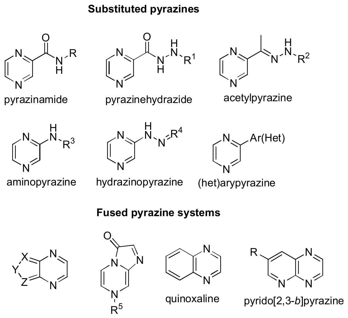
Figure 2.
Molecular structure of N-(quinoline-8-yl)pyrazine-2-carboxamide sensor 1 and proposed interaction mode with Zn²⁺ upon addition of Zn(ClO4)2×6H2O.
Figure 2.
Molecular structure of N-(quinoline-8-yl)pyrazine-2-carboxamide sensor 1 and proposed interaction mode with Zn²⁺ upon addition of Zn(ClO4)2×6H2O.
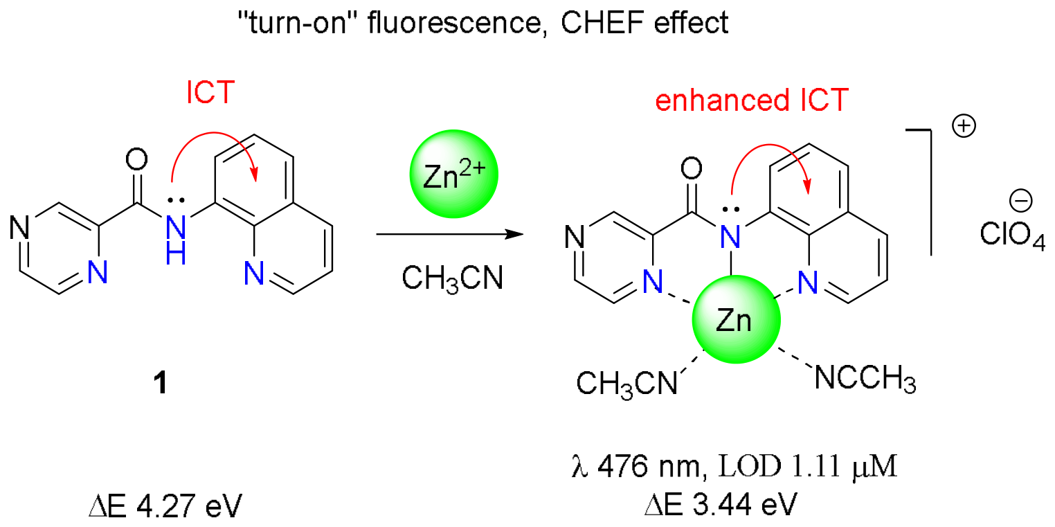
Figure 3.
Molecular structure of a pyrazine-2-carboxamide-based fluorescent sensor 2 and proposed interaction mode with Cu²⁺ or Pb²⁺ upon addition of CuCl2×2H2O or PbCl2, respectively.
Figure 3.
Molecular structure of a pyrazine-2-carboxamide-based fluorescent sensor 2 and proposed interaction mode with Cu²⁺ or Pb²⁺ upon addition of CuCl2×2H2O or PbCl2, respectively.
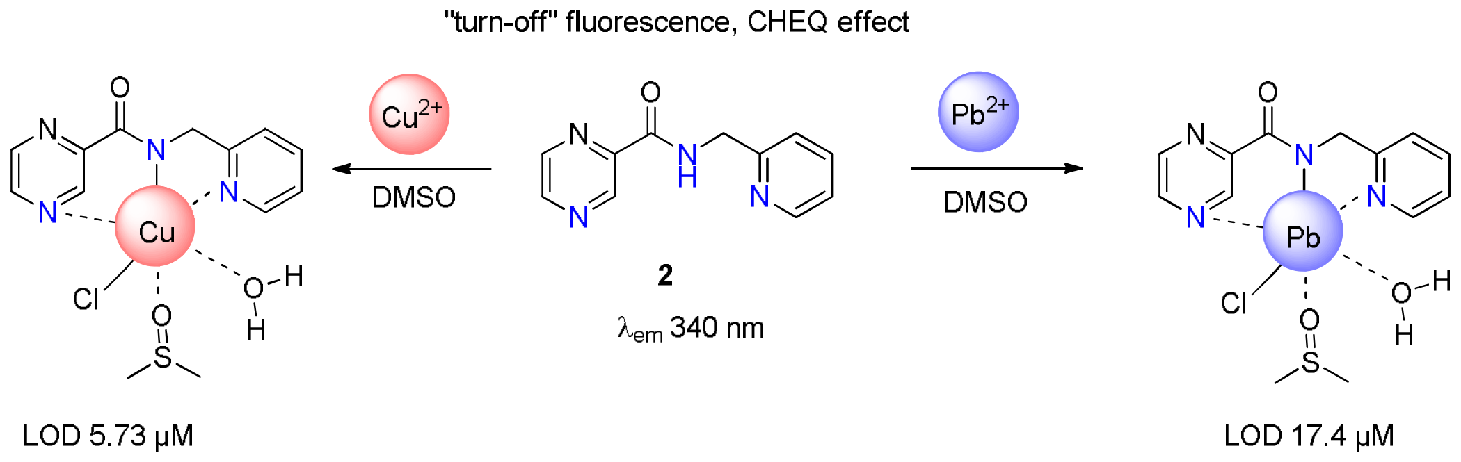
Figure 4.
Molecular structure of N-(2-halophenyl)pyrazine-2-carboxamides 3а,b and proposed interaction mode with Fe3+ upon addition of FeCl3×6H2O.
Figure 4.
Molecular structure of N-(2-halophenyl)pyrazine-2-carboxamides 3а,b and proposed interaction mode with Fe3+ upon addition of FeCl3×6H2O.
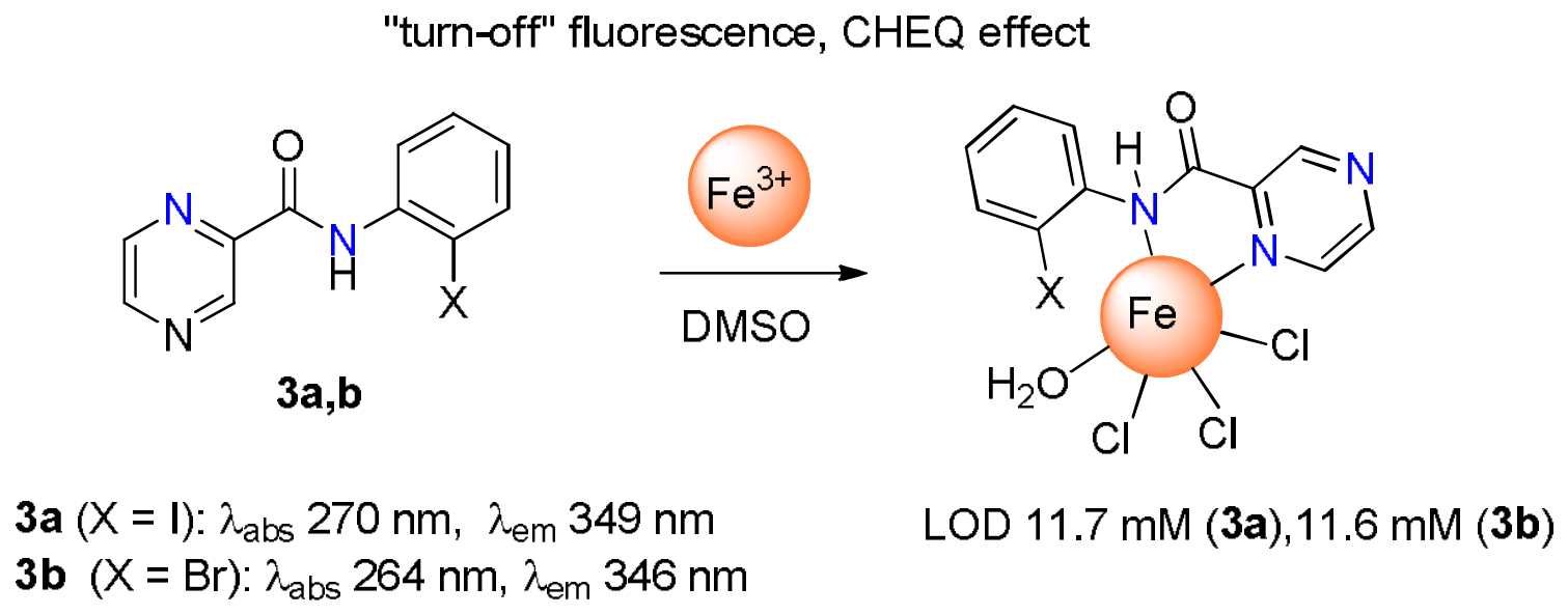
Figure 5.
Molecular structure of pyrazine-2-carboxamides-based sensors 4a-c and their sensing properties toward Cu2+.
Figure 5.
Molecular structure of pyrazine-2-carboxamides-based sensors 4a-c and their sensing properties toward Cu2+.
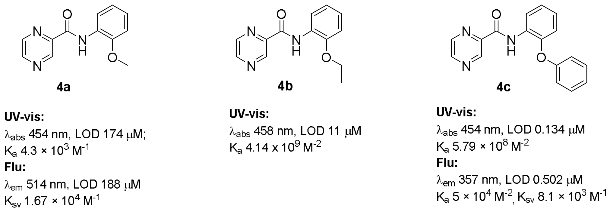
Scheme 1.
Synthesis of 4a-Cu2+ complexes.
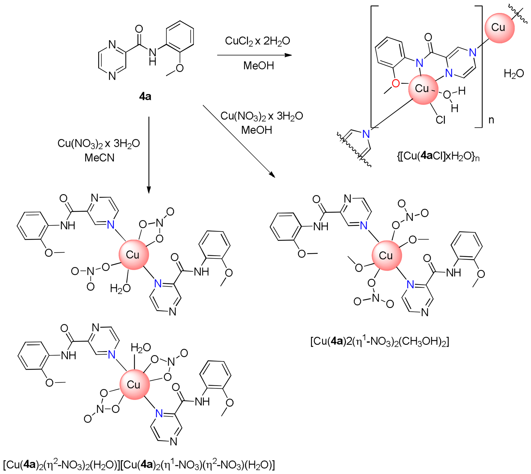
Scheme 2.
Synthesis of 4c-Cu2+ complex.
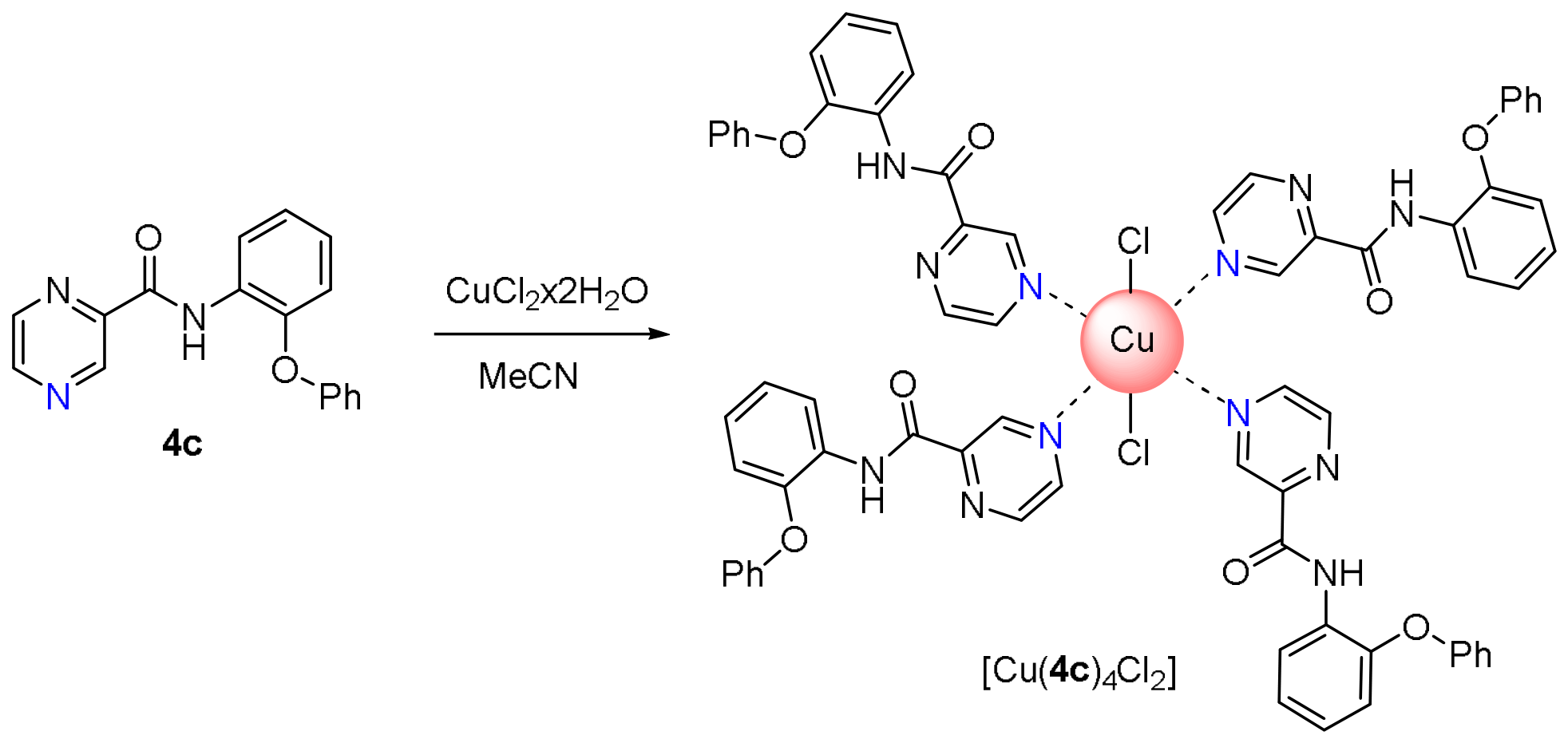
Figure 6.
Molecular structure of hydrazone 5 based on pyrazine-2-carbohydrazide and N-ethylcarbazole-3-carbaldehyde and proposed interaction mode with Сu2+ cation upon addition of Cu(NO3)2.
Figure 6.
Molecular structure of hydrazone 5 based on pyrazine-2-carbohydrazide and N-ethylcarbazole-3-carbaldehyde and proposed interaction mode with Сu2+ cation upon addition of Cu(NO3)2.
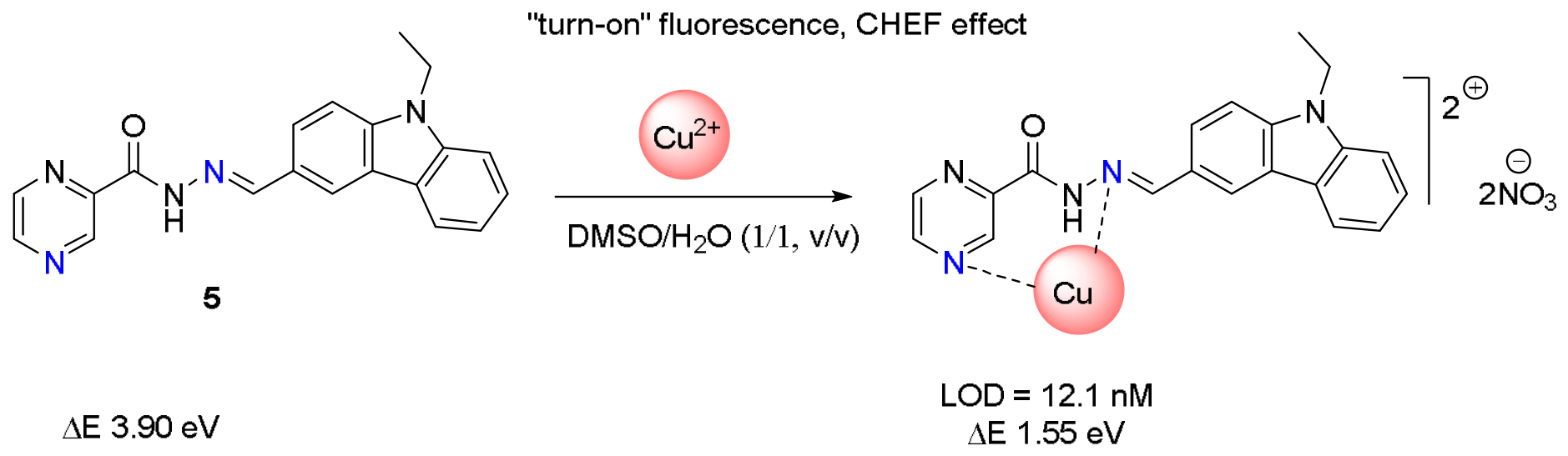
Figure 7.
Molecular structure of chromone-based Schiff base 8 and the proposed interaction mode with Cu2+ upon addition of Cu(NO3)2×H₂O.
Figure 7.
Molecular structure of chromone-based Schiff base 8 and the proposed interaction mode with Cu2+ upon addition of Cu(NO3)2×H₂O.
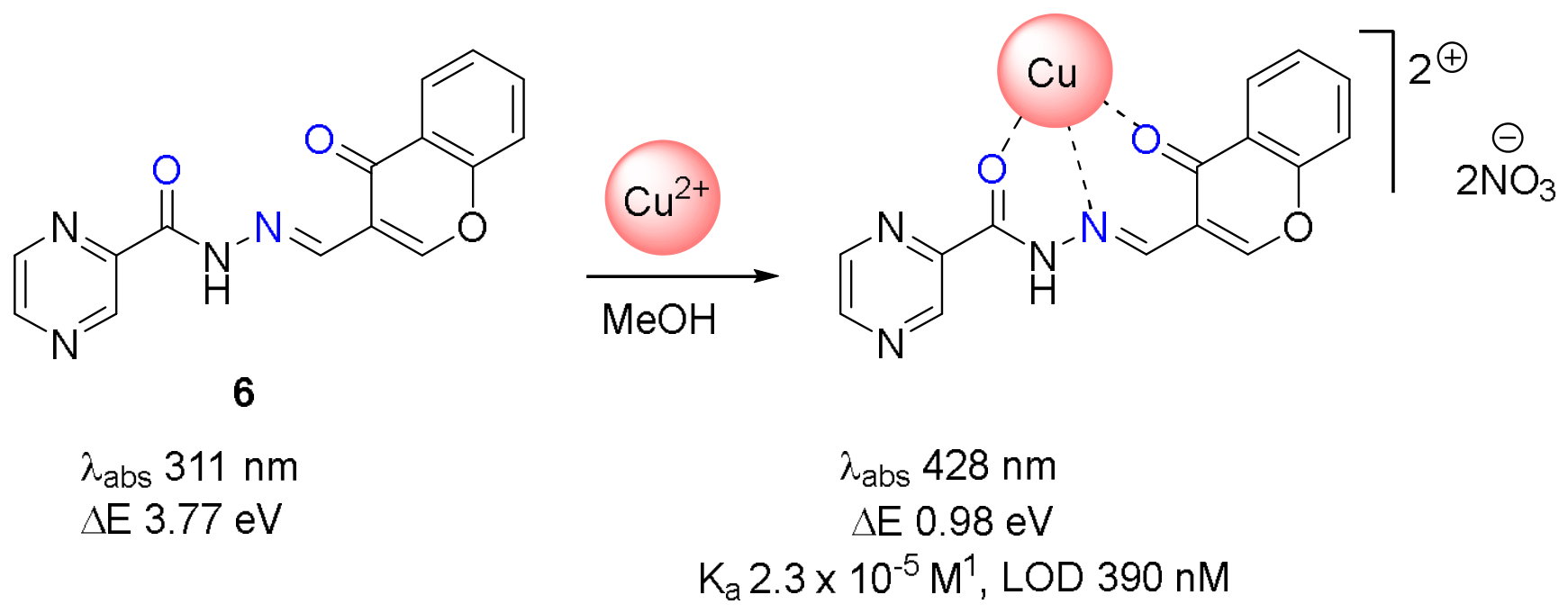
Figure 8.
Pyrazine-based 1,2,3-triazole-linked organosilane 7 and proposed its binding mode with Cu2+ upon addition of CuСl2.
Figure 8.
Pyrazine-based 1,2,3-triazole-linked organosilane 7 and proposed its binding mode with Cu2+ upon addition of CuСl2.
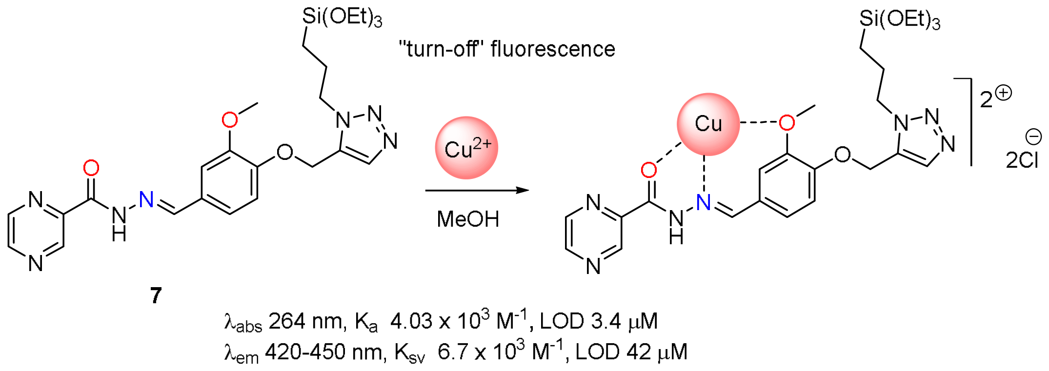
Figure 9.
Chemosensor 8, constructed from 3-amino-pyrazine-2-carbohydrazide and 7-diethylamino-3-formylcoumarin, and its binding mode with Zn²⁺ upon addition of Zn(NO3)2.
Figure 9.
Chemosensor 8, constructed from 3-amino-pyrazine-2-carbohydrazide and 7-diethylamino-3-formylcoumarin, and its binding mode with Zn²⁺ upon addition of Zn(NO3)2.
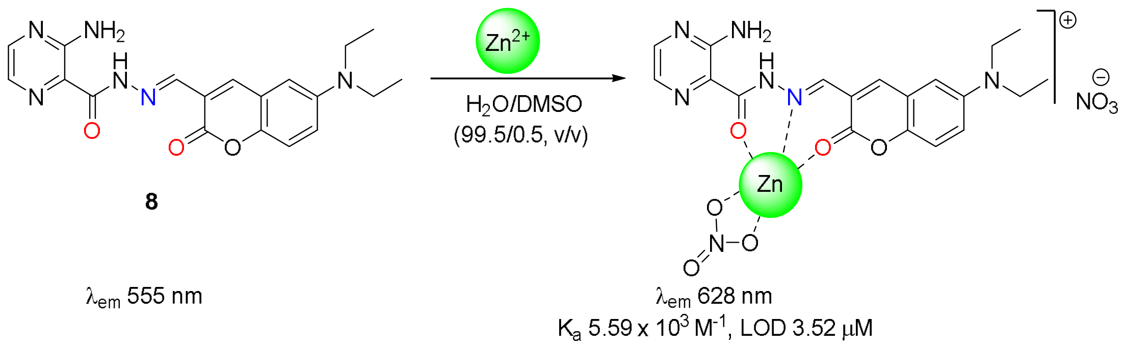
Figure 10.
Chemosensor 9 and proposed interaction mode of 9 with Zn2+. The nature of anion X- is not specified.
Figure 10.
Chemosensor 9 and proposed interaction mode of 9 with Zn2+. The nature of anion X- is not specified.
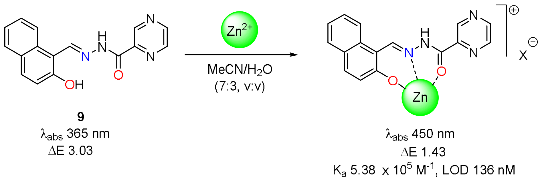
Scheme 3.
Schematic illustration of the binding interactions of pyrazine-based sensor 10 with Al3+, Mg2+, and Zn2+ ions upon addition of their nitrate salts.
Scheme 3.
Schematic illustration of the binding interactions of pyrazine-based sensor 10 with Al3+, Mg2+, and Zn2+ ions upon addition of their nitrate salts.
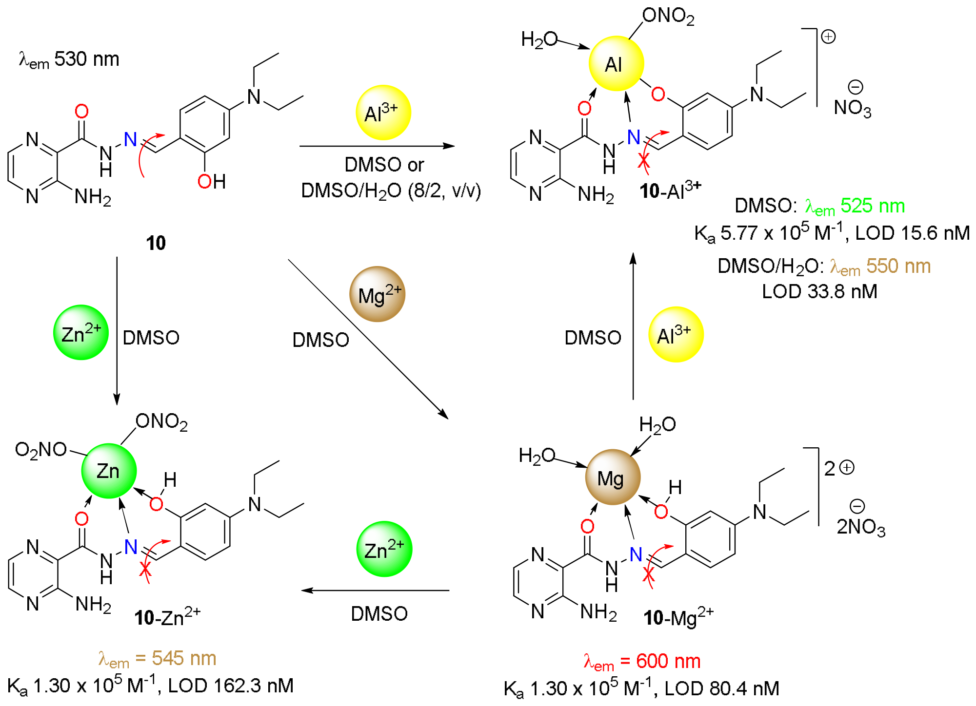
Scheme 4.
Molecular structure of sensor 11 and the plausible mechanism for fluorescent turn-on upon addition of H2O or Al(NO3)3.
Scheme 4.
Molecular structure of sensor 11 and the plausible mechanism for fluorescent turn-on upon addition of H2O or Al(NO3)3.
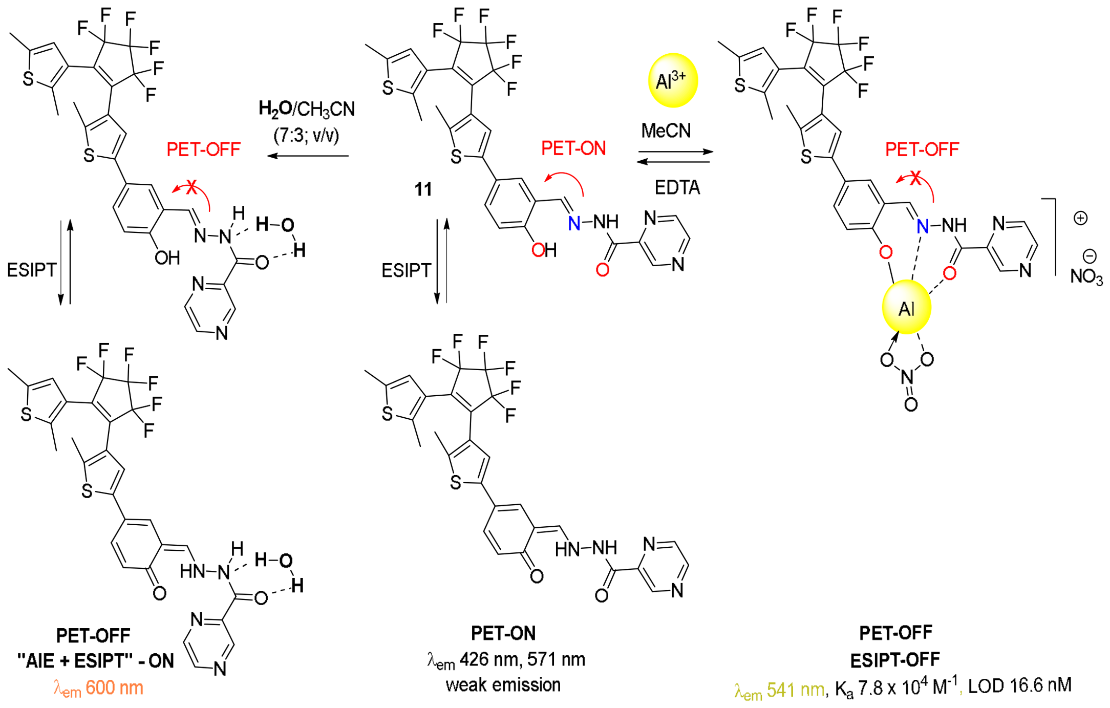
Figure 11.
Vitamin B6-based fluorescent sensor 12 and its proposed interaction with Al3+ upon subsequent addition of Al(NO3)3.
Figure 11.
Vitamin B6-based fluorescent sensor 12 and its proposed interaction with Al3+ upon subsequent addition of Al(NO3)3.
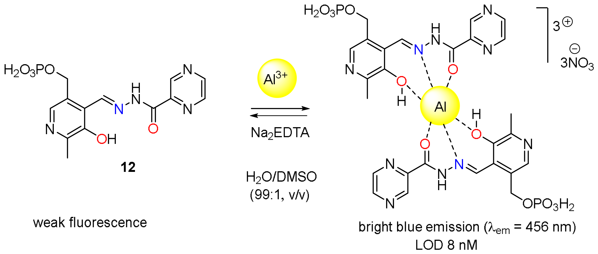
Figure 12.
Thiazole-derived pyrazin-2-carbohydrazide chemosensor 13 and its proposed interaction with Ag+ cation upon addition of AgNO3.
Figure 12.
Thiazole-derived pyrazin-2-carbohydrazide chemosensor 13 and its proposed interaction with Ag+ cation upon addition of AgNO3.
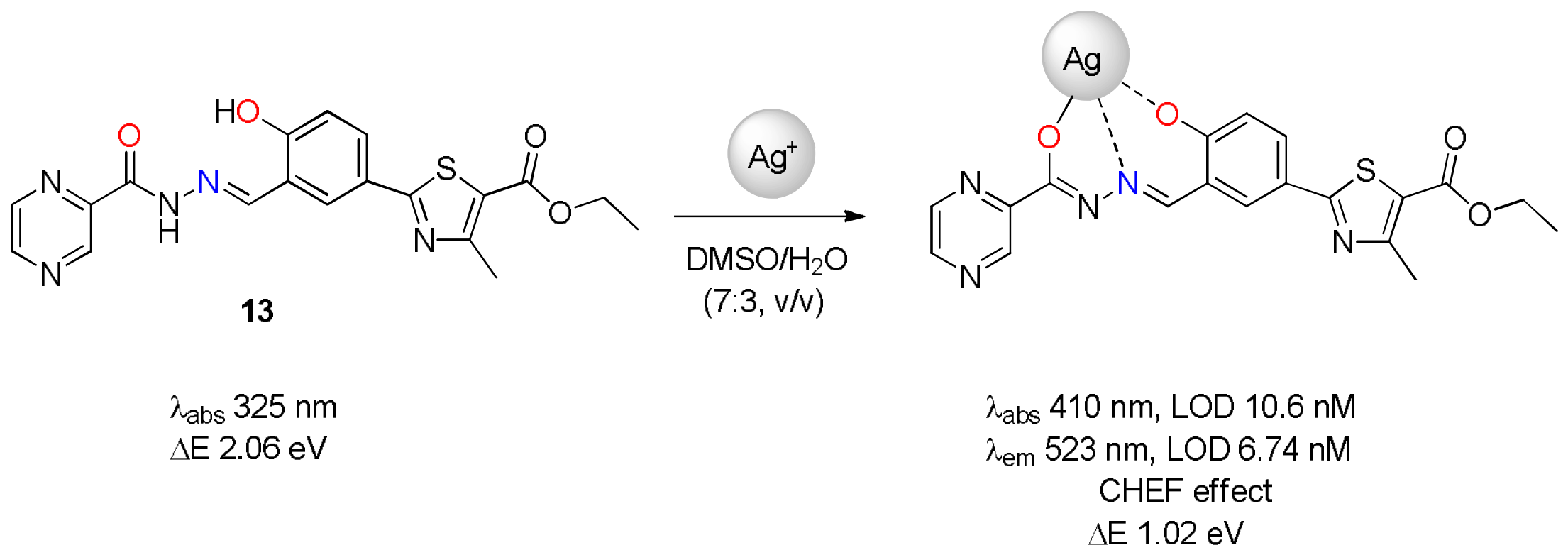
Scheme 5.
Molecular structure of BODIPY pyrazine-attached Schiff base chemosensor 14 and proposed mechanism for Al3+ sensing upon addition of Al(NO3)3 (>1.0 equiv.).
Scheme 5.
Molecular structure of BODIPY pyrazine-attached Schiff base chemosensor 14 and proposed mechanism for Al3+ sensing upon addition of Al(NO3)3 (>1.0 equiv.).
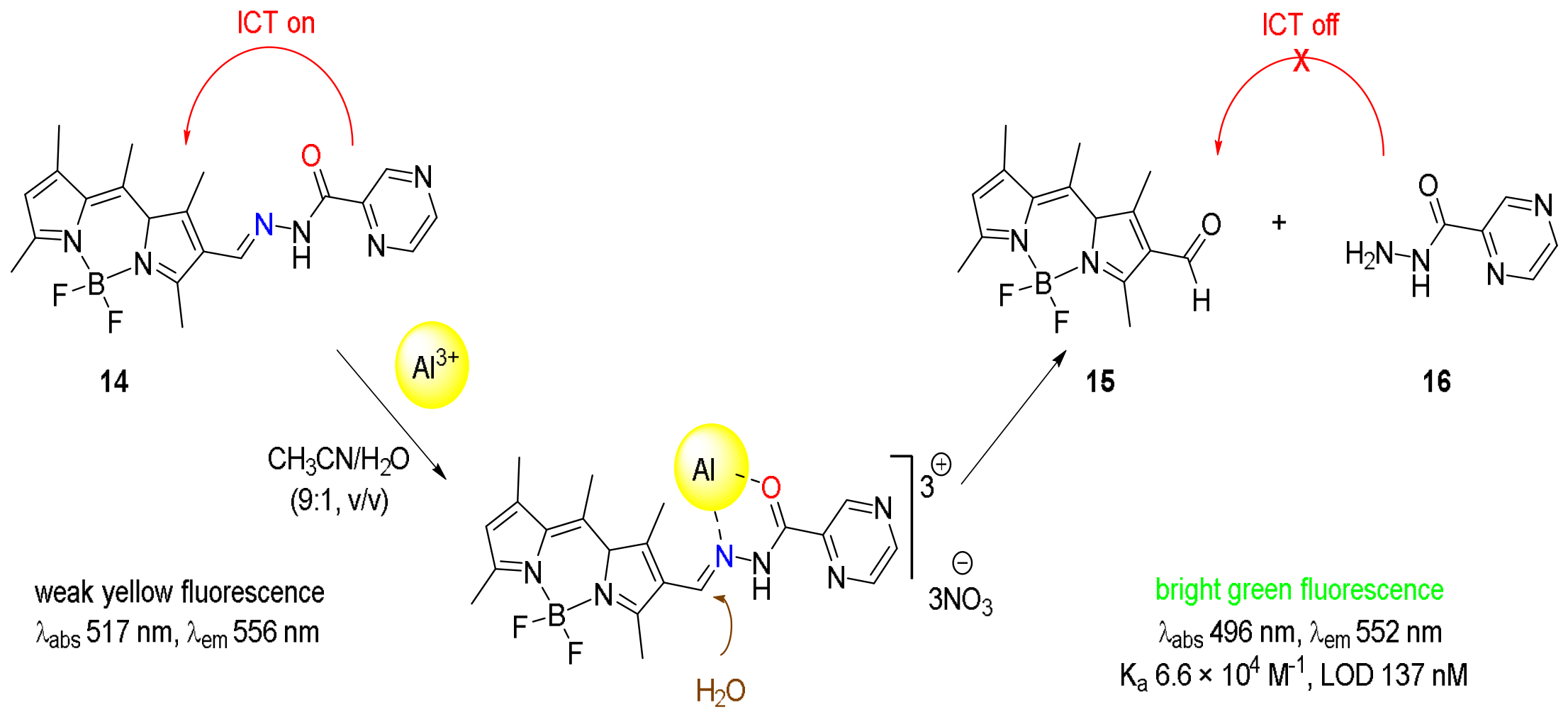
Figure 13.
Proposed binding mode of (2-acetylpyrazine- (8′-hydroxyquinolineyl-2′-acetyl)hydrazones 17a and 2-acetylpyrazine(2′-furanformyl)hydrazine 17b with Al3+. The nature of anion X- is not indicated.
Figure 13.
Proposed binding mode of (2-acetylpyrazine- (8′-hydroxyquinolineyl-2′-acetyl)hydrazones 17a and 2-acetylpyrazine(2′-furanformyl)hydrazine 17b with Al3+. The nature of anion X- is not indicated.
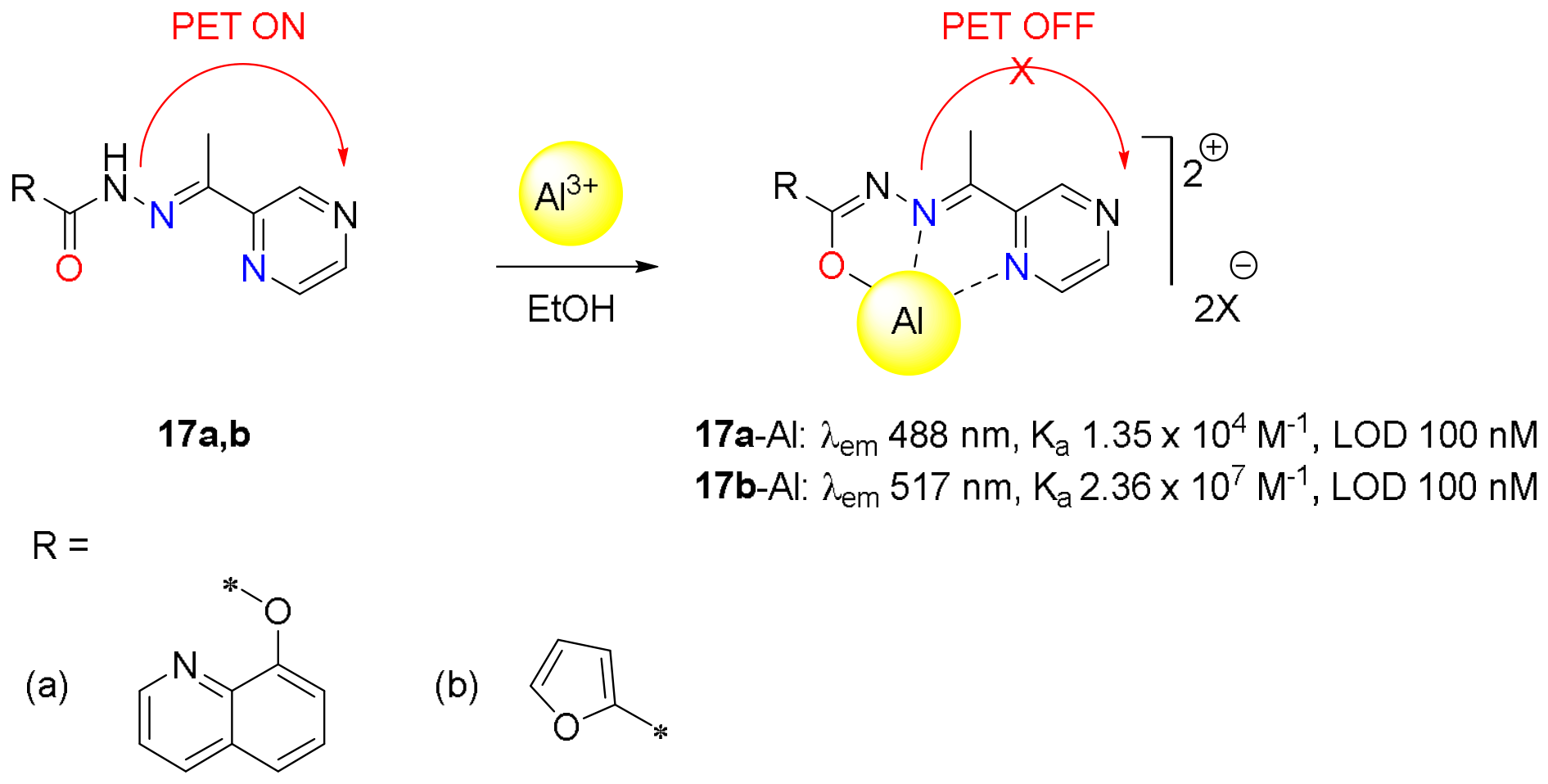
Figure 14.
(a) Fluorescence spectra of 17b (50 µM) upon addition of Al3+ (1 equiv.) and other metal ions (5 equiv.) in ethanol with an excitation at 382 nm; (b) Fluorescence spectra of 18 (50 µM) upon the titration of Al3+ (0.1, 0.2, 0.3, 0.4, 0.5, 0.6, 0.7, 0.8, 0.9, 1.0, 1.2, 1.4, 1.6, 1.8, 2.0 equiv., respectively) in ethanol with an excitation at 382 nm. Reproduced with permission of Royal Society of Chemistry [72].
Figure 14.
(a) Fluorescence spectra of 17b (50 µM) upon addition of Al3+ (1 equiv.) and other metal ions (5 equiv.) in ethanol with an excitation at 382 nm; (b) Fluorescence spectra of 18 (50 µM) upon the titration of Al3+ (0.1, 0.2, 0.3, 0.4, 0.5, 0.6, 0.7, 0.8, 0.9, 1.0, 1.2, 1.4, 1.6, 1.8, 2.0 equiv., respectively) in ethanol with an excitation at 382 nm. Reproduced with permission of Royal Society of Chemistry [72].
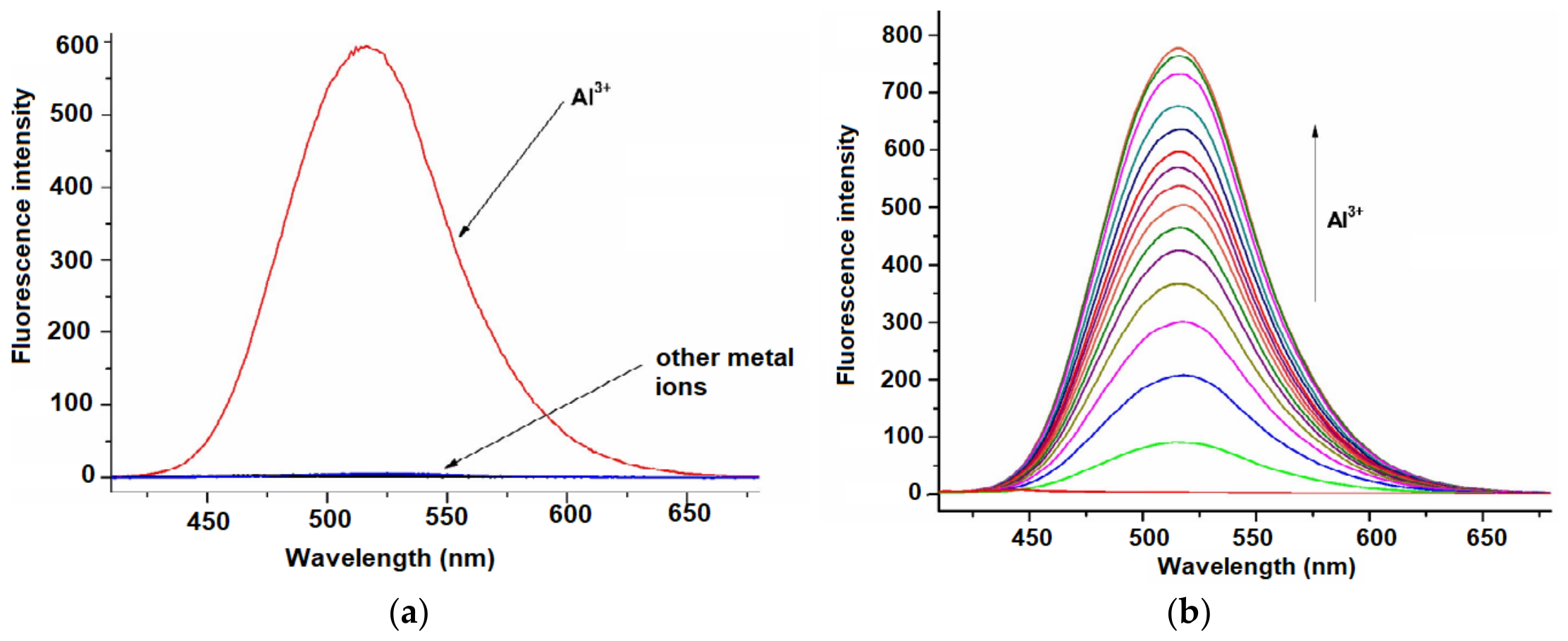
Figure 15.
Pyrene-based fluorescent sensor 18 and its proposed mechanism for Co2+ sensing upon addition of CoCl2.
Figure 15.
Pyrene-based fluorescent sensor 18 and its proposed mechanism for Co2+ sensing upon addition of CoCl2.
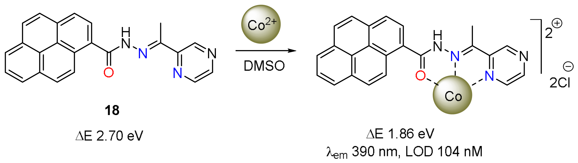
Figure 16.
Proposed binding mode of sensor 19 with Co2+ upon addition of CoCl2.
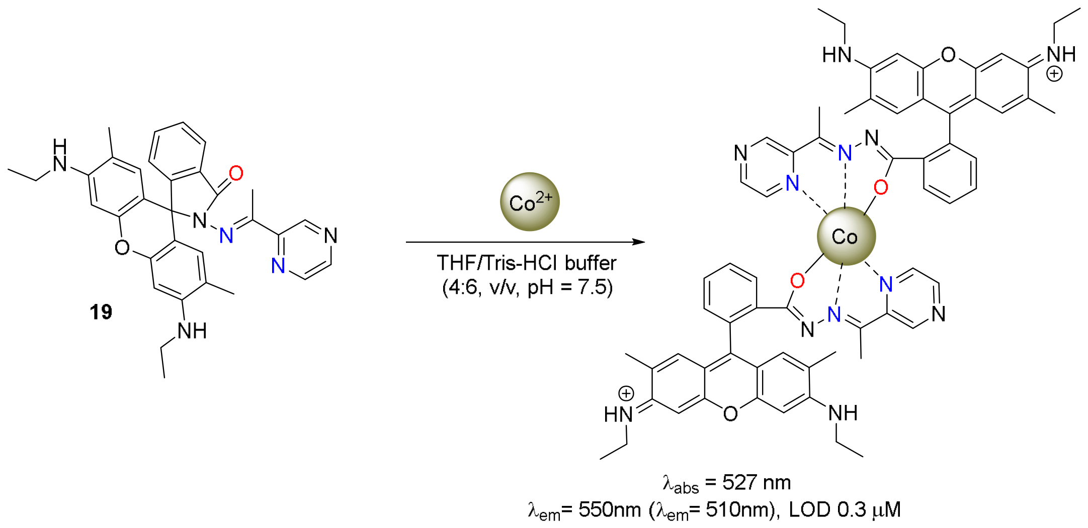
Figure 17.
Proposed binding mode of 3-amino-2-pyrazinecarboxylic acid 20 with Сr3+ and the crystal structure of [(C5H4N3O2)3Cr] according to X-ray diffraction data.
Figure 17.
Proposed binding mode of 3-amino-2-pyrazinecarboxylic acid 20 with Сr3+ and the crystal structure of [(C5H4N3O2)3Cr] according to X-ray diffraction data.
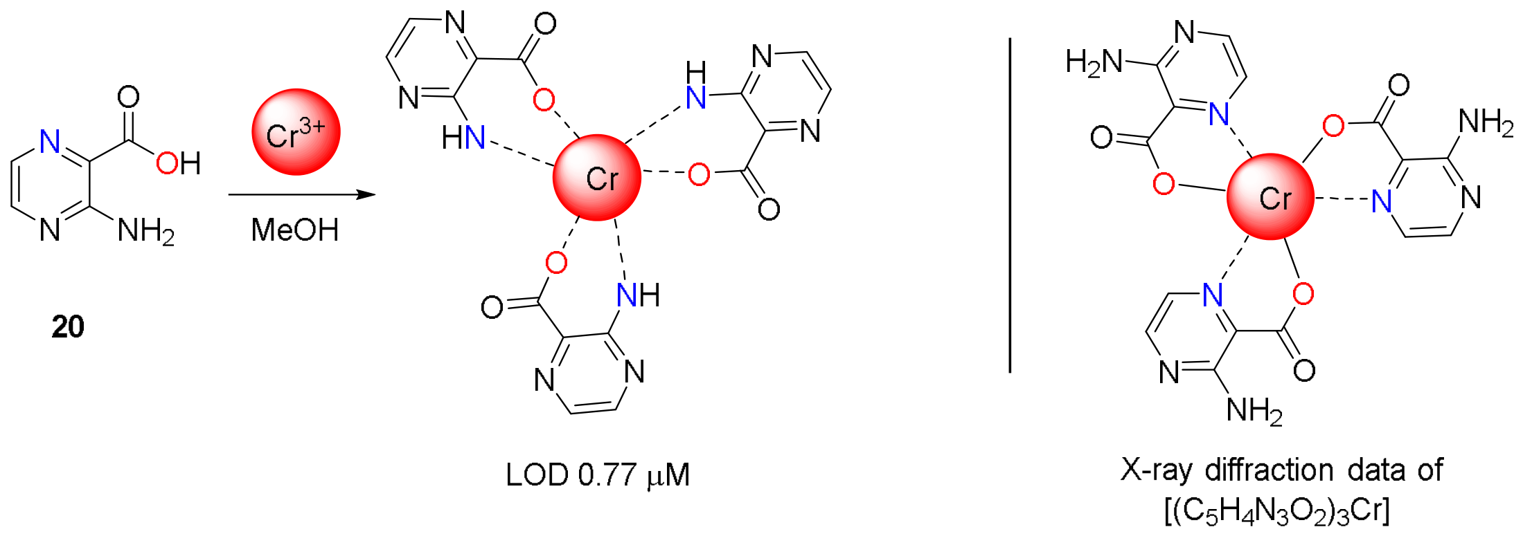
Figure 18.
Molecular structure of sensor 21 and its proposed binding mode with Hg2+ and Ni2+ upon addition of their chlorides.
Figure 18.
Molecular structure of sensor 21 and its proposed binding mode with Hg2+ and Ni2+ upon addition of their chlorides.
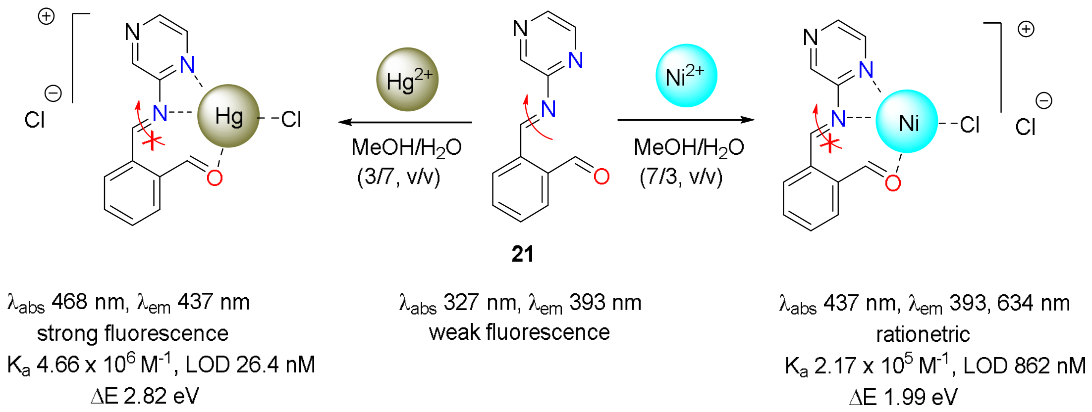
Figure 19.
Molecular structure of sensor 22 and its proposed binding mode with Al3+ upon addition of Al2(SO4)3.
Figure 19.
Molecular structure of sensor 22 and its proposed binding mode with Al3+ upon addition of Al2(SO4)3.
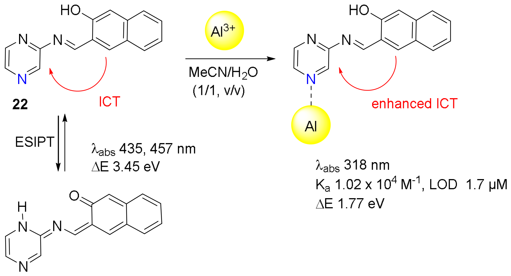
Scheme 6.
Tautomerization of sensor 23 and the proposed coordination mode with Cu²⁺ upon addition of Cu(NO3)2.
Scheme 6.
Tautomerization of sensor 23 and the proposed coordination mode with Cu²⁺ upon addition of Cu(NO3)2.
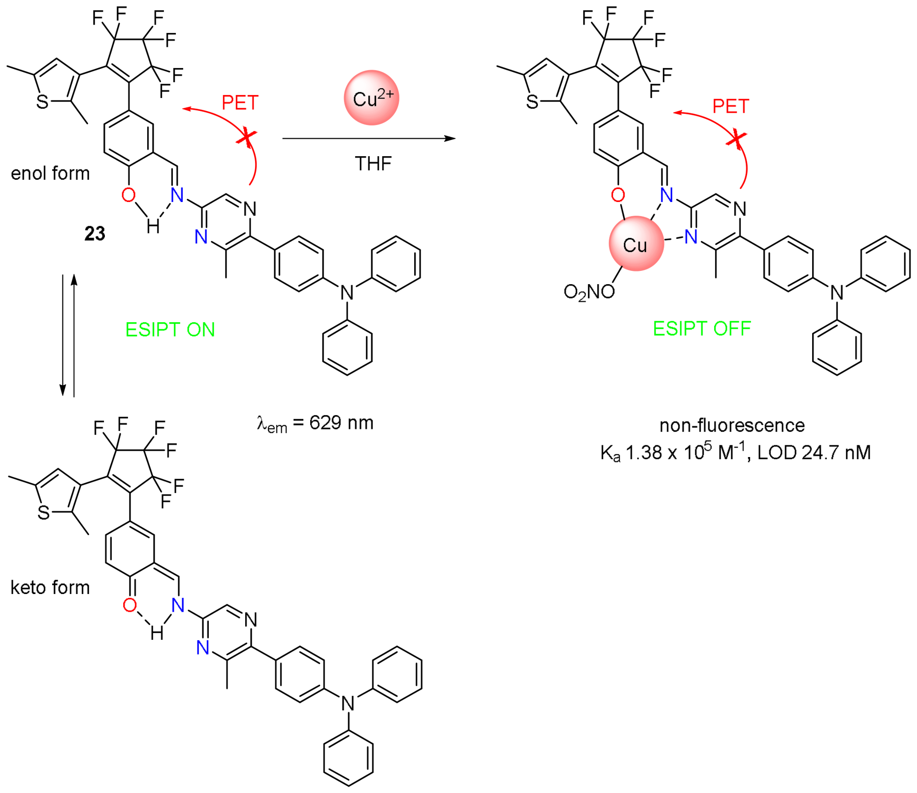
Figure 20.
Molecular structure of chemosensor 24 and its proposed binding mode with Al3+ and Zn2+ upon addition of Al(ClO4)3 or Zn(ClO4)2.
Figure 20.
Molecular structure of chemosensor 24 and its proposed binding mode with Al3+ and Zn2+ upon addition of Al(ClO4)3 or Zn(ClO4)2.
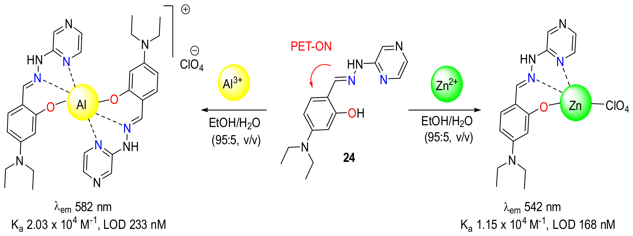
Figure 21.
(a) Fluorescence spectrum changes of 24 (50 μM) on addition of various metal ions (2.0 equiv.) in ethanol–water (95:5, v/v) (ex = 366 nm). (b) The color changes of the 24-coated cotton swab in the absence and presence of Al3+ and Zn2+. Reproduced from [81] licensed under Creative Commons BY 4.0.
Figure 21.
(a) Fluorescence spectrum changes of 24 (50 μM) on addition of various metal ions (2.0 equiv.) in ethanol–water (95:5, v/v) (ex = 366 nm). (b) The color changes of the 24-coated cotton swab in the absence and presence of Al3+ and Zn2+. Reproduced from [81] licensed under Creative Commons BY 4.0.
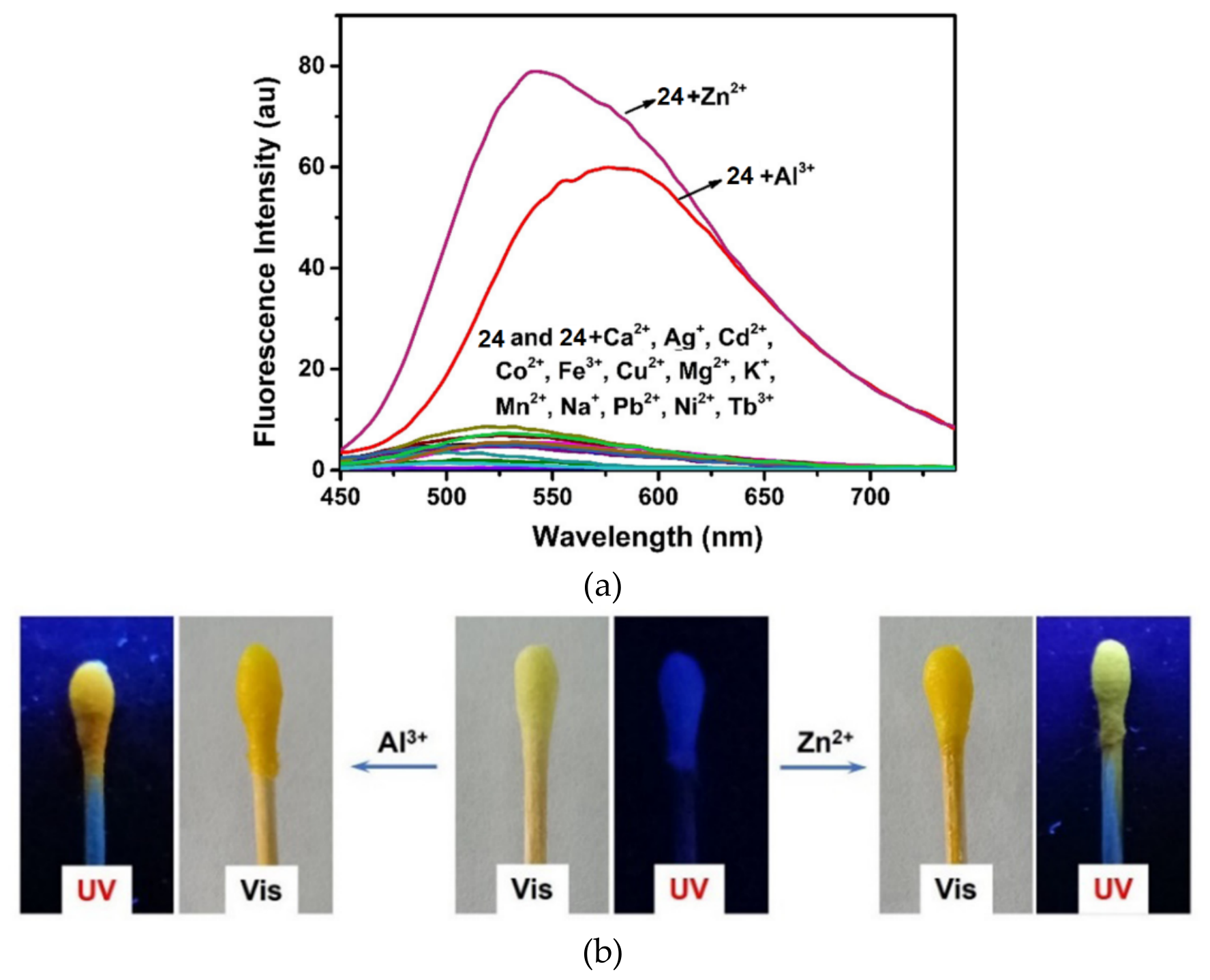
Figure 22.
Molecular structure of sensors 25–27.
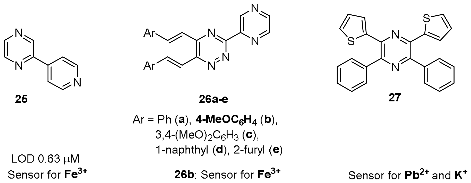
Figure 23.
Molecular structure of dual-mode coordinated chemosensor 28 based on pyrazine ligand.
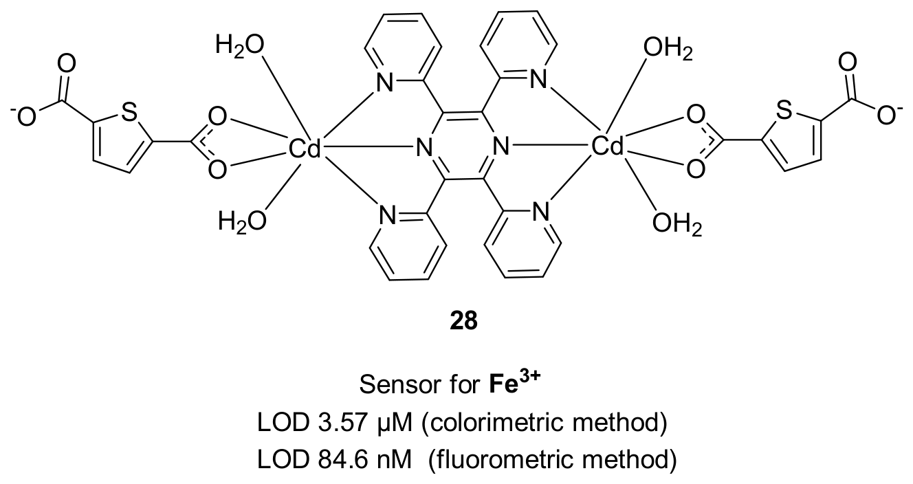
Figure 24.
Molecular structure of fluorescein-based chemosensors 29a-c and the proposed Zn²⁺-binding upon addition of ZnCl2 at pH 7.0.
Figure 24.
Molecular structure of fluorescein-based chemosensors 29a-c and the proposed Zn²⁺-binding upon addition of ZnCl2 at pH 7.0.
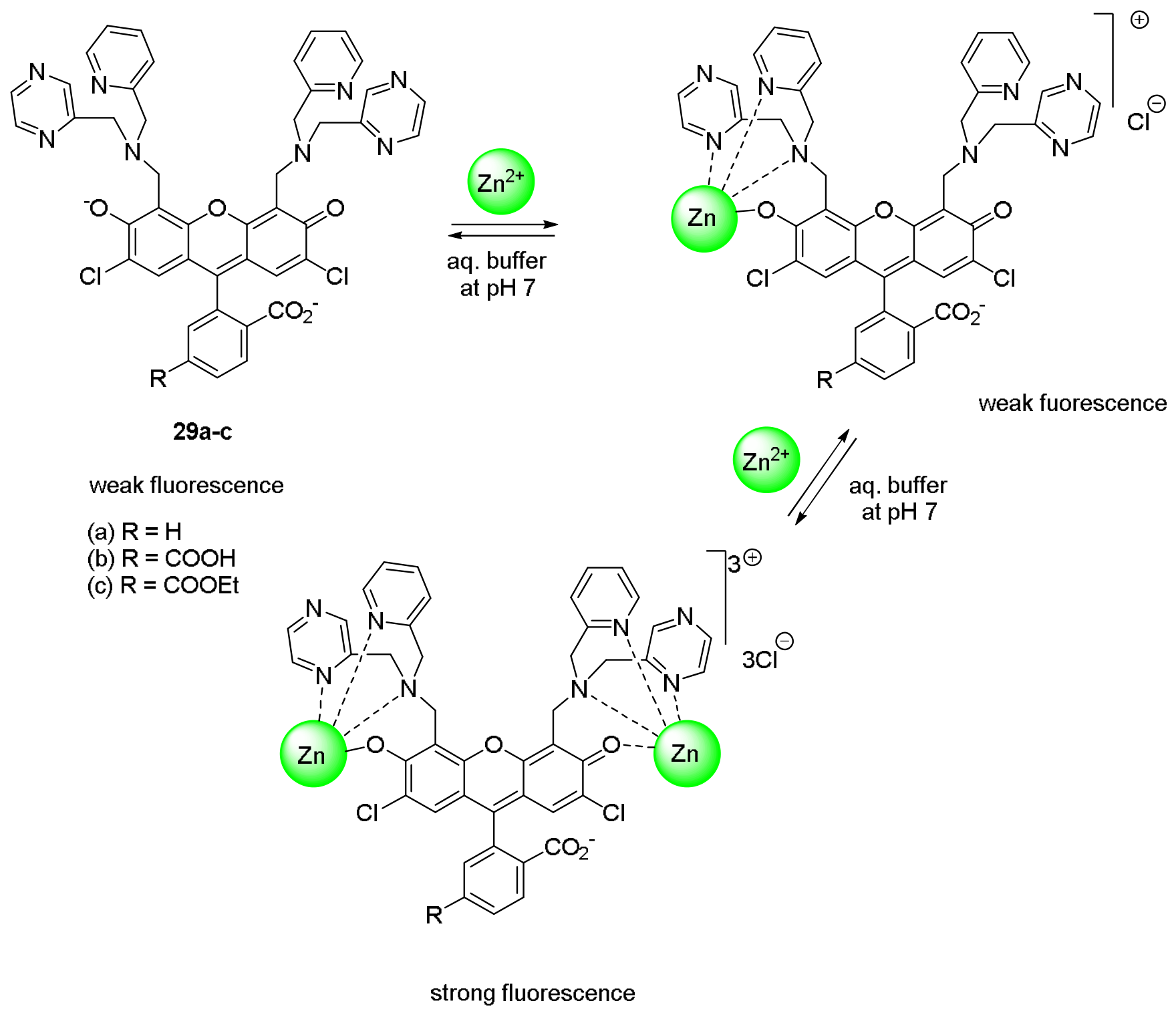
Figure 25.
Molecular structure of carbazole functionalized pyrazine conjugated polymers 30a-c.
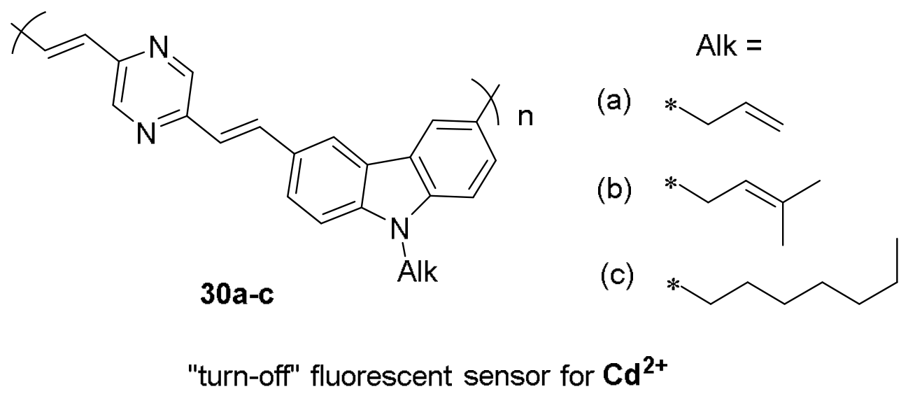
Figure 26.
A ninhydrin based chemosensor 31 and its proposed binding mode with Hg2+ upon addition of (CH3COO)2Hg.
Figure 26.
A ninhydrin based chemosensor 31 and its proposed binding mode with Hg2+ upon addition of (CH3COO)2Hg.
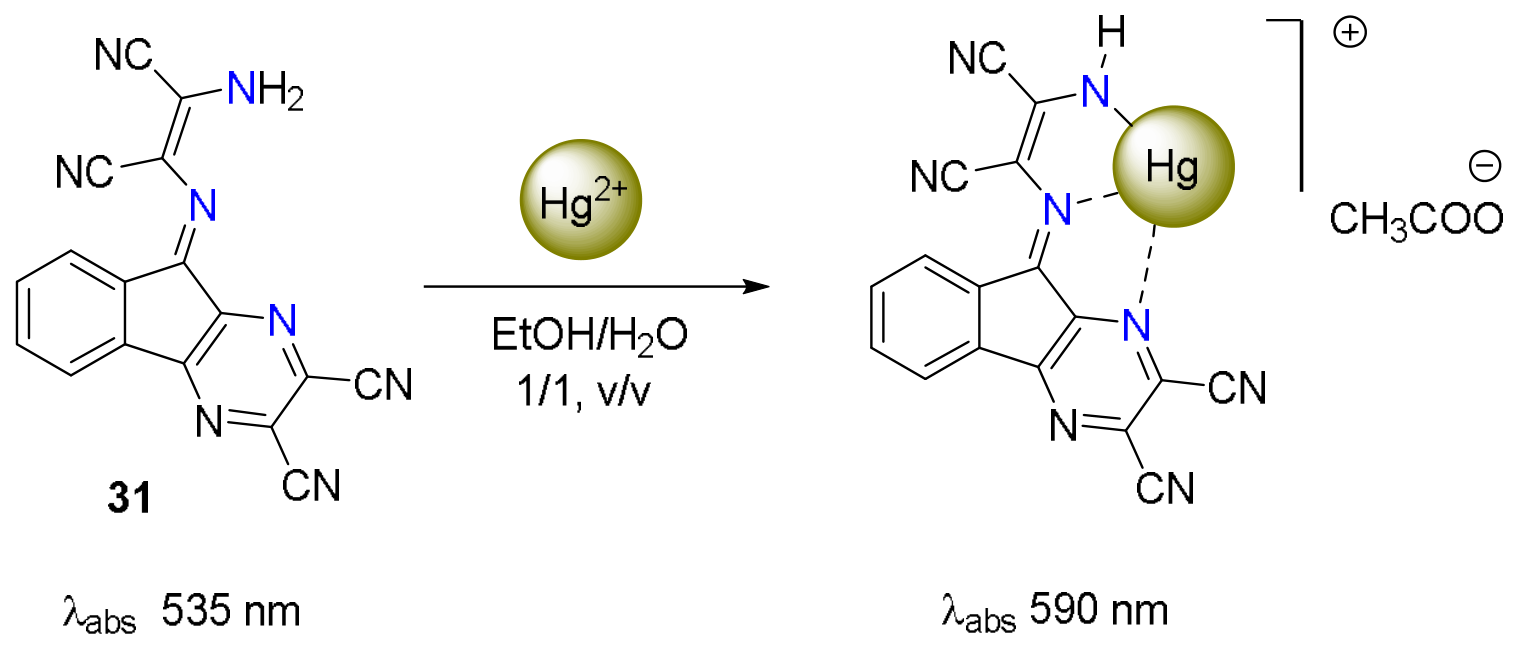
Figure 27.
Molecular structure of 1H-imidazo [4,5-b]pyrazine sensor 32a-c and proposed binding mode with Pd2+ upon addition of PdCl2.
Figure 27.
Molecular structure of 1H-imidazo [4,5-b]pyrazine sensor 32a-c and proposed binding mode with Pd2+ upon addition of PdCl2.
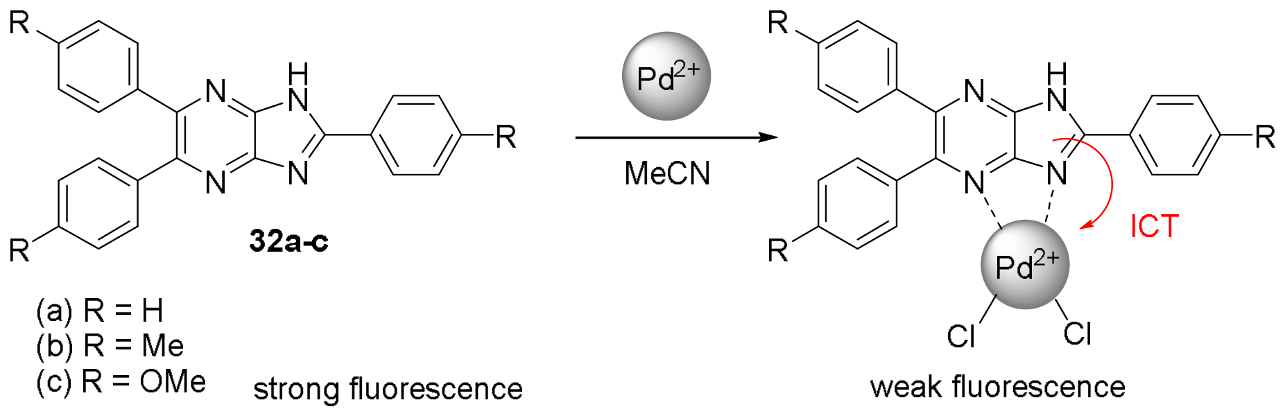
Figure 28.
Molecular structures of imidazo [1,2-a]pyrazin-3(7H)-one sensors 33a-e and 34a,b and schematic representation of their interaction with various metal cations upon addition of perchlorate or trifluoromethanesulfonate salts (Xn-).
Figure 28.
Molecular structures of imidazo [1,2-a]pyrazin-3(7H)-one sensors 33a-e and 34a,b and schematic representation of their interaction with various metal cations upon addition of perchlorate or trifluoromethanesulfonate salts (Xn-).
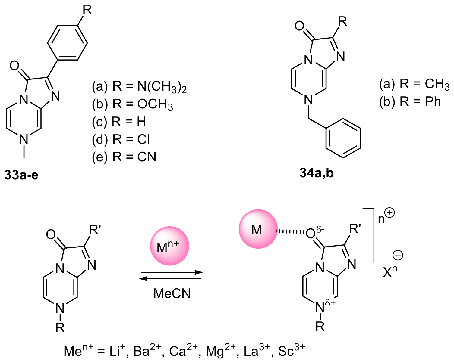
Figure 29.
Molecular structure of the D-π-A copolymers 35a and 35b.
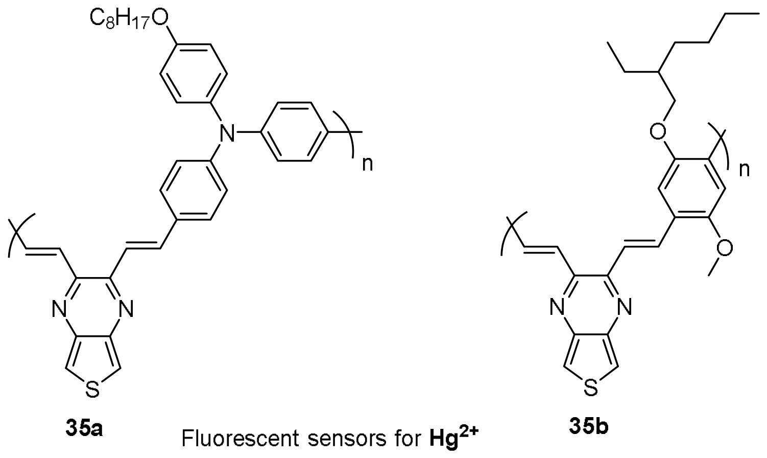
Figure 30.
Molecular structure of 2,3-bis(6-chloropyridin-2-yl)-6-fluoroquinoxaline 36 and its binding mode with Fe3+ upon addition of FeCl3×6H2O.
Figure 30.
Molecular structure of 2,3-bis(6-chloropyridin-2-yl)-6-fluoroquinoxaline 36 and its binding mode with Fe3+ upon addition of FeCl3×6H2O.
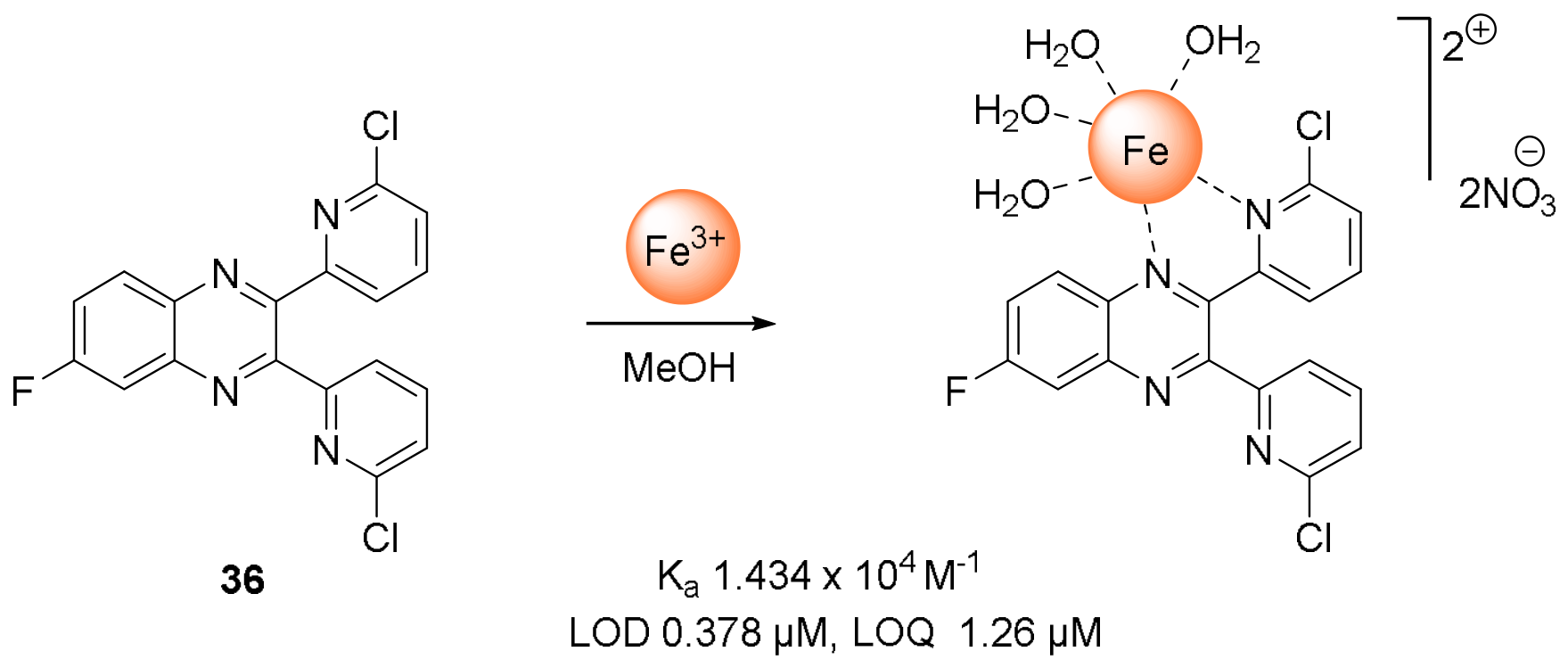
Figure 31.
Molecular structure of 2,3-dichloro-6-(2,7-dihydroxynaphth-1-ylazo)quinoxaline 37 and its binding mode with Sb3+ upon addition of potassium antimony tartrate.
Figure 31.
Molecular structure of 2,3-dichloro-6-(2,7-dihydroxynaphth-1-ylazo)quinoxaline 37 and its binding mode with Sb3+ upon addition of potassium antimony tartrate.
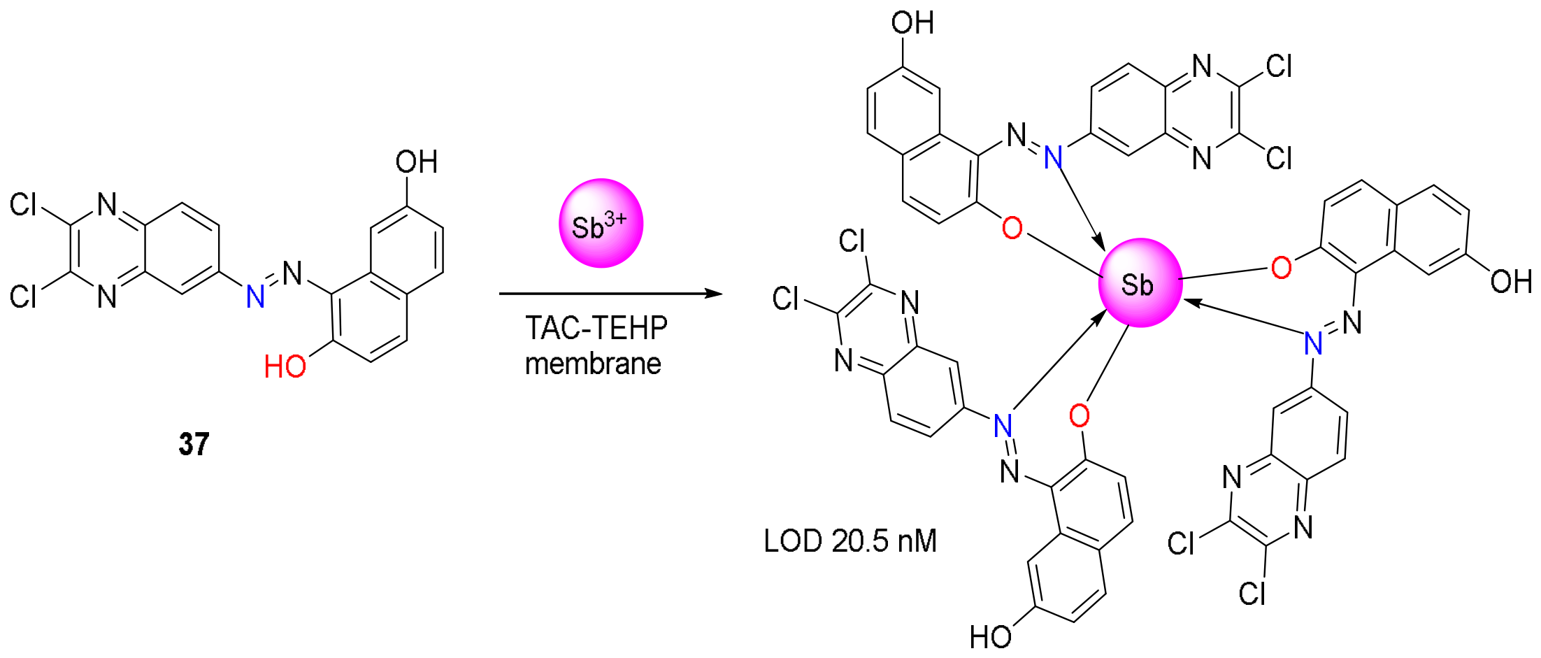
Figure 32.
Molecular structure of sensor 38 and its binding mode with ZnCl2.
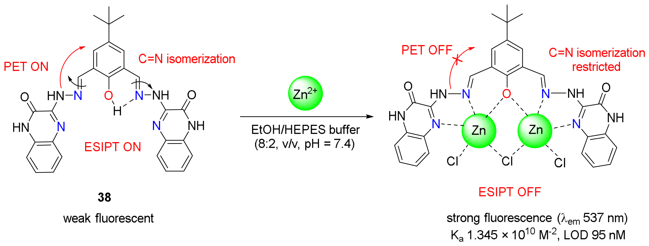
Figure 33.
Quinoxaline–triazole conjugate 39 as a selective turn-off fluorescent and colorimetric sensor for Fe³⁺ in methanol.
Figure 33.
Quinoxaline–triazole conjugate 39 as a selective turn-off fluorescent and colorimetric sensor for Fe³⁺ in methanol.
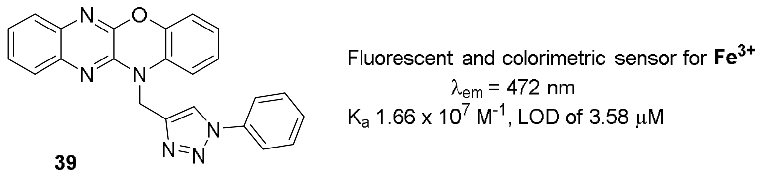
Figure 34.
Molecular structure of sensor 40 and its binding mode with Cu2+ upon addition of CuCl2.
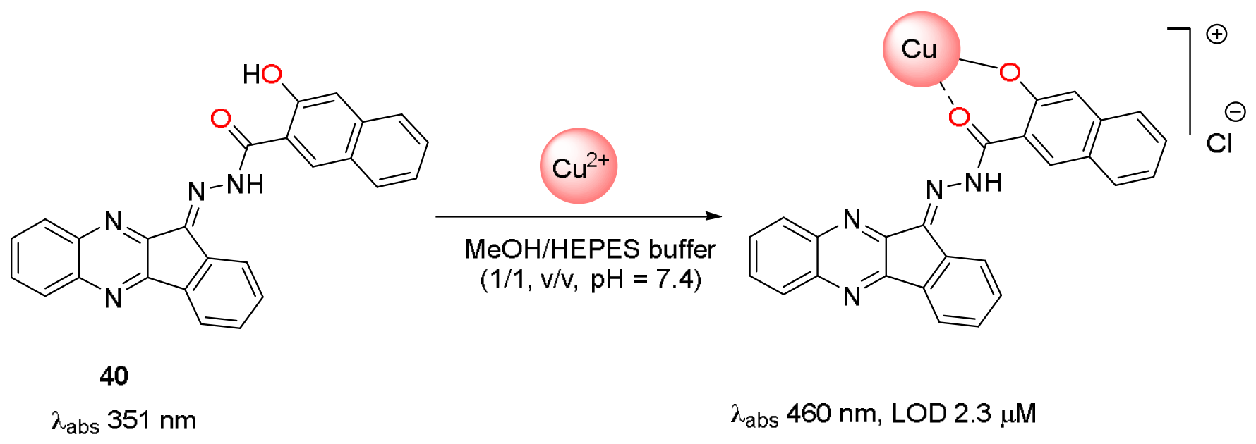
Figure 35.
(a) Molecular structure of quinoxaline–thiosemicarbazide conjugate 41; (b) proposed binding modes of 41 with metal cations in DMSO/H2O (8/2, v/v) upon addition of the corresponding nitrate or chloride salt.
Figure 35.
(a) Molecular structure of quinoxaline–thiosemicarbazide conjugate 41; (b) proposed binding modes of 41 with metal cations in DMSO/H2O (8/2, v/v) upon addition of the corresponding nitrate or chloride salt.
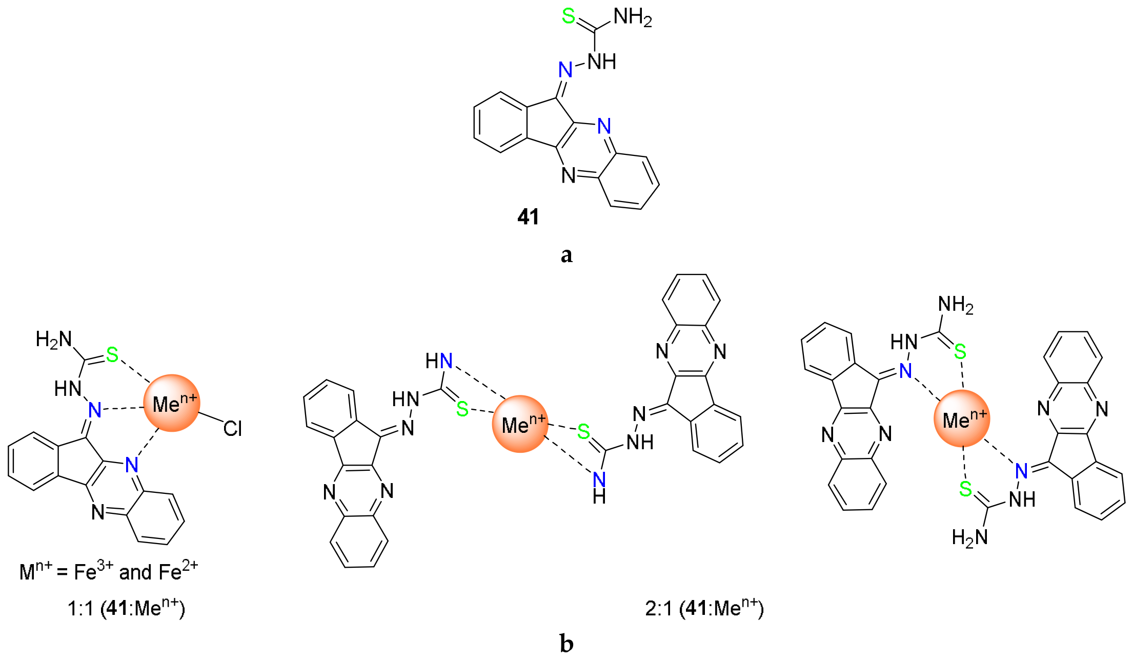
Figure 36.
Molecular structure of 2,3-di(1H-2-pyrrolyl)pyrido [2,3-b]pyrazine 42 and its binding mode with NiCl2.
Figure 36.
Molecular structure of 2,3-di(1H-2-pyrrolyl)pyrido [2,3-b]pyrazine 42 and its binding mode with NiCl2.
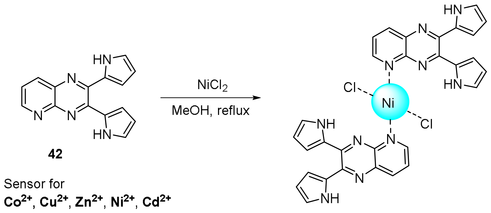
Figure 37.
Molecular structure of tetraphenylethene-substituted pyrido [2,3-b]pyrazine 43 and its binding mode with Hg2+ upon addition of Hg(NO3)2.
Figure 37.
Molecular structure of tetraphenylethene-substituted pyrido [2,3-b]pyrazine 43 and its binding mode with Hg2+ upon addition of Hg(NO3)2.
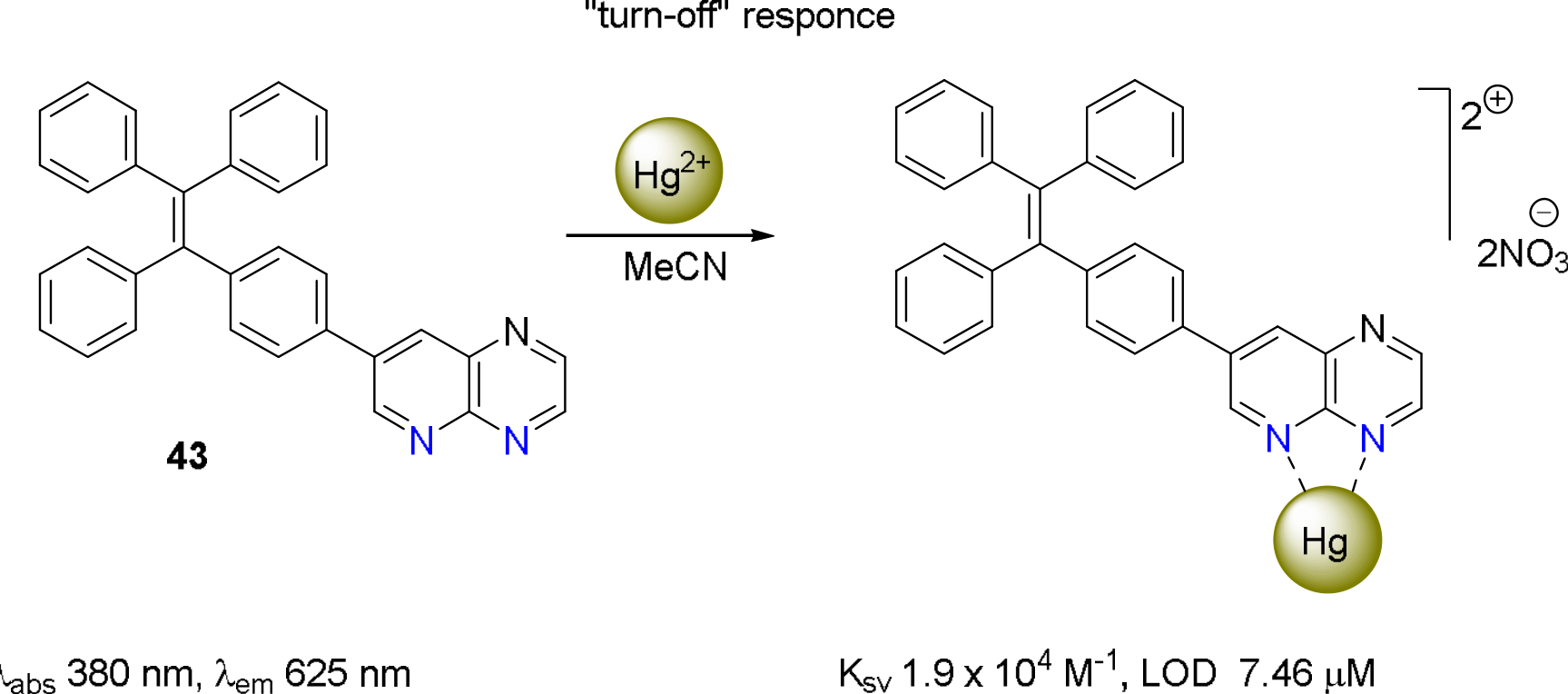
Figure 38.
(a) Fluorescence spectra of sensor 43 (2.0 × 10⁻5 mol L-1) towards various cations, excitation wavelength 380 nm; (b) Fluorescence photographs of sensor 43 after addition of various metal ions (7.0 equiv.) under 365 nm light. Reproduced with permission of Royal Society of Chemistry [102].
Figure 38.
(a) Fluorescence spectra of sensor 43 (2.0 × 10⁻5 mol L-1) towards various cations, excitation wavelength 380 nm; (b) Fluorescence photographs of sensor 43 after addition of various metal ions (7.0 equiv.) under 365 nm light. Reproduced with permission of Royal Society of Chemistry [102].
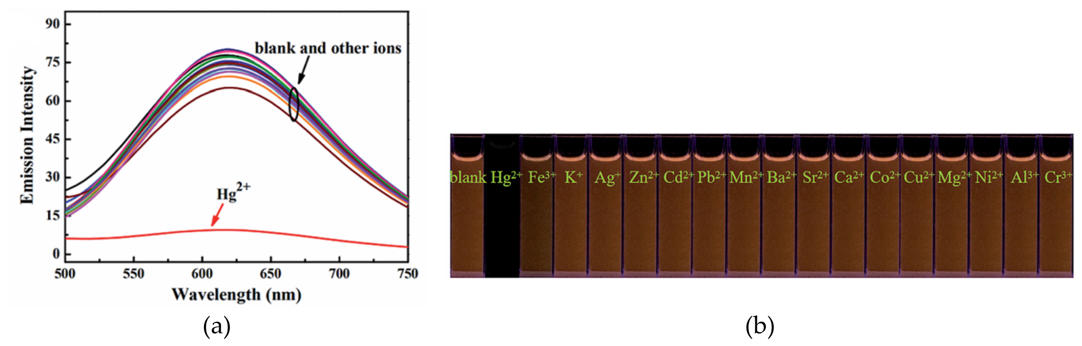
Scheme 7.
Photochromism of diarylethenes 44a,b with a styrene-linked pyrido [2,3-b]pyrazine unit.
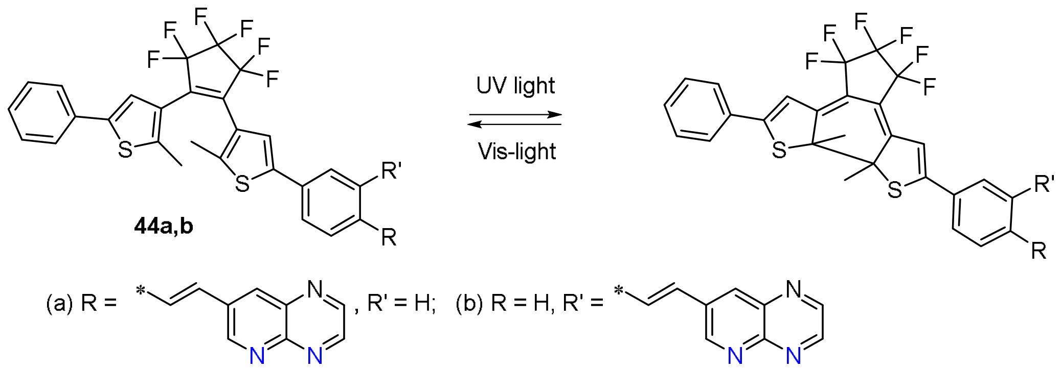
Scheme 8.
Dual-controlled fluorescent switching of photochromic diarylethene-pyrido [2,3-b]pyrazine derivative 44a in response to Hg2+/Sn2+ and UV-vis stimuli upon addition of Hg(NO3)2 or SnCl2.
Scheme 8.
Dual-controlled fluorescent switching of photochromic diarylethene-pyrido [2,3-b]pyrazine derivative 44a in response to Hg2+/Sn2+ and UV-vis stimuli upon addition of Hg(NO3)2 or SnCl2.
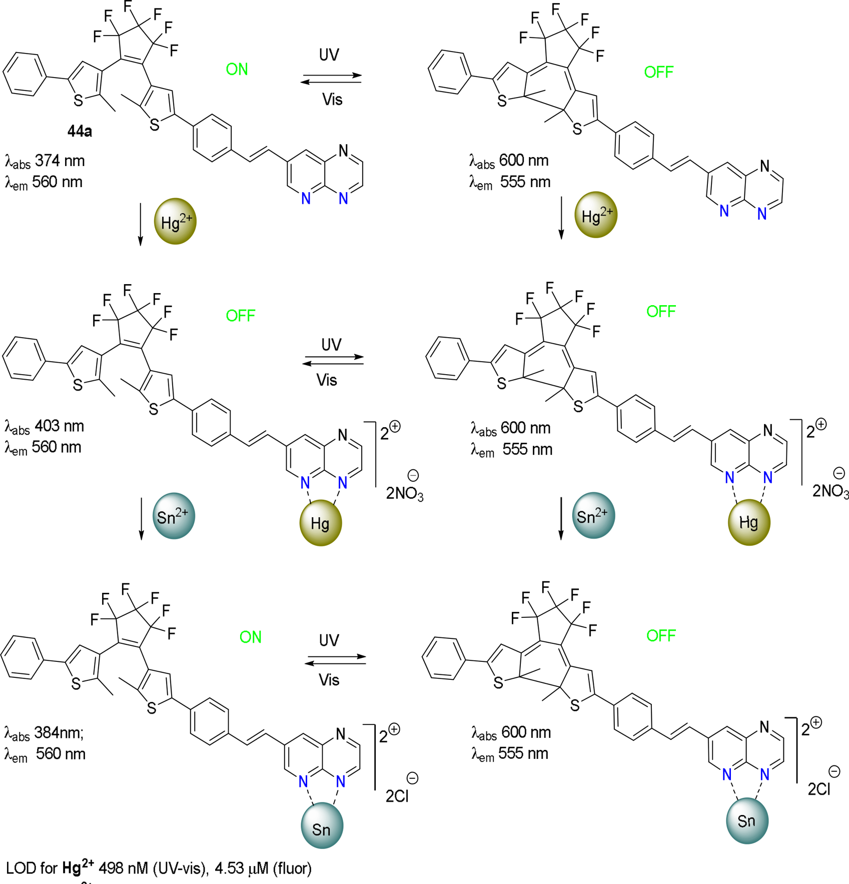
Figure 39.
Molecular structure of pyridopyrazine derivatives 45a-c and interaction pattern of 45a with ZnCl2.
Figure 39.
Molecular structure of pyridopyrazine derivatives 45a-c and interaction pattern of 45a with ZnCl2.
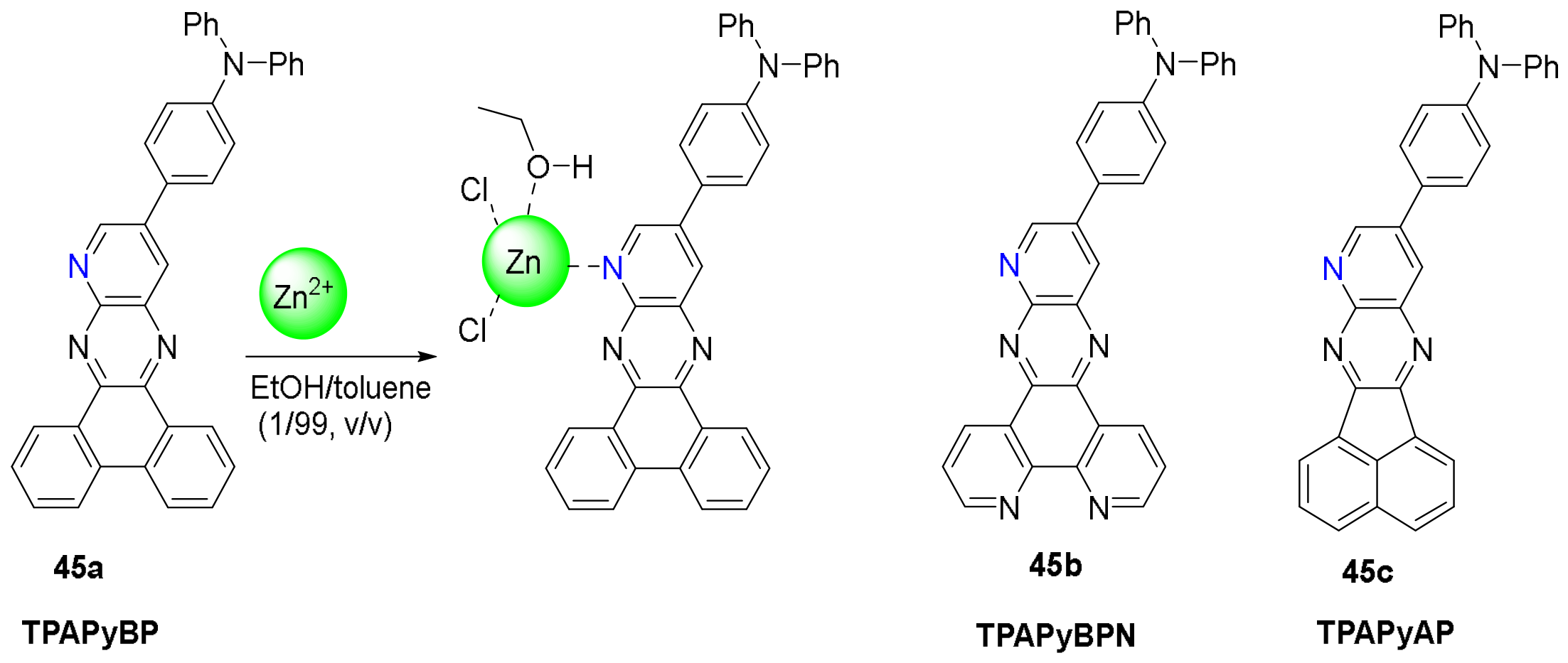
Figure 40.
(a) PL measurements of 45a, TPAPyBP (1.3×10⁻⁴ M) with the addition of ZnCl2 from 0 to 2.0 equiv. (λex = 487 nm); (b) Samples in daylight and excited by UV torch (λex = 360 nm) of 45a, TPAPyBP ZnCl2 from 0 to 2.0 equiv.; (c) Combined view of the single crystal structures of 45a and its Zn2+-complex (TPAPyBP and Zn(TPAPyBP)Cl2), and the corresponding frontier molecular orbitals (isovalue: 0.02) calculated using single crystal geometry in the gas phase at the PBE0/6-31G(d,p) level. Reproduced with permission of Wiley-VCH GmbH under the terms of the Creative Commons CC BY license [104].
Figure 40.
(a) PL measurements of 45a, TPAPyBP (1.3×10⁻⁴ M) with the addition of ZnCl2 from 0 to 2.0 equiv. (λex = 487 nm); (b) Samples in daylight and excited by UV torch (λex = 360 nm) of 45a, TPAPyBP ZnCl2 from 0 to 2.0 equiv.; (c) Combined view of the single crystal structures of 45a and its Zn2+-complex (TPAPyBP and Zn(TPAPyBP)Cl2), and the corresponding frontier molecular orbitals (isovalue: 0.02) calculated using single crystal geometry in the gas phase at the PBE0/6-31G(d,p) level. Reproduced with permission of Wiley-VCH GmbH under the terms of the Creative Commons CC BY license [104].
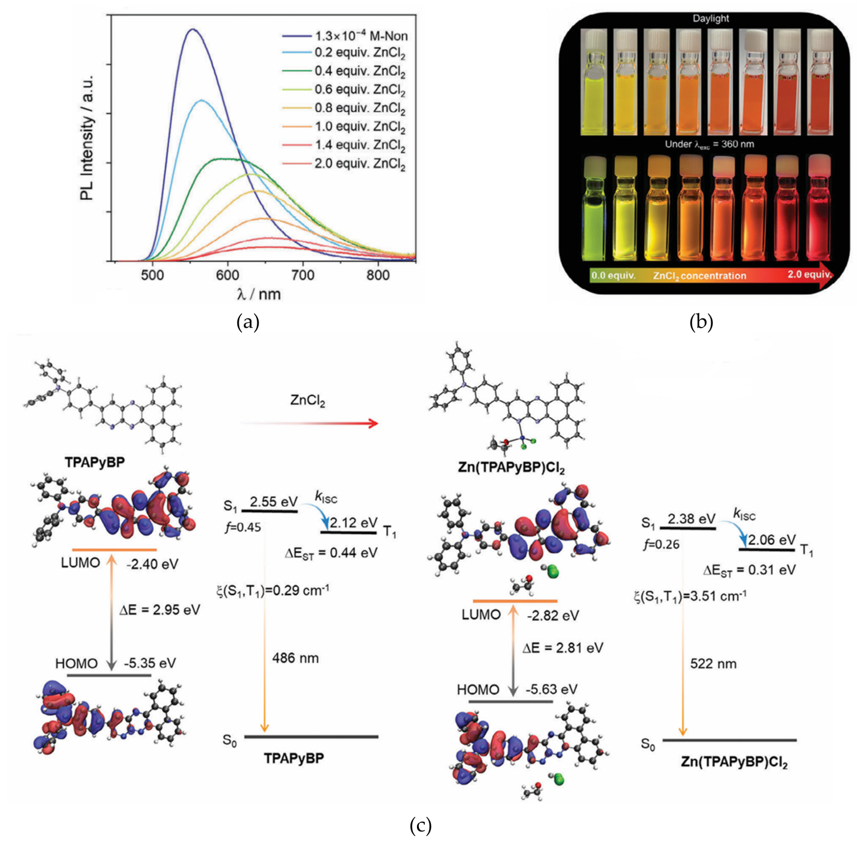
Disclaimer/Publisher’s Note: The statements, opinions and data contained in all publications are solely those of the individual author(s) and contributor(s) and not of MDPI and/or the editor(s). MDPI and/or the editor(s) disclaim responsibility for any injury to people or property resulting from any ideas, methods, instructions or products referred to in the content. |
© 2026 by the authors. Licensee MDPI, Basel, Switzerland. This article is an open access article distributed under the terms and conditions of the Creative Commons Attribution (CC BY) license.
Copyright: This open access article is published under a Creative Commons CC BY 4.0 license, which permit the free download, distribution, and reuse, provided that the author and preprint are cited in any reuse.