

Submitted:
15 September 2026
Posted:
16 September 2026
You are already at the latest version
Abstract
Background/Objectives: Lumpy skin disease, a highly contagious Capripoxvirus infection, causes significant economic losses in cattle worldwide. In Ethiopia, diagnosis primarily relies on virus neutralization tests and PCR, highlighting the need for scalable serological tools suitable for large-volume surveillance. The objective of this study was to produce and functionally characterize egg-yolk-derived IgY antibodies against the lumpy skin disease virus (LSDV) vaccinal strain as a proof-of-concept for future serological assay development. Methods: An experimental study was conducted from January to May 2024 over 42 days. The vaccinal virus was adapted on the Vero cell line and titrated after two passages in Minimum Essential Medium. Chickens (n = 10) were allocated into two groups (six experimental and four control). The experimental group received the LSDV vaccinal strain with a booster on day 15, while the control group received a placebo. Weekly serum and egg samples were collected. IgY was extracted from egg yolk using polyethylene glycol precipitation followed by dialysis purification. The protein concentration, molecular weight, and functional activity were assessed using NanoDrop spectrophotometry, SDS-PAGE, and virus neutralization tests (VNTs), respectively. A statistical analysis was performed using linear regression, repeated-measures ANOVA, and a linear mixed-effects model in R (version 4.4.1), with p < 0.05 considered statistically significant. Results: Total protein concentration increased significantly over time (p = 0.0239), with significant differences observed across weeks (p < 0.0001). The virus neutralization assays demonstrated a significantly higher neutralizing activity of egg-yolk-derived IgY compared to serum-derived antibodies (p < 0.0002). Conclusions: This study demonstrates the feasibility of producing functionally active IgY antibodies against LSDV in egg yolk. However, as the protein quantification was based on total protein measurement and antigen-specific validation was not performed, further studies are required to confirm specificity and optimize their application in diagnostic assay development.
Keywords:
chicken egg yolk
; immunoglobulin Y
; lumpy skin disease virus
; polyclonal antibodies
; virus neutralization test
1. Introduction
1.1. Background
Lumpy skin disease (LSD) is caused by the lumpy skin disease virus (LSDV) from the family Poxviridae, genus Capripoxvirus (CapV), the Neethling strain prototype [1]. It is characterized by fever; nodules on the skin, mucous membranes and internal organs; emaciation; enlarged lymph nodes; edema of the skin; and sometimes death [2]. The disease is a highly contagious transboundary infection that causes significant economic losses in the livestock industry as it can cause a temporary reduction in milk production, temporary or permanent sterility in bulls, damage to hides, and, occasionally, death [3].
Lumpy skin disease virus is endemic to Ethiopia, first reported in 1983 in the northwestern part of Ethiopia in the Amhara National Regional State, surrounding the southwest part of Lake Tana [4]. Because of the wide distribution of the disease and the size and structure of the cattle population in Ethiopia, LSDV is one of the most economically important livestock diseases in the country [5]. A study conducted in central and northwestern Ethiopia estimated the median total economic loss of an LSD outbreak at USD 1,176 per affected herd, with losses of USD 489 and USD 2,735 reported for subsistence and commercial farms, respectively[6]
Mammalian polyclonal antibodies (pAbs), monoclonal antibodies (mAbs), and avian polyclonal antibodies (IgY) have been extensively adopted as valuable tools for assay-specific target development and detection in basic research, diagnostics, and biomarker discovery [7].
Avian polyclonal antibodies (IgY) can be produced from chickens using non-invasive protocols, offering several advantages over mammalian antibodies. IgY antibodies enable the detection of conserved epitopes across species, making them particularly suitable for targeting proteins with high sequence similarity [8,9].
Egg-yolk-derived antibodies provide a high yield per animal, and the harvesting process is simple, ethical, and non-invasive [10]. Purified IgY antibodies exhibit higher avidity and sensitivity, broader epitope recognition, and reduced interference in immunoassays compared with mammalian IgG antibodies. [11].
Despite these advantages, the application of IgY technology to lumpy skin disease virus (LSDV) remains limited. In this context, the present study was designed as a proof-of-concept to investigate the production, purification, and functional characterization of egg-yolk-derived IgY antibodies against the LSDV vaccinal strain. Specifically, this study aimed to evaluate the kinetics of IgY production following immunization and to assess the functional activity of the generated antibodies using in vitro assays. The findings of this study provide preliminary evidence supporting the feasibility of IgY-based approaches and highlight their potential application in the future development of diagnostic tools for LSDV.
Despite the potential advantages of IgY technology, its application for generating antibodies against LSDV has not been investigated in Ethiopia. Therefore, this study aimed to produce and functionally characterize egg-yolk-derived IgY antibodies against the LSDV vaccinal strain as a proof-of-concept for their potential use in the development of serological diagnostic assays.
1.2. Objectives
1.2.1. General Objective
To generate and functionally characterize the polyclonal IgY antibodies produced in chickens immunized with the lumpy skin disease virus (LSDV) vaccinal strain, and to evaluate their potential for future application in LSDV-related research and serological assay development.
1.2.2. Specific Objectives
To isolate and purify IgY antibodies from chicken egg yolk following immunization with the LSDV vaccinal strain.
To evaluate the temporal antibody response and production kinetics of IgY following immunization.
To assess the virus-neutralizing activity of egg-yolk-derived IgY against the LSDV vaccinal strain.
2. Materials and Methods
2.1. Study Site
The experimental work was conducted at the Animal Health Institute (AHI), Sebeta, Oromia, Ethiopiaand the AU-PANVAC Laboratory in Bishoftu, Ethiopia.
2.2. Study Design
The experimental study was conducted over 42 days between January and May 2024 to produce and functionally characterize egg-yolk-derived IgY antibodies against the LSDV vaccinal strain as a proof-of-concept for future serological assay development. Following virus adaptation and titration, the chickens were randomly allocated into an experimental group and a control group. The experimental group received two doses of the LSDV vaccinal strain, while the control group received phosphate-buffered saline (PBS) as a placebo. The injections were administered on days 0 and 15 into the pectoral muscles [12] (Supplementary Figure S2).
2.3. Experimental Chickens
Ten healthy 52-week-old Bovans Brown layer chicks with no history of vaccination, from the same hatch batch were bought from a local farm in Sebeta town and managed in controlled environments for housing, inoculation, and sample collection. The chickens were allocated into two groups and housed separately in mesh wire enclosures. For a successful recovery from the stress caused by transportation, feed, and environmental alterations, the chickens were given a week to acclimate before the trial commenced. Chickens were managed in a good state of welfare: they were healthy, comfortable, well-nourished, safe, able to express natural behaviors, and free from pain, fear, and distress The study was approved by the College of Veterinary Medicine and Animal Science, University of Gondar Research Ethics Review Committee (CVMASC-RERC; Ref. No. CVMASC/UoG/RERC/26/04/2023), and all procedures involving experimental animals were conducted in accordance with the approved protocol.
2.4. Sample Size
The sample size was determined based on feasibility and alignment with previous exploratory IgY immunization and kinetic studies, which commonly employ small experimental groups due to the ethical and logistical considerations associated with animal-based research. A total of ten (n = 10) egg-laying chickens were included and randomly allocated into an experimental group (n = 6) and a control group (n = 4), and housed under controlled conditions to minimize bias.
2.5. Preparation, Adaptation, and Titration of Lumpy Skin Disease Virus Vaccine
Preparation of Vero Cell Culture for LSDV Adaptation
Vero cells (obtained from the Virus Isolation and Cell Culture Laboratory of Animal Health Institute; Sebeta, Oromia, Ethiopia) were cultured in Minimum Essential Medium (MEM) supplemented with 10% (v/v) fetal bovine serum (FBS), 2% (v/v) streptomycin sulfate, and Amphotericin B (250 µg/mL) [14]. The cells were incubated at 37 °C in a humidified atmosphere with 5% CO2 and allowed to reach approximately 70% confluence within 3 days before viral inoculation.
2.6. Titration of Lumpy Skin Disease Virus Vaccinal Strain
A live lumpy skin disease virus (LSDV) vaccine was obtained from the National Veterinary Institute (NVI). The virus was therefore harvested and passaged into fresh Vero cells. In the subsequent passage, characteristic CPEs were observed within 7 days [2], after which the virus was harvested and used for titration and downstream applications (see Supplementary Figure S1).
The viral titer was determined by tenfold serial dilution in 96-well microtitration plates, and the median tissue culture infectious dose (TCID50) was calculated using the Reed–Muench method [15]. The final viral stock used for immunization was standardized to 103 TCID50/mL.
2.7. Immunization of Chickens, Sample Collection, and IgY Extraction
Immunization and Sample Collection
The chickens in the experimental group were immunized intramuscularly with 103 TCID50/mL of LSDV into both pectoral muscles [12], while the control birds received 1 mL of PBS. A booster dose was administered on day 15 using the same virus dose and placebo.
Eggs and blood samples were collected weekly from day 7 to day 42 (Table 1). The eggs were stored at 4 °C, and approximately 3 mL of blood was collected from the brachial vein of each chicken (see Supplementary Figure S3).
2.8. Extraction of Chicken IgY
Fresh, dark-brown-shelled eggs were processed using a modified extraction protocol as described in [17].The egg yolks were separated from the egg whites using an egg strainer and transferred into 50 mL Falcon tubes. Each egg yielded approximately 15 mL of yolk (V1).
The yolk was mixed with PBS at a 1:2 ratio, yielding a final volume of approximately 45 mL. The mixture was vortexed, followed by the addition of 3.5% (w/v) PEG 6000 to the total volume (V2), and vortexed again. The solution was then agitated in a thermoshaker incubator at 300 rpm for 10 min, at room temperature followed by centrifugation at 4 °C and 3500 rpm for 15 min. The supernatant was carefully decanted and filtered through folded filter paper into a sterile tube, producing a new volume (V3).
Afterward, 8.5% (w/v) PEG 6000 was added to the volume (V3), and the mixture was vortexed and incubated on a thermoshaker at 300 rpm. The tubes were then centrifuged at 4 °C and 3500 rpm for 15 min, and the supernatant was discarded. The resulting pellet was resuspended and homogenized in 1 mL of PBS using a glass rod, and PBS was added to adjust the total volume to 10 mL (V4).
The solution was further treated with 12% (w/v) PEG 6000, mixed on a thermoshaker at 300 rpm for 10 min, and centrifuged again at 4 °C and 3500 rpm for 15 min. The supernatant was discarded, and the pellet was resuspended in 1.8 mL of PBS, mixed thoroughly, and transferred into a 2 mL cryovial for storage (see Supplementary Figure S4).
Finally, the extracts were purified using a Membra-Cel® (Serva Electrophoresis GmbH, Heidelberg, Germany) dialysis membrane with a 14 kDa molecular weight cut-off (MWCO) to remove PEG 6000 and further refine the IgY preparation against PBS.
2.9. Purification of the IgY Extract
The IgY extracts were purified using a Membra-Cel® dialysis membrane with a 14 kDa molecular weight cut-off (MWCO). The tubing was pretreated with 5 mM EDTA and thoroughly rinsed with distilled water prior to use. The IgY extract was loaded into the membrane and dialyzed overnight against 0.1% saline under continuous stirring, followed by an additional 3 h of dialysis against phosphate-buffered saline (PBS). The purified IgY was then recovered and stored at −20 °C until further analysis.
2.10. Detection and Quantification of Antibodies
Virus Neutralization Test
The virus neutralization test (VNT) was performed to evaluate the functional activity of IgY antibodies against LSDV by assessing their ability to inhibit viral infection in Vero cells. This assay enabled the detection and comparison of neutralizing polyclonal antibodies (pAbs) in both serum- and egg-yolk-derived IgY samples.
The serially diluted IgY samples were incubated with a standardized viral dose prior to inoculation onto the Vero cells. Each dilution was tested in duplicate (two wells per dilution), and the presence or absence of cytopathic effects (CPEs) was recorded following incubation. Neutralization endpoints were determined based on the proportion of wells protected from CPEs across serial dilutions. When partial neutralization was observed between consecutive dilutions, the endpoint titer was estimated using an interpolation approach.
2.11. Protein Concentration and Purity
The total protein concentration and purity of the preparationwere determined using a NeoDot microvolume spectrophotometer (NeoBiotech Co., Ltd., South Korea) by measuring absorbance at 280 nm. Phosphate-buffered saline (PBS) was used as the blank. For each measurement, 2 µL of egg-yolk-derived protein preparationsample was loaded onto the pedestal, and readings were recorded according to the manufacturer’s instructions. Proteinpurity was assessed based on the A260/A280 absorbance ratio and UV spectral profile (see Supplementary Figure S5).
2.12. SDS-PAGE Analysis
Reducing SDS-PAGE was performed to determine the purity and molecular weight of purified IgY. The samples (10 µg) were separated on NuPAGE™ Novex® Bis-Tris Mini Gels using the XCell SureLock™ Mini-Cell system, Invitrogen (Thermo Fisher Scientific: Carlsbad, CA, USA). A prestained protein molecular weight marker (BenchMark™ Protein Ladder, Invitrogen (Thermo Fisher Scientific: Carlsbad, CA, USA)) was included for size estimation. Electrophoresis was conducted at 200 V for approximately 2 h, and protein bands were visualized by Coomassie-based staining according to the manufacturer’s instructions.2.13. Data Management and Analysis
Data were analyzed using R version 4.4.1. Changes in IgY concentration over time were evaluated using linear regression and repeated-measures ANOVA, with week treated as the within-subject factor. When significant differences were detected, post hoc comparisons were performed. For the virus neutralization analysis, log-transformed neutralization titers were treated as the dependent variable and analyzed using a linear mixed-effects model, with week and antibody source (egg yolk vs. serum) included as fixed effects and individual chickens included as random effects to account for repeated measurements. Assumptions of normality and sphericity were assessed and met prior to analysis. A p-value of <0.05 was considered statistically significant (see Supplementary Table S1).
3. Results
3.1. Virus Titration
The viral stock had a calculated titer of 105 TCID50/mL. For immunization, the virus was diluted 1:100 to obtain a final concentration of 103 TCID50/mL.
3.2. Quantification of IgY Concentration
IgY concentrations were determined weekly using a microvolume spectrophotometer at 280 nm. In the experimental group, mean IgY levels increased markedly from 0.023 ± 0.004 mg/mL at Week 1 to a peak of 0.961 ± 0.059 mg/mL at Week 5, followed by a slight decline to 0.837 ± 0.055 mg/mL at Week 6 (Figure 1) (see Supplementary Table S3). In contrast, the control group maintained consistently low IgY concentrations throughout the study period (range: 0.027–0.043 mg/mL) (see Supplementary Table S4).
A linear regression analysis demonstrated a significant increase in IgY concentration over time in the experimental group (slope = 0.162, p = 0.024), whereas no significant trend was observed in the control group (slope = 0.001, p = 0.301) (Table 2, Figure 1).
A repeated-measures ANOVA was conducted to assess changes in IgY concentration over time in the experimental group. A significant effect of time was observed (p < 0.0001). Assumptions of normality (Shapiro–Wilk test) and sphericity (Mauchly’s test) were satisfied (p > 0.05); therefore, no corrections were applied. Post hoc comparisons indicated significant differences between weeks (see Supplementary Table S2). These findings demonstrate a time-dependent IgY response following immunization, consistent with an active humoral immune response to the LSDV vaccinal strain.
3.3. Confirmation of IgY Identity
3.4. Evaluating the Virus Neutralization Efficacy of IgY
Virus neutralization testing demonstrated that IgY exhibited effective antiviral activity, with the endpoint titer estimated at approximately 1:125 (log10 ≈ 2.10), based on interpolation between adjacent dilution levels showing partial neutralization. No neutralizing activity was detected in the serum or egg-yolk preparations from the control group at any of the tested dilutions.
Neutralization titers were expressed as log10 reciprocal dilution values. Egg-yolk-derived IgY exhibited a progressive increase in neutralizing activity over time, reaching peak levels at Week 5, which corresponded to the highest recorded IgY concentration, followed by a modest decline thereafter.
In contrast, serum-derived antibodies showed lower neutralization titers, with a maximum of approximately 1:50 (log10 ≈ 1.70) and an earlier decline over time. The linear mixed-effects model analysis confirmed a statistically significant difference in neutralization responses between the egg-yolk-derived and serum-derived antibody preparations across the study period (p < 0.0001).
Overall, egg-yolk-derived IgY demonstrated stronger and more sustained neutralizing activity against lumpy skin disease virus compared to serum-derived antibodies (Figure 3).
4. Discussion
Immunoglobulin Y technology is a scalable production technology that started its foundation in 1980 [18] and was rapidly adopted in developing countries and regions such as South America and India, as well as Japan, Germany, and other developed countries [19]. It is highly relevant in immunodiagnostics, playing a crucial role in the development of immunoassays such as ELISA due to its specificity and sensitivity [20]. Its relevance extends to disease control and serosurveillance, where IgY is potentially used for the detection, prevention, and treatment of human and animal infections [21].
In this study, we demonstrated the feasibility of producing and functionally characterizing IgY antibodies against the LSDV vaccine strain using a polyethylene glycol (PEG)-based extraction method [22] and dialysis purification protocols [23]. The generated egg-yolk-derived antibody preparationswere characterized by SDS-PAGE, quantified spectrophotometrically, and evaluated for functional activity using virus neutralization tests. The results provide proof-of-concept evidence that the immunization of chickens with the LSDV antigen can induce the production of biologically active IgY antibodies capable of neutralizing the virus in vitro. This expands the application of IgY technology to an economically important transboundary livestock disease and highlights its potential as a scalable platform for antibody production.
The SDS-PAGE analysis was suggestive of the characteristic IgY subunit profile, with distinct bands corresponding to the heavy (~65 kDa) and light (~25 kDa) chains, consistent with the expected molecular structure of avian IgY. Comparable molecular weights have been reported in previous IgY studies targeting Nosema ceranae [24], avian influenza viruses (H5/H9) [25], and rotaviruses [26], demonstrating structural consistency across antigen systems. Similar IgY isolation and profiling patterns were also described in comparative studies of immunized chicken breeds [27]. Progressive increases in band intensity from Week 2 onward paralleled the quantified rise in IgY concentration, reflecting active humoral immune maturation following primary and booster immunization [26]. The absence of detectable bands during the first week is consistent with the known kinetics of primary antibody responses in chickens, where IgY becomes measurable after the initial lag phase [28]. The marked enhancement in band intensity following booster administration further supports effective secondary immune stimulation and amplified polyclonal antibody production [29]. Collectively, these findings confirm the structural integrity and time-dependent accumulation of functional IgY in egg yolk.
IgY quantification demonstrated a progressive increase in mean egg yolk antibody concentration over the study period, with peak levels observed following booster immunization and a subsequent decline toward the final week. Linear regression confirmed a significant time-dependent increase in IgY production across six weeks, consistent with the expected kinetics of primary and secondary humoral responses. These findings align with previous reports describing time-dependent increases in IgY production against ricin and Clostridium botulinum neurotoxins [30], as well as studies documenting the cyclical patterns of antibody titer elevation and decline following immunization [31]. Together, these results confirm that immunization with the LSDV vaccine strain induces a robust and dynamic IgY response in chickens.
The functional virus neutralization analysis demonstrated that the increased IgY concentrations observed in egg yolk translated into superior neutralizing activity against LSDV compared with serum-derived antibodies. The mixed-effects modeling of dilution endpoints further confirmed significantly higher neutralization titers in yolk IgY. This functional advantage is biologically consistent with the preferential accumulation of IgY in egg yolk, where antibody levels are typically higher than in serum. Similar observations have been reported in previous IgY-based studies targeting viral pathogens [32], supporting egg yolk as a reliable source of high-potency polyclonal antibodies [8].
Notably, direct within-animal comparisons between serum-derived antibodies and egg-yolk-derived IgY remain limited in the literature. In this study, the inclusion of paired serum and egg yolk samples from the same immunized chickens enabled a controlled evaluation of their relative functional performance. The comparatively lower and delayed neutralization response observed in serum further supports the preferential accumulation and functional enrichment of virus-specific antibodies in egg yolk. These findings provide additional insight into the differential distribution and activity of IgY across biological compartments, although further studies are needed to validate these observations across larger sample sizes and different viral systems.
Although this study demonstrated the production and functional neutralization capacity of egg-yolk-derived IgY against lumpy skin disease virus, several important limitations should be acknowledged.
First, IgY quantification was based on absorbance at 280 nm, which measures total protein concentration rather than IgY specifically. As a result, the reported values may include non-IgY proteins and should be interpreted as approximate estimates rather than precise antibody concentrations.
Second, functional characterization relied primarily on virus neutralization testing without complementary antigen-specific assays such as ELISA. Therefore, while neutralizing activity was demonstrated, the specificity and binding characteristics of the antibodies were not directly evaluated.
Third, purification was performed using polyethylene glycol precipitation followed by dialysis, which may provide lower purity compared to chromatographic methods.
Finally, the relatively small sample size, although consistent with exploratory IgY studies and supported by a repeated-measures design, may limit the generalizability of the findings.
Additionally, total protein also increased during the later time points; therefore, the observed increase in bands consistent with the expected IgY profile cannot be attributed exclusively to IgY production. Furthermore, SDS-PAGE was not performed on the control-group samples; therefore, the observed changes in bands consistent with the expected IgY profile cannot be attributed specifically to IgY.
These limitations indicate that the present study should be considered a preliminary proof-of-concept. Future studies incorporating antigen-specific immunoassays, advanced purification techniques, and larger sample sizes are required to further validate and optimize the application of IgY for diagnostic purposes.5. Conclusions and Recommendations
This study demonstrates the production, purification, and functional characterization of egg-yolk-derived IgY against the LSDV vaccinal strain. IgY-associated neutralizing activity was detectable from the second week post-immunization, with peak activity observed at Week 5. The SDS-PAGE analysis indicated the structural integrity of the purified IgY, while virus neutralization testing demonstrated its functional activity. Egg-yolk-derived IgY exhibited stronger and more sustained neutralizing activity compared to serum-derived antibodies, consistent with its accumulation in egg yolk.
Collectively, these findings provide a proof-of-concept foundation for the potential use of IgY in future LSDV serological assay development. However, additional studies incorporating antigen-specific immunoassays, improved purification strategies, and broader immunological characterization are required to further validate and optimize this approach.
Supplementary Materials
The following supporting information can be downloaded at the website of this paper posted on Preprints.org, Figure S1: Vero cell culture, second passage of lumpy skin disease virus (LSDV), and viral titration; Figure S2: Inoculation of LSDV vaccinal strain into both pectoral muscles of chickens; Figure S3: Egg and serum sample collection and preparation; Figure S4: Visual documentation of IgY extraction from chicken egg yolk using PEG 6000; Figure S5: Quantification of IgY concentration using a NanoDrop microvolume spectrophotometer; Figure S6: Uncropped SDS-PAGE gel image of purified IgY; Table S1: Status of repeated-measures ANOVA assumptions for mean IgY concentrations across weeks; Table S2: Post hoc pairwise comparisons of weekly IgY production means; Table S3: Weekly IgY concentrations (mg/mL) of experimental chickens with individual and mean values; Table S4: Weekly IgY concentrations (mg/mL) of negative control experimental chickens injected with PBS, with individual and mean values.
Author Contributions
F.W. and D.N. contributed equally to this work, including study design and manuscript drafting. They supervised the experimental animals and performed laboratory tests. Additionally, F.W. analyzed the data and wrote and prepared the manuscript; S.M. and A.S. provided critical insights and valuable guidance throughout the study; J.d.D.B. conducted laboratory analyses and contributed to the interpretation of results, and T.B conducted laboratory analyses and contributed to the interpretation. All authors have read and agreed to the published version of the manuscript.
Funding
This research received no external funding. No APC funding was provided.
Institutional Review Board Statement
The use of experimental animals in this research was approved by the College of Veterinary Medicine and Animal Science UoG Research Ethics Review Committee (CVMASC-RERC) with reference: CVMASC/UoG/RERC/26/04/2023.:
Institutional Review Board Statement
The use of experimental animals in this research was approved by the College of Veterinary Medicine and Animal Science UoG Research Ethics Review Committee (CVMASC-RERC) with reference: CVMASC/UoG/RERC/26/04/2023.
Data Availability Statement
The datasets used and/or analyzed during the current study are available from the corresponding author upon reasonable request.
Acknowledgments
The authors would like to thank the Animal Health Institute, Ethiopia, for their logistical support in procuring experimental animals and granting full access to their cell culture laboratory. We are also grateful to AU-PANVAC for their invaluable assistance in conducting the molecular identification of IgY. Special thanks to the Ministry of Labour and Skills for providing financial support in the form of pocket money during the research period. Additionally, we acknowledge the use of a Large Language Model (LLM) for assistance in refining the grammar and improving the clarity of the text during the preparation of the manuscript.
Conflicts of Interest
The authors declare that the research was conducted in the absence of any commercial or financial relationships that could be construed as potential conflicts of interest.
Abbreviations
The following abbreviations are used in this manuscript:
| ANOVA | Analysis of Variance |
| CapV | Capripoxvirus |
| CPE | Cytopathic Effect |
| ELISA | Enzyme-linked Immunosorbent Assay |
| Ig | Immunoglobulin |
| LSD(V) | Lumpy Skin Disease Virus |
| mAb | Monoclonal Antibody |
| pAbs | Polyclonal Antibodies |
| PBS | Phosphate-Buffered Saline |
| PEG, MW | Molecular Weight of Polyethylene Glycol |
| SDS-PAGE | Sodium Dodecyl Sulfate-Polyacrylamide Gel Electrophoresis |
| TCID50 | Tissue Culture Infectious Dose |
| VNT | Virus Neutralization Test |
| WOAH | World Organization for Animal Health |
References
- Al-Salihi, K.A. Lumpy Skin disease. Mirror Res. Vet. Sci. Anim. 2014, 3, 6–23.
- WOAH. Lumpy Skin Disease. In Terrestrial Manual; WOAH: Paris, France, 2023; pp. 128–133.
- Kiplagat, S.K.; Kitala, P.M.; Onono, J.O.; Beard, P.M.; Lyons, N.A. Risk Factors for Outbreaks of Lumpy Skin Disease and the Economic Impact in Cattle Farms of Nakuru County, Kenya. Front. Vet. Sci. 2020, 7, 259. [CrossRef]
- Mebratu, G.Y.; Kassa, B.; Fikre, Y.; Berhanu, B. Observation on the outbreak of lumpy skin disease in Ethiopia. Rev. D’élev. Med. Vet. Des. Pays Trop. 1984, 37, 395–399.
- Gari, G.; Waret-Szkuta, A.; Grosbois, V.; Jacquiet, P.; Roger, F. Risk factors associated with observed clinical lumpy skin disease in Ethiopia. Epidemiol. Infect. 2010, 138, 1657–1666. [CrossRef]
- Ascoli, C.A.; Aggeler, B. Overlooked benefits of using polyclonal antibodies. Biotechniques 2018, 65, 127–136. [CrossRef]
- Kovacs-nolan, J.; Mine, Y. Egg Yolk Antibodies for Passive Immunity. Annu. Rev. Food Sci. Technol. 2012, 3, 163–184. [CrossRef]
- Müller, S.; Schubert, A.; Zajac, J.; Dyck, T.; Oelkrug, C. IgY antibodies in human nutrition for disease prevention. Nutr. J. 2015, 14, 109. [CrossRef]
- Michael, A.; Meenatchisundaram, S.; Parameswari, G.; Subbraj, T.; Selvakumaran, R.; Ramalingam, S. Chicken egg yolk antibodies (IgY) as an alternative to mammalian antibodies. Indian J. Sci. Technol. 2010, 3, 468–474. [CrossRef]
- Pereira, E.P.V.; van Tilburg, M.F.; Florean, E.O.P.T.; Guedes, M.I.F. Egg yolk antibodies (IgY)and their applications in human and veterinary health: A review. Int. Immunopharmacol. 2019, 73, 293–303. [CrossRef]
- Schade, R.; Staak, C.; Hendriksen, C.; Erhard, M.; Hugl, H.; Koch, G.; Larsson, A.; Pollmann, W.; Van Regenmortel, M.; Rijke, E.; et al. The production of avian (egg yolk) antibodies: IgY. The report and recommendations of ECVAM workshop 21. In ATLA Alternatives to Laboratory Animals; FRAME: Nottingham, UK, 1996; pp. 925–934. [CrossRef]
- NCC. National Chicken Council Animal Welfare Guidelines; NCC: Washington, DC, USA, 2020; pp. 1–19.
- Sandell, L.; Sakai, D. Mammalian cell culture. In Current Protocols in Essential Laboratory Techniques; John Wiley & Sons, Inc.: Hoboken, NJ, USA, 2011; pp. 1–32.
- Lei, C.; Yang, J.; Hu, J.; Sun, X. On the Calculation of TCID50 for Quantitation of Virus Infectivity. Virol. Sin. 2021, 36, 141–144. [CrossRef]
- Hamal, K.R.; Burgess, S.C.; Pevzner, I.Y.; Erf, G.F. Maternal antibody transfer from dams to their egg yolks, egg whites, and chicks in meat lines of chickens. Poult. Sci. 2006, 85, 1364–1372. [CrossRef]
- Morishita, T.Y. Poultry Blood Collection; Western University of Health Sciences: Pomona, CA, USA, 2019; pp. 1–3.
- Madera-Contreras, A.M.; Solano-Texta, R.; Cisneros-Sarabia, A.C.; Bautista-Santos, I.B.; Vences-Velázquez, G.V.; Vences-Velázquez, A.V.; Cortés-Sarabia, K.C. Optimized method for the extraction of contaminant-free IgY antibodies from egg yolk using PEG 6000. MethodsX 2022, 9, 101874. [CrossRef]
- Hawkins, M. et al. (2025) “Mass production of IgY-containing tablets for COVID-19 transmission control,” Scientific Reports, 15(1), pp. 1–15. Available at: . [CrossRef]
- Zhang, X.Y.; Vieira-Pires, R.S.; Morgan, P.M. IgY-Technology: Production and Application of Egg Yolk Antibodies; Springer: Cham, Switzerland, 2021; pp. 1–70. [CrossRef]
- Yadoung, S.; Ishimatsu, R.; Xu, Z.L.; Sringarm, K.; Pata, S.; Thongkham, M.; Chantara, S.; Pattarawarapan, M.; Hongsibsong, S. Development of IgY-Based Indirect Competitive ELISA for the Detection of Fluoroquinolone Residues in Chicken and Pork Samples. Antibiotics 2022, 11, 1512. [CrossRef]
- Lee, L.; Samardzic, K.; Wallach, M.; Frumkin, L.R.; Mochly-Rosen, D. Immunoglobulin Y for Potential Diagnostic and Therapeutic Applications in Infectious Diseases. Front. Immunol. 2021, 12, 696003. [CrossRef]
- Polson, A.; von Wechmar, M.B.; van Regenmortel, M.H.V. Isolation of viral igy antibodies from yolks of immunized hens. Immunol. Commun. 1980, 9, 475–493. [CrossRef]
- Pauly, D.; Chacana, P.A.; Calzado, E.G.; Brembs, B.; Schade, R. Igy technology: Extraction of chicken antibodies from egg yolk by polyethylene glycol (PEG) precipitation. J. Vis. Exp. 2011, 51, 3084.
- Açık, M.N.; Karagülle, B.; Yakut, S.; Öztürk, Y.; Kutlu, M.A.; Kalın, R.; Çetinkaya, B. Production, characterization and therapeutic efficacy of egg yolk antibodies specific to Nosema ceranae. PLoS ONE 2024, 19, e0297864. [CrossRef]
- Nahla, M.; Abdelfattah, N.; Ibrahim, M. Characterization of chicken IgY produced against H5 and H9 avian influenza viruses. Alex. J. Vet. Sci. 2018, 59, 79. [CrossRef]
- Bentes, G.A.; Lanzarini, N.M.; Guimarães, J.R.; Heinemann, M.B.; Volotão, E.d.M.; da Silva, A.d.S.; Heneine, L.G.D.; de Oliveira, J.M.; Pinto, M.A. Production and Evaluation of Chicken Egg Yolk Immunoglobulin (IgY) against Human and Simian Rotaviruses. Viruses 2022, 14, 1995. [CrossRef]
- Amro, W.A.; Al-Qaisi, W.; Al-Razem, F. Production and purification of IgY antibodies from chicken egg yolk. J. Genet. Eng. Biotechnol. 2018, 16, 99–103. [CrossRef]
- Ramakrishnan, S.; Kappala, D. Avian infectious bronchitis virus. In Recent Advances in Animal Virology; Springer: Singapore, 2019, pp. 301–319. [CrossRef]
- Agurto-Arteaga, A.; Poma-Acevedo, A.; Rios-Matos, D.; Choque-Guevara, R.; Montesinos-Millán, R.; Montalván, Á.; Isasi-Rivas, G.; Cauna-Orocollo, Y.; Cauti-Mendoza, M.d.G.; Pérez-Martínez, N.; et al. Preclinical Assessment of IgY Antibodies Against Recombinant SARS-CoV-2 RBD Protein for Prophylaxis and Post-Infection Treatment of COVID-19. Front. Immunol. 2022, 13, 881604. [CrossRef]
- Pauly, D.; Dorner, M.; Zhang, X.; Hlinak, A.; Dorner, B.; Schade, R. Monitoring of laying capacity, immunoglobulin Y concentration, and antibody titer development in chickens immunized with ricin and botulinum toxins over a two-year period. Poult. Sci. 2009, 88, 281–290. [CrossRef]
- Schade, R.; Gutierrez Calzado, E.; Sarmiento, R.; Chacana, P.A.; Porankiewicz-Asplund, J.; Terzolo, H.R. Chicken egg yolk antibodies (IgY-technology): A review of progress in production and use in research and human and veterinary medicine. ATLA 2005, 33, 129–154. [CrossRef]
- Silim, A.; Venne, D. Comparison of egg-yolk and serum antibody titers to four avian viruses by enzyme-linked immunosorbent assay using paired field samples. Avian Dis. 1989, 33, 643–648. [CrossRef]
Figure 1.
Weekly kinetics of IgY concentration based on estimated marginal means.
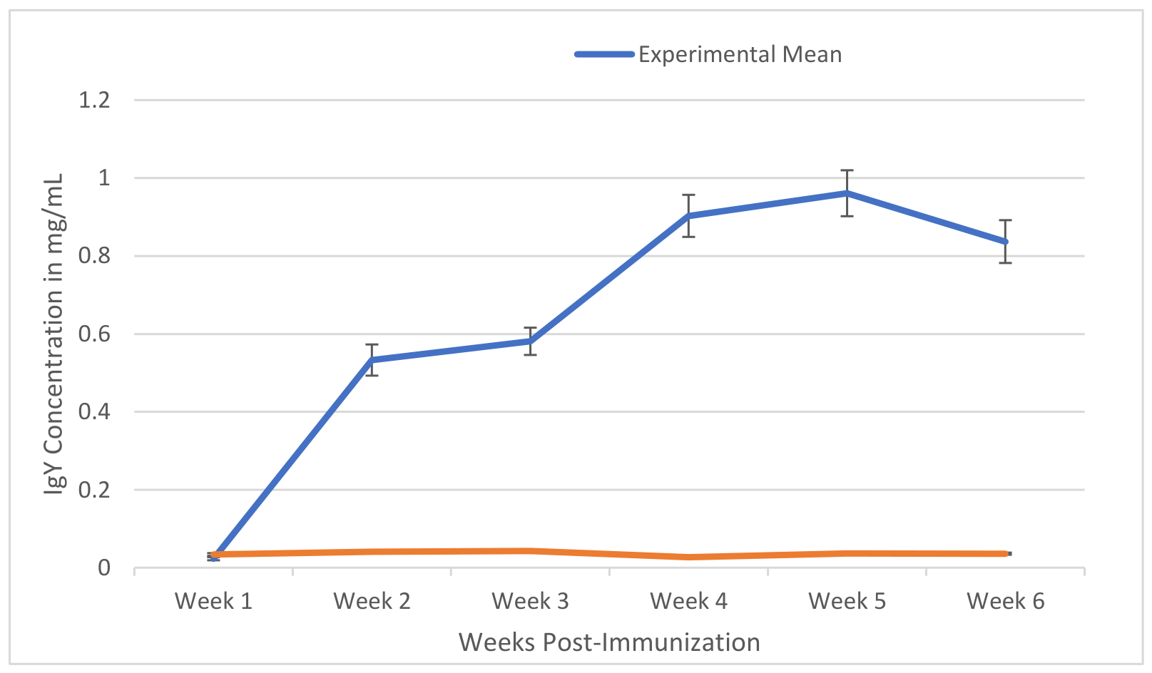
Figure 2.
SDS-PAGE profiling of IgY antibody production in egg yolk under reducing conditions. Key: Lane M: Bench Mark™ Protein Ladder; Lane 1: IgY from egg yolk (Week 1); Lane 2: IgY from egg yolk (Week 2); Lane 3: IgY from egg yolk (Week 3); Lane 4: IgY from egg yolk (Week 4); Lane 5: IgY from egg yolk (Week 5); Lane 6: IgY from egg yolk (Week 6); HC: Heavy chain; LC: Light chain; kDa: Kilodalton.
Figure 2.
SDS-PAGE profiling of IgY antibody production in egg yolk under reducing conditions. Key: Lane M: Bench Mark™ Protein Ladder; Lane 1: IgY from egg yolk (Week 1); Lane 2: IgY from egg yolk (Week 2); Lane 3: IgY from egg yolk (Week 3); Lane 4: IgY from egg yolk (Week 4); Lane 5: IgY from egg yolk (Week 5); Lane 6: IgY from egg yolk (Week 6); HC: Heavy chain; LC: Light chain; kDa: Kilodalton.
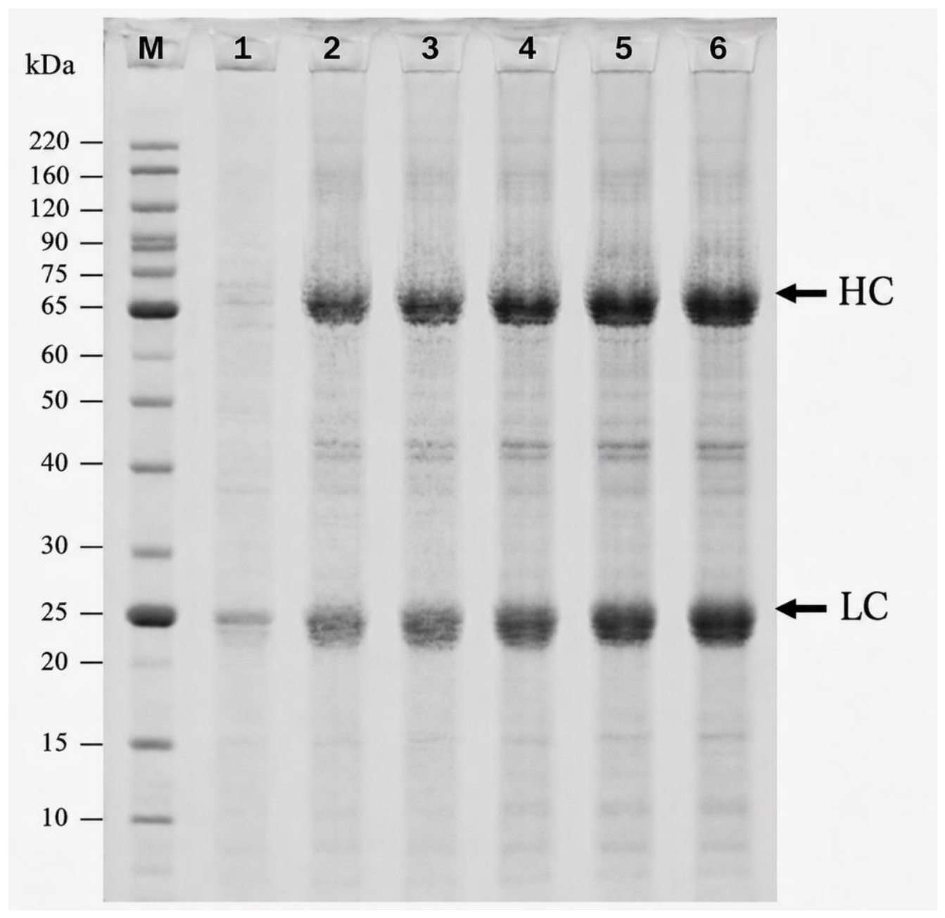
Figure 3.
Comparison of log10 neutralization titers of IgY derived from egg yolk and serum.
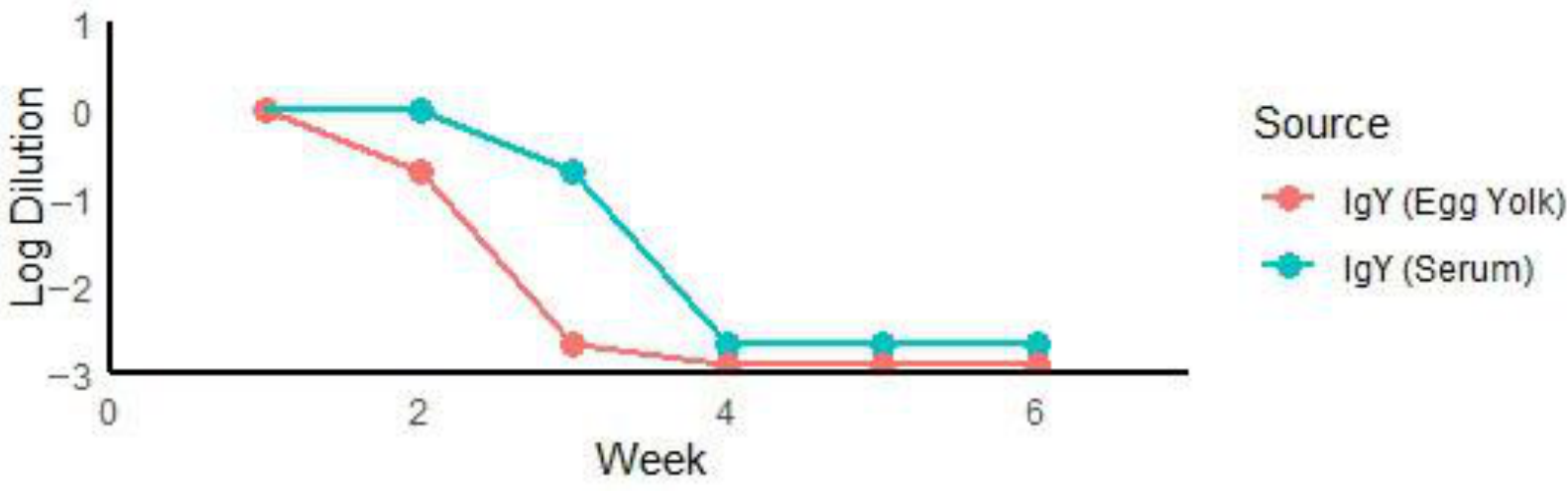

Table 1.
Immunization and sample collection.
| Day | Treatment Group (n = 6) | Control Group (n = 4) |
|---|---|---|
| 0 | First vaccine dose administered | First placebo dose administered |
| 7 | Blood sample collected | Blood sample collected |
| 15 | Second vaccine dose + sampling | Second placebo dose + sampling |
| 21 | Blood sample collected | Blood sample collected |
| 28 | Blood sample collected | Blood sample collected |
| 35 | Blood sample collected | Blood sample collected |
| 42 | Blood sample collected | Blood sample collected |
Blood samples were collected weekly for IgY quantification.
Table 2.
Linear regression analysis of weekly IgY concentration dynamics: slope comparison between the experimental and control groups.
Table 2.
Linear regression analysis of weekly IgY concentration dynamics: slope comparison between the experimental and control groups.
| Group | Variable | Coefficient | Std. Error | t-Value | p-Value | 95% Confidence Interval |
|---|---|---|---|---|---|---|
| Experimental | Week | 0.162 | 0.046 | 3.55 | 0.024 | 0.035–0.289 |
| Constant (_cons) | 0.072 | 0.178 | 0.40 | 0.707 | −0.423–0.566 | |
| Control | Week | 0.001 | 0.0009 | 1.19 | 0.301 | −0.0001–0.003 |
| Constant (_cons) | 0.036 | 0.0035 | 10.24 | 0.001 | −0.026–0.040 |
Keywords: Std. Err.: standard error; t: t statistic; p > |t|: p value;; Neg. Conc. IgY: Concentration of Control; Expo. Conc. IgY: Concentration of Exposed; _cons: Constant.
Disclaimer/Publisher’s Note: The statements, opinions and data contained in all publications are solely those of the individual author(s) and contributor(s) and not of MDPI and/or the editor(s). MDPI and/or the editor(s) disclaim responsibility for any injury to people or property resulting from any ideas, methods, instructions or products referred to in the content. |
© 2026 by the authors. Licensee MDPI, Basel, Switzerland. This article is an open access article distributed under the terms and conditions of the Creative Commons Attribution (CC BY) license (http://creativecommons.org/licenses/by/4.0/).
Copyright: This open access article is published under a Creative Commons CC BY 4.0 license, which permit the free download, distribution, and reuse, provided that the author and preprint are cited in any reuse.