
Submitted:
15 September 2026
Posted:
16 September 2026
You are already at the latest version
Abstract
Background/Objectives: Rehabilitation in active rheumatoid arthritis (RA) remains challenging, particularly regarding the dosing of physical agent modalities during in-flammatory activity. We evaluated changes in clinical disease activity, inflammatory markers, and Power Doppler (PD) activity in patients with RA undergoing individual-ized multimodal rehabilitation. Methods: In this retrospective longitudinal study, 29 patients with RA were assessed at baseline and after 8 weeks. The program comprised 10 supervised sessions of individualized electrotherapy and therapeutic exercise over 2 weeks, followed by 6 weeks of home-based exercise. Background disease-modifying an-tirheumatic therapy was unchanged; 16 patients received ESR-guided methylpredni-solone. Outcomes included DAS28-ESR and its components, C-reactive protein (CRP), and patient-level PD grade across a standardized joint set. Results: DAS28-ESR de-creased from 6.05 ± 0.87 to 4.03 ± 0.82, with significant reductions in all its components and in CRP (all p < 0.001). PD grade decreased from 2.38 ± 0.62 to 1.69 ± 0.66 (p < 0.001); 18 patients (62.1%) improved and none worsened. Changes in DAS28-ESR and PD grade were not correlated (ρ = 0.181, p = 0.348). In the 13 patients without gluco-corticoids, DAS28-ESR decreased from 5.22 to 3.41 (p < 0.001) and PD grade from 2.38 to 1.77 (p = 0.016). Conclusions: Improvements across clinical, laboratory, and ultra-sound domains were observed during individualized rehabilitation delivered along-side pharmacological treatment, while the magnitude of clinical and ultrasound changes was not significantly correlated. Because of the uncontrolled design and con-comitant glucocorticoid and NSAID therapy, the contribution of rehabilitation cannot be isolated; controlled studies of dose-adapted rehabilitation in active RA, prescribed by the rehabilitation physician in collaboration with the rheumatologist, are warranted.
Keywords:
rheumatoid arthritis
; rehabilitation
; electrotherapy
; physical and rehabilitation medicine
; DAS28-ESR
; Power Doppler ultrasonography
; disease activity
1. Introduction
Rheumatoid arthritis (RA) is a chronic systemic inflammatory autoimmune disease characterized by persistent synovitis, progressive structural joint damage, pain, and functional impairment [1].
Despite major advances in pharmacological management with conventional synthetic, biological, and targeted disease-modifying antirheumatic drugs (DMARDs), optimization of physical function and preservation of mobility remain important components of comprehensive disease management [2].
Rehabilitation, particularly therapeutic exercise and physical therapy, represents an important adjunct to pharmacological treatment, with evidence supporting benefits in physical function, disease activity, and quality of life in patients with RA [3].
Assessment of RA activity is inherently multidimensional. Composite clinical indices such as the Disease Activity Score in 28 joints (DAS28) integrate tender and swollen joint counts, the patient’s global assessment, and an inflammatory marker and are widely used to quantify disease activity and therapeutic response [4]. However, clinical composite indices may not fully reflect local synovial inflammatory activity, and discrepancies between clinical findings and persistent subclinical inflammation may occur [5].
Musculoskeletal ultrasonography provides a complementary method for evaluating inflammatory and structural joint involvement in RA [6]. In particular, Power Doppler ultrasonography allows semiquantitative assessment of synovial vascularity and may provide additional information on active synovial inflammation beyond that obtained from clinical examination and systemic inflammatory markers [7]. Consequently, the combined longitudinal assessment of DAS28 and Power Doppler findings may provide a more comprehensive characterization of changes in disease activity [5,8].
Although rehabilitation is increasingly recognized as an important component of comprehensive RA management, evidence regarding its contribution to inflammatory disease activity remains comparatively limited [3,9]. Previous studies of comprehensive rehabilitation have reported favorable clinical and functional outcomes, including changes in disease activity, but the combined longitudinal assessment of clinical, systemic inflammatory, and ultrasound parameters during rehabilitation has received substantially less attention [3,10].
To our knowledge, the longitudinal evolution of composite clinical disease activity, systemic inflammatory markers, and Power Doppler ultrasound findings during structured multimodal rehabilitation in RA has been insufficiently investigated. Therefore, the present study aimed to evaluate changes in clinical disease activity, systemic inflammatory markers, and Power Doppler activity over an 8-week period comprising a supervised multimodal rehabilitation phase followed by home-based therapeutic exercise. A secondary objective was to examine the relationship between changes in DAS28-ESR and Power Doppler activity and to explore whether the observed longitudinal changes differed according to concomitant glucocorticoid exposure.
2. Materials and Methods
2.1. Study Design and Setting
This was a retrospective, single-centre, longitudinal observational study with a paired pre–post design. Data were extracted from the medical records of patients with rheumatoid arthritis (RA) who underwent a multimodal rehabilitation program in Ambulatory Medical Rehabilitation Department Craiova, România, between 1 June 2023 and 30 June 2026. Each patient was assessed at two time points: at baseline, before initiation of the rehabilitation program (T1), and at the end of the 8-week observation period (T2). The study is reported in accordance with the Strengthening the Reporting of Observational Studies in Epidemiology (STROBE) statement [11]; the completed checklist is provided in Table S1.
2.2. Participants
Patients were identified from the clinical practice of the attending physical and rehabilitation medicine (PRM) physician; no external referral pathway was involved. Inclusion criteria were age ≥ 18 years; a diagnosis of RA previously established by a rheumatologist according to the 2010 American College of Rheumatology/European League Against Rheumatism (ACR/EULAR) classification criteria [12]; treatment with a conventional synthetic or biological DMARD for at least 6 months; absence of contraindications to the prescribed rehabilitation procedures; absence of another identifiable cause of the inflammatory syndrome; and completion of the prescribed program with complete clinical, laboratory, and ultrasound data at T1 and T2. Exclusion criteria were decompensated comorbidities contraindicating physical rehabilitation (including decompensated cardiac, renal, respiratory, or hepatic failure), active malignancy, active infection, extensive skin lesions precluding the application of physical modalities, and other concomitant autoimmune diseases. All patients who met these criteria during the study period were included in the analysis. No patient was excluded, lost to follow-up, or discontinued the intervention
2.3. Multimodal Rehabilitation Program
The rehabilitation program comprised 10 supervised sessions administered on weekdays (Monday to Friday) over 2 weeks, followed by a 6-week home-based therapeutic exercise program. During the supervised phase, individualized therapeutic exercise was combined with physical agent modalities selected according to the anatomical site, the predominant clinical manifestations, and the presence or absence of local inflammatory signs. All interventions were prescribed by the PRM physician.
The therapeutic exercise program consisted of low-intensity active isotonic exercises performed without resistance and within the functional range of motion required for activities of daily living rather than at the maximum range of joint motion. Targeted isometric exercises were also incorporated, particularly for patients with active inflammation, to maintain muscle activation and function while minimizing joint mobilization and mechanical loading. The exercise program was individually adapted according to joint involvement, pain, swelling, and functional status and included active mobilization without resistance, exercises using a therapeutic stick, and unloaded stationary cycling. Patients were instructed in the appropriate exercise techniques and joint protection principles and were advised to continue these exercises at home.
The primary aim of the physical therapy program was to preserve functional joint alignment and counteract characteristic deforming tendencies, particularly ulnar deviation and flexion contractures of the fingers, as well as varus and flexion deformities of the knees in the lower limbs.
Electrotherapy was individualized according to clinical presentation. Inflammatory hand involvement was treated with wrist–hand interferential current therapy (IFC; 100 Hz for 10 min followed by 90–100 Hz for 10 min) combined with transcutaneous electrical nerve stimulation (TENS; 20 min). Knee inflammation with suprapatellar bursal effusion was treated with IFC (100 Hz for 5 min followed by 0–100 Hz for 10 min) combined with diadynamic currents (DDC; diphase fixed, 5 + 5 min, and long period, 5 + 5 min). Therapeutic ultrasound (0.5 W/cm², continuous mode, 5 min) was used for knee or shoulder pain without local inflammatory signs. Bicipital tendinitis was treated with IFC combined with low-level laser therapy (3 J/cm², 10 Hz, four application points), and cervicodorsolumbar pain with descending longitudinal galvanic current (20 min). For all electrical currents, intensity was adjusted to the patient’s perception of vibration. Massage was not part of the protocol.
Attendance at supervised sessions was recorded for each patient. Adherence to the home-based exercise program (weeks 3–8) was not monitored between the end of the supervised phase and the T2 visit.
2.4. Concomitant Pharmacological Treatment
All patients were receiving background DMARD therapy at baseline, consisting of methotrexate, leflunomide, etanercept, adalimumab, or combined leflunomide and methotrexate. Background DMARD therapy remained unchanged throughout the 8-week observation period.
Additional systemic glucocorticoid therapy was prescribed according to an erythrocyte sedimentation rate (ESR)-based clinical protocol. Patients with a baseline ESR of 50–79 mm/h received oral methylprednisolone 32 mg/day for 5 days, and patients with an ESR ≥ 80 mm/h received intravenous methylprednisolone pulse therapy (1 g/day for 3 consecutive days); patients with an ESR < 50 mm/h did not receive additional glucocorticoids. In all treated patients, glucocorticoid therapy was initiated during the first week of the observation period. Glucocorticoid exposure (route, daily dose, duration, and week of initiation) was extracted from the medical records as actually administered.
The patients received a 10-day course of a non-steroidal anti-inflammatory drug (NSAID): naproxen 550 mg twice daily (n = 13), aceclofenac 100 mg twice daily (n = 10), or extended-release diclofenac 100 mg once daily (n = 6). Use of simple analgesics was not systematically documented.
2.5. Clinical and Laboratory Assessment
Clinical and laboratory assessments were performed at T1 and T2 by the attending physician. Clinical disease activity was assessed using the 28-joint tender joint count (TJC28) and swollen joint count (SJC28). The patient’s global assessment of health (GH) was recorded on a 0–100 mm visual analogue scale, with higher values indicating poorer perceived health. Laboratory assessment included ESR (mm/h), C-reactive protein (CRP; mg/L), rheumatoid factor (RF; IU/mL), and anti-cyclic citrullinated peptide (anti-CCP) antibodies (U/mL). RF and anti-CCP were considered positive at values > 14 IU/mL and > 10 U/mL, respectively; results recorded as 0 corresponded to seronegative (below-threshold) results.
Disease activity was calculated using the four-variable Disease Activity Score in 28 joints (DAS28-ESR) [4,13]:
where ESR is expressed in mm/h, GH in mm (0–100), and ln denotes the natural logarithm. Disease activity was categorized as remission (< 2.6), low (≥ 2.6 to ≤ 3.2), moderate (> 3.2 to ≤ 5.1), or high (> 5.1). The individual change between assessments (ΔDAS28-ESR) was calculated for each patient. Clinical response at T2 was classified according to the European Alliance of Associations for Rheumatology (EULAR) response criteria: a good response was defined as a decrease in DAS28-ESR > 1.2 with a T2 value ≤ 3.2; a moderate response as a decrease > 1.2 with a T2 value > 3.2, or a decrease > 0.6 to ≤ 1.2 with a T2 value ≤ 5.1; and all other cases as no response [13].
DAS28-ESR = 0.56 × √TJC28 + 0.28 × √SJC28 + 0.70 × ln(ESR) + 0.014 × GH
Baseline covariates extracted from the medical records included age, sex, disease duration (years from the rheumatologist’s diagnosis to T1), Steinbrocker radiographic stage [14], comorbidities (arterial hypertension, diabetes mellitus, dyslipidaemia, osteoporosis, thyroid disease, and other documented conditions), and smoking status at T1.
2.6. Ultrasound Assessment
Musculoskeletal ultrasonography with Power Doppler (PD) was performed at T1 and T2 by the attending physician using a MyLab 50 ultrasound system (Esaote) equipped with a 12–18 MHz linear-array transducer. The same standardized set of 28 joints was examined in every patient at both time points: bilateral wrists (radiocarpal joints), metacarpophalangeal joints 1–5, proximal interphalangeal joints 1–5 (including the thumb interphalangeal joint), shoulders, knees, and ankles. Doppler settings were kept constant for each anatomical region between T1 and T2. In the three patients with previous knee arthroplasty, the prosthetic knee could not be assessed for synovial PD signal; the prosthetic knee therefore did not contribute to the patient-level PD grade.
PD signal was graded semiquantitatively at each examined joint using the EULAR–OMERACT 0–3 scale [15]: grade 0, no PD signal; grade 1, up to three single spots, one confluent spot plus up to two single spots, or up to two confluent spots; grade 2, signal exceeding grade 1 but present in < 50% of the synovial area; and grade 3, signal present in > 50% of the synovial area. For each patient and time point, the highest PD grade observed among the examined joints was retained as the patient-level PD grade. The change between assessments (ΔPD) was calculated, and patients were classified as improved, unchanged, or worsened according to whether the patient-level PD grade decreased, remained unchanged, or increased between T1 and T2.
Structural involvement was characterized by the presence or absence of ultrasound-detected bone erosions. As erosive status did not change during the observation period, it was recorded as a single dichotomous variable and was not analysed as a longitudinal outcome. The sonographer was aware of the assessment time point and of the clinical data. Ultrasound images were not archived; therefore, blinded re-reading and assessment of inter- or intra-observer reliability were not possible.
2.7. Data Sources, Outcomes, and Bias
Data were extracted retrospectively from the medical records using a standardized extraction form with a predefined codebook. Information not documented in the records was coded as not determined and was not imputed. The primary outcomes were the changes in DAS28-ESR and in patient-level PD grade between T1 and T2. Secondary outcomes were the changes in TJC28, SJC28, GH, ESR, CRP, RF, and anti-CCP, the distribution of DAS28-ESR activity categories, and the EULAR response at T2.
Potential sources of bias included confounding by concomitant pharmacological treatment, particularly ESR-guided glucocorticoid therapy and NSAID use; regression to the mean, given that patients were treated during a period of high disease activity; and measurement bias, because clinical and ultrasound assessments were performed by the same non-blinded physician. Confounding by glucocorticoid therapy was addressed through a stratified analysis (Section 2.9); the remaining sources of bias are considered in the Discussion.
2.8. Study Size
No a priori sample size calculation was performed; the sample comprised all eligible patients treated during the study period. With 29 paired observations, the study had 80% power to detect a standardized paired effect size (Cohen’s d for paired data) of approximately 0.54 at a two-sided α of 0.05.
2.9. Statistical Analysis
Continuous variables were summarized as mean ± standard deviation (SD) or median and interquartile range [IQR], according to their distribution, and categorical variables as absolute frequencies and percentages. Normality of continuous variables and of paired differences was assessed using the Shapiro–Wilk test.
Changes between T1 and T2 were analysed using the paired-samples t-test when paired differences were normally distributed and the Wilcoxon signed-rank test otherwise. PD grade was treated as an ordinal variable and analysed using the Wilcoxon signed-rank test. Associations between clinical and ultrasound activity were assessed using Spearman’s rank correlation coefficient (ρ), between DAS28-ESR and PD grade at T1 and at T2 and between their longitudinal changes (ΔDAS28-ESR and ΔPD). Exploratory correlations were examined between ΔPD and changes in TJC28, SJC28, GH, ESR, and CRP.
To address confounding by glucocorticoid therapy, a stratified analysis was performed according to glucocorticoid exposure, comparing patients who received any additional glucocorticoid therapy (oral or intravenous) with those who did not. Baseline characteristics of the two strata were compared using the independent-samples t-test or Mann–Whitney U test for continuous variables and Fisher’s exact test for categorical variables. Within-stratum T1–T2 changes were tested as described above, and between-stratum differences in the magnitude of change were assessed using the Mann–Whitney U test. As an exploratory analysis, changes were also compared across the three exposure categories (none, oral, intravenous) using the Kruskal–Wallis test. Because glucocorticoid allocation was determined by baseline ESR, the strata did not overlap in baseline ESR, which precluded statistical adjustment for this variable; the stratified analysis was therefore considered descriptive.
Exploratory comparisons of ΔDAS28-ESR and ΔPD across background DMARD categories were performed using the Kruskal–Wallis test, and between patients with and without erosions using the Mann–Whitney U test. No correction for multiple comparisons was applied; secondary and subgroup analyses were regarded as exploratory. All tests were two-sided, and p < 0.05 was considered statistically significant. Analyses were performed in Python (version 3.13.5) using pandas (version 2.2.3), NumPy (version 2.3.5), and SciPy (version 1.17.0).
2.10. Ethical Considerations
The study was conducted in accordance with the Declaration of Helsinki and was approved by the Ethics Committee of Ambulatory Medical Rehabilitation Department Craiova (approval no. 187, 18 August 2026). At the initial clinical evaluation, all patients provided written informed consent permitting the use of their medical data for future research. The retrospective analysis was performed after ethical approval had been obtained. All patient data were handled in accordance with applicable confidentiality and data protection requirements.
3. Results
3.1. Participants
Twenty-nine patients with RA were assessed for eligibility during the study period, and all were included. All patients completed the 10 supervised rehabilitation sessions and the T2 assessment; no patient was lost to follow-up or discontinued the intervention. Complete data for the primary and secondary outcomes were available for all 29 patients. The flow of participants through the study is presented in Figure 1.
3.2. Baseline Characteristics
The study population comprised 23 women (79.3%) and 6 men (20.7%), with a mean age of 47.4 ± 9.5 years (range 28–66). The median disease duration was 6.0 [IQR 3.0–8.0] years (range 1–22). Eleven patients (37.9%) had Steinbrocker stage II, 15 (51.7%) stage III, and 3 (10.3%) stage IV disease, and ultrasound-detected erosions were present in 19 (65.5%).
At least one comorbidity was documented in 15 patients (51.7%), most frequently arterial hypertension (n = 12; 41.4%), dyslipidaemia (n = 10; 34.5%), osteoporosis, chronic ischaemic heart disease, and knee osteoarthritis (n = 9 each; 31.0%). Three patients (10.3%) had undergone knee arthroplasty. Rheumatoid hip involvement (coxitis) was documented in one patient (3.4%). No patient had documented diabetes mellitus or thyroid disease. Six patients (20.7%) were current smokers.
Background therapy consisted of methotrexate in 9 patients (31.0%), leflunomide in 8 (27.6%), etanercept in 6 (20.7%), adalimumab in 5 (17.2%), and combined leflunomide and methotrexate in 1 (3.4%). Additional glucocorticoid therapy was administered to 16 patients (55.2%): oral methylprednisolone to 7 (24.1%) and intravenous methylprednisolone pulse therapy to 9 (31.0%); the remaining 13 patients (44.8%) received no additional glucocorticoids. All patients received a 10-day NSAID course (naproxen, n = 13; aceclofenac, n = 10; diclofenac, n = 6).
Baseline disease activity was predominantly high. The mean DAS28-ESR was 6.05 ± 0.87; 25 patients (86.2%) had high and 4 (13.8%) moderate disease activity, and none had low disease activity or remission. The median ESR was 50 [IQR 40–80] mm/h. Patient-level PD grade was 3 in 13 patients (44.8%), 2 in 14 (48.3%), and 1 in 2 (6.9%); no patient had grade 0. Baseline characteristics are presented in Table 1.
Because glucocorticoid therapy was allocated according to baseline ESR, the two exposure strata did not overlap in baseline ESR (30–40 mm/h in patients without glucocorticoids vs. 50–120 mm/h in those with glucocorticoids; p < 0.001). The strata did not differ significantly in age, sex, disease duration, disease stage, comorbidities, smoking status, or baseline PD grade; however, biological DMARD therapy was more frequent among patients who did not receive glucocorticoids (8/13, 61.5% vs. 3/16, 18.8%; p = 0.027) (Table S2).
3.3. Changes in Clinical and Laboratory Disease Activity
Clinical disease activity and systemic inflammatory markers improved significantly between T1 and T2 (Table 2).
The median TJC28 decreased from 12 [IQR 8–14] to 4 [IQR 2–6] and the median SJC28 from 6 [IQR 2–8] to 1 [IQR 1–2], while the median GH decreased from 60 [IQR 50–70] mm to 30 [IQR 20–30] mm (all p < 0.001). The median ESR decreased from 50 [IQR 40–80] to 30 [IQR 20–30] mm/h and the median CRP from 32 [IQR 16–64] to 16 [IQR 8–24] mg/L (both p < 0.001). The mean DAS28-ESR decreased from 6.05 ± 0.87 to 4.03 ± 0.82, corresponding to a mean reduction of 2.02 points (p < 0.001). Individual DAS28-ESR trajectories are shown in Figure 2.
3.4. Disease Activity Categories and EULAR Response
The distribution of DAS28-ESR activity categories shifted substantially during follow-up (Table 3). At 8 weeks, 2 patients (6.9%) remained in the high disease activity category, whereas 22 (75.9%) had moderate and 5 (17.2%) low disease activity; no patient met the DAS28-ESR remission criterion at either assessment. According to the EULAR response criteria, 5 patients (17.2%) achieved a good response and 24 (82.8%) a moderate response; no patient was classified as a non-responder. The distribution of disease activity categories at T1 and T2 and the EULAR response at T2 are presented in Table 3.
3.5. Changes in Power Doppler Activity
Patient-level PD grade, assessed across the same evaluable joints at both time points, decreased from 2.38 ± 0.62 at T1 to 1.69 ± 0.66 at T2 (Wilcoxon signed-rank test, p < 0.001). PD grade decreased in 18 patients (62.1%) and remained unchanged in 11 (37.9%); no patient showed an increase. Among the 13 patients with grade 3 at baseline, 2 improved to grade 1, 9 to grade 2, and 2 remained at grade 3. Among the 14 patients with grade 2, 6 improved to grade 1 and 8 remained unchanged. Of the 2 patients with grade 1, one improved to grade 0 and one remained unchanged. Changes in patient-level PD grade are summarized in Table 4.
Individual transitions in patient-level PD grade between T1 and T2 are illustrated in Figure 3.
3.6. Relationship Between Clinical Disease Activity and Power Doppler Activity
At baseline, DAS28-ESR showed a positive but non-significant correlation with PD grade (ρ = 0.342, p = 0.070), and no significant correlation was observed at 8 weeks (ρ = 0.108, p = 0.579). ΔDAS28-ESR was not significantly correlated with ΔPD (ρ = 0.181, p = 0.348), and no significant correlations were found between ΔPD and changes in TJC28, SJC28, GH, ESR, or CRP (all p > 0.05). Thus, although both measures decreased at the group level, the magnitude of individual changes was not significantly correlated. The correlations between DAS28-ESR and patient-level PD grade are summarized in Table 5.
3.7. Stratified Analysis According to Glucocorticoid Exposure
Sixteen patients (55.2%) received additional glucocorticoid therapy (oral, n = 7; intravenous, n = 9), whereas 13 (44.8%) did not. At baseline, mean ESR was 36.2 mm/h in patients without glucocorticoids and 77.8 mm/h in those with glucocorticoids, whereas mean PD grade was identical in the two strata (2.38). Baseline characteristics according to additional glucocorticoid exposure are presented in Table S2.
Among the 13 patients who did not receive glucocorticoids, mean DAS28-ESR decreased from 5.22 ± 0.36 to 3.41 ± 0.61 (p < 0.001), mean ESR from 36.2 to 19.2 mm/h (p < 0.001), mean CRP from 15.8 to 9.2 mg/L (p = 0.031), and mean PD grade from 2.38 to 1.77 (p = 0.016). Among the 16 patients who received glucocorticoids, mean DAS28-ESR decreased from 6.73 ± 0.48 to 4.54 ± 0.60 (p < 0.001), mean ESR from 77.8 to 33.8 mm/h (p < 0.001), mean CRP from 59.8 to 30.5 mg/L (p < 0.001), and mean PD grade from 2.38 to 1.62 (p = 0.001). TJC28, SJC28, and GH also decreased significantly in both strata (all p < 0.001).
The reduction in DAS28-ESR did not differ significantly between strata (−1.82 vs. −2.19; p = 0.181), nor did the change in PD grade (−0.62 vs. −0.75; p = 0.518). Reductions in ESR (−16.9 vs. −44.1 mm/h) and CRP (−6.5 vs. −29.3 mg/L) were larger in the glucocorticoid stratum (both p < 0.001), which had higher baseline systemic inflammatory activity. In the exploratory three-category comparison, ΔDAS28-ESR (none, −1.82; oral, −1.93; intravenous, −2.39; p = 0.069) and ΔPD (−0.62, −0.86, and −0.67, respectively; p = 0.701) did not differ significantly. Changes in disease activity, systemic inflammatory markers, and patient-level PD grade according to glucocorticoid exposure are summarized in Table 6.
3.8. Exploratory Analyses According to Background Therapy and Erosive Status
The magnitude of DAS28-ESR improvement did not differ significantly across background DMARD categories (Kruskal–Wallis test, p = 0.186), nor did the change in PD grade (p = 0.581). No significant differences were observed between patients with (n = 19) and without (n = 10) ultrasound-detected erosions in baseline or 8-week DAS28-ESR, baseline or 8-week PD grade, or the magnitude of change in either measure (all p > 0.05). Given the small number of patients in individual categories, these analyses are exploratory.
3.9. Changes in Serological Markers
At baseline, 26 patients (89.7%) were RF-positive and 19 (65.5%) were anti-CCP-positive; 3 patients (10.3%) were seronegative for both antibodies, and serological status did not change during follow-up. RF levels showed a small but statistically significant decrease, from a median of 78 [IQR 64–84] IU/mL at T1 to 70 [IQR 60–80] IU/mL at T2 (Wilcoxon signed-rank test, p = 0.002), whereas anti-CCP levels remained stable (median 80 [IQR 0–120] U/mL at both time points; p = 0.106). Changes in serological markers between T1 and T2 are summarized in Table 7.
4. Discussion
The present study evaluated longitudinal changes in clinical disease activity, systemic inflammatory markers, and ultrasound PD activity in patients with RA undergoing a multimodal rehabilitation program within routine clinical management. The main findings were a significant improvement in clinical disease activity and inflammatory markers over the 8-week observation period, accompanied by a significant reduction in PD activity, with no patient showing worsening of PD grade. However, the magnitude of improvement in DAS28-ESR was not significantly correlated with the change in PD grade. Significant improvements in DAS28-ESR, its components, CRP, and PD grade were also observed among the 13 patients who did not receive additional glucocorticoid therapy, although the observational design does not allow the effects of rehabilitation to be separated from those of background pharmacological treatment, concomitant NSAID use, regression to the mean, natural disease variability, or other potential confounders.
4.1. Changes in Clinical Disease Activity and Rehabilitation Approach
The marked improvement in clinical disease activity observed during follow-up is particularly relevant given the high baseline inflammatory burden of the cohort. DAS28-ESR decreased from 6.05 ± 0.87 to 4.03 ± 0.82, and the favorable evolution was consistent across multiple disease activity domains rather than being confined to the composite score, with parallel improvements in tender and swollen joint counts, general health assessment, and systemic inflammatory markers. The shift from high toward moderate and low disease activity, together with the absence of EULAR non-responders, further supports the clinical relevance and consistency of this evolution [13].
Nevertheless, most patients remained in the moderate disease activity category and none achieved remission, indicating substantial improvement rather than achievement of the recommended therapeutic target of remission or low disease activity [16].
Pharmacological treatment undoubtedly contributed to this evolution. More than half of the patients (55.2%) received a short course of methylprednisolone during the first week of the observation period, and all patients received a 10-day NSAID course; both can reduce joint tenderness and swelling, patient-reported global health, and acute-phase reactants within the time frame studied. Background DMARD therapy remained unchanged, and significant improvements were also observed in the 13 patients who did not receive additional glucocorticoids. The convergence of findings across several clinical and inflammatory outcomes raises the possibility that the individualized multimodal rehabilitation program may also have contributed to the favorable evolution. This interpretation is consistent with evidence supporting exercise and rehabilitation as complementary components of pharmacological management in RA, although the extent to which these interventions contribute to disease activity control remains uncertain [9,17]. The independent contribution of rehabilitation cannot, however, be established within the present observational design.
4.2. Changes in Power Doppler Activity
An important finding was the significant reduction in PD activity over the same observation period. Mean patient-level PD grade decreased from 2.38 ± 0.62 to 1.69 ± 0.66, with 18 of 29 patients (62.1%) improving and no patient worsening. Because the same standardized joint set was examined in every patient at both time points, with constant Doppler settings, these changes cannot be explained by differences in the joints assessed.
This finding is relevant because PD provides information on synovial vascularity and therefore represents an imaging dimension of inflammatory activity distinct from the clinical components incorporated into DAS28 [18]. The parallel reduction in clinical disease activity, systemic inflammatory markers, and PD activity suggests that the improvement observed during follow-up extended across clinical, laboratory, and imaging domains.
These ultrasound findings nevertheless require cautious interpretation. The study was not designed to determine whether the reduction in PD activity was attributable specifically to rehabilitation. All patients continued background DMARD therapy and used NSAIDs, while additional glucocorticoids were administered to patients with greater baseline inflammatory activity. Both DMARD and glucocorticoid therapy may influence PD-detected synovial activity [19,20]. Moreover, ultrasound was performed by the same physician who performed the clinical assessment, who was aware of the time point, and images were not archived for blinded re-reading; measurement bias favoring improvement therefore cannot be excluded. The absence of a control group receiving comparable pharmacological management without rehabilitation prevents causal attribution of the PD changes to the rehabilitation program.
4.3. Relationship Between DAS28-ESR and Power Doppler Activity
One of the most interesting findings was the lack of a significant association between clinical and ultrasound changes at the individual level. Although both DAS28-ESR and PD activity decreased significantly for the cohort as a whole, baseline DAS28-ESR was only weakly to moderately associated with PD grade and did not reach statistical significance (ρ = 0.342, p = 0.070). No significant association was observed at follow-up (ρ = 0.108, p = 0.579), and ΔDAS28-ESR was not significantly correlated with ΔPD (ρ = 0.181, p = 0.348).
These findings are consistent with previous studies reporting discordance between clinical disease activity and ultrasound-detected synovial inflammation in RA [21,22]. Clinical composite scores and PD ultrasonography may therefore capture partially different but complementary dimensions of disease activity. Changes in PD grade were also not significantly correlated with changes in TJC28, SJC28, GH, ESR, or CRP, further indicating that improvement across clinical, laboratory, and ultrasound domains may not occur proportionally at the individual-patient level. This potential discordance may have practical relevance for disease monitoring and supports the integration of clinical composite indices, inflammatory biomarkers, and musculoskeletal ultrasound, particularly when clinical and imaging findings diverge [23,24].
Nevertheless, the absence of statistically significant correlations in a cohort of 29 patients should not be interpreted as evidence of true independence between these measures. The limited sample size reduces statistical power, and the patient-level PD grade, which condenses information from 28 joints into the highest grade observed, is a four-level ordinal variable with a restricted range that may not capture improvement in joints other than the most active one.
4.4. Influence of Glucocorticoid Therapy
The stratified analysis according to glucocorticoid exposure provides additional context for interpreting the longitudinal findings. Glucocorticoids were allocated according to an ESR-based protocol, so that treated patients had markedly higher baseline ESR, with no overlap between strata (30–40 vs. 50–120 mm/h). Because glucocorticoids can reduce both clinical inflammatory activity and PD-detected synovial vascularity [25], and because ESR is itself a component of DAS28-ESR, glucocorticoid exposure represents an important potential confounder, particularly for the change in DAS28-ESR.
Significant reductions in DAS28-ESR, its components, CRP, and PD grade were observed not only in glucocorticoid-treated patients but also in the 13 patients who did not receive additional glucocorticoids, in whom DAS28-ESR decreased from 5.22 to 3.41 and PD grade from 2.38 to 1.77. Moreover, the magnitude of improvement in DAS28-ESR (p = 0.181) and PD grade (p = 0.518) did not differ significantly between strata, and no significant gradient was observed across the three exposure categories. These findings indicate that longitudinal improvement was also observed in patients who did not receive additional glucocorticoids; however, the non-randomized allocation and baseline differences between strata preclude attribution of these changes to rehabilitation. As expected, reductions in ESR and CRP were larger in the glucocorticoid stratum, consistent with both the higher baseline values and the pharmacological effect of glucocorticoids on acute-phase reactants.
Two characteristics of the strata are relevant to this interpretation. First, baseline PD grade was identical in the two strata (mean 2.38), so the comparison of PD changes is less affected by baseline imbalance than the comparison of DAS28-ESR changes. Second, patients who did not receive glucocorticoids had lower baseline ESR and were more frequently receiving biological DMARDs (61.5% vs. 18.8%), which may itself have influenced their trajectory of disease activity. Conversely, the markedly elevated baseline ESR in the glucocorticoid stratum makes larger reductions in ESR and DAS28-ESR more likely through regression to the mean.
These findings do not demonstrate an independent effect of rehabilitation. Glucocorticoid allocation was not randomized but determined by baseline inflammatory activity, the non-overlapping ESR distributions precluded statistical adjustment, and all patients continued background DMARD therapy and used NSAIDs. The contribution of rehabilitation therefore cannot be isolated from pharmacological treatment, regression to the mean, natural fluctuations in disease activity, or other unmeasured factors.
4.5. Background Therapy, Erosive Status, and Serological Markers
Exploratory analyses did not identify significant differences in DAS28-ESR or PD improvement according to background antirheumatic treatment. Similarly, the presence of ultrasound-detected erosions was not associated with significant differences in baseline or follow-up DAS28-ESR, PD activity, or their longitudinal changes. These findings should be interpreted cautiously because of the small number of patients within individual treatment categories; the study was not powered to compare DMARD or biological treatment strategies, and these analyses should be regarded as exploratory and hypothesis-generating.
RF showed a small but statistically significant reduction during follow-up, whereas anti-CCP levels remained essentially stable. In contrast to the marked changes in clinical disease activity, acute-phase reactants, and PD activity, serological markers showed limited short-term variation over the 8-week observation period. Previous longitudinal observations have similarly shown that changes in RF and anti-CCP during treatment may differ in magnitude and in their relationship with clinical treatment response [26].
4.6. Implications for Rehabilitation in Active Rheumatoid Arthritis
The present findings raise the possibility that appropriately prescribed rehabilitation may have a role within the integrated management of patients with active RA, rather than being considered only after inflammatory activity has been controlled. Although the observational design does not allow the contribution of rehabilitation to be separated from pharmacological treatment, the favorable evolution observed across clinical, laboratory, and ultrasound domains provides a rationale for further investigating this approach.
The role of electrotherapy in RA remains controversial. Current ACR recommendations conditionally recommend against electrotherapy because of the limited and low-certainty evidence supporting improvements in pain and physical function, rather than evidence that appropriately prescribed electrotherapy aggravates inflammatory activity [9]. This distinction is particularly relevant when considering rehabilitation during active disease.
From a rehabilitation perspective, the present approach emphasizes dose selection rather than the indiscriminate application of physical modalities. The biological effects of electrotherapy depend on the selected modality, its parameters and dose, the site of application, and the biological state of the target tissue. Accordingly, analgesic, vasculotrophic, and resorptive protocols were selected according to clinical and paraclinical findings and the intended therapeutic effect, with electrical intensity maintained at the perceptual level of vibration during active inflammatory manifestations. The objective was therefore not merely to select an electrotherapy modality, but to individualize its parameters and dose according to inflammatory status and therapeutic objective. Recent evidence on physiotherapeutic interventions in RA supports the potential value of multimodal approaches, while emphasizing the need for better standardized intervention protocols and higher-quality controlled studies [27].
The same principle of individualized prescription guided therapeutic exercise. Isotonic and isometric exercises were combined to preserve functional joint mobility while providing gentle muscle strengthening to support joint stability and alignment and to limit deforming tendencies, without excessive mechanical loading of painful or swollen joints. Targeted isometric hand exercises were also used in selected patients with active local inflammation, with the aim of maintaining muscle activation while minimizing joint mobilization and mechanical loading. Although the present study was not designed to determine the independent effect of this specific exercise component, its use illustrates the individualized adaptation of therapeutic exercise to inflammatory status. Current recommendations support individualized physical activity and therapeutic exercise as integral components of RA management [28].
The present findings should not be interpreted as evidence that electrotherapy or rehabilitation directly suppresses rheumatoid synovitis. Rather, they raise the hypothesis that individually prescribed and appropriately dosed rehabilitation may be integrated into the management of selected patients with active RA without necessarily requiring complete inflammatory control before its initiation. Testing this hypothesis will require controlled studies capable of distinguishing the contribution of rehabilitation from that of pharmacological treatment. Such an approach requires close interdisciplinary collaboration between the rheumatologist and the physical and rehabilitation medicine physician, integrating disease-modifying pharmacological management with rehabilitation prescribed according to clinical and paraclinical inflammatory status, joint involvement, and functional needs.
4.7. Strengths and Limitations
The principal strength of this study is the multidimensional longitudinal assessment combining DAS28-ESR and its individual components with systemic inflammatory markers and PD ultrasonography. The paired design allowed clinical, laboratory, and ultrasound changes to be examined within the same patients; ultrasound was performed on the same standardized joint set in all patients at both time points, with constant Doppler settings; follow-up was complete, with no attrition and full attendance at the supervised sessions; and glucocorticoid exposure was extracted from the records as actually administered. A further strength is the pragmatic, individualized rehabilitation approach, in which exercise and electrotherapy parameters were prescribed according to clinical and paraclinical inflammatory status, joint involvement, and functional needs, reflecting routine practice.
Several limitations should be acknowledged. First, the retrospective single-centre design, the small sample drawn from the practice of a single physician, and the absence of an a priori sample size calculation limit statistical power and generalizability. Second, the absence of a control group receiving comparable pharmacological management without rehabilitation prevents causal attribution of the observed improvements to the rehabilitation program, and regression to the mean cannot be excluded in a cohort treated during high disease activity. Third, the multimodal and individualized nature of the intervention precludes determination of the contribution of individual components. Fourth, concomitant pharmacological treatment represents an important confounder: ESR-guided glucocorticoids were administered to 55.2% of patients, the non-overlapping ESR distributions between strata precluded statistical adjustment, biological DMARD use was unevenly distributed between strata, and all patients received a 10-day NSAID course. Fifth, clinical and ultrasound assessments were performed by the same physician, who was aware of the time point and clinical data; images were not archived, precluding blinded re-reading and reliability assessment. In addition, the exact Doppler parameters (pulse repetition frequency, gain, and wall filter) were not documented. Sixth, adherence to the home-based exercise program was not monitored, so the rehabilitation dose actually received during weeks 3–8 is unknown. Finally, the 8-week follow-up does not allow assessment of the persistence of the observed changes.
Despite these limitations, the concordant group-level improvement across clinical, systemic inflammatory, and PD outcomes, together with the individual-level discordance between DAS28-ESR and PD changes, provides a rationale for controlled studies investigating individually prescribed multimodal rehabilitation as part of the integrated management of active RA, alongside multidimensional disease monitoring.
5. Conclusions
In this retrospective longitudinal study, patients with active RA undergoing individualized multimodal rehabilitation alongside pharmacological treatment showed significant improvements across clinical, systemic inflammatory, and PD outcomes over 8 weeks, with no worsening of PD activity. No significant correlation was observed between changes in DAS28-ESR and PD grade, indicating that the magnitude of clinical and ultrasound changes was not significantly correlated. Significant improvements in DAS28-ESR, CRP, and PD grade were also observed in the 13 patients who did not receive additional glucocorticoids, although this subgroup had lower baseline inflammatory activity.
Given the observational design, small sample size, absence of a control group, and concomitant glucocorticoid and NSAID therapy, the independent contribution of rehabilitation cannot be established. Nevertheless, the findings provide a rationale for controlled investigation of whether individually prescribed, dose-adapted multimodal rehabilitation—including electrotherapy prescribed according to clinical and paraclinical inflammatory status—may be integrated into the management of active RA. Such an approach requires close collaboration between rheumatology and physical and rehabilitation medicine and the integration of clinical, laboratory, and ultrasound assessment for individualized patient management.
Supplementary Materials
The following supporting information can be downloaded at the website of this paper posted on Preprints.org.
Author Contributions
Conceptualization, C.O.D. and A.M.B.; methodology, C.O.D., M.C.V. and M.M.; software, C.V.D., A.M.P.G. and E.A.T.; validation, E.V.I., M.C.V. and M.M.; formal analysis, C.O.D. and E.V.I.; investigation, A.M.B. and M.M.; resources, C.V.D., A.M.P.G. and E.A.T.; data curation, C.V.D., A.M.P.G. and E.A.T.; writing—original draft preparation, C.O.D. and A.M.B.; writing—review and editing, C.O.D., A.M.B. and M.C.V.; visualization, C.O.D. and M.C.V.; supervision, C.O.D. and M.M.; project administration, C.O.D. and A.M.B.; funding acquisition, C.O.D. and E.V.I. All authors have read and agreed to the published version of the manuscript.
Funding
This research received no external funding.
Institutional Review Board Statement
The retrospective study was conducted in accordance with the Declaration of Helsinki and was approved by the Ethics Committee of Ambulatory Medical Rehabilitation Department Craiova (Approval No. 187, 18 August 2026).
Informed Consent Statement
Informed Consent Statement: Written informed consent permitting the use of medical data for potential future research purposes was obtained from all patients at their initial clinical evaluation.
Data Availability Statement
The data supporting the findings of this study are available from the corresponding author upon reasonable request. The data are not publicly available due to privacy and confidentiality considerations related to the study participants.
Acknowledgments
During the preparation of this manuscript, the authors used ChatGPT (OpenAI GPT-5.6 Sol) solely for language editing, including grammar, spelling, punctuation, structure, and formatting. The authors critically reviewed and edited all AI-assisted output and take full responsibility for the accuracy, integrity, and content of this manuscript.
Conflicts of Interest
The authors declare no conflicts of interest.
Abbreviations
The following abbreviations are used in this manuscript:
| ACR | American College of Rheumatology |
| anti-CCP | anti-cyclic citrullinated peptide antibodies |
| CRP | C-reactive protein |
| DAS28-ESR | Disease Activity Score in 28 joints using erythrocyte sedimentation rate |
| DDC | diadynamic currents |
| DMARD | disease-modifying antirheumatic drug |
| ESR | erythrocyte sedimentation rate |
| EULAR | European Alliance of Associations for Rheumatology |
| GC | glucocorticoid |
| GH | patient’s global assessment of health |
| IFC | interferential current therapy |
| IQR | interquartile range |
| NSAID | non-steroidal anti-inflammatory drug |
| OMERACT | Outcome Measures in Rheumatology |
| PD | Power Doppler |
| PRM | physical and rehabilitation medicine |
| RA | rheumatoid arthritis |
| RF | rheumatoid factor |
| SD | standard deviation |
| SJC28 | 28-joint swollen joint count |
| STROBE | Strengthening the Reporting of Observational Studies in Epidemiology |
| TENS | transcutaneous electrical nerve stimulation |
| TJC28 | 28-joint tender joint count |
References
- Jahid, M.; Khan, K.U.; Rehan-Ul-Haq; Ahmed, R.S. Overview of Rheumatoid Arthritis and Scientific Understanding of the Disease. Mediterr. J. Rheumatol. 2023, 34, 284–291. [CrossRef]
- Radu, A.F.; Bungau, S.G. Management of Rheumatoid Arthritis: An Overview. Cells 2021, 10, 2857. [CrossRef]
- Alapa, N.; Tafuto, B.; Mittal, R. Impact of Rehabilitation Therapy on Disease Activity, Function, and Quality of Life in Rheumatoid Arthritis: A Systematic Review. Cureus 2025, 17, e92627. [CrossRef]
- Johnson, T.M.; Michaud, K.; England, B.R. Measures of Rheumatoid Arthritis Disease Activity. Arthritis Care Res. 2020, 72, 4–26. [CrossRef]
- Baker, K.F.; Thompson, B.; Lendrem, D.W.; Scadeng, A.; Pratt, A.G.; Isaacs, J.D. Lack of association between clinical and ultrasound measures of disease activity in rheumatoid arthritis remission. Ther. Adv. Musculoskelet. Dis. 2020, 12, 1759720X20915322. [CrossRef]
- de Agustín de Oro, J.J.; Soria, L.M.; Fernandez, A.P.; Torrente Segarra, V. Ultrasound in the evaluation of rheumatoid arthritis. Eur. J. Rheumatol. 2024, 11, S277–S282. [CrossRef]
- Carotti, M.; Salaffi, F.; Manganelli, P.; Salera, D.; Simonetti, B.; Grassi, W. Power Doppler sonography in the assessment of synovial tissue of the knee joint in rheumatoid arthritis: A preliminary experience. Ann. Rheum. Dis. 2002, 61, 877–882. [CrossRef]
- Coras, R.; Sturchio, G.A.; Bru, M.B.; Fernandez, A.S.; Farietta, S.; Badia, S.C.; Diez, B.R.; de Agustín de Oro, J.J. Analysis of the correlation between disease activity score 28 and its ultrasonographic equivalent in rheumatoid arthritis patients. Eur. J. Rheumatol. 2020, 7, 118–123. [CrossRef]
- England, B.R.; Smith, B.J.; Baker, N.A.; Barton, J.L.; Oatis, C.A.; Guyatt, G.; Anandarajah, A.; Carandang, K.; Chan, K.K.; Constien, D.; et al. 2022 American College of Rheumatology Guideline for Exercise, Rehabilitation, Diet, and Additional Integrative Interventions for Rheumatoid Arthritis. Arthritis Care Res. 2023, 75, 1603–1615. [CrossRef]
- Szewczyk, D.; Sadura-Sieklucka, T.; Sokołowska, B.; Księżopolska-Orłowska, K. Improving the quality of life of patients with rheumatoid arthritis after rehabilitation irrespective of the level of disease activity. Rheumatol. Int. 2021, 41, 781–786. [CrossRef]
- von Elm, E.; Altman, D.G.; Egger, M.; Pocock, S.J.; Gøtzsche, P.C.; Vandenbroucke, J.P. The Strengthening the Reporting of Observational Studies in Epidemiology (STROBE) statement: Guidelines for reporting observational studies. Lancet 2007, 370, 1453–1457. [CrossRef]
- Aletaha, D.; Neogi, T.; Silman, A.J.; Funovits, J.; Felson, D.T.; Bingham, C.O., 3rd; Birnbaum, N.S.; Burmester, G.R.; Bykerk, V.P.; Cohen, M.D.; et al. 2010 Rheumatoid arthritis classification criteria: An American College of Rheumatology/European League Against Rheumatism collaborative initiative. Arthritis Rheum. 2010, 62, 2569–2581. [CrossRef]
- Fransen, J.; van Riel, P.L. The Disease Activity Score and the EULAR response criteria. Rheum. Dis. Clin. North Am. 2009, 35, 745–757. [CrossRef]
- Steinbrocker, O.; Traeger, C.H.; Batterman, R.C. Therapeutic criteria in rheumatoid arthritis. JAMA 1949, 140, 659–662. [CrossRef]
- D’Agostino, M.A.; Terslev, L.; Aegerter, P.; Backhaus, M.; Balint, P.; Bruyn, G.A.; Filippucci, E.; Grassi, W.; Iagnocco, A.; Jousse-Joulin, S.; et al. Scoring ultrasound synovitis in rheumatoid arthritis: A EULAR-OMERACT ultrasound taskforce—Part 1: Definition and development of a standardised, consensus-based scoring system. RMD Open 2017, 3, e000428. [CrossRef]
- Smolen, J.S.; Landewé, R.B.M.; Bergstra, S.A.; Kerschbaumer, A.; Sepriano, A.; Aletaha, D.; Caporali, R.; Edwards, C.J.; Hyrich, K.L.; Pope, J.E.; et al. EULAR recommendations for the management of rheumatoid arthritis with synthetic and biological disease-modifying antirheumatic drugs: 2022 update. Ann. Rheum. Dis. 2023, 82, 3–18. [CrossRef]
- Sobejana, M.; van der Esch, M.; van den Hoek, J.; Kitas, G.; van der Leeden, M.; Nurmohamed, M.T.; Metsios, G.S. Do Disease-Modifying Anti-rheumatic Drugs and Exercise Therapy Have a Combined Effect on Disease Activity in Patients with RA? A Scoping Review. Curr. Rheumatol. Rep. 2023, 25, 69–81. [CrossRef]
- Fukae, J.; Tanimura, K.; Atsumi, T.; Koike, T. Sonographic synovial vascularity of synovitis in rheumatoid arthritis. Rheumatology 2014, 53, 586–591. [CrossRef]
- Teh, J.; Stevens, K.; Williamson, L.; Leung, J.; McNally, E.G. Power Doppler ultrasound of rheumatoid synovitis: Quantification of therapeutic response. Br. J. Radiol. 2003, 76, 875–879. [CrossRef]
- Naredo, E.; Collado, P.; Cruz, A.; Palop, M.J.; Cabero, F.; Richi, P.; Carmona, L.; Crespo, M. Longitudinal power Doppler ultrasonographic assessment of joint inflammatory activity in early rheumatoid arthritis: Predictive value in disease activity and radiologic progression. Arthritis Rheum. 2007, 57, 116–124. [CrossRef]
- Fakhfakh, R.; Elamri, N.; Baccouche, K.; Laataoui, S.; Zeglaoui, H.; Bouajina, E. Ultrasound remission in patients with rheumatoid arthritis in clinical remission. Reumatologia 2021, 59, 378–385. [CrossRef]
- Brown, A.K.; Conaghan, P.G.; Karim, Z.; Quinn, M.A.; Ikeda, K.; Peterfy, C.G.; Hensor, E.; Wakefield, R.J.; O'Connor, P.J.; Emery, P. An explanation for the apparent dissociation between clinical remission and continued structural deterioration in rheumatoid arthritis. Arthritis Rheum. 2008, 58, 2958–2967. [CrossRef]
- Hameed, B.; Pilcher, J.; Heron, C.; Kiely, P.D. The relation between composite ultrasound measures and the DAS28 score, its components and acute phase markers in adult RA. Rheumatology 2008, 47, 476–480. [CrossRef]
- Zufferey, P.; Courvoisier, D.S.; Nissen, M.J.; Möller, B.; Brulhart, L.; Ziswiler, H.R.; Tamborrini, G.; Ciurea, A.; D'Agostino, M.A.; Finckh, A. Discordances between clinical and ultrasound measurements of disease activity among RA patients followed in real life. Joint Bone Spine 2020, 87, 57–62. [CrossRef]
- Seymour, M.W.; Kelly, S.; Beals, C.R.; Malice, M.-P.; Bolognese, J.A.; Dardzinski, B.J.; Cheng, A.S.; Cummings, C.E.; Smugar, S.S.; McClinton, C.; et al. Ultrasound of metacarpophalangeal joints is a sensitive and reliable endpoint for drug therapies in rheumatoid arthritis: Results of a randomized, two-center placebo-controlled study. Arthritis Res. Ther. 2012, 14, R198. [CrossRef]
- Mikuls, T.R.; O'Dell, J.R.; Stoner, J.A.; Parrish, L.A.; Arend, W.P.; Norris, J.M.; Holers, V.M. Association of rheumatoid arthritis treatment response and disease duration with declines in serum levels of IgM rheumatoid factor and anti-cyclic citrullinated peptide antibody. Arthritis Rheum. 2004, 50, 3776–3782. [CrossRef]
- Sundus, H.; Khan, Z.A.; Rashid, H.; Agarwal, A.; Khan, S.A. Effect of Different Physiotherapeutic Interventions in Patients With Rheumatoid Arthritis: A Systematic Review and Meta-Analysis. Musculoskelet. Care 2025, 23, e70173. [CrossRef]
- Rausch Osthoff, A.K.; Vlieland, T.P.M.V.; Ferreira, R.J.O.; Amarnani, R.; Battista, S.; Orlinov, D.; Boyadzhieva, V.; Brodin, N.; Cox, A.; Davergne, T.; et al. EULAR recommendations for physical activity in people with inflammatory arthritis and osteoarthritis: 2025 update. Ann. Rheum. Dis. 2026, 85, 1026–1038. [CrossRef]
Figure 1.
STROBE flow diagram of participant identification, inclusion, follow-up, and analysis.
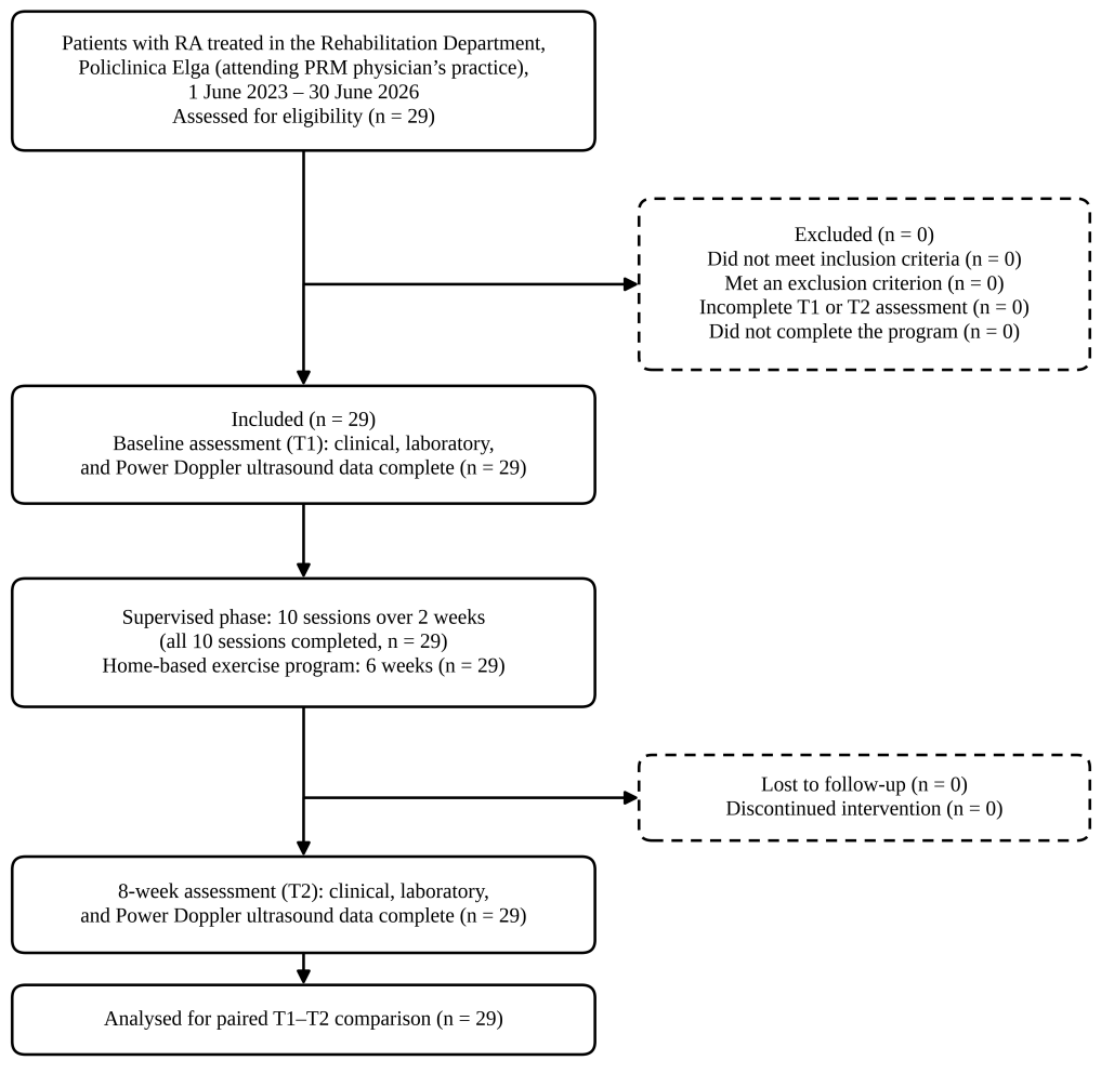
Figure 2.
Individual DAS28-ESR trajectories between baseline (T1) and 8 weeks (T2) (n = 29). Grey lines and open circles represent individual patients; the bold line indicates the group mean. Dashed horizontal lines indicate the DAS28-ESR thresholds of 2.6, 3.2, and 5.1, delimiting remission and low, moderate, and high disease activity.
Figure 2.
Individual DAS28-ESR trajectories between baseline (T1) and 8 weeks (T2) (n = 29). Grey lines and open circles represent individual patients; the bold line indicates the group mean. Dashed horizontal lines indicate the DAS28-ESR thresholds of 2.6, 3.2, and 5.1, delimiting remission and low, moderate, and high disease activity.
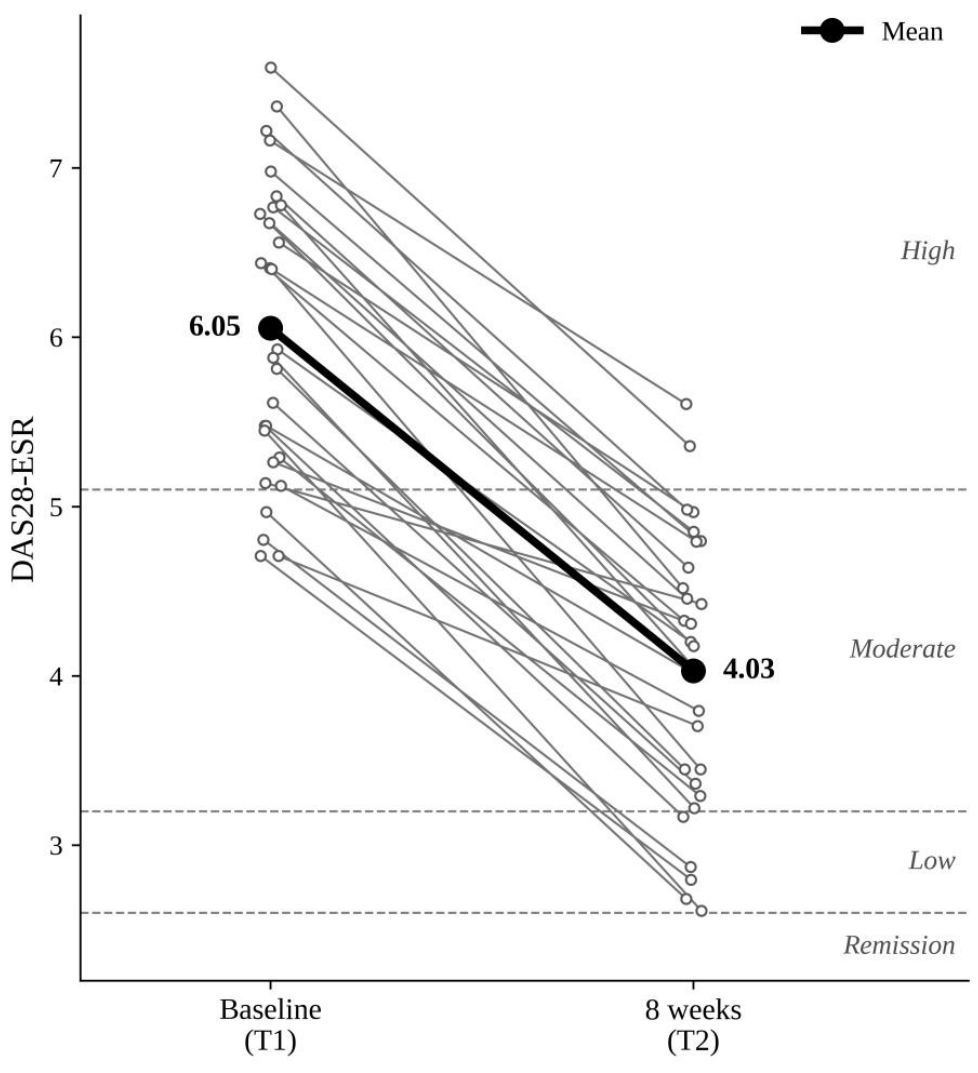
Figure 3.
Alluvial plot of individual transitions in patient-level Power Doppler grade between baseline (T1) and 8 weeks (T2) (n = 29). Band width is proportional to the number of patients; band shading indicates the baseline grade.
Figure 3.
Alluvial plot of individual transitions in patient-level Power Doppler grade between baseline (T1) and 8 weeks (T2) (n = 29). Band width is proportional to the number of patients; band shading indicates the baseline grade.
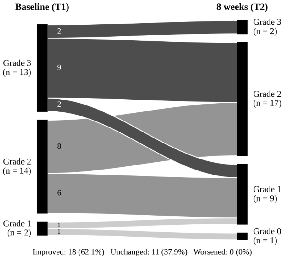

Table 1.
Baseline demographic, clinical, and treatment characteristics of the study population (n = 29).
Table 1.
Baseline demographic, clinical, and treatment characteristics of the study population (n = 29).
| Characteristic | Value |
|---|---|
| Age, years, mean ± SD (range) | 47.4 ± 9.5 (28–66) |
| Female sex, n (%) | 23 (79.3) |
| Disease duration, years, median [IQR] (range) | 6.0 [3.0–8.0] (1–22) |
| Steinbrocker radiographic stage, n (%) | |
| Stage II | 11 (37.9) |
| Stage III | 15 (51.7) |
| Stage IV | 3 (10.3) |
| Ultrasound-detected erosions, n (%) | 19 (65.5) |
| Comorbidities, n (%) | |
| Any comorbidity | 15 (51.7) |
| Arterial hypertension | 12 (41.4) |
| Dyslipidaemia | 10 (34.5) |
| Osteoporosis | 9 (31.0) |
| Chronic ischaemic heart disease | 9 (31.0) |
| Knee osteoarthritis | 9 (31.0) |
| Previous knee arthroplasty | 3 (10.3) |
| Diabetes mellitus | 0 |
| Thyroid disease | 0 |
| Current smoker, n (%) | 6 (20.7) |
| Background DMARD therapy, n (%) | |
| Methotrexate | 9 (31.0) |
| Leflunomide | 8 (27.6) |
| Etanercept | 6 (20.7) |
| Adalimumab | 5 (17.2) |
| Leflunomide + methotrexate | 1 (3.4) |
| Additional glucocorticoid therapy, n (%) | |
| None | 13 (44.8) |
| Oral methylprednisolone, 32 mg/day for 5 days | 7 (24.1) |
| Intravenous methylprednisolone, 1 g/day for 3 days | 9 (31.0) |
| NSAID course (10 days), n (%) | 29 (100) |
| Naproxen 550 mg twice daily | 13 (44.8) |
| Aceclofenac 100 mg twice daily | 10 (34.5) |
| Diclofenac (extended-release) 100 mg once daily | 6 (20.7) |
| Baseline disease activity | |
| DAS28-ESR, mean ± SD | 6.05 ± 0.87 |
| High disease activity (DAS28-ESR > 5.1), n (%) | 25 (86.2) |
| Moderate disease activity (DAS28-ESR 3.2–5.1), n (%) | 4 (13.8) |
| ESR, mm/h, median [IQR] | 50 [40–80] |
| Patient-level PD grade, mean ± SD | 2.38 ± 0.62 |
Abbreviations: DAS28-ESR, Disease Activity Score in 28 joints using erythrocyte sedimentation rate; DMARD, disease-modifying antirheumatic drug; ESR, erythrocyte sedimentation rate; IQR, interquartile range; NSAID, non-steroidal anti-inflammatory drug; PD, Power Doppler; SD, standard deviation.
Table 2.
Changes in clinical disease activity and inflammatory markers between baseline and 8-week follow-up (n = 29).
Table 2.
Changes in clinical disease activity and inflammatory markers between baseline and 8-week follow-up (n = 29).
| Outcome | Baseline (T1) | 8 weeks (T2) | p-value |
|---|---|---|---|
| TJC28, median [IQR] | 12 [8–14] | 4 [2–6] | < 0.001 |
| SJC28, median [IQR] | 6 [2–8] | 1 [1–2] | < 0.001 |
| GH, mm, median [IQR] | 60 [50–70] | 30 [20–30] | < 0.001 |
| ESR, mm/h, median [IQR] | 50 [40–80] | 30 [20–30] | < 0.001 |
| CRP, mg/L, median [IQR] | 32 [16–64] | 16 [8–24] | < 0.001 |
| DAS28-ESR, mean ± SD | 6.05 ± 0.87 | 4.03 ± 0.82 | < 0.001 |
Note: Paired comparisons were performed using the paired-samples t-test or the Wilcoxon signed-rank test, as appropriate. Abbreviations: CRP, C-reactive protein; DAS28-ESR, Disease Activity Score in 28 joints using erythrocyte sedimentation rate; ESR, erythrocyte sedimentation rate; GH, patient’s global assessment of health; IQR, interquartile range; SD, standard deviation; SJC28, 28-joint swollen joint count; TJC28, 28-joint tender joint count.
Table 3.
DAS28-ESR disease activity categories at baseline and 8-week follow-up and EULAR response at 8-week follow-up (n = 29).
Table 3.
DAS28-ESR disease activity categories at baseline and 8-week follow-up and EULAR response at 8-week follow-up (n = 29).
| Category | Baseline, n (%) | 8 weeks, n (%) |
|---|---|---|
| DAS28-ESR category | ||
| High disease activity | 25 (86.2) | 2 (6.9) |
| Moderate disease activity | 4 (13.8) | 22 (75.9) |
| Low disease activity | 0 | 5 (17.2) |
| Remission | 0 | 0 |
| EULAR response | ||
| Good response | — | 5 (17.2) |
| Moderate response | — | 24 (82.8) |
| No response | — | 0 |
Abbreviations: DAS28-ESR, Disease Activity Score in 28 joints using erythrocyte sedimentation rate; EULAR, European Alliance of Associations for Rheumatology.
Table 4.
Changes in patient-level Power Doppler grade between baseline and 8-week follow-up (n = 29).
Table 4.
Changes in patient-level Power Doppler grade between baseline and 8-week follow-up (n = 29).
| Transition (T1 → T2) | n (%) |
|---|---|
| Grade 1 → Grade 0 | 1 (3.4) |
| Grade 1 → Grade 1 | 1 (3.4) |
| Grade 2 → Grade 1 | 6 (20.7) |
| Grade 2 → Grade 2 | 8 (27.6) |
| Grade 3 → Grade 1 | 2 (6.9) |
| Grade 3 → Grade 2 | 9 (31.0) |
| Grade 3 → Grade 3 | 2 (6.9) |
| Improved | 18 (62.1) |
| Unchanged | 11 (37.9) |
| Worsened | 0 |
Note: Patient-level PD grade was defined as the highest grade among the examined joints. Abbreviations: PD, Power Doppler. Abbreviations: PD, Power Doppler.
Table 5.
Associations between DAS28-ESR and Power Doppler grade (n = 29).
| Association | Spearman ρ | p-value |
| DAS28-ESR T1 vs. PD grade T1 | 0.342 | 0.070 |
| DAS28-ESR T2 vs. PD grade T2 | 0.108 | 0.579 |
| ΔDAS28-ESR vs. ΔPD grade | 0.181 | 0.348 |
Abbreviations: DAS28-ESR, Disease Activity Score in 28 joints using erythrocyte sedimentation rate; PD, Power Doppler; T1, baseline assessment; T2, 8-week assessment; Δ, change from T1 to T2; ρ, Spearman’s rank correlation coefficient.
Table 6.
Changes in disease activity, inflammatory markers, and Power Doppler grade between baseline and 8-week follow-up according to glucocorticoid exposure.
Table 6.
Changes in disease activity, inflammatory markers, and Power Doppler grade between baseline and 8-week follow-up according to glucocorticoid exposure.
| Outcome | No GC T1 (n = 13) | No GC T2 | Within p | GC T1 (n = 16) | GC T2 | Within p | Between Δ p |
|---|---|---|---|---|---|---|---|
| DAS28-ESR | 5.22 ± 0.36 | 3.41 ± 0.61 | < 0.001 | 6.73 ± 0.48 | 4.54 ± 0.60 | < 0.001 | 0.181 |
| ESR, mm/h | 36.2 ± 5.1 | 19.2 ± 10.2 | < 0.001 | 77.8 ± 24.4 | 33.8 ± 10.3 | < 0.001 | < 0.001 |
| CRP, mg/L | 15.8 ± 10.1 | 9.2 ± 4.7 | 0.031 | 59.8 ± 32.1 | 30.5 ± 21.5 | < 0.001 | < 0.001 |
| PD grade | 2.38 ± 0.51 | 1.77 ± 0.44 | 0.016 | 2.38 ± 0.72 | 1.62 ± 0.81 | 0.001 | 0.518 |
Note: Values are presented as mean ± SD. Within-stratum p-values refer to T1–T2 comparisons using the paired-samples t-test or Wilcoxon signed-rank test, as appropriate; the Wilcoxon signed-rank test was used for PD grade. Between-stratum p-values compare the magnitude of change (Δ) using the Mann–Whitney U test. Abbreviations: CRP, C-reactive protein; DAS28-ESR, Disease Activity Score in 28 joints using erythrocyte sedimentation rate; ESR, erythrocyte sedimentation rate; GC, additional glucocorticoid therapy; PD, Power Doppler; SD, standard deviation; T1, baseline assessment; T2, 8-week assessment; Δ, change from T1 to T2.
Table 7.
Changes in serological markers between baseline and 8-week follow-up.
| Serological marker | T1, Median [IQR] | T2, Median [IQR] | p-value |
| RF | 78 [64–84] | 70 [60–80] | 0.002 |
| anti-CCP | 80 [0–120] | 80 [0–120] | 0.106 |
Note: Paired comparisons were performed using the Wilcoxon signed-rank test. Positivity thresholds were RF > 14 IU/mL and anti-CCP > 10 U/mL. Abbreviations: anti-CCP, anti-cyclic citrullinated peptide antibodies; IQR, interquartile range; RF, rheumatoid factor; T1, baseline assessment; T2, 8-week assessment.
Disclaimer/Publisher’s Note: The statements, opinions and data contained in all publications are solely those of the individual author(s) and contributor(s) and not of MDPI and/or the editor(s). MDPI and/or the editor(s) disclaim responsibility for any injury to people or property resulting from any ideas, methods, instructions or products referred to in the content. |
© 2026 by the authors. Licensee MDPI, Basel, Switzerland. This article is an open access article distributed under the terms and conditions of the Creative Commons Attribution (CC BY) license (http://creativecommons.org/licenses/by/4.0/).
Copyright: This open access article is published under a Creative Commons CC BY 4.0 license, which permit the free download, distribution, and reuse, provided that the author and preprint are cited in any reuse.