

Submitted:
16 September 2026
Posted:
16 September 2026
You are already at the latest version
Abstract
Nitric oxide (NO) is a central regulator of cardiovascular function, influencing vascular tone, platelet activity, and inflammatory signaling. Despite decades of work defining its molecular pathways, translation into consistent clinical application has remained uneven, with findings across experimental, biomarker, and therapeutic studies often appearing inconsistent.Part of this variability reflects how NO behaves under different biological conditions. Its effects are shaped by where it is produced, how much is generated, and the biochemical environment in which it operates. Under some conditions, NO supports vascular homeostasis, while in others it contributes to oxidative and nitrosative stress, a pattern we refer to as the nitric oxide paradox.This review revisits commonly used NO-related biomarkers, including nitrate/nitrite (NOx), asymmetric dimethylarginine (ADMA), and 3-nitrotyrosine, and considers why their clinical interpretation remains inconsistent. It also examines the limitations of NO-based therapies, where short half-life, systemic exposure, and limited control over delivery continue to constrain outcomes.Rather than viewing these findings as conflicting, this article considers them as reflections of how NO signaling shifts across different physiological contexts. We organize NO signaling according to four interacting determinants: enzymatic source, magnitude and duration of production, cellular compartment, and the surrounding redox environment. Framing NO biology in this way may help explain variability in both diagnostic and therapeutic settings and clarify where current approaches remain limited.
Keywords:
nitric oxide
; cardiovascular disease
; endothelial dysfunction
; oxidative stress
; nitric oxide signaling
; reactive nitrogen species
; NO bioavailability
; vascular inflammation
; nitric oxide paradox
; biomarkers
1. Introduction
Nitric oxide (NO) is a small gaseous signaling molecule with important roles across multiple organ systems, particularly in the cardiovascular system [1]. Although it was identified as a chemical entity in the 18th century, its biological significance became clear much later, when it was recognized as the endothelium-derived relaxing factor responsible for vascular smooth muscle relaxation, a discovery that led to the 1998 Nobel Prize in Physiology or Medicine.
As a reactive free radical, NO is short-lived in biological systems, with a half-life ranging from milliseconds to seconds depending on the surrounding redox environment and its rapid interactions with oxygen, superoxide, and metalloproteins [2]. Its small size and lipophilic nature allow it to diffuse readily across cellular membranes, enabling both intercellular and intracellular signaling [3].
NO is synthesized enzymatically by nitric oxide synthases (NOS), primarily in endothelial cells, neurons, and macrophages [4,5]. Under physiological conditions, tightly regulated production supports vascular homeostasis. When this regulation is disrupted, signaling can shift toward oxidative and nitrosative stress, with consequences that vary depending on the biological setting [6].
Within the cardiovascular system, NO influences vascular tone, myocardial contractility, endothelial–leukocyte interactions, barrier function, and platelet activity. Disruption of these processes, whether through reduced availability or altered signaling, contributes to the development of cardiovascular disease [7,8].
Impaired NO production, altered metabolism, and reduced bioavailability contribute to endothelial dysfunction, an early and critical event in a range of cardiovascular conditions, including hypertension, coronary artery disease, atherosclerosis, diabetes, and heart failure [9,10]. Despite extensive work outlining NO signaling pathways, translation into clinical application has remained uneven, with conflicting findings across experimental models, biomarker studies, and therapeutic trials.
Part of this difficulty appears to lie in how NO behaves under different biological conditions. Its effects are influenced by the enzymatic source, the extent and duration of production, and interactions within the local redox environment [7,11]. These factors are not always considered explicitly, which may contribute to variability in both experimental outcomes and clinical interpretation. Therapeutic strategies targeting NO pathways, whether aimed at enhancing synthesis or modulating downstream signaling, have therefore shown inconsistent efficacy across clinical settings [7,11].
These inconsistencies are often described as the “nitric oxide paradox,” where NO can exert either protective or harmful effects depending on the biological context. Rather than representing contradictory findings, this pattern may reflect distinct physiological and pathological states shaped by specific combinations of these factors.
In this context, NO signaling is better viewed as a dynamic system rather than a uniform process. Changes in production and local biochemical conditions can shift signaling behavior, complicating the interpretation of NO-related biomarkers and limiting the predictability of therapeutic responses.
In this review, we examine these challenges by integrating mechanistic, clinical, and translational observations. We organize NO signaling according to four interacting determinants: enzymatic source, magnitude and duration of production, cellular compartment, and the surrounding redox environment. We focus on how variability in NO signaling contributes to inconsistencies in experimental findings, biomarker interpretation, and therapeutic outcomes, and consider the factors that continue to limit its effective use as both a biomarker and a therapeutic target in cardiovascular disease (Figure 1).
2. Molecular Basis of Context-Dependent No Signaling
2.1. No Biosynthesis and Nos Isoforms
NO synthesis is mediated by nitric oxide synthases (NOS), which convert L-arginine to L-citrulline while generating NO [12]. Although often described as a single enzymatic step, this process is influenced by several factors, including substrate availability, cofactor balance, and regulatory control of enzyme activity. These inputs collectively determine both the amount and duration of NO production, allowing signaling to adapt to physiological demands [13].
A key determinant of NO bioavailability is the regulation of L-arginine metabolism. Arginase converts L-arginine into urea and ornithine and competes directly with NOS for substrate across vascular and metabolic tissues [14]. Increased arginase activity limits L-arginine availability for endothelial NOS (eNOS), promoting conditions that favor eNOS uncoupling and increased reactive oxygen species generation [15]. Under these conditions, superoxide production becomes more prominent, contributing to oxidative stress and vascular remodeling.
NO biosynthesis therefore reflects the interaction of multiple regulatory layers rather than a single linear pathway. Changes in substrate availability, enzymatic activity, and the surrounding metabolic environment can alter signaling behavior, with downstream effects extending beyond vascular tone. These shifts have been linked to conditions such as hypertension and neurocognitive dysfunction, highlighting the sensitivity of NO pathways to metabolic imbalance.
Tetrahydrobiopterin (BH4) is critical for maintaining proper NOS function by stabilizing the enzyme and supporting efficient coupling of NADPH oxidation to NO production [16]. This depends in part on NOS structural organization, as cofactor binding and domain interactions directly influence enzymatic coupling and overall NO output (Figure 2). When BH4 levels decline, NOS becomes prone to uncoupling, leading to superoxide generation rather than NO and contributing to oxidative stress and endothelial dysfunction [17].
eNOS is expressed as a homodimer, with each monomer consisting of a C-terminal reductase domain and an N-terminal oxygenase domain connected by a calmodulin (CaM)-binding linker region. The reductase domain (orange) contains the electron-donating cofactors NADPH, flavin adenine dinucleotide (FAD), and flavin mononucleotide (FMN), arranged in a defined electron transfer chain from NADPH → FAD → FMN. The oxygenase domain (blue) harbors the substrate-binding site for L-arginine, the ferric heme prosthetic group (Fe³⁺), and a binding pocket for the essential cofactor tetrahydrobiopterin (BH4), which is positioned at the dimer interface and contributes to structural stabilization of the active homodimer. Calmodulin, activated upon Ca²⁺ elevation, binds at the interface between the reductase and oxygenase domains of each monomer and acts as a conformational switch that facilitates electron transfer from FMN to the heme center. Notably, eNOS employs a cross-monomer electron transfer mechanism, whereby electrons from the FMN of one monomer are donated to the ferric heme of the opposing monomer (indicated by crossing arrows), a structural feature critical for catalytic activity. BH4 located at the homodimer interface further stabilizes this architecture and supports productive oxygen activation for NO synthesis. eNOS is localized to the cytoplasmic face of endothelial plasma membrane caveolae; the nucleus is not shown for clarity.
BH4, tetrahydrobiopterin; C, C-terminus; CaM, calmodulin; Ca²⁺, calcium ion; FAD, flavin adenine dinucleotide; FMN, flavin mononucleotide; N, N-terminus; NADPH, nicotinamide adenine dinucleotide phosphate (reduced); Fe³⁺, ferric iron (heme center). Figure created using Biorender.com
Under pathological conditions, this shift becomes more pronounced over time. Reduced cofactor availability favors the generation of reactive oxygen species, altering the balance of signaling within the local environment (Figure 3). These changes reflect a transition in signaling behavior rather than a simple reduction in NO levels.
Three major NOS isoforms contribute to NO production (Table 1): endothelial NOS (eNOS or NOS3), neuronal NOS (nNOS or NOS1), and inducible NOS (iNOS or NOS2). These isoforms differ in expression patterns, regulation, and output, all of which influence downstream signaling outcomes [18]. eNOS and nNOS are constitutively expressed and Ca2+-dependent, producing relatively low levels of NO under physiological conditions. In contrast, iNOS is induced by inflammatory stimuli and generates higher, sustained levels of NO independent of Ca2+ regulation.
These isoforms support distinct modes of signaling. eNOS-derived NO is typically localized and transient, contributing to vascular homeostasis through regulation of vascular tone and inhibition of smooth muscle proliferation [19,20]. In contrast, iNOS generates high-output NO over extended periods, particularly during inflammation, increasing the likelihood of reactive nitrogen species formation and tissue injury [21,30]. nNOS contributes to more localized signaling in neuronal and cardiac tissues, including modulation of calcium handling and myocardial contractility, although these effects vary with physiological conditions [22,23].
These differences underscore that NO signaling does not operate as a uniform process. Variations in enzymatic source, production profile, and local biochemical conditions shape how signaling is expressed, and whether it supports physiological function or contributes to pathology.
2.2. No Signaling
In the cardiovascular system, NO signaling operates through both cGMP-dependent and alternative pathways, contributing to vascular relaxation, myocardial function, and metabolic regulation [24]. The downstream effects vary with the conditions under which NO is generated, including its site of production and the local biochemical environment.
NO diffuses rapidly from its site of production to nearby target cells, enabling both autocrine and paracrine signaling [25]. Its small size and lipophilic nature allow it to cross cellular membranes without requiring specific transporters [25], allowing it to act over short distances without dedicated delivery systems.
One of the most well-characterized pathways involves activation of soluble guanylate cyclase (sGC), although NO signaling is not restricted to this route. Both cGMP-dependent and cGMP-independent mechanisms contribute to downstream cardiovascular responses (Figure 4). Binding of NO to the heme group of the β subunit activates sGC, leading to conversion of guanosine triphosphate (GTP) to cyclic guanosine monophosphate (cGMP) [24,25,26].
Protein kinase G (PKG), a serine/threonine kinase, mediates many of the downstream effects of this pathway. It promotes vasodilation by activating myosin light chain phosphatase and lowering intracellular calcium through modulation of calcium channels [19,27], resulting in relaxation of vascular smooth muscle cells and improved blood flow. The extent of this response depends on the level and persistence of NO signaling, and under some conditions the response becomes attenuated or shifts toward structural changes in the vessel wall.
The NO–cGMP pathway is further regulated by phosphodiesterases (PDEs), particularly PDE5, PDE6, and PDE9, which degrade cGMP and limit signal duration [19,25]. Thrombospondin-1 provides an additional regulatory input by inhibiting sGC activity and influencing calcium signaling, thereby reducing NO-mediated responses [25]. Under normal conditions, these mechanisms constrain signaling within a functional range, but disruption can lead to either reduced or excessive NO activity, with consequences for vascular function.
NO also signals independently of cGMP through post-translational modification of proteins. S-nitrosylation, which involves attachment of NO to cysteine residues, alters protein function and affects processes such as mitochondrial activity and metabolic regulation [24]. The extent of S-nitrosylation depends on local NO availability and redox conditions, linking these modifications to the cellular environment.
Interactions with other gaseous mediators further influence NO signaling. Hydrogen sulphide (H₂S), for example, can enhance NO activity by promoting eNOS function, stabilizing its dimeric form, and facilitating NO release from nitrite under hypoxic conditions [28]. These interactions suggest a broader network of gasotransmitters contributing to vascular responses, although their relative contribution under different physiological and pathological conditions remains unclear.
NO signaling is ultimately terminated through oxidation to nitrite and nitrate or through reactions with reactive oxygen species, particularly superoxide, leading to formation of peroxynitrite [25]. As this occurs, signaling shifts toward oxidative and nitrosative processes that contribute to cellular dysfunction.
3. Endothelial Function and No
3.1. No and Endothelial Function
The endothelial glycocalyx is a polysaccharide-rich layer that extends from endothelial cells and plays an important role in mechanotransduction, vascular permeability, and inflammatory signaling [29]. It is composed mainly of proteoglycans and glycoproteins, including syndecans and glypicans, which contribute to cellular signaling and the sensing of shear stress [29,30]. Through this structure, mechanical forces are translated into biochemical signals that influence endothelial NO production, linking blood flow to vascular responses.
Endothelial cells form a specialized monolayer lining the vascular lumen, serving as the interface between circulating blood and underlying smooth muscle cells. They regulate vascular tone, permeability, immune responses, and hemostasis [31]. These functions depend in part on coordinated NO signaling, which integrates mechanical, metabolic, and inflammatory inputs within the vascular wall.
NO contributes to endothelial function through vasodilatory, anti-inflammatory, and antithrombotic actions. It diffuses from endothelial cells to vascular smooth muscle cells, promoting relaxation through cGMP-dependent signaling pathways [26,32,33].
NO also counteracts vasoconstrictors such as endothelin-1, helping maintain hemodynamic stability under physiological conditions [34]. These effects are typically associated with low, tightly regulated levels of eNOS-derived NO. In addition, NO regulates endothelial permeability by controlling the movement of fluids, solutes, and immune cells across the vascular barrier [35]. This process supports tissue homeostasis but becomes less controlled under inflammatory conditions. NO also reduces the expression of adhesion molecules such as VCAM-1 and ICAM-1, limiting leukocyte adhesion and vascular inflammation [36].
The effects of NO in this setting depend on its bioavailability and the local redox environment. Interactions with reactive oxygen species influence whether signaling supports endothelial stability or contributes to oxidative stress.
3.2. Dysregulation of No Signaling in Endothelial Dysfunction and Cardiovascular Diseases
Disruption of NO signaling is a central feature of endothelial dysfunction, which often appears early in the development of cardiovascular disease. Reduced NO bioavailability contributes to impaired vasodilation, increased vascular inflammation, and altered hemostasis [37]. These changes reflect alterations in how NO is regulated within the vascular wall, rather than a simple decline in NO levels alone.
Oxidative stress is a major contributor to this process. Interactions with reactive oxygen species and changes in cofactor availability interfere with NO signaling [38,39]. Over time, this favors the formation of reactive nitrogen species, reinforcing pathways that contribute to endothelial dysfunction. In some cases, these changes develop gradually as redox balance becomes increasingly unstable.
Differences in enzymatic source add further complexity. NO signaling does not uniformly decrease in disease states. While endothelial NO production is often reduced, iNOS activity may increase under inflammatory conditions, leading to higher and less regulated NO generation [37]. This divergence between eNOS and iNOS activity alters the overall signaling profile, and both insufficient and excessive NO can contribute to disease progression depending on the context.
This pattern is observed across multiple cardiovascular conditions. In preeclampsia, reduced NO bioavailability is associated with impaired placentation and endothelial dysfunction, contributing to increased vascular resistance and hypertension [38]. Similar imbalances are present in other cardiovascular diseases, where the relative contributions of NO deficiency and excess vary with disease stage, inflammatory status, and redox conditions [37,38,39]. These differences make it difficult to interpret NO signaling in a uniform way across clinical settings.
Across these conditions, NO signaling does not follow a single direction of change. Reduced availability impairs vascular function, whereas dysregulated or excessive production promotes oxidative and nitrosative stress [37,38,39,40,41]. These patterns complicate the interpretation of NO-related biomarkers and limit the predictability of therapeutic strategies targeting NO pathways [42,43].
4. No and Vascular Inflammation
4.1. No in Vascular Inflammation
Vascular inflammation is a defining feature of cardiovascular disease and involves lipid accumulation, immune cell infiltration, and structural changes within the vessel wall. NO contributes to vascular homeostasis by regulating vasodilation, platelet function, and inflammatory signaling [28].
Reduced NO availability is associated with a shift toward a pro-inflammatory vascular environment. Leukocyte adhesion increases, cytokine production rises, and endothelial activation becomes more pronounced [36]. These changes are particularly evident when eNOS-derived NO is diminished, as localized anti-inflammatory signaling is no longer maintained.
eNOS-derived NO suppresses inflammatory signaling in part through inhibition of nuclear factor kappa B (NF-κB) activation [44,45]. Under oxidative stress, however, eNOS function becomes impaired, often in association with enzyme uncoupling and disruption of cofactor balance [38,39]. This contributes to increased reactive oxygen species accumulation and reinforcement of pro-inflammatory signaling pathways.
Inflammatory conditions can also increase iNOS expression in response to cytokines and lipopolysaccharide [46,47]. In contrast to eNOS, iNOS generates sustained NO levels independent of calcium regulation. While this supports host defense in the short term, prolonged activation alters the signaling environment. Reactive nitrogen species (RNS) accumulate, increasing the likelihood of cellular injury and tissue damage [40,41].
RNS are formed through interactions between NO and reactive oxygen species and can modify proteins, lipids, and nucleic acids. These reactions contribute to protein nitration, lipid peroxidation, and DNA damage [40,41]. Peroxynitrite, in particular, disrupts protein function, membrane integrity, and mitochondrial activity.
As these molecular changes progress, they begin to affect cellular processes more broadly. Enzymatic activity becomes less stable, signaling pathways are altered, and energy metabolism is disrupted, contributing to endothelial dysfunction and tissue injury [40,41]. RNS-mediated modifications can also sustain inflammatory signaling through activation of stress-responsive pathways.
In this setting, NO signaling becomes part of a more reactive environment shaped by redox imbalance. Interactions between NO and reactive oxygen species influence whether cellular responses remain adaptive or contribute to pathology, complicating both mechanistic interpretation and therapeutic targeting.
4.2. Limitations of No-Based Therapeutic Strategies
Therapeutic strategies targeting NO pathways have been explored to modulate vascular inflammation. NO-releasing compounds can reduce cytokine production, limit inflammatory mediator release, and protect against apoptosis in experimental models [42]. These effects, however, depend on how NO is delivered. The magnitude, timing, and localization of exposure influence outcomes, and poorly controlled NO delivery can exacerbate tissue injury [42,43].
Increasing NO availability alone does not reproduce the spatial and temporal characteristics of physiological signaling. As a result, therapeutic responses are often inconsistent across experimental and clinical settings.
4.3. Toward Controlled and Targeted No Delivery
Recent approaches focus on improving delivery rather than simply increasing NO levels. Nanoparticle-based systems, liposomes, and controlled-release platforms allow for more localized and sustained NO release [51], which may improve stability and reduce systemic exposure.
These strategies aim to approximate the dynamics of physiological signaling by controlling both the magnitude and duration of NO exposure.
Even with these advances, replicating endogenous NO signaling in vivo remains challenging. Targeting efficiency varies, release kinetics are not always predictable, and interactions with the local redox environment can alter outcomes. These factors continue to limit translational success and indicate that further refinement of delivery strategies is required.
5. No and Platelet Function
Platelets are small, anucleated cells derived from megakaryocytes that play a central role in hemostasis. Following vascular injury, they adhere to exposed subendothelial components, become activated, and aggregate to form a hemostatic plug. Disruption of platelet regulation contributes to both thrombosis and bleeding disorders. The balance between pro- and anti-aggregatory signals is therefore critical, with NO acting as an important regulator [52,53].
NO inhibits platelet activation, adhesion, and aggregation primarily through cGMP-dependent signaling pathways [54]. Endothelial cells represent the main source of NO acting on platelets, with production influenced by shear stress and physiological stimuli [55]. Platelets may also generate small amounts of NO, although the functional significance of this contribution remains unclear [56].
Within platelets, NO activates soluble guanylate cyclase, increasing cGMP levels and triggering downstream signaling through PKG [57]. PKG lowers intracellular calcium, inhibits granule secretion, and prevents integrin activation, thereby limiting platelet aggregation and thrombus formation [58]. When this pathway is disrupted, platelet activity shifts toward a prothrombotic state, particularly in the setting of endothelial dysfunction.
NO also influences platelet-derived mediators that affect vascular and myocardial function. It can modulate platelet activating factor (PAF) pathways and alter calcium handling through S-nitrosylation, reducing calcium overload during ischemic injury [53,59]. The extent to which these mechanisms contribute to clinical outcomes remains uncertain.
In addition to its signaling effects, NO can limit oxidative modification of lipoproteins, including low-density lipoproteins (LDL), which contribute to atherosclerotic plaque formation [60]. Under conditions of redox imbalance, however, NO may participate in reactive nitrogen species formation, contributing to oxidative damage rather than preventing it.
Therapeutic strategies targeting NO pathways have been explored to reduce thrombotic risk. NO-releasing derivatives of existing drugs, such as NO-aspirin (NCX-4016), have shown enhanced antiplatelet activity and reduced gastrointestinal toxicity in experimental models [61,62], although consistent long-term clinical benefit has not been established.
Conventional NO donors, including nitroglycerin and sodium nitroprusside, are widely used to improve blood flow and relieve ischemia [63]. Their use, however, is limited by short half-life, systemic hypotension, and the development of nitrate tolerance [64], highlighting the difficulty of maintaining controlled NO exposure.
More recent approaches focus on targeted delivery. NO-donating compounds and nanotechnology-based systems, including nanoparticles and liposomes, allow more localized and sustained release [65,66,67,68,69,70], improving control over both the magnitude and duration of NO signaling.
NO-releasing biomaterials, such as vascular graft coatings and hydrogels, have also been developed to reduce platelet adhesion and prevent restenosis [71]. These strategies reflect the importance of spatial and temporal control of NO signaling in shaping therapeutic outcomes.
6. No and Vascular Aging
Vascular aging is characterized by progressive structural and functional changes in the vasculature, including arterial stiffening, reduced endothelial function, impaired regulation of blood flow, and chronic low-grade inflammation. These changes contribute to age-related cardiovascular conditions such as hypertension, atherosclerosis, and stroke [72]. A consistent feature across these processes is a decline in NO bioavailability and signaling, reflecting both reduced production and altered regulation within the vascular wall.
Multiple mechanisms contribute to vascular aging, including oxidative stress, mitochondrial dysfunction, cellular senescence, and chronic inflammation [73]. Among these, oxidative stress is particularly important. An imbalance between reactive oxygen species production and antioxidant defenses reduces NO availability and interferes with endothelial signaling [74,75].
Interactions between NO and reactive oxygen species, together with cofactor imbalance, favor the formation of reactive nitrogen species [74,75,76]. These changes contribute to oxidative damage, mitochondrial dysfunction, and vascular injury.
Oxidative stress also affects NO production directly. Depletion of BH4 promotes eNOS uncoupling, increasing superoxide generation and further reducing NO availability [74,75]. Over time, this creates a self-reinforcing process in which oxidative stress and impaired NO signaling sustain each other.
The functional consequences are reflected in reduced vasodilatory capacity, increased vascular stiffness, and amplification of inflammatory signaling [77,78]. In addition to reduced NO levels, vascular responsiveness to physiological stimuli declines, indicating broader disruption of NO signaling pathways.
NO also contributes to angiogenesis through vascular endothelial growth factor (VEGF) signaling and regulation of hypoxia-inducible factor-1α (HIF-1α) [79]. With aging, reduced NO availability is associated with impaired angiogenic responses, which may limit tissue repair and regeneration.
Sex-related differences further influence these processes. Premenopausal women generally exhibit higher NO bioavailability compared to age-matched males, partly due to estrogen-mediated regulation of eNOS [80,81]. In contrast, increased oxidative stress in males has been associated with reduced NO signaling and more rapid vascular aging [82].
Several strategies have been explored to improve NO availability in this setting. Dietary nitrate supplementation provides an alternative pathway for NO generation through the nitrate–nitrite–NO axis [83,84], with some studies reporting improvements in endothelial function and oxidative balance [85,86]. These responses vary and appear to depend on baseline metabolic and cardiovascular status.
Regular aerobic exercise can enhance NO signaling. Increased shear stress during exercise stimulates eNOS activity and supports vascular remodeling [77]. Exercise has also been associated with partial restoration of angiogenic responses through VEGF–eNOS signaling pathways [87].
Polyphenolic compounds such as curcumin and resveratrol have been investigated for their ability to improve NO bioavailability by reducing oxidative stress and supporting eNOS activity [88,89]. Clinical effects remain variable, in part due to differences in bioavailability and dosing.
Supplementation with L-arginine and B vitamins has also been examined as a means of supporting NO production by increasing substrate availability and reducing levels of asymmetric dimethylarginine (ADMA) [75,90]. As with other interventions, responses are not uniform and appear to depend on underlying metabolic and disease conditions.
In vascular aging, reduced NO availability contributes to functional decline, but restoring NO signaling is not straightforward. Outcomes depend on the interaction between production, redox balance, and enzymatic regulation over time.

Table 2.
Selected interventions that preserve nitric oxide signaling during vascular aging.
| Prevention strategy | Study type | Participants | Duration | Study groups | Effect on NO signaling | Outcome | Reference |
|---|---|---|---|---|---|---|---|
| Nitrate supplementation | Animal-based experimental (mice) | 98–100-week-old mice | 4 weeks | Beetroot juice vs. water | Increased NO bioavailability, reduced oxidative stress | ↑ Plasma nitrate | [85] |
| Nitrate supplementation | Randomized controlled trial (double-blind, crossover) | Adults (67 ± 3 y) | 4 weeks | Inorganic nitrate supplementation vs. placebo | Increased plasma nitrate and nitrite; reduced retrograde and oscillatory shear | Improved conduit artery shear profile and increased basal forearm blood flow | [86] |
| Aerobic exercise | Animal-based experimental (rats) | 21-month-old rats | 8 weeks | Swim training vs. sedentary | Restoration of VEGF–Akt–eNOS signaling pathway | ↑ Cardiac angiogenesis | [87] |
| Curcumin supplementation | Randomized controlled trial (double-blind, parallel) | Adults (45–74 y) | 12 weeks | Curcumin vs. placebo | Increased NO bioavailability, reduced oxidative stress | ↑ Forearm resistance-artery endothelial function and brachial artery flow-mediated dilation | [88] |
| Resveratrol | Randomized controlled trial (double-blind, crossover) | 18-month-old mice | 6 months | Resveratrol vs. normal feed | Increased eNOS activity via SIRT1 activation | ↑ p-eNOS/eNOS ratio; ↓ Nox2 and Nox4 | [91] |
| L-arginine and vitamin B supplementation | Randomized controlled trial (double-blind, prospective) | Adults (40–65 y) with mild hypertension | 3 months | Dietary intervention vs. placebo | Increased substrate availability for NO synthesis; reduced oxidative stress via homocysteine metabolism | ↑ Endothelial function | [90] |
NO, nitric oxide; eNOS, endothelial nitric oxide synthase; VEGF, vascular endothelial growth factor; SIRT1, sirtuin 1; NOx, nitrate/nitrite.
7. Genetic Variations in No Metabolism
Genetic variation in NO-related pathways influences NO production, bioavailability, and signaling, with implications for cardiovascular disease risk. NO is generated by three NOS isoforms, NOS1, NOS2, and NOS3, which differ in tissue distribution and patterns of NO production [92,93]. Variation across these genes can alter downstream physiological responses, although the effects are not always consistent.
NO availability is also influenced by the L-arginine pathway. Arginases (ARG1 and ARG2) compete with NOS for L-arginine, limiting substrate availability for NO synthesis, while dimethylarginine dimethylaminohydrolases (DDAH1 and DDAH2) regulate NO production indirectly by metabolizing asymmetric dimethylarginine (ADMA), an endogenous NOS inhibitor [94]. These pathways introduce multiple points of regulation rather than a single site of control. Key genetic variations affecting NO regulation across these systems are summarized in Table 3.
Variants within these pathways have been studied in relation to cardiovascular disease, but reported effects are often modest and not consistently reproduced across populations [94]. Differences in genetic background, environmental exposure, and study design likely contribute to this variability.
Among NOS genes, variants in NOS3 have received the most attention. Polymorphisms such as Glu298Asp (rs1799983) and T-786C (rs2070744) have been associated with altered eNOS activity and NO production [20]. Links to hypertension, coronary artery disease, and preeclampsia have been reported in some cohorts, although findings vary across studies [20,95,96,97,98]. The impact of these variants appears to depend on additional factors, including oxidative stress, comorbidities, and environmental influences.
In comparison, variants in NOS1 and NOS2 show weaker and less consistent associations with cardiovascular outcomes and are more often linked to neurological or inflammatory conditions [93]. Their contribution to cardiovascular risk appears indirect, which limits their value as standalone predictors.
Variation in arginase and DDAH pathways further complicates interpretation. Polymorphisms in ARG1 and ARG2 have been associated with altered enzyme activity and reduced NO bioavailability in some populations [94]. Variants in DDAH1 and DDAH2 may influence cardiovascular risk by affecting ADMA levels and NOS inhibition [94]. These associations remain difficult to interpret and require validation in larger and more diverse cohorts.
Overall, genetic variation across NO-related pathways appears to have modest and variable effects on cardiovascular risk. These effects are influenced by interactions with metabolic, environmental, and disease-related factors.
From a translational standpoint, genetic data alone provide limited insight into NO activity in vivo. Interpretation is more informative when considered alongside functional, biochemical, and clinical measures, particularly in settings where NO signaling is influenced by multiple interacting factors.
8. No as A Biomarker for Cardiovascular Disease
Cardiovascular biomarkers are widely used to assess disease risk, support diagnosis, and monitor therapeutic responses. In conditions with clearly defined mechanisms, single markers can be informative. In cardiovascular disease, however, interpretation is more complex and often requires a combination of markers [99,100]. Within this context, NO and its related metabolites have been examined as indicators of vascular function and disease progression.
Direct measurement of NO is limited by its short half-life and rapid reactivity. As a result, most studies rely on indirect measures such as nitrate and nitrite (NOx), along with related molecules including asymmetric dimethylarginine (ADMA), 3-nitrotyrosine, and myeloperoxidase (MPO) [101]. Representative studies highlighting variability in these markers are summarized in Table 4. These measurements reflect downstream products of NO metabolism rather than real-time signaling activity.
Reduced NO bioavailability has been associated with impaired vasodilation and increased disease severity in coronary artery disease [102]. Similar findings have been reported in heart failure, where NO signaling is linked to oxidative stress and NADPH oxidase activity [103].
Findings related to NOx levels are less consistent. In some studies, higher concentrations are associated with increased cardiovascular mortality and adverse outcomes [105,107]. Elevated NOx does not necessarily indicate increased NO signaling and may instead reflect compensatory changes, altered metabolism, or contributions from non-endothelial sources.
Population-based studies illustrate this variability. Higher circulating NOx levels have been linked to increased mortality risk in certain cohorts [105], whereas others, including the Framingham Offspring Study, report no clear association between plasma nitrate levels and cardiovascular disease [106]. Differences in diet, renal function, systemic inflammation, and measurement approaches likely contribute to these discrepancies [105,106].
Additional biomarkers provide complementary information. 3-nitrotyrosine reflects protein nitration and nitrosative stress [108], ADMA functions as an endogenous NOS inhibitor associated with endothelial dysfunction and cardiovascular risk [109,110,111,112], and MPO contributes to oxidative reactions that reduce NO availability [113,114,115]. These markers represent different aspects of NO-related biology rather than a single pathway.
NO-related biomarkers therefore do not provide a direct measure of signaling activity. Instead, they reflect multiple processes, including NO metabolism, oxidative stress, and vascular dysfunction. Differences across studies are influenced by variation in biological and clinical conditions.
From a translational perspective, this limits the use of NO as a standalone biomarker. Interpretation is more informative when multiple markers are considered alongside clinical and functional assessments.
The coexistence of reduced NO availability and increased metabolic byproducts contributes to findings that appear inconsistent across studies. These patterns reflect differences in how NO is produced, processed, and measured in vivo, rather than opposing biological effects.
9. Conclusions
Nitric oxide remains a central regulator of cardiovascular physiology, influencing vascular tone, platelet function, inflammation, and endothelial homeostasis. Despite extensive work defining its production and signaling pathways, translation into consistent clinical use has been uneven. This gap appears less related to gaps in mechanism and more to the difficulty of interpreting NO behavior within complex and changing physiological systems.
In this review, we examined how differences in where NO is produced, how it is generated, and the conditions in which it operates influence downstream effects. Rather than functioning as a single, predictable mediator, NO shifts its behavior across physiological and pathological settings.
This becomes evident across multiple areas of cardiovascular biology. Reduced NO availability can impair vascular function, while sustained or dysregulated production contributes to oxidative and nitrosative stress. Similar patterns appear in endothelial regulation, vascular inflammation, platelet function, and vascular aging, although the balance between these effects varies across conditions.
These features complicate clinical interpretation. NO-related biomarkers often yield inconsistent findings because they reflect indirect aspects of NO metabolism rather than functional signaling. Therapeutic approaches face similar limitations. Increasing NO levels alone does not reproduce the spatial and temporal characteristics of endogenous signaling, which may account for the variability observed in clinical outcomes.
Taken together, these observations indicate that NO signaling does not follow a single direction of change. Apparent contradictions in the literature are often a result of differences in biological context rather than opposing mechanisms.
Progress in this area may depend less on identifying new pathways and more on improving how existing knowledge is applied. Approaches that account for how NO is generated, how long it persists, and how it interacts with the surrounding environment may provide more consistent insights. This includes refining delivery strategies, improving interpretation of biomarker data, and integrating genetic and biochemical information with clinical context.
A clearer understanding of these relationships may help bring greater consistency to how NO is interpreted and targeted in cardiovascular disease.
Author Contributions
K.A., R.N.Z., A.K., Y.X., Z.X., and K.K. contributed to the writing and development of the manuscript. All authors contributed to the preparation of figures and tables. Y.X. led the design and finalization of all figures. K.K. supervised the project and performed critical revision and editing of the manuscript. All authors reviewed and approved the final version of the manuscript. All authors take complete responsibility for the integrity of the work done in this manuscript.
Funding
We did not use any funding sources for this manuscript.; Transparency statement; KK affirms that this manuscript is an honest, accurate, and transparent account of the study being reported; that no important aspects of the study have been omitted; and that any discrepancies from the study as planned have been explained.
Data Availability Statement
Data sharing not applicable – no new data generated, or the article describes entirely theoretical research.
Conflicts of Interest
Authors have no conflict of interest to declare with the contents of this article.
References
- Hollenberg, N.K. Organ systems dependent on nitric oxide and the potential for nitric oxide-targeted therapies in related diseases. J. Clin. Hypertens. (Greenwich) 2006, 8((12) Suppl 4, 63–73. [Google Scholar] [CrossRef] [PubMed]
- Bredt, D.S. Endogenous nitric oxide synthesis: biological functions and pathophysiology. Free Radic. Res. 1999, 31(6), 577–96. [Google Scholar] [CrossRef] [PubMed]
- Khan, M.; et al. Nitric Oxide Acts as a Key Signaling Molecule in Plant Development under Stressful Conditions. Int. J. Mol. Sci. 2023, 24(5). [Google Scholar] [CrossRef] [PubMed]
- Chen, K.; Pittman, R.N.; Popel, A.S. Nitric oxide in the vasculature: where does it come from and where does it go? A quantitative perspective. Antioxid. Redox Signal 2008, 10(7), 1185–98. [Google Scholar] [CrossRef] [PubMed]
- Armentano, G.M.; et al. Nitric Oxide Plays a Dual Role in Cardiorenal Syndrome in Vitro Model. Cell Physiol. Biochem 2024, 58(1), 33–48. [Google Scholar] [CrossRef] [PubMed]
- Förstermann, U.; Xia, N.; Li, H. Roles of Vascular Oxidative Stress and Nitric Oxide in the Pathogenesis of Atherosclerosis. Circ. Res. 2017, 120(4), 713–735. [Google Scholar] [CrossRef] [PubMed]
- Loscalzo, J. G. Welch, Nitric oxide and its role in the cardiovascular system. Prog. Cardiovasc Dis. 1995, 38(2), 87–104. [Google Scholar] [CrossRef] [PubMed]
- Liu, V.W.; Huang, P.L. Cardiovascular roles of nitric oxide: a review of insights from nitric oxide synthase gene disrupted mice. Cardiovasc Res. 2008, 77(1), 19–29. [Google Scholar] [CrossRef] [PubMed]
- Wang, H.; et al. Nitric oxide directly promotes vascular endothelial insulin transport. Diabetes 2013, 62(12), 4030–42. [Google Scholar] [CrossRef] [PubMed]
- Naseem, K.M. The role of nitric oxide in cardiovascular diseases. Mol. Asp. Med. 2005, 26(1-2), 33–65. [Google Scholar] [CrossRef] [PubMed]
- Abolfazli, S.; et al. Regulatory effects of curcumin on nitric oxide signaling in the cardiovascular system. Nitric Oxide 2024, 143, 16–28. [Google Scholar] [CrossRef] [PubMed]
- Zhu, H.Y.; Hong, F.F.; Yang, S.L. The Roles of Nitric Oxide Synthase/Nitric Oxide Pathway in the Pathology of Vascular Dementia and Related Therapeutic Approaches. Int. J. Mol. Sci. 2021, 22(9). [Google Scholar] [CrossRef] [PubMed]
- Andrabi, S.M.; et al. Nitric Oxide: Physiological Functions, Delivery, and Biomedical Applications. Adv. Sci. (Weinh) 2023, 10(30), e2303259. [Google Scholar] [CrossRef] [PubMed]
- Ren, Y.; et al. Arginase: Biological and Therapeutic Implications in Diabetes Mellitus and Its Complications. Oxid. Med. Cell Longev. 2022, 2419412. [Google Scholar] [CrossRef] [PubMed]
- Mahdi, A.; Kövamees, O.; Pernow, J. Improvement in endothelial function in cardiovascular disease - Is arginase the target? Int. J. Cardiol. 2020, 301, 207–214. [Google Scholar] [CrossRef] [PubMed]
- Alderton, W.K.; Cooper, C.E.; Knowles, R.G. Nitric oxide synthases: structure, function and inhibition. Biochem J. 2001, 357 Pt 3, 593–615. [Google Scholar] [CrossRef]
- Bisconti, A.V.; et al. No effect of acute tetrahydrobiopterin (BH(4)) supplementation on vascular dysfunction in the old. J. Appl. Physiol. (1985) 2022, 132(3), 773–784. [Google Scholar] [CrossRef] [PubMed]
- Arzumanian, V.; et al. [Mechanisms of nitric oxide synthesis and action in cells]. In Medicina (Kaunas); 2003; Volume 39, 6, pp. 535–41. [Google Scholar]
- Farah, C.; Michel, L.Y.M.; Balligand, J.L. Nitric oxide signalling in cardiovascular health and disease. Nat. Rev. Cardiol. 2018, 15(5), 292–316. [Google Scholar] [CrossRef] [PubMed]
- Oliveira-Paula, G.H.; Lacchini, R.; Tanus-Santos, J.E. Endothelial nitric oxide synthase: From biochemistry and gene structure to clinical implications of NOS3 polymorphisms. Gene 2016, 575 2 Pt 3, 584–99. [Google Scholar] [CrossRef] [PubMed]
- Cinelli, M.A.; et al. Inducible nitric oxide synthase: Regulation, structure, and inhibition. Med. Res. Rev. 2020, 40(1), 158–189. [Google Scholar] [CrossRef] [PubMed]
- Solanki, K.; et al. The expanding roles of neuronal nitric oxide synthase (NOS1). PeerJ 2022, 10, e13651. [Google Scholar] [CrossRef] [PubMed]
- Ally, A.; et al. Role of neuronal nitric oxide synthase on cardiovascular functions in physiological and pathophysiological states. Nitric Oxide 2020, 102, 52–73. [Google Scholar] [CrossRef] [PubMed]
- Lau, B.; et al. Endogenous S-nitrosocysteine proteomic inventories identify a core of proteins in heart metabolic pathways. Redox Biol. 2021, 47, 102153. [Google Scholar] [CrossRef] [PubMed]
- Lundberg, J.O.; Weitzberg, E. Nitric oxide signaling in health and disease. Cell 2022, 185(16), 2853–2878. [Google Scholar] [CrossRef] [PubMed]
- Roy, R.; et al. Dysfunctional and Dysregulated Nitric Oxide Synthases in Cardiovascular Disease: Mechanisms and Therapeutic Potential. Int. J. Mol. Sci. 2023, 24(20). [Google Scholar] [CrossRef] [PubMed]
- Akanji, M.A.; et al. Nitric Oxide Balance in Health and Diseases: Implications for New Treatment Strategies. Open Biochem. J. 2020, 14, 25–32. [Google Scholar] [CrossRef]
- Kida, M.; et al. Hydrogen sulfide increases nitric oxide production with calcium-dependent activation of endothelial nitric oxide synthase in endothelial cells. Eur. J. Pharm. Sci. 2013, 48(1-2), 211–5. [Google Scholar] [CrossRef] [PubMed]
- Vittum, Z.; Cocchiaro, S.; Mensah, S.A. Basal endothelial glycocalyx's response to shear stress: a review of structure, function, and clinical implications. Front Cell Dev. Biol. 2024, 12, 1371769. [Google Scholar] [CrossRef] [PubMed]
- Foote, C.A.; et al. Endothelial Glycocalyx. Compr. Physiol. 2022, 12(4), 3781–3811. [Google Scholar] [CrossRef]
- Cyr, A.R.; et al. Nitric Oxide and Endothelial Dysfunction. Crit. Care Clin. 2020, 36(2), 307–321. [Google Scholar] [CrossRef] [PubMed]
- Gresele, P.; Momi, S.; Guglielmini, G. Nitric oxide-enhancing or -releasing agents as antithrombotic drugs. Biochem. Pharmacol. 2019, 166, 300–312. [Google Scholar] [CrossRef] [PubMed]
- Chirkov, Y.Y.; Nguyen, T.H.; Horowitz, J.D. Impairment of Anti-Aggregatory Responses to Nitric Oxide and Prostacyclin: Mechanisms and Clinical Implications in Cardiovascular Disease. Int. J. Mol. Sci. 2022, 23(3). [Google Scholar] [CrossRef] [PubMed]
- Genovesi, S.; et al. Relationship between endothelin and nitric oxide pathways in the onset and maintenance of hypertension in children and adolescents. Pediatr. Nephrol. 2022, 37(3), 537–545. [Google Scholar] [CrossRef] [PubMed]
- Hellenthal, K.E.M.; Brabenec, L.; Wagner, N.M. Regulation and Dysregulation of Endothelial Permeability during Systemic Inflammation. Cells 2022, 11(12). [Google Scholar] [CrossRef] [PubMed]
- Singh, V.; et al. ICAM-1 and VCAM-1: Gatekeepers in various inflammatory and cardiovascular disorders. Clin. Chim. Acta 2023, 548, 117487. [Google Scholar] [CrossRef] [PubMed]
- Janaszak-Jasiecka, A.; et al. Endothelial dysfunction due to eNOS uncoupling: molecular mechanisms as potential therapeutic targets. Cell Mol. Biol. Lett. 2023, 28(1), 21. [Google Scholar] [CrossRef] [PubMed]
- Guerby, P.; et al. Role of oxidative stress in the dysfunction of the placental endothelial nitric oxide synthase in preeclampsia. Redox Biol. 2021, 40, 101861. [Google Scholar] [CrossRef] [PubMed]
- Scioli, M.G.; et al. Oxidative Stress and New Pathogenetic Mechanisms in Endothelial Dysfunction: Potential Diagnostic Biomarkers and Therapeutic Targets. J. Clin. Med. 2020, 9(6). [Google Scholar] [CrossRef] [PubMed]
- Kim, M.E.; Lee, J.S. Advances in the Regulation of Inflammatory Mediators in Nitric Oxide Synthase: Implications for Disease Modulation and Therapeutic Approaches. Int. J. Mol. Sci. 2025, 26(3). [Google Scholar] [CrossRef] [PubMed]
- Kumar, A.; et al. Mitigating oxidative stress in red blood cells with quercetin-loaded liposomes: a promising strategy. RSC Pharm. 2025, 2(6), 1471–1478. [Google Scholar] [CrossRef]
- H.U. Current approaches of anti-inflammatory-dependent antiaging strategies., in Molecular Basis and Emerging Strategies for Anti-aging Interventions.; 2018; pp. 355–372. [Google Scholar]
- Navale, G.R.; Singh, S.; Ghosh, K. NO donors as the wonder molecules with therapeutic potential: Recent trends and future perspectives. Coord. Chem. Rev. 2023, 481, 215052. [Google Scholar] [CrossRef]
- Mollace, V.; Tavernese, A.; Mollace, R. The essential role of a "healthy" relationship between caveolin-1 and endothelial nitric oxide synthase in counteracting vascular inflammation and atherothrombosis. Kardiol. Pol. 2020, 78(2), 96–97. [Google Scholar] [CrossRef] [PubMed]
- Gliozzi, M.; et al. Modulation of Nitric Oxide Synthases by Oxidized LDLs: Role in Vascular Inflammation and Atherosclerosis Development. Int. J. Mol. Sci. 2019, 20(13). [Google Scholar] [CrossRef] [PubMed]
- Griffiths, C.; Garthwaite, J. The shaping of nitric oxide signals by a cellular sink. J. Physiol. 2001, 536 Pt 3, 855–62. [Google Scholar] [CrossRef] [PubMed]
- Farahani, A.; et al. Inducible nitric oxide synthase (iNOS): More than an inducible enzyme? Rethinking the classification of NOS isoforms. Pharmacol. Res. 2025, 216, 107781. [Google Scholar] [CrossRef] [PubMed]
- Burov, O.N.; et al. Mechanisms of nitric oxide generation in living systems. Nitric Oxide 2022, 118, 1–16. [Google Scholar] [CrossRef] [PubMed]
- Thadani, U. Challenges with nitrate therapy and nitrate tolerance: prevalence, prevention, and clinical relevance. Am. J. Cardiovasc Drugs 2014, 14(4), 287–301. [Google Scholar] [CrossRef] [PubMed]
- Münzel, T.; Gori, T. Nitrate therapy and nitrate tolerance in patients with coronary artery disease. Curr. Opin. Pharmacol. 2013, 13(2), 251–9. [Google Scholar] [CrossRef] [PubMed]
- Tortella Fuentes, G.; et al. Nanoparticle-Based Nitric Oxide Donors: Exploring Their Antimicrobial and Anti-Biofilm Capabilities. Antibiotics 2024, 13(11). [Google Scholar] [CrossRef] [PubMed]
- Pepke-Zaba, J.; et al. Inhaled nitric oxide as a cause of selective pulmonary vasodilatation in pulmonary hypertension. Lancet 1991, 338(8776), 1173–4. [Google Scholar] [CrossRef] [PubMed]
- Russo, I.; et al. Platelets and Cardioprotection: The Role of Nitric Oxide and Carbon Oxide. Int. J. Mol. Sci. 2023, 24(7). [Google Scholar] [CrossRef] [PubMed]
- Loscalzo, J. Nitric Oxide Insufficiency, Platelet Activation, and Arterial Thrombosis. Circ. Res. 2001, 88(8), 756–762. [Google Scholar] [CrossRef] [PubMed]
- Blanton, R.M. cGMP Signaling and Modulation in Heart Failure. J. Cardiovasc Pharmacol. 2020, 75(5), 385–398. [Google Scholar] [CrossRef] [PubMed]
- Gkaliagkousi, E.; Ritter, J.; Ferro, A. Platelet-Derived Nitric Oxide Signaling and Regulation. Circ. Res. 2007, 101(7), 654–662. [Google Scholar] [CrossRef] [PubMed]
- Gambaryan, S.; Tsikas, D. A review and discussion of platelet nitric oxide and nitric oxide synthase: do blood platelets produce nitric oxide from L-arginine or nitrite? Amino Acids 2015, 47(9), 1779–93. [Google Scholar] [CrossRef] [PubMed]
- Preedy, M.E.J.; Baliga, R.S.; Hobbs, A.J. Multiplicity of Nitric Oxide and Natriuretic Peptide Signaling in Heart Failure. J. Cardiovasc Pharmacol. 2020, 75(5), 370–384. [Google Scholar] [CrossRef] [PubMed]
- Subramanian, H.; et al. Phosphorylation of CalDAG-GEFI by protein kinase A prevents Rap1b activation. J. Thromb. Haemost. 2013, 11(8), 1574–82. [Google Scholar] [CrossRef] [PubMed]
- Penna, C.; et al. Platelet-activating factor induces cardioprotection in isolated rat heart akin to ischemic preconditioning: role of phosphoinositide 3-kinase and protein kinase C activation. Am. J. Physiol. Heart Circ. Physiol. 2005, 288(5), H2512–20. [Google Scholar] [CrossRef] [PubMed]
- Hogg, N.; et al. Inhibition of low-density lipoprotein oxidation by nitric oxide. Potential role in atherogenesis. FEBS Lett. 1993, 334(2), 170–4. [Google Scholar] [CrossRef] [PubMed]
- Michelson, A.D. Antiplatelet therapies for the treatment of cardiovascular disease. Nat. Rev. Drug Discov. 2010, 9(2), 154–69. [Google Scholar] [CrossRef] [PubMed]
- Furchgott, R.F.; Zawadzki, J.V. The obligatory role of endothelial cells in the relaxation of arterial smooth muscle by acetylcholine. Nature 1980, 288(5789), 373–6. [Google Scholar] [CrossRef] [PubMed]
- Wallace, J.L.; et al. In vivo antithrombotic effects of a nitric oxide-releasing aspirin derivative, NCX-4016. Thromb. Res. 1999, 93(1), 43–50. [Google Scholar] [CrossRef] [PubMed]
- Vilahur, G.; et al. Effects of a novel platelet nitric oxide donor (LA816), aspirin, clopidogrel, and combined therapy in inhibiting flow- and lesion-dependent thrombosis in the porcine ex vivo model. Circulation 2004, 110(12), 1686–93. [Google Scholar] [CrossRef] [PubMed]
- Divakaran, S. J. Loscalzo, The Role of Nitroglycerin and Other Nitrogen Oxides in Cardiovascular Therapeutics. JACC 2017, 70(19), 2393–2410. [Google Scholar] [CrossRef] [PubMed]
- Oyelaja, O.; et al. Applications of Nanotechnology in the Field of Cardiology. Cureus 2024, 16(4), e58059. [Google Scholar] [CrossRef] [PubMed]
- Nahar, K.; et al. Liposomal Aerosols of Nitric Oxide (NO) Donor as a Long-Acting Substitute for the Ultra-Short-Acting Inhaled NO in the Treatment of PAH. Pharm. Res. 2016, 33(7), 1696–710. [Google Scholar] [CrossRef] [PubMed]
- Gothwal, A.; et al. Dendrimers as an Effective Nanocarrier in Cardiovascular Disease. Curr. Pharm. Des. 2015, 21(30), 4519–26. [Google Scholar] [CrossRef] [PubMed]
- Ge, X.; et al. Advances in metal-organic frameworks for cardiovascular therapy: from structural design to preclinical applications. Coord. Chem. Rev. 2025, 544, 216971. [Google Scholar] [CrossRef]
- Tabish, T.A.; et al. Nitric Oxide Releasing Nanomaterials for Cardiovascular Applications. JACC Basic Transl. Sci. 2024, 9(5), 691–709. [Google Scholar] [CrossRef] [PubMed]
- Seals, D.R.; Alexander, L.M. Vascular aging. J. Appl. Physiol. (1985) 2018, 125(6), 1841–1842. [Google Scholar] [CrossRef] [PubMed]
- van der Linden, J.; et al. Model Systems to Study the Mechanism of Vascular Aging. Int. J. Mol. Sci. 2023, 24(20). [Google Scholar] [CrossRef] [PubMed]
- Rossman, M.J.; et al. Healthy lifestyle-based approaches for successful vascular aging. J. Appl. Physiol. (1985) 2018, 125(6), 1888–1900. [Google Scholar] [CrossRef] [PubMed]
- Poeggeler, B.; et al. Nitric Oxide as a Determinant of Human Longevity and Health Span. Int. J. Mol. Sci. 2023, 24(19). [Google Scholar] [CrossRef] [PubMed]
- Pourbagher-Shahri, A.M.; et al. An Overview of NO Signaling Pathways in Aging. Molecules 2021, 26(15). [Google Scholar] [CrossRef] [PubMed]
- Ya, J.; Bayraktutan, U. Vascular Ageing: Mechanisms, Risk Factors, and Treatment Strategies. Int. J. Mol. Sci. 2023, 24(14). [Google Scholar] [CrossRef] [PubMed]
- Oliveira, A.C.; et al. Vascular Aging and Arterial Stiffness. Arq. Bras. Cardiol. 2022, 119(4), 604–615. [Google Scholar] [PubMed]
- Zhang, J.; et al. Therapeutic potential of nitric oxide in vascular aging due to the promotion of angiogenesis. Chem. Biol. Drug Des. 2023, 102(2), 395–407. [Google Scholar] [CrossRef] [PubMed]
- Cattaneo, M.G.; et al. Sex-specific eNOS activity and function in human endothelial cells. Sci. Rep. 2017, 7(1), 9612. [Google Scholar] [CrossRef] [PubMed]
- Pabbidi, M.R.; et al. Sex differences in the vascular function and related mechanisms: role of 17β-estradiol. Am. J. Physiol. Heart Circ. Physiol. 2018, 315(6), H1499–h1518. [Google Scholar] [CrossRef] [PubMed]
- Barros, P.R.; et al. Vascular Aging in Rodent Models: Contrasting Mechanisms Driving the Female and Male Vascular Senescence. Front Aging 2021, 2, 727604. [Google Scholar] [CrossRef] [PubMed]
- Rocha, B.S. The Nitrate-Nitrite-Nitric Oxide Pathway on Healthy Ageing: A Review of Pre-clinical and Clinical Data on the Impact of Dietary Nitrate in the Elderly. Front Aging 2021, 778467. [Google Scholar] [CrossRef] [PubMed]
- Jones, T.; et al. The Effects of Beetroot Juice on Blood Pressure, Microvascular Function and Large-Vessel Endothelial Function: A Randomized, Double-Blind, Placebo-Controlled Pilot Study in Healthy Older Adults. Nutrients 2019, 11(8). [Google Scholar] [CrossRef] [PubMed]
- Tawa, M.; Nakagawa, K.; Ohkita, M. Effects of beetroot juice supplementation on vascular functional and structural changes in aged mice. Physiol. Rep. 2023, 11(12), e15755. [Google Scholar] [CrossRef] [PubMed]
- Casey, D.P.; Bock, J.M. Inorganic nitrate supplementation attenuates conduit artery retrograde and oscillatory shear in older adults. Am. J. Physiol. Heart Circ. Physiol. 2021, 320(3), H991–h998. [Google Scholar] [CrossRef] [PubMed]
- Iemitsu, M.; et al. Exercise training improves aging-induced downregulation of VEGF angiogenic signaling cascade in hearts. Am. J. Physiol. Heart Circ. Physiol. 2006, 291(3), H1290–8. [Google Scholar] [CrossRef] [PubMed]
- Santos-Parker, J.R.; et al. Curcumin supplementation improves vascular endothelial function in healthy middle-aged and older adults by increasing nitric oxide bioavailability and reducing oxidative stress. Aging 2017, 9(1), 187–208. [Google Scholar] [CrossRef] [PubMed]
- Li, H.; et al. Resveratrol and Vascular Function. Int. J. Mol. Sci. 2019, 20(9). [Google Scholar] [CrossRef] [PubMed]
- Menzel, D.; et al. L-Arginine and B vitamins improve endothelial function in subjects with mild to moderate blood pressure elevation. Eur. J. Nutr. 2018, 57(2), 557–568. [Google Scholar] [CrossRef] [PubMed]
- Roth, L.; et al. Nitric oxide donor molsidomine favors features of atherosclerotic plaque stability and reduces myocardial infarction in mice. Vasc. Pharmacol. 2019, 118-119, 106561. [Google Scholar] [CrossRef] [PubMed]
- Wang, X.L.; Wang, J. Endothelial nitric oxide synthase gene sequence variations and vascular disease. Mol. Genet Metab. 2000, 70(4), 241–51. [Google Scholar] [CrossRef] [PubMed]
- Silkoff, P.E.; et al. Marked flow-dependence of exhaled nitric oxide using a new technique to exclude nasal nitric oxide. Am. J. Respir. Crit. Care Med. 1997, 155(1), 260–7. [Google Scholar] [CrossRef] [PubMed]
- Shaheen, G.; et al. Association of endothelial nitric oxide synthase gene variants with preeclampsia. Reprod. Health 2021, 18(1), 163. [Google Scholar] [CrossRef] [PubMed]
- Rai, H.; et al. Glu298Asp variant of the endothelial nitric oxide synthase gene and acute coronary syndrome or premature coronary artery disease: A systematic review and meta-analysis. Nitric Oxide 2023, 138-139, 85–95. [Google Scholar] [CrossRef] [PubMed]
- Huang, G.; et al. Endothelial nitric oxide synthase polymorphism and venous thromboembolism: A meta-analysis of 9 studies involving 3993 subjects. Phlebology 2021, 36(10), 797–808. [Google Scholar] [CrossRef] [PubMed]
- Xu, X.; et al. Association of endothelial nitric oxide synthase intron 4a/b gene polymorphisms and hypertension: a systematic review and meta-analysis. J. Int. Med. Res. 2021, 49(11), 300060520979230. [Google Scholar] [CrossRef] [PubMed]
- Thupakula, S.; et al. Emerging biomarkers for the detection of cardiovascular diseases. Egypt Heart J. 2022, 74(1), 77. [Google Scholar] [CrossRef] [PubMed]
- Gumanova, N.G.; et al. Serum biomarkers, including nitric oxide metabolites (NOx), for prognosis of cardiovascular death and acute myocardial infarction in an ESSE-RF case-control cohort with 6.5-year follow up. Sci. Rep. 2022, 12(1), 18177. [Google Scholar] [CrossRef] [PubMed]
- Martin, K.R.; Bloomer, R.J. Plasma Nitrate and Nitrite as Biological Indicators of Health and Disease in Nutritional Studies, in Biomarkers in Nutrition; Patel, V.B., Preedy, V.R., Eds.; Springer International Publishing: Cham, 2022; pp. 85–112. [Google Scholar]
- Adam, L.N.; et al. Exploring nitric oxide as a crucial prognostic biomarker of coronary artery disease. Prostaglandins Other Lipid Mediat 2023, 165, 106717. [Google Scholar] [CrossRef] [PubMed]
- Bonafede, R.J.; et al. Nitric oxide: A new possible biomarker in heart failure? Relationship with pulmonary hypertension secondary to left heart failure. Clin. Investig. Arterioscler. 2017, 29(3), 120–126. [Google Scholar] [CrossRef]
- Gumanova, N.G.; et al. Elevated levels of serum nitrite and nitrate, NOx, are associated with increased total and cardiovascular mortality in an 8-year follow-up study. Eur. J. Clin. Invest 2019, 49(3), e13061. [Google Scholar] [CrossRef] [PubMed]
- Bahadoran, Z.; et al. Serum nitric oxide metabolites and hard clinical endpoints: a population-based prospective study. Scand. Cardiovasc J. 2019, 53(4), 176–182. [Google Scholar] [CrossRef] [PubMed]
- Maas, R.; et al. Plasma Nitrate and Incidence of Cardiovascular Disease and All-Cause Mortality in the Community: The Framingham Offspring Study. J. Am. Heart Assoc. 2017, 6(11). [Google Scholar] [CrossRef] [PubMed]
- Shishehbor, M.H.; et al. Association of nitrotyrosine levels with cardiovascular disease and modulation by statin therapy. Jama 2003, 289(13), 1675–80. [Google Scholar] [CrossRef] [PubMed]
- Bouras, G.; et al. Asymmetric Dimethylarginine (ADMA): a promising biomarker for cardiovascular disease? Curr. Top. Med. Chem. 2013, 13(2), 180–200. [Google Scholar] [CrossRef] [PubMed]
- Sibal, L.; et al. The Role of Asymmetric Dimethylarginine (ADMA) in Endothelial Dysfunction and Cardiovascular Disease. Curr. Cardiol. Rev. 2010, 6(2), 82–90. [Google Scholar] [CrossRef] [PubMed]
- Shivkar, R.R.; Abhang, S.A. Ratio Of Serum Asymmetric Dimethyl Arginine (ADMA)/ Nitric Oxide in Coronary Artery Disease patients. J. Clin. Diagn. Res. 2014, 8(8), Cc04–6. [Google Scholar] [CrossRef] [PubMed]
- Goudhaman, L.; et al. Association of Serum Asymmetric Dimethylarginine with the Severity of Coronary Artery Disease: A Pilot Study. Rep. Biochem Mol. Biol. 2021, 10(2), 302–306.s. [Google Scholar] [CrossRef] [PubMed]
- Ramachandra, C.J.A.; et al. Myeloperoxidase As a Multifaceted Target for Cardiovascular Protection. Antioxid. Redox Signal 2020, 32(15), 1135–1149. [Google Scholar] [CrossRef] [PubMed]
- Meuwese, M.C.; et al. Serum myeloperoxidase levels are associated with the future risk of coronary artery disease in apparently healthy individuals: the EPIC-Norfolk Prospective Population Study. J. Am. Coll. Cardiol. 2007, 50(2), 159–65. [Google Scholar] [PubMed]
- Schindhelm, R.K.; et al. Myeloperoxidase: a useful biomarker for cardiovascular disease risk stratification? Clin. Chem. 2009, 55(8), 1462–70. [Google Scholar] [CrossRef] [PubMed]
- Kim, E.N.; et al. The protective effect of resveratrol on vascular aging by modulation of the renin-angiotensin system. Atherosclerosis 2018, 270, 123–131. [Google Scholar] [CrossRef] [PubMed]
- Yavuz, C.; et al. Serum nitric oxide level could be a predictive biomarker for detection of critical ischaemia duration. Biomarkers 2013, 18(2), 116–20. [Google Scholar] [CrossRef] [PubMed]
Figure 1.
The Nitric oxide paradox in cardiovascular disease. Nitric oxide (NO) synthesis and signaling are tightly regulated by NOS activity, cofactor availability, and the surrounding redox environment. Under physiological conditions, low-output NO maintains vascular homeostasis through vasodilatory, anti-thrombotic, anti-inflammatory, and anti-proliferative effects. In contrast, oxidative stress, NOS uncoupling, and reduced NO bioavailability contribute to endothelial dysfunction and cardiovascular pathology. This context-dependent shift between protective and pathological NO signaling, referred to as the nitric oxide paradox, influences disease progression, biomarker interpretation, and therapeutic responses in cardiovascular disease. Figure created using Biorender.com.
Figure 1.
The Nitric oxide paradox in cardiovascular disease. Nitric oxide (NO) synthesis and signaling are tightly regulated by NOS activity, cofactor availability, and the surrounding redox environment. Under physiological conditions, low-output NO maintains vascular homeostasis through vasodilatory, anti-thrombotic, anti-inflammatory, and anti-proliferative effects. In contrast, oxidative stress, NOS uncoupling, and reduced NO bioavailability contribute to endothelial dysfunction and cardiovascular pathology. This context-dependent shift between protective and pathological NO signaling, referred to as the nitric oxide paradox, influences disease progression, biomarker interpretation, and therapeutic responses in cardiovascular disease. Figure created using Biorender.com.
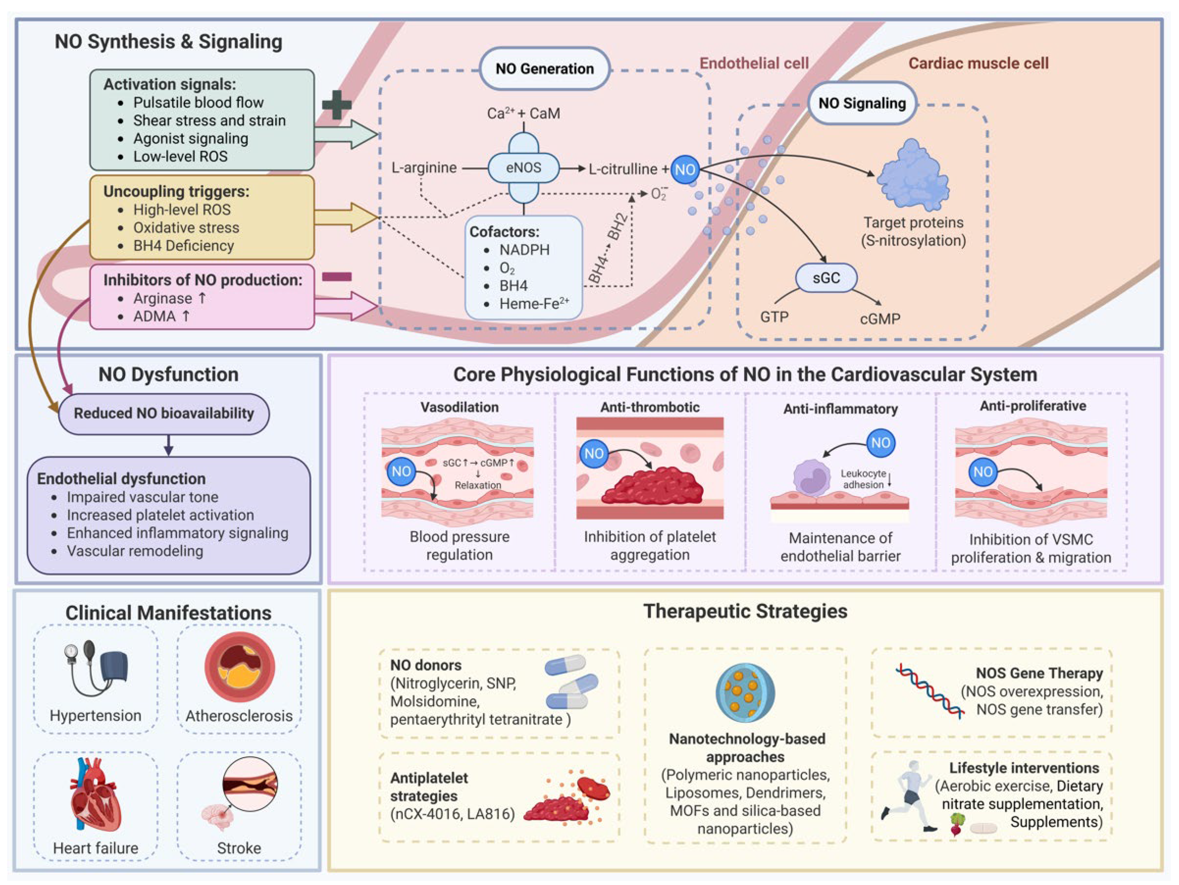
Figure 2.
Structural organization of the nitric oxide synthase (NOS) in the endothelial cell.
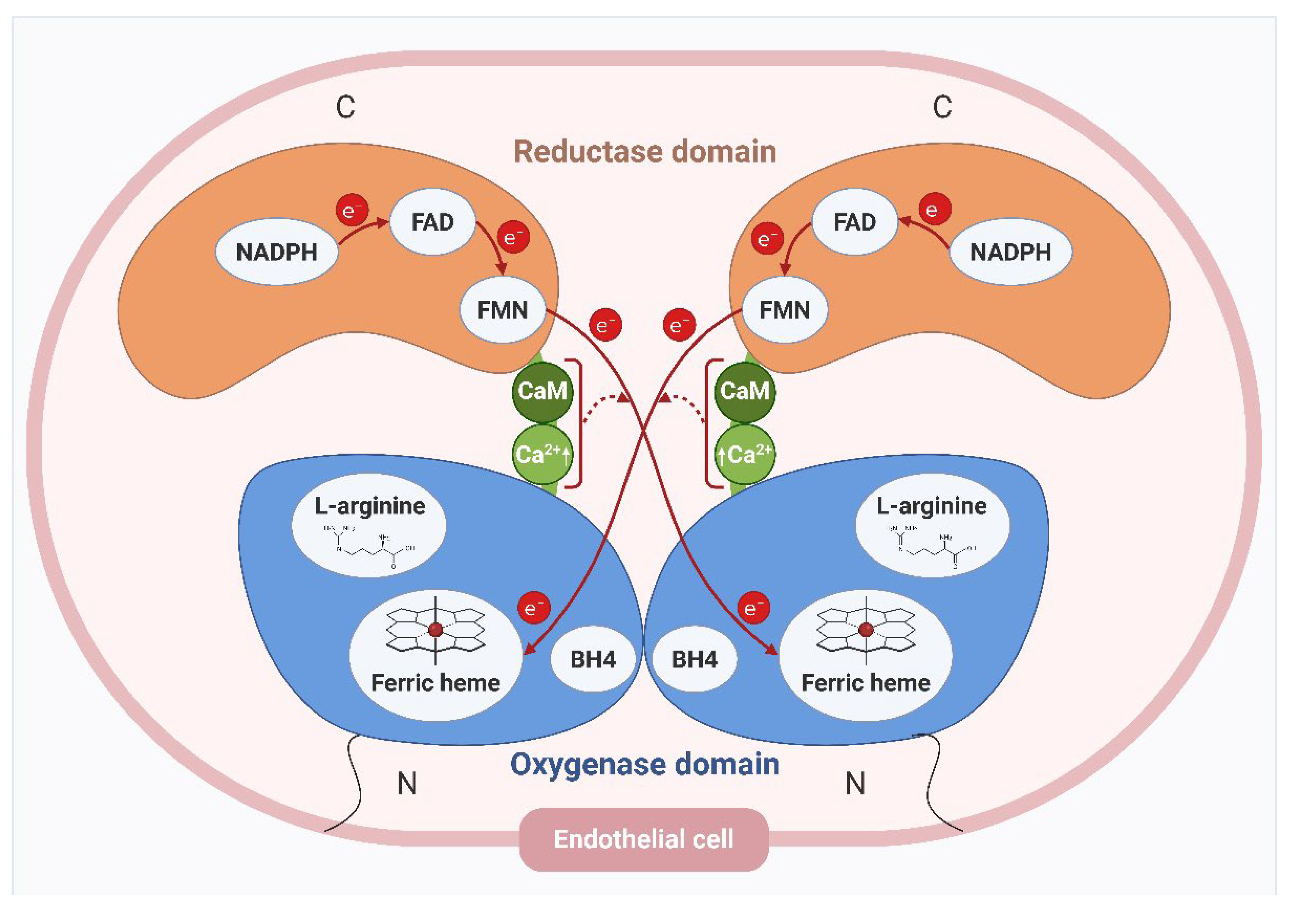
Figure 3.
Coupled and uncoupled states of nitric oxide synthase (NOS) in endothelial cells. (A) Coupled state. Under physiological activation signals - including pulsatile blood flow, laminar shear stress, agonist-receptor signaling, and low-level reactive oxygen species (ROS) — eNOS operates in a coupled configuration. Electrons derived from NADPH are sequentially transferred through FAD and FMN in the reductase domain, and subsequently donated in a cross-monomer fashion to the ferric heme center of the opposing subunit. In the presence of the essential cofactor tetrahydrobiopterin (BH4), the heme iron coordinates molecular oxygen (O₂) to form the Fe³⁺–O₂–BH4 ternary complex, which catalyzes the five-electron oxidation of L-arginine to yield nitric oxide (NO) and L-citrulline. NO diffuses out of the endothelial cell and mediates vasodilation and cytoprotection. eNOS is localized to the cytoplasmic face of endothelial plasma membrane caveolae; the nucleus is not shown for clarity. (B) Uncoupled state. Under pathological conditions — including high-level ROS, oxidative stress, and BH4 deficiency — BH4 is oxidized to the inactive form dihydrobiopterin (BH2), destabilizing the Fe³⁺–O₂ complex and disrupting productive electron transfer to L-arginine. Electrons instead leak to molecular oxygen, generating superoxide (O₂•⁻) rather than NO. The concurrent production of residual NO and O₂•⁻ leads to rapid formation of peroxynitrite (ONOO⁻), a highly reactive nitrogen species that exacerbates oxidative stress, further depletes BH4, and perpetuates eNOS uncoupling — shifting the enzyme from a vasoprotective to a vasotoxic phenotype. eNOS is localized to the cytoplasmic face of endothelial plasma membrane caveolae; the nucleus is not shown for clarity. BH2, dihydrobiopterin; BH4, tetrahydrobiopterin; C, C-terminus; CaM, calmodulin; Ca²⁺, calcium; FAD, flavin adenine dinucleotide; FMN, flavin mononucleotide; N, N-terminus; NADPH, nicotinamide adenine dinucleotide phosphate (reduced); NO, nitric oxide; ONOO⁻, peroxynitrite; O₂⁻, superoxide anion; ROS, reactive oxygen species. Figure created using Biorender.com.
Figure 3.
Coupled and uncoupled states of nitric oxide synthase (NOS) in endothelial cells. (A) Coupled state. Under physiological activation signals - including pulsatile blood flow, laminar shear stress, agonist-receptor signaling, and low-level reactive oxygen species (ROS) — eNOS operates in a coupled configuration. Electrons derived from NADPH are sequentially transferred through FAD and FMN in the reductase domain, and subsequently donated in a cross-monomer fashion to the ferric heme center of the opposing subunit. In the presence of the essential cofactor tetrahydrobiopterin (BH4), the heme iron coordinates molecular oxygen (O₂) to form the Fe³⁺–O₂–BH4 ternary complex, which catalyzes the five-electron oxidation of L-arginine to yield nitric oxide (NO) and L-citrulline. NO diffuses out of the endothelial cell and mediates vasodilation and cytoprotection. eNOS is localized to the cytoplasmic face of endothelial plasma membrane caveolae; the nucleus is not shown for clarity. (B) Uncoupled state. Under pathological conditions — including high-level ROS, oxidative stress, and BH4 deficiency — BH4 is oxidized to the inactive form dihydrobiopterin (BH2), destabilizing the Fe³⁺–O₂ complex and disrupting productive electron transfer to L-arginine. Electrons instead leak to molecular oxygen, generating superoxide (O₂•⁻) rather than NO. The concurrent production of residual NO and O₂•⁻ leads to rapid formation of peroxynitrite (ONOO⁻), a highly reactive nitrogen species that exacerbates oxidative stress, further depletes BH4, and perpetuates eNOS uncoupling — shifting the enzyme from a vasoprotective to a vasotoxic phenotype. eNOS is localized to the cytoplasmic face of endothelial plasma membrane caveolae; the nucleus is not shown for clarity. BH2, dihydrobiopterin; BH4, tetrahydrobiopterin; C, C-terminus; CaM, calmodulin; Ca²⁺, calcium; FAD, flavin adenine dinucleotide; FMN, flavin mononucleotide; N, N-terminus; NADPH, nicotinamide adenine dinucleotide phosphate (reduced); NO, nitric oxide; ONOO⁻, peroxynitrite; O₂⁻, superoxide anion; ROS, reactive oxygen species. Figure created using Biorender.com.
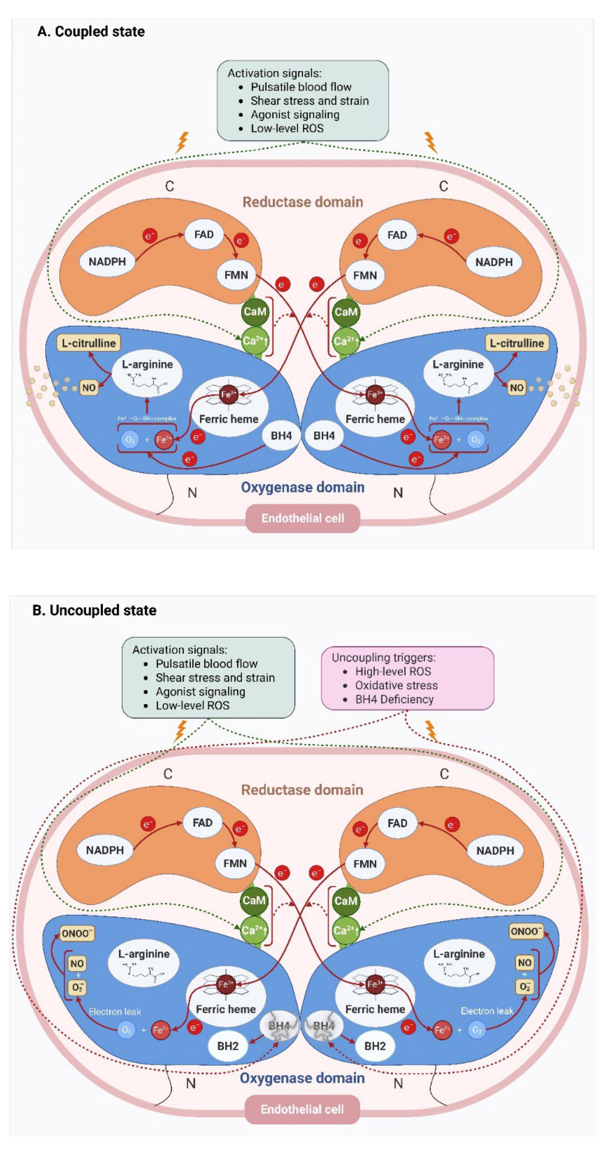
Figure 4.
Nitric oxide (NO) signaling pathways in Cardiomyocyte. NO produced by endothelial nitric oxide synthase (eNOS) in the endothelial cell diffuses into adjacent cardiomyocytes and exerts its effects through two principal mechanisms: cyclic guanosine monophosphate (cGMP)-dependent and cGMP-independent signaling. (A) cGMP-dependent NO signaling pathway. NO enters the cardiac myocyte and binds to the heme iron (Fe) of soluble guanylyl cyclase (sGC), inducing a conformational change that markedly increases its catalytic activity. Activated sGC converts guanosine triphosphate (GTP) to the second messenger cGMP, the intracellular concentration of which is negatively regulated by phosphodiesterases 5, 6, and 9 (PDE-5/6/9) through cGMP hydrolysis. Elevated cGMP activates protein kinase G type 1 (PKG-1), which promotes cardiac myocyte relaxation through two convergent mechanisms: (i) activation of myosin light chain phosphatase, leading to dephosphorylation of the myosin light chain (MLC) and reduced actomyosin cross-bridge formation; and (ii) phosphorylation of sarcolemmal calcium channels, resulting in reduced intracellular Ca²⁺ concentration and subsequent decrease in Ca²⁺-dependent contractile activation. The nucleus is not shown for clarity. (B) cGMP-independent NO signaling: S-nitrosylation. NO can also signal independently of cGMP through the post-translational modification of proteins via S-nitrosylation. In this pathway, NO reacts with the free thiol group (–SH) of a reactive cysteine residue within a target protein to form a protein S-nitrosothiol (–SNO). This covalent modification alters the conformation and activity of the target protein, thereby modulating a broad range of downstream cellular processes in the cardiac myocyte without engaging the sGC–cGMP–PKG axis. The nucleus is not shown for clarity. Figure created using Biorender.com Abstract figure- Strategies to preserve nitric oxide signaling and vascular health during aging. Lifestyle and pharmacological interventions, including exercise, healthy diet, antioxidants, and NO-targeted therapies, may improve endothelial function, enhance nitric oxide bioavailability, reduce oxidative stress, and support vascular signaling pathways. Collectively, these approaches aim to counter age-associated vascular dysfunction and maintain vascular integrity during cardiovascular aging. Figure created using Biorender.com.
Figure 4.
Nitric oxide (NO) signaling pathways in Cardiomyocyte. NO produced by endothelial nitric oxide synthase (eNOS) in the endothelial cell diffuses into adjacent cardiomyocytes and exerts its effects through two principal mechanisms: cyclic guanosine monophosphate (cGMP)-dependent and cGMP-independent signaling. (A) cGMP-dependent NO signaling pathway. NO enters the cardiac myocyte and binds to the heme iron (Fe) of soluble guanylyl cyclase (sGC), inducing a conformational change that markedly increases its catalytic activity. Activated sGC converts guanosine triphosphate (GTP) to the second messenger cGMP, the intracellular concentration of which is negatively regulated by phosphodiesterases 5, 6, and 9 (PDE-5/6/9) through cGMP hydrolysis. Elevated cGMP activates protein kinase G type 1 (PKG-1), which promotes cardiac myocyte relaxation through two convergent mechanisms: (i) activation of myosin light chain phosphatase, leading to dephosphorylation of the myosin light chain (MLC) and reduced actomyosin cross-bridge formation; and (ii) phosphorylation of sarcolemmal calcium channels, resulting in reduced intracellular Ca²⁺ concentration and subsequent decrease in Ca²⁺-dependent contractile activation. The nucleus is not shown for clarity. (B) cGMP-independent NO signaling: S-nitrosylation. NO can also signal independently of cGMP through the post-translational modification of proteins via S-nitrosylation. In this pathway, NO reacts with the free thiol group (–SH) of a reactive cysteine residue within a target protein to form a protein S-nitrosothiol (–SNO). This covalent modification alters the conformation and activity of the target protein, thereby modulating a broad range of downstream cellular processes in the cardiac myocyte without engaging the sGC–cGMP–PKG axis. The nucleus is not shown for clarity. Figure created using Biorender.com Abstract figure- Strategies to preserve nitric oxide signaling and vascular health during aging. Lifestyle and pharmacological interventions, including exercise, healthy diet, antioxidants, and NO-targeted therapies, may improve endothelial function, enhance nitric oxide bioavailability, reduce oxidative stress, and support vascular signaling pathways. Collectively, these approaches aim to counter age-associated vascular dysfunction and maintain vascular integrity during cardiovascular aging. Figure created using Biorender.com.
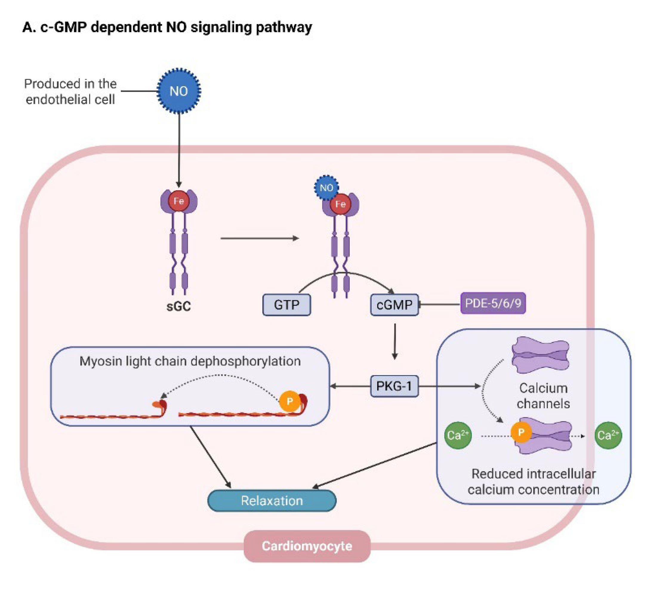
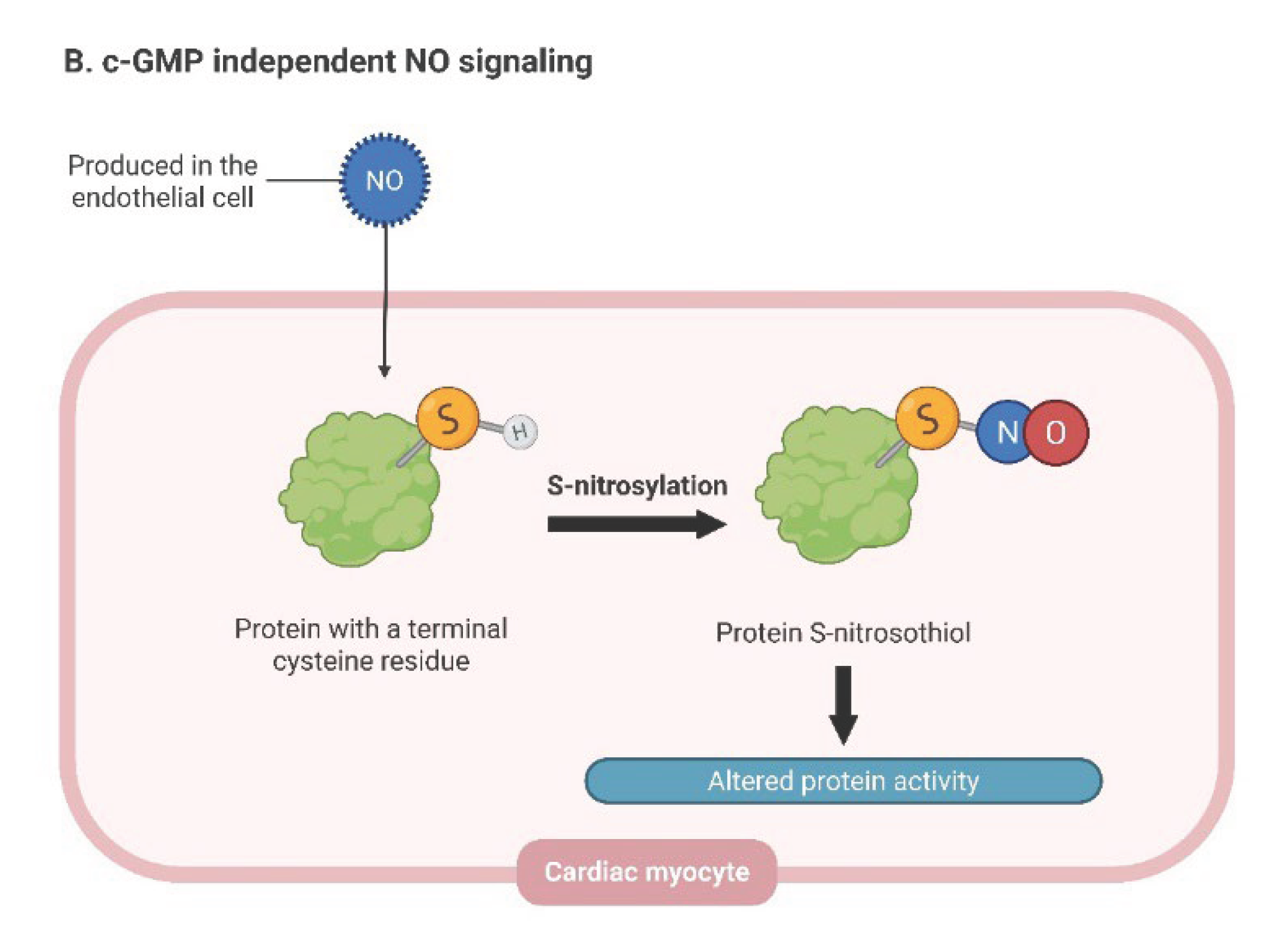
Table 1.
Comparison of the three isoforms of NOS enzymes.
| Property | NOS1 (nNOS) | NOS2 (iNOS) | NOS3 (eNOS) |
|---|---|---|---|
| Name | Neuronal nitric oxide synthase | Inducible nitric oxide synthase | Endothelial nitric oxide synthase |
| Chromosomal location | 12q24.2 | 17q11.2 | 7q35–7q36 |
| Primary location | Nervous system and skeletal muscle | Immune cells | Vascular endothelium, RBCs, platelets |
| Molecular weight (kDa) | 161 | 130 | 133 |
| Expression pattern | Constitutive | Inducible | Constitutive |
| Regulation | Calcium-dependent | Calcium-independent | Calcium-dependent |
| Stimulus | Increased intracellular calcium (neuronal activation) | Pro-inflammatory cytokines, bacterial lipopolysaccharide | Shear stress, pulsatile blood flow, reactive oxygen species |
| Physiological function | Synaptic plasticity, neuromuscular signaling, smooth muscle relaxation | Host defense and inflammatory signaling | Regulation of vascular tone and endothelial function |
| Pathophysiological relevance | Neurodegenerative disorders (e.g., Alzheimer’s disease, Parkinson’s disease), epilepsy | Chronic inflammation, autoimmune and neuroinflammatory disorders | Hypertension, atherosclerosis, coronary artery disease, heart failure |
| NO signaling profile | Low, localized NO production | High, sustained NO production | Low, tightly regulated NO production |
NOS, nitric oxide synthases; RBC, red blood cells; ROS, reactive oxygen species; CNS, central nervous system.
Table 3.
Genetic modulation of nitric oxide signaling in cardiovascular disease.
| Gene | Variant (Example) | Functional Effect | Cardiovascular Association | Effect on NO Signaling (Framework) |
|---|---|---|---|---|
| NOS3 (eNOS) | Glu298Asp (rs1799983) | Reduced eNOS activity and NO production | Hypertension, coronary artery disease, preeclampsia | ↓ Baseline NO production (source/magnitude) |
| NOS3 (eNOS) | T-786C (rs2070744) | Decreased NOS3 promoter activity → reduced NO synthesis | Endothelial dysfunction, hypertension | ↓ NO synthesis (source) |
| DDAH1 / DDAH2 | Various polymorphisms | Reduced DDAH activity → ↑ ADMA accumulation | Endothelial dysfunction, cardiovascular risk | ↓ NO availability via NOS inhibition (magnitude) |
| ARG1 | Polymorphisms affecting enzyme activity | Increased arginase activity → L-arginine depletion | Vascular dysfunction, metabolic syndrome | ↓ Substrate availability → eNOS uncoupling (magnitude/redox) |
| ARG2 | Variants linked to metabolic traits | Altered arginine metabolism | Type 2 diabetes, vascular dysfunction | Indirect ↓ NO via metabolic and substrate imbalance |
NOS, nitric oxide synthase; NOS1, neuronal NOS; NOS2, inducible NOS; NOS3, endothelial NOS; ARG, arginase; DDAH, dimethylarginine dimethylaminohydrolase; ADMA, asymmetric dimethylarginine.
Table 4.
Representative studies evaluating nitric oxide-related biomarkers in cardiovascular disease.
Table 4.
Representative studies evaluating nitric oxide-related biomarkers in cardiovascular disease.
| Biomarker | Study type | Disease | Participants (n, age) | Measurement method | Key findings | Interpretation within nitric oxide paradox | Reference |
|---|---|---|---|---|---|---|---|
| Serum NO | Case control study | CAD | 70 (52 M, 18 F; 41–85 y) | Colorimetric assay | Lower serum NO levels in CAD; trend of reduced NO activity with increasing CAD severity | Reduced serum NO is consistent with impaired endothelial signaling but does not distinguish decreased production from increased oxidative consumption | [102] |
| Plasma NOx levels | Cross sectional observational study | Heart failure | 30 (23 M, 7 F; 53 y) | Griess method | Reduction in NO bioavailability and increase in NADPH oxidase activity compared to healthy control group | Lower NOx may reflect reduced NO bioavailability, although interpretation depends on concurrent oxidative stress and metabolic context | [103] |
| Plasma NOx levels | Randomized, controlled, single-blinded, interventional animal study | Ischaemia | 40 male rats | Griess method | NO levels peaked at first 2 hours of ischemia, reaching normal levels at 6th hour (established tissue ischemia) | Time-dependent variation suggests NOx reflects dynamic ischemic responses rather than steady-state NO signaling | [104] |
| Serum NOx levels | 8 year follow up study at Moscow | Cardiovascular mortality | 1869 (546 M, 1323 F; >55 y) | Griess method | Mean serum NOx concentration – 58 µM (over 70 µM in mortality groups); below 40 µM not associated with mortality | Elevated NOx may reflect increased oxidative metabolism or compensatory turnover rather than enhanced bioactive NO signaling | [105] |
| Serum NOx levels | Case control, 6.5 year follow up study | Acute myocardial infarction and cardiovascular mortality | 222 (134 M, 88 F; mean age 44.5±16 y) | Griess method | NOx levels were independently associated with AMI; NOx showed better predictive power for AMI than traditional biomarkers | Increased NOx does not necessarily indicate preserved endothelial function and may instead reflect dysregulated NO metabolism | [100] |
| Plasma nitrate | Prospective community based study | CVD incidence and mortality | 2855 (46% M, 54% F; 59 y) | Gas chromatography–mass spectrometry | Higher plasma nitrate was linked to increased risk of death but was not associated with incidence of CVD | Plasma nitrate reflects multiple physiological and dietary influences, limiting its specificity as a marker of functional NO signaling | [106] |
| Serum NOx levels | Prospective population based study (Iran) | Cardiovascular mortality | 3520 (1414 M, 2106 F; mean age 44.5±16 y) | Spectrophotometry | Elevated serum NOx levels (>30.5 and 32.5 µmol/L) increase the risk of CVD and all-cause mortality by 98% and 52%, respectively, in individuals with traditional risk factors like hypertension, history of CVD, and type 2 diabetes | Increased NOx may indicate altered NO turnover rather than improved signaling capacity | [107] |
| Nitrotyrosine | Case-control study | CAD | 100 (54 M, 46 F; ≥21 y) | Stable isotope dilution liquid chromatography | Plasma protein-bound nitrotyrosine levels were higher in CAD patients compared to healthy control group | Elevated nitrotyrosine reflects increased reactive nitrogen species formation and a shift toward redox-driven NO dysfunction | [108] |
| ADMA | Cross-sectional study | CAD | 40 (22 M, 18 F; 30–60 y) | ELISA | Increased ADMA levels are significantly linked to CAD, correlating with extent and severity of CAD | Increased ADMA reduces NO production through endogenous NOS inhibition, limiting signaling capacity | [109] |
NO, nitric oxide; NOx, nitrate/nitrite; CAD, coronary artery disease; CVD, cardiovascular disease; AMI, acute myocardial ischemia; ADMA, asymmetric dimethyl arginin.
Disclaimer/Publisher’s Note: The statements, opinions and data contained in all publications are solely those of the individual author(s) and contributor(s) and not of MDPI and/or the editor(s). MDPI and/or the editor(s) disclaim responsibility for any injury to people or property resulting from any ideas, methods, instructions or products referred to in the content. |
© 2026 by the authors. Licensee MDPI, Basel, Switzerland. This article is an open access article distributed under the terms and conditions of the Creative Commons Attribution (CC BY) license (http://creativecommons.org/licenses/by/4.0/).
Copyright: This open access article is published under a Creative Commons CC BY 4.0 license, which permit the free download, distribution, and reuse, provided that the author and preprint are cited in any reuse.