

Submitted:
15 September 2026
Posted:
15 September 2026
You are already at the latest version
Abstract
Osteoclasts are the primary bone-resorbing cells responsible for maintaining skeletal homeostasis through balanced bone remodeling. Accumulating evidence suggests that oxidative stress is an emerging regulator of osteoclast differentiation, metabolic reprogramming, and bone resorption, arising from excessive reactive oxygen species (ROS) and insufficient antioxidant defenses. Dysregulated redox signaling contributes to osteoporosis, rheumatoid arthritis, periodontitis, periapical lesions, and other osteolytic disorders. This review summarizes recent advances in the molecular mechanisms linking oxidative stress to osteoclast metabolism, focusing on ROS sources, mitochondrial dysfunction, redox-sensitive signaling pathways, endogenous antioxidant systems, and emerging antioxidant-based therapies. Oxidative stress regulates osteoclastogenesis through interconnected signaling networks, including NF-κB, MAPKs, PI3K/Akt, AMPK, mTOR, NFATc1, and Keap1/Nrf2. Under both physiological and pathological conditions, osteoclast function is regulated by mitochondrial dynamics, mitophagy, and epigenetic mechanisms. Although both synthetic and natural antioxidants have been shown to reduce pathological osteoclast activity in preclinical studies, limitations in bioavailability, target specificity, and long-term efficacy hinder clinical translation. Advances in multi-omics technologies, single-cell analyses, biomarker discovery, precision medicine, and targeted antioxidant delivery may facilitate the development of effective therapies for oxidative stress–related bone disorders.
Keywords:
oxidative stress
; osteoclast metabolism
; reactive oxygen species
; antioxidants
; Nrf2
; mitochondrial dysfunction
; NFATc1
; bone remodeling
; osteoporosis
; arthritis
1. Introduction
Bone remodeling is a continuous process that maintains mineral homeostasis, repairs microdamage, and protects skeletal integrity through the coordinated action of osteoclasts, osteoblasts, and osteocytes [1‒3]. Disruption of this coupling leads to excessive bone loss and skeletal deterioration in conditions such as osteoporosis, rheumatoid arthritis (RA), periodontitis, peri-implantitis, periapical lesions, and osteolytic bone metastases [4,5,6]. Among the mechanisms underlying these conditions, oxidative stress has been identified as a key factor that accelerates osteoclast differentiation and contributes to pathological bone loss.
Osteoclasts are multinucleated cells that develop from monocyte/macrophage precursors upon stimulation with macrophage colony-stimulating factor (M-CSF) and receptor activator of nuclear factor-κB (RANK) ligand (RANKL). RANKL binding to RANK triggers multiple intracellular cascades that culminate in the activation of nuclear factor of activated T cell 1 (NFATc1), the master transcription factor controlling osteoclast differentiation and function [7,8,9,10]. Mature osteoclasts enable the acidification and degradation of mineralized bone matrix through specialized structures, such as the sealing zone and ruffled border [7,11]. To meet the high bioenergetic demands of bone resorption, osteoclast differentiation necessitates extensive metabolic reprogramming that extends beyond classical cytokine signaling and is characterized by increased glycolysis, mitochondrial oxidative phosphorylation, glutamine utilization, and lipid metabolism [12,13,14,15].
Reactive oxygen species (ROS) are essential signaling molecules that regulate immune responses, cell differentiation, and proliferation under physiological conditions. However, excessive ROS production or insufficient antioxidant defenses disrupt cellular redox homeostasis, resulting in oxidative stress that damages macromolecules, impairs mitochondrial function, and promotes inflammatory signaling [16,17,18,19]. Nicotinamide adenine dinucleotide phosphate (NADPH) oxidases, mitochondrial respiration, and other oxidase systems are major sources of ROS, including superoxide anions, hydrogen peroxide, and hydroxyl radicals [16,17,20]. A growing body of evidence suggests that oxidative stress influences iron metabolism, autophagy, mitochondrial function, inflammatory responses, and several redox-sensitive pathways, thereby contributing to osteoclastogenesis [15,17,21,22,23,24,25]. Therefore, maintaining redox homeostasis is crucial for ongoing physiological bone remodeling.
The Kelch-like ECH-associated protein 1 (Keap1)/nuclear factor erythroid 2-related factor 2 (Nrf2) signaling pathway has been identified as a key endogenous defense system against oxidative stress. Under basal conditions, Keap1 sequesters Nrf2 and directs it toward proteasomal degradation. Oxidative stress disrupts this interaction, enabling Nrf2 to translocate to the nucleus and induce the expression of cytoprotective and antioxidant genes that restore redox balance [26,27,28,29,30]. In experimental models of osteoporosis, Nrf2 activation attenuated oxidative stress, inhibited osteoclast differentiation, and reduced pathological bone resorption in mice. However, recent studies suggest that rather than completely eliminating ROS, therapeutic approaches should aim to restore redox homeostasis, given that physiological ROS are also necessary for normal osteoclastogenesis. Furthermore, recent developments have expanded our understanding of osteoclast biology by revealing the important roles of mitochondrial dynamics, metabolic adaptation, epigenetic regulation, and non-coding RNAs (ncRNAs) in oxidative stress–mediated bone remodeling. Meanwhile, CRISPR-based functional genomics, single-cell transcriptomics, spatial transcriptomics, and multi-omics techniques have uncovered previously unrecognized osteoclast heterogeneity and identified new molecular targets implicated in pathological bone resorption [15,31,32,33,34]. These innovations are reshaping our understanding of redox regulation in skeletal biology and creating new possibilities for precision medicine.
Despite these developments, several challenges remain to be addressed. Most mechanistic evidence has been derived from experimental models, and pharmacokinetic limitations, poor target specificity, poor bioavailability of antioxidant compounds, and a lack of robust clinical trials have impeded clinical translation [17,30,35]. Inconsistencies between studies may reflect differences in disease models and methods used to assess oxidative stress. Integrating redox biology, osteoimmunology, bone metabolism, advanced omics technologies, and precision medicine is essential for overcoming these constraints. In this review, we integrate recent advances in understanding the role of oxidative stress in osteoclast metabolism and bone resorption, with an emphasis on ROS production, metabolic reprogramming, redox-sensitive signaling pathways, endogenous antioxidant systems, epigenetic regulation, and antioxidant-based therapeutic approaches. Additionally, we outline current limitations, emerging technologies, and potential strategies for developing precise antioxidant therapies for bone disorders associated with oxidative stress.
2. Osteoclast Biology
Osteoclasts, the sole cells responsible for physiological bone resorption, are highly specialized multinucleated cells derived from hematopoietic monocyte/macrophage precursors. Through ongoing bone remodeling, osteoclasts interact with osteoblasts and osteocytes to preserve skeletal homeostasis. M-CSF and RANKL are the primary regulators of osteoclast differentiation. RANKL initiates signaling cascades that converge on NFATc1, whereas M-CSF promotes precursor survival, proliferation, and RANK receptor expression [7,8,9,10]. NFATc1 activation induces osteoclast-specific genes, including tartrate-resistant acid phosphatase (TRAP), cathepsin K (CTSK), dendritic cell-specific transmembrane protein (DC-STAMP), ATP6V0D2, and calcitonin receptor (CTR). Taken together, these genes control cell fusion, cytoskeletal organization, and bone resorption.
The maturation of osteoclast precursors, which fuse to form multinucleated cells with specialized structures such as the sealing zone and ruffled border, enables acidification of the resorption lacuna and enzymatic breakdown of the mineralized bone matrix [7,11]. Although osteoclastogenesis depends on cytokine-mediated signaling, growing evidence suggests that differentiation and activation also require substantial metabolic reprogramming. To meet the high energy and biosynthetic demands associated with multinucleation, cytoskeletal remodeling, proton secretion, and matrix degradation, osteoclasts undergo coordinated changes in glucose, lipid, amino acid, and mitochondrial metabolism. In addition to producing the required adenosine triphosphate (ATP), these metabolic pathways produce intermediates that modulate redox homeostasis, intracellular signaling, and epigenetic modifications. This establishes cellular metabolism as a key regulator of osteoclast differentiation and function, extending beyond its role in energy production [12,13,14,15].
3. Metabolic Reprogramming During Osteoclastogenesis
Metabolic reprogramming has emerged as a fundamental regulator of osteoclast differentiation and function rather than merely a consequence of cellular activation (Table 1). Recent transcriptomic and metabolomic analyses have revealed dynamic reprogramming of glucose, lipid, amino acid, and mitochondrial metabolism during osteoclast differentiation, underscoring metabolism as an active determinant of osteoclast fate and bone-resorptive capacity that extends beyond energy production [13,14,15,36].
3.1. Glycolysis and Glucose Metabolism
During osteoclast differentiation, glycolysis rapidly increases to provide ATP and the metabolic intermediates required for precursor fusion and bone resorption. According to recent in vitro studies, RANKL stimulation increases the expression of important glycolytic enzymes, such as hexokinase 2 (HK2), phosphofructokinase (PFK), and pyruvate kinase M2 (PKM2), thereby accelerating glycolytic flux and increasing glucose uptake through the induction of glucose transporters, especially glucose transporter type 1 (GLUT1) [36,37,38,39]. In addition to ATP production, glycolytic intermediates support the biosynthesis of lipids, amino acids, and nucleotides required for rapid cell division. Glycolytic metabolism was found to be essential for osteoclastogenesis rather than merely a consequence of differentiation, as pharmacological inhibition of glycolysis markedly suppressed osteoclast differentiation, reduced the formation of TRAP-positive multinucleated cells, and attenuated bone-resorptive activity. Similarly, recent animal studies have demonstrated that inhibition of glycolytic enzymes reduces pathological bone loss, whereas increased glycolytic activity is associated with enhanced osteoclast formation in inflammatory bone diseases [37,38]. These findings indicate that glycolysis is a crucial metabolic pathway linking RANKL signaling to osteoclast differentiation.
3.2. Mitochondrial Oxidative Phosphorylation and Bioenergetics
During osteoclast maturation, mitochondrial oxidative phosphorylation (OXPHOS) is the main source of sustained ATP production required for proton transport, cytoskeletal organization, and extracellular matrix degradation. Recent evidence suggests that the c-Myc–PGC-1β signaling axis plays a central role in driving the increased mitochondrial biogenesis, respiratory capacity, and oxidative phosphorylation that accompany osteoclast differentiation [13,15,40]. Mitochondrial respiration is a pivotal source of intracellular ROS and is crucial for osteoclast function. Excessive ROS levels can cause mitochondrial dysfunction and oxidative damage, thereby compromising osteoclast homeostasis, whereas moderate ROS levels act as secondary messengers that enhance RANKL-dependent signaling and promote physiological NFATc1 activation [21,25,40]. Consequently, mitochondrial quality control mechanisms, including mitophagy, fusion–fission dynamics, and mitochondrial biogenesis, are increasingly recognized as crucial for maintaining physiological osteoclast activity and mitochondrial integrity.
3.3. Fatty Acid and Amino Acid Metabolism
Recent metabolomic studies suggest that osteoclast differentiation is supported by metabolic substrates other than glucose. Fatty acid oxidation (FAO) plays a key role in ATP production, particularly under nutrient-limited conditions or during prolonged osteoclast activation [13,41]. FAO supports mitochondrial oxidative metabolism and glycolysis by supplying acetyl-CoA to the tricarboxylic acid (TCA) cycle. Similarly, osteoclast bioenergetics and biosynthesis depend heavily on glutamine metabolism. In vitro studies have shown that glutamine maintains intracellular redox balance during differentiation by replenishing TCA cycle intermediates through anaplerosis, supporting nucleotide synthesis, and amino acid and glutathione biosynthesis [14,42]. These findings suggest that the coordinated use of glucose, fatty acids, and amino acids provides the metabolic flexibility required to maintain osteoclast function under various physiological and pathological conditions.
3.4. Metabolic Plasticity and Microenvironmental Adaptation
Osteoclast metabolism is highly dynamic and varies depending on the stage of development and the microenvironment. According to recent studies, osteoclasts shift between OXPHOS, glycolysis, and other fuel sources in response to oxidative stress, inflammatory cytokines, oxygen tension, and nutrient availability [15,41]. Nutrient-sensing pathways that integrate extracellular stimuli with intracellular metabolic programs, such as adenosine monophosphate (AMP)-activated protein kinase (AMPK), mechanistic target of rapamycin (mTOR), hypoxia-inducible factor-1α (HIF-1α), and sirtuin signaling, contribute to the regulation of metabolic plasticity. In animal models of osteoporosis, inflammatory arthritis, diabetes, and aging, osteoclasts undergo substantial metabolic remodeling, characterized by altered mitochondrial function, elevated glycolysis, and disrupted redox homeostasis [18,25,41]. Under stressful circumstances, these adaptive responses allow osteoclasts to maintain their bone-resorptive activity. However, when metabolic regulation is dysregulated, these adaptations may also contribute to pathological bone loss.
3.5. Metabolic Reprogramming as a Therapeutic Target
Current evidence suggests that metabolic reprogramming is a key regulator of osteoclast survival, activation, and differentiation. During osteoclastogenesis, glycolysis, OXPHOS, FAO, and amino acid metabolism function as interrelated metabolic networks that regulate energy production, biosynthesis, and redox homeostasis. Nevertheless, the relative contributions of individual metabolic pathways appear to vary according to developmental stage, disease context, and microenvironment, highlighting the complexity of osteoclast metabolic regulation. Recent developments in metabolomics, CRISPR-based functional genomics, spatial transcriptomics, stable isotope tracing, and single-cell RNA sequencing have begun to define stage-specific metabolic programs and reveal previously unknown therapeutic targets [31,32,33,34]. Combining these advanced technologies with redox biology is expected to yield a more comprehensive understanding of osteoclast metabolism and accelerate the development of metabolism-based therapeutic approaches for oxidative stress–associated osteolytic disorders.
4. Sources of Oxidative Stress in Osteoclasts
ROS are continuously generated during cellular metabolism and function as essential secondary messengers that regulate cell proliferation, differentiation, apoptosis, and immune responses. Physiological ROS levels in osteoclasts are essential for normal RANKL-induced osteoclast differentiation and bone remodeling. However, excessive ROS production or insufficient antioxidant defenses can disrupt redox homeostasis, resulting in oxidative stress that accelerates bone resorption, promotes osteoclastogenesis, and causes pathological bone loss. Numerous intracellular and microenvironmental factors, including NADPH oxidases (NOXs), mitochondrial dysfunction, endoplasmic reticulum (ER) stress, iron dysregulation, inflammatory cytokines, hypoxia, aging, metabolic disorders, cigarette smoking, and chronic inflammatory diseases such as periodontitis, have been implicated in oxidative stress in osteoclasts [43,44,45,46]. Under pathological conditions, these pathways interact to form an integrated redox network that enhances osteoclast activation rather than functioning independently (Table 2).
4.1. NADPH Oxidases: The Primary Enzymatic Source of ROS
The NOX family is the primary enzymatic source of ROS among intracellular ROS-generating systems during osteoclastogenesis. Recent studies have demonstrated that NOX1, NOX2, and NOX4 are expressed during osteoclast differentiation. NOX4 supports osteoclast maturation and bone resorption, whereas NOX1 and NOX2 regulate early precursor commitment [43,44]. Nuclear factor kappa B (NF-κB), mitogen-activated protein kinase (MAPK), and NFATc1 signaling pathways are amplified by NOX-dependent ROS production following RANKL stimulation. Pharmacological inhibition or genetic deletion of NOX isoforms markedly reduces intracellular ROS accumulation, suppresses osteoclast formation, and protects against inflammatory and osteoporotic bone loss, as observed in animal studies [44,45,46]. These findings highlight NOX enzymes as promising therapeutic targets for oxidative stress–associated skeletal diseases.
4.2. Mitochondrial ROS and Bioenergetic Stress
Mitochondria are another important source of intracellular ROS during osteoclast differentiation. To meet the high energy demands of proton secretion, cytoskeletal remodeling, and matrix degradation, osteoclast maturation is accompanied by increased mitochondrial biogenesis and OXPHOS, as discussed in the previous section. Recent mechanistic evidence suggests that increased mitochondrial respiration can increase mitochondrial ROS production, which promotes RANKL-induced signaling under physiological circumstances but becomes harmful when ROS accumulate excessively [21,40,47,48]. Overproduction of mitochondrial ROS accelerates pathological bone resorption by impairing respiratory function, disrupting the mitochondrial membrane potential, promoting mitochondrial DNA damage, and amplifying inflammatory signaling. Therefore, mitochondrial quality control mechanisms, such as mitophagy, fusion–fission dynamics, and mitochondrial biogenesis, are considered crucial for preserving bioenergetic homeostasis and limiting oxidative damage [40,47].
4.3. ER Stress and Iron Dysregulation
Recent research suggests that the ER also contributes to oxidative stress during osteoclastogenesis. Prolonged ER stress triggers the unfolded protein response (UPR), which disrupts calcium homeostasis, increases ROS production, and activates inflammatory signaling pathways that promote osteoclast differentiation [46,49,50]. Although brief ER stress promotes adaptive cellular responses, prolonged activation exacerbates oxidative damage and disrupts cellular homeostasis [46]. Iron metabolism is another key factor regulating osteoclast redox status. Several animal and human studies have shown that iron overload increases osteoclast differentiation, accelerates bone loss in osteoporosis and inflammatory bone diseases, and promotes ROS generation via Fenton chemistry [51,52]. Ferroptosis, a type of cell death driven by iron-dependent lipid peroxidation, has recently emerged as a possible modulator of skeletal remodeling [49,52]. Despite mounting evidence that ferroptosis-related pathways affect osteoclast function through interactions among iron metabolism, lipid peroxidation, and oxidative stress, their precise role in osteoclast biology remains unclear.
4.4. Inflammatory and Microenvironmental Drivers of Oxidative Stress
In addition to intracellular ROS-producing systems, pathological microenvironmental factors substantially increase oxidative stress during osteoclastogenesis. Pro-inflammatory cytokines, such as tumor necrosis factor (TNF)-α, interleukin (IL)-1β, IL-6, and IL-17, interact with RANKL to enhance ROS production, trigger osteoclastogenic signaling pathways, and impair endogenous antioxidant defenses. This interaction contributes to a vicious cycle of inflammation and bone loss [18,21,53]. Hypoxia increases mitochondrial ROS levels during prolonged oxygen deprivation and further stimulates metabolic remodeling via HIF-1α. Likewise, cellular senescence, reduced antioxidant capacity, and mitochondrial dysfunction are hallmarks of aging that contribute to a pro-oxidant microenvironment that promotes osteoclast activation [25,41,54].
Environmental exposures and metabolic disorders exacerbate oxidative stress. Chronic hyperglycemia increases ROS production through mitochondrial dysfunction, advanced glycation end-product formation, and persistent inflammation, thereby enhancing osteoclast activity in diabetes mellitus [55,56]. Periodontal pathogens promote ROS production and pro-inflammatory cytokine release, thereby increasing RANKL expression and accelerating alveolar bone destruction, while cigarette smoking introduces exogenous oxidants that exacerbate oxidative stress and inflammatory signaling [57,58].
Collectively, these findings indicate that oxidative stress in osteoclasts results from the coordinated interaction of pathological microenvironmental stimuli and intracellular ROS-generating systems, rather than from a single source. This integrated understanding may provide new opportunities for developing precise antioxidant therapies that selectively suppress pathological bone resorption while preserving physiological skeletal remodeling.
5. Redox Signaling Networks Regulating Osteoclastogenesis
ROS are considered vital intracellular signaling molecules rather than merely detrimental metabolic byproducts. During RANKL-induced osteoclastogenesis, physiological ROS function as secondary messengers that modulate signaling pathways involved in differentiation, metabolism, and bone resorption. However, excessive ROS disrupt redox homeostasis, resulting in sustained activation of osteoclastogenic signaling and pathological bone loss. ROS orchestrate an interconnected network of redox-sensitive pathways, including NF-κB, MAPKs, PI3K/Akt, mTOR, AMPK, calcium/calcineurin, NFATc1, cAMP response element-binding protein (CREB), c-Fos, forkhead box O (FOXO), and Keap1/Nrf2 (Figure 1), which collectively integrate inflammatory, metabolic, and oxidative signals to regulate osteoclast formation and function, as demonstrated in recent in vitro and animal studies [2,18,35,43].
5.1. NF-κB Signaling
The NF-κB pathway is one of the earliest and most critical signaling cascades activated during osteoclast differentiation [57]. Recent mechanistic studies have demonstrated that physiological ROS generated by NADPH oxidases and mitochondria function as secondary messengers that facilitate NF-κB activation, whereas sustained oxidative stress prolongs NF-κB activation and accelerates pathological bone resorption [18,43]. RANKL binding to RANK activates tumor necrosis factor receptor-associated factor 6 (TRAF6), which then activates transforming growth factor-β-activated kinase 1 (TAK1) and the IκB kinase (IKK) complex, leading to IκB degradation and nuclear translocation of the p65/p50 NF-κB heterodimer [59]. Following the induction of c-Fos and NFATc1 expression, activated NF-κB promotes expression of osteoclast-specific genes, such as TRAP, CTSK, DC-STAMP, MMP-9, and ATP6V0D2 [60,61]. Further in vivo studies have demonstrated that osteoclast formation and inflammatory bone loss are markedly reduced by pharmacological inhibition of NF-κB or suppression of intracellular ROS. It is important to emphasize that therapeutic strategies should restore redox balance rather than eliminate ROS signaling, as complete NF-κB blockade or indiscriminate ROS removal may impair physiological bone remodeling and immune function [2].
5.2. MAPK Signaling Pathways
Oxidative stress is transduced into transcriptional programs that regulate osteoclast differentiation via MAPK signaling. Precursor proliferation, survival, cytoskeletal remodeling, and osteoclast maturation are all coordinated by ROS through redox-sensitive kinase cascades, as demonstrated in experimental studies [62]. While p38 stimulates NFATc1 expression and osteoclast-specific gene transcription, JNK activates c-Jun and AP-1, whereas ERK mainly supports cell survival. [62,63]. Interestingly, osteoclastogenesis is consistently suppressed by MAPK inhibition; however, the relative contribution of each MAPK pathway varies depending on the microenvironment and disease context [63,64].
5.3. PI3K/Akt/mTOR and AMPK: Linking Redox Signaling with Metabolism
ROS also modulate the nutrient-sensing pathways that link osteoclast differentiation to metabolism. Recent studies have shown that activation of the PI3K/Akt/mTOR axis promotes osteoclast formation by enhancing glycolysis, mitochondrial activity, protein synthesis, and cell survival [35,36]. During oxidative and energy stress, AMPK serves as a metabolic checkpoint that limits excessive anabolic activity. Osteoclasts can integrate intracellular energy with redox signaling through crosstalk between these pathways, thereby facilitating metabolic adaptation during bone resorption and differentiation [65,66].
5.4. Calcium Signaling and NFATc1 Activation
Calcium signaling is a major downstream convergence point of redox-sensitive signaling pathways. Previous studies have shown that calcineurin, which dephosphorylates NFATc1 and promotes its nuclear translocation, is activated by ROS-induced calcium oscillations [43]. NFATc1 promotes multinucleation and bone-resorptive activity by inducing the expression of osteoclast-specific genes, such as TRAP, CTSK, DC-STAMP, ATP6V0D2, and MMP-9, in conjunction with c-Fos, AP-1, and CREB [59,60].
IRF8 is a well-established negative regulator of osteoclast differentiation. Suppression of Irf8 expression promotes the induction of NFATc1, the master transcription factor of osteoclastogenesis [67]. Subsequent studies demonstrated that RANKL-RANK signaling induces ROS production, which enhances mitochondrial biogenesis through PGC-1β [68]. Furthermore, increased mitochondrial biogenesis suppresses Irf8 expression via DNA methylation [69]. Taken together, these findings indicate that RANKL-RANK signaling promotes ROS production, leading to enhanced mitochondrial biogenesis and epigenetic suppression of Irf8, thereby relieving the inhibitory effect of IRF8 on NFATc1 expression and facilitating osteoclast differentiation.
5.5. Integrated Redox Signaling Networks and Therapeutic Perspectives
To regulate osteoclast differentiation and bone resorption, ROS coordinate the activities of NF-κB, MAPKs, PI3K/Akt/mTOR, AMPK, calcium signaling, and antioxidant pathways that converge on the regulation of NFATc1. Instead of operating independently, redox-sensitive pathways form interconnected signaling networks that integrate oxidative, inflammatory, and metabolic signals.
In preclinical studies, natural antioxidants, such as curcumin, resveratrol, quercetin, epigallocatechin gallate (EGCG), and sulforaphane, have been shown to suppress osteoclastogenesis by reducing oxidative stress, inhibiting NF-κB and MAPK signaling, and activating Nrf2-mediated antioxidant defenses [70‒73]. Recently, Yang et al. demonstrated that Spirulina platensis, a naturally occurring microorganism used as a food ingredient, exerted potent therapeutic effects against RA by activating Nrf2 and suppressing the protein levels of NFATc1, c-Fos, and cathepsin K [74]. Despite these encouraging results, limitations in bioavailability and target specificity, as well as a lack of long-term clinical evidence, hinder the clinical translation of identified natural antioxidants. Further research is required to better understand the spatiotemporal regulation of redox signaling and to develop targeted antioxidant therapies for osteolytic diseases.
6. Nrf2 and Endogenous Antioxidant Systems
6.1. FOXO–Nrf2 Crosstalk and Endogenous Antioxidant Signaling
Major endogenous antioxidant systems that limit excessive ROS accumulation include the Keap1/Nrf2 pathway and FOXO transcription factors. According to recent mechanistic and animal studies, FOXO and Nrf2 activation limits pathological osteoclast activation by upregulating antioxidant enzymes, maintaining mitochondrial integrity, and reducing oxidative damage [26,27,28,29,30,66,73]. However, therapeutic modulation of antioxidant signaling should maintain basal redox signaling while preventing chronic oxidative stress because physiological ROS are necessary for normal osteoclastogenesis.
6.2. Keap1–Nrf2 Signaling in Redox Homeostasis
Balanced bone remodeling requires preservation of intracellular redox equilibrium. Excessive ROS triggers inflammatory signaling, mitochondrial dysfunction, and pathological bone resorption, although physiological levels of ROS are necessary for RANKL-induced osteoclast differentiation [18,75]. Recent in vitro and animal studies suggest that the Keap1–Nrf2 pathway is a key regulator of endogenous antioxidant defense during osteoclastogenesis [76,77]. Under physiological conditions, Keap1 sequesters Nrf2 in the cytoplasm, thereby facilitating its ubiquitination and proteasomal degradation. Oxidative or electrophilic stress disrupts the Keap1–Nrf2 complex, allowing Nrf2 to translocate to the nucleus, bind to antioxidant response elements, and induce the expression of several cytoprotective and antioxidant genes [76,78].
6.3. Nrf2-Regulated Antioxidant Enzymes
Heme oxygenase-1 (HO-1), NAD(P) quinone oxidoreductase-1 (NQO1), superoxide dismutase (SOD), catalase, glutathione peroxidase (GPx), thioredoxin (Trx), and peroxiredoxins (Prxs) are among the antioxidant enzymes and redox-regulatory proteins induced by Nrf2 activation [79,80,81]. Collectively, these effectors contribute to the regulation of redox-sensitive signaling pathways involved in osteoclast differentiation, limit excessive ROS accumulation, and maintain mitochondrial homeostasis. Among these effectors, HO-1 has been identified as a crucial mediator of Nrf2-dependent skeletal protection. Recent mechanistic studies have shown that HO-1 reduces inflammatory cytokine production, suppresses NF-κB activation, and inhibits RANKL-induced osteoclastogenesis. Taken together, the NQO1, SOD, catalase, GPxs, and Trx/Prx systems help preserve physiological ROS signaling and protect against oxidative damage [72,78,80,81].
6.4. Role of Nrf2 in Osteoclastogenesis and Bone Diseases
In animal models of osteoporosis, RA, periodontitis, and diabetic bone disease, experimental evidence consistently shows that activation of the Keap1–Nrf2 pathway suppresses intracellular ROS accumulation, maintains mitochondrial function, inhibits NF-κB and MAPK signaling, and attenuates osteoclast differentiation [18,81,82]. Conversely, Nrf2 deficiency compromises skeletal homeostasis by increasing oxidative stress, promoting osteoclastogenesis, and accelerating bone loss, thereby highlighting the crucial role of Nrf2 in maintaining redox homeostasis [77,78,82]. Physiological ROS are essential for normal osteoclast differentiation. Therefore, therapeutic modulation targeting Nrf2 should aim to restore redox balance rather than completely suppress ROS signaling.
Nrf2 is a promising therapeutic target for oxidative stress–related bone diseases owing to its crucial role in endogenous antioxidant defense. Preclinical studies have demonstrated that natural antioxidants, including sulforaphane, curcumin, resveratrol, quercetin, EGCG, and melatonin, suppress osteoclastogenesis by activating Nrf2 signaling while inhibiting NF-κB-mediated inflammation [71‒73,81]. New approaches, such as targeted antioxidant delivery systems and synthetic Nrf2 activators, may enhance therapeutic efficacy. However, limited long-term clinical evidence, pharmacokinetic variability, and poor bioavailability remain major barriers to their clinical translation.
7. Epigenetic and Non-Coding RNA Regulation of Oxidative Stress in Osteoclastogenesis
Epigenetic regulation has emerged as a critical mechanism that links oxidative stress to osteoclast differentiation and pathological bone resorption. Unlike genetic changes, epigenetic modifications control gene expression without altering the DNA sequence, allowing osteoclasts to adapt rapidly to metabolic, oxidative, and inflammatory stresses. According to recent transcriptomic, epigenomic, and functional studies, DNA methylation, histone modifications, RNA methylation, and ncRNAs interact extensively with redox-sensitive signaling pathways, such as NF-κB, MAPKs, PI3K/Akt, AMPK, and Nrf2, to regulate osteoclast metabolism, differentiation, and survival. Dysregulation of these epigenetic and non-coding RNA mechanisms has been implicated in the pathogenesis of RA, periodontitis, osteoporosis, periapical lesions, and other osteolytic disorders.
7.1. ncRNAs Regulating Osteoclast Redox Homeostasis
ncRNAs are among the most well-studied post-transcriptional and epigenetic regulators of osteoclastogenesis. Numerous microRNAs (miRNAs) that influence osteoclast differentiation by targeting elements of the RANKL-RANK signaling cascade, such as TRAF6, NF-κB, c-Fos, NFATc1, and Nrf2, have been identified in recent studies [83‒85]. miRNAs can stimulate or inhibit osteoclast formation and bone resorption depending on their downstream targets. Competing endogenous RNAs (ceRNAs) can sequester miRNAs, thereby modulating oxidative stress, mitochondrial function, inflammatory signaling, and antioxidant responses. Long non-coding RNAs (lncRNAs) and circular RNAs also contribute to the complexity of redox regulation [84‒86]. Recent studies suggest these ncRNA-mediated regulatory networks help osteoclasts adapt to physiological and pathological conditions by fine-tuning intracellular redox homeostasis.
7.2. DNA, Histone, and RNA Modifications
Chromatin remodeling provides an additional layer of redox regulation to ncRNA activity during osteoclastogenesis. Recent research has shown that oxidative stress alters DNA methylation patterns and the activity of histone-modifying enzymes, causing transcriptional changes that affect osteoclast-specific genes, inflammatory responses, and antioxidant defenses [87,88]. Histone acetylation and methylation regulate chromatin accessibility, thereby modulating the expression of NFATc1, c-Fos, and other transcription factors required for osteoclast differentiation. In addition, epigenetic regulation is an important component of skeletal biology. N6-methyladenosine (m6A), which controls RNA stability, splicing, translation, and degradation, is the most abundant internal mRNA modification. Recent mechanistic studies have indicated that m6A methylation integrates cellular metabolism, redox signaling, and osteoclast differentiation, representing an emerging area of research in bone biology, although the relationship between m6A and oxidative stress remains unclear [89].
7.3. Crosstalk Between Epigenetics and Redox Signaling
Epigenetic mechanisms interact closely with traditional redox-sensitive signaling pathways rather than acting independently. Epigenetic modifications can regulate the expression of antioxidant genes and signaling mediators involved in NF-κB, MAPKs, PI3K/Akt, AMPK, and Keap1–Nrf2 pathways, while oxidative stress can, in turn, alter epigenetic enzyme activity [90‒92]. According to recent multi-omics studies, osteoclasts can coordinate stage-specific differentiation and bone-resorptive activity by integrating inflammatory, metabolic, and oxidative cues through bidirectional epigenetic–redox crosstalk [61,93,94]. These findings emphasize that epigenetic regulation is a dynamic interface linking osteoclast transcriptional programs to environmental stimuli.
7.4. Therapeutic Potential and Future Perspectives
Because epigenetic modifications are reversible, they represent promising therapeutic targets for treating oxidative stress–associated bone diseases. Preclinical studies have demonstrated that miRNA-based treatments, lncRNA-targeting strategies, histone deacetylase inhibitors, DNA methyltransferase inhibitors, and RNA therapeutics inhibit osteoclastogenesis by restoring redox homeostasis and reducing inflammatory signaling [95‒99]. New strategies, such as targeted RNA delivery and CRISPR-based epigenome editing, have expanded the therapeutic potential of epigenetic interventions [100]. Nevertheless, most evidence remains preclinical, and further research is required to establish the clinical relevance and therapeutic efficacy of these epigenetic strategies.
8. Antioxidant Therapeutics Targeting Osteoclast Oxidative Stress
8.1. Natural Antioxidants and Phytochemicals
Natural products constitute the largest class of antioxidant compounds investigated for osteolytic diseases owing to their pleiotropic biological activities (Figure 2, Table 3). In recent in vitro and animal studies, polyphenols, flavonoids, and other phytochemicals, such as resveratrol, curcumin, quercetin, EGCG, sulforaphane, luteolin, apigenin, and baicalein, have been shown to suppress osteoclast differentiation by reducing intracellular ROS, inhibiting NF-κB, MAPK, and NFATc1 signaling, and activating endogenous antioxidant pathways such as Keap1–Nrf2 [65‒68,70‒73,92‒94,101‒103]. Numerous phytochemicals exert multitarget protective effects against pathological bone resorption by enhancing mitochondrial function, reducing inflammatory cytokine production, and modulating metabolic pathways. Despite promising preclinical results, poor aqueous solubility, low oral bioavailability, rapid metabolism, and limited bone accumulation remain barriers to their clinical use.
8.2. Dietary and Endogenous Antioxidants
Skeletal redox homeostasis is supported by endogenous antioxidants and dietary micronutrients. Melatonin protects bones by directly scavenging ROS, improving mitochondrial function, and upregulating antioxidant enzymes such as GPx, catalase, and SOD [104]. Likewise, vitamin C supports collagen synthesis and osteoblast differentiation while reducing oxidative stress [105], and vitamin E protects membrane lipids from lipid peroxidation and suppresses inflammatory signaling [106]. Although experimental evidence consistently demonstrates the beneficial effects of antioxidants, clinical outcomes remain variable owing to differences in nutritional status, disease severity, dosage, and treatment duration.
8.3. Synthetic Antioxidants and Mitochondria-Targeted Therapies
Synthetic antioxidants with enhanced pharmacological characteristics have been developed to overcome the limitations of natural compounds. In recent preclinical investigations, N-acetylcysteine (NAC), edaravone, Tempol, dimethyl fumarate, MitoQ, MitoTEMPO, and SS-31 have been shown to reduce oxidative stress through complementary mechanisms, such as direct ROS scavenging, glutathione replenishment, activation of Nrf2 signaling, and selective targeting of mitochondrial ROS [107‒111]. These agents may offer advantages in terms of pharmacokinetic properties, target specificity, and dosing control compared with some naturally occurring antioxidants. However, further research is required to determine long-term safety, optimal dosing regimens, and clinical efficacy.
8.4. Nanotechnology and Targeted Drug Delivery
Antioxidant compounds often have limited therapeutic efficacy owing to their poor bioavailability, rapid systemic clearance, and insufficient accumulation in bone tissue. Nanotechnology-based delivery platforms, including liposomes, polymeric nanoparticles, hydrogels, exosomes, and bone-targeting nanocarriers, may improve antioxidant delivery to bone tissue by enhancing drug stability, extending circulation time, improving bone-specific delivery, and reducing systemic toxicity [112,113]. Furthermore, ROS-responsive nanoparticles that release therapeutic agents in oxidative microenvironments have been actively studied for applications in bone regeneration, periodontitis, osteoporosis, and peri-implantitis [114,115]. These nanoparticles may provide opportunities for precise and localized antioxidant therapies.
8.5. Clinical Translation and Future Perspectives
Despite the encouraging preclinical evidence, relatively few randomized clinical trials have evaluated the efficacy of antioxidant therapies in osteoporosis, RA, periodontitis, and other osteolytic disorders. Clinical translation is hampered by heterogeneity in disease mechanisms, patient populations, dosage schedules, and treatment durations, as well as by the absence of standardized oxidative stress biomarkers. Future therapeutic strategies should prioritize biomarker-guided patient selection, targeted drug delivery, combination therapies, and precise modulation of redox signaling rather than indiscriminate ROS suppression. Additionally, the development of safe and efficient antioxidant therapies for oxidative stress–associated bone diseases is anticipated to be accelerated through the integration of single-cell and spatial multi-omics, artificial intelligence-assisted drug discovery, nanomedicine, and personalized medicine.
9. Current Limitations, Clinical Challenges, and Future Directions
Despite substantial progress in understanding the role of oxidative stress in osteoclast metabolism, several limitations have impeded the translation of experimental results into successful clinical treatments. Although numerous in vitro and animal studies have identified ROS as key regulators of osteoclast differentiation and bone resorption, many aspects of redox biology remain poorly understood. Furthermore, the development of targeted antioxidant therapies is hampered by the complexity of oxidative stress signaling, disease heterogeneity, and limited clinical evidence.
9.1. Mechanistic and Experimental Limitations
Despite considerable advancements in the understanding of redox-sensitive signaling pathways, the molecular thresholds that distinguish physiological ROS signaling from pathological oxidative stress remain poorly defined. Furthermore, the relative contributions of mitochondrial ROS, NADPH oxidase-derived ROS, ER stress, and iron-dependent oxidative pathways vary depending on the disease context and stage of osteoclast development [43,44,45,46,47,48,49,51,52]. Recent single-cell studies have revealed substantial heterogeneity among osteoclast precursors, suggesting that distinct osteoclast subsets may exhibit unique metabolic and redox programs that remain incompletely characterized [116,117]. Moreover, most mechanistic evidence is derived from rodent and cell culture models that often use varying experimental protocols and only partially recapitulate human skeletal diseases, thereby limiting the comparability and clinical relevance of findings across studies.
9.2. Challenges in Clinical Translation
Although preclinical studies have yielded encouraging results, the clinical application of antioxidant therapies remains limited. In experimental models, osteoclastogenesis has been effectively suppressed by natural antioxidants such as resveratrol, curcumin, quercetin, sulforaphane, EGCG, and melatonin, as well as synthetic antioxidants such as NAC, MitoQ, MitoTEMPO, and dimethyl fumarate [70‒73,107,109,110]. However, the lack of well-designed randomized clinical trials, poor bioavailability, unfavorable pharmacokinetics, and limited bone-specific delivery remain major barriers to their clinical application. Furthermore, indiscriminate antioxidant therapy may compromise skeletal homeostasis because physiological ROS are necessary for normal bone remodeling [22,48,109]. This highlights the need to selectively modulate redox signaling rather than eliminate ROS indiscriminately.
9.3. Biomarker Discovery and Patient Heterogeneity
Another major challenge is the lack of reliable biomarkers that accurately reflect oxidative stress and predict therapeutic responses. Imaging and traditional bone turnover markers are the mainstays of current clinical evaluations; however, they offer little insight into intracellular redox status. Multi-omics studies have identified numerous transcriptomic, proteomic, metabolomic, and epigenetic candidates associated with osteoclast activity, but few have undergone extensive clinical validation [93‒96]. Furthermore, oxidative stress levels and treatment outcomes may vary according to age, sex, genetic background, metabolic disorders, smoking, nutritional status, systemic inflammation, and microbiome composition, underscoring the necessity of biomarker-guided patient stratification.
9.4. Future Directions
Future research should integrate single-cell and spatial multi-omics, CRISPR-based functional genomics, advanced redox imaging, and artificial intelligence to identify stage-specific mechanisms that regulate osteoclast metabolism and oxidative stress [93,100,118]. Biomimetic platforms such as bone organoids and bone-on-a-chip models, along with targeted antioxidant delivery systems and biomarker-guided precision medicine, may enhance therapeutic specificity and accelerate clinical translation. Collectively, these multidisciplinary approaches may facilitate the development of personalized antioxidant therapies capable of selectively suppressing pathological bone resorption while preserving physiological skeletal remodeling.
10. Literature Search and Methodology
To identify relevant studies published up to June 2026, we conducted a comprehensive literature search using PubMed, Scopus, Web of Science, and Google Scholar. The search strategy combined Medical Subject Headings (MeSH) terms and free-text keywords, including osteoclasts, osteoclastogenesis, oxidative stress, reactive oxygen species, redox signaling, mitochondria, NADPH oxidase, Nrf2, NF-κB, MAPK, PI3K/Akt, AMPK, calcium signaling, epigenetics, mitophagy, bone remodeling, osteoporosis, periodontitis, rheumatoid arthritis, and antioxidant therapy. Peer-reviewed original research articles, meta-analyses, and systematic reviews were prioritized based on their relevance and methodological quality. Reference lists of relevant publications were manually screened to identify additional pertinent publications. Studies unrelated to osteoclast biology, oxidative stress, or musculoskeletal diseases were excluded. The selected literature was critically synthesized to provide an updated overview of the molecular mechanisms, metabolic regulation, antioxidant defense systems, therapeutic advances, current limitations, and future directions in the redox regulation of osteoclast biology.
11. Conclusions
Oxidative stress plays a key role in regulating osteoclast metabolism, differentiation, and bone-resorptive activity through coordinated interactions among redox-sensitive signaling pathways, mitochondrial function, metabolic reprogramming, and endogenous antioxidant defenses. Normal bone remodeling depends on physiological ROS, while excessive ROS accumulation disrupts redox homeostasis and contributes to pathological bone loss in osteoporosis, RA, periodontitis, and other osteolytic diseases. Although several promising antioxidant-based therapeutic approaches have been identified, clinical translation remains limited owing to inadequate clinical evidence, poor bioavailability, and an incomplete mechanistic understanding. Considering the central role of oxidative stress in pathological osteoclastogenesis, restoring redox homeostasis rather than indiscriminately scavenging ROS may represent a more appropriate therapeutic strategy for maintaining skeletal health. The continued integration of mechanistic research, emerging technologies, and well-designed clinical studies may facilitate the development of targeted antioxidant therapies for oxidative stress–associated bone diseases.
Author Contributions
Conceptualization, M. I. A. and E. S.; data curation, M. I. A., F. F., T. R. K., F.S., and M. H.; writing—original draft preparation, M. I. A., F. F., T. R. K., F. S., and M. H.; writing—review and editing, M. I. A., Y. K., Y. S., and E. S.; visualization, M. I. A.; supervision, M. I. A. and E. S.; and project administration, E. S. All authors have read and agreed to the published version of this manuscript.
Funding
This study was funded by JSPS KAKENHI, Grant Numbers 19K10055 (E.S.) and 23K09152 (E.S.).
Institutional Review Board Statement
Not applicable.
Informed Consent Statement
Not applicable.
Data Availability Statement
No new data were created or analyzed in this study. Data sharing is not applicable to this study.
Conflicts of Interest
The authors declare no conflict of interest.
Abbreviations
The following abbreviations are used in this manuscript:
| M-CSF | Macrophage Colony-Stimulating Factor |
| RA | Rheumatoid Arthritis |
| RANK | Receptor Activator of Nuclear Factor-κB |
| RANKL | Receptor Activator of Nuclear Factor-κB Ligand |
| NFATc1 | Nuclear Factor of Activated T Cells 1 |
| ROS | Reactive Oxygen Species |
| NADPH | Nicotinamide Adenine Dinucleotide Phosphate |
| Keap1 | Kelch-Like ECH-associated Protein 1 |
| Nrf2 | Nuclear Factor erythroid 2-related Factor 2 |
| TRAP | Tartrate-Resistant Acid Phosphatase |
| CTSK | Cathepsin K |
| DC-STAMP | Dendritic Cell-Specific Transmembrane Protein |
| CTR | Calcitonin Receptor |
| ATP | Adenosine Triphosphate |
| HK2 | Glycolytic Enzymes Like Hexokinase 2 |
| PFK | Phosphofructokinase |
| PKM2 | Pyruvate Kinase M2 |
| GTT1 | Glucose Transporter Type 1 |
| OXPHOS | Oxidative Phosphorylation |
| FAO | Fatty Acid Oxidation |
| TCA | Tricarboxylic Acid |
| AMP | Adenosine Monophosphate |
| AMPK | AMP-Activated Protein Kinase |
| mTOR | Mechanistic Target of Rapamycin |
| HIF-1α | Hypoxia-Inducible Factor-1α |
| NOXs | NADPH Oxidases |
| ER | Endoplasmic Reticulum |
| NF-κB | Nuclear Factor Kappa B |
| UPR | Unfolded Protein Response |
| CREB | cAMP Response Element-Binding Protein |
| TRAF6 | Tumor Necrosis Factor Receptor-Associated Factor 6 |
| TAK1 | Transforming Growth Factor-β-Activated Kinase 1 |
| EGCG | Epigallocatechin Gallate |
| AREs | Antioxidant Response Elements |
| HO-1 | Heme Oxygenase-1 |
| NQO1 | NADPH Quinone Oxidoreductase-1 |
| SODs | Superoxide Dismutases |
| GPxs | Glutathione Peroxidases |
| Trx | Thioredoxins |
| Prxs | Peroxiredoxins |
| ncRNAs | Non-Coding RNAs |
| miRNAs | MicroRNAs |
| ceRNAs | Competing Endogenous RNAs |
| lncRNAs | Long Non-Coding RNAs |
| circRNAs | Circular RNAs |
| NAC | N-acetylcysteine |
References
- Tao, H.; Li, X.; Wang, Q.; Yu, L.; Yang, P.; Chen, W.; Yang, X.; Zhou, J.; Geng, D. Redox signaling and antioxidant defense in osteoclasts. Free Radic. Biol. Med. 2024, 212, 403–414. [Google Scholar] [CrossRef] [PubMed]
- Boyle, W. J.; Simonet, W. S.; Lacey, D. L. Osteoclast differentiation and activation. Nature 2003, 423, 337–342. [Google Scholar] [CrossRef] [PubMed]
- Teitelbaum, S. L. Bone resorption by osteoclasts. Science 2000, 289, 1504–1508. [Google Scholar] [CrossRef] [PubMed]
- Teitelbaum, S. L. Osteoclasts: what do they do and how do they do it? Am. J. Pathol. 2007, 170, 427–435. [Google Scholar] [CrossRef] [PubMed]
- Asagiri, M.; Takayanagi, H. The molecular understanding of osteoclast differentiation. Bone 2007, 40, 251–264. [Google Scholar] [CrossRef] [PubMed]
- Feng, X.; McDonald, J. M. Disorders of bone remodeling. Annu. Rev. Pathol. 2011, 6, 121–145. [Google Scholar] [CrossRef] [PubMed]
- Novack, D. V.; Teitelbaum, S. L. The osteoclast: friend or foe? Annu. Rev. Pathol. 2008, 3, 457–484. [Google Scholar] [CrossRef] [PubMed]
- Takayanagi, H. Osteoimmunology: shared mechanisms and crosstalk between the immune and bone systems. Nat. Rev. Immunol. 2007, 7, 292–304. [Google Scholar] [CrossRef] [PubMed]
- Takayanagi, H. RANKL as the master regulator of osteoclast differentiation. J. Bone Miner. Metab. 2021, 39, 13–18. [Google Scholar] [CrossRef] [PubMed]
- Kim, J. H.; Kim, N. Signaling Pathways in Osteoclast Differentiation. Chonnam Med. J. 2016, 52, 12–17. [Google Scholar] [CrossRef] [PubMed]
- Cappariello, A.; Maurizi, A.; Veeriah, V.; Teti, A. The Great Beauty of the osteoclast. Arch. Biochem. Biophys. 2014, 558, 70–78. [Google Scholar] [CrossRef] [PubMed]
- Kim, J. M.; Lin, C.; Stavre, Z.; Greenblatt, M. B.; Shim, J. H. Osteoblast-Osteoclast Communication and Bone Homeostasis. Cells 2020, 9, 2073. [Google Scholar] [CrossRef] [PubMed]
- Park-Min, K. H. Metabolic reprogramming in osteoclasts. Semin. Immunopathol. 2019, 41, 565–572. [Google Scholar] [CrossRef] [PubMed]
- Lemma, S.; Sboarina, M.; Porporato, P. E.; Zini, N.; Sonveaux, P.; Di Pompo, G.; Baldini, N.; Avnet, S. Energy metabolism in osteoclast formation and activity. Int. J. Biochem. Cell Biol. 2016, 79, 168–180. [Google Scholar] [CrossRef] [PubMed]
- Ghosh-Choudhary, S.; Liu, J.; Finkel, T. Metabolic Regulation of Cell Fate and Function. Trends Cell Biol. 2020, 30, 201–212. [Google Scholar] [CrossRef] [PubMed]
- Sies, H. Oxidative stress: a concept in redox biology and medicine. Redox Biol. 2015, 4, 180–183. [Google Scholar] [CrossRef] [PubMed]
- Pignolo, R. J.; Law, S. F.; Chandra, A. Bone Aging, Cellular Senescence, and Osteoporosis. JBMR Plus 2021, 5, e10488. [Google Scholar] [CrossRef] [PubMed]
- Agidigbi, T. S.; Kim, C. Reactive Oxygen Species in Osteoclast Differentiation and Possible Pharmaceutical Targets of ROS-Mediated Osteoclast Diseases. Int. J. Mol. Sci. 2019, 20, 3576. [Google Scholar] [CrossRef] [PubMed]
- Callaway, D. A.; Jiang, J. X. Reactive oxygen species and oxidative stress in osteoclastogenesis, skeletal aging and bone diseases. J. Bone Miner. Metab. 2015, 33, 359–370. [Google Scholar] [CrossRef] [PubMed]
- Schieber, M.; Chandel, N. S. ROS function in redox signaling and oxidative stress. Curr. Biol. 2014, 24, R453–R462. [Google Scholar] [CrossRef] [PubMed]
- Ashtar, M.; Tenshin, H.; Teramachi, J.; Bat-Erdene, A.; Hiasa, M.; Oda, A.; Tanimoto, K.; Shimizu, S.; Higa, Y.; Harada, T.; Oura, M.; Sogabe, K.; Nakamura, S.; Fujii, S.; Sumitani, R.; Miki, H.; Udaka, K.; Takahashi, M.; Kagawa, K.; Endo, I.; Abe, M. The Roles of ROS Generation in RANKL-Induced Osteoclastogenesis: Suppressive Effects of Febuxostat. Cancers 2020, 12, 929. [Google Scholar] [CrossRef] [PubMed]
- Marques-Carvalho, A.; Kim, H. N.; Almeida, M. The role of reactive oxygen species in bone cell physiology and pathophysiology. Bone Rep. 2023, 19, 101664. [Google Scholar] [CrossRef] [PubMed]
- Lee, N. K.; Choi, Y. G.; Baik, J. Y.; Han, S. Y.; Jeong, D. W.; Bae, Y. S.; Kim, N.; Lee, S. Y. A crucial role for reactive oxygen species in RANKL-induced osteoclast differentiation. Blood 2005, 106, 852–859. [Google Scholar] [CrossRef] [PubMed]
- Baek, K. H.; Oh, K. W.; Lee, W. Y.; Lee, S. S.; Kim, M. K.; Kwon, H. S.; Rhee, E. J.; Han, J. H.; Song, K. H.; Cha, B. Y.; Lee, K. W.; Kang, M. I. Association of oxidative stress with postmenopausal osteoporosis and the effects of hydrogen peroxide on osteoclast formation in human bone marrow cell cultures. Calcif. Tissue Int. 2010, 87, 226–235. [Google Scholar] [CrossRef] [PubMed]
- Qin, Z.; Zhu, X.; Shen, Y.; Ling, H.; Ruan, N.; Ye, W.; Xu, Y.; Gan, X. An emerging role of mitochondrial quality control in bone metabolism: from molecular mechanisms to targeted therapeutic interventions. Cell. Mol. Life Sci. 2025, 82, 291. [Google Scholar] [CrossRef] [PubMed]
- Wang, L.; Liang, Y.; Zhou, X.; Tian, Y.; Miao, Z.; Ko, C. C.; Hu, X. Nrf2 differentially regulates osteoclast and osteoblast differentiation for bone homeostasis. Biochem. Biophys. Res. Commun. 2023, 674, 19–26. [Google Scholar] [CrossRef] [PubMed]
- Che, J.; Yang, X.; Jin, Z.; Xu, C. Nrf2: A promising therapeutic target in bone-related diseases. Biomed. Pharmacother. 2023, 168, 115748. [Google Scholar] [CrossRef] [PubMed]
- Kanzaki, H.; Shinohara, F.; Kanako, I.; Yamaguchi, Y.; Fukaya, S.; Miyamoto, Y.; Wada, S.; Nakamura, Y. Molecular regulatory mechanisms of osteoclastogenesis through cytoprotective enzymes. Redox Biol. 2016, 8, 186–191. [Google Scholar] [CrossRef] [PubMed]
- Sun, P.; Wang, Z.; Chen, S.; Chen, X.; Zhou, F.; Zhang, C.; Yang, J.; Huang, Y.; Lin, Y. Imbalance of Bone Homeostasis Caused by Nrf2 Deficiency Leads to Bone Loss in OVX Rats. Stem Cells Int. 2025, 7214250. [Google Scholar] [CrossRef] [PubMed]
- Dong, Y.; Kang, H.; Peng, R.; Liu, Z.; Liao, F.; Hu, S. A.; Ding, W.; Wang, P.; Yang, P.; Zhu, M.; Wang, S.; Wu, M.; Ye, D.; Gan, X.; Li, F.; Song, K. A clinical-stage Nrf2 activator suppresses osteoclast differentiation via the iron-ornithine axis. Cell Metab. 2024, 36, 1679–1695.e6. [Google Scholar] [CrossRef] [PubMed]
- Gao, H.; Zhao, Y.; Zhao, L.; Wang, Z.; Yan, K.; Gao, B.; Zhang, L. The Role of Oxidative Stress in Multiple Exercise-Regulated Bone Homeostasis. Aging Dis. 2023, 14, 1555–1582. [Google Scholar] [CrossRef] [PubMed]
- Han, J.; Yang, K.; An, J.; Jiang, N.; Fu, S.; Tang, X. The Role of NRF2 in Bone Metabolism - Friend or Foe? Front Endocrinol. 2022, 13, 813057. [Google Scholar] [CrossRef] [PubMed]
- Li, J.; Cao, H.; Zhou, X.; Guo, J.; Zheng, C. Advances in the study of traditional Chinese medicine affecting bone metabolism through modulation of oxidative stress. Front. Pharmacol. 2023, 14, 1235854. [Google Scholar] [CrossRef] [PubMed]
- Kansara, M.; Tsang, M.; Kodjabachian, L.; Sims, N. A.; Trivett, M. K.; Ehrich, M.; Dobrovic, A.; Slavin, J.; Choong, P. F.; Simmons, P. J.; Dawid, I. B.; Thomas, D. M. Wnt inhibitory factor 1 is epigenetically silenced in human osteosarcoma, and targeted disruption accelerates osteosarcomagenesis in mice. J. Clin. Investig. 2009, 119, 837–851. [Google Scholar] [CrossRef] [PubMed]
- Li, B.; Lee, W. C.; Song, C.; Ye, L.; Abel, E. D.; Long, F. Both aerobic glycolysis and mitochondrial respiration are required for osteoclast differentiation. FASEB J. 2020, 34, 11058–11067. [Google Scholar] [CrossRef] [PubMed]
- Stegen, S.; Carmeliet, G. Metabolic regulation of skeletal cell fate and function. Nat. Rev. Endocrinol. 2024, 20, 399–413. [Google Scholar] [CrossRef] [PubMed]
- Arnett, T. R.; Orriss, I. R. Metabolic properties of the osteoclast. Bone 2018, 115, 25–30. [Google Scholar] [CrossRef] [PubMed]
- Liu, J.; Zhang, X.; Hou, H.; Ouyang, J.; Dai, J. Advances in Osteoblast and Mitochondrial Dynamics and Their Transfer in Osteoporosis. J. Cell. Mol. Med. 2024, 28, e70299. [Google Scholar] [CrossRef] [PubMed]
- Kachler, K.; Andreev, D.; Thapa, S.; Royzman, D.; Gießl, A.; Karuppusamy, S.; Llerins Perez, M.; Liu, M.; Hofmann, J.; Gessner, A.; Meng, X.; Rauber, S.; Steinkasserer, A.; Fromm, M.; Schett, G.; Bozec, A. Acod1-mediated inhibition of aerobic glycolysis suppresses osteoclast differentiation and attenuates bone erosion in arthritis. Ann. Rheum. Dis. 2024, 83, 1691–1706. [Google Scholar] [CrossRef] [PubMed]
- Bae, S.; Lee, M. J.; Mun, S. H.; Giannopoulou, E. G.; Yong-Gonzalez, V.; Cross, J. R.; Murata, K.; Giguère, V.; van der Meulen, M.; Park-Min, K. H. MYC-dependent oxidative metabolism regulates osteoclastogenesis via nuclear receptor ERRα. J. Clin. Invest. 2017, 127, 2555–2568. [Google Scholar] [CrossRef] [PubMed]
- Da, W.; Tao, L.; Zhu, Y. The Role of Osteoclast Energy Metabolism in the Occurrence and Development of Osteoporosis. Front. Endocrinol. 2021, 12, 675385. [Google Scholar] [CrossRef] [PubMed]
- Bertels, J. C.; He, G.; Long, F. Metabolic reprogramming in skeletal cell differentiation. Bone Res. 2024, 12, 57. [Google Scholar] [CrossRef] [PubMed]
- Kim, M. S.; Yang, Y. M.; Son, A.; Tian, Y. S.; Lee, S. I.; Kang, S. W.; Muallem, S.; Shin, D. M. RANKL-mediated reactive oxygen species pathway that induces long lasting Ca2+ oscillations essential for osteoclastogenesis. J. Biol. Chem. 2010, 285, 6913–6921. [Google Scholar] [CrossRef] [PubMed]
- Wang, C.; Wang, G.; Song, F.; Zhao, J.; Liu, Q.; Xu, J. Antioxidant enzyme Prdx1 inhibits osteoclastogenesis via suppressing ROS and NFATc1 signaling pathways. J. Cell. Physiol. 2024, 239, e31431. [Google Scholar] [CrossRef] [PubMed]
- Shi, C.; Chen, L.; Li, J.; Shi, T.; Yang, C.; Zhao, L. The pivotal role of mitochondria in osteoporosis: From pathogenesis to future therapies. Biochem. Biophys. Rep. 2025, 45, 102407. [Google Scholar] [CrossRef] [PubMed]
- Liu, Y.; Xu, C.; Gu, R.; Han, R.; Li, Z.; Xu, X. Endoplasmic reticulum stress in diseases. MedComm 2024, 5, e701. [Google Scholar] [CrossRef] [PubMed]
- Wang, Z.; Yan, Q.; Wang, Z.; Hu, Z.; Wang, C.; Zhang, X.; Gao, X.; Bai, X.; Chen, X.; Zhang, L.; Lv, D.; Liu, H.; Chen, Y. Ferroptosis and its implications in bone-related diseases. PeerJ 2024, 12, e18626. [Google Scholar] [CrossRef] [PubMed]
- Alam, M. I.; Mae, M.; Farhana, F.; Oohira, M.; Yamashita, Y.; Ozaki, Y.; Sakai, E.; Yoshimura, A. NLRP3 Inflammasome Negatively Regulates RANKL-Induced Osteoclastogenesis of Mouse Bone Marrow Macrophages but Positively Regulates It in the Presence of Lipopolysaccharides. Int. J. Mol. Sci. 2022, 23, 6096. [Google Scholar] [CrossRef] [PubMed]
- Murusuri, A. B.; Tang, J. The role of ferroptosis in osteoporosis: a cellular perspective on osteoblast, osteoclast and osteocyte dysfunction. Front. Endocrinol. 2026, 17, 1742656. [Google Scholar] [CrossRef] [PubMed]
- Okamoto, K.; Nakashima, T.; Shinohara, M.; Negishi-Koga, T.; Komatsu, N.; Terashima, A.; Sawa, S.; Nitta, T.; Takayanagi, H. Osteoimmunology: The Conceptual Framework Unifying the Immune and Skeletal Systems. Physiol. Rev. 2017, 97, 1295–1349. [Google Scholar] [CrossRef] [PubMed]
- Xiao, W.; Beibei, F.; Guangsi, S.; Yu, J.; Wen, Z.; Xi, H.; Youjia, X. Iron overload increases osteoclastogenesis and aggravates the effects of ovariectomy on bone mass. J. Endocrinol. 2015, 226, 121–134. [Google Scholar] [CrossRef] [PubMed]
- Zhu, Y.; Wu, X.; Peng, X.; He, H.; Hu, Z.; Xiao, L. The role of ferroptosis in osteoporosis: from pathogenic mechanisms to natural product-driven therapeutic innovations. Front. Med. 2026, 12, 1713327. [Google Scholar] [CrossRef] [PubMed]
- Zhou, P.; Zheng, T.; Zhao, B. Cytokine-mediated immunomodulation of osteoclastogenesis. Bone 2022, 164, 116540. [Google Scholar] [CrossRef] [PubMed]
- Almeida, M.; Han, L.; Martin-Millan, M.; Plotkin, L. I.; Stewart, S. A.; Roberson, P. K.; Kousteni, S.; O'Brien, C. A.; Bellido, T.; Parfitt, A. M.; Weinstein, R. S.; Jilka, R. L.; Manolagas, S. C. Skeletal involution by age-associated oxidative stress and its acceleration by loss of sex steroids. J. Biol. Chem. 2007, 282, 27285–27297. [Google Scholar] [CrossRef] [PubMed]
- Cavati, G.; Pirrotta, F.; Merlotti, D.; Ceccarelli, E.; Calabrese, M.; Gennari, L.; Mingiano, C. Role of Advanced Glycation End-Products and Oxidative Stress in Type-2-Diabetes-Induced Bone Fragility and Implications on Fracture Risk Stratification. Antioxidants 2023, 12, 928. [Google Scholar] [CrossRef] [PubMed]
- Wang, B.; Vashishth, D. Advanced glycation and glycoxidation end products in bone. Bone 2023, 176, 116880. [Google Scholar] [CrossRef] [PubMed]
- Chapple, I. L.; Matthews, J. B. The role of reactive oxygen and antioxidant species in periodontal tissue destruction. Periodontol. 2000 2007, 43, 160–232. [Google Scholar] [CrossRef] [PubMed]
- Liu, W.; Guo, D. Oxidative stress in periodontitis and the application of antioxidants in treatment: a narrative review. Front. Physiol. 2025, 16, 1485367. [Google Scholar] [CrossRef] [PubMed]
- Boyce, B. F.; Li, J.; Yao, Z.; Xing, L. Nuclear Factor-Kappa B Regulation of Osteoclastogenesis and Osteoblastogenesis. Endocrinol. Metab. 2023, 38, 504–521. [Google Scholar] [CrossRef] [PubMed]
- Kim, K.; Lee, S. H.; Ha Kim, J.; Choi, Y.; Kim, N. NFATc1 induces osteoclast fusion via up-regulation of Atp6v0d2 and the dendritic cell-specific transmembrane protein (DC-STAMP). Mol. Endocrinol. 2008, 22, 176–185. [Google Scholar] [CrossRef] [PubMed]
- Zhu, S.; Yan, M. Q.; Masson, A.; Chen, W.; Li, Y. P. Cell signaling and transcriptional regulation of osteoclast lineage commitment, differentiation, bone resorption and diseases. Cell Discov. 2026, 12, 6. [Google Scholar] [CrossRef] [PubMed]
- Lee, K.; Chung, Y. H.; Ahn, H.; Kim, H.; Rho, J.; Jeong, D. Selective Regulation of MAPK Signaling Mediates RANKL-dependent Osteoclast Differentiation. Int. J. Biol. Sci. 2016, 12, 235–245. [Google Scholar] [CrossRef] [PubMed]
- Lee, K.; Seo, I.; Choi, M. H.; Jeong, D. Roles of Mitogen-Activated Protein Kinases in Osteoclast Biology. Int. J. Mol. Sci. 2018, 19, 3004. [Google Scholar] [CrossRef] [PubMed]
- Xu, C.; Wei, Z.; Dong, X.; Xing, J.; Meng, X.; Qiu, Y.; Zhou, H.; Zheng, W.; Xu, Z.; Huang, S.; Xia, W.; Lv, L.; Jiang, H.; Wang, W.; Zhao, X.; Liu, Z.; Akimoto, Y.; Zhao, B.; Wang, S.; Hu, Z. A p38 MAP kinase inhibitor suppresses osteoclastogenesis and alleviates ovariectomy-induced bone loss through the inhibition of bone turnover. Biochem. Pharmacol. 2024, 226, 116391. [Google Scholar] [CrossRef] [PubMed]
- Tanaka, M.; Inoue, H.; Takahashi, N.; Uehara, M. AMPK negatively regulates RANKL-induced osteoclast differentiation by controlling oxidative stress. Free Radic. Biol. Med. 2023, 205, 107–115. [Google Scholar] [CrossRef] [PubMed]
- Shao, D.; Oka, S.; Liu, T.; Zhai, P.; Ago, T.; Sciarretta, S.; Li, H.; Sadoshima, J. A redox-dependent mechanism for regulation of AMPK activation by Thioredoxin1 during energy starvation. Cell Metab. 2014, 19, 232–245. [Google Scholar] [CrossRef] [PubMed]
- Zhao, B.; Takami, M.; Yamada, A.; Wang, X.; Koga, T.; Hu, X.; Tamura, T.; Ozato, K.; Choi, Y.; Ivashkiv, L. B.; Takayanagi, H.; Kamijo, R. Interferon regulatory factor-8 regulates bone metabolism by suppressing osteoclastogenesis. Nat. Med. 2009, 15, 1066–1071. [Google Scholar] [CrossRef] [PubMed]
- Ishii, K. A.; Fumoto, T.; Iwai, K.; Takeshita, S.; Ito, M.; Shimohata, N.; Aburatani, H.; Taketani, S.; Lelliott, C. J.; Vidal-Puig, A.; Ikeda, K. Coordination of PGC-1beta and iron uptake in mitochondrial biogenesis and osteoclast activation. Nat. Med. 2009, 15, 259–266. [Google Scholar] [CrossRef] [PubMed]
- Nishikawa, K.; Iwamoto, Y.; Kobayashi, Y.; Katsuoka, F.; Kawaguchi, S.; Tsujita, T.; Nakamura, T.; Kato, S.; Yamamoto, M.; Takayanagi, H.; Ishii, M. DNA methyltransferase 3a regulates osteoclast differentiation by coupling to an S-adenosylmethionine-producing metabolic pathway. Nat. Med. 2009, 21, 281–287. [Google Scholar] [CrossRef] [PubMed]
- Ke, D.; Xu, H.; Han, J.; Dai, H.; Wang, X.; Luo, J.; Yu, Y.; Xu, J. Curcumin suppresses RANKL-induced osteoclast precursor autophagy in osteoclastogenesis by inhibiting RANK signaling and downstream JNK-BCL2-Beclin1 pathway. Biomed. J. 2024, 47, 100605. [Google Scholar] [CrossRef] [PubMed]
- Shakibaei, M.; Buhrmann, C.; Mobasheri, A. Resveratrol-mediated SIRT-1 interactions with p300 modulate receptor activator of NF-kappaB ligand (RANKL) activation of NF-kappaB signaling and inhibit osteoclastogenesis in bone-derived cells. J. Biol. Chem. 2011, 286, 11492–11505. [Google Scholar] [CrossRef] [PubMed]
- Louka, P.; Orriss, I. R.; Pitsillides, A. A. Stable Sulforaphane Targets the Early Stages of Osteoclast Formation to Engender a Lasting Functional Blockade of Osteoclastogenesis. Cells 2024, 13, 165. [Google Scholar] [CrossRef] [PubMed]
- Sakai, E.; Farhana, F.; Yamaguchi, Y.; Tsukuba, T. Potentials of Natural Antioxidants from Plants as Antiosteoporotic Agents. In Studies in Natural Products Chemistry; Atta-ur-Rahman, Ed.; Elsevier: Amsterdam, The Netherlands, 2022; Volume 72, pp. 1–28. [Google Scholar] [CrossRef]
- Yang, X.; Rong, K.; Fu, S.; Yang, Y.; Liu, S.; Zhang, C.; Xu, K.; Zhang, K.; Zhu, Y.; Hao, Y.; Zhao, J.; Fu, J. Engineered Spirulina platensis for treating rheumatoid arthritis and restoring bone homeostasis. Nat. Commun. 2025, 16, 4434. [Google Scholar] [CrossRef] [PubMed]
- Zhang, H.; Zhao, R.; Wang, X.; Qi, Y.; Sandai, D.; Wang, W.; Song, Z.; Liang, Q. Interruption of mitochondrial symbiosis is associated with the development of osteoporosis. Front. Endocrinol. 2025, 16, 1488489. [Google Scholar] [CrossRef] [PubMed]
- Kanzaki, H.; Shinohara, F.; Kajiya, M.; Kodama, T. The Keap1/Nrf2 protein axis plays a role in osteoclast differentiation by regulating intracellular reactive oxygen species signaling. J. Biol. Chem. 2013, 288(32), 23009–23020. [Google Scholar] [CrossRef] [PubMed]
- Chen, Z.; Yao, H.; Encarnacion, A. M.; Jeong, J.; Choi, Y.; Park, S.; Lee, S.; Lee, T. Novel Inhibitor of Keap1-Nrf2 Protein-Protein Interaction Attenuates Osteoclastogenesis In Vitro and Prevents OVX-Induced Bone Loss In Vivo. Antioxidants 2024, 13, 850. [Google Scholar] [CrossRef] [PubMed]
- Yu, C.; Xiao, J. H. The Keap1-Nrf2 System: A Mediator between Oxidative Stress and Aging. Oxid. Med. Cell. Longev. 2021, 2021, 6635460. [Google Scholar] [CrossRef] [PubMed]
- Baird, L.; Yamamoto, M. The Molecular Mechanisms Regulating the KEAP1-NRF2 Pathway. Mol. Cell. Biol. 2020, 40, e00099-20. [Google Scholar] [CrossRef] [PubMed]
- Ma, Q. Role of nrf2 in oxidative stress and toxicity. Annu. Rev. Pharmacol. Toxicol. 2013, 53, 401–426. [Google Scholar] [CrossRef] [PubMed]
- Loboda, A.; Damulewicz, M.; Pyza, E.; Jozkowicz, A.; Dulak, J. Role of Nrf2/HO-1 system in development, oxidative stress response and diseases: an evolutionarily conserved mechanism. Cell. Mol. Life Sci. 2016, 73, 3221–3247. [Google Scholar] [CrossRef] [PubMed]
- Narimiya, T.; Kanzaki, H.; Yamaguchi, Y.; Wada, S.; Katsumata, Y.; Tanaka, K.; Tomonari, H. Nrf2 activation in osteoblasts suppresses osteoclastogenesis via inhibiting IL-6 expression. Bone Rep. 2019, 11, 100228. [Google Scholar] [CrossRef] [PubMed]
- Hrdlicka, H. C.; Lee, S. K.; Delany, A. M. MicroRNAs are Critical Regulators of Osteoclast Differentiation. Curr. Mol. Biol. Rep. 2019, 5, 65–74. [Google Scholar] [CrossRef] [PubMed]
- Jiménez-Ortega, R. F.; Aparicio-Bautista, D. I.; Becerra-Cervera, A.; Ortega-Meléndez, A. I.; Patiño, N.; Rivera-Paredez, B.; Hidalgo-Bravo, A.; Velázquez-Cruz, R. The Regulatory Role of Long Non-Coding RNAs in the Development and Progression of Osteoporosis. Int. J. Mol. Sci. 2025, 26, 4273. [Google Scholar] [CrossRef] [PubMed]
- Silva, W. J.; Cruz, A.; Duque, G. MicroRNAs and their Modulatory Effect on the Hallmarks of Osteosarcopenia. Curr. Osteoporos. Rep. 2024, 22, 458–470. [Google Scholar] [CrossRef] [PubMed]
- Aurilia, C.; Donati, S.; Palmini, G.; Miglietta, F.; Iantomasi, T.; Brandi, M. L. The Involvement of Long Non-Coding RNAs in Bone. Int. J. Mol. Sci. 2021, 22, 3909. [Google Scholar] [CrossRef] [PubMed]
- Sharma, G.; Sultana, A.; Abdullah, K. M.; Pothuraju, R.; Nasser, M. W.; Batra, S. K.; Siddiqui, J. A. Epigenetic regulation of bone remodeling and bone metastasis. Semin. Cell Dev. Biol. 2024, 154, 275–285. [Google Scholar] [CrossRef] [PubMed]
- Li, K.; Han, J.; Wang, Z. Histone modifications centric-regulation in osteogenic differentiation. Cell Death Discov. 2021, 7, 91. [Google Scholar] [CrossRef] [PubMed]
- Wang, X.; Zhao, B. S.; Roundtree, I. A.; Lu, Z.; Han, D.; Ma, H.; Weng, X.; Chen, K.; Shi, H.; He, C. N(6)-methyladenosine Modulates Messenger RNA Translation Efficiency. Cell 2015, 161, 1388–1399. [Google Scholar] [CrossRef] [PubMed]
- Huang, M.; Wu, Q.; Jiang, Z. H. Epigenetic Alterations under Oxidative Stress in Stem Cells. Oxid. Med. Cell. Longev. 2022, 2022, 6439097. [Google Scholar] [CrossRef] [PubMed]
- Cheng, D.; Wu, R.; Guo, Y.; Kong, A. N. Regulation of Keap1-Nrf2 signaling: The role of epigenetics. Curr. Opin. Toxicol. 2016, 1, 134–138. [Google Scholar] [CrossRef] [PubMed]
- Liu, S.; Liu, J.; Wang, Y.; Deng, F.; Deng, Z. Oxidative Stress: Signaling Pathways, Biological Functions, and Disease. MedComm 2025, 6, e70268. [Google Scholar] [CrossRef] [PubMed]
- Li, H.; Xu, W. X.; Tan, J. C.; Hong, Y. M.; He, J.; Zhao, B. P.; Zhou, J. A.; Zheng, Y. M.; Lei, M.; Zheng, X. Q.; Ding, J.; Liu, N. N.; Gao, J. J.; Zhang, C. Q.; Wang, H. Single-cell multi-omics identify novel regulators required for osteoclastogenesis during aging. iScience 2024, 27, 110734. [Google Scholar] [CrossRef] [PubMed]
- Terkawi, M. A.; Matsumae, G.; Shimizu, T.; Takahashi, D.; Kadoya, K.; Iwasaki, N. Interplay between Inflammation and Pathological Bone Resorption: Insights into Recent Mechanisms and Pathways in Related Diseases for Future Perspectives. Int. J. Mol. Sci. 2022, 23, 1786. [Google Scholar] [CrossRef] [PubMed]
- Holoch, D.; Moazed, D. RNA-mediated epigenetic regulation of gene expression. Nat. Rev. Genet. 2015, 16, 71–84. [Google Scholar] [CrossRef] [PubMed]
- Yang, Y.; Yujiao, W.; Fang, W.; Linhui, Y.; Ziqi, G.; Zhichen, W.; Zirui, W.; Shengwang, W. The roles of miRNA, lncRNA and circRNA in the development of osteoporosis. Biol. Res. 2020, 53, 40. [Google Scholar] [CrossRef] [PubMed]
- Zhang, L.; Zhang, L.; You, H.; Sun, S.; Liao, Z.; Zhao, G.; Chen, J. Inhibition of osteoclastogenesis by histone deacetylase inhibitor Quisinostat protects mice against titanium particle-induced bone loss. Eur. J. Pharmacol. 2021, 904, 174176. [Google Scholar] [CrossRef] [PubMed]
- John, A. A.; Xie, J.; Yang, Y. S.; Kim, J. M.; Lin, C.; Ma, H.; Gao, G.; Shim, J. H. AAV-mediated delivery of osteoblast/osteoclast-regulating miRNAs for osteoporosis therapy. Mol. Ther. Nucleic Acids 2022, 29, 296–311. [Google Scholar] [CrossRef] [PubMed]
- Liu, Y.; Zhu, Z. X.; Zboinski, E. K.; Qiu, W.; Lian, J.; Liu, S.; Van Dyke, T. E.; Johansson, H. E.; Tu, Q.; Luo, E.; Chen, J. J. Long non-coding RNA APDC plays important regulatory roles in metabolism of bone and adipose tissues. RNA Biol. 2023, 20, 836–846. [Google Scholar] [CrossRef] [PubMed]
- Ji, R.; Chen, Q.; Zhang, Y. Emerging trends in gene and cell therapy: CRISPR in DNA editing and beyond. Cell Rep. Med. 2026, 7, 102459. [Google Scholar] [CrossRef] [PubMed]
- Du, J.; Qin, W.; Wen, F.; Zhao, D.; Yin, X.; Guo, Z.; Feng, Q.; Gu, E.; Pan, Z.; Wang, L. Luteolin's Potential in Managing Osteoporosis and Bone Metabolism Disorders: Preclinical Insights. Drug Des. Devel. Ther. 2025, 19, 9715–9732. [Google Scholar] [CrossRef] [PubMed]
- Chen, J.; Liao, X.; Wu, L. The Protective Activity of Apigenin Against Bone and Cartilage Diseases. Clin. Interv. Aging 2025, 20, 1235–1251. [Google Scholar] [CrossRef] [PubMed]
- Yi, N.; Mi, Y.; Xu, X.; Li, N.; Zeng, F.; Yan, K.; Tan, K.; Kuang, G.; Lu, M. Baicalein Alleviates Osteoarthritis Progression in Mice by Protecting Subchondral Bone and Suppressing Chondrocyte Apoptosis Based on Network Pharmacology. Front. Pharmacol. 2022, 12, 788392. [Google Scholar] [CrossRef] [PubMed]
- Wang, X.; Liang, T.; Zhu, Y.; Qiu, J.; Qiu, X.; Lian, C.; Gao, B.; Peng, Y.; Liang, A.; Zhou, H.; Yang, X.; Liao, Z.; Li, Y.; Xu, C.; Su, P.; Huang, D. Melatonin prevents bone destruction in mice with retinoic acid-induced osteoporosis. Mol. Med. 2019, 25, 43. [Google Scholar] [CrossRef] [PubMed]
- Abdulhameed, Elaf Akram; Rani, K. G. A.; AlGhalban, F. M.; Neel, A.; Khalifa, N.; Khalil, K. A.; Omar, M.; Samsudin, Ab Rani. Managing Oxidative Stress Using Vitamin C to Improve Biocompatibility of Polycaprolactone for Bone Regeneration In Vitro. ACS Omega 2024, 9, 31776–31788. [Google Scholar] [CrossRef] [PubMed]
- Molavi Vasei, F.; Zamanian, M. Y.; Golmohammadi, M.; Mahmoodi, M.; Khademalhosseini, M.; Tavakoli, T.; Esmaeili, O. S.; Zarei, S.; Reza Mirzaei, M.; Hajizadeh, M. R. The Impact of Vitamin E Supplementation on Oxidative Stress, Cognitive Functions, and Aging-Related Gene Expression in Aged Mice. Food Sci. Nutr. 2024, 12, 9834–9845. [Google Scholar] [CrossRef] [PubMed]
- Chen, L.; Wang, G.; Wang, Q.; Liu, Q.; Sun, Q.; Chen, L. N-acetylcysteine prevents orchiectomy-induced osteoporosis by inhibiting oxidative stress and osteocyte senescence. Am. J. Transl. Res. 2019, 11, 4337–4347. Available online: https://pmc.ncbi.nlm.nih.gov/articles/PMC6684909/. [PubMed]
- Jiang, C.; Gong, Y.; Wu, X.; Chen, J.; Chen, Y.; Chen, J.; Tang, F.; Fang, Z.; Bao, Y.; Ye, J.; Wang, Z.; Hong, Z. Therapeutic effect of edaravone on osteoarthritis: targeting NRF2 signaling and mitochondrial function. J. Orthop. Transl. 2025, 52, 220–232. [Google Scholar] [CrossRef] [PubMed]
- Rossetto, I. M. U.; Santos, F. R.; da Silva, H. M.; Minatel, E.; Mesquitta, M.; Salvador, M. J.; Montico, F.; Cagnon, V. H. A. Tempol effect on oxidative and mitochondrial markers in preclinical models for prostate cancer. Toxicol. Res. 2024, 13, tfae056. [Google Scholar] [CrossRef] [PubMed]
- Chen, W.; Guo, C.; Jia, Z.; Wang, J.; Xia, M.; Li, C.; Li, M.; Yin, Y.; Tang, X.; Chen, T.; Hu, R.; Chen, Y.; Liu, X.; Feng, H. Inhibition of Mitochondrial ROS by MitoQ Alleviates White Matter Injury and Improves Outcomes after Intracerebral Haemorrhage in Mice. Oxid. Med. Cell. Longev. 2020, 2020, 8285065. [Google Scholar] [CrossRef] [PubMed]
- Zhang, G.; Du, X.; Zhao, Y.; Zhi, W.; Wang, L.; Bai, X.; He, L.; Xie, H.; Liu, J.; Han, C.; Liu, W. Dual action of SS31 via NRF2/HO-1: countering oxidative stress and metabolic dysfunction in ovarian aging. J. Ovarian Res. 2026, 19, 159. [Google Scholar] [CrossRef] [PubMed]
- Huzum, B.; Lungu, I. I.; Alexa, O.; Sirbu, P. D.; Cionca, V. D.; Corciova, A.; Lungu, A.; Hancianu, M.; Serban, I. L.; Cioanca, O. Nanotechnology in Osteogenesis and Inflammation Management: Metal-Organic Frameworks, Metal Complexes, and Biomaterials for Bone Restoration. Biomedicines 2025, 13, 1597. [Google Scholar] [CrossRef] [PubMed]
- Burdușel, A. C.; Andronescu, E. Lipid Nanoparticles and Liposomes for Bone Diseases Treatment. Biomedicines 2022, 10, 3158. [Google Scholar] [CrossRef] [PubMed]
- He, W.; Xu, T.; Wang, M.; Ni, N.; Su, Y.; Fan, X. ROS-scavenging nanomaterials as emerging tools for bone tissue regeneration: A comprehensive review of recent progress. Acta Pharm. Sin. B 2025, 15, 6274–6306. [Google Scholar] [CrossRef] [PubMed]
- Sui, L.; Wang, J.; Xiao, Z.; Yang, Y.; Yang, Z.; Ai, K. ROS-Scavenging Nanomaterials to Treat Periodontitis. Front. Chem. 2020, 8, 595530. [Google Scholar] [CrossRef] [PubMed]
- Zhang, G.; Lin, W.; Guo, L.; Li, Z.; Xiang, Y.; Wang, L.; Yan, X. Single-cell technologies drive mechanistic insights and therapeutic translation in skeletal system research. iScience 2026, 29, 115356. [Google Scholar] [CrossRef] [PubMed]
- Lin, P.; Gan, Y. B.; He, J.; Lin, S. E.; Xu, J. K.; Chang, L.; Zhao, L. M.; Zhu, J.; Zhang, L.; Huang, S.; Hu, O.; Wang, Y. B.; Jin, H. J.; Li, Y. Y.; Yan, P. L.; Chen, L.; Jiang, J. X.; Liu, P. Advancing skeletal health and disease research with single-cell RNA sequencing. Mil. Med. Res. 2024, 11, 33. [Google Scholar] [CrossRef] [PubMed]
- Anzalone, A. V.; Koblan, L. W.; Liu, D. R. Genome editing with CRISPR-Cas nucleases, base editors, transposases and prime editors. Nat. Biotechnol. 2020, 38, 824–844. [Google Scholar] [CrossRef] [PubMed]
Figure 1.
Signaling pathways connecting ROS formation with osteoclastogenesis and bone resorption. ROS generated by NADPH oxidases (NOX), mitochondrial dysfunction, endoplasmic reticulum (ER) stress, and Ca²⁺ signaling interact with inflammatory cytokines to activate redox signaling pathways via NF-κB, MAPKs, PI3K/Akt/mTOR, AMPK, and Ca²⁺/calcineurin pathways. These pathways regulate the expression of NFATc1 and osteoclast-specific genes TRAP, CTSK, DC-STAMP, ATP6V0D2, and MMP-9. In turn, the Keap1-Nrf2-dependent antioxidant response pathway induces the expression of antioxidant genes, including HO-1, NQO1, SOD, catalase, GPx, Trx, and Prx, thereby limiting ROS accumulation and oxidative stress.
Figure 1.
Signaling pathways connecting ROS formation with osteoclastogenesis and bone resorption. ROS generated by NADPH oxidases (NOX), mitochondrial dysfunction, endoplasmic reticulum (ER) stress, and Ca²⁺ signaling interact with inflammatory cytokines to activate redox signaling pathways via NF-κB, MAPKs, PI3K/Akt/mTOR, AMPK, and Ca²⁺/calcineurin pathways. These pathways regulate the expression of NFATc1 and osteoclast-specific genes TRAP, CTSK, DC-STAMP, ATP6V0D2, and MMP-9. In turn, the Keap1-Nrf2-dependent antioxidant response pathway induces the expression of antioxidant genes, including HO-1, NQO1, SOD, catalase, GPx, Trx, and Prx, thereby limiting ROS accumulation and oxidative stress.
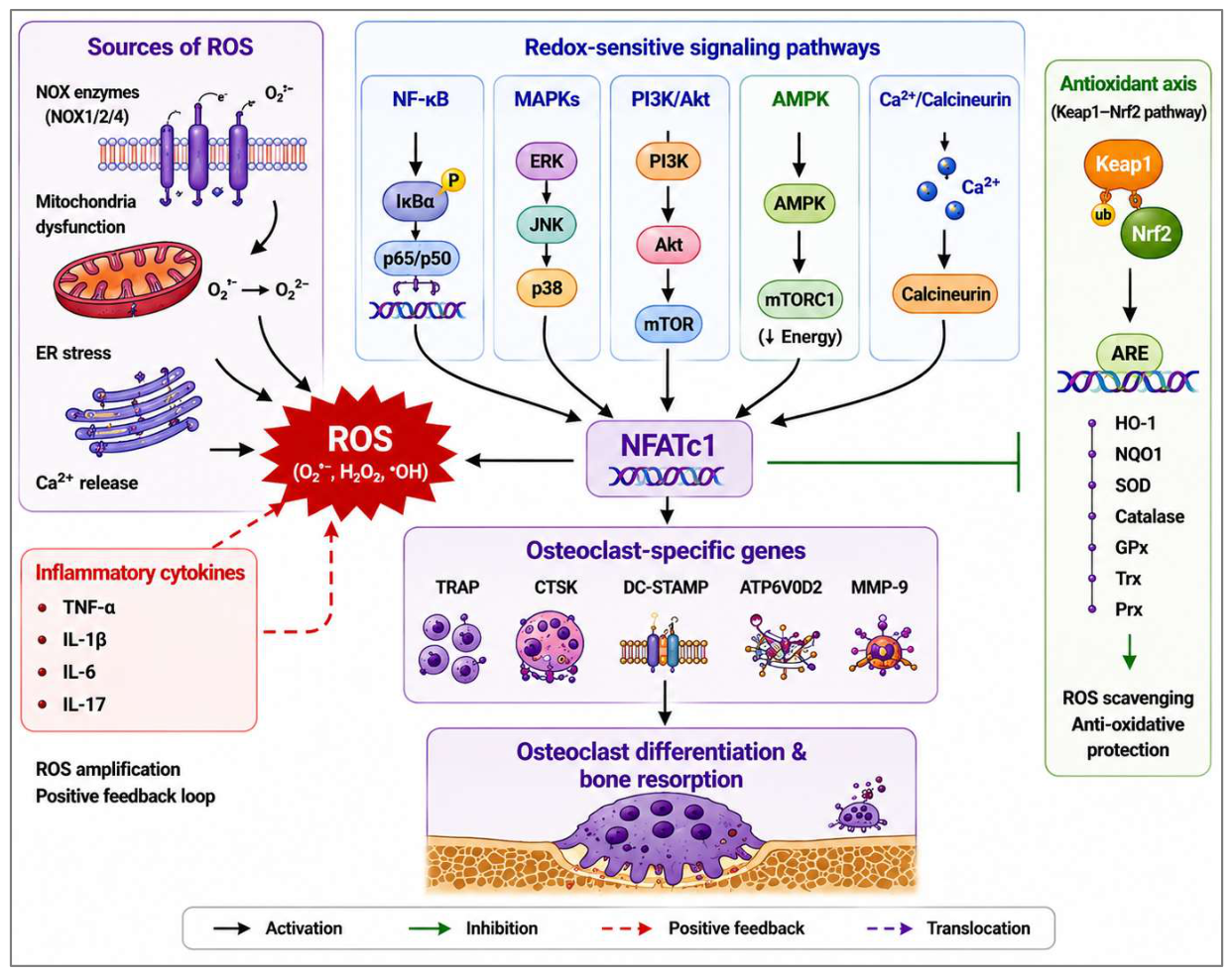
Figure 2.
Summary of antioxidant-based therapies aimed at reducing oxidative stress and osteoclast-mediated bone resorption. Main therapeutic targets include ROS produced by NOX, mitochondrial ROS, NF-κB, MAPKs, PI3K/Akt/mTOR, NFATc1, and the Keap1–Nrf2 signaling pathway. Antioxidant therapies include natural antioxidants (curcumin, resveratrol, quercetin, EGCG, sulforaphane, and melatonin), synthetic antioxidants (NAC, tempol, edaravone, and dimethyl fumarate), mitochondrial antioxidants (MitoQ, MitoTEMPO, and SS-31), as well as innovative drug delivery systems such as nanoparticles, liposomes, hydrogels, and exosomes. They may help to reduce ROS levels, attenuate redox-sensitive inflammatory and osteoclastogenic signaling, inhibit osteoclastogenesis and osteoclastic activity, and protect bone tissues. Nevertheless, clinical utility is limited by low bioavailability, insufficient bone-specific targeting, lack of validated biomarkers, and patient heterogeneity. Redox modulation may represent a more appropriate therapeutic strategy than ROS elimination.
Figure 2.
Summary of antioxidant-based therapies aimed at reducing oxidative stress and osteoclast-mediated bone resorption. Main therapeutic targets include ROS produced by NOX, mitochondrial ROS, NF-κB, MAPKs, PI3K/Akt/mTOR, NFATc1, and the Keap1–Nrf2 signaling pathway. Antioxidant therapies include natural antioxidants (curcumin, resveratrol, quercetin, EGCG, sulforaphane, and melatonin), synthetic antioxidants (NAC, tempol, edaravone, and dimethyl fumarate), mitochondrial antioxidants (MitoQ, MitoTEMPO, and SS-31), as well as innovative drug delivery systems such as nanoparticles, liposomes, hydrogels, and exosomes. They may help to reduce ROS levels, attenuate redox-sensitive inflammatory and osteoclastogenic signaling, inhibit osteoclastogenesis and osteoclastic activity, and protect bone tissues. Nevertheless, clinical utility is limited by low bioavailability, insufficient bone-specific targeting, lack of validated biomarkers, and patient heterogeneity. Redox modulation may represent a more appropriate therapeutic strategy than ROS elimination.
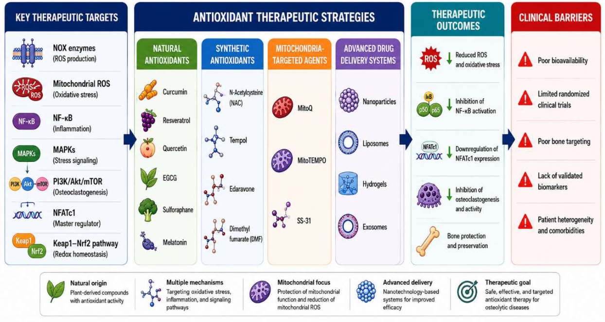

Table 1.
Major metabolic pathways regulating osteoclast differentiation and function.
| Metabolic pathway | Key regulators/enzymes | Primary function | Role in osteoclastogenesis | Therapeutic implications |
| Glycolysis | GLUT1, HK2, PFKFB3, LDHA | Rapid ATP production and biosynthesis | Supports precursor proliferation, differentiation, multinucleation, and bone resorption | Glycolytic inhibitors suppress osteoclast formation |
| Mitochondrial oxidative phosphorylation (OXPHOS) | ETC complexes, PGC-1β, TFAM | Sustained ATP generation | Provides energy for mature osteoclast function and generates mitochondrial ROS | Modulation of mitochondrial function may reduce excessive osteoclast activity |
| Fatty acid oxidation | CPT1, PPARs | Alternative energy source | Supports mitochondrial metabolism during metabolic stress | Potential metabolic target in inflammatory bone diseases |
| Glutamine metabolism | GLS, GLUD, TCA cycle enzymes | Anaplerosis and redox balance | Maintains the tricarboxylic acid cycle (TCA) activity, biosynthesis, and antioxidant capacity | Glutaminase inhibition reduces osteoclast differentiation |
| Metabolic plasticity | AMPK, mTOR, HIF-1α | Adaptation to nutrient and oxygen availability | Coordinates metabolic remodeling during physiological and pathological conditions | Emerging target for precision metabolic therapy |
Table 2.
Key sources of ROS and redox-sensitive signaling pathways in osteoclasts.
| ROS source | Major mediators | Activated signaling pathways | Biological effects | Associated diseases |
| NADPH oxidases | NOX1, NOX2, NOX4 | NF-κB, MAPKs, and NFATc1 | Initiates RANKL-mediated ROS production and osteoclast differentiation | Osteoporosis, arthritis, and periodontitis |
| Mitochondria | Electron transport chain | PI3K/Akt, AMPK, and mTOR | ATP production, mitochondrial ROS generation, and metabolic remodeling | Aging and osteoporosis |
| Endoplasmic reticulum stress | PERK, IRE1α, ATF6 | UPR and NF-κB | Calcium dysregulation and oxidative stress | Inflammatory bone diseases |
| Iron metabolism | Fenton reaction, ferroptosis | Lipid peroxidation pathways | ROS amplification and oxidative damage | Iron overload-associated bone loss |
| Inflammatory cytokines | TNF-α, IL-1β, IL-6, IL-17 | NF-κB and MAPKs | Enhance ROS generation and osteoclast activation | Rheumatoid arthritis and periodontitis |
| Hypoxia | HIF-1α | HIF signaling, mitochondrial adaptation | Alters metabolism and increases ROS production | Periodontitis, bone defects |
| Environmental/ metabolic factors | Aging, diabetes, smoking | Multiple redox pathways | Chronic oxidative stress and enhanced bone resorption | Osteoporosis and diabetic bone disease |
Table 3.
Antioxidant therapeutics targeting oxidative stress-induced pathological bone loss.
| Antioxidant/strategy | Primary target | Major mechanism | Evidence | Clinical status |
| Curcumin | NF-κB, Nrf2 | Suppresses ROS and inflammatory signaling | In vitro and animal studies | Limited clinical evidence |
| Resveratrol | SIRT1, Nrf2 | Antioxidant, anti-inflammatory, and mitochondrial protection | In vitro and animal studies | Early clinical studies |
| Quercetin | NF-κB, MAPKs | ROS scavenging and inhibition of osteoclastogenesis | In vitro and animal studies | Limited |
| EGCG | NF-κB, RANKL signaling | Reduces oxidative stress and inflammation | In vitro and animal studies | Limited |
| Sulforaphane | Keap1–Nrf2 | Activates endogenous antioxidant defenses | Animal studies | Emerging |
| Melatonin | Mitochondria, antioxidant enzymes | Improves mitochondrial function and redox homeostasis | Animal and clinical studies | Promising |
| N-acetylcysteine (NAC) | Glutathione | Restores intracellular antioxidant capacity | Experimental and clinical studies | Widely used antioxidant |
| MitoQ/ MitoTEMPO | Mitochondria | Selective scavenging of mitochondrial ROS | Animal studies | Experimental |
| Dimethyl fumarate | Nrf2 | Activates endogenous antioxidant signaling | Preclinical studies | Approved for other indications |
| Nanoparticle-based delivery | Bone tissue | Improves bioavailability and targeted delivery | Preclinical studies | Emerging technology |
Disclaimer/Publisher’s Note: The statements, opinions and data contained in all publications are solely those of the individual author(s) and contributor(s) and not of MDPI and/or the editor(s). MDPI and/or the editor(s) disclaim responsibility for any injury to people or property resulting from any ideas, methods, instructions or products referred to in the content. |
© 2026 by the authors. Licensee MDPI, Basel, Switzerland. This article is an open access article distributed under the terms and conditions of the Creative Commons Attribution (CC BY) license.
Copyright: This open access article is published under a Creative Commons CC BY 4.0 license, which permit the free download, distribution, and reuse, provided that the author and preprint are cited in any reuse.