

Submitted:
14 September 2026
Posted:
15 September 2026
You are already at the latest version
Abstract
Extracellular vesicles (EVs) are membrane-enclosed structures released by virtually all cell types and are increasingly recognized as important mediators of intercellular communication in physiological and pathological conditions. Transient receptor potential (TRP) channels are widely expressed cation-permeable channels involved in the detection and integration of chemical, thermal, and mechanical stimuli, as well as in the regulation of intracellular Ca2+ signaling. Given the ubiquitous expression of both EVs and TRP channels, increasing evidence points to a functional interplay between them. In this review, we summarize the currently available literature on the interactions between TRP channels and EVs. We first discuss the role of TRP channels in EV release, focusing on the contribution of plasma membrane and intracellular TRP channels to Ca2+-dependent vesicle shedding and exocytosis. We then review evidence for the presence of TRP channels in EVs and their potential transfer to recipient cells, including their emerging use as biomarkers of disease. Finally, we discuss how EVs can modulate TRP channel activity and expression in recipient cells through their protein, lipid, and nucleic acid cargo. These interactions have been implicated in diverse processes, including neuronal communication, inflammation, cancer progression, chemoresistance, vascular dysfunction, and tissue injury. Overall, the available evidence supports a bidirectional relationship in which TRP channels regulate EV release and composition, while EVs can in turn modify TRP channel signaling in recipient cells. Further studies are required to determine the molecular mechanisms underlying this crosstalk and its physiological and pathological significance, which may provide new opportunities for disease biomarkers and therapeutic interventions.
Keywords:
TRP channel
; extracellular vesicle
; EV shedding
; target cells
1. Introduction
Extracellular vesicles (EVs) are cell-derived lipid membrane-enclosed structures released by all cell types to the extracellular space and into biological fluids. After their discovery decades ago, EVs were thought to be a means to dispose of cellular waste, but it is now universally accepted that they are critically involved in wide variety of physiological and pathological processes, e.g., intercellular communication. Their signaling role is defined by the membrane composition and the carried cargo, as they can transport lipids, proteins, and genetic material both to neighboring cells and to distal regions in the body. The contents packaged in EVs are cell type-specific and influenced by cellular conditions. The intercellular communication mediated by EVs has several advantages, such as effectiveness (EVs allow cells to exchange membrane, protein, and mRNA), target cell selectivity (only cells with certain receptors allow fusion of the EVs), and long-distance signaling (the vesicles can travel long distance within a lumen, as in the case of epithelial primary cilia) [1,2].
Despite the great heterogeneity between different types of EVs, these structures can be broadly divided into three categories based on the release mechanism: exosomes, microvesicles, and apoptotic bodies. Exosomes are the smallest of EVs, with sizes ranging between 30 and 150 nm. They are endosomal products, being released as intraluminal vesicles (ILVs) upon the fusion of multivesicular bodies (MVBs) with the plasma membrane via the endosomal sorting complex required for transport (ESCRT) pathway. [2] Exosomes carry cellular markers and contribute to the exchange of molecules such as lipids and proteins (as constitutive components of their membrane) and nucleic acids mostly in the form of non-coding RNA (ncRNA). Compared to the plasma membrane, the exosomal membrane is enriched in cholesterol, sphingomyelin, saturated phosphatidylcholine, and phosphatidylethanolamine [3]. Microvesicles (MVs) are larger structures (50 – 1000 nm) released in the extracellular medium via outward budding and fission of the plasma membrane. MVs involved in cell-to-cell communication between cancer cells and other neighboring cells are referred to as ectosomes and can have larger diameters (up to 10 μm). Besides their role in protein and microRNA exchange between cells, MVs are believed to also remove misfolded proteins, cytotoxic agents, and metabolic waste from the cell. Because they are produced directly from the plasma membrane, MV contain mostly cellular membrane lipids and proteins [4]. Finally, apoptotic bodies (ABs) have the largest in size (>1 μM) and represent membrane-enclosed fragments released by cells undergoing apoptosis. Unlike the other two types of EVs, ABs contain nuclear fragments and intracellular organelles such as mitochondria [5]. Although ABs are cellular fragments, they do not present the usual cellular membrane asymmetry, as the outer leaflet is enriched in phosphatidylserine [6]. The phospholipid profile of EV membranes differs from that of the plasma membrane and phospholipid redistribution and loss of plasma membrane asymmetry are among the first steps in the process of EV genesis. However, because microvesicles, unlike exosomes, result from the direct blebbing of the cell, MVs resemble the plasma membrane structure more closely compared to exosomes. The most researched types of EVs are exosomes and microvesicles, as they are implicated in disease progression and can be used as biomarkers for diagnosis. In fact, ABs are considered by part of the scientific community as a subcategory of microvesicles, as they are both generated from plasma membrane blebbing [7]. Even though ABs can carry important signals to neighboring cells, their relevance for clinical investigations is less significant than in the case of exosomes and MVs.
EVs exert their signaling role by interacting with the plasma membrane of other cells and by releasing their cargo into the target cells. The membrane of EVs is decorated with a range of lipids and proteins that have the role to bind to specific receptors on the surface of target cells, and can thus be used as markers to identify and isolate these structures. Some of the proteins expressed ubiquitously in EV membranes are tetraspanins (CD9, CD63, CD81), integrins, syntenin-1, heat shock proteins, and lipoproteins. Other tissue-specific markers are also used to differentiate between different EVs [8]. The lumen of the vesicles also contains signaling molecules such as non-coding RNAs, especially microRNA, that can induce phenotypic changes in the recipient cells [9]. Depending on the membrane architecture and size of EVs, but also on the proteins and glycoproteins found on the surface of target cells, EVs can be internalized by recipient cells via different pathways. Besides direct fusion with the plasma membrane and immediate cargo release into the cytosol, EVs can be endocytosed via multiple mechanisms (phagocytosis, macropinocytosis, clathrin-mediated endocytosis, caveolin-mediated endocytosis, and lipid raft-mediated endocytosis), leading to fusion with the endosomal membrane upon endocytic uptake (Figure 1). EVs deliver their cargo by active and passive uptake mechanisms. The endocytosis of small vesicles (<75 nm) involves energy-dependent pathways in a greater proportion than for larger vesicles (>75 nm), but it is generally accepted that EV uptake involves, at least to a certain extent, active energy-dependent mechanisms [10,11].
The roles of EVs range from maintaining homeostasis (by being involved in secretory autophagy and by mediating adaptive responses to pathogens) to promoting pathological processes [9,12,13]. Upon interaction with recipient cells, EVs can influence physiological processes in target cells and even change their phenotype [1,14]. In the context of cancer, EV release is dysregulated and enhanced. Tumor-derived EVs can alter the composition and behavior of the tumor microenvironment, promoting angiogenesis and immune system evasion. By entering blood circulation, EVs can migrate to distal regions thereby promoting metastasis. Exosome cargo can be influenced by signals received from the cell of origin. Tumor cells secrete exosomes enriched in angiogenic and metastatic factors due to being exposed to hypoxia. This suggests the ability of tumor cells to adapt to the hypoxic tumor environment by stimulating angiogenesis and metastasis to a more favorable environment [9,13,15,16,17]. Within the nervous system, disruption of intercellular communication underlies neurodegenerative diseases such as Alzheimer’s and Parkinson’s disease by disrupting the clearance of toxic misfolded proteins and facilitating their accumulation and/or spreading in the brain. EVs can cross the blood-brain barrier, raising the possibility of their us for delivery of therapeutic agents against brain tumors [18,19]. Within the cardiovascular system, exosomes from erythrocytes have been shown to reduce cardiac ischemia-reperfusion injury and exosomes from platelets have an anti-thrombotic effect, whereas platelet-derived microvesicles promote angiogenesis. Moreover, exosomes released by cardiac progenitor cells are known to have cardioprotective properties [20]. EVs are present in various biological fluids, such as blood, urine, and cerebrospinal fluid. Since their release is enhanced and dysregulated in diseases, they can be used as markers for different pathologies. For example, EV concentrations in blood samples have shown potential as markers in liquid biopsies for multiple types of cancer, while the level and cargo of urinary exosomes can help determine the onset and progression of ADPKD [21,22].
Transient receptor potential (TRP) channels are a superfamily of cation-permeable channels expressed ubiquitously throughout the body. TRP channel activation leads to cell depolarization and increase in intracellular Ca2+ concentration, and are thereby implicated in a plethora of physiological processes, such as body homeostasis and adaptive responses to noxious stimuli, and have been identified as key players in various diseases [23,24,25,26,27]. Based on the homology of the amino acid sequences, human TRP channels can be classified in 6 subfamilies: TRPC (canonical, 7 members), TRPV (vanilloid, 6 members), TRPM (melastatin, 8 members), TRPP (polycystin, 3 members), TRPML (mucolipin, 3 members) and TRPA (ankyrin, 1 member). All TRP channels share some main structural features: they are transmembrane proteins with 6 transmembrane domains (S1-S6), with intracellular C- and N-termini and a cation-permeable pore situated between S5 and S6. Functional TRP channels consist of four identical (homotetramers) or similar (heterotetramers) units [28]. TRP channels are mainly expressed in the plasma membrane of cells, but some channels are also localized in the membrane of intracellular organelles, such as the lysosome and the endoplasmic reticulum [29,30].
Multiple TRP channels are promiscuous sensory receptors, detecting a wide variety of chemical, thermal and mechanical stimuli. Chemical activators include both endogenous (TRPM3 activation by the neurosteroid pregnenolone sulfate [31], TRPV4 activation by the phospholipid hydrolysis product arachidonic acid [32], TRPV1 activation by endocannabinoids [33]) and exogenous pungent and noxious compounds (TRPA1 activation by acrolein [34], TRPV1 activation by capsaicin [35], TRPV4 activation by bacterial lipopolysaccharides [36]). The activation of TRP channels by chemical stimuli is highly specific, although some compounds can act on several members at different concentrations. For example, TRPA1 can be activated by the consecrated TRPM8 agonist menthol at concentrations higher than the EC50 value for TRPM8 [37,38]. Moreover, TRP channels are also sensors of a wide range of temperatures, from noxious cold [39] to noxious heat [27]. The mechanosensory function is by far the most intriguing and controversial activation mode of TRP channels. Some TRPs have been shown to respond to mechanical stimuli such as fluid shear stress, membrane tension, and changes in cell volume and osmolarity and it has even been proposed that mechanosensation is an intrinsic universal property within the TRP family [40,41,42].
Given the universal property of vesicle shedding by all cells and the ubiquitous TRP channel expression, as well as the wide range of TRP channel activators, the interaction between these two types of structures is very probable. The literature on EV-TRP interaction is still very limited, but recent research has pointed out a bidirectional regulation of TRP channels by EVs and vice versa. The activity of certain TRP channels regulates the release of EVs from neurons, allowing communication with other cell types within the nervous system [18,43,44,45]. Moreover, TRPML channels located in the membrane of endosomes and lysosomes can trigger the fusion of multivesicular bodies with the plasma membrane, facilitating the release of exosomes [46,47,48,49]. On the other hand, exosomes and microvesicles can modulate the activity of TRP channels in recipient cells either by inducing TRP channel-mediated signaling or by regulating the expression levels via microRNA exchange or by direct transfer of functional channels [50,51,52,53]. Besides regulating and being regulated by EVs, TRP channels have also been shown to be transported on the surface or in the lumen of these lipid structures [54,55,56,57]. The expression of TRP channels in EVs circulating in biological fluids has allowed them to be identified as biomarkers of various diseases [58].There is comprehensive literature showing that EVs interact with recipient cells via specific key-lock interactions between proteins decorating the surface of the vesicles and receptors in the plasma membrane of cells. However, other types of interactions and the implication of TRP channels should not be excluded. For instance, the inherently mechanical interactions of EVs with target cells (membrane fusion, indentation, and membrane disruption) might be sensed by the poly-modal TRP channels. This review aims to summarize the currently available literature on the interactions between extracellular vesicles and TRP channels.
2. The Role of TRP Channels in EV Shedding by Donor Cells
The budding process of microvesicles involves multiple signaling pathways including the elevation of intracellular Ca2+ levels. EV biogenesis requires the disruption of the phospholipid asymmetry in the plasma membrane. The membrane asymmetry is ensured by Ca2+-modulated translocases that maintain the right distribution of phospholipids within the membrane (phosphatidylserine and phosphatidylethanolamine enriched in the inner leaflet and phosphatidylcholine and sphingomyelin enriched in the outer leaflet). This asymmetry collapses when intracellular Ca2+ levels rise [59]. TRP channels are cation-permeable channels that allow the influx of Ca2+ ions when activated. This section describes how Ca2+ signaling mediated by TRP channels results in EV release from the cells.
2.1. The Role of TRPV1 Channels in EV Shedding
Extracellular vesicles are of great importance in the nervous system. They facilitate intercellular communication between neuronal cells (neuron-neuron), but also bidirectional interactions between neuronal cells and other cell types (neuron-muscle cell, neuron-microglia, neuron-astrocyte, neuron-Schwann cell, etc.). Upon an increase in local intracellular Ca2+ levels, synaptic vesicles fuse with the plasma membrane of pre-synaptic neurons to release neurotransmitters in the synaptic cleft, allowing the exertion of critical functions within the body. Unlike synaptic vesicles, EVs are released intact into the extracellular space by fusion of MVB with the plasma membrane or by membrane budding. The exchange of EVs between neurons and other cell types ensures proper development and regeneration of the nervous system, clearance of inactive synapses and waste products, and proper myelination. The release of EVs is tightly linked to the expression and activity of TRP channels, especially members of the vanilloid (TRPV) family, as they regulate the communication between various cell types in the central and peripheral nervous system. [18]
In the nervous system, TRPV1 is highly expressed in microglial cells. As resident macrophages, microglia play an important role in brain development and in maintaining brain homeostasis during life and aging. Microglial cells interact with neurons and TRPV1 is a critical ion channel for microglial-neuronal communication. The communication between the two cell types is facilitated by the exchange of neurotransmitter-containing vesicles (200 nm). The activation of TRPV1 in microglia triggers Ca2+ influx, which results in vesicle fusion and exocytosis in the extracellular medium. Capsaicin has been shown to increase postsynaptic currents frequency and microglia-to-neuron communication via EV release. However, TRPV1 is not the sole player in this process. Purinergic stimulation by ATP via the P2X7 receptor also results in microvesicle delivery to neurons, increasing the frequency of postsynaptic currents. EV shedding by microglia is a continuous process ensuring basal levels of glutamatergic transmission and the activation of either TRPV1 or P2X7 increases the EV release rate by 50%. [43]
The communication between macrophages and neurons is not unidirectional. The bidirectional EV exchange between neurons and macrophagic cells is illustrated in Figure 2. Peripheral nerve injury leads to the upregulation of microRNA (miR-21-5p) in DRG neuron cell bodies. Upon TRPV1 activation, DRG neurons release exosomes (75 nm) loaded with miR-21-5p. One week after the injury, macrophages infiltrate into DRGs in high numbers. The endocytosis of exosomes by macrophages leads to increased miRNA levels inside the macrophages, triggering nociceptive hypersensitivity underlined by a pro-inflammatory phenotype. [44] These two studies pinpoint TRPV1 as a central player in the communication between neurons and resident and infiltrating macrophages by facilitating the release of vesicles containing important signaling molecules.
2.2. The Role of TRPML Channels in EV Shedding
The TRPML1 and TRPML2 members are highly expressed in the lysosomes, forming homomers that can sense and control the acidification of the lysosomal lumen, thus maintaining its function. TRPML3 forms homomers in the endoplasmic reticulum, but it can also be expressed in lysosomes as hetero-multimers with TRPML1 or TRPML2 [60].
As one of the main proteins expressed in lysosomes, TRPML1 facilitates the fusion of late endosomes, MVBs and autophagosomes with lysosomes and lysosomal exocytosis, regulates the lysosomal pH and its activation leads to Ca2+ mobilization from lysosomes, making TRPML1 an essential protein for exosomal release. In the adipose tissue, TRPML1 is known to be a regulator of obesity-related diseases. Adipocytes are responsible for maintaining energy homeostasis by controlling energy consumption and acting as a source of proteins and cytokines. Adipogenesis is regulated by both hormones and extrinsic factors secreted from exosomes. Adipogenic differentiation is mediated by adipocyte-derived exosomes and their cargo. In response to adipogenic stimuli, TRPML1 levels in OP9 pre-adipocytes are increased. When OP9 pre-adipocytes were exposed to exosomes isolated from adipocytes in the absence of adipogenic stimuli, the rate of lipid synthesis was enhanced compared to cells treated with the control vehicle (PBS). Activation of TRPML1 stimulates exosomal exocytosis, facilitates the release of exosomes from adipocytes, and promotes adipogenesis. This seemingly counterintuitive mechanism can be explained by the fact that exosomes present in lysosomes can escape lysosomal degradation, possibly by neutralizing the lysosomal pH and thus being released via lysosomal exocytosis. On the other hand, the deletion of TRPML1 decreases lipid synthesis and leads to impaired differentiation by reducing the levels of lysosomal-associated membrane protein 1 (LAMP1) and attenuating lysosomal trafficking to the membrane. The exosomes released by adipocytes contain microRNAs and miR-450a-5p accumulation in rat pre-adipocytes leads to enhanced adipogenesis and adipogenic differentiation. [46]
TRPML1 is also abundant in the lysosomes of podocytes, where it mediates Ca2+ release from lysosomes involved in Ca2+-dependent lysosome trafficking and autophagic flux. TRPML1 plays a protective role against podocyte injury and podocytopathy and its activation by sphingolipids determines lysosome trafficking. Sphingomyelin, a type of sphingolipid found in mammalian cell membranes, is converted to ceramide in the presence of acid sphingomyelinase. Acid ceramidase in lysosomes converts ceramide into sphingosine. TRPML1 can be activated by the acid ceramidase (AC) product sphingosine, promoting lysosome trafficking, the fusion of lysosomes with MVBs, and MVBs degradation. This results in reduced exosome release, hence reduced podocyte injury. Inhibition of AC or TRPML1 suppresses lysosome-MVB interaction, reduces autophagy, and increases exosome release, thus promoting podocyte injury. [60,61] Similar results were found when the α-subunit of AC was genetically deleted in podocytes, resulting in elevated levels of urinary exosomes. Inhibition of lysosomal function results in increased exosome release from neurons, epithelial cells, and vascular cells. Sphingosine can rescue TRPML1 activity. [62] Moreover, hyperhomocysteinemia, characterized by elevated levels of homocysteine (Hcy), is a condition in which TRPML1 is inhibited due to an NADPH oxidase-dependent increase in ROS. TRPML1 inhibition leads to the activation of the NLRP3 inflammasome, resulting in glomerular inflammation and sclerosis due to the increased release of exosomes and IL-1β and IL-18 cytokines. The TRPML1-dependent EV release allows exosomes to be used as a marker of glomerular disease, as they trigger phenotypic changes in neighboring cells and participate in the development of albuminuria and glomerular degeneration. [47]
Arterial medial calcification (AMC) is a condition triggered by increased free serum levels of inorganic phosphate (Pi). Elevated levels of Pi inhibit the MVB-lysosome interaction, leading to increased Ca2+ deposition and EV (40-140 nm) secretion. Knocking-out AC in mice results in more severe AMC and enhanced EV markers in arterial media compared to wild-type animals. The TRPML1-mediated EV release due to low levels of AC precedes the development of AMC. AC in the plasma prevents the development of AMC by reducing the levels of secreted EVs. Under stress conditions induced by phosphate stimulation, the release of EVs is enhanced, allowing the development of AMC. [63] Proper TRPML1 function seems to play a protective role against podocyte injury and podocytopathy, glomerular degeneration, as well as arterial medial calcification.
The low-density lipoprotein receptor (LDLR) does not only mediate the endocytosis of LDL. The Japanese encephalitis virus (JEV) infects the cells by binding to the same receptor and LDLR/ligand complexes are internalized every 10 minutes. Under normal conditions, LDLR is degraded inside the lysosomes and is recycled back to the cell membrane. Due to their role in lysosome function, TRPML channels are involved in the degradation of LDLR. Berbamine, a known Ca2+ channel blocker, compromises the endolysosomal trafficking of LDLR due to its TRPML inhibitory function. As a result, LDLR is secreted in EVs instead of being degraded. Hence, berbamine can offer protection against JEV by decreasing the plasma membrane expression levels of LDLR, and TRPML channels regulate the activity of host viral infection via LDLR-containing EVs release. [64]
TRPML2 is a cation-permeable ion channel localized in late endosomes and lysosomes (together with the other two members of the TRPML family), but also in the plasma membrane. However, TRPML2 has not yet been identified in secretory vesicles. TRPML2 is activated by drops in pH and regulates vesicle fusion and scission via Ca2+ release from endolysosomes. It is believed that activation of mucolipins in endosomes and lysosomes causes Ca2+ release from the lumen into the surrounding cytosolic space that triggers their fusion with the plasma membrane or if already fused, their scission. [48]
The autonomous cell defense system relies on antimicrobial peptides and proteases, reactive oxygen species (ROS), and nitric oxide synthases (NOS), as well as on the acidic environment of the lysosome. The lysosomal function is regulated by vacuolar ATP-ases, which control the pH, and TRPML channels, including TRPML3, which is responsible for regulating the ionic flux. Upon infection of bladder epithelial cells, E. coli activates the autophagy pathway, but it neutralizes the lysosomal pH, thus escaping degradation. TRPML3 is a pH-sensitive ion channel that can sense the increase in pH, initiating Ca2+ efflux, lysosomal exocytosis, and expulsion of vesicle-encased bacteria. The vesicles expressed Caveolin-1 (present in membranes), ESCRT proteins, and tetraspanin CD63 (present in exosomes) and components of the autophagy pathway but no intracellular organelle markers, allowing them to be identified as exosomes. TRPML1 also plays a role in bacterial expulsion from lysosomes, but smaller. In contrast to TRPML1, which is active in acidic environments, TRPML3 is activated by neutral pH. [49]
Besides its role in bacterial expulsion in exosomes, TRPML3 has also been identified as a modulator of drug resistance in non-small cell lung cancer (NSCLC). NSCLC cells resistant to gefitinib shed more exosomes compared to the sensitive controls. The enhanced exosomal release was associated with an elevated expression of TRPML3 in the resistant cells and was a result of increased lysosomal exocytosis. Gefitinib elevates lysosomal pH, triggering Ca2+ release into the cytoplasm via TRPML3, which acts as a sensor of lysosomal pH. This results in lysosomal trafficking and exosome exocytosis. Moreover, TRPML3 deficiency resulted in reduced cell proliferation, suggesting its involvement in drug resistance in tumors and cancer development. [65] The contribution of TRPML channels to EV release is summarized in Figure 3.
2.3. The Role of TRPM Channels in EV Shedding
TRPM8 is a cold-activated ion channel found in cold-sensing neurons. It resides both in the plasma membrane where they directly respond to temperature drops below 18oC, as well as in the endoplasmic reticulum where they play a crucial role in intracellular Ca2+ signaling, contributing to calcium-induced calcium release (CICR). Under normal conditions, amyloid-beta induces Ca2+ influx via voltage-gated Ca2+ channels and TRPM8 in the plasma membrane, leading to CICR due to TRPM8 in the ER (Figure 4). The increase in depolarization triggers EV release. In a numerical model of exosomal release in Alzheimer’s disease, the shedding of exosomes from neurons is a temperature-dependent process and TRPM8 mediates the exosome release rate in cold-stimulated neurons. According to the model, higher temperatures allow the enhanced release of exosomes. [66]
The B-Raf protein, encoded by the BRAF gene, is a protein involved in cell growth. Some human cancers, including melanomas, show BRAF mutations that usually occur at the V600 residue. [67] BRAF inhibitors vemurafenib and dabrafenib are drugs routinely used to treat metastatic melanomas with BRAF mutations. These drugs have been shown to increase total protein and RNA content in all types of EVs (exosomes, microvesicles, and apoptotic bodies), in particular increased levels of miR-211-5p, and caused significant changes in RNA profiles of target cells. The increase in miR-211-5p represents the strategy of melanoma cells to overcome the anti-tumor effects of the drug, as this confers resistance to the cells. Vemurafenib increases the melanocyte-inducing transcription factor (MITF) expression via the down-regulation of pERK1/2 in host cells. This leads to the activation of the TRPM1 gene and to increased TRPM1 levels, which triggers the activation of the survival pathway and a subsequent increase in miR-211-5p (Figure 4). As EVs are microRNA carriers, vesicles released by the host cells contain high levels of this microRNA which is transferred to target cells, offering them resistance to vemurafenib. TRPM1 is an ion channel expressed in melanomas that is inversely correlated with the metastatic potential of melanoma cells, acting as a tumor suppressor. BRAF inhibitors show initial antitumoral effects, but drug-resistant clones appear soon. [28,68] This study sheds light on the role of TRPM1 in the development of vemurafenib resistance and highlights the role of EVs downstream of TRPM1 in cancer progression.
Vitamin K-dependent matrix Gla protein (MGP) is an inhibitor of vascular calcification (VC). To become active, MGP needs to be phosphorylated and carboxylated. The absence or inactivity of MGP has been shown to be a driver of vascular calcification. High phosphate and Ca2+ levels induce an increased release of MGP-containing matrix vesicles (a type of membrane-derived extracellular vesicle), leading to decreased levels of available protein and vascular calcification. TRPM7 is a magnesium-permeable ion channel. Upon cell entry via TRPM7, Mg2+ can exert protective roles by upregulating the expression of MGP and preventing the MGP levels to drop below the level needed to trigger vascular calcification (Figure 4). [69]
2.4. The Role of TRPC in EV Shedding
In the context of platelet activation, thrombin is a procoagulant that exerts its role by inducing Ca2+ mobilization upon binding to PAR1 and PAR4 receptors in arterial cells’ wall. Intracellular store depletion activates the calcium-sensitive TRPC6 channel, triggering the activation of platelets via secretion of EVs containing α- and δ-granules (Figure 4). [70]
It is known that extracellular Ca2+ entry results in enhanced EV production in all cell types, and skeletal muscle cells in particular. Besides Ca2+ influx, mitochondrial respiration and, hence, physical exercise have also been shown to stimulate the EV release from muscle cells. The muscular secretome promotes, among others, myogenesis. Moreover, 10 min exposure to low amplitude and low frequency pulsed electromagnetic fields (PEMF) can enhance mitochondrial respiration and induce myogenesis in mice by stimulating the production of muscle-derived EVs (Figure 4). These effects are (partially) mediated by TRPC1, whose activation has been linked to chondrogenesis, neurogenesis, and myogenesis. Exposure to PEMFs enhances TRPC1 expression in a direction-dependent manner. Streptomycin, 2-APB, and SKF-96365, three TRPC1 blockers, prevent the production of the myogenic conditioned medium upon exposure to PEMFs. Hence, the release of myogenic soluble factors, in particular EVs (100-200 nm), from skeletal muscle cells is enhanced upon exposure to short PEMF in a TRPC1-dependent fashion. [71]
2.5. The Role of TRPP in EV Shedding
Although no longer considered a TRP channel, PC1 forms functional complexes with TRPP1 and has also been shown to influence the release of exosomes. Loss of PC1 in mouse inner medullary collecting duct cells reduces autophagy completion due to decreased lysosomal acidification induced by defective, highly active calpain proteases (CAPN). Of note, these effects can be rescued upon reintroduction of P1 into the cells. [72]
3. TRP Channel Expression in EVs
Proteins can be exchanged via EVs either as a constituent part of their membrane or by being transported in the luminal cargo. EVs have been shown to transport TRP channels from donor to target cells. This may result in an overall enhanced cellular signaling in the recipient cell, mediated by the increased expression of TRP channels. The expression of some TRP channels is altered in various pathologies and this can reflect in the vesicular expression level. Because EVs can enter the blood circulation, the expression levels of TRPs in the circulating EVs can be used as a disease marker, given that adequate normalization and control protocols are available. The expression of different TRP channels on/in EVs is illustrated in Figure 5.
3.1. TRPV Expression in EVs
Posterior urethral valves (PUV) are the most common cause of lower urinary tract obstruction in males. TRPV4 is a mechanosensitive channel with a role in flow sensing, expressed in the collecting duct. TRPV4 levels in this region and whole urine and urine exosomes were decreased in obstructive fetal kidneys compared to control. Moreover, TRPV4 expression in urinary exosomes was correlated with the glomerular filtration rate. This would allow TRPV4 levels in urine samples to be used as a specific biomarker for posterior urethral valves. However, TRPV4 levels did not correlate with the protein-to-creatinine ratio. Hence, more research is needed to determine whether or not TRPV4 levels are a good indicator of PUV. [54]
TRPV6 is found in the epididymal epithelium, where it regulates Ca2+ concentration. TRPV6 is known to play an important role in sperm maturation and motility. Mice lacking TRPV6 develop asthenozoospermia, which is known to be a common cause of male infertility. Seminal EVs play roles such as spermatozoa motility, immunomodulation, regulation of the capacitation process, and antioxidant protection. A study comparing normal and asthenozoospermic extracellular vesicles (50-500 nm) found a markedly reduced TRPV6 expression in vesicles isolated from patients, suggesting its importance for sperm motility. [73]
3.2. TRPP Expression in EVs
The TRPP group includes three members in mammals: TRPP1 (currently also known as PC2, previously known as TRPP2), TRPP2 (previously known as TRPP3), and TRPP3 (previously known as TRPP5). PC2 is co-expressed with PC1 (no longer considered a member of the TRPP family). They form functional complexes composed of one PC1 unit and 3 PC2 units that localize in the primary cilia, and the PC2 function is nevertheless influenced by PC1. PC1 and PC2 are localized in all tissues. At the cellular level, PC1 is expressed in the plasma membrane and PC2 predominantly in the endoplasmic reticulum, but also in the plasma membrane in lower amounts. In C. elegans, PC1 and PC2 are named LOV-1 and PKD2, respectively. [74] These two proteins are among the most investigated ones in the context of TRP expression in extracellular vesicles. LOV-1 and PKD-2 are present in the ciliated sensory neurons of the nematode, from where they can be secreted in microvesicles (100 nm). The microvesicles containing PKD-2 are only secreted by the male-specific neurons of the worm and the release takes place without the breakage of the cilium. [75] EV release outside the worm plays an important role in mating, while EV release inside the worm is involved in gastrulation in the embryo, cuticle formation during larval development, extrinsic repair of injured neurons, and neuronal waste expulsion in adults. The vesicles are released from the cilium upon mechanical stimulation and the release is regulated by the conserved intraflagellar transport machinery. The release sites at the level of the cilium are the periciliary compartment at the base, with roles in sorting proteins between ciliary and cell body destinations, and the axoneme, acting as a transition zone. [76] When C. elegans males are mounted on coverslips covered with a soft agarose gel layer, the release of PKD-2-containing vesicles is reduced. [77]
LOV-1- and PKD-2-containing EVs are released from the cilium base and can exit through the cuticular pore, from where they are directly exposed to the environment, the cilium base, and the cilium. Transmission electron microscopy images identified EVs in the extracellular lumen formed by glial cells. The vesicular release from the cilium is dependent on KLP-6, a kinesin that regulates the morphology and transport of mitochondria in neuronal cells. EVs shedded by the cilium contain several TRP channels-encoding mRNAs. However, they only contain LOV-1 and PKD-2 proteins and no specific RNA. Primary cilia are not only responsible for EV release but also for their uptake. [78] The release of PKD-2 vesicles requires intraflagellar transport 88 (IFT88) and kinesin-3 KLP-6 and induces animal communication and mating behavior, such as tail chasing and tail circling, male reversal, sex drive, response to mating contact and vulva location. PKD-2 is co-expressed with LOV-1 (location of the vulva) in the primary cilium membrane and they are both responsible for triggering species-specific mating behaviors. PKD-2 is crucial for this function, as PKD-2 deficient EVs fail to trigger the specific behavior in C. elegans and PC2 knock-out in mice is lethal. [75,76] CIL-7 is a myristoylated protein that is essential for the release of PKD-2-containing EVs and CIL-7 loss in C. elegans leads to male mating behavioral defects, excessive EV accumulation, and failure to release PKD-2 vesicles. [79] C. elegans represents a good animal model to study polycystin-related diseases in humans, as PC1/LOV-1 and PC2/PKD-2 act in the same genetic pathway and function in sensory capacity in C. elegans, as well as in humans. Moreover, literature shows that both mammalian kidney epithelium and C. elegans male sensory neurons shed TRPP-containing vesicles. [78]
Human urinary exosomes contain PC1 and PC2. [55] Subfractionation studies identified 3 types of vesicles: large vesicles (200 nm), exosomes (100 nm), and glomerular membrane vesicles (70 nm). PC1 expression was the highest in the exosome-containing fraction. [56] Mutations in PC1 and PC2 proteins lead to the development of autosomal dominant polycystic kidney disease (ADPKD). Urinary exosomes of around 100 nm containing the two proteins are secreted by the ciliated cells within the kidney. Biotinylation experiments showed that in mouse models of ADPKD, EVs localize preferentially on cilia in greater amounts compared to the control and this is an adhesive rather than a budding event. [22] The exosomes that only interact with the surface of the cilia are mostly cleared rapidly (10 min). In ADPKD patients, the secreted exosomes carry defective copies of PC1 and PC2. Patients with PC1 mutations show different expression levels in exosomes; PC1 and PC2 levels were reduced to 54% and 53% respectively, while transmembrane protein 2 (TMEM2) levels were 2.1 times higher. PC1/TMEM2 and PC2/TMEM2 ratios can be correlated with total kidney volume and can be used to distinguish between control kidneys and kidneys with mutations. In the case of PC1 individuals, PC1/TMEM2 ratio is lower (9.98) compared to normal (23.7), and for PC2 patients PC1/TMEM2 value is decreased (1.83) compared to normal (4.6). TMEM2 is present on PC1- and PC2-positive exosomes, which allows it to be used as an internal standard. The expression levels of PC1 and PC2 proteins can be thus used to design stable (throughout a lifetime) ratiometric markers to diagnose ADPKD disease. [58,76]
3.3. TRPC Expression in EVs
The literature body described multiple ways in which TRPC channels play a role in cancer progression: knock-outs of TRPC1/4/5 show inhibited vascularization represented by decreased tube formation, TRPC1 knock-down suppresses cell migration and proliferation, TRPC4 is upregulated in hypoxia, and TRPC5 is expressed in EVs and is implicated in the development of chemoresistance. These roles point to the potentially beneficial use of TRPC channel modulators. TRPC inhibitors are unlikely to cause adverse effects, but they might have positive effects, such as suppression of innate fear, visceral pain, and cardiac remodeling. [80]
Microvesicles released by C2C12 murine myoblasts have been shown to contain TRPC1 channels and respond to pulsed low amplitude electromagnetic fields (PEMF) with TRPC1-mediated Ca2+ influx. PEMF activates the TRPC1/mitochondrial axis underlying cell expansion and mitohormetic survival adaptations by enhancing mitochondrial respiratory capacity and proliferative responses. Mitochondrial dysfunction leads to cell senescence and quiescence. Microvesicles (200 nm) secreted by unaffected cells provide sufficient molecular machinery to restore the proliferative and mitochondrial function in TRPC1 knock-down cells. Microvesicles secreted by C2C12 cells are enriched in sphingomyelin (SM), the products of which are implicated in the modulation of TRPC1 function. Notably, SM is also a major constituent of lipid rafts, where TRPC1 resides. [57] Urinary EV samples showed that glomerular membrane vesicles collected from patients with nephrotic syndrome express TRPC6, but not the ones isolated from healthy individuals. [56]
In many cases, chemotherapy potency decreases in time due to chemoresistance. Endothelial cells have been shown to acquire resistance to the drug via the tumor microenvironment. EVs, and microvesicles in particular, released in the microenvironment, are known for their role in intercellular communication. P-glycoprotein (P-gp) is a protein that offers cells resistance to therapeutic drugs. P-gp expression is modulated by the Ca2+-permeable channel TRPC5 via the activation of the NFATc3 pathway. Both P-gp and TRPC5 overexpression are associated with multi-drug resistance. MCF-7 cells are resistant to Adriamycin (MCF-7/ADM) and have been shown to shed microvesicles containing P-gp and TRPC5. In contrast to normal MCF-7 cells, where Adriamycin (Doxorubicin) is accumulated in the nucleus, in resistant cells Adriamycin is accumulated in vesicles (50-500 nm) in the cytoplasm. Moreover, cell surface vesicles are much more abundant in MCF-7/ADM compared to wild-type cells. Healthy human microvascular endothelial cells (HMEC) exposed to EVs secreted by resistant adenocarcinoma cells acquired drug chemoresistance via the transfer of TRPC5 and the subsequent activation of the NFATc3 pathway and additional P-gp expression. Healthy HMEC cells do not express either P-gp, or TRPC5, but the proteins can be identified in the cells exposed to MCF-7/ADM microvesicles. The morphology of HMEC cells upon EV treatment is also changed dramatically, from flat, elongated, and spindle-like to various shapes. This study shows that chemoresistance can be transferred from MCF-7/ADM to normal HMEC cells via EV cargo, in particular TRPC5 and P-gp. Treatment with microvesicles results in increased P-gp expression in recipient cells, both via direct transfer and via TRPC5-modulated expression. The acquired chemoresistance can be reversed by blocking the TRPC5 channels. [53]
Elevated TRPC5 levels were found in EVs isolated from breast cancer patients’ plasma and the expression levels in circulating exosomes were correlated with expression levels in breast cancer cells and with tumor response, meaning that higher TRPC5 levels were associated with less tumor shrinkage. The differential expression of TRPC5 between the two groups (Adriamycin therapy responders vs. non-responders) allows TRPC5 expression levels in circulating exosomes to be used as a chemoresistance marker. Calculating the ratio between TRPC5 levels after 2 cycles of chemotherapy and TRPC5 levels prior to chemotherapy can determine the progression rate of cancer, with a cut-off value of 1.09. An increased value is associated with faster progression and hence more resistance. [81] Another study identified TRPC5 as a breast cancer exosome marker since the protein levels in blood plasma were correlated with progression-free survival, disease-free survival, and overall survival. [82]
The TRPC4/5 hetero-multimer seems to be implicated in drug resistance, the transmission of drug resistance through EVs, tumor vascularization, and cancer cell death by activation with englerin A. [80] Chemotherapy resistance is a major problem for patients with breast cancer. TRPC5 is responsible for EV formation and can be transferred between cells via EVs. MCF-7 adenocarcinoma cells develop resistance to Adriamycin by trapping Adriamycin in the EV lumen. TRPC5 accumulation in target cells triggers increases in cytosolic Ca2+ levels, which stimulates the production of multidrug efflux transporter P-glycoprotein (P-gp) due to nuclear translocation of activated T-cells isoform c3 (NFATc3). TRPC5 is involved in growth factor-regulated vesicular trafficking through PI3K, Rac1, and PIP-5-kinase–mediated pathways. Resistant MCF-7 cells have higher expression of TRPC5, greatly upregulated TRPC5 and P-gp in EVs and great enhancement of Adriamycin-containing EVs on the cell surface. TRPC5-specific siRNA inhibited EV generation and led to drug accumulation in the nucleus, supporting the role of the channel in the development of drug resistance. TRPC5 expression levels in EVs from periphery blood samples can be used as an indicator of breast cancer progression. According to this study, Adriamycin-resistant cells can induce a resistant phenotype in non-resistant cells by transferring TRPC5- and P-gp-containing EV. [83] TRPC5 plays the same role in the development of colorectal cancer. 5-Fluorouracil is a drug commonly used in the treatment of colorectal cancer and in many cases, patients develop resistance to this kind of chemotherapy. Like in adenocarcinoma, colorectal cancer cells show increased levels of TRPC5 and P-gp. Due to its role, P-gp inhibition is a promising strategy against chemotherapy resistance. [84]
3.4. TRPA1 Expression in EVs
Besides TRPC1, microvesicles released by C2C12 cells also contain TRPA1, but not TRPV1 and TRPV2. The presence of TRPA1 in the vesicles has been confirmed via exposure to menthol. TRPA1 has also been shown to contribute to mitochondrial function in adipocytes, but its function in C2C12 cells has not been studied. [57]
3.5. TRPM4 Expression in EVs
The intracellular Ca2+ levels are tightly regulated in neuronal cells via the activity of multiple Ca2+-permeable channels in the plasma membrane, as well as in the membrane of organelles, such as mitochondria and endoplasmic reticulum. The fine Ca2+ homeostasis mechanisms are disrupted in pathologies such as schizophrenia and major depressive disorder. TRPM4 is a Na+-permeable, Ca2+-impermeable ion channel activated by increased intracellular Ca2+ levels. Plasma EVs from patients with first episodes of psychosis and healthy controls have been compared in terms of expression levels of proteins that play a role in mitochondrial function. The levels of TRPM4, Na+/Ca2+ exchanger, and leucine zipper EF-hand containing transmembrane 1 protein (LETM1) in plasma EVs from neurons were significantly reduced in EVs from psychotic patients compared to controls, while levels of mitochondrial voltage-dependent L-type Ca channel subunit α-1C (CANCA-1C) were enhanced. TRPM4 has been shown to form complexes with N-methyl-D-aspartate receptor 1 (NMDAR1), which play a significant role in maintaining mitochondrial Ca2+ homeostasis. Reduced levels of the complex may result in reduced mitochondrial Ca2+ uptake and may induce neuronal toxicity. [85] The differential expression of TRPM4 in plasma EVs from first-episode schizophrenia patients compared to healthy controls may allow TRPM4 to be used as a disease marker in routine laboratory blood tests in the future.
4. The Role of EVs in Regulating TRP Activity in Target Cells
Extracellular vesicles can modulate the TRP channel activity in target cells either by inducing TRP channel-mediated Ca2+ signaling, or by altering TRP channel expression levels. EVs carry various cargoes such as proteins, lipids, and nucleic acids. Small interfering RNA or silencing RNA (siRNA) can be transferred to target cells upon EV internalization and cargo release. siRNAs can modulate the expression of specific genes in the recipient cells, resulting in decreased expression levels of corresponding proteins. As a result, the expression levels of specific TRP channels can be altered by siRNA-carrying exosomes upon interaction with the target cells. Figure 7 serves as a summary of the effects of EVs on the modulation of TRP channel activity and expression.
4.1. Regulation of TRPP Activity
Epithelial mesenchymal transition (EMT) plays an important role in the invasion and metastasis of head and neck cancer. EMT is known to be mediated by PC2 (TRPP1) in human laryngeal squamous cell carcinoma and is characterized by decreased levels of E-cadherin and increased levels of vimentin. The increased TRPP1 expression can be downregulated by specific siRNA molecules. FaDu cells (a cell line originating from human pharyngeal squamous cell carcinoma) transfected with PC2 siRNA showed higher levels of E-cadherin and lower levels of vimentin compared to the control. The same reversing result was obtained when FaDu cells were treated with exosomes (50-100 nm) carrying PC2 siRNA. The complexes of PC2 siRNA and HEK293T exosomes are stable and can be taken up by FaDu cells, resulting in a successful knock-down of TRPP1 in target cells. [86]
Mutations in the PKD1 or PKD2 genes encoding the PC1 and PC2 proteins represent the cause of autosomal dominant polycystic kidney disease (ADPKD). The development of cysts and cell-to-cell communication within the kidney and within 3D cultures of mouse inner medullary-collecting duct 3 cells can be (partially) explained by exchanging exosomes (110 nm). Urinary exosomes from ADPKD patients stimulate cell proliferation and cystogenesis in 3D collagen cell cultures after 2 days in vitro and in mouse kidneys after 24h in vivo. Exosomes from cystic renal epithelial cells induce PC1 (a former member of the TRPP family) down-regulation by transferring miR-200s and miR-21 to target cells, inducing cyst growth and fibrosis. Exposure to cyst renal epithelial cells exosomes increases levels of cell proliferation markers and the expression of cyst growth-related miRNA. Moreover, exosome treatment leads to fibroblast activation, cytokine and chemokine release (TNF-α, IL-6, MCP-1), and recruitment of macrophages. [52]
4.2. Regulation of TRPA1 Activity
Lung adenocarcinoma (LUAD) is a type of cancer whose progression and metastasis are modulated by TRPA1, together with fibroblast growth factor receptor 2 (FGFR2). The N-terminus of TRPA1 binds to the C-terminus of FGFR2, resulting in FGFR2 dimerization. The dimerization activates FGFR2 and inhibits TRPA1, presumably due to conformational changes in the structure of the proteins. This results in LUAD progression and metastasis due to the activation of PLC-γ1 and MAPK/ERK pathways, responsible for cell invasion and cell proliferation, respectively. LUAD can form metastases in the brain. Exosomes (30-100 nm) secreted by brain astrocytes and loaded with miR-142-3p microRNA are transferred to cancer cells, where they cause TRPA1 depletion. Since FGFR2 needs TRPA1 to exert its function, the targeting of cancer cells by astrocyte-derived exosomes has protective results against cancer progression in the brain. Because of its role in cancer metastasis, TRPA1 is used as an invasive and prognostic marker in lung adenocarcinoma patients. [87]
Atopic dermatitis (AD) is a pruritic skin disorder often affecting animals, but also humans, that share very similar pathways with the canine disease. AD is characterized by high levels of IL-31, which stimulate the expression of TRPA1 via the JAK/STAT pathway. TRPA1 is one of the TRP channel family members involved in itch (together with TRPV1 and TRPM3). The main treatment for AD involves immunosuppressive drugs, but some dogs are not responsive or relapse after some time. Mesenchymal stem cells exert an immunosuppressive effect by generating extracellular vesicles. Canine adipose stem cells (ASCs) and ASCs-derived exosomes (180 nm) have been shown to share similar immunomodulatory abilities and to reduce AD pruritus by decreasing the expression of TRPA1 in mice. The exact underlying molecular mechanisms have not yet been investigated. However, ASCs and ASC exosomes show therapeutic potential against AD in animals, as well as in humans. [88]
4.3. Regulation of TRPM7 Activity
Ischemic stroke can be modeled in vitro by culturing brain microvascular endothelial cells (BMECs) under conditions of oxygen and glucose deprivation. Adipose-derived stem cells (ADSCs) have been demonstrated to promote cerebral vascular remodeling after stroke. BMECs exposed to exosomes secreted by ADSCs show enhanced angiogenesis reflected by increased migration distance and tube length. BMECs showed increased levels of miR-181b-5p and miR-212-5p microRNAs. When exposed selectively to exosomes containing only one type of microRNA, the expression levels of TRPM7 were only downregulated in the group treated with miR-181b-5p-containing exosomes, showing that this microRNA can modulate the levels of TRPM7 in recipient cells. The effect of the microRNA could be (partially) reversed when TRPM7 was overexpressed in BMECs. Previous studies pointed out the role of TRPM7 in the adhesion and tube formation of vascular endothelial cells. These results demonstrate that exosomes from ADSCs can alter the expression levels of TRPM7 in BMECs via their cargo, leading to angiogenesis after oxygen-glucose deprivation and confirming their potential therapeutic role for recovery after ischemic stroke. [89]
Along the same lines, TRPM7 down-regulation can alleviate neuronal injury following intracerebral hemorrhage (ICH). Mesenchymal stem cells are believed to be a potentially successful therapy method against ICH, but their survival rate following transplantation is poor. Previous studies have reported that miR-21 is down-regulated in the blood and brain tissues of patients with ICH. This microRNA can target TRPM7 and has been found to enhance the survival of MSCs, improving their recovery function in rats with ICH. miR-21 can be transported to neurons via exosomes released by MSCs, where it down-regulates the expression levels of TRPM7, preventing ionic overload and neuronal apoptosis via the activation of the NF-kβ pathway. Thus, overexpressing miR-21 in MSC increases their post-transplantation survival rate and allows the endocytosis of miR-21-containing exosomes by neurons, ultimately improving survival and recovery of neurological function. [90] TRPM7 down-regulation via exosomes carrying specific microRNAs released by stem cells can provide an effective strategy for neuroprotection and treatment of cerebrovascular diseases, particularly against ischemic stroke, as well as against stroke triggered by intracerebral hemorrhage.
EVs also play an important role in the progression of glioblastoma by promoting angiogenesis in vitro. EVs from U87 glioblastoma cells can be taken up by human microvascular endothelial cells (HMECs) and regulate cell migration, tumor growth, and endothelial sprouting. Chloride intracellular channel 1 (CLIC1) is overexpressed in U87 cells, as well as in the vesicles shedded by these cells. CLIC1 is known for having pathological implications in cell proliferation, motility, and angiogenesis. Apart from CLIC1, glioblastoma EVs carry miR-5096. Interestingly, higher miR-5096 has been correlated with increased numbers of CLIC1-containing EVs from U87 cells. Enhanced EV secretion in the presence of miR-5096 might be underlined by increased filopodia outgrowth induced by this microRNA. EVs transfer from U87 cells to HMECs facilitates the transfer of miR-5096, as well as CLIC1. Upon exposure to glioblastoma exosomes, cytosolic Ca2+ spikes regulated by TRPM7 and increased CLIC1 levels can be observed in HMECs, leading to endothelial sprouting in matrigel after 24h. TRPM7 knock-down leads to decreased CLIC1 levels in recipient cells, confirming its role in EV cargo delivery and new blood vessel formation. In control HMECs, CLIC1 is expressed in perinuclear areas, while in EV-treated HMECs it is mostly present in the plasma membrane and cytosol, suggesting the delivery via extracellular vesicles. Notably, miR-5096 does not change CLIC1 levels in U87 and HMEC cells and TRPM7 levels are also not altered by U87 conditioned medium. This study points to the capability of EVs to trigger Ca2+ signals in recipient cells and to deliver CLIC1-loaded EVs from U87 cells to HMECs, facilitating glioblastoma angiogenesis. This process is enhanced by higher levels of miR-5096. Figure 6 illustrates the possible interplay between extracellular EVs and TRPM7 activation. [50]
4.4. Modulation of TRPV Activity
Over the past years, the strategy against tumors consists in ensuring normal tumor vascularization for more efficient chemotherapy delivery. Tumor angiogenesis results in aberrant blood vessel growth, with short and collapsed tubes that cannot sustain proper blood circulation. It is known that tumor cells release EVs in the tumor microenvironment and induce angiogenesis by communicating with endothelial cells. Normal human endothelial cells (NEC) exposed to conditioned medium from human lung tumors develop a tumor-like phenotype due to the EVs (50-200 nm) released by the tumor cells. TRPV4 is a mechanosensitive ion channel that is a critical player in cancer progression. The role of TRPV4 in metastasis is highlighted by its ability to induce blebbing in breast cancer and activate the ERK pathway in hepatocellular carcinoma, while down-regulation results in reduced metastasis and cell proliferation. TRPV4 activation results in normalized tumor vasculature and improved cancer therapy. TRPV4 expression and activity are reduced in endothelial cells repeatedly exposed to tumor EVs, explaining the abnormal angiogenesis within the tumor microenvironment, while TRPV4 activation normalizes tumor vasculature. The change in phenotype is underlined by the activation of pathways responsible for cell movement and extracellular matrix remodeling. The EV cargo is responsible for TRPV4 down-regulation has not yet been identified. However, previous studies demonstrated that increased levels of miR-203 result in lower TRPV4 expression in chondrocytes and hepatic stellate cells. These studies highlight the fact that tumor EVs regulate TRPV4 protein levels in healthy recipient cells, induce a tumor-like phenotype, and lead to aberrant angiogenesis, preventing efficient delivery of cancer therapy. [14,91]
Mechanical ventilation (MV) is known to cause pulmonary edema and lung inflammation due to endothelial barrier destruction and pulmonary barrier hyperpermeability. TRPV4 is a mechanosensitive channel widely expressed in the lungs, whose activation leads to vascular permeability and hence, undesired outcomes associated with mechanical ventilation. During mechanical ventilation, the serum glucocorticoid-regulated kinase 1 (SGK1) is activated, which further activates TRPV4, resulting in increased permeability and cytokine release due to Ca2+ influx. Counterintuitively, obesity has been associated with lower mortality risk in MV patients. A recent study has determined that adipose tissue-derived exosomes, which also act as adipokines and can reach distal tissues, can have protective roles against MV-induced lung injury. MV can be mimicked in vitro by culturing cells on elastic membranes and exposing them to cyclic stretching. Both in vivo and in vitro experiments have concluded that pulmonary microvascular endothelial cells exposed to adipose-derived stem cells (ADSC) exosomes show decreased levels of TRPV4 RNA and protein expression. Down-regulation of TRPV4 facilitates the increased expression of adherens molecules (β-cadherin and VE-cadherin, decreased pulmonary endothelial barrier hyperpermeability, decreased inflammatory responses (assessed in terms of TNF-α and IL-6 levels) and ultimately decreased ventilator-induced lung injury. Although the exact mechanism underlying TRPV4 down-regulation by adipocyte-derived exosomes is unknown, microRNAs might play an important role in modulating the protein levels in recipient cells. [92] Because the isolation of exosomes from ADSCs is a time-consuming, inefficient, and costly process, the same group investigated a new type of adipose-derived bioactive material, lipoaspirate nanoparticles (Lipo-NPs). Lipo-NPs (30-150 nm) are non-cell culture-derived EVs obtained from the adipose tissue collected during liposuction. Lipo-NPs can exert the same TRPV4-mediated protective effects against ventilator-induced lung injury (VILI) as ADSC-derived exosomes. Application of Lipo-NPs induced a dose-dependent decrease in TRPV4 expression levels, that were elevated as a result of mechanical ventilation in mice. The protective effects were reduced by the application of a specific TRPV4 agonist, both in vitro and in vivo. The application of a TRPV4-specific antagonist offered no additional protection to Lipo-NPs. Overall, Lipo-NPs increased cell survival and reduced lung injury induced by mechanical ventilation by decreasing the expression of the mechanosensory channel TRPV4. [93]
Intimal hyperplasia, the abnormal accumulation of cells in the vascular tunica intima, is characterized by increased cell number due to the proliferation and migration of vascular wall cells, predominantly vascular smooth muscle cells (VSMCs), following intimal injury. The migration is the result of combined extracellular matrix components, peptide growth factors, cytokines, RNA molecules, mechanical factors, and Ca2+ ion signaling. In vitro, intimal injury can be mimicked by exposing VSMCs to collagen I. Exposure to collagen I also activates circulating platelets, which secrete microvesicles (100-600 nm). Platelet microvesicles (PMVs) are key players in hypertension, atherosclerosis, and thrombin formation and can transfer chemokine receptor-4 to target cells. PMVs induce large Ca2+ increases and Ca2+ oscillations in VSMCs, characterized by an influx of ions from the extracellular medium. TRPV4 inhibition reduces the Ca2+ increase, the frequency of oscillations, and cell migration. In contrast, inhibition of L-type voltage-dependent calcium channels did not have any effects. Exposure of VSMCs to PMVs under conditions of intimal injury results in Ca2+ oscillations due to TRPV4 activation and upregulation (from 5 min to 24 h). The binding of Ca2+ ions to calmodulin activates the PI3K/CAMKII/MLCK pathway, leading to posttranscriptional regulation of matrix metalloproteinase 9 (MMP9) and cell migration. The underlying mechanisms of TRPV4 activation by microvesicles are not yet elucidated. However, possible pathways include modulation of TRPV4 by regulation of P2Y1 purinergic receptor induced by transforming growth factor-beta 1 (TGFB1) in PMVs and regulation of protein kinase A (PKA) induced by chemokine (C-C motif) ligand 5 (CCL5) and CD36 glycoprotein in PMVs. [51]
Acne vulgaris is a common inflammatory skin disease induced by Cutibacterium acnes. Normal human epidermal keratinocytes (HNEKs) exposed to C. acnes-derived extracellular vesicles (CEVs) showed elevated expression of inflammatory markers, such as IL-6, IL-8, and TNF-α. Cannabidiol (CBD), a phytocannabinoid with well-known anti-inflammatory properties, could bring the inflammatory markers in HNEKs exposed to CEVs close to normal values. Moreover, TRPV1 levels were up-regulated upon CEV exposure and CBD down-regulated the CEVs-promoted TRPV1 expression. Blockage of TRPV1 by capsazepine induced a similar inflammation inhibitory effect to CBD and exerted a potentiated anti-inflammatory effect in combination with CBD. Although no mechanistic explanation for the enhanced TRPV1 levels upon exposure to CEVs was offered, these results show that TRPV1 is up-regulated in target HNEK cells exposed to CEVs and suggests that CBD, due to its ability to lower TRPV1 levels, might represent a potential treatment for acne in the future. [94]
4.5. Modulation of TRPC3 Activity
Colorectal cancer is one of the main causes of death induced by cancers. Exosomes derived from human colorectal carcinoma cells (HCT116) induce the differentiation of mesenchymal stem cells (MSC) into MSC-transformed cancer-associated fibroblasts (MT-CAFs) by activating the TRPC3 ion channel. TRPC3 is present in the plasma membrane and mitochondrial inner membrane and was reported to promote the migration and invasion of several different types of tumors, being expressed in higher amounts in colorectal tissues compared to normal healthy tissues. Upon exposure to HCT116-derived exosomes, TRPC3 expression levels in MSCs increase, leading to increased intracellular Ca2+ levels starting at day 3 after exposure. The elevated Ca2+ concentration leads to the activation of the NF-kB signaling pathway, triggering the transformation of MSCs into CAFs. This results in cell growth, migration, and invasion of HCT116 cells by CAFs. The increased expression of TRPC3 in CAFs upon colorectal-derived EVs raised the possibility that TRPC3 levels might be correlated with disease prognosis in patients. However, TRPC3 expression in colorectal cancer patients correlates poorly with cancer prognosis and the increased expression in patients correlates with poorer prognosis compared to patients with lower TRPC3 levels. Even though TRPC3 expression does not seem to be a good biomarker in colorectal cancer, TRPC3 might represent a potential therapeutic target for the treatment of colon cancer. [95]
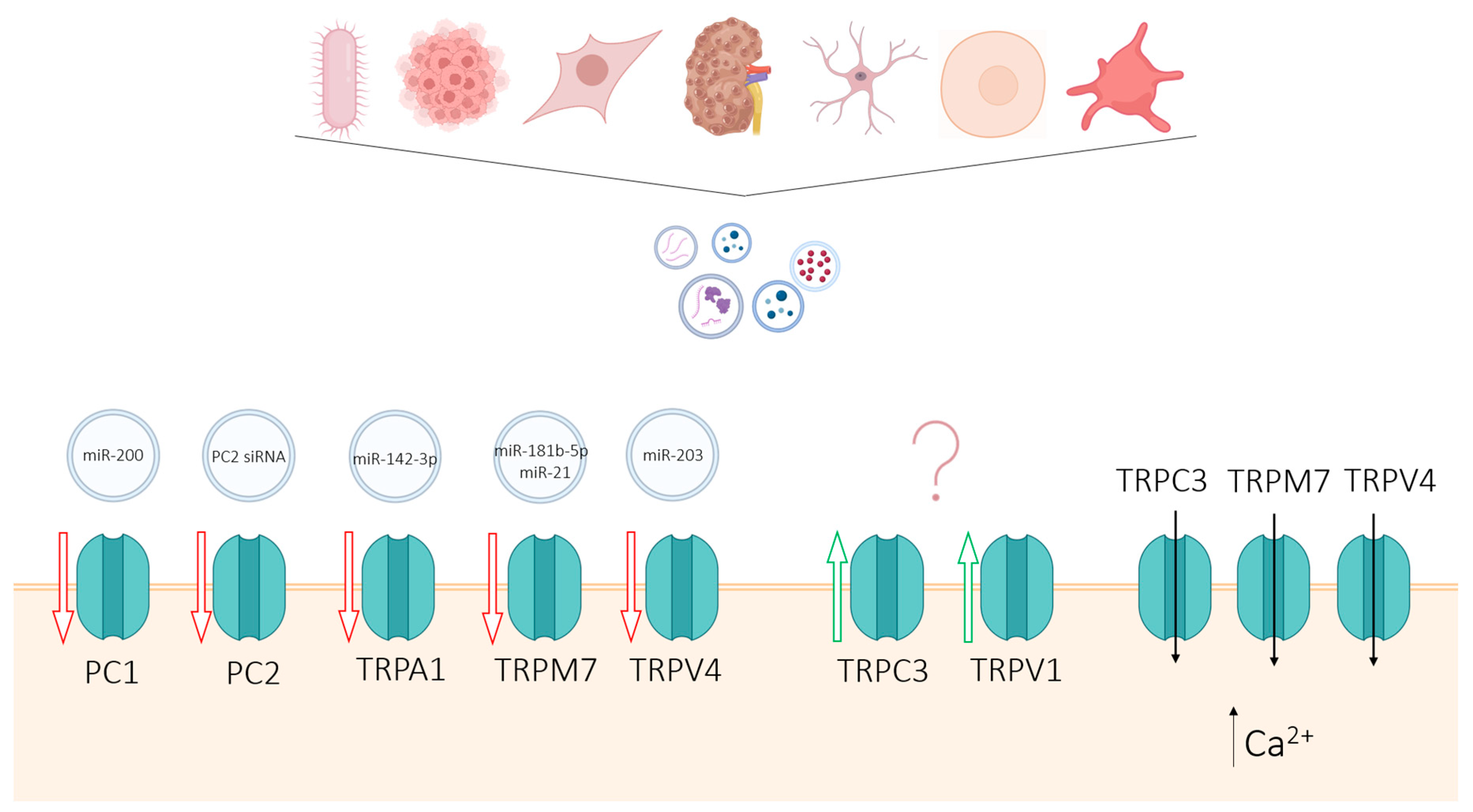
Figure 7.
Modulation of TRP channel activity and expression by various EVs in the recipient cells. EVs released by different cell types/tissues/organs can trigger a series of effects in target cells: down-regulation of TRP channel expression (PC1, PC2, TRPA1, TRPM7, TRPV4), up-regulation of TRP channel expression (TRPC3, TRPV1), and TRP channel activation (TRPC3, TRPM7, TRPV4). It is believed that in some cases these effects are mediated by specific microRNAs transported as cargo by the EVs, but further research is needed to fully understand the underlying mechanisms of the modulation of TRP channel activity and expression by EVs.
Figure 7.
Modulation of TRP channel activity and expression by various EVs in the recipient cells. EVs released by different cell types/tissues/organs can trigger a series of effects in target cells: down-regulation of TRP channel expression (PC1, PC2, TRPA1, TRPM7, TRPV4), up-regulation of TRP channel expression (TRPC3, TRPV1), and TRP channel activation (TRPC3, TRPM7, TRPV4). It is believed that in some cases these effects are mediated by specific microRNAs transported as cargo by the EVs, but further research is needed to fully understand the underlying mechanisms of the modulation of TRP channel activity and expression by EVs.
5. Concluding Remarks
Although extracellular vesicles and TRP channels are both extensively researched topics of great interest, the literature on the interplay between the two is not yet very broad. The interactions between EVs and TRPs can be split into three major domains: the role of TRP channels in EVs release, TRP channel expression in EVs, and, lastly, the role of EVs in the modulation of TRP channel activity and expression.
The biogenesis of EVs is a complex process, involving multiple subcellular signaling pathways based on the release modality of the EVs (exosomes vs. microvesicles) and being highly dependent on the current state of the cell. EV release is enhanced under specific conditions, in particular under pathological circumstances. One of the roles of EVs is removing harmful or unneeded elements (i.e., viruses, bacterial fragments, cellular waste) from the cells. Exosomes are generated within the endosomal pathway upon the fusion of early endosomes with a lysosome. The low pH of the lysosome allows the components of the endosome to be partially or fully degraded. Although not the only proteins involved in the process, TRPML channels in the lysosomal membrane (together with H+-ATPases) play a major role in maintaining lysosomal function. Dysregulation of lysosomal TRPML activity results in elevated lysosomal pH values, hindering the fusion of MVBs with the lysosome, resulting in the release of ILVs as exosomes. However, most TRP channels are expressed in the plasma membrane, where their activation by various agonists results in Ca2+ influx. EV shedding is a Ca2+-dependent process and elevated local intracellular Ca2+ levels can trigger the fusion of multivesicular bodies with the plasma membrane (in the case of exosome release) and/or plasma membrane budding (in the case of microvesicle release). Although the exact molecular mechanisms have not yet been elucidated, EV release has been shown to be influenced by the activity of TRPV1, TRPM1, TRPM8, and TRPC6 due to Ca2+ influx and cell depolarization mediated by these channels.
Extracellular vesicles are of paramount importance in intercellular signaling, as they transfer various cargo, such as lipids, nucleic acids, and proteins (both in the lumen, as well as on the membrane) between the donor and the recipient cells. Several studies have identified TRP channels to be delivered among cells via EVs. Upon internalization by recipient cells, TRP channel expression levels can be upregulated, leading to altered cellular signaling and phenotypic changes. Because TRP channel expression can be altered in certain diseases, their expression in EVs isolated from various biological fluids (blood, urine, semen) can be used as disease markers, allowing for a less invasive diagnosis. Among the TRP channels identified in EVs are members of the TRPA, TRPC, TRPV, and TRPP family.
Besides proteins, EVs contain nucleic acids in the form of non-coding, microRNAs or siRNAs. microRNAs and siRNAs are short strands of RNA that act as post-transcriptional gene expression regulators. Delivery of non-coding (mostly micro) RNA to target cells via EVs results in altered expression of various proteins, including TRP channels. While some studies explain the TRP channel down-regulation by linking specific miRNAs carried by EVs to the observed effects, in some cases the altered expression levels are only hypothetically linked to the transfer of miRNA. Hence, TRP channel expression levels in recipient cells can be both up-regulated and down-regulated by EV uptake, via direct protein delivery to the target cell or transfer of miRNA. EVs do not only alter the expression levels of TRP channels in target cells but they have also been shown to modulate their activity. EV delivery has been shown to induce TRPM7- and TRPV4-mediated Ca2+ signaling in human microvascular endothelial cells and vascular smooth muscle cells.
The incipient body of literature related to the interplay between extracellular vesicles and TRP channels shows growing interest in the topic. More research is needed to understand how EVs and TRPs influence each other and what role this interaction plays in the big scheme of physiological and pathophysiological signaling.
More in-depth studies are required to elucidate how EVs and TRP channels regulate each other and what are the implications therein. Understanding this cross-talk may facilitate understanding the role of EVs in TRP-channel mediated diseases and vice versa, discovering better markers for various pathologies and developing new and more efficient therapies against disease.
Author Contributions
Conceptualization, A. Milici and K. Talavera; Funding Acquisition, K. Talavera; Investigation, A. Milici and K. Talavera; Project Administration, K. Talavera; Supervision, K. Talavera; Writing-original draft, A Milici; Writing-review and editing, A Milici and K. Talavera.
Acknowledgments
This work was supported by a grants of the Research Foundation Flanders FWO (G0AAL24N). The authors declare no competing financial interests.
References
- G. van Niel, G. D’Angelo, and G. Raposo, ‘Shedding light on the cell biology of extracellular vesicles’, Nat Rev Mol Cell Biol, vol. 19, no. 4, pp. 213–228, Apr. 2018. [CrossRef] [PubMed]
- N. P. Hessvik and A. Llorente, ‘Current knowledge on exosome biogenesis and release’, Cellular and Molecular Life Sciences, vol. 75, no. 2, p. 193, Jan. 2018. [CrossRef] [PubMed]
- C. Villarroya-Beltri, F. Baixauli, C. Gutiérrez-Vázquez, F. Sánchez-Madrid, and M. Mittelbrunn, ‘SORTING IT OUT: REGULATION OF EXOSOME LOADING’. [CrossRef] [PubMed]
- E. van der Pol, A. N. Böing, P. Harrison, A. Sturk, and R. Nieuwland, ‘Classification, Functions, and Clinical Relevance of Extracellular Vesicles’, Pharmacol Rev, vol. 64, no. 3, pp. 676–705, Jul. 2012. [CrossRef] [PubMed]
- C. D. Gregory and I. Dransfield, ‘Apoptotic Tumor Cell-Derived Extracellular Vesicles as Important Regulators of the Onco-Regenerative Niche’, Front Immunol, vol. 9, no. MAY, p. 1111, May 2018. [CrossRef] [PubMed]
- P. A. Leventis and S. Grinstein, ‘The distribution and function of phosphatidylserine in cellular membranes’, Annu Rev Biophys, vol. 39, no. 1, pp. 407–427, Jun. 2010. [CrossRef] [PubMed]
- G. van Niel and G. Raposo, ‘Shedding light on the cell biology of extracellular vesicles’, 2018. [CrossRef] [PubMed]
- C. Théry et al., ‘Minimal information for studies of extracellular vesicles 2018 (MISEV2018): a position statement of the International Society for Extracellular Vesicles and update of the MISEV2014 guidelines’, J Extracell Vesicles, vol. 7, no. 1, Jan. 2018. [CrossRef] [PubMed]
- G. W. L. Eng, Y. Zheng, D. W. T. Yap, A. Y. T. Teo, and J. K. Cheong, ‘Autophagy and ncRNAs: Dangerous Liaisons in the Crosstalk between the Tumor and Its Microenvironment’, Cancers (Basel), vol. 14, no. 1, Jan. 2021. [CrossRef] [PubMed]
- A. U. Andar, R. R. Hood, W. N. Vreeland, D. L. Devoe, and P. W. Swaan, ‘Microfluidic preparation of liposomes to determine particle size influence on cellular uptake mechanisms’, Pharm Res, vol. 31, no. 2, pp. 401–413, Feb. 2014. [CrossRef] [PubMed]
- L. A. Mulcahy, R. C. Pink, and D. R. F. Carter, ‘Routes and mechanisms of extracellular vesicle uptake’, J Extracell Vesicles, vol. 3, no. 1, 2014. [CrossRef] [PubMed]
- X. B. Li, Z. R. Zhang, H. J. Schluesener, and S. Q. Xu, ‘Role of exosomes in immune regulation’, J Cell Mol Med, vol. 10, no. 2, pp. 364–375, 2006. [CrossRef] [PubMed]
- M. Logozzi et al., ‘High Levels of Exosomes Expressing CD63 and Caveolin-1 in Plasma of Melanoma Patients’, PLoS One, vol. 4, no. 4, Apr. 2009. [CrossRef] [PubMed]
- B. Guarino et al., ‘Tumor-Derived Extracellular Vesicles Induce Abnormal Angiogenesis via TRPV4 Downregulation and Subsequent Activation of YAP and VEGFR2’, Front Bioeng Biotechnol, vol. 9, Dec. 2021. [CrossRef] [PubMed]
- J. E. Park et al., ‘Hypoxic tumor cell modulates its microenvironment to enhance angiogenic and metastatic potential by secretion of proteins and exosomes’, Mol Cell Proteomics, vol. 9, no. 6, pp. 1085–1099, Jun. 2010. [CrossRef] [PubMed]
- M. Record, K. Carayon, M. Poirot, and S. Silvente-Poirot, ‘Exosomes as new vesicular lipid transporters involved in cell–cell communication and various pathophysiologies’, Biochimica et Biophysica Acta (BBA) - Molecular and Cell Biology of Lipids, vol. 1841, no. 1, pp. 108–120, Jan. 2014. [CrossRef] [PubMed]
- P. Kharaziha, S. Ceder, Q. Li, and T. Panaretakis, ‘Tumor cell-derived exosomes: a message in a bottle’, Biochim Biophys Acta, vol. 1826, no. 1, pp. 103–111, Aug. 2012. [CrossRef] [PubMed]
- V. Zappulli, C. A. Maguire, and X. O. Breakefield, ‘Extracellular vesicles and intercellular communication within the nervous system The Journal of Clinical Investigation’, J Clin Invest, vol. 126, no. 4, pp. 1198–1207, 2016. [CrossRef] [PubMed]
- A. F. Hill, ‘Extracellular Vesicles and Neurodegenerative Diseases’, J Neurosci, vol. 39, no. 47, pp. 9269–9273, Nov. 2019. [CrossRef] [PubMed]
- S. M. Davidson et al., ‘Circulating blood cells and extracellular vesicles in acute cardioprotection’, Cardiovasc Res, vol. 115, no. 7, pp. 1156–1166, Jun. 2019. [CrossRef] [PubMed]
- A. Becker, B. K. Thakur, J. M. Weiss, H. S. Kim, H. Peinado, and D. Lyden, ‘Extracellular Vesicles in Cancer: Cell-to-Cell Mediators of Metastasis’, Cancer Cell, vol. 30, no. 6, pp. 836–848, Dec. 2016. [CrossRef] [PubMed]
- M. C. Hogan et al., ‘Characterization of PKD protein-positive exosome-like vesicles’, J Am Soc Nephrol, vol. 20, no. 2, pp. 278–288, Feb. 2009. [CrossRef] [PubMed]
- J. P. Decuypere et al., ‘Interdependent regulation of polycystin expression influences starvation-induced autophagy and cell death’, Int J Mol Sci, vol. 22, no. 24, p. 22, Dec. 2021. [CrossRef]
- M. Freichel et al., ‘TRP Channels in the Heart’, Neurobiology of TRP Channels, pp. 149–185, Jun. 2017. [CrossRef] [PubMed]
- Y. A. Alpizar et al., ‘TRPV4 activation triggers protective responses to bacterial lipopolysaccharides in airway epithelial cells’, Nat Commun, vol. 8, no. 1, 2017. [CrossRef] [PubMed]
- Y. Karashima et al., ‘TRPA1 acts as a cold sensor in vitro and in vivo’, Proc Natl Acad Sci U S A, vol. 106, no. 4, p. 1273, Jan. 2009. [CrossRef] [PubMed]
- Vandewauw et al., ‘A TRP channel trio mediates acute noxious heat sensing’, Nature, vol. 555, no. 7698, pp. 662–666, Mar. 2018. [CrossRef] [PubMed]
- V. Flockerzi and B. Nilius, ‘TRPs: Truly remarkable proteins’, Handb Exp Pharmacol, vol. 222, 2014. [CrossRef] [PubMed]
- Fliniaux, E. Germain, V. Farfariello, and N. Prevarskaya, ‘TRPs and Ca2+ in cell death and survival’, Cell Calcium, vol. 69, pp. 4–18, Jan. 2018. [CrossRef] [PubMed]
- X. Zhang, M. Hu, Y. Yang, and H. Xu, ‘Organellar TRP channels’, Nat Struct Mol Biol, vol. 25, no. 11, pp. 1009–1018, Nov. 2018. [CrossRef] [PubMed]
- C. Harteneck, ‘Pregnenolone Sulfate: From Steroid Metabolite to TRP Channel Ligand’, Molecules, vol. 18, no. 10, p. 12012, Oct. 2013. [CrossRef] [PubMed]
- S. Cao et al., ‘Transient receptor potential vanilloid 4 (TRPV4) activation by arachidonic acid requires protein kinase A-mediated phosphorylation’, J Biol Chem, vol. 293, no. 14, pp. 5307–5322, Apr. 2018. [CrossRef] [PubMed]
- C. Muller, P. Morales, and P. H. Reggio, ‘Cannabinoid Ligands Targeting TRP Channels’, Front Mol Neurosci, vol. 11, Jan. 2018. [CrossRef] [PubMed]
- E. Andrè et al., ‘Cigarette smoke–induced neurogenic inflammation is mediated by α,β-unsaturated aldehydes and the TRPA1 receptor in rodents’, Journal of Clinical Investigation, Jun. 2008. [CrossRef] [PubMed]
- M. J. Caterina, M. A. Schumacher, M. Tominaga, T. A. Rosen, J. D. Levine, and D. Julius, ‘The capsaicin receptor: a heat-activated ion channel in the pain pathway’, Nature, vol. 389, no. 6653, pp. 816–824, 1997. [CrossRef] [PubMed]
- Y. A. Alpizar et al., ‘TRPV4 Mediates Acute Bladder Responses to Bacterial Lipopolysaccharides’, Front Immunol, vol. 11, 2020. [CrossRef] [PubMed]
- B. Boonen et al., ‘Activation of Drosophila melanogaster TRPA1 Isoforms by Citronellal and Menthol’, Int J Mol Sci, vol. 22, no. 20, Oct. 2021. [CrossRef] [PubMed]
- H. Yamamura, S. Ugawa, T. Ueda, A. Morita, and S. Shimada, ‘TRPM8 activation suppresses cellular viability in human melanoma’, Am J Physiol Cell Physiol, vol. 295, no. 2, Aug. 2008. [CrossRef] [PubMed]
- G. M. Story et al., ‘ANKTM1, a TRP-like channel expressed in nociceptive neurons, is activated by cold temperatures’, Cell, vol. 112, no. 6, pp. 819–829, Mar. 2003. [CrossRef] [PubMed]
- A. P. Christensen and D. P. Corey, ‘TRP channels in mechanosensation: direct or indirect activation?’, Nat Rev Neurosci, vol. 8, no. 7, pp. 510–521, Jul. 2007. [CrossRef] [PubMed]
- B. Startek, B. Boonen, K. Talavera, and V. Meseguer, ‘TRP Channels as Sensors of Chemically-Induced Changes in Cell Membrane Mechanical Properties’, Int J Mol Sci, vol. 20, no. 2, Jan. 2019. [CrossRef] [PubMed]
- C. Liu and C. Montell, ‘Forcing Open TRP channels: mechanical gating as a unifying activation mechanism’, 2015. [CrossRef] [PubMed]
- M. C. Marrone et al., ‘TRPV1 channels are critical brain inflammation detectors and neuropathic pain biomarkers in mice’, Nat Commun, vol. 8, 2017. [CrossRef] [PubMed]
- R. Simeoli et al., ‘Exosomal cargo including microRNA regulates sensory neuron to macrophage communication after nerve trauma’, Nat Commun, vol. 8, no. 1, Dec. 2017. [CrossRef] [PubMed]
- A. Z. C. Rodrigues, Z. M. Wang, M. L. Messi, and O. Delbono, ‘Sympathomimetics regulate neuromuscular junction transmission through TRPV1, P/Q- and N-type Ca 2+ channels’, Mol Cell Neurosci, vol. 95, pp. 59–70, Mar. 2019. [CrossRef] [PubMed]
- M. S. Kim, S. Muallem, S. H. Kim, K. B. Kwon, and M. S. Kim, ‘Exosomal release through TRPML1-mediated lysosomal exocytosis is required for adipogenesis’, Biochem Biophys Res Commun, vol. 510, no. 3, pp. 409–415, Mar. 2019. [CrossRef] [PubMed]
- G. Li, D. Huang, N. Li, J. K. Ritter, and P. L. Li, ‘Regulation of TRPML1 channel activity and inflammatory exosome release by endogenously produced reactive oxygen species in mouse podocytes’, Redox Biol, vol. 43, Jul. 2021. [CrossRef] [PubMed]
- García-Añoveros and T. Wiwatpanit, ‘TRPML2 and mucolipin evolution’, Handb Exp Pharmacol, vol. 222, pp. 647–658, 2014. [CrossRef] [PubMed]
- Y. Miao, G. Li, X. Zhang, H. Xu, and S. N. Abraham, ‘A TRP Channel Senses Lysosome Neutralization by Pathogens to Trigger Their Expulsion’, Cell, vol. 161, no. 6, pp. 1306–1319, Jun. 2015. [CrossRef] [PubMed]
- D. Thuringer, G. Chanteloup, P. Winckler, and C. Garrido, ‘The vesicular transfer of CLIC1 from glioblastoma to microvascular endothelial cells requires TRPM7’, Oncotarget, vol. 9, no. 70, pp. 33302–33311, Sep. 2018. [CrossRef] [PubMed]
- S. S. Li et al., ‘Platelet-derived microvesicles induce calcium oscillations and promote VSMC migration via TRPV4’, Theranostics, vol. 11, no. 5, pp. 2410–2423, Jan. 2021. [CrossRef] [PubMed]
- H. Ding, L. X. Li, P. C. Harris, J. Yang, and X. Li, ‘Extracellular vesicles and exosomes generated from cystic renal epithelial cells promote cyst growth in autosomal dominant polycystic kidney disease’. [CrossRef] [PubMed]
- Y. Dong et al., ‘Tumor endothelial expression of P-glycoprotein upon microvesicular transfer of TrpC5 derived from adriamycin-resistant breast cancer cells’, Biochem Biophys Res Commun, vol. 446, no. 1, pp. 85–90, Mar. 2014. [CrossRef] [PubMed]
- P. Trnka, L. Ivanova, M. J. Hiatt, and D. G. Matsell, ‘Urinary biomarkers in obstructive nephropathy’, Clin J Am Soc Nephrol, vol. 7, no. 10, pp. 1567–1575, Oct. 2012. [CrossRef] [PubMed]
- W. A. Lea et al., ‘Analysis of the polycystin complex (PCC) in human urinary exosome-like vesicles (ELVs)’, Sci Rep, vol. 10, no. 1, Dec. 2020. [CrossRef] [PubMed]
- C. Hogan et al., ‘Subfractionation, characterization, and in-depth proteomic analysis of glomerular membrane vesicles in human urine’, Kidney Int, vol. 85, no. 5, pp. 1225–1237, 2014. [CrossRef] [PubMed]
- F. Kurth et al., ‘Cell-Derived Vesicles as TRPC1 Channel Delivery Systems for the Recovery of Cellular Respiratory and Proliferative Capacities’, Adv Biosyst, vol. 4, no. 11, p. 2000146, Nov. 2020. [CrossRef] [PubMed]
- C. Hogan et al., ‘Identification of Biomarkers for PKD1 Using Urinary Exosomes’, J Am Soc Nephrol, vol. 26, no. 7, pp. 1661–1670, Jul. 2015. [CrossRef] [PubMed]
- Taylor, I. Azimi, G. Monteith, and M. Bebawy, ‘Ca 2+ mediates extracellular vesicle biogenesis through alternate pathways in malignancy’, J Extracell Vesicles, vol. 9, no. 1, Jan. 2020. [CrossRef] [PubMed]
- G. Li, D. Huang, J. Hong, O. M. Bhat, X. Yuan, and P. L. Li, ‘Control of lysosomal TRPML1 channel activity and exosome release by acid ceramidase in mouse podocytes’, Am J Physiol Cell Physiol, vol. 317, no. 3, pp. C481–C494, 2019. [CrossRef] [PubMed]
- G. Li and P.-L. Li, ‘Lysosomal TRPML1 Channel: Implications in Cardiovascular and Kidney Diseases’, Adv Exp Med Biol, vol. 1349, pp. 275–301, 2021. [CrossRef] [PubMed]
- G. Li et al., ‘Abnormal podocyte TRPML1 channel activity and exosome release in mice with podocyte-specific Asah1 gene deletion’, Biochim Biophys Acta Mol Cell Biol Lipids, vol. 1866, no. 2, Feb. 2021. [CrossRef] [PubMed]
- O. M. Bhat et al., ‘Arterial Medial Calcification through Enhanced small Extracellular Vesicle Release in Smooth Muscle-Specific Asah1 Gene Knockout Mice’, Sci Rep, vol. 10, no. 1, Dec. 2020. [CrossRef] [PubMed]
- Huang et al., ‘Berbamine inhibits Japanese encephalitis virus (JEV) infection by compromising TPRMLs-mediated endolysosomal trafficking of low-density lipoprotein receptor (LDLR)’, Emerg Microbes Infect, vol. 10, no. 1, pp. 1257–1271, 2021. [CrossRef] [PubMed]
- S. Kim, S. H. Yang, and M. S. Kim, ‘TRPML3 enhances drug resistance in non-small cell lung cancer cells by promoting Ca2+-mediated lysosomal trafficking’, Biochem Biophys Res Commun, vol. 627, pp. 152–159, Oct. 2022. [CrossRef] [PubMed]
- H. Shaheen, S. Singh, and R. Melnik, ‘A Neuron-Glial Model of Exosomal Release in the Onset and Progression of Alzheimer’s Disease’, Front Comput Neurosci, vol. 15, Sep. 2021. [CrossRef] [PubMed]
- H. Davies et al., ‘Mutations of the BRAF gene in human cancer’, Nature, vol. 417, no. 6892, pp. 949–954, Jun. 2002. [CrossRef] [PubMed]
- T. R. Lunavat et al., ‘BRAF V600 inhibition alters the microRNA cargo in the vesicular secretome of malignant melanoma cells’, Proc Natl Acad Sci U S A, vol. 114, no. 29, pp. E5930–E5939, Jul. 2017. [CrossRef] [PubMed]
- E. Houben, A. Neradova, L. J. Schurgers, and M. Vervloet, ‘The influence of phosphate, calcium and magnesium on matrix Gla-protein and vascular calcification: a systematic review’, G Ital Nefrol, vol. 33, no. 6, Nov. 2016.
- E. Lopez et al., ‘Relationship between calcium mobilization and platelet α- and δ-granule secretion. A role for TRPC6 in thrombin-evoked δ-granule exocytosis’, Arch Biochem Biophys, vol. 585, pp. 75–81, Nov. 2015. [CrossRef] [PubMed]
- C. J. K. Wong et al., ‘Brief exposure to directionally-specific pulsed electromagnetic fields stimulates extracellular vesicle release and is antagonized by streptomycin: A potential regenerative medicine and food industry paradigm’, Biomaterials, vol. 287, p. 121658, Aug. 2022. [CrossRef] [PubMed]
- Peintner et al., ‘Loss of PKD1/polycystin-1 impairs lysosomal activity in a CAPN (calpain)-dependent manner’, Autophagy, vol. 17, no. 9, pp. 2384–2400, Sep. 2021. [CrossRef] [PubMed]
- Y. Lin et al., ‘Proteomic analysis of seminal extracellular vesicle proteins involved in asthenozoospermia by iTRAQ’, Mol Reprod Dev, vol. 86, no. 9, pp. 1094–1105, Sep. 2019. [CrossRef] [PubMed]
- J. Hu and P. C. Harris, ‘Regulation of polycystin expression, maturation and trafficking’, Cell Signal, vol. 72, Aug. 2020. [CrossRef] [PubMed]
- J. Wang et al., ‘C. elegans ciliated sensory neurons release extracellular vesicles that function in animal communication’, Curr Biol, vol. 24, no. 5, pp. 519–525, Mar. 2014. [CrossRef] [PubMed]
- J. D. Walsh, O. Boivin, and M. M. Barr, ‘What about the males? the C. elegans sexually dimorphic nervous system and a CRISPR-based tool to study males in a hermaphroditic species’, J Neurogenet, vol. 34, no. 3–4, pp. 1–12, 2020. [CrossRef] [PubMed]
- J. Wang, I. A. Nikonorova, A. Gu, P. W. Sternberg, and M. M. Barr, ‘Release and targeting of polycystin-2-carrying ciliary extracellular vesicles’, Curr Biol, vol. 30, no. 13, pp. R755–R756, Jul. 2020. [CrossRef] [PubMed]
- R. O’Hagan, J. Wang, and M. M. Barr, ‘Mating behavior, male sensory cilia, and polycystins in Caenorhabditis elegans’, Semin Cell Dev Biol, vol. 33, pp. 25–33, 2014. [CrossRef] [PubMed]
- J. E. Maguire et al., ‘Myristoylated CIL-7 regulates ciliary extracellular vesicle biogenesis’, Mol Biol Cell, vol. 26, no. 15, pp. 2823–2832, Aug. 2015. [CrossRef] [PubMed]
- H. J. Gaunt, N. S. Vasudev, and D. J. Beech, ‘Transient receptor potential canonical 4 and 5 proteins as targets in cancer therapeutics’, Eur Biophys J, vol. 45, no. 7, pp. 611–620, Oct. 2016. [CrossRef] [PubMed]
- T. Wang et al., ‘Increasing circulating exosomes-carrying TRPC5 predicts chemoresistance in metastatic breast cancer patients’, Cancer Sci, vol. 108, no. 3, pp. 448–454, Mar. 2017. [CrossRef] [PubMed]
- Wang et al., ‘Effect of exosome biomarkers for diagnosis and prognosis of breast cancer patients’, Clin Transl Oncol, vol. 20, no. 7, pp. 906–911, Jul. 2018. [CrossRef] [PubMed]
- X. Ma et al., ‘Essential role for TrpC5-containing extracellular vesicles in breast cancer with chemotherapeutic resistance’, Proc Natl Acad Sci U S A, vol. 111, no. 17, pp. 6389–6394, Apr. 2014. [CrossRef] [PubMed]
- T. Wang et al., ‘Inhibition of Transient Receptor Potential Channel 5 Reverses 5-Fluorouracil Resistance in Human Colorectal Cancer Cells *’, 2014. [CrossRef] [PubMed]
- E. J. Goetzl, V. H. Srihari, M. Mustapic, D. Kapogiannis, and G. R. Heninger, ‘Abnormal levels of mitochondrial Ca 2+ channel proteins in plasma neuron-derived extracellular vesicles of early schizophrenia’, The FASEB Journal, vol. 36, no. 8, Aug. 2022. [CrossRef] [PubMed]
- C. Wang et al., ‘Exosome-delivered TRPP2 siRNA inhibits the epithelial-mesenchymal transition of FaDu cells’, Oncol Lett, vol. 17, no. 2, pp. 1953–1961, Feb. 2019. [CrossRef] [PubMed]
- J. Berrout et al., ‘TRPA1-FGFR2 binding event is a regulatory oncogenic driver modulated by miRNA-142-3p’, Nat Commun, vol. 8, no. 1, Dec. 2017. [CrossRef] [PubMed]
- S. Y. Kim et al., ‘Mesenchymal Stem Cells and Extracellular Vesicles Derived from Canine Adipose Tissue Ameliorates Inflammation, Skin Barrier Function and Pruritus by Reducing JAK/STAT Signaling in Atopic Dermatitis’, Int J Mol Sci, vol. 23, no. 9, p. 4868, May 2022. [CrossRef] [PubMed]
- Y. Yang, Y. Cai, Y. Zhang, J. Liu, and Z. Xu, ‘Exosomes Secreted by Adipose-Derived Stem Cells Contribute to Angiogenesis of Brain Microvascular Endothelial Cells Following Oxygen-Glucose Deprivation In Vitro Through MicroRNA-181b/TRPM7 Axis’, J Mol Neurosci, vol. 65, no. 1, pp. 74–83, May 2018. [CrossRef] [PubMed]
- H. Zhang, Y. Wang, Q. Lv, J. Gao, L. Hu, and Z. He, ‘MicroRNA-21 Overexpression Promotes the Neuroprotective Efficacy of Mesenchymal Stem Cells for Treatment of Intracerebral Hemorrhage’, Front Neurol, vol. 9, no. NOV, Nov. 2018. [CrossRef] [PubMed]
- K. Narayanan et al., ‘Extracellular Vesicles From Pathological Microenvironment Induce Endothelial Cell Transformation and Abnormal Angiogenesis via Modulation of TRPV4 Channels’, 2019. [CrossRef] [PubMed]
- Q. Yu et al., ‘Adipose-derived exosomes protect the pulmonary endothelial barrier in ventilator-induced lung injury by inhibiting the TRPV4/Ca 2+ signaling pathway’, Am J Physiol Lung Cell Mol Physiol, vol. 318, no. 4, pp. L723–L741, Apr. 2020. [CrossRef] [PubMed]
- Q. Yu et al., ‘Protective effects of adipose-derived biogenic nanoparticles on the pulmonary microvascular endothelial barrier in mice with ventilator-induced lung injury via the TRPV4/ROCK1 signalling pathway’, Pulm Pharmacol Ther, vol. 73–74, p. 102123, Jun. 2022. [CrossRef] [PubMed]
- Z. Jiang et al., ‘Cannabidiol Inhibits Inflammation Induced by Cutibacterium acnes-Derived Extracellular Vesicles via Activation of CB2 Receptor in Keratinocytes’, J Inflamm Res, vol. Volume 15, pp. 4573–4583, Aug. 2022. [CrossRef] [PubMed]
- C. Xue et al., ‘Mesenchymal stem cells derived from adipose accelerate the progression of colon cancer by inducing a MT-CAFs phenotype via TRPC3/NF-KB axis’, Stem Cell Res Ther, vol. 13, no. 1, p. 335, Dec. 2022. [CrossRef] [PubMed]
Figure 1.
Uptake pathways involved in EV internalization by recipient cells [11].
Figure 1.
Uptake pathways involved in EV internalization by recipient cells [11].
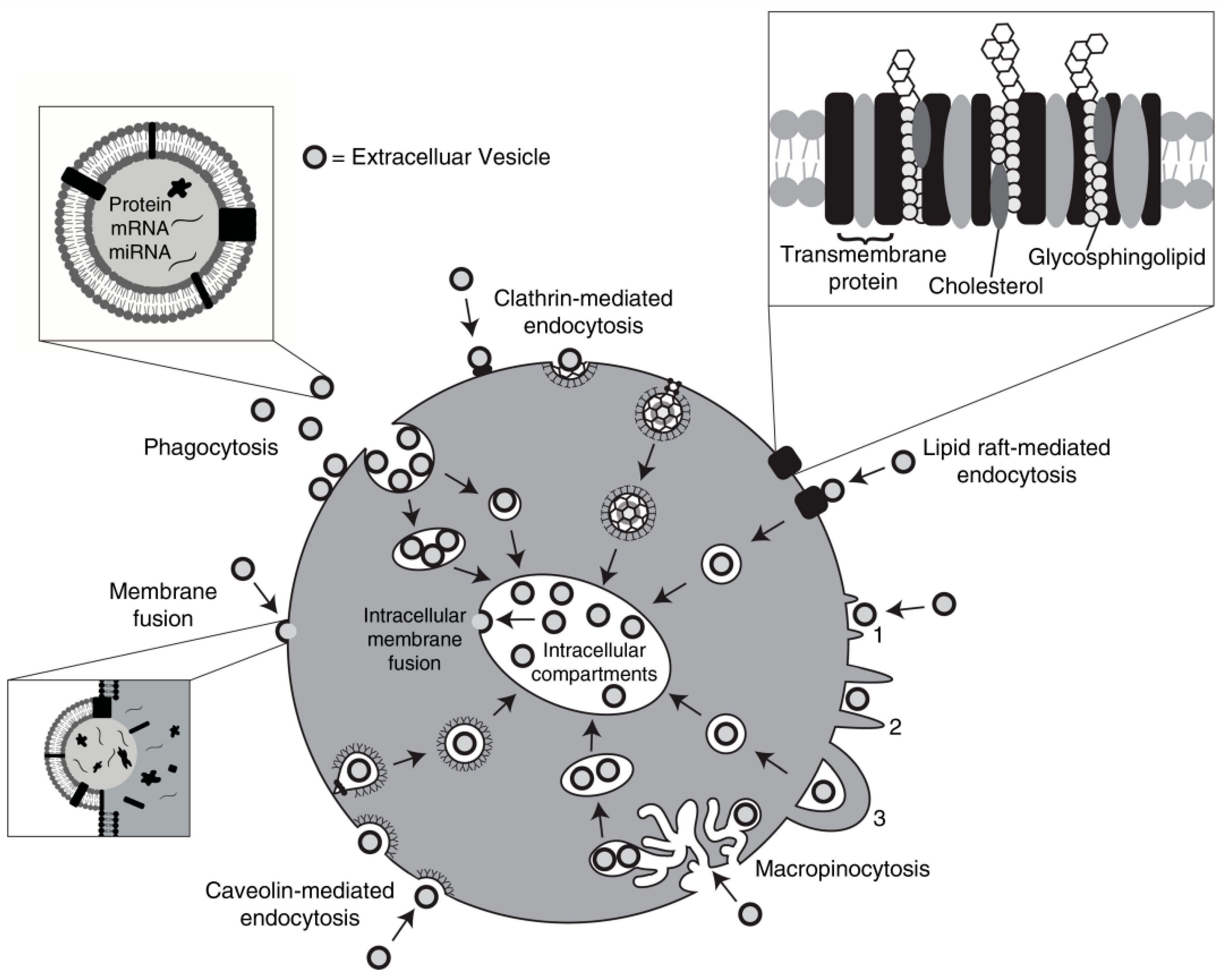
Figure 2.
Bidirectional EV exchange between neurons and macrophages. Microglial EVs modulate the postsynaptic current frequency in neurons, while EVs released by neurons induce a pro-inflammatory phenotype in macrophages upon nerve injury.
Figure 2.
Bidirectional EV exchange between neurons and macrophages. Microglial EVs modulate the postsynaptic current frequency in neurons, while EVs released by neurons induce a pro-inflammatory phenotype in macrophages upon nerve injury.
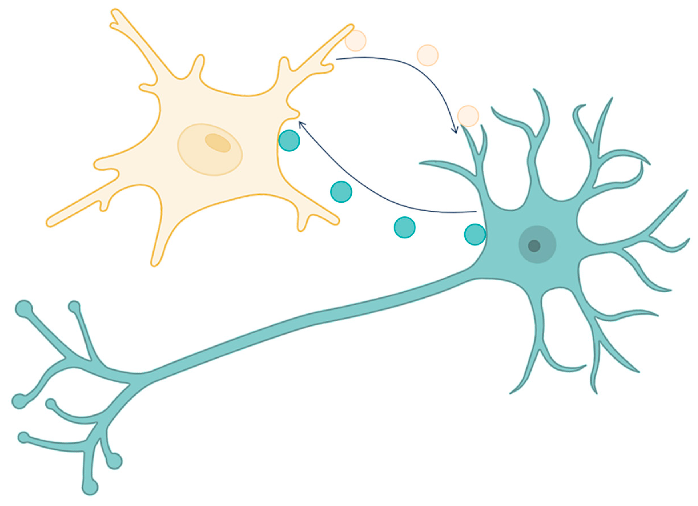
Figure 3.
Lysosomal TRPML channels play a role in maintaining lysosomal acidification for correct lysosomal function. TRPML1 activation by acidic pH triggers the fusion of MVBs with lysosomes, resulting in reduced exosome release. Increased levels of acid ceramidase promote TRPML1 activation via the degradation of ceramide to sphingosine. Inhibition of acid ceramidase (or of TRPML1 directly) hinders the fusion of MVBs with lysosomes, preventing their degradation and resulting in increased EV release. The endocytosis of E. coli and the anti-tumor drug Gefitinib leads to TRPML3 activation via neutralization of lysosomal pH. TRPML3 activation results in the fusion of the lysosome with the plasma membrane and lysosomal exocytosis of the cargo. Uptake of Gefitinib-containing EVs by neighboring cells induces drug resistance.
Figure 3.
Lysosomal TRPML channels play a role in maintaining lysosomal acidification for correct lysosomal function. TRPML1 activation by acidic pH triggers the fusion of MVBs with lysosomes, resulting in reduced exosome release. Increased levels of acid ceramidase promote TRPML1 activation via the degradation of ceramide to sphingosine. Inhibition of acid ceramidase (or of TRPML1 directly) hinders the fusion of MVBs with lysosomes, preventing their degradation and resulting in increased EV release. The endocytosis of E. coli and the anti-tumor drug Gefitinib leads to TRPML3 activation via neutralization of lysosomal pH. TRPML3 activation results in the fusion of the lysosome with the plasma membrane and lysosomal exocytosis of the cargo. Uptake of Gefitinib-containing EVs by neighboring cells induces drug resistance.
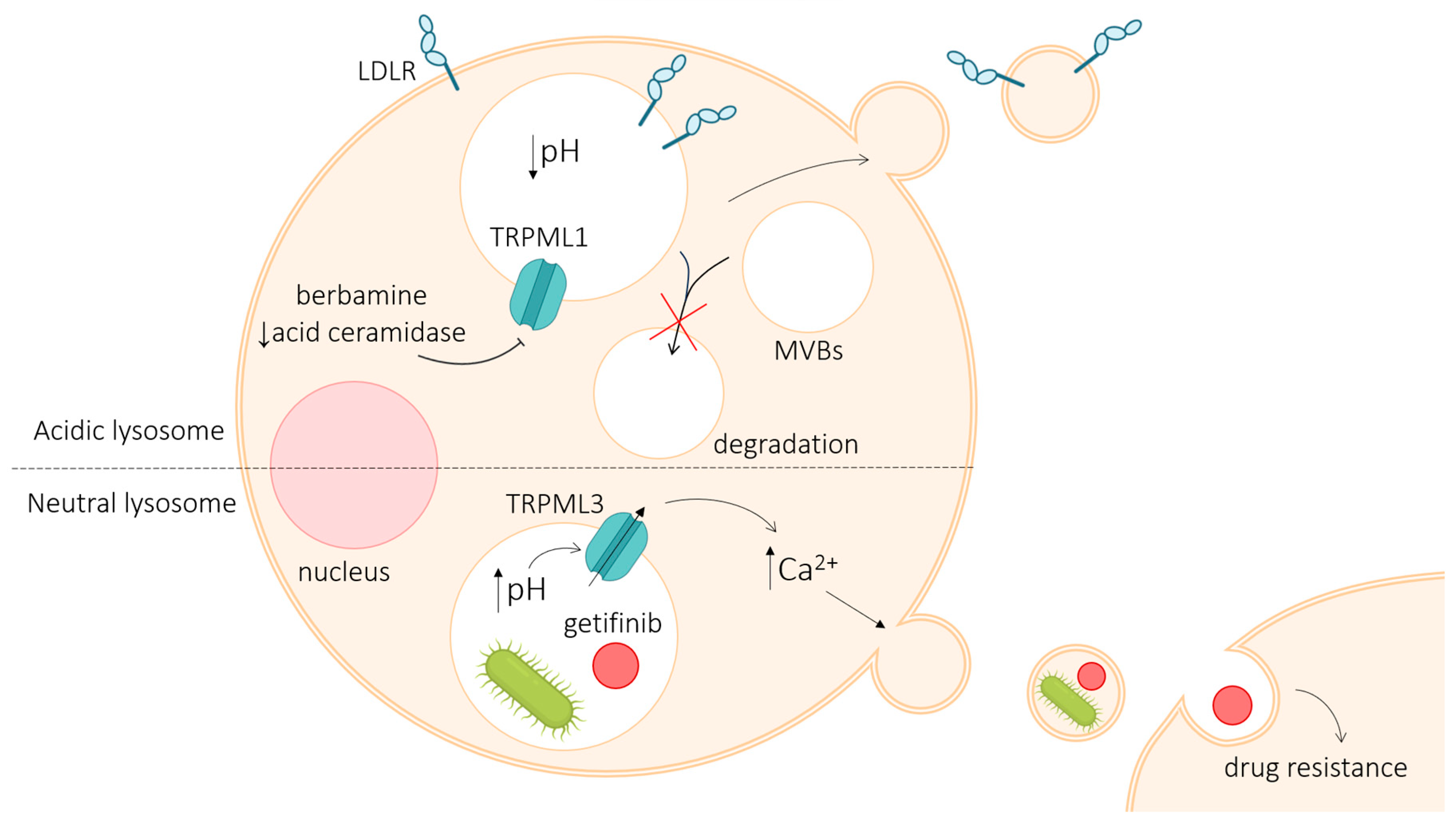
Figure 4.
Involvement of different TRP channels in EV release. Activation and/or up-regulation of TRP channel expression in target cells leads to elevated intracellular Ca2+ levels (Ca2+ influx or mobilization from intracellular stores), promoting the release of EVs from mother cells.
Figure 4.
Involvement of different TRP channels in EV release. Activation and/or up-regulation of TRP channel expression in target cells leads to elevated intracellular Ca2+ levels (Ca2+ influx or mobilization from intracellular stores), promoting the release of EVs from mother cells.
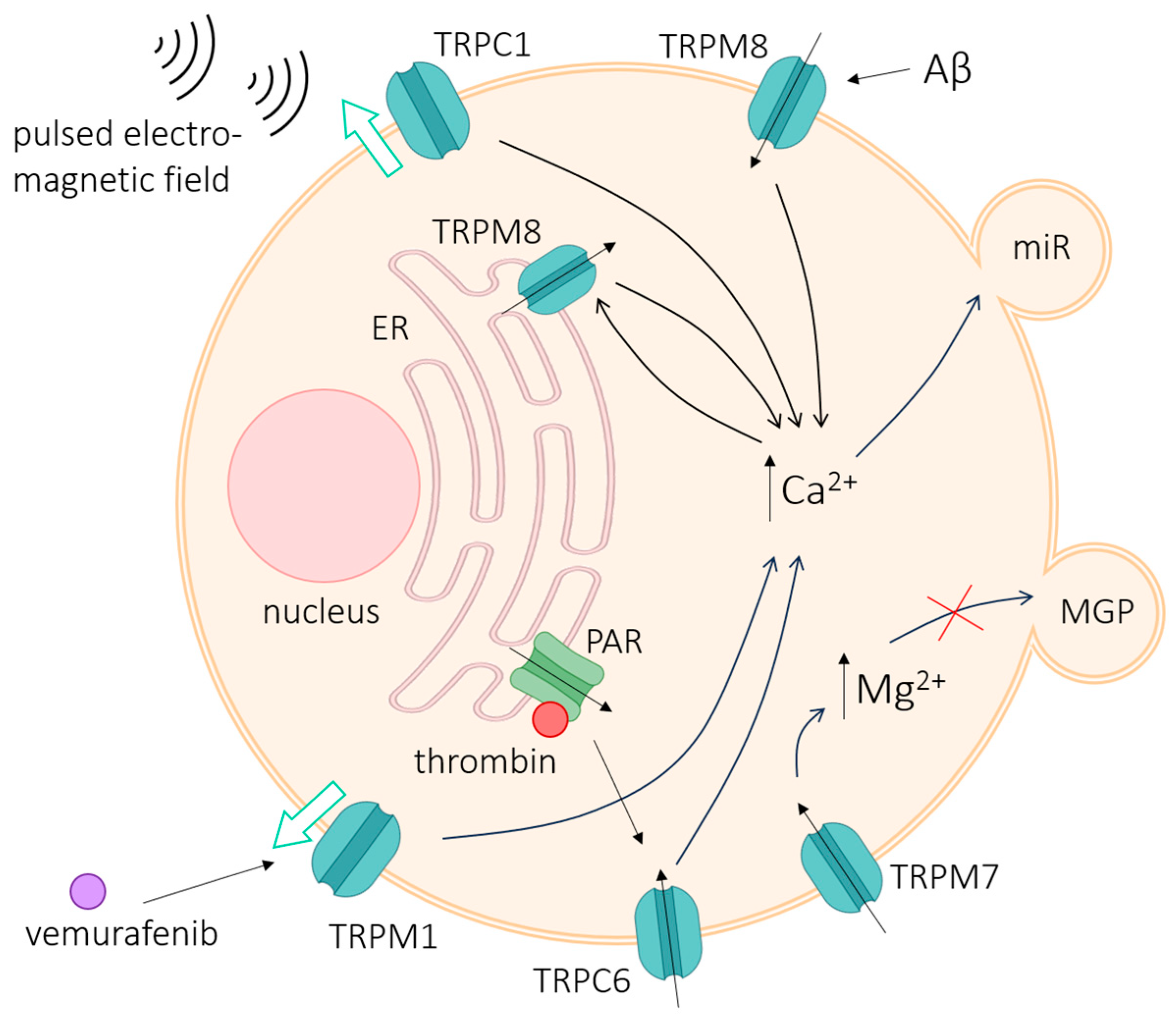
Figure 5.
Expression in TRP channels in seminal (blue), urinary (yellow) and blood (red) EVs. The expression/amount of specific TRP channels on the surface of EVs isolated from biological fluids can be used as diagnostic tools in various diseases.
Figure 5.
Expression in TRP channels in seminal (blue), urinary (yellow) and blood (red) EVs. The expression/amount of specific TRP channels on the surface of EVs isolated from biological fluids can be used as diagnostic tools in various diseases.
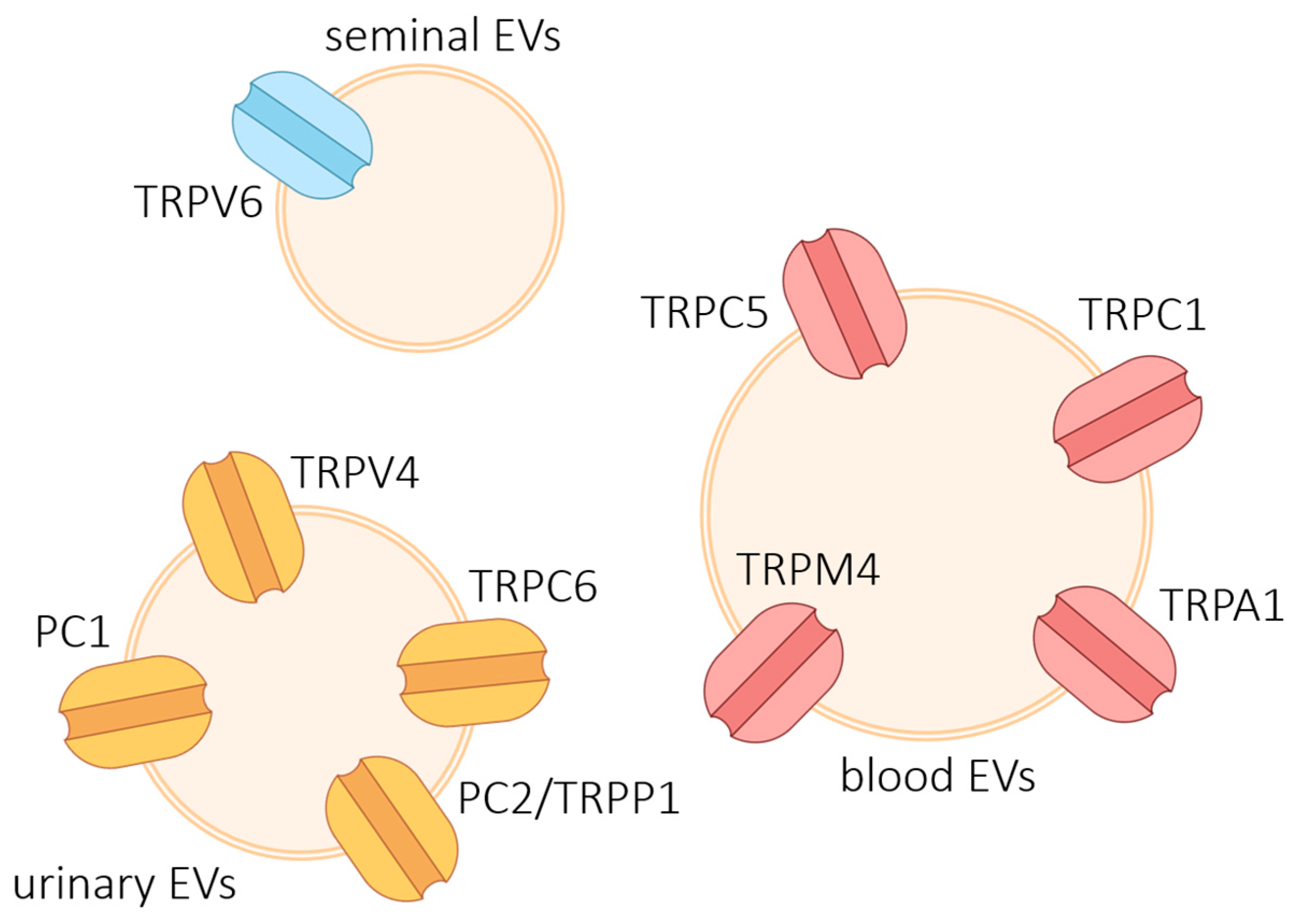
Figure 6.
Generation of Ca2+ signals by EVs in recipient cells via TRPM7. Membrane adhesion of EVs is believed to activate TRPM7, leading to Ca2+ influx. In turn, Ca2+ influx into the cell facilitates further endocytosis and membrane fusion of EVs, leading to content release inside the recipient cell. [50].
Figure 6.
Generation of Ca2+ signals by EVs in recipient cells via TRPM7. Membrane adhesion of EVs is believed to activate TRPM7, leading to Ca2+ influx. In turn, Ca2+ influx into the cell facilitates further endocytosis and membrane fusion of EVs, leading to content release inside the recipient cell. [50].
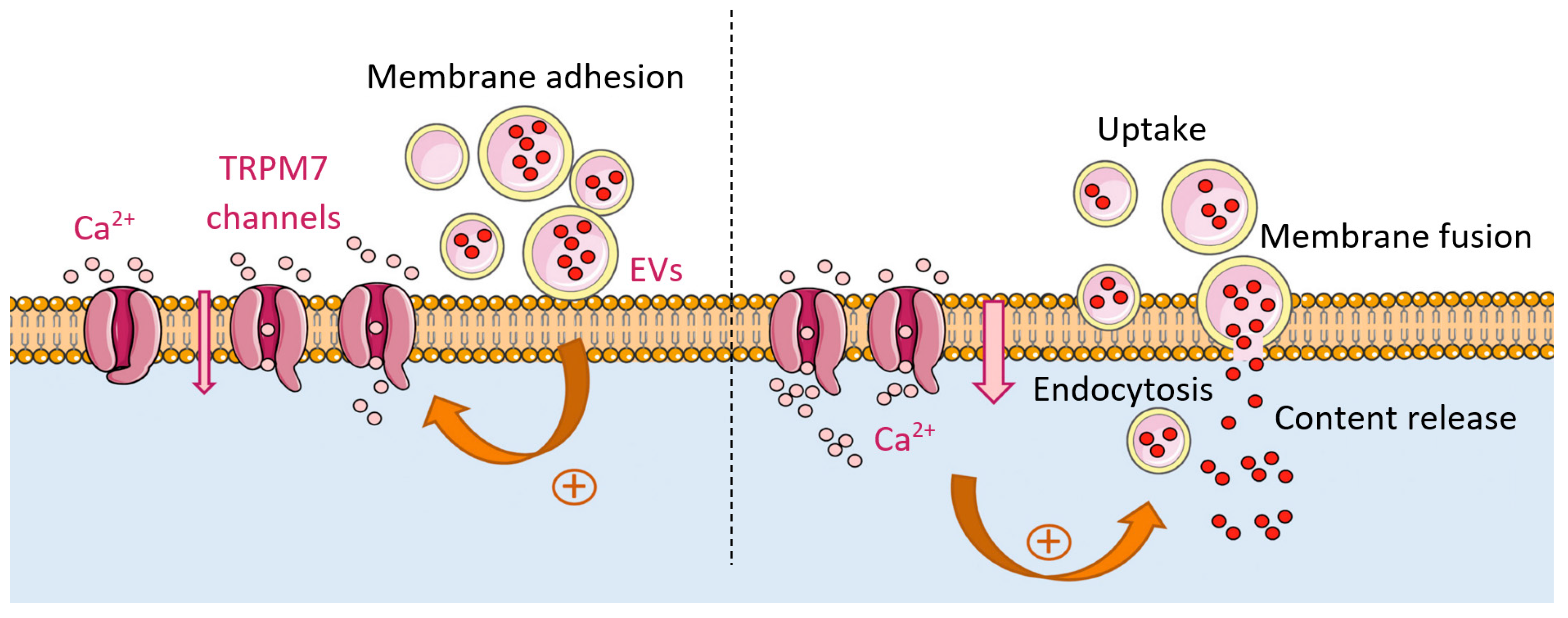
Disclaimer/Publisher’s Note: The statements, opinions and data contained in all publications are solely those of the individual author(s) and contributor(s) and not of MDPI and/or the editor(s). MDPI and/or the editor(s) disclaim responsibility for any injury to people or property resulting from any ideas, methods, instructions or products referred to in the content. |
© 2026 by the authors. Licensee MDPI, Basel, Switzerland. This article is an open access article distributed under the terms and conditions of the Creative Commons Attribution (CC BY) license (http://creativecommons.org/licenses/by/4.0/).
Copyright: This open access article is published under a Creative Commons CC BY 4.0 license, which permit the free download, distribution, and reuse, provided that the author and preprint are cited in any reuse.