

Submitted:
14 September 2026
Posted:
15 September 2026
You are already at the latest version
Abstract
Podocalyxin (PODXL) is a potential diagnostic biomarker and therapeutic target in various tumors. Development of anti-PODXL mAbs has been limited by concerns about potential reactivity with normal tissues. To minimize on-target, off-tumor toxicities and adverse effects, we developed cancer-specific mAbs (CasMabs) against PODXL and demonstrated antitumor efficacy in human tumor xenograft models. In this study, we evaluated humPcMab-60-DXd, an anti-PODXL CasMab conjugated with DXd, a deriv-ative of the topoisomerase I inhibitor exatecan. humPcMab-60-DXd retained reactivity with the pancreatic ductal adenocarcinoma cell line MIA PaCa-2 and inhibited cell pro-liferation in vitro. In the MIA PaCa-2 xenograft model, humPcMab-60-DXd showed su-perior antitumor efficacy compared with humPcMab-60. Furthermore, humP-cMab-60-DXd exhibited dose-dependent antitumor activity without reducing mice body weight. These results provide the first observation of antitumor efficacy for an an-ti-PODXL CasMab-drug conjugate, which could facilitate the clinical development of CasMabs for tumor therapy.
Keywords:
PODXL
; cancer-specific monoclonal antibody
; antibody-drug conjugate
; Dxd
; pancreatic cancer
1. Introduction
Podocalyxin (PODXL) is a heavily glycosylated cell-surface protein in the CD34 antigen family [1]. Although the unglycosylated PODXL core protein is approximately 53 kDa, extensive N- and O-linked glycosylation increases its size to approximately 150–200 kDa [2]. In normal tissues, PODXL supports the biology of several specialized cell populations and is detected in vascular and lymphatic endothelial cells [3], renal podocytes [4], and early hematopoietic progenitors [5]. The embryonic lethality observed in PODXL-deficient mice underscores PODXL’s physiological importance, indicating an essential role in normal development and tissue homeostasis [6]. In contrast, PODXL is frequently dysregulated in human cancers. Increased PODXL expression has been reported in diverse malignancies, including pancreatic ductal adenocarcinoma (PDAC) [7], colorectal cancer [8], renal cell carcinoma [9], oral squamous cell carcinoma [10], and breast cancer [11]. Importantly, accumulating clinical evidence indicates that PODXL expression is associated with unfavorable outcomes. High levels of PODXL have been linked to reduced disease-free survival, cancer-specific survival, and overall survival in patients with PDAC, colorectal cancer, glioblastoma, renal cell carcinoma, and urothelial bladder cancer [12]. These observations suggest that aberrant PODXL expression may contribute to malignant progression and identify PODXL as a potential biomarker and therapeutic target across multiple cancer types.
PODXL expression is strongly induced during epithelial–mesenchymal transition (EMT) and contributes to the metastatic behavior of cancer cells [10]. In PDAC, PODXL promotes the extravasation of mesenchymal-type tumor cells through its interaction with the cytoskeletal adaptor ezrin, which activates signaling pathways, including Rac1, RhoA, Cdc42, phosphatidylinositol-3 kinase, and mitogen-activated protein kinase, and thereby enhances cell motility [13,14]. This interaction also facilitates morphological changes toward an invasive phenotype [14]. Gain- and loss-of-function studies have implicated PODXL in tumor cell migration, invasion, stemness, and metastasis across multiple cancer types [13,15]. These properties make PODXL an attractive target for cancer therapy [13,16].
The clinical application of therapeutic mAbs is often limited by on-target/off-tumor toxicities resulting from target antigen expression in normal tissues [17,18]. For instance, EGFR-targeting mAbs frequently induce dermatological toxicities that can impair patients’ quality of life and interfere with treatment adherence [19]. Cardiotoxicity represents a major safety concern associated with HER2-targeted therapies [20]. Because PODXL is expressed not only in tumor cells but also in several normal cells, including vascular and lymphatic endothelial cells [3] and kidney podocytes [4], therapeutic antibodies directed against PODXL may similarly cause on-target/off-tumor toxicities. Therefore, antibodies that preferentially recognize PODXL in tumor cells while recognizing it little or not at all in normal cells would be highly desirable for safe therapeutic targeting of PODXL.
Our group has developed cancer-specific mAbs (CasMabs) against various tumor antigens, including PODXL [21], podoplanin [22], and HER2 [23]. We screened an anti-PODXL CasMab, PcMab-60, from over 100 hybridoma clones by flow cytometry for differential reactivity to cancer and normal cell lines [21]. A humanized IgG1-type PcMab-60 (humPcMab-60) reacted to PDAC and colorectal cancer cell lines, but not to a normal endothelial cell line in flow cytometry. Furthermore, humPcMab-60 induced antibody-dependent cellular cytotoxicity and complement-dependent cytotoxicity against PODXL-expressing cancer lines and showed antitumor activity in xenograft models [24], suggesting that humPcMab-60 could be a promising mAb-based tool in tumor therapy.
Trastuzumab deruxtecan (T-DXd) is a HER2-directed antibody-drug conjugate (ADC) composed of trastuzumab and DXd, a derivative of the topoisomerase I inhibitor exatecan, which is attached through a cleavable maleimide–GGFG peptide linker [25]. Following HER2 binding and receptor-mediated internalization, T-DXd is transported to lysosomes, where proteolytic cleavage of the linker releases the cytotoxic payload DXd [25]. The drug is conjugated to cysteine residues generated by reduction of the four interchain disulfide bonds of the human IgG1 antibody, allowing attachment of up to eight drugs per antibody and thereby achieving a relatively high and controlled drug-to-antibody ratio (DAR) [26]. Efficient linker cleavage occurs following intracellular trafficking to lysosomes, where the peptide is selectively degraded by lysosomal proteases [25]. Furthermore, the high membrane permeability of DXd facilitates a pronounced bystander effect, allowing T-DXd to eliminate not only HER2-high tumor cells but also adjacent cells with low or undetectable HER2 expression [25,27]. This property is particularly advantageous in tumors characterized by heterogeneous HER2 expression. It broadens the potential therapeutic applicability of T-DXd across tumors with varying levels of HER2 [28,29].
In this study, we generated an anti-PODXL CasMab-DXd conjugate, humPcMab-60-DXd, and evaluated the anti-tumor efficacy in a PDAC xenograft model.
2. Results
2.1. Flow Cytometric Analysis of humPcMab-60 and humPcMab-60-Dxd in CHO/PODXL and MIA PaCa-2 Cell Lines
humPcMab-60 exhibited cancer-specific reactivity and exerted antitumor efficacy in the MIA PaCa-2 xenograft model [24]. To investigate the antitumor activity of humPcMab-60-ADC, DXd was conjugated to humPcMab-60 through a cleavable maleimide–GGFG peptide linker (DAR8, Figure 1A). We assessed reactivity to PODXL-positive cells by flow cytometry. The humPcMab-60-Dxd exhibited a similar reactivity to CHO/PODXL and MIA PaCa-2 compared to humPcMab-60 (Figure 1B). Both humPcMab-60-Dxd and humPcMab-60 did not recognize CHO-K1 (Figure 1B). We also prepared humCvMab-62 and humCvMab-62-Dxd as control IgG1 and control IgG1-Dxd, respectively. These control mAbs did not react to CHO/PODXL, CHO-K1, and MIA PaCa-2 (Figure 1C).
2.2. Cellular cytotoxicity elicited by humPcMab-60-Dxd.
We investigated cellular cytotoxicity elicited by humPcMab-60, control IgG1-Dxd, and humPcMab-60-Dxd in vitro. MIA PaCa-2 cells were treated with the above mAbs for 7 days, and cell viability was measured compared to the control. As shown in Figure 2, humPcMab-60-Dxd showed dose-dependent cytotoxicity against MIA PaCa-2, whereas humPcMab-60 did not. The IC50 of humPcMab-60-Dxd was 0.69 µg/mL. The control IgG1-Dxd did not exhibit cytotoxicity until 1.6 µg/mL but showed weak cytotoxicity at 6.3 µg/mL.
2.3. Antitumor activity of humPcMab-60-Dxd against MIA PaCa-2 xenograft
We investigated the antitumor activity of humPcMab-60-Dxd against MIA PaCa-2 xenografts, compared with control IgG1-Dxd and humPcMab-60. MIA PaCa-2 was subcutaneously injected into BALB/c nude mice. After the size of the xenograft tumor reached ~150 mm3, 100 μg of humPcMab-60-Dxd, control IgG1-Dxd, or humPcMab-60 was intraperitoneally injected into each mouse on days 0 and 6 (arrows). Figure 3A showed the xenograft volume on days 0, 3, 6, 11, and 14. The humPcMab-60-Dxd administration resulted in a significant reduction in MIA PaCa-2 xenografts on days 6 (p < 0.01), 11 (p < 0.01), and 14 (p < 0.01) compared with that of control IgG1-Dxd (Figure 3A). Furthermore, the humPcMab-60-Dxd administration exhibited more potent antitumor efficacy compared to humPcMab-60 on day 14 (p < 0.01, Figure 3A). On day 14, humPcMab-60-Dxd significantly reduced tumor weight in the MIA PaCa-2 xenograft (56% reduction; p < 0.01, Figure 3B). Furthermore, the tumor weight in the humPcMab-60-Dxd-treated group was significantly reduced compared to that in the humPcMab-60-treated group (p < 0.05, Figure 3B). Figure 3 B shows the resected MIA PaCa-2 xenograft tumors on day 14. Tumor-bearing mice did not lose body weight with treatment with humPcMab-60-Dxd, control IgG1-Dxd, or humPcMab-60 (Figure 4C).
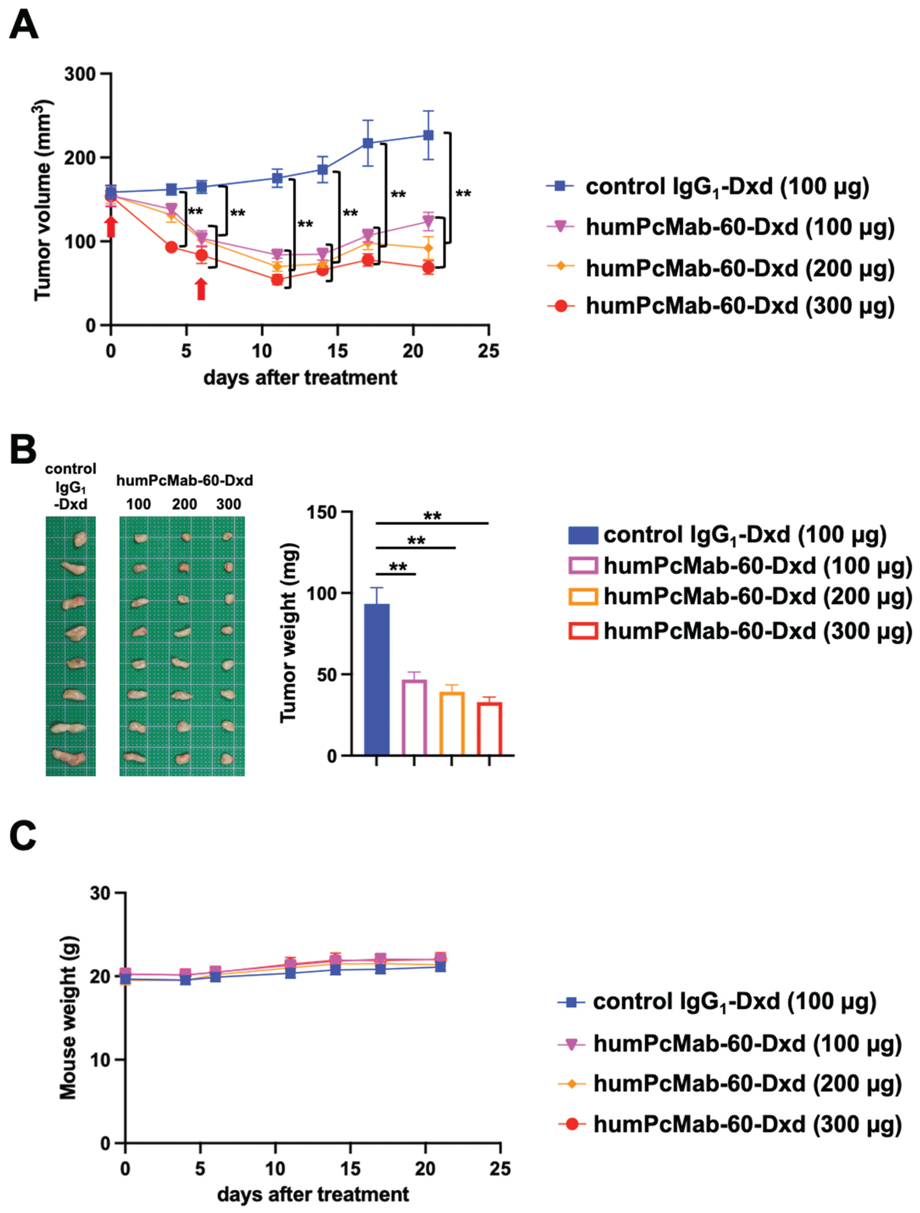
Dose-dependent antitumor efficacy of humPcMab-60-Dxd was next investigated. MIA PaCa-2 was subcutaneously injected into BALB/c nude mice. After the size of the xenograft tumor reached ~150 mm3, control IgG1-Dxd or humPcMab-60-Dxd (100, 200, and 300 μg) was intraperitoneally injected into each mouse on days 0 and 6 (arrows). Figure 4A showed the xenograft volume on days 0, 4, 6, 11, 14, 17, and 21. The humPcMab-60-Dxd administration (300 µg) resulted in a significant reduction in MIA PaCa-2 xenografts from day 4 to 21 (p < 0.01) compared with that of control IgG1-Dxd (Figure 4A). The humPcMab-60-Dxd administration (100 and 200 µg) also showed a significant reduction from day 6 to 21 (p < 0.01) compared with that of control IgG1-Dxd (Figure 4A). On day 21, humPcMab-60-Dxd showed a significant, dose-dependent reduction in MIA PaCa-2 xenograft tumor weight compared with control IgG1-Dxd (Figure 4B). Figure 4 B shows the resected MIA PaCa-2 xenograft tumors on day 21. Tumor-bearing mice did not lose body weight after treatment with a high dose of humPcMab-60-Dxd (Figure 4C). These results indicated that humPcMab-60-Dxd exerted antitumor efficacy against MIA PaCa-2 xenografts without any toxicity in BALB/c nude mice.
3. Discussion
Pancreatic cancer remains a major cause of cancer-related mortality in the United States. In 2026, it was estimated to represent the fourth leading cause of cancer death among men and the third among women [30]. PDAC, the predominant histological subtype of pancreatic cancer, is characterized by an aggressive clinical course and remains one of the most lethal malignancies, with a 5-year overall survival rate of only approximately 10% [31]. Accumulating evidence indicates that PODXL contributes to the aggressive behavior of cancer cells. In particular, elevated PODXL expression has been linked to unfavorable clinical outcomes across several malignancies. It is associated with reduced overall survival, particularly in PDAC [32]. Beyond its prognostic association, PODXL expression increases during EMT [14], suggesting a role in acquiring metastatic properties. Indeed, PODXL has been shown to facilitate the transendothelial migration of mesenchymal PDAC cells [33]. This activity is mediated by interaction with ezrin, a cytoskeletal linker protein, which promotes the morphological changes required for tumor cells to adopt an invasive phenotype capable of extravasation [33]. Therefore, PODXL contributes to tumor malignant progression, which facilitates the development of therapeutic mAbs.
In this study, we showed the antitumor efficacy of an anti-PODXL CasMab-Dxd conjugate, humPcMab-60-Dxd, in a pancreatic cancer xenograft model. The parental mAb, humPcMab-60, was previously shown to exert antibody-dependent cellular cytotoxicity, complement-dependent cytotoxicity, and antitumor effects in the MIA PaCa-2 xenograft model [24]. The humPcMab-60-Dxd exhibited more potent antitumor efficacy compared to that of humPcMab-60 (Figure 3), suggesting that Dxd-mediated cytotoxicity contributed to the antitumor effect. Given that the antitumor activity was demonstrated in only a single human cancer cell line model, further studies using a broader range of PODXL-positive cell lines and heterogeneous tumor models are needed to validate its therapeutic potential. Furthermore, humPcMab-60-Dxd showed dose-dependent antitumor efficacy without reducing mouse body weight (Figure 4), suggesting that these doses did not induce noticeable adverse effects. Because PcMab-60 does not recognize mouse PODXL, further toxicity evaluation in human normal cell lines is essential.
Our group and others have developed the anti-PODXL mAbs which recognize tumor-associated epitopes that are absent or less abundant in normal tissues [34]. A clone, PODO447, exhibited highly selective binding to a tumor-associated glycosylated PODXL without detectable reactivity toward normal tissues. Studies using glycosylation-deficient cell lines demonstrated that its epitope consists of an O-linked core 1 glycan presented within the structural framework of the PODXL polypeptide [35]. In contrast, the PcMab-60 epitope, corresponding to residues 109–116 (RGGGSGNP), contains potential sites for both N- and O-linked glycosylation. Notably, PcMab-60 binds a synthetic, nonglycosylated peptide encompassing this region in both enzyme-linked immunosorbent assay and surface plasmon resonance assays [36]. These findings suggest that glycosylation may not be required for PcMab-60 binding. Further studies are essential to establish a strategy to identify human cancers expressing the cancer-specific PODXL epitopes.
PODO447 was coupled to monomethyl auristatin E (MMAE), a tubulin polymerization inhibitor with a valine-citrulline proteolytically cleavable linker, resulting in PODO447-vedotin [37]. In a cellular cytotoxicity assay like Figure 2, PODO447-vedotin exhibits toxicity against a broad range of PODXL-positive cancer cell lines, including MIA PaCa-2. The cytotoxic effect of PODO447-vedotin on MIA PaCa-2 was potent (IC50 ~ 0.1 µg/ml). Furthermore, the antitumor effect on MIA PaCa-2 xenograft bearing nude mice was observed by bi-weekly intravenous administration of PODO447-vedotin at a dose of 4 mg/kg [37]. Although the cytotoxic effect of humPcMab-60-Dxd on MIA PaCa-2 was moderate (IC50 ~ 0.69 µg/ml, Figure 2), significant antitumor efficacy was achieved by weekly intraperitoneal administration of humPcMab-60-Dxd at a dose of 5 mg/kg (100 µg/mice, Figure 3 and Figure 4). DXd has high membrane permeability and shows a bystander effect [38], which might enhance antitumor efficacy. Further studies are needed to determine the optimal route of administration for humPcMab-60-Dxd.
PDAC remains one of the most lethal malignancies, with a 5-year overall survival rate of approximately 12%, largely owing to late diagnosis and the high frequency of advanced disease at presentation [39]. Despite extensive efforts to develop molecular targeted therapies, effective targeted treatment options for PDAC remained limited for decades [40]. Therefore, identifying novel tumor-associated targets that can be therapeutically exploited is an important strategy for improving PDAC treatment. Because more than 90% of PDAC cases have mutations in the KRAS gene, researchers have evaluated various RAS inhibitors in clinical studies [41]. Daraxonrasib, a RAS (ON) inhibitor, demonstrated significant, unprecedented improvements in both progression-free and overall survival for patients with metastatic PDAC [42,43]. A study has already revealed mechanisms of acquired resistance to daraxonrasib that converge on RAS–MAPK signaling, including RAF alterations and HER2 amplifications [44]. Furthermore, RAS–MAPK inhibitors upregulated HER2 expression in pancreatic cancer cell lines, including MIA PaCa-2 harboring KRASG12C. Therefore, AMG-510 (a KRAS G12C inhibitor) plus T-Dxd showed promising preclinical efficacy in the MIA PaCa-2 xenograft model [45]. Because PODXL is expressed in mesenchymal PDAC, which is characterized by resistance to therapies [46], a combination therapy of a KRAS inhibitor and humPcMab-60-Dxd may improve treatment for refractory PDAC.
4. Materials and Methods
4.1. Cell Lines
A PDAC cell line (MIA PaCa-2) was obtained from the Cell Resource Center for the Biomedical Research Institute of Development, Aging, and Cancer of Tohoku University (Miyagi, Japan). MIA PaCa-2 was cultured in Dulbecco’s modified Eagle’s medium (DMEM) (Nacalai Tesque, Inc., Kyoto, Japan) supplemented with 10% heat-inactivated fetal bovine serum (FBS; Thermo Fisher Scientific Inc., Waltham, MA, USA), 100 units/mL of penicillin, 100 μg/mL of streptomycin, and 0.25 μg/mL of amphotericin B (Nacalai Tesque, Inc.) at 37 °C in a humidified atmosphere containing 5% CO2 and 95% air.
4.2. Antibodies
Mouse anti-PODXL CasMab PcMab-60 (IgM, kappa) was previously generated [21]. To generate the humanized antibodies (humPcMab-60), the complementarity-determining regions (CDRs) of the variable heavy (VH) chains of PcMab-60 were grafted onto human IgG framework sequences (accession number: KF698734) and cloned into the pCAG-Neo expression vector containing the human IgG1 heavy-chain constant region (CH). Likewise, the CDRs of the variable light (VL) chains were grafted onto human IgG VL framework sequences (accession number: U41645) and cloned into the pCAG-Ble vector containing the human kappa light-chain constant region (CL). The resulting expression vectors were introduced into ExpiCHO-S cells using the ExpiCHO Expression System for production of humPcMab-60. As a control human IgG1 mAb (control IgG1), humCvMab-62 was generated from CvMab-62, an anti-SARS-CoV-2 spike protein S2 subunit mAb [47], using the same humanization procedure. We purified all antibodies using Ab-Capcher (ProteNova Co., Ltd., Kagawa, Japan). The humPcMab-60 and humCvMab-62 were conjugated with DXd through the cleavable maleimide–GGFG peptide linker (DAR8, FUJIFILM Toyama Chemical Co., Ltd., Toyama, Japan), which were designated as humPcMab-60-Dxd and control IgG1-Dxd, respectively.
4.3. Flow Cytometry
MIA PaCa-2 was washed with blocking buffer [0.1% BSA in phosphate-buffered saline (PBS)] and treated with primary mAbs for 30 min at 4 °C, followed by treatment with anti-human IgG conjugated with fluorescein isothiocyanate (Sigma-Aldrich Corp., St. Louis, MO, USA). We collected fluorescence data (5000 events) using an SA3800 Cell Analyzer (Sony Corp., Tokyo, Japan). We gated cells on the dot plot based on side scatter and forward scatter, and analyzed fluorescence intensity using FlowJo software (BD Biosciences, Franklin Lakes, NJ, USA).
4.4. Measurement of cell viability
MIA PaCa-2 (5,000 cells) was seeded in a 96-well plate (Corning, Inc., New York, NY, USA). Then, the humCvMab-62-DXd or humPcMab-60-DXd was added at the indicated concentrations. After 7 days, cell viability was measured using the CellTiter-Glo® 2.0 Cell Viability Assay and a GloMax luminometer (Promega, Madison, WI, USA).
4.5. Antitumor Activity of humPcMab-60
Animal experiments were performed in accordance with applicable guidelines and regulations, with appropriate measures taken to minimize pain, distress, and suffering. The Institutional Committee for Experiments of the Institute of Microbial Chemistry (Numazu, Japan) reviewed and approved the experimental protocol (approval no. 2026-012 and 2026-035). We maintained the mice under specific pathogen-free conditions. We maintained mice on an 11 h light/13 h dark cycle throughout the experimental period. We supplied food and water ad libitum. Throughout the experimental period, we monitored the mice daily for general health and well-being. A reduction in body weight exceeding 25% of the initial body weight and/or a maximal tumor size of >3,000 mm3 were predetermined as a humane endpoint for euthanasia.
MIA PaCa-2 was mixed with Matrigel Matrix Growth Factor Reduced (BD Biosciences). Subcutaneous injections (5 × 10⁶ cells/mouse) were then given to the left flanks of BALB/c nude mice (Japan SLC, Inc., Shizuoka, Japan). On the seventh post-inoculation day, 100 µg of humCvMab-62-DXd (control hIgG1-DXd) (n = 8), 100 µg of hum PcMab-60-DXd (n = 8), or 100 µg of hum PcMab-60 (n = 8) in 100 µL PBS were administered intraperitoneally (day 0). We gave additional antibody injections on day 6. We assessed tumor diameter on days 0, 3, 6, 11, and 14. In the dose-dependent study, 100 µg of humCvMab-62-DXd (control hIgG1-DXd) (n = 8) or hum PcMab-60-DXd (100, 200, and 300 µg, n = 8) were administered intraperitoneally (day 0). We gave additional antibody injections on day 6. We assessed tumor diameter on days 0, 4, 6, 11, 14, 17, and 21. Tumor volume was calculated using the formula: volume = W2 × L/2, where W represents the short diameter and L the long diameter. We also assessed mice weight on the indicated days. After observations were complete, we sacrificed the mice and assessed tumor weights after excision.
Data are presented as the mean ± standard error of the mean (SEM). Statistical analyses were performed using two-way ANOVA followed by Tukey’s post hoc test for tumor volume and mouse weight. One-way ANOVA followed by Tukey’s post hoc test was also conducted for tumor weight. A p-value < 0.05 was considered statistically significant.
5. Conclusions
An anti-PODXL CasMab-Dxd conjugate showed antitumor efficacy in a pancreatic cancer MIA PaCa-2 xenograft model. Because we demonstrated antitumor effects in a human cancer cell line model, further investigation in more PODXL-positive cell line models and heterogeneous tumor models is essential. Although we observed low toxicity in mice, we have not evaluated toxicity to normal human cells. Our results provide the first evidence of antitumor efficacy for the anti-PODXL CasMab-Dxd conjugate, which could facilitate the clinical development of CasMabs for tumor therapy.
Author Contributions
Conceptualization, M.K.K. and Y.K.; methodology, M.K.K. and T.O.; validation, H.S. and Y.K.; investigation, T.O. and H.S.; data curation, H.S.; writing—original draft preparation, H.S.; writing—review and editing, Y.K.; project administration, Y.K.; funding acquisition, H.S. and Y.K. All authors have read and agreed to the published version of the manuscript.
Funding
This research was supported in part by the Japan Agency for Medical Research and Development (AMED) under Grant Numbers JP26am0521010 (to Y.K.), JP26ama121008 (to Y.K.), JP25ama221153 (to Y.K.), and JP25ama221339 (to Y.K.), and by the Japan Society for the Promotion of Science (JSPS) Grants-in-Aid for Scientific Research (KAKENHI) grant no. 25K10553 (to Y.K.) and 26K02289 (to H.S.).
Institutional Review Board Statement
The Institutional Committee for Experiments of the Institute of Microbial Chemistry approved animal experiments (approval no. 2026-012 and 2026-035, Approval Date: March 6, 2026 and June 19, 2026, respectively).
Informed Consent Statement
Not applicable.
Data Availability Statement
The data presented in this study are available in the article.
Conflicts of Interest
The authors declare no conflicts of interest.
References
- Doyonnas, R.; Nielsen, J.S.; Chelliah, S.; Drew, E.; Hara, T.; Miyajima, A.; McNagny, K.M. Podocalyxin is a CD34-related marker of murine hematopoietic stem cells and embryonic erythroid cells. Blood 2005, 105, 4170–4178. [Google Scholar] [CrossRef] [PubMed]
- Nielsen, J.S.; McNagny, K.M. Novel functions of the CD34 family. J. Cell Sci. 2008, 121, 3683–3692. [Google Scholar] [CrossRef] [PubMed]
- Kriehuber, E.; Breiteneder-Geleff, S.; Groeger, M.; Soleiman, A.; Schoppmann, S.F.; Stingl, G.; Kerjaschki, D.; Maurer, D. Isolation and characterization of dermal lymphatic and blood endothelial cells reveal stable and functionally specialized cell lineages. J. Exp. Med. 2001, 194, 797–808. [Google Scholar] [CrossRef] [PubMed]
- Kerjaschki, D.; Sharkey, D.J.; Farquhar, M.G. Identification and characterization of podocalyxin--the major sialoprotein of the renal glomerular epithelial cell. J. Cell Biol. 1984, 98, 1591–1596. [Google Scholar] [CrossRef] [PubMed]
- McNagny, K.M.; Pettersson, I.; Rossi, F.; Flamme, I.; Shevchenko, A.; Mann, M.; Graf, T. Thrombomucin, a novel cell surface protein that defines thrombocytes and multipotent hematopoietic progenitors. J. Cell Biol. 1997, 138, 1395–1407. [Google Scholar] [CrossRef] [PubMed]
- Doyonnas, R.; Kershaw, D.B.; Duhme, C.; Merkens, H.; Chelliah, S.; Graf, T.; McNagny, K.M. Anuria, omphalocele, and perinatal lethality in mice lacking the CD34-related protein podocalyxin. J. Exp. Med. 2001, 194, 13–27. [Google Scholar] [CrossRef] [PubMed]
- Ney, J.T.; Zhou, H.; Sipos, B.; Büttner, R.; Chen, X.; Klöppel, G.; Gütgemann, I. Podocalyxin-like protein 1 expression is useful to differentiate pancreatic ductal adenocarcinomas from adenocarcinomas of the biliary and gastrointestinal tracts. Hum. Pathol. 2007, 38, 359–364. [Google Scholar] [CrossRef] [PubMed]
- Larsson, A.; Johansson, M.E.; Wangefjord, S.; Gaber, A.; Nodin, B.; Kucharzewska, P.; Welinder, C.; Belting, M.; Eberhard, J.; Johnsson, A.; et al. Overexpression of podocalyxin-like protein is an independent factor of poor prognosis in colorectal cancer. Br. J. Cancer 2011, 105, 666–672. [Google Scholar] [CrossRef] [PubMed]
- Hsu, Y.H.; Lin, W.L.; Hou, Y.T.; Pu, Y.S.; Shun, C.T.; Chen, C.L.; Wu, Y.Y.; Chen, J.Y.; Chen, T.H.; Jou, T.S. Podocalyxin EBP50 ezrin molecular complex enhances the metastatic potential of renal cell carcinoma through recruiting Rac1 guanine nucleotide exchange factor ARHGEF7. Am. J. Pathol. 2010, 176, 3050–3061. [Google Scholar] [CrossRef] [PubMed]
- Lin, C.W.; Sun, M.S.; Wu, H.C. Podocalyxin-like 1 is associated with tumor aggressiveness and metastatic gene expression in human oral squamous cell carcinoma. Int. J. Oncol. 2014, 45, 710–718. [Google Scholar] [CrossRef] [PubMed]
- Forse, C.L.; Yilmaz, Y.E.; Pinnaduwage, D.; O’Malley, F.P.; Mulligan, A.M.; Bull, S.B.; Andrulis, I.L. Elevated expression of podocalyxin is associated with lymphatic invasion, basal-like phenotype, and clinical outcome in axillary lymph node-negative breast cancer. Breast Cancer Res. Treat. 2013, 137, 709–719. [Google Scholar] [CrossRef] [PubMed]
- He, S.; Du, W.; Li, M.; Yan, M.; Zheng, F. PODXL might be a new prognostic biomarker in various cancers: a meta-analysis and sequential verification with TCGA datasets. BMC Cancer 2020, 20, 620. [Google Scholar] [CrossRef] [PubMed]
- Le Tran, N.; Wang, Y.; Nie, G. Podocalyxin in Normal Tissue and Epithelial Cancer. Cancers 2021, 13. [Google Scholar] [CrossRef] [PubMed]
- Fröse, J.; Chen, M.B.; Hebron, K.E.; Reinhardt, F.; Hajal, C.; Zijlstra, A.; Kamm, R.D.; Weinberg, R.A. Epithelial-Mesenchymal Transition Induces Podocalyxin to Promote Extravasation via Ezrin Signaling. Cell Rep. 2018, 24, 962–972. [Google Scholar] [CrossRef] [PubMed]
- Tamayo-Orbegozo, E.; Amo, L.; Díez-García, J.; Amutio, E.; Riñón, M.; Alonso, M.; Arana, P.; Maruri, N.; Larrucea, S. Emerging Role of Podocalyxin in the Progression of Mature B-Cell Non-Hodgkin Lymphoma. Cancers 2020, 12. [Google Scholar] [CrossRef] [PubMed]
- Snyder, K.A.; Hughes, M.R.; Hedberg, B.; Brandon, J.; Hernaez, D.C.; Bergqvist, P.; Cruz, F.; Po, K.; Graves, M.L.; Turvey, M.E.; et al. Podocalyxin enhances breast tumor growth and metastasis and is a target for monoclonal antibody therapy. Breast Cancer Res. 2015, 17, 46. [Google Scholar] [CrossRef] [PubMed]
- Park, M.; Kim, J.W. Updates on the mechanisms of toxicities associated with monoclonal antibodies targeting growth factor signaling and immune cells in cancer. Toxicol. Res. 2024, 40, 335–348. [Google Scholar] [CrossRef] [PubMed]
- Guan, M.; Zhou, Y.P.; Sun, J.L.; Chen, S.C. Adverse events of monoclonal antibodies used for cancer therapy. BioMed Res. Int. 2015, 2015, 428169. [Google Scholar] [CrossRef] [PubMed]
- Holcmann, M.; Sibilia, M. Mechanisms underlying skin disorders induced by EGFR inhibitors. Mol. Cell Oncol. 2015, 2, e1004969. [Google Scholar] [CrossRef] [PubMed]
- Copeland-Halperin, R.S.; Liu, J.E.; Yu, A.F. Cardiotoxicity of HER2-targeted therapies. Curr. Opin. Cardiol. 2019, 34, 451–458. [Google Scholar] [CrossRef] [PubMed]
- Kaneko, M.K.; Ohishi, T.; Kawada, M.; Kato, Y. A cancer-specific anti-podocalyxin monoclonal antibody (60-mG(2a)-f) exerts antitumor effects in mouse xenograft models of pancreatic carcinoma. Biochem Biophys. Rep. 2020, 24, 100826. [Google Scholar] [CrossRef] [PubMed]
- Kato, Y.; Kaneko, M.K. A cancer-specific monoclonal antibody recognizes the aberrantly glycosylated podoplanin. Sci. Rep. 2014, 4, 5924. [Google Scholar] [CrossRef] [PubMed]
- Kaneko, M.K.; Suzuki, H.; Kato, Y. Establishment of a Novel Cancer-Specific Anti-HER2 Monoclonal Antibody H(2)Mab-250/H(2)CasMab-2 for Breast Cancers. Monoclon. Antib. Immunodiagn. Immunother. 2024, 43, 35–43. [Google Scholar] [CrossRef] [PubMed]
- Suzuki, H.; Ohishi, T.; Nakamura, T.; Yanaka, M.; Handa, S.; Tanaka, T.; Kaneko, M.K.; Kato, Y. A Cancer-Specific Anti-Podocalyxin Monoclonal Antibody (humPcMab-60) Demonstrated Antitumor Efficacy in Pancreatic and Colorectal Cancer Xenograft Models. Antibodies 2025, 14. [Google Scholar] [CrossRef] [PubMed]
- Ogitani, Y.; Aida, T.; Hagihara, K.; Yamaguchi, J.; Ishii, C.; Harada, N.; Soma, M.; Okamoto, H.; Oitate, M.; Arakawa, S.; et al. DS-8201a, A Novel HER2-Targeting ADC with a Novel DNA Topoisomerase I Inhibitor, Demonstrates a Promising Antitumor Efficacy with Differentiation from T-DM1. Clin. Cancer Res. 2016, 22, 5097–5108. [Google Scholar] [CrossRef] [PubMed]
- Chen, R.; Ren, Z.; Bai, L.; Hu, X.; Chen, Y.; Ye, Q.; Hu, Y.; Shi, J. Novel antibody-drug conjugates based on DXd-ADC technology. Bioorg Chem. 2024, 151, 107697. [Google Scholar] [CrossRef] [PubMed]
- Nakada, T.; Masuda, T.; Naito, H.; Yoshida, M.; Ashida, S.; Morita, K.; Miyazaki, H.; Kasuya, Y.; Ogitani, Y.; Yamaguchi, J.; et al. Novel antibody drug conjugates containing exatecan derivative-based cytotoxic payloads. Bioorg Med. Chem. Lett. 2016, 26, 1542–1545. [Google Scholar] [CrossRef] [PubMed]
- Shadab, A.; Al-Shdefat, R.I.K. Redefining breast cancer: therapeutic opportunities in HER2-low and emerging molecular subtypes. Clin. Transl. Oncol. 2026. [Google Scholar] [CrossRef] [PubMed]
- Etessami, J.D.; Dalla Via, G.; Ferraro, E.; Trapani, D.; Curigliano, G.; Marra, A. Treatment of breast cancer brain metastases in the era of novel drugs. Cancer Treat. Rev. 2026, 148, 103179. [Google Scholar] [CrossRef] [PubMed]
- Siegel, R.L.; Kratzer, T.B.; Wagle, N.S.; Sung, H.; Jemal, A. Cancer statistics, 2026. CA Cancer J. Clin. 2026, 76, e70043. [Google Scholar] [CrossRef] [PubMed]
- Kamisawa, T.; Wood, L.D.; Itoi, T.; Takaori, K. Pancreatic cancer. Lancet 2016, 388, 73–85. [Google Scholar] [CrossRef] [PubMed]
- Wang, J.; Zhao, Y.; Qi, R.; Zhu, X.; Huang, C.; Cheng, S.; Wang, S.; Qi, X. Prognostic role of podocalyxin-like protein expression in various cancers: A systematic review and meta-analysis. Oncotarget 2017, 8, 52457–52464. [Google Scholar] [CrossRef] [PubMed]
- Dallas, M.R.; Chen, S.H.; Streppel, M.M.; Sharma, S.; Maitra, A.; Konstantopoulos, K. Sialofucosylated podocalyxin is a functional E- and L-selectin ligand expressed by metastatic pancreatic cancer cells. Am. J. Physiol. Cell Physiol. 2012, 303, C616–624. [Google Scholar] [CrossRef] [PubMed]
- Pinho, S.S.; Reis, C.A. Glycosylation in cancer: mechanisms and clinical implications. Nat. Rev. Cancer 2015, 15, 540–555. [Google Scholar] [CrossRef] [PubMed]
- Canals Hernaez, D.; Hughes, M.R.; Dean, P.; Bergqvist, P.; Samudio, I.; Blixt, O.; Wiedemeyer, K.; Li, Y.; Bond, C.; Cruz, E.; et al. PODO447: a novel antibody to a tumor-restricted epitope on the cancer antigen podocalyxin. J. Immunother. Cancer 2020, 8. [Google Scholar] [CrossRef] [PubMed]
- Asano, T.; Suzuki, H.; Kaneko, M.K.; Kato, Y. Epitope Mapping of a Cancer-Specific Anti-Podocalyxin Monoclonal Antibody (PcMab-60) Using Enzyme-Linked Immunosorbent Assay and Surface Plasmon Resonance. Monoclon. Antib. Immunodiagn. Immunother. 2021, 40, 227–232. [Google Scholar] [CrossRef] [PubMed]
- Canals Hernaez, D.; Hughes, M.R.; Li, Y.; Mainero Rocca, I.; Dean, P.; Brassard, J.; Bell, E.M.; Samudio, I.; Mes-Masson, A.M.; Narimatsu, Y.; et al. Targeting a Tumor-Specific Epitope on Podocalyxin Increases Survival in Human Tumor Preclinical Models. Front Oncol. 2022, 12, 856424. [Google Scholar] [CrossRef] [PubMed]
- Ogitani, Y.; Hagihara, K.; Oitate, M.; Naito, H.; Agatsuma, T. Bystander killing effect of DS-8201a, a novel anti-human epidermal growth factor receptor 2 antibody-drug conjugate, in tumors with human epidermal growth factor receptor 2 heterogeneity. Cancer Sci. 2016, 107, 1039–1046. [Google Scholar] [CrossRef] [PubMed]
- Rahib, L.; Coffin, T.; Kenner, B. Factors Driving Pancreatic Cancer Survival Rates. Pancreas 2025, 54, e530–e536. [Google Scholar] [CrossRef] [PubMed]
- Mannucci, A.; Goel, A. Advances in pancreatic cancer early diagnosis, prevention, and treatment: The past, the present, and the future. CA Cancer J. Clin. 2026, 76, e70035. [Google Scholar] [CrossRef] [PubMed]
- Riedl, J.M.; Matsubara, H.; McNeil, R.; Patel, P.S.; Fece de la Cruz, F.; Gulhan, D.C.; Corcoran, R.B. Emerging landscape of KRAS inhibitors in cancer treatment. Cancer Cell 2026, 44, 471–497. [Google Scholar] [CrossRef] [PubMed]
- Wolpin, B.M.; Park, W.; Garrido-Laguna, I.; Spira, A.; Starodub, A.; Sommerhalder, D.; Punekar, S.R.; Barve, M.; Pelster, M.; Herzberg, B.; et al. Daraxonrasib in Previously Treated Advanced RAS-Mutated Pancreatic Cancer. N Engl. J. Med. 2026, 394, 1790–1802. [Google Scholar] [CrossRef] [PubMed]
- O’Reilly, E.M.; Wainberg, Z.A.; Hendifar, A.E.; Borad, M.J.; Pietrantonio, F.; Pant, S.; Hammel, P.; Cremolini, C.; Manji, G.A.; Oberstein, P.E.; et al. Daraxonrasib or Chemotherapy in Previously Treated Metastatic Pancreatic Cancer. N Engl. J. Med. 2026, 395, 325–337. [Google Scholar] [CrossRef] [PubMed]
- Aronchik, I.; Kar, S.; Zhuang, Y.; Ahler, E.; Lai, L.P.; Seshadri, V.; Yang, Y.C.; Bulle, A.; Menard, M.; Nayak, B.; et al. Acquired resistance to the RAS(ON) multi-selective inhibitor daraxonrasib guides rational combination therapy strategies in pancreatic cancer. Nat. Med. 2026, 32, 2865–2877. [Google Scholar] [CrossRef] [PubMed]
- Bulle, A.; Liu, P.; Seehra, K.; Bansod, S.; Chen, Y.; Zahra, K.; Somani, V.; Khawar, I.A.; Chen, H.P.; Dodhiawala, P.B.; et al. Combined KRAS-MAPK pathway inhibitors and HER2-directed drug conjugate is efficacious in pancreatic cancer. Nat. Commun. 2024, 15, 2503. [Google Scholar] [CrossRef] [PubMed]
- Dardare, J.; Witz, A.; Merlin, J.L.; Bochnakian, A.; Toussaint, P.; Gilson, P.; Harlé, A. Epithelial to Mesenchymal Transition in Patients with Pancreatic Ductal Adenocarcinoma: State-of-the-Art and Therapeutic Opportunities. Pharmaceuticals 2021, 14. [Google Scholar] [CrossRef] [PubMed]
- Inoue, T.; Yamamoto, Y.; Sato, K.; Okemoto-Nakamura, Y.; Shimizu, Y.; Ogawa, M.; Onodera, T.; Takahashi, Y.; Wakita, T.; Kaneko, M.K.; et al. Overcoming antibody-resistant SARS-CoV-2 variants with bispecific antibodies constructed using non-neutralizing antibodies. iScience 2024, 27, 109363. [Google Scholar] [CrossRef] [PubMed]
Figure 1.
Flow cytometric analysis of humPcMab-60 and humPcMab-60-Dxd in CHO/PODXL and MIA PaCa-2 cell lines. (A) Production of humPcMab-60-Dxd (DAR8) from humPcMab-60. (B) CHO/PODXL, CHO-K1, and MIA PaCa-2 were treated with 6.25 μg/mL of humPcMab-60 or humPcMab-60-Dxd (red line) or blocking buffer (black line, negative control). (C) CHO/PODXL, CHO-K1, and MIA PaCa-2 were treated with 6.25 μg/mL of control IgG1-Dxd (humCvMab-62-Dxd) or control IgG1 (humCvMab-62) at 6.25 μg/mL (red line) or blocking buffer (black line, negative control). We treated these cells with fluorescein isothiocyanate-conjugated anti-human IgG. We collected fluorescence data using the SA3800 Cell Analyzer and analyzed them in FlowJo software.
Figure 1.
Flow cytometric analysis of humPcMab-60 and humPcMab-60-Dxd in CHO/PODXL and MIA PaCa-2 cell lines. (A) Production of humPcMab-60-Dxd (DAR8) from humPcMab-60. (B) CHO/PODXL, CHO-K1, and MIA PaCa-2 were treated with 6.25 μg/mL of humPcMab-60 or humPcMab-60-Dxd (red line) or blocking buffer (black line, negative control). (C) CHO/PODXL, CHO-K1, and MIA PaCa-2 were treated with 6.25 μg/mL of control IgG1-Dxd (humCvMab-62-Dxd) or control IgG1 (humCvMab-62) at 6.25 μg/mL (red line) or blocking buffer (black line, negative control). We treated these cells with fluorescein isothiocyanate-conjugated anti-human IgG. We collected fluorescence data using the SA3800 Cell Analyzer and analyzed them in FlowJo software.
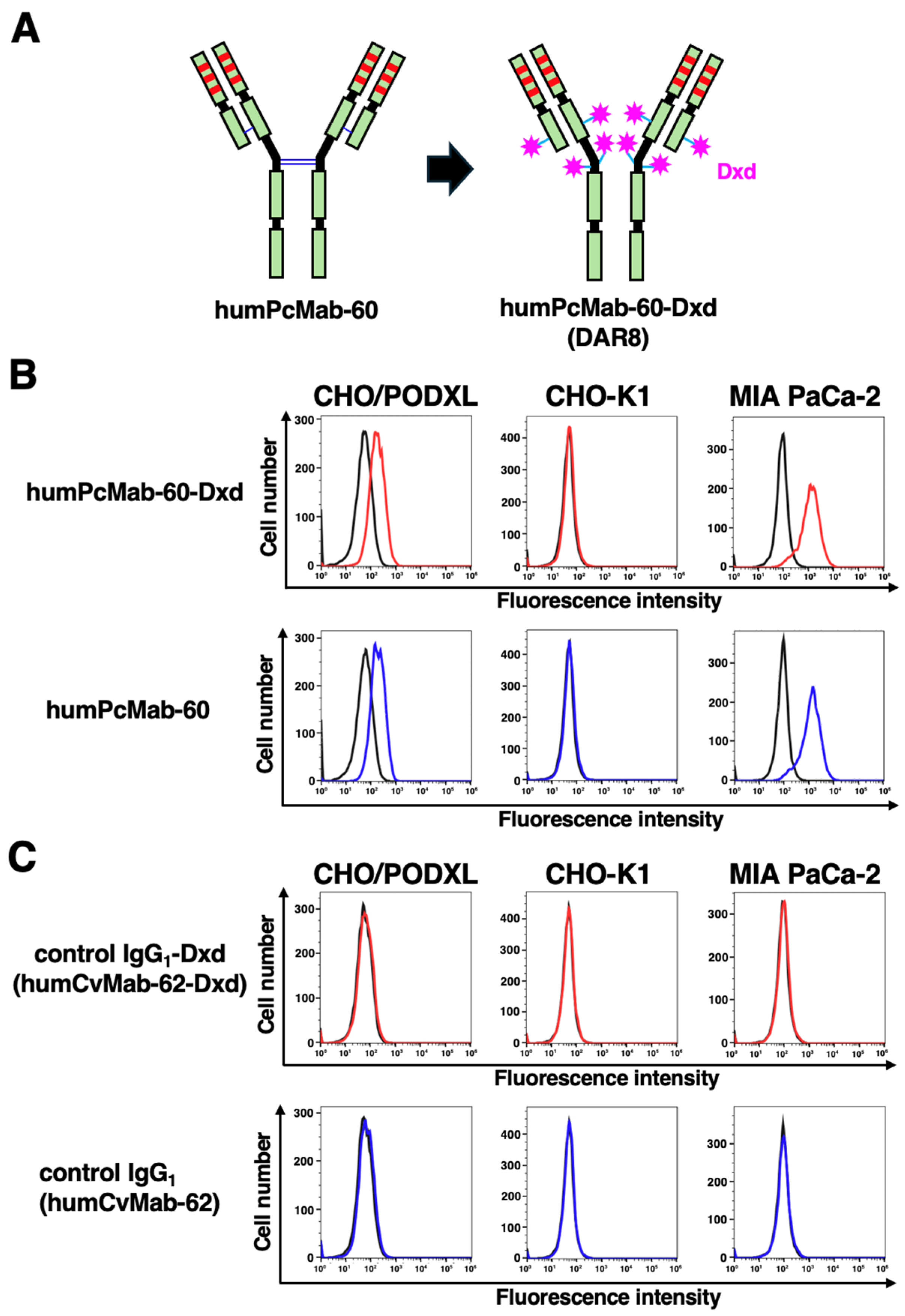
Figure 2.
Measurement of cell viability in the presence of control IgG1-Dxd, humPcMab-60-Dxd, or humPcMab-60. MIA PaCa-2 cells were seeded in a 96-well plate, and control IgG1-Dxd, humPcMab-60-Dxd, or humPcMab-60 were added at the indicated concentrations (n = 3). After 7 days, we measured cell viability using the CellTiter-Glo® 2.0 Cell Viability Assay and a GloMax luminometer.
Figure 2.
Measurement of cell viability in the presence of control IgG1-Dxd, humPcMab-60-Dxd, or humPcMab-60. MIA PaCa-2 cells were seeded in a 96-well plate, and control IgG1-Dxd, humPcMab-60-Dxd, or humPcMab-60 were added at the indicated concentrations (n = 3). After 7 days, we measured cell viability using the CellTiter-Glo® 2.0 Cell Viability Assay and a GloMax luminometer.
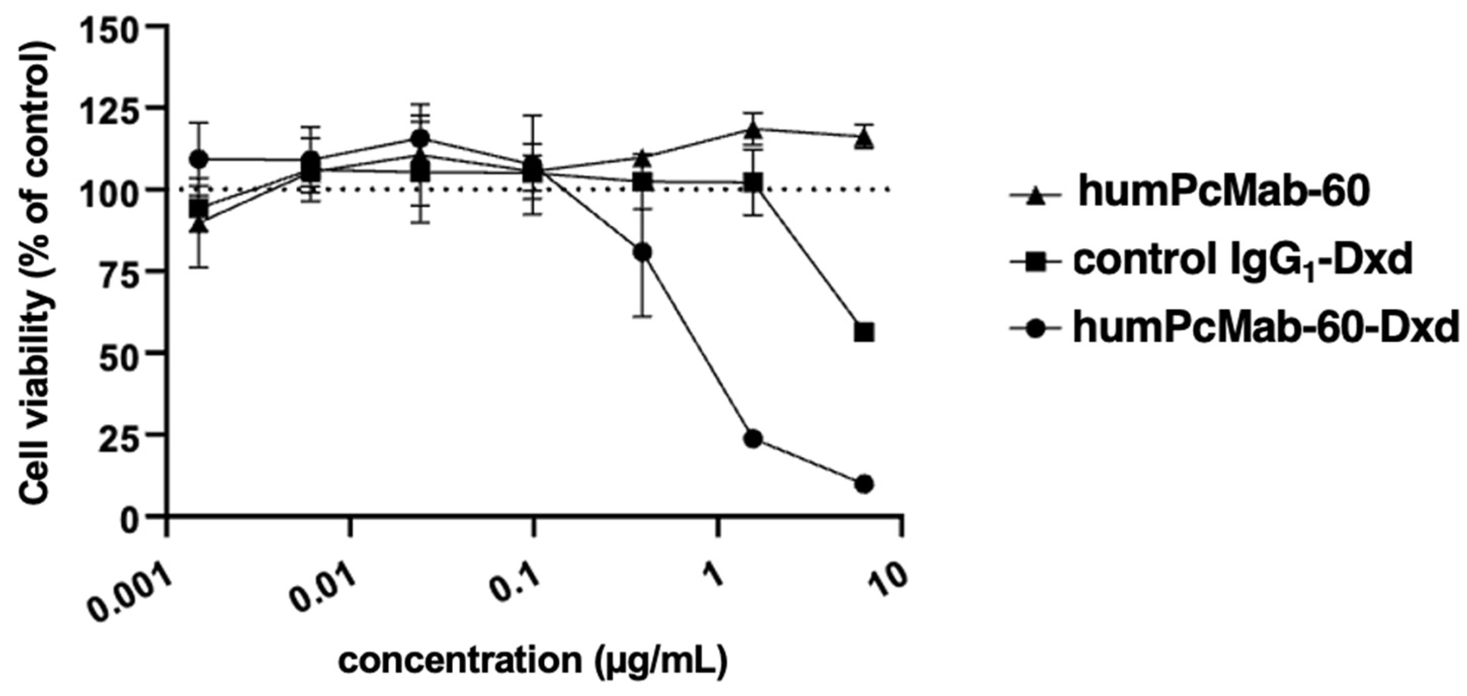
Figure 3.
Antitumor activity of humPcMab-60-Dxd against MIA PaCa-2 xenografts. (A) MIA PaCa-2 was subcutaneously injected into BALB/c nude mice. After a week, control IgG1-Dxd (100 μg, n = 8), humPcMab-60 (100 μg, n = 8), or humPcMab-60-Dxd (100 μg, n = 8) was intraperitoneally injected into each mouse on days 0 and 6 (arrows). Tumor volume is shown as mean ± SEM. ** p < 0.01 (two-way ANOVA followed by Tukey’s post hoc test). (B) The mice were euthanized on day 14. We measured tumor appearance (left) and weight (right) of MIA PaCa-2 xenografts. Values are presented as the mean ± SEM. * p < 0.05, ** p < 0.01 (One-way ANOVA followed by Tukey’s post hoc test). Each grid line is 1 cm. (C) Body weight (mean ± SEM) of xenograft-bearing mice treated with the mAbs. There is no significant difference (two-way ANOVA followed by Tukey’s post hoc test).
Figure 3.
Antitumor activity of humPcMab-60-Dxd against MIA PaCa-2 xenografts. (A) MIA PaCa-2 was subcutaneously injected into BALB/c nude mice. After a week, control IgG1-Dxd (100 μg, n = 8), humPcMab-60 (100 μg, n = 8), or humPcMab-60-Dxd (100 μg, n = 8) was intraperitoneally injected into each mouse on days 0 and 6 (arrows). Tumor volume is shown as mean ± SEM. ** p < 0.01 (two-way ANOVA followed by Tukey’s post hoc test). (B) The mice were euthanized on day 14. We measured tumor appearance (left) and weight (right) of MIA PaCa-2 xenografts. Values are presented as the mean ± SEM. * p < 0.05, ** p < 0.01 (One-way ANOVA followed by Tukey’s post hoc test). Each grid line is 1 cm. (C) Body weight (mean ± SEM) of xenograft-bearing mice treated with the mAbs. There is no significant difference (two-way ANOVA followed by Tukey’s post hoc test).
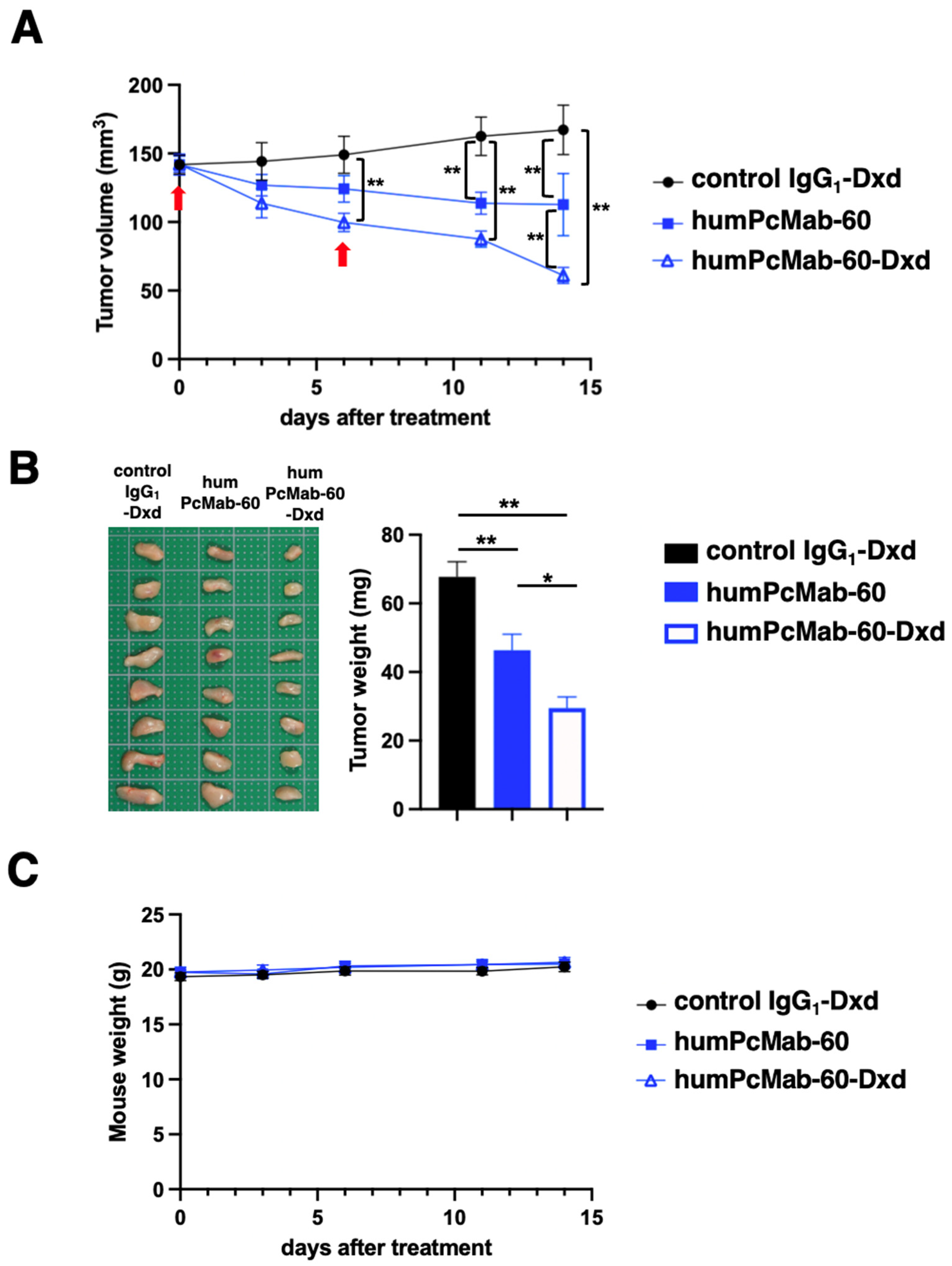
Figure 4.
Dose-dependent antitumor activity of humPcMab-60-Dxd against MIA PaCa-2 xenografts. (A) MIA PaCa-2 was subcutaneously injected into BALB/c nude mice. After a week, control IgG1-Dxd (100 μg, n = 8) or humPcMab-60-Dxd (100, 200, and 300 μg, n = 8) were intraperitoneally injected into each mouse on days 0 and 6 (arrows). Tumor volume is shown as mean ± SEM. ** p < 0.01 (two-way ANOVA followed by Tukey’s post hoc test). (B) The mice were euthanized on day 21. We measured tumor appearance (left) and weight (right) of MIA PaCa-2 xenografts. Values are presented as the mean ± SEM. ** p < 0.01 (One-way ANOVA followed by Tukey’s post hoc test). Each grid line is 1 cm. (C) Body weight (mean ± SEM) of xenograft-bearing mice treated with the mAbs. There is no significant difference (two-way ANOVA followed by Tukey’s post hoc test).
Figure 4.
Dose-dependent antitumor activity of humPcMab-60-Dxd against MIA PaCa-2 xenografts. (A) MIA PaCa-2 was subcutaneously injected into BALB/c nude mice. After a week, control IgG1-Dxd (100 μg, n = 8) or humPcMab-60-Dxd (100, 200, and 300 μg, n = 8) were intraperitoneally injected into each mouse on days 0 and 6 (arrows). Tumor volume is shown as mean ± SEM. ** p < 0.01 (two-way ANOVA followed by Tukey’s post hoc test). (B) The mice were euthanized on day 21. We measured tumor appearance (left) and weight (right) of MIA PaCa-2 xenografts. Values are presented as the mean ± SEM. ** p < 0.01 (One-way ANOVA followed by Tukey’s post hoc test). Each grid line is 1 cm. (C) Body weight (mean ± SEM) of xenograft-bearing mice treated with the mAbs. There is no significant difference (two-way ANOVA followed by Tukey’s post hoc test).
Disclaimer/Publisher’s Note: The statements, opinions and data contained in all publications are solely those of the individual author(s) and contributor(s) and not of MDPI and/or the editor(s). MDPI and/or the editor(s) disclaim responsibility for any injury to people or property resulting from any ideas, methods, instructions or products referred to in the content. |
© 2026 by the authors. Licensee MDPI, Basel, Switzerland. This article is an open access article distributed under the terms and conditions of the Creative Commons Attribution (CC BY) license (http://creativecommons.org/licenses/by/4.0/).
Copyright: This open access article is published under a Creative Commons CC BY 4.0 license, which permit the free download, distribution, and reuse, provided that the author and preprint are cited in any reuse.