Preprint
Article

This version is not peer-reviewed.

Combined Salmonella and Pasteurella multocida Antigens, Single-Dose and Adsorbed in Aluminum Hydroxide-Chitosan, Protect Swiss Mice Against Lethal Challenge

Submitted:

14 September 2026

Posted:

15 September 2026

You are already at the latest version

Abstract

Avian cholera and typhoid are major bacterial diseases in the poultry industry, causing reduced productivity and economic losses. Here, we developed a single-dose, bivalent, inactivated vaccine containing Pasteurella multocida and Salmonella spp. antigens adsorbed to an aqueous matrix based on chitosan, aluminum hydroxide, and saponin. Different concentrations of the experimental vaccine were evaluated in mice to determine the optimal single-dose concentration. Vaccine efficacy was assessed by survival rates; the experimental vaccine provided complete protection, whereas 60% of animals immunized with a commercial vaccine survived a challenge with 1×10¹⁰ CFU/mL of P. multocida and 5×10¹⁰ CFU/mL of Salmonella spp. Subsequently, experiments were conducted to determine the cytokine profiles of animals immunized with the experimental vaccine (single-dose vs. double-dose regimens) followed by a challenge. Pre-challenge cytokine analysis revealed similar profiles for IL-2, IL-4, IL-10, IL-17, IFN-γ, and TNF-α across groups; however, IL-6 levels were significantly higher in the single-dose experimental vaccine group (G1) than in the commercial vaccine group (G2). In the post-challenge comparative study, the cytokine response induced by the single-dose vaccine was robust and stable, showing no negative interference following the Salmonella spp. challenge, although the double-dose regimen did boost cellular and regulatory cytokines (IFN-γ, TNF-α, and IL-10) specifically against Salmonella spp. Conversely, no significant differences in cytokine profiles were observed among the treatments for groups challenged with P. multocida or for unchallenged groups. The single-dose regimen proved fully sufficient to confer complete protective immunity against both pathogens. The results demonstrated the potential of the single-dose bivalent inactivated vaccine as a promising strategy for the simultaneous prevention of avian typhoid and cholera.

Keywords: 
;  ;  ;  ;  ;  

1. Introduction

Fowl cholera and fowl typhoid are bacterial diseases with major economic impact, causing significant losses to poultry production in various regions of the world. Fowl cholera is caused by Pasteurella multocida, a pathogen capable of infecting different animal species; it causes hepatic lesions and sudden death in infected chickens, posing a serious threat to the poultry industry. It is classified into five capsular serogroups (A, B, D, E, and F), with serogroups A and F (and more rarely D) primarily associated with disease cases [1]. On the other hand, Salmonella spp. infections remain a persistent challenge, as they can lead to secondary mutations and virulence reversion, as well as severe reactions in immunocompromised animals [2,3]. The persistence and dissemination of these agents within poultry production systems present additional control challenges. In the case of fowl cholera, wild birds act as important reservoirs for P. multocida, facilitating the spread of the agent to commercial flocks [4]. Furthermore, once introduced into a farm, the bacterium can establish persistent infections and become endemic, hindering eradication efforts and fostering recurrent outbreaks [5]. Similarly, fowl typhoid remains endemic in various regions of Central America, South America, Africa, and Southeast Asia, posing an obstacle to the sustainability of poultry production and to the international trade of poultry and poultry products [2]. Inactivated vaccines are generally well-tolerated by host organisms and serve as key strategies for controlling infectious diseases in poultry farming, contributing to reduced mortality, improved production metrics, and decreased economic losses associated with infectious outbreaks [3]. However, commercially available inactivated vaccines still face limitations regarding the breadth and duration of the induced immune response [6]. Strategies involving antigen selection and adsorption methods utilizing novel immunomodulatory adjuvants show promise for enhancing vaccine efficacy. Adjuvants can boost antigen immunogenicity, prolong the immune response, stimulate cellular and humoral immunity mechanisms, and reduce antigenic competition in combination vaccines [7,8,9]. Research into novel adjuvants highlights the use of chitosan, valued for its mucoadhesive properties and ability to facilitate interactions between antigens and immune system cells. Saponins enhance the simultaneous stimulation of cellular and humoral responses, contributing to mechanisms associated with both Th1 and Th2 profiles when combined with aluminum hydroxide matrixes; the latter are predominantly used in commercial vaccines due to their role in inducing Th2-type responses [10,11,12,13,14,15].
Therefore, this study proposes an experimental vaccine utilizing inactivated Salmonella spp. antigens and P. multocida adsorbed onto a matrix composed of chitosan, aluminum hydroxide, and saponin, aiming to enhance the immune response and extend the protection conferred by immunization in murine models. The proposal seeks to contribute to the development of more efficient vaccination strategies for the poultry industry, with the potential to reduce the handling associated with repeated administrations and optimize immunization programs for poultry.

2. Materials and Methods

2.1. Bacterial Strains and LD50 Determination

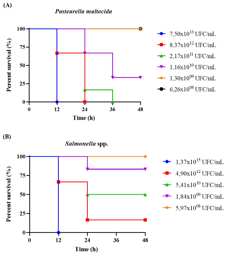
The P. multocida and Salmonella spp. bacterial strains used in this study were provided by the Avian Medicine Laboratory at the State University of Londrina, Brazil. The isolates were cultured in Brain Heart Infusion (BHI) broth (Merck®) in an orbital shaking incubator for 12 h at 37 °C and 180 rpm. Cells were harvested using a benchtop centrifuge (model 80-2B) at 3,000 rpm for 10 min; the cells were collected and resuspended in phosphate-buffered saline (PBS, pH 7.2), a process repeated three times. The median lethal dose (LD50) of P. multocida and Salmonella spp. was determined according to the Reed and Muench method [16,17]. To determine the LD50 of P. multocida, mice (n = 6 per group) were inoculated intraperitoneally with 200 µL of active bacterial suspension at concentrations of 7.50×10¹³; 8.37×10¹²; 2.17×10¹¹; 1.16×10¹⁰; 1.30×10⁹; and 6.26×10⁸ CFU/mL. For the LD50 determination of Salmonella spp., animals were distributed into five groups (n = 6 per group) and inoculated intraperitoneally with the same volume (200 µL) of active suspension at concentrations of 1.37×10¹⁵; 4.90×10¹²; 5.41×10¹⁰; 1.84×10⁹; and 5.97×10⁸ CFU/mL. The animals were monitored for 48 hours, with events and clinical signs recorded, to determine the LD50.

2.2. Ethical/legal Requirements and Use of Animals in Experiments

Mice weighing 20–24 g were obtained from the Federal University of Goiás (UFG) and housed in the preclinical testing room of the Biomolecules and Vaccines Laboratory (LaBVac) at the Federal University of Tocantins (UFT), Brazil. The animals were kept in pathogen-free primary enclosures with ad libitum access to food and water, maintained at a controlled temperature (24 °C), with odor and lighting controls adjusted to a 12-hour light/dark cycle. All animals used in this experiment were previously dewormed via access to an Ivermectin® solution (1:20 dilution) for 7 days. The experiments were conducted in accordance with ethical recommendations established by the law governing procedures for the scientific use of animals. They were approved by the Ethics Committee on Animal Use (CEUA) of the Federal University of Tocantins (protocol no. 23101.002359/2020-31). Every effort was made to avoid undue suffering or pain; the animals were monitored for clinical status, changes in body weight, and clinical signs such as lethargy, hypothermia, and/or respiratory distress. The onset of such clinical signs was treated as a death event and recorded to determine the cumulative mortality for each experimental group and subsequently calculate the LD50. At the end of the observation period, the animals were euthanized using ketamine (300 mg/kg) (Vetbrands®, Brazil) and xylazine hydrochloride (22.5 mg/kg) (Syntec®, Brazil).

2.3. Antigen Production, Inactivation, and Vaccine Formulation

The vaccine antigen was obtained from inactivated P. multocida and Salmonella spp. bacterial cells derived from pathogen cultures in BHI broth. Inactivation was achieved by adding a 0.6% (v/v) formalin solution under agitation at 150 rpm for 48 hours at 37 °C. Inactivation efficacy was verified by plating 1 mL of each inactivated sample onto BHI agar, followed by incubation for 24 hours at 37 °C. The adjuvant used was prepared by first solubilizing chitosan powder (Polymar Ltd.a, 81% degree of deacetylation) in 0.8% (v/v) acetic acid and 0.9% (w/v) saline solution, using a 2:1 ratio of chitosan to aluminum hydroxide (DEUSDARÁ et al., 2023), supplemented with saponin at a final concentration of 500 ng/mL. The experimental vaccine was formulated with both antigens at a concentration of 109 cells/mL, using a ratio of 25% (v/v) adjuvant and 10% (v/v) saponin.

2.4. Definition of Vaccine Dose

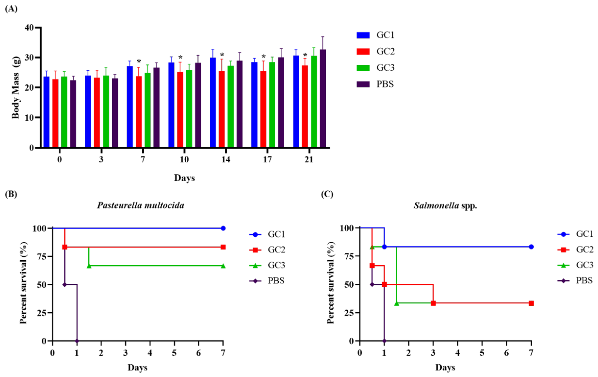
To determine the dose of the experimental vaccine, mice were assigned to four experimental groups of 12 animals each, designated GC1, GC2, GC3, and PBS. Groups GC1, GC2, and GC3 received a single intramuscular dose of 250 µL, 125 µL, and 50 µL of the experimental vaccine, respectively. The PBS group received a single 250 µL dose of PBS and served as the negative control. Animals were monitored daily from day 1 to day 21 post-immunization and evaluated for clinical signs including changes in feed intake, lethargy, prostration, and diarrhea as well as variations in body weight (Figure 1-A). Following this analysis period, the groups were subdivided and challenged intraperitoneally with 200 µL of a suspension containing the respective viable bacteria at a concentration corresponding to the median lethal dose (LD50) previously determined for each pathogen. A second study was designed based on data obtained from the standardization of the experimental vaccine dose. Thus, the vaccine dose defined in the previous study was compared with the vaccination protocol used by the manufacturer of a commercial vaccine employed in the poultry industry against both pathogens (Figure 1-B). Mice were assigned to four experimental groups of 12 animals each: experimental vaccine (single 250 µL dose); commercial vaccine (two 250 µL doses with a 14-day interval); adjuvant (single 250 µL dose of the formulation without bacterial antigen); and negative control (250 µL of PBS). Twenty-one days after immunization, the animals were challenged intraperitoneally with 200 µL of a virulent bacterial suspension of P. multocida and Salmonella spp. Following the challenge, the animals were monitored daily for seven days, with clinical signs and mortality recorded.

2.5. Cytokine Determination Assay

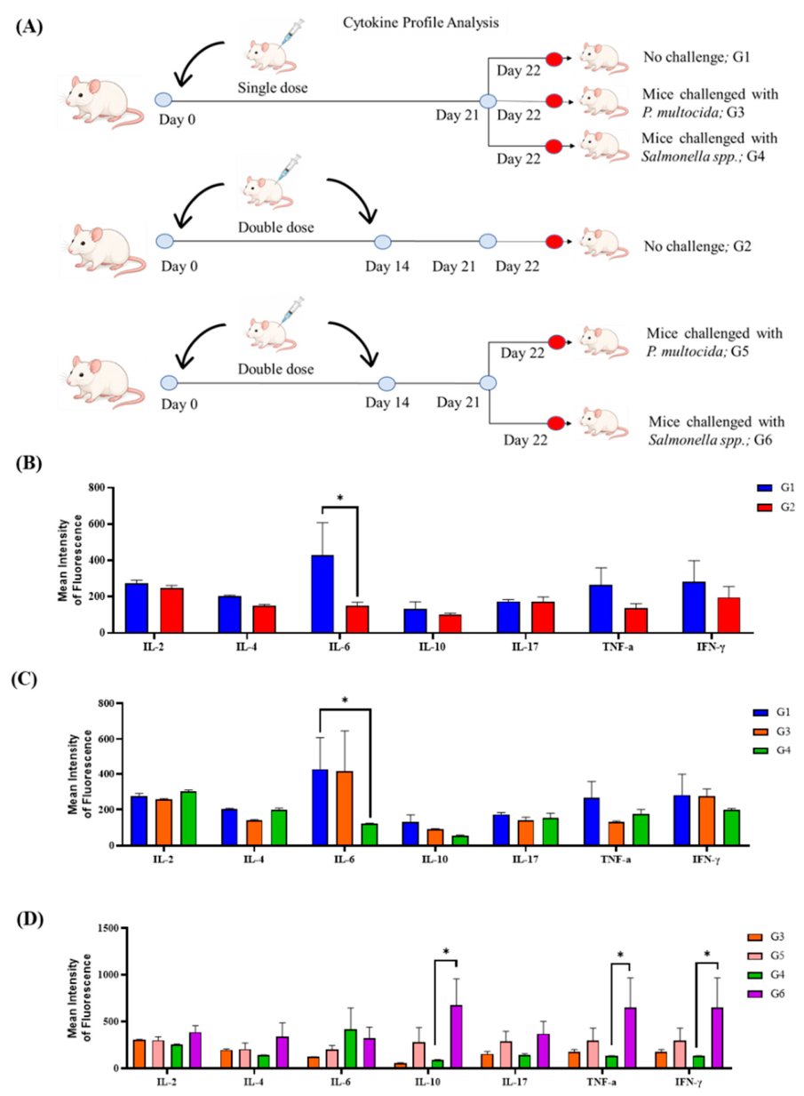
Cytokine analysis of animals was carried out and distributed into six experimental groups: G1 and G2, composed of animals immunized, respectively, with a single dose of the experimental vaccine and two doses of the commercial vaccine, without subsequent challenge; G3 and G4, immunized with a single dose of the experimental vaccine and challenged with P. multocida and Salmonella spp., respectively; and G5 and G6, immunized with two doses of the experimental vaccine and subsequently challenged with P. multocida and Salmonella spp., respectively. Blood plasma samples from the unchallenged groups were collected 21 days after the first immunization. In contrast, in the groups subjected to the experimental challenge, collection was carried out three days after inoculation of the pathogens. Plasma was obtained by centrifuging whole blood in a microcentrifuge (Microcentrifuge G-L, IKA®) at 15,000 rpm for 30 minutes. After separation, the samples were stored at 2 °C until flow cytometry analysis. The concentrations of interleukin-2 (IL-2), IL-4, IL-6, IL-10, IL-17, Tumor Necrosis Factor alpha (TNF-α), and interferon-gamma (IFN-γ) were determined using Cytometric Bead Array (CBA) technology. For this analysis, the BD™ CBA Mouse Th1/Th2/Th17 kit was used, following the manufacturer’s instructions. Data were acquired on a flow cytometer (FACSCanto™ II), and results were expressed as mean fluorescence intensity (mean fluorescence intensity—MFI).

2.6. Statistical Analysis

Statistical tests were performed using GraphPad Prism 8.0 software. The log-rank test was used to compare two or more groups with censored data, defined as individual death, evaluated via Kaplan-Meier analysis. Results are presented as mean values ± SD. ANOVA was used to assess statistical differences between groups. Statistical significance was set at p < 0.05.

3. Results

3.1. Determination of the Lethal Dose of Salmonella Spp. and P. Multocida in Murine Models

The virulence of P. multocida strains in Swiss mice resulted in clinical signs consistent with avian cholera at concentrations ranging from 7.5×10¹³ to 2.17×10¹¹, leading to the most acute form of the disease and 100% mortality in the group (Figure 2-A). Conversely, animals inoculated with concentrations equal to or lower than 1.16×10¹⁰ included survivors throughout the experimental period (Figure 2-A), indicating a lethal infection dose corresponding to 95 LD50 (1×10¹⁰ CFU/mL). Regarding the LD50 of Salmonella spp., all animals inoculated with a concentration of 1.37×10¹⁵ died within 12 hours of observation; lower doses showed progressively higher survival rates as the bacterial infection concentration was reduced (Figure 2-B), indicating a lethal infection dose corresponding to 40 LD50 (5×10¹⁰ CFU/mL).

3.2. Evaluation of Vaccine Dosage Using Murine Models

In the preliminary study of vaccine dosages GC1 (250 µL), GC2 (125 µL), and GC3 (50 µL), the evaluation of the experimental animals’ mean body mass revealed no significant changes in weight gain throughout the 21-day post-immunization period. At the end of this period, a statistically significant difference (p < 0.05) was observed only between the GC2 group and the control group. In contrast, the GC1 and GC3 groups showed no differences, thereby demonstrating the formulation’s safety and the maintenance of the animals’ zootechnical performance (Figure 3-A). Regarding the survival rate of animals immunized and challenged with the P. multocida LD50, the GC1 group exhibited 100% survival, representing the highest observed survival rate. In contrast, animals in the GC2 and GC3 groups showed survival rates of 83% and 66%, respectively (p < 0.05) (Figure 3-B). Meanwhile, the survival rate of animals immunized and challenged with Salmonella spp. showed a progressive decline over the monitoring period, varying according to the administered dose of the experimental vaccine; the GC1 group demonstrated the highest survival rate (83%), whereas the GC2 and GC3 groups showed a more marked reduction over time, resulting in 33% survival in both groups (p < 0.05) (Figure 3-C).
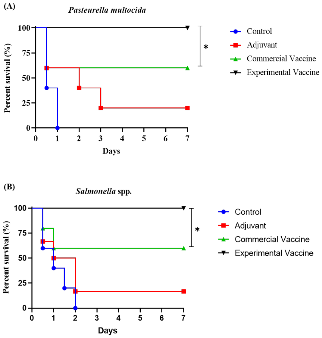
With the vaccine dosage established, the experimental vaccine provided 100% survival for animals challenged with the LD50 of P. multocida and Salmonella spp, during the seven-day post-challenge observation period. In contrast, animals immunized with the commercial vaccine showed a survival rate of 60%, whereas those that received only the adjuvant formulation showed a survival rate of 16%. No surviving animals were observed in the control group inoculated with PBS (p < 0.05) (Figure 4A,B).

3.3. Determination of the Cytokine Profile of Animals Immunized with Experimental and Commercial Vaccines, with and Without Challenge, in Murine Models

Evaluation of the cytokine profile post-immunization and before experimental challenge (Figure 5-A) revealed slight differences between the single-dose immunization group (G1) and the commercial double-dose vaccine group (G2) (Figure 5-B); however, among the cytokines evaluated, IL-6 showed a significant difference (p<0.05), with higher levels in the single-dose group (G1). We also investigated the effect of bacterial challenge with either P. multocida (G3) or Salmonella spp. (G4) on the immune response induced by immunization. Data showed no significant differences for most of the cytokines evaluated (Figure 5-C). However, it was again observed that IL-6 levels were significantly higher in immunized, unchallenged animals compared to the group immunized and subsequently challenged with P. multocida (p < 0.05), whereas Salmonella spp. (G4) did not interfere with cytokine stimulation before or after experimental challenge; this indicates that the primary stimulus was driven by immunization, without further stimulation interference following the experimental challenge (Figure 5-D). We also conducted a comparative study of immunization protocols and cytokine stimulation data using single-dose (G3 and G4) and double-dose (G5 and G6) experimental vaccines, assessing the period post-immunization and following the respective bacterial challenges to contribute to the understanding of the protective immune response against both pathogens (Figure 5-D). Here, we observed that animals immunized with either the single-dose or double-dose experimental vaccine protocols were protected following the challenge test, with 100% survival; this demonstrates that the stimulated cytokine levels conferred a protective response even when the animals were challenged with either pathogen. Following challenge, animals that received two vaccine doses exhibited enhanced cytokine stimulation. Of note were the groups challenged with Salmonella spp., where the two-dose regimen resulted in significantly higher (p < 0.05) concentrations of IFN-γ, TNF-α, and IL-10 compared to the single-dose group; this indicates greater activation of cellular and regulatory immunological mechanisms associated with the response to intracellular pathogens. In contrast, no significant differences were observed between the evaluated vaccination protocols in the groups challenged with P. multocida.

4. Discussion

Inactivated vaccines serve as an alternative that enables broader coverage against infectious agents through simultaneous antigen exposure, stimulates the herd’s immune system, offers logistical efficiency and reduced transport costs, and improves animal management. However, in some cases, they may induce weaker immune responses; therefore, it is important to devise strategies, such as using adjuvants to enhance immunogenicity [18]. Commonly used adjuvants include saponin [19], chitosan [20], and aluminum hydroxide [21]. In this study, we developed a single-dose bivalent inactivated vaccine targeting Salmonella spp. and P. multocida, pathogens that cause significant economic losses in the global poultry industry. Data from a murine model demonstrated the high efficacy of the experimental vaccine, ensuring complete protection following challenge with virulent strains of Salmonella spp. (Figure 4-A) and P. multocida (Figure 4-B), and showing superior performance compared to the reference commercial vaccine, which requires a two-dose regimen.
Given the sanitary and economic significance of these diseases, developing effective vaccination strategies is crucial for controlling fowl typhoid and fowl cholera in poultry production systems. Various approaches, such as vaccines using Salmonella spp. strains attenuated via SptP gene deletion, radiation-inactivated vaccines, inactivated vaccines combined with L-proline supplementation, and the use of adjuvants derived from bacterial DNA have been reported to enhance humoral and cellular immune responses in murine models [22,23,24,25]. Consequently, data from numerous studies reinforce the potential of inactivated vaccines as a cost-effective disease control strategy, corroborating the findings of the present study.
In this context, the route of administration can also provide insight into the immune response. The intramuscular immunization route adopted in this study may facilitate local antigen retention at the injection site, promoting the recruitment of antigen-presenting cells and the activation of the systemic immune response in regional lymph nodes [26]. Conversely, the intraperitoneal challenge widely used in experimental models of bacterial infection mimics the systemic dissemination of pathogens; this makes the systemic immunity induced by intramuscular vaccination biologically relevant and capable of conferring protection in this experimental model [27]. Regarding the cytokine profile, IFN-γ levels increased significantly in the group immunized with a double dose of the experimental vaccine and subsequently challenged with Salmonella spp. (G6) (Figure 5-C); this pattern mirrored the TNF-α levels observed in the immunized animals of group G6, suggesting the involvement of immunological mechanisms characteristic of a Th1 response. Such mechanisms are crucial for containing intracellular pathogens, as they promote macrophage activation and enhance cell-mediated immunity [28]. Activation of this immunological pathway is recognized as a key mechanism in the elimination of intracellular pathogens, contributing to the control of bacterial replication and the establishment of a more efficient protective response against Salmonella spp. Infection [29].
IL-17 levels in group G6 do not allow us to confirm the involvement of the Th17 response in the conditions evaluated (p > 0.05), although the observed pattern suggests a possible role for this immunological mechanism. This response has been linked to the recruitment of inflammatory cells to sites of infection and the amplification of immunity against invasive bacteria [28]. The increase in IL-10 observed in group G6 compared to G4 suggests that administering double doses of the experimental vaccine promoted a more pronounced modulation of the immune response following the Salmonella spp. challenge. IL-10 plays a key regulatory role, contributing to the control of inflammation and limiting tissue damage resulting from the immune response [30].
Although this profile indicates active engagement of the adaptive immune response, it is worth noting that the group immunized with a single dose (G4) despite showing no statistically significant changes in cytokine levels after the challenge (p > 0.05) (Figure 5-B) achieved a survival rate equivalent to that of the double-dose group, with both groups showing 100% protection. This finding suggests that the single-dose was capable of establishing sufficient protective immunity to control the Salmonella spp. infection without requiring intense systemic inflammatory activation; this can be interpreted as a more efficiently modulated immune response, wherein the immunity established by vaccination obviated the need for extensive recruitment of pro-inflammatory mediators following the challenge [31]. From this perspective, cytokine stability in the single-dose group does not indicate an absence of immune response but rather evidence of effective infection control with a lower inflammatory cost—a desirable characteristic for vaccine formulations, especially considering that single-dose regimens reduce animal stress, operational costs, and vaccination coverage failures associated with multidose schedules in the field [26]. In the group challenged with Salmonella spp., levels of IL-6 and the other cytokines evaluated (IL-2, IL-4, IL-10, IL-17, TNF-α, and IFN-γ) remained stable relative to pre-challenge levels (p > 0.05); this is biologically consistent with an immune response efficiently modulated by vaccination, wherein previously established immunological memory obviated the need for secondary systemic inflammatory activation, as described in murine models of vaccine-mediated protection against Salmonella [31,32]. The stability of plasma cytokines post-challenge observed in vaccinated animals aligns with studies demonstrating distinct immunological profiles between immunized and non-immunized animals following a DL50 challenge, often characterized by the absence of exacerbated systemic inflammatory amplification in protected groups [33].
Immunization against P. multocida and the resulting cytokine profile indicate stimulation characteristic of a response to a predominantly extracellular pathogen; this suggests that mechanisms linked to the humoral response may have contributed to animal protection, given the high survival rate and effective protection observed during the experimental challenge [34]. In non-challenged animals, the group receiving the experimental vaccine (G1) exhibited basal plasma IL-6 levels significantly higher than those of the group receiving the commercial vaccine (G2) (p < 0.05) (Figure 5-B). This effect is attributed to the immunostimulatory potential of the adjuvant used—composed of chitosan, aluminum hydroxide, and saponin corroborating findings by Zhao et al. [35], who observed elevated IL-6 levels in mice immunized with a bivalent pasteurellosis vaccine delivered via chitosan nanoparticles.
The data demonstrate that the evaluated bivalent vaccine successfully induced an immune response and provided effective protection against Salmonella spp. and P. multocida in the study model; this protection is associated with the modulation of immunological mechanisms linked to cellular and regulatory responses. Such findings reinforce the potential of this experimental vaccine as a promising strategy for developing bivalent vaccines aimed at minimizing the impact of poultry diseases. Future studies in the target species will be necessary to expand knowledge regarding the efficacy and safety of the vaccine developed in this pilot study. Additionally, further characterization of the vaccine-induced immune response, potentially including comparative studies of specific antibodies in murine and avian models, is warranted to assess its direct application for disease control in the poultry industry.

5. Conclusions

The results demonstrate that the single-dose bivalent inactivated vaccine developed in this study possesses high protective potential against Salmonella spp. and P. multocida challenges in a murine model, conferring 100% survival to immunized animals. Although the two-dose regimen increased cellular and regulatory cytokines post-challenge with Salmonella spp., the single-dose immunization was sufficient to activate the immunological mechanisms required for complete protection. Among the formulations evaluated, the 250 µL dose exhibited the best protective performance and an excellent safety profile, without affecting animal weight gain. Taken together, the data highlight the potential of combining chitosan, aluminum hydroxide, and saponin as a promising adjuvant platform, providing a basis for future studies in the target avian species—an essential step to bridge the immunological differences between the models.

Author Contributions

Hélio S. Brito: Writing—Original Draft, Data Curation, Investigation, Formal Analysis, Validation, Methodology, Conceptualization; Marcela O. Ferreira: Writing—Review & Editing; Gessyk M. Marques: Writing—Review & Editing; Tullio T. Deusdará: Writing—Review & Editing, Methodology; Wellington S. Moura: Writing—Review & Editing, Frederico Eugenio: Writing—Review & Editing; Edson W.S. Cangussu: Writing—Review & Editing Benedito Albuquerque: Methodology, Validation; Gil R. Santos: Writing—Review & Editing; Marcos G. Silva: Methodology, Validation, Writing—Review & Editing; Ana A. S. Baptista: Resources; Vladimir P. Nascimento: Resources; Igor V. Brandi: Writing—Review & Editing; Luis A. M. Mariúba: Writing—Review & Editing; Kelvison F. Viana: Writing—Review & Editing; Lucas S. S. Santos: Writing—Review & Editing; Vanessa M. Chapla: Writing—Review & Editing; Raynne O. Alves: Writing—Review & Editing; Paulo A. Nogueira: Conceptualization, Writing—Review & Editing; Keoma D. P. Cangussu: Resources, Review & Editing; Yuri O. Chaves: Methodology, Validation, Resources; Alex S. R. Cangussu: Writing—Review & Editing, Project Administration, Methodology, Resources, Supervision, Funding Acquisition.

Funding

This research was supported by the National Council of Scientific and Technological Development (CNPq, Brazil), Funding Authority for Studies and Projects (FINEP, Brazil) and ANTIGEN LTDA, projects: CNPq RHAE 424435/2021 and CNPq RHAE 441791/2024; FINEP 03.22.0448.00/2025. ASRC (CNPq 427304/2018-0, CNPq 305589/2021-0, CNPq 309054/2025-7). The Graduate Program for Biotechnology and the Graduate Program for Biodiversity and Biotechnology of the Legal Amazon from Federal University of Tocantins.

Institutional Review Board Statement

In this study, all procedures involving animals were approved by the Ethics Committee on Animal Use (CEUA) of the Federal University of Tocantins, Brazil (Approval No. 23101.002359/2020-31). All experiments were conducted in strict accordance with the guidelines of the National Council for the Control of Animal Experimentation (CONCEA), and all animal care and experimental protocols strictly adhered to relevant national and institutional standards regarding the use of laboratory animals.

Data Availability Statement

The original contributions presented in this study are included in the article. Further inquiries can be directed to the corresponding author.

Conflicts of Interest

Authors A.S.R.C. and K.D.P.C. are shareholders of Antigen Ltd.a., a company dedicated to the research, development, and innovation of veterinary products. The other authors declare no conflicts of interest.

References

  1. Wilkie, I.W.; Harper, M.; Boyce, J.D.; Adler, B. Pasteurella Multocida: Diseases and Pathogenesis. In Assessment & Evaluation in Higher Education; 2012; Vol. 37, pp. 1–22 ISBN 0260-2938.
  2. Foley, S.L.; Johnson, T.J.; Ricke, S.C.; Nayak, R.; Danzeisen, J. Salmonella Pathogenicity and Host Adaptation in Chicken-Associated Serovars. Microbiol. Mol. Biol. Rev. 2013, 77, 582–607. [CrossRef]
  3. Gebauer, J.; Tesařík, R.; Králová, N.; Havlíčková, H.; Matiašovic, J. Salmonella Typhimurium-Based Inactivated Vaccine Containing a Wide Spectrum of Bacterial Antigens Which Mimics Protein Expression Changes during Different Stages of an Infection Process. Vet. Microbiol. 2023, 282. [CrossRef]
  4. van Dijk, J.G.B.; Iverson, S.A.; Gilchrist, H.G.; Harms, N.J.; Hennin, H.L.; Love, O.P.; Buttler, E.I.; Lesceu, S.; Foster, J.T.; Forbes, M.R.; et al. Herd Immunity Drives the Epidemic Fadeout of Avian Cholera in Arctic-Nesting Seabirds. Sci. Rep. 2021, 11, 1–10. [CrossRef]
  5. Tang, Q.; Li, W.; Dai, N.; Gao, Y.; Han, Y.; Cheng, G.; Gu, C. The Role of Necroptosis, Apoptosis, and Inflammation in Fowl Cholera-Associated Liver Injury in a Chicken Model. Avian Dis. 2017, 61, 491–502. [CrossRef]
  6. Uribe-Diaz, S.; Santamaria, J.M.; Hargis, B.M.; Kwon, Y.M.; Vuong, C.N.; Erf, G.F. Decoding Poultry Immune Responses to Salmonella Vaccines: Current Advances and Future Directions for next-Generation Vaccine Development. Poult. Sci. 2025, 104, 105884. [CrossRef]
  7. Bonam, S.R.; Partidos, C.D.; Halmuthur, S.K.M.; Muller, S. An Overview of Novel Adjuvants Designed for Improving Vaccine Efficacy. Trends Pharmacol. Sci. 2017, 38, 771–793. [CrossRef] [PubMed]
  8. Del Giudice, G.; Rappuoli, R.; Didierlaurent, A.M. Correlates of Adjuvanticity: A Review on Adjuvants in Licensed Vaccines. Semin. Immunol. 2018, 39, 14–21. [CrossRef] [PubMed]
  9. Shi, S.; Zhu, H.; Xia, X.; Liang, Z.; Ma, X.; Sun, B. Vaccine Adjuvants: Understanding the Structure and Mechanism of Adjuvanticity. Vaccine 2019, 37, 3167–3178. [CrossRef] [PubMed]
  10. Aucouturier, J.; Dupuis, L.; Ganne, V. Adjuvants Designed for Veterinary and Human Vaccines. Vaccine 2001, 19, 2666–2672. [CrossRef]
  11. Drane, D.; Gittleson, C.; Boyle, J.; Maraskovsky, E. ISCOMATRIXTM Adjuvant for Prophylactic and Therapeutic Vaccines. Expert Rev. Vaccines 2007, 6, 761–772. [CrossRef]
  12. Illum, L. Nanoparticulate Systems for Nasal Delivery of Drugs: A Real Improvement over Simple Systems? J. Pharm. Sci. 2007, 96, 473–483. [CrossRef]
  13. Zeng, Z. Recent Advances of Chitosan Nanoparticles as Drug Carriers. Int. J. Nanomedicine 2011, 765. [CrossRef]
  14. Samal, S.K.; Dash, M.; Vlierberghe, S. Van; Kaplan, D.L.; Chiellini, E.; Blitterswijk, C. Van; Moroni, L.; Dubruel, P. Cationic Polymers and Their Therapeutic Potential. Chem. Soc. Rev. 2012, 41, 7147–7194. [CrossRef]
  15. Deusdará, T.T.; Félix, M.K.C.; de S. Brito, H.; Cangussu, E.W.S.; de S. Moura, W.; Albuquerque, B.; Silva, M.G.; dos Santos, G.R.; de Morais, P.B.; da Silva, E.F.; et al. Using an Aluminum Hydroxide–Chitosan Matrix Increased the Vaccine Potential and Immune Response of Mice against Multi-Drug-Resistant Acinetobacter Baumannii. Vaccines 2023, 11. [CrossRef]
  16. Ding, L.; Lu, J.; Zhu, Y.; Qi, Q.; Yan, G.; Shan, Z.; Lu, Y.; Xu, H. Integrated Genomic and Phenotypic Characterization of Representative Salmonella Pullorum and Salmonella Enteritidis Isolated from a Poultry Farm. Front. Microbiol. 2026, 17. [CrossRef]
  17. Reed, L.J.; Muench, H. A Simple Method of Estimating Fifty per Cent Endpoints. Am. J. Epidemiol. 1938, 27, 493–497. [CrossRef]
  18. Ghadimipour, R.; Ghorbanpoor, M.; Gharibi, D.; Mayahi, M.; Jabbary, A.R. Effects of Selected Adjuvants on Immunogenicity and Protectivity of Pasteurella Multocida Bacterin Vaccine in Chickens. Arch. Razi Inst. 2021, 76, 741–749. [CrossRef]
  19. David, S.C.; Lau, J.; Singleton, E. V; Babb, R.; Davies, J.; Hirst, T.R.; McColl, S.R.; Paton, J.C.; Alsharifi, M. The Effect of Gamma-Irradiation Conditions on the Immunogenicity of Whole-Inactivated Influenza A Virus Vaccine. Vaccine 2017, 35, 1071–1079. [CrossRef]
  20. Ayub, A.; Usman, M.; Ihsan, A.; Ain, Q.; Awan, A.B.; Wajid, M.; Ali, A.; Haque, A.; Iqbal, M.; Sarwar, Y. Immunological Characterization of Chitosan Adjuvanted Outer Membrane Proteins of Salmonella Enterica Serovar Typhi as Multi-Epitope Typhoid Vaccine Candidate. Mol. Biol. Rep. 2022, 49, 7377–7387. [CrossRef]
  21. Hem, S.L.; HogenEsch, H. Relationship between Physical and Chemical Properties of Aluminum-Containing Adjuvants and Immunopotentiation. Expert Rev. Vaccines 2007, 6, 685–698. [CrossRef]
  22. Ren, W.; Zou, L.; Ruan, Z.; Li, N.; Wang, Y.; Peng, Y.; Liu, G.; Yin, Y.; Li, T.; Hou, Y.; et al. Dietary L-Proline Supplementation Confers Immunostimulatory Effects on Inactivated Pasteurella Multocida Vaccine Immunized Mice. Amino Acids 2013, 45, 555–561. [CrossRef]
  23. Bahadur Basnet, H.; Kwon, H.; Cho, S.-H.; Kim, S.; Yoo, H.; Park, Y.; Yoon, S.; Shin, N.; Youn, H. Reproduction of Fowl Typhoid by Respiratory Challenge with Salmonella Gallinarum. Avian Dis. 2008, 52, 156–159. [CrossRef]
  24. Lin, Z.; Tang, P.; Jiao, Y.; Kang, X.; Li, Q.; Xu, X.; Sun, J.; Pan, Z.; Jiao, X. Immunogenicity and Protective Efficacy of a Salmonella Enteritidis SptP Mutant as a Live Attenuated Vaccine Candidate. BMC Vet. Res. 2017, 13, 1–9. [CrossRef]
  25. Homayoon, M.; Tahamtan, Y.; Kargar, M.; Hosseini, S.M.H.; Akhavan Sepahy, A. Pasteurella Multocida Inactivated with Ferric Chloride and Adjuvanted with Bacterial DNA Is a Potent and Efficacious Vaccine in Balb/c Mice. J. Med. Microbiol. 2018, 67, 1383–1390. [CrossRef]
  26. Zhao, T.; Cai, Y.; Jiang, Y.; He, X.; Wei, Y.; Yu, Y.; Tian, X. Vaccine Adjuvants: Mechanisms and Platforms. Signal Transduct. Target. Ther. 2023, 8. [CrossRef]
  27. Walker, G.T.; Gerner, R.R.; Nuccio, S.P.; Raffatellu, M. Murine Models of Salmonella Infection. Curr. Protoc. 2023, 3, 1–34. [CrossRef]
  28. Acevedo-Villanueva, K.Y.; Akerele, G.O.; Hakeem, W.G. Al; Renu, S.; Shanmugasundaram, R.; Selvaraj, R.K. A Novel Approach against Salmonella: A Review of Polymeric Nanoparticle Vaccines for Broilers and Layers. Vaccines 2021, 9. [CrossRef]
  29. Abreu, R.; Essler, L.; Giri, P.; Quinn, F. Interferon-Gamma Promotes Iron Export in Human Macrophages to Limit Intracellular Bacterial Replication. PLoS One 2020, 15, 1–19. [CrossRef]
  30. Martinez-Espinosa, I.; Serrato, J.A.; Ortiz-Quintero, B. Role of Il-10-Producing Natural Killer Cells in the Regulatory Mechanisms of Inflammation during Systemic Infection. Biomolecules 2022, 12. [CrossRef]
  31. Vinod, N.; Noh, H.B.; Oh, S.; Ji, S.; Park, H.J.; Lee, K.S.; Kim, S.C.; Park, H.O.; Yang, J.S.; Choi, C.W. A Salmonella Typhimurium Ghost Vaccine Induces Cytokine Expression in Vitro and Immune Responses in Vivo and Protects Rats against Homologous and Heterologous Challenges. PLoS One 2017, 12, 1–18. [CrossRef]
  32. Wang, N.; Scott, T.A.; Kupz, A.; Shreenivas, M.M.; Peres, N.G.; Hocking, D.M.; Yang, C.; Jebeli, L.; Beattie, L.; Groom, J.R.; et al. Vaccine-Induced Inflammation and Inflammatory Monocytes Promote CD4+ T Cell-Dependent Immunity against Murine Salmonellosis. PLoS Pathog. 2023, 19, 1–28. [CrossRef]
  33. Emami, S.; Westerlund, E.; Rojas Converso, T.; Johansson-Lindbom, B.; Persson, J.J. Protection Acquired upon Intraperitoneal Group a Streptococcus Immunization Is Independent of Concurrent Adaptive Immune Responses but Relies on Macrophages and IFN-γ. Virulence 2025, 16, 1–16. [CrossRef]
  34. Homayoon, M.; Tahamtan, Y.; Kargar, M. The Comparison Detection of Cytokines (IL-6 and IL-12) from Spleen Cells and Serums in Balb/c Mice after Immunization with Killed P. Multocida Vaccines Co-Formulated with Bacterial DNAs as Adjuvant. Arch. Clin. Microbiol. 2020, 11. [CrossRef]
  35. Zhao, Z.Z.; Zhang, H.B.; Chen, Q.; Su, D.; Xie, Z.; Wang, Y.Y.; Yang, Y.; Wang, Z.Z.; Li, J.L.; Wu, K.Y.; et al. Promotion of Immunity of Mice to Pasteurella Multocida and Hog Cholera Vaccine by Pig Interleukin-6 Gene and CpG Motifs. Comp. Immunol. Microbiol. Infect. Dis. 2009, 32, 191–205. [CrossRef]
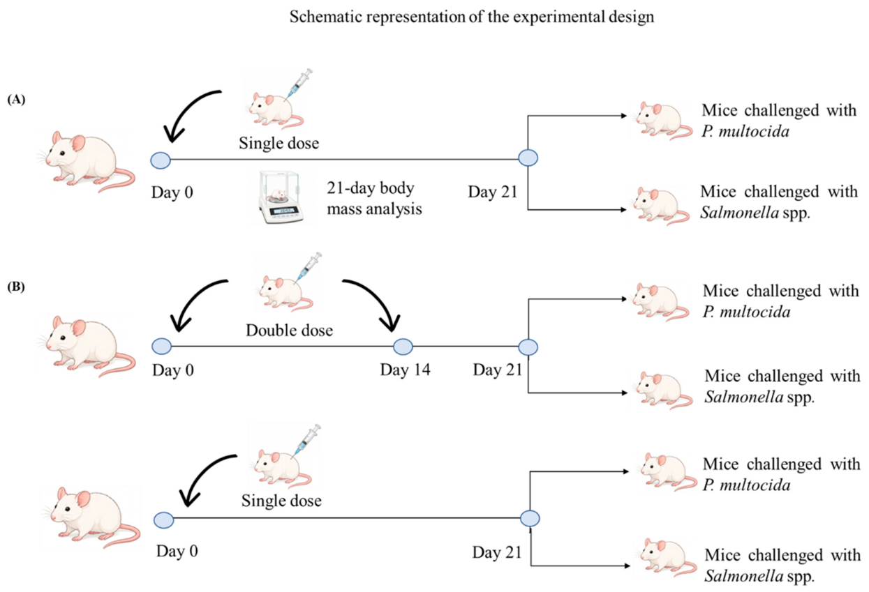
Figure 1. Schematic representation of the experimental design. (A) Protocol used to determine the optimal dose of the experimental vaccine, including single-dose immunization, monitoring of mice body weight over a 21-day period, and assessment of protective efficacy following experimental challenge with P. multocida or Salmonella spp. (B) Comparison of the immunization protocols used for the experimental vaccine (single dose) and the commercial vaccine (two doses administered on days 0 and 14), followed by assessment of protective efficacy after experimental challenge with P. multocida or Salmonella spp.
Preprints 233381 g001
Figure 2. Standardization of the median lethal dose (LD50) of P. multocida and Salmonella spp. in mice. Kaplan-Meier survival curves following intraperitoneal inoculation with increasing concentrations (108 to 1013 CFU/mL) of (A) P. multocida and (108 to 1015 CFU/mL) (B) Salmonella spp. Mortality was monitored every 12 hours for two days post-inoculation. LD50 values were calculated using the Reed-Muench method, determining the doses used in subsequent challenge assays.
Preprints 233381 g002
Figure 3. Groups GC1, GC2, GC3, and PBS were immunized and monitored for body weight over 21 days, then subdivided for bacterial challenges. (A) Variation in mouse body weight during the 21 days following immunization with the experimental vaccine. GC1: 250 µL; GC2: 125 µL; GC3: 50 µL; PBS: 250 µL of PBS. (B) Survival analysis of groups GC1, GC2, GC3, and PBS following challenge with P. multocida, and (C) survival analysis of groups GC1, GC2, GC3, and PBS following challenge with Salmonella spp. * = Statistically significant difference (p < 0.05).
Preprints 233381 g003
Figure 4. Protective efficacy of the experimental vaccine against bacterial challenges. Kaplan-Meier survival curves following challenge with the LD50 of (A) P. multocida and (B) Salmonella spp. The experimental groups consisted of unvaccinated animals (control, blue line —●—), animals immunized only with the adjuvant solution (red line —■—), animals immunized with the commercial vaccine using a two-dose regimen (green line —▲—), and animals immunized with the experimental vaccine using a single-dose regimen (black line —▼—). The animals (n = 12/group initially; n = 6/group per pathogen) were challenged 21 days after immunization in the single-dose protocol or seven days after the second immunization in the two-dose protocol. Survival was monitored daily for seven days following the challenge.
Preprints 233381 g004
Figure 5. Plasma cytokine profile in mice under different vaccination regimens and bacterial challenges. (A) Experimental group scheme: G1, experimental vaccine (single-dose, no challenge); G2, commercial vaccine (double dose, no challenge); G3 and G4, experimental vaccine (single-dose) challenged with P. multocida or Salmonella spp., respectively; G5 and G6, experimental vaccine (double-dose) challenged with P. multocida or Salmonella spp., respectively. (B) Plasma concentrations of IL-2, IL-4, IL-6, IL-10, IL-17, TNF-α, and IFN-γ (G1 vs. G2) during the pre-challenge period. (C) Effect of bacterial challenge on the immune response induced by the single dose of the experimental vaccine (G1 vs. G3 and G4). (D) Comparison of the post-challenge cytokine profile between single-dose (G3, G4) and double-dose (G5, G6) regimens. Data presented as mean ± SEM. *p < 0.05 indicates a statistically significant difference (two-way ANOVA followed by Tukey’s test).
Preprints 233381 g005
Disclaimer/Publisher’s Note: The statements, opinions and data contained in all publications are solely those of the individual author(s) and contributor(s) and not of MDPI and/or the editor(s). MDPI and/or the editor(s) disclaim responsibility for any injury to people or property resulting from any ideas, methods, instructions or products referred to in the content.
Copyright: This open access article is published under a Creative Commons CC BY 4.0 license, which permit the free download, distribution, and reuse, provided that the author and preprint are cited in any reuse.