

Submitted:
14 September 2026
Posted:
15 September 2026
You are already at the latest version
Abstract
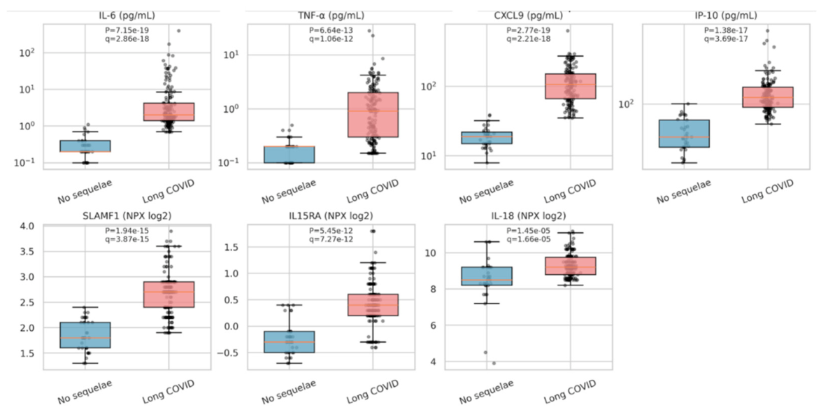
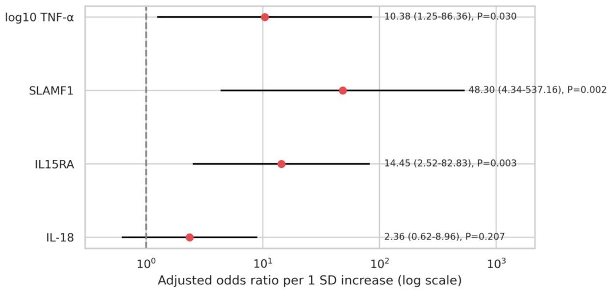
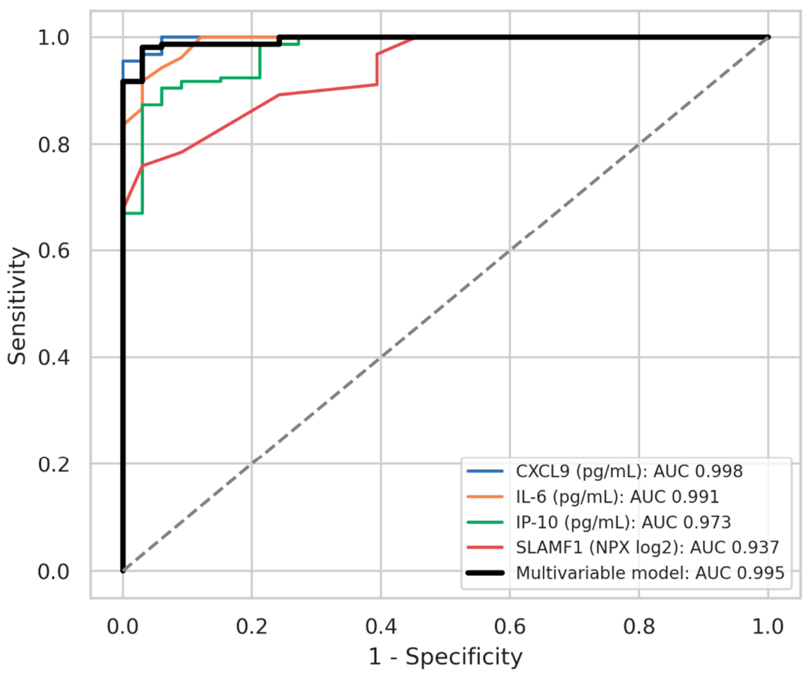
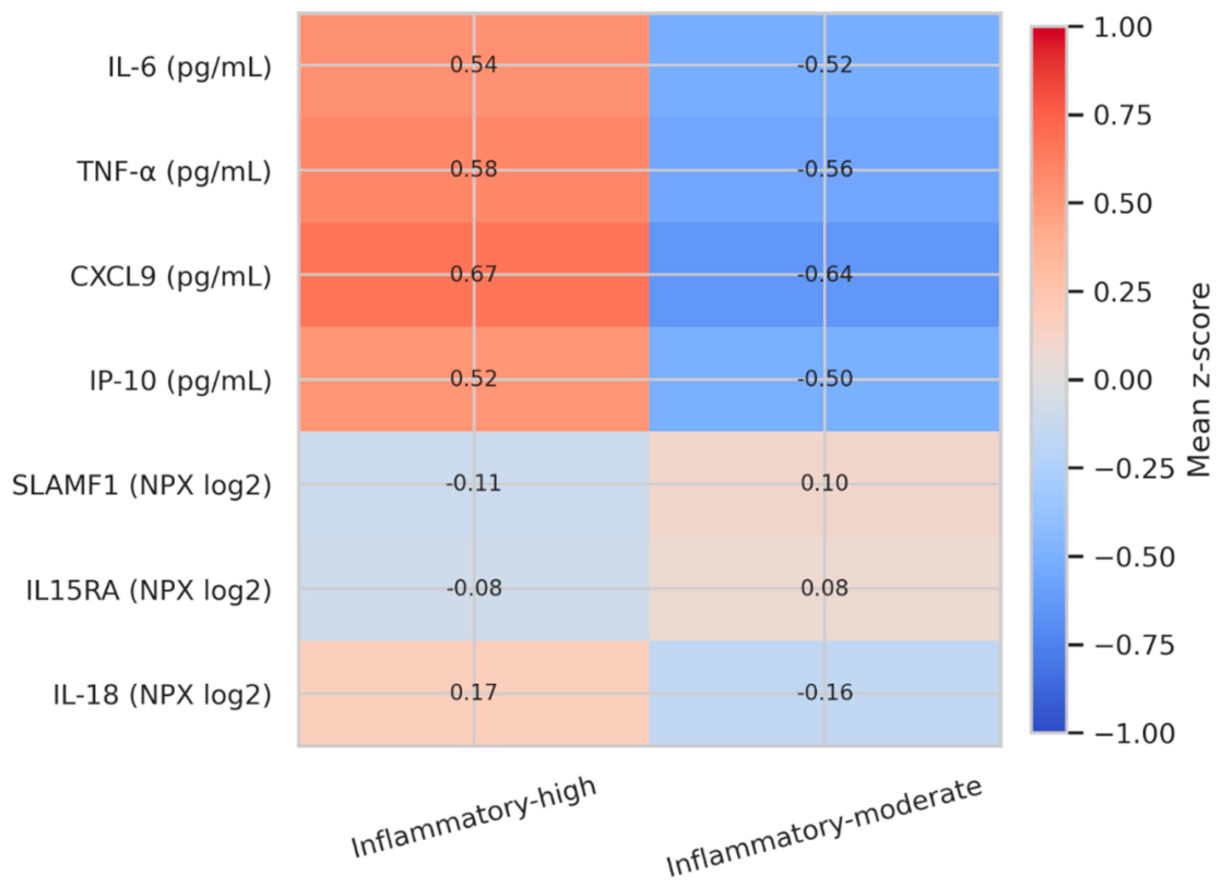
Background: Long COVID is clinically heterogeneous; objective biomarkers for diagnosis and biological stratification remain incompletely defined. Methods: We conducted a cross-sectional analysis of 191 de-identified post-COVID participants (158 with long COVID; 33 without sequelae), assessing eight biomarkers. Values below detection limits were imputed as half the assay-specific threshold. Analyses included two-sided Mann–Whitney U tests with Benjamini–Hochberg false-discovery-rate (FDR) correction, logistic regression, receiver operating characteristic analysis with stratified bootstrap confidence intervals (B=1,000), and Ward hierarchical clustering. Results: The long COVID group showed significantly higher IL-6, TNF-α, CXCL9, IP-10, SLAMF1, IL15RA, and IL-18 levels (all FDR q<0.001), whereas IFN-λ3 was not discriminatory (P=0.658). CXCL9 showed the highest single-marker performance (AUC 0.998, 95% confidence interval [CI] 0.989–1.000), followed by IL-6 (AUC 0.991) and IP-10 (AUC 0.973). In the stable multivariable model, higher log10-transformed TNF-α (adjusted odds ratio [aOR] 10.38, 95% CI 1.25–86.36; P=0.030), SLAMF1 (aOR 48.30, 95% CI 4.34–537.16; P=0.002), and IL15RA (aOR 14.45, 95% CI 2.52–82.83; P=0.003) remained independently associated with long COVID, whereas IL-18 was not significant (P=0.207). The model achieved an AUC of 0.995. Clustering identified high-inflammatory and moderate-inflammatory long COVID subtypes (n=79 each). Conclusions: Persistent inflammatory chemokine and cytokine elevation characterizes long COVID, with strong discrimination by CXCL9 and IL-6. Biomarker-defined subtypes support biological heterogeneity and may guide future patient stratification.
Keywords:
1. Introduction
2. Materials and Methods
2.1. Study Design and Analytical Dataset
2.2. Biomarkers and Data Handling
2.3. Statistical Analysis
3. Results
3.1 Study Population and Biomarker Completeness
3.2. Univariate Biomarker Comparison
3.3. Multivariable Logistic Regression
3.4. Diagnostic Performance
3.5. Long COVID Subtype Analysis
4. Discussion
4.1. Limitations
5. Conclusions
Author Contributions
Funding
Institutional Review Board Statement
Informed Consent Statement
Data Availability Statement
Acknowledgments
Conflicts of Interest
References
- Soriano, J.B.; Murthy, S.; Marshall, J.C.; Relan, P.; Diaz, J.V.; WHO Clinical Case Definition Working Group on Post-COVID-19 Condition. A clinical case definition of post-COVID-19 condition by a Delphi consensus. Lancet Infect Dis 2022, 22, e102–e107. [CrossRef]
- Sudre, C.H.; Murray, B.; Varsavsky, T.; Graham, M.S.; Penfold, R.S.; Bowyer, R.C.; Pujol, J.C.; Klaser, K.; Antonelli, M.; Canas, L.S.; et al. Attributes and predictors of long COVID. Nat Med 2021, 27, 626–631. [CrossRef]
- Xie, Y.; Bowe, B.; Al-Aly, Z. Burdens of post-acute sequelae of COVID-19 by severity of acute infection, demographics and health status. Nat Commun 2021, 12, 6571. [CrossRef]
- Phetsouphanh, C.; Darley, D.R.; Wilson, D.B.; Howe, A.; Munier, C.M.L.; Patel, S.K.; Juno, J.A.; Burrell, L.M.; Kent, S.J.; Dore, G.J.; et al. Immunological dysfunction persists for 8 months following initial mild-to-moderate SARS-CoV-2 infection. Nat Immunol 2022, 23, 210–216. [CrossRef]
- Lai, Y.J.; Liu, S.H.; Manachevakul, S.; Lee, T.A.; Kuo, C.T.; Bello, D. Biomarkers in long COVID-19: a systematic review. Front Med (Lausanne) 2023, 10, 1085988. [CrossRef]
- Thomas, C.; Faghy, M.A.; Chidley, C.; Phillips, B.E.; Bewick, T.; Ashton, R.E. Blood biomarkers of long COVID: a systematic review. Mol Diagn Ther 2024, 28, 537–574. [CrossRef]
- Yin, K.; Peluso, M.J.; Luo, X.; Thomas, R.; Shin, M.G.; Neidleman, J.; Andrew, A.; Young, K.C.; Ma, T.; Hoh, R.; et al. Long COVID manifests with T cell dysregulation, inflammation and an uncoordinated adaptive immune response to SARS-CoV-2. Nat Immunol 2024, 25, 218–225. [CrossRef]
- Phetsouphanh, C.; Jacka, B.; Ballouz, S.; Jackson, K.J.L.; Wilson, D.B.; Manandhar, B.; Klemm, V.; Tan, H.X.; Wheatley, A.; Aggarwal, A.; et al. Improvement of immune dysregulation in individuals with long COVID at 24-months following SARS-CoV-2 infection. Nat Commun 2024, 15, 3315. [CrossRef]
- Ai, J.; Guo, J.; Lin, K.; Cai, J.; Zhang, H.; Zhu, F.; Sun, G.; Xue, Q.; Zhu, K.; Yang, Y.; et al. Integrated multi-omics characterization across clinically relevant subgroups of long COVID. Natl Sci Rev 2024, 12, nwae410. [CrossRef]
- Gao, Y.; Cai, C.; Adamo, S.; Biteus, E.; Kamal, H.; Dager, L.; Miners, K.L.; Llewellyn-Lacey, S.; Ladell, K.; Amratia, P.S.; et al. Identification of soluble biomarkers that associate with distinct manifestations of long COVID. Nat Immunol 2025, 26, 692–705. [CrossRef]
- Aid, M.; Boero-Teyssier, V.; McMahan, K.; Dang, R.; Doyle, M.; Belabbaci, N.; Borducchi, E.; Collier, A.Y.; Mullington, J.; Barouch, D.H. Long COVID involves activation of proinflammatory and immune exhaustion pathways. Nat Immunol 2026, 27, 61–71. [CrossRef]
- Kumar, S.; Li, C.; Zhou, L.; Zhan, Q.; Alaswad, A.; Volland, S.; Costa, B.; Krooss, S.A.; Klefenz, I.; Schmaus, H.; et al. A distinct monocyte transcriptional state links systemic immune dysregulation to pulmonary impairment in long COVID. Nat Immunol 2026, 27, 200–212. [CrossRef]
- Olivarria G, Lane TE. Evaluating the role of chemokines and chemokine receptors involved in coronavirus infection. Expert Rev Clin Immunol. 2022, 18, 57–66. [CrossRef]
- Wolszczak-Biedrzycka, B.; Cieślikiewicz, B.; Studniarz, F.; Dąbrowski, Ł.; Fąs, M.; Matyszkiewicz-Suchodolska, K.; Harasimowicz, M.; Dorf, J. Chemokines as potential biomarkers for predicting the course of COVID-19—a review of the literature. Front Immunol 2025, 16, 1662643. [CrossRef]
- Talla, A.; Vasaikar, S.V.; Szeto, G.L.; Lemos, M.P.; Czartoski, J.L.; MacMillan, H.; Moodie, Z.; Cohen, K.W.; Fleming, L.B.; Thomson, Z.; et al. Persistent serum protein signatures define an inflammatory subcategory of long COVID. Nat Commun 2023, 14, 3417. [CrossRef]
- Quiroga, R.; Sanhueza, S.; Sepúlveda, C.; Antilef, B.; Muñoz, C.; Cabrera, C.; Fraga, M.; Llerena, F.; Lamperti, L.; Barría, M.I.; et al. CXCL9 and CXCL10 support the exacerbated humoral response in recovered COVID-19 patients who developed acute respiratory distress syndrome by promoting plasma cell differentiation, whereas CXCL9 also induces CD40L and CXCR3 upregulation on T helper cells. Front Immunol 2025, 16, 1684704. [CrossRef]
- Ruhl, L.; Pink, I.; Kühne, J.F.; Beushausen, K.; Keil, J.; Christoph, S.; Sauer, A.; Boblitz, L.; Schmidt, J.; David, S.; et al. Endothelial dysfunction contributes to severe COVID-19 in combination with dysregulated lymphocyte responses and cytokine networks. Signal Transduct Target Ther 2021, 6, 418. [CrossRef]
- Ruhl, L.; Pink, I.; Chichelnitskiy, E.; Drick, N.; Sauer, A.; Boblitz, L.; Beushausen, K.; Keil, J.; Ullrich, A.L.; Schmidt, J.; et al. Post-acute sequelae patients with severe COVID-19 history show a prolonged inflammatory, vascular injury pattern. Eur J Immunol 2026, 56, e70169. [CrossRef]
- Wu, F.; Mu, W.C.; Markov, N.T.; Fuentealba, M.; Halaweh, H.; Senchyna, F.; Manwaring-Mueller, M.N.; Winer, D.A.; Furman, D. Immunological biomarkers of aging. J Immunol 2025, 214, 889–902. [CrossRef]
- Singh, A.; Schurman, S.H.; Bektas, A.; Kaileh, M.; Roy, R.; Wilson, D.M.; Sen, R.; Ferrucci, L. Aging and inflammation. Cold Spring Harb Perspect Med 2024, 14, a041197. [CrossRef]
- Seo, D.H.; Corr, M.; Patel, S.; Lui, L.Y.; Cauley, J.A.; Evans, D.; Mau, T.; Lane, N.E. Chemokine CXCL9, a marker of inflammaging, is associated with changes of muscle strength and mortality in older men. Osteoporos Int 2024, 35, 1789–1796. [CrossRef]
- Muri, J.; Cecchinato, V.; Cavalli, A.; Shanbhag, A.A.; Matkovic, M.; Biggiogero, M.; Maida, P.A.; Moritz, J.; Toscano, C.; Ghovehoud, E.; et al. Autoantibodies against chemokines post-SARS-CoV-2 infection correlate with disease course. Nat Immunol 2023, 24, 604–611. [CrossRef]
- Yin, J.X.; Agbana, Y.L.; Sun, Z.S.; Fei, S.W.; Zhao, H.Q.; Zhou, X.N.; Chen, J.H.; Kassegne, K. Increased interleukin-6 is associated with long COVID-19: a systematic review and meta-analysis. Infect Dis Poverty 2023, 12, 43. [CrossRef]
- Obeagu, E.I. Taming the cytokine storm: therapeutic strategies for post-acute sequelae of SARS-CoV-2 infection. Health Sci Rep 2026, 9, e72174. [CrossRef]
- Espín, E.; Yang, C.; Shannon, C.P.; Assadian, S.; He, D.; Tebbutt, S.J. Cellular and molecular biomarkers of long COVID: a scoping review. EBiomedicine 2023, 91, 104552. [CrossRef]
- Hein, Z.M.; Thazin, K.S.; Kumar, S.; Che Ramli, M.D.; Che Mohd Nassir, C.M.N. Immunomodulatory mechanisms underlying neurological manifestations in long COVID: implications for immune-mediated neurodegeneration. Int J Mol Sci 2025, 26, 6214. [CrossRef]
- Abbas, U.; Laghari, R.N.; Ahmed, I.; Musawwir, U.A.; Riaz, H.; Anwar, K.; Hussain, N.; Mubeen, M.; Khan, M.; Khalid, M.U.; et al. Acute SARS-CoV-2 viral load and systemic inflammation are associated with neuropsychiatric and musculoskeletal symptoms in long COVID. PLoS One 2026, 21, e0346978. [CrossRef]
- Bachiller, S.; Vitallé, J.; Camprubí-Ferrer, L.; García, M.; Gallego, I.; López-García, M.; Galvá, M.I.; Cañizares, J.; Rivas-Jeremías, I.; Díaz-Mateos, M.; et al. SARS-CoV-2 post-acute sequelae linked to inflammation via extracellular vesicles. Front Immunol 2025, 16, 1501666. [CrossRef]
- Maciel, A.B.S.; Pinto, A.S.; Maia Silva, B.; Goulart, C.L.; Silva, L.F.A.; Chaves, A.S.; Mouta, G.S.; Sato, C.M.S.; Valente, J.; Mwangi, V.I.; et al. Inflammatory discoveries two years after acute severe COVID-19: a longitudinal biomarker profile assessment in long COVID individuals in the Brazilian Amazon. Front Immunol 2024, 15, 1520193. [CrossRef]
- Omdal, R.; Lenning, O.B.; Jonsson, G.; Kvaløy, J.T.; Di Molfetta, G.; Tan, K.; Benedet, A.L.; Ashton, N.J.; Braut, G.S.; Zetterberg, H.; et al. Long-COVID: assessment of circulating markers suggests no cerebral neuronal damage, neuroinflammation or systemic inflammation—a controlled study. Sci Rep 2026, 16, 11856. [CrossRef]
- Detre, C.; Keszei, M.; Romero, X.; Tsokos, G.C.; Terhorst, C. SLAM family receptors and the SLAM-associated protein (SAP) modulate T cell functions. Semin Immunopathol 2010, 32, 157–171. [CrossRef]
- Yigit, B.; Wang, N.; Herzog, R.W.; Terhorst, C. SLAMF receptors: immune regulators in health and disease. Clin Immunol 2018, 204, 3–13. [CrossRef]
- Farhangnia, P.; Ghomi, S.M.; Mollazadehghomi, S.; Nickho, H.; Akbarpour, M.; Delbandi, A.A. SLAM-family receptors come of age as a potential molecular target in cancer immunotherapy. Front Immunol 2023, 14, 1174138. [CrossRef]
- Mortier, E.; Advincula, R.; Kim, L.; Chmura, S.; Barrera, J.; Reizis, B.; Malynn, B.A.; Ma, A. Macrophage- and dendritic-cell-derived interleukin-15 receptor alpha supports homeostasis of distinct CD8+ T cell subsets. Immunity 2009, 31, 811–822. [CrossRef]
- Guo, Y.; Luan, L.; Patil, N.K.; Wang, J.; Bohannon, J.K.; Rabacal, W.; Fensterheim, B.A.; Hernandez, A.; Sherwood, E.R. IL-15 enables septic shock by maintaining NK cell integrity and function. J Immunol 2017, 198, 1320–1333. [CrossRef]

|
Marker |
Long COVID, median [IQR] |
No sequelae, median [IQR] |
P value |
FDR q |
Rank-biserial r |
AUC |
| CXCL9 | 106.00 [65.90–151.00] |
19.00 [15.00–22.00] |
5.09×10⁻¹⁹ | 2.86×10⁻¹⁸ | 0.987 | 0.998 |
| IL-6 | 2.00 [1.40–4.25] |
0.20 [0.20–0.40] |
7.15×10⁻¹⁹ | 2.86×10⁻¹⁸ | 0.983 | 0.991 |
| IP-10 | 118.00 [92.22–152.75] |
44.00 [34.00–67.00] |
1.38×10⁻¹⁷ | 3.69×10⁻¹⁷ | 0.946 | 0.973 |
| SLAMF1 | 2.70 [2.40–2.90] |
1.80 [1.60–2.10] |
1.94×10⁻¹⁵ | 3.87×10⁻¹⁵ | 0.875 | 0.937 |
| IL15RA | 0.40 [0.20–0.60] |
−0.30 [−0.50–−0.10] |
5.45×10⁻¹² | 8.73×10⁻¹² | 0.760 | 0.880 |
| TNF-α | 0.91 [0.30–2.00] |
0.20 [0.10–0.20] |
3.99×10⁻¹¹ | 5.32×10⁻¹¹ | 0.731 | 0.866 |
| IL-18 | 9.20 [8.80–9.78] |
8.50 [8.20–9.20] |
1.49×10⁻⁵ | 1.70×10⁻⁵ | 0.479 | 0.739 |
| IFN-λ3 | 1.50 [1.50–1.50] |
1.50 [1.50–1.50] |
0.658 | 0.658 | 0.006 | 0.503 |
| Predictor | Adjusted OR (per 1 SD) | 95% CI | P value | Sig. |
| TNF-α (log10) | 10.38 | 1.25–86.36 | 0.030 | * |
| SLAMF1 | 48.30 | 4.34–537.16 | 0.002 | ** |
| IL15RA | 14.45 | 2.52–82.83 | 0.003 | ** |
| IL-18 | 2.36 | 0.62–8.96 | 0.207 | ns |
| Marker/model | AUC (95% CI) | Optimal cutoff | Sensitivity | Specificity |
| CXCL9 (pg/mL) | 0.998 (0.989–1.000) | 40.3 pg/mL | 94.9% | 100.0% |
| Multivariable model | 0.995 (0.983–0.999) | 0.737 | 96.8% | 97.0% |
| IL-6 (pg/mL) | 0.991 (0.978–0.999) | 1.0 pg/mL | 91.8% | 97.0% |
| IP-10 (pg/mL) | 0.973 (0.948–0.993) | 79.5 pg/mL | 90.5% | 93.9% |
| SLAMF1 (NPX log2) | 0.937 (0.900–0.969) | 2.4 NPX | 75.9% | 97.0% |
| IL15RA (NPX log2) | 0.880 (0.814–0.933) | 0.1 NPX | 89.2% | 81.8% |
| TNF-α (pg/mL) | 0.866 (0.809–0.911) | 0.2 pg/mL | 88.0% | 81.8% |
| IL-18 (NPX log2) | 0.739 (0.630–0.848) | 8.5 NPX | 99.4% | 45.5% |
| IFN-λ3 (pg/mL) | 0.503 (0.500–0.509) | 7.1 pg/mL | 0.6% | 100.0% |
| Marker | High-inflammatory (n=79) | Moderate-inflammatory (n=79) | P value | FDR q |
| CXCL9 | 146.00 [104.50–207.00] | 72.90 [50.60–106.50] | 1.02×10⁻¹² | 6.67×10⁻¹² |
| TNF-α | 1.52 [0.89–3.00] | 0.32 [0.21–0.97] | 2.23×10⁻¹² | 6.67×10⁻¹² |
| IL-6 | 4.00 [1.90–17.80] | 1.50 [1.10–2.05] | 2.86×10⁻¹² | 6.67×10⁻¹² |
| IP-10 | 141.00 [110.50–173.50] | 102.00 [84.90–120.00] | 6.39×10⁻⁹ | 1.12×10⁻⁸ |
| SLAMF1 | 2.70 [2.20–2.90] | 2.70 [2.45–2.90] | 0.046 | 0.064 |
| IL-18 | 9.20 [8.85–9.80] | 9.10 [8.80–9.50] | 0.104 | 0.122 |
| IL15RA | 0.40 [0.20–0.60] | 0.40 [0.20–0.60] | 0.527 | 0.527 |
Disclaimer/Publisher’s Note: The statements, opinions and data contained in all publications are solely those of the individual author(s) and contributor(s) and not of MDPI and/or the editor(s). MDPI and/or the editor(s) disclaim responsibility for any injury to people or property resulting from any ideas, methods, instructions or products referred to in the content. |
© 2026 by the authors. Licensee MDPI, Basel, Switzerland. This article is an open access article distributed under the terms and conditions of the Creative Commons Attribution (CC BY) license (http://creativecommons.org/licenses/by/4.0/).