

Submitted:
12 September 2026
Posted:
15 September 2026
You are already at the latest version
Abstract
Background/Objectives: Indocyanine green fluorescence angiography (ICG-FA) images bowel perfusion during colorectal cancer resection. Randomised trials have not established a consistent reduction in anastomotic leak (AL), so attention has shifted to how the test should be deployed. We examined the determinants of ICG-driven intraoperative decision change. Methods: Retrospective analysis of 379 consecutive patients (273 colorectal cancer, 106 benign disease) undergoing elective colorectal resection with primary anastomosis and intraoperative ICG-FA at six Spanish centres. The outcome was an ICG-prompted change of the intended transection line. Associations were examined by logistic regression, with permutation testing and case-mix adjustment for between-centre comparisons. Results: ICG-FA changed the operative plan in 46 patients (12.1%), identically in cancer and benign disease (12.1% versus 12.3%). No patient-level preoperative characteristic was associated with decision change (all ten candidates p > 0.20); odds ratios for malignant diagnosis, rectal location and neoadjuvant therapy were within 0.03 of unity. Two factors did predict it: left-sided or rectal resection versus right hemicolectomy (15.0% versus 4.3%; OR 3.96, 95% CI 1.38-11.37) and treating centre (0-18.1%; p = 0.021), both persisting in a mutually adjusted model (adjusted OR 6.75, 2.21-20.60 and 5.25, 2.38-11.58). AL occurred in 25 patients (6.6%); neoadjuvant therapy (adjusted OR 3.73) and rectal location (2.64) were independent predictors, decision change was not (1.88, 0.65-5.41). The higher crude leak rate after revision (10.9% versus 6.0%) reflects confounding by indication and must not be read as harm. Conclusions: Whether ICG-FA changes a colorectal cancer operation is determined by anatomical segment and treating centre, not by the patient's preoperative risk profile. Patient-level selection is therefore not feasible whereas segment-based targeting is, and the between-centre variation indicates that the intervention as delivered is not uniform.
Keywords:
colorectal cancer
; indocyanine green
; fluorescence angiography
; near-infrared imaging
; anastomotic leak
; intraoperative decision-making
; practice variation
; real-world evidence
1. Introduction
Anastomotic leak (AL) is the most feared complication after colorectal resection with primary anastomosis. Reported rates range from roughly 3-8% after right or left colectomy to as high as 15-17% after low anterior resection, and AL is associated with increased short-term morbidity and mortality, prolonged hospital stay, permanent stoma formation, and worse long-term oncological outcomes [1,2,3,4,5].
Inadequate perfusion of the anastomotic ends is one of the few modifiable determinants of AL. Conventional intraoperative assessment of bowel viability relies on serosal colour, marginal arterial pulsation, and bleeding at the transection line. These signs are subjective and poorly reproducible, and surgeons have limited accuracy in predicting AL from clinical judgement alone [1,6,7]. Indocyanine green fluorescence angiography (ICG-FA) provides a real-time, near-infrared assessment of tissue perfusion after intravenous injection of ICG, and has been rapidly adopted as an adjunct to visual judgement in colorectal cancer surgery [8,9,10,11,12].
The efficacy question appears to be settling in an unhelpful direction. The single-arm PILLAR II study reported a leak rate of only 1.4% and showed that ICG-FA changed the transection line in a meaningful proportion of cases [13]. The randomised PILLAR III trial and several subsequent randomised studies failed to demonstrate a statistically significant reduction in AL [14,15,16], and the largest contemporary trials in rectal and colorectal cancer surgery — EssentiAL, AVOID, IntAct and the Finnish multicentre trial — have likewise been negative for their primary endpoint [17,18,19,20]. Meta-analyses continue to report a modest pooled protective effect concentrated in left-sided and rectal resections [21,22,23,24,25,26,27], but the most recent synthesis, which incorporated AVOID and IntAct and applied trial sequential analysis, tempers that conclusion further [29].
Attention has therefore shifted from whether ICG-FA works to how it should be deployed, and two questions have become central. The first is whether the patients in whom the test will alter the operation can be identified in advance; if they can, selective use is rational, and if they cannot, routine use is the only coherent policy. A recent single-centre series of 302 low anterior resections reported that patients whose plan was changed were older, had a higher body mass index, more often received neoadjuvant therapy and had lower tumours, and concluded that ICG-FA identifies a definable high-risk group [28]. That finding has not been examined in a multicentre cohort spanning both colonic and rectal resections. The second question is whether the test is interpreted and acted upon consistently. Surgeons shown identical colonic fluorescence recordings disagree substantially about perfusion adequacy [30], but whether that experimental disagreement translates into differing operative behaviour between centres working under a common protocol has not been quantified.
We therefore analysed a consecutive, real-world, six-centre cohort of colorectal resections performed with ICG-FA in order to determine (i) how frequently ICG-FA changes the intraoperative plan, (ii) which patient-level, operative and organisational factors predict that change, (iii) how much the rate of change varies between centres and whether such variation is explained by case mix, and (iv) the determinants of AL in this setting.
2. Materials and Methods
2.1. Study Design and Population
We performed a retrospective analysis of a prospectively maintained multicentre database of consecutive patients who underwent elective colorectal resection with primary anastomosis and intraoperative ICG-FA at six Spanish hospitals. Adult patients (>= 18 years) operated on for colorectal cancer, endoscopically unresectable polyps, diverticular disease, or inflammatory bowel disease were eligible. Patients undergoing purely diagnostic procedures or resections without a primary anastomosis were excluded.
The analysis population comprised all 379 consecutive eligible patients, of whom 273 (72.0%) were operated on for colorectal adenocarcinoma. Benign indications were retained rather than excluded because bowel perfusion physiology is independent of the indication for resection, and because excluding diverticular and inflammatory disease would remove precisely those anastomoses in which perfusion is most variable, introducing selection bias into an analysis whose subject is the perfusion assessment itself. All principal analyses were repeated in the colorectal cancer subgroup (n = 273) and are reported alongside the full-cohort estimates throughout (Table S2).
The study was conducted in accordance with the Declaration of Helsinki and approved by the Institutional Review Board of the coordinating centre, Hospital Universitario Virgen del Rocío, Seville, Spain (act number 19/16; approved on 12 April 2019), with reciprocal acceptance at each participating centre. Patient informed consent was waived owing to the retrospective design and the use of fully de-identified data. Participating centres are de-identified in this report because centre-level process data are presented. The study is reported in accordance with the STROBE statement for observational studies (Table S1). No generative artificial intelligence tools were used in the generation of the scientific content of this manuscript.
2.2. ICG-FA Protocol and Definition of Decision Change
Near-infrared fluorescence imaging was performed with the commercially available systems in use at each centre (Karl Storz D-Light P [Karl Storz SE & Co. KG, Tuttlingen, Germany], n = 292; Stryker 1588 AIM System [Stryker Corp., Kalamazoo, MI, USA], n = 84; Olympus VISERA ELITE II [Olympus Corp., Tokyo, Japan], n = 3). After completion of the vascular ligation and immediately before creation of the anastomosis, indocyanine green was administered as an intravenous bolus of 2.5 mg per institutional protocol, and perfusion of the proximal and distal bowel ends was assessed in near-infrared mode.
A change of surgical plan was defined as any ICG-prompted modification of the intended procedure relative to the point selected by conventional assessment, most commonly revision of the proximal or distal transection line. Perfusion was judged qualitatively by the operating surgeon; no quantitative fluorescence metric was applied, and no central adjudication of the decision was performed. Re-injection of ICG was performed at the surgeon's discretion when perfusion required reassessment after a revised transection.
For the analysis of anatomical segment, procedures were grouped into right hemicolectomy and left-sided or rectal resection (left hemicolectomy, sigmoidectomy, and anterior or rectal resection), reflecting the difference between ileocolic and inferior mesenteric arterial supply and the watershed territory at the splenic flexure.
2.3. Outcomes and Definitions
The principal outcome for the analyses of decision-making was the ICG-driven change of surgical plan. The principal clinical outcome was anastomotic leak, defined and graded according to the International Study Group of Rectal Cancer classification [33]. Secondary outcomes included intra-abdominal abscess, paralytic ileus, surgical-site infection, lower gastrointestinal bleeding, cardiovascular complications, transfusion, reintervention (for leak or for other causes), percutaneous abscess drainage, 30-day readmission, length of hospital stay, and postoperative morbidity graded by the Clavien-Dindo classification [34]. Baseline demographic, comorbidity, tumour, and operative variables were recorded prospectively.
2.4. Statistical Analysis
Continuous variables are reported as median (interquartile range, IQR) and categorical variables as frequency (percentage). Groups were compared using the Mann-Whitney U test for continuous variables and the chi-square or Fisher exact test for categorical variables, as appropriate.
All analyses were complete-case. Missing data were infrequent for every variable entered into a model (age, 1 of 379; body mass index, 9; ASA class, 8; hypertension and diabetes mellitus, 2; neoadjuvant therapy, 2; protective ileostomy, 13), and the exact denominator for each variable is given in the footnote to Table 1. Multiple imputation was not undertaken because the proportion of missing values among modelled covariates was below 3% in all but one instance, and because the variable with substantially incomplete recording — Clavien-Dindo grade, available for 81 of 379 patients — was reported descriptively only and never entered a model; imputing an outcome that is missing for 79% of the cohort would generate estimates driven almost entirely by the imputation model rather than by observed data.
The association of each candidate variable with ICG-driven change of plan was estimated by univariable logistic regression, and the factors that were significant univariably were entered together with age, body mass index, malignant diagnosis and neoadjuvant therapy into a mutually adjusted model. Between-centre heterogeneity in the rate of decision change was tested by a Monte-Carlo permutation test (20,000 permutations) and by the asymptotic chi-square test, and quantified by the intraclass correlation coefficient (ICC) using the Donner and Klar analysis-of-variance estimator. To determine whether between-centre differences reflected case mix, a multivariable logistic model compared the highest-volume centre with the remaining five, adjusting for procedure family, neoadjuvant therapy, age and body mass index.
For AL, univariable logistic regression was used to estimate the association of candidate predictors. A parsimonious multivariable model was specified a priori and limited to three covariates by the conventional ten-events-per-variable rule. The choice of covariates was made on causal rather than statistical grounds. Neoadjuvant therapy and body mass index are preoperative characteristics that plausibly act on leak risk directly, and were therefore admissible confounders. Protective ileostomy and conversion to open surgery, although both strongly associated with AL univariably, were deliberately excluded: each is an intraoperative decision or event that occurs after — and frequently in response to — the same perfusion assessment that defines the exposure, so each lies on the causal pathway between exposure and outcome. Conditioning on such an intermediate would introduce over-adjustment and potential collider bias rather than control confounding. Rectal location was entered in a separate sensitivity model in place of neoadjuvant therapy because of collinearity between the two. Odds ratios (OR) are presented with 95% confidence intervals (CI); a two-sided p < 0.05 was considered significant. Analyses were performed in R version 4.3.1 (R Foundation for Statistical Computing, Vienna, Austria).
3. Results
3.1. Cohort Characteristics
A total of 379 patients from six centres were analysed; the study flow is summarised in Figure 1. The median age was 67 years (IQR 57-74), 225 patients (59.4%) were male, and the median body mass index was 26.8 kg/m2 (IQR 24.4-29.7). The indication was colorectal adenocarcinoma in 273 patients (72.0%) and benign disease in 106 (28.0%: endoscopically unresectable polyps, n = 52; diverticular disease, n = 39; Crohn disease, n = 5; other, n = 10). The lesion was located in the rectum in 115 patients (30.3%), and 72 of 377 patients with available data (19.1%) received neoadjuvant therapy. Most procedures were performed by a minimally invasive approach (open surgery, 4.0%; conversion, 1.6%). Baseline and operative characteristics, stratified by the occurrence of AL, are shown in Table 1.
3.2. ICG-FA Prompted a Change of Plan in One Operation in Eight
ICG-FA prompted a change of surgical plan in 46 patients (12.1%), most frequently a revision of the transection line before anastomosis. Re-injection of ICG was performed in 5 patients (1.3%).
The rate of decision change was almost identical in patients operated on for cancer and for benign disease (33/273, 12.1% versus 13/106, 12.3%; OR 0.98, 95% CI 0.50-1.95; p = 0.96), and in patients who did or did not receive neoadjuvant therapy (12.5% versus 12.1%).
3.3. No Patient-Level Preoperative Characteristic Predicted Decision Change
None of ten candidate patient-level preoperative variables was associated with ICG-driven change of plan (Figure 2, upper panel). The smallest p value across all ten was 0.21 (age), and the odds ratios for the three characteristics most often used clinically to define a high-risk anastomosis were within 0.03 of unity: malignant diagnosis 0.98 (95% CI 0.50-1.95; p = 0.96), rectal location 1.00 (0.51-1.96; p = 0.99), and neoadjuvant therapy 1.03 (0.48-2.25; p = 0.93). The same pattern was observed when the analysis was restricted to the 273 patients with colorectal cancer, in whom the smallest p value was 0.41 (Table S2).
Because a recent single-centre series of low anterior resections reported that patients whose plan was changed were older, heavier, more often irradiated and had lower tumours [28], we repeated the analysis in the 115 patients with rectal lesions, the subgroup most comparable to that report. Neoadjuvant therapy remained unassociated with decision change (OR 0.87, 95% CI 0.28-2.71; p = 0.82), as did age (0.81 per 10 years, 0.50-1.32) and body mass index (1.03 per 5 units, 0.54-2.00); this subgroup contained only 14 decision changes, however, and is underpowered to exclude a modest association.
3.4. Anatomical Segment and Treating Centre Predicted Decision Change
Two factors were associated with decision change (Figure 2, lower panel). The first was the anatomical segment resected. The rate of ICG-driven change rose progressively from right hemicolectomy (4/94, 4.3%) through sigmoidectomy (5/63, 7.9%) and anterior or rectal resection (15/116, 12.9%) to left hemicolectomy (21/95, 22.1%) (Figure S2). Pooling the three left-sided and rectal categories, the plan was changed in 41 of 274 operations (15.0%) compared with 4 of 94 right hemicolectomies (4.3%) (OR 3.96, 95% CI 1.38-11.37; p = 0.011).
The second was the treating centre. The proportion of operations in which ICG-FA changed the plan ranged from 0% to 18.1% across the six centres (Table 3, Figure 3); among the four centres contributing at least 30 patients the range was 4.7% to 18.1%. Heterogeneity was statistically significant (chi-square = 13.3; asymptotic p = 0.021; Monte-Carlo permutation p = 0.024), and the intraclass correlation coefficient for decision change across centres was 0.032. Compared with the other five centres combined, the highest-volume centre revised the plan substantially more often (34/188, 18.1% versus 12/191, 6.3%; OR 3.29, 95% CI 1.65-6.58; p < 0.001), and the difference was undiminished after adjustment for procedure family, neoadjuvant therapy, age and body mass index (adjusted OR 5.75, 95% CI 2.13-15.57; p < 0.001; Figure 3), indicating that it was not a consequence of case mix.
Both associations were independent of one another and of case mix. In a mutually adjusted model including anatomical segment, centre, neoadjuvant therapy, age, body mass index and malignant diagnosis, the adjusted OR was 6.75 (95% CI 2.21-20.60; p < 0.001) for left-sided or rectal resection and 5.25 (2.38-11.58; p < 0.001) for the highest-volume centre, whereas neoadjuvant therapy (1.37, 0.55-3.39), body mass index (1.09, 0.76-1.56) and malignant diagnosis (0.89, 0.40-1.99) remained null. Age became weakly associated after adjustment (1.41 per 10 years, 1.03-1.93; p = 0.033) although it was not associated univariably; given the number of comparisons we regard this as hypothesis-generating rather than as a usable selection criterion. The corresponding centre estimate in the colorectal cancer subgroup was 4.81 (1.68-13.76; p = 0.003), although the six-way heterogeneity test did not reach significance in that smaller subgroup (p = 0.10; Table S2).
Camera system did not explain the between-centre difference. The highest-volume centre used two imaging platforms over the study period, permitting a within-centre comparison: the rate of decision change was 19.0% (16/84) with one system and 15.8% (16/101) with the other (Fisher exact p = 0.70; adjusted OR 1.17, 95% CI 0.52-2.63; p = 0.71). Because the two platforms were used in successive time periods at that centre, this comparison is a before-and-after rather than a randomised one.
3.5. Postoperative Outcomes
AL occurred in 25 patients (6.6%): 21 of 273 patients with cancer (7.7%) and 4 of 106 with benign disease (3.8%). Other complications included paralytic ileus in 34 patients (9.0%), intra-abdominal abscess in 18 (4.7%), surgical-site infection in 16 (4.2%), lower gastrointestinal bleeding in 12 (3.2%), and cardiovascular complications in 12 (3.2%). Reintervention for leak was required in 12 patients (3.2%), and 22 patients (5.8%) were readmitted within 30 days. The median length of stay was 5 days (IQR 4-8).
Clavien-Dindo grading was available for only 81 of 379 patients (21.4%); among these, 24 (29.6% of graded patients) had grade III or higher morbidity. Because grading was recorded for approximately one patient in five, and because completeness differed between centres, these figures are descriptive only and were not used in any comparative analysis. Of the 25 anastomotic leaks, 12 (48.0%) required reintervention and were therefore graded IIIb or higher, while the remainder were managed conservatively or by percutaneous drainage. ISREC grading was recorded for 24 of the 25 leaks (grade A, 9; B, 7; C, 8).
3.6. Predictors of Anastomotic Leak
On univariable analysis, neoadjuvant therapy (OR 3.75, 95% CI 1.62-8.65; p = 0.002), rectal location (OR 2.68, 1.18-6.06; p = 0.018), conversion to open surgery (OR 7.61, 1.32-43.74; p = 0.023), and creation of a protective ileostomy (OR 3.54, 1.52-8.25; p = 0.003) were associated with AL (Table 2, Figure S1). In the multivariable model, neoadjuvant therapy remained the only independent predictor (adjusted OR 3.73, 95% CI 1.60-8.66; p = 0.002). Because neoadjuvant therapy and rectal location were strongly collinear (60% of rectal cases received neoadjuvant therapy versus 1% of colonic cases), rectal location was significant in the sensitivity model in which it replaced neoadjuvant therapy (adjusted OR 2.64, 1.16-6.01; p = 0.021). Estimates in the colorectal cancer subgroup were closely concordant (neoadjuvant therapy adjusted OR 3.50, 1.40-8.72, p = 0.007; Table S2).
ICG-driven change of plan was not independently associated with AL (adjusted OR 1.88, 95% CI 0.65-5.41; p = 0.24). The crude leak rate was higher in the revised group than in the unrevised group (5/46, 10.9% versus 20/333, 6.0%), and when examined in strata defined by neoadjuvant status (Figure 4) the leak rate among patients who did not receive neoadjuvant therapy was 3.7% (10/268) when the plan was unchanged and 10.8% (4/37) when it was revised, whereas among irradiated patients the leak rate was high irrespective of decision change (15.9%, 10/63, versus 11.1%, 1/9). This direction of association is expected under confounding by indication and must not be interpreted as harm attributable to revision; the reasoning is set out in the Discussion.
4. Discussion
In this real-world, six-centre cohort of 379 colorectal resections performed with intraoperative ICG-FA, of which 273 were for cancer, fluorescence imaging altered the operative plan in 12.1% of cases. The central finding is a dissociation: whether the test changed the operation was determined by where in the colon or rectum the operation was taking place and by which centre was performing it, and not by any characteristic of the patient. The adjusted odds of a decision change were nearly seven times higher in left-sided and rectal resections than in right hemicolectomy, and five times higher at the highest-volume centre than at the other five, whereas malignant diagnosis, rectal location as a patient-level descriptor, neoadjuvant therapy, body mass index, ASA class and comorbidity were all null.
The practical implication for patient selection is direct. A policy of reserving ICG-FA for anastomoses judged high-risk before the operation cannot work, because the preoperative risk profile carries no information about whether the test will change anything. Our data do not, however, support the stronger claim that ICG-FA is unpredictable in every respect: the segment being resected discriminates well, with a five-fold gradient from right hemicolectomy to left hemicolectomy. Segment-based targeting is therefore feasible where patient-based targeting is not, and the gradient we observed is physiologically coherent — the peak rate in left hemicolectomy corresponds to the marginal artery watershed at the splenic flexure, while the ileocolic supply of the right colon is comparatively robust. It also aligns with the pooled randomised evidence, in which any protective signal has consistently concentrated in left-sided and rectal resections [24,25,27].
Our patient-level null result is at variance with a recent single-centre series of 302 low anterior resections, in which the group whose plan was changed was older, had a higher body mass index, more often received neoadjuvant chemotherapy and had lower tumours, and in which neoadjuvant chemotherapy and lower tumour location survived multivariable adjustment [28]. Three considerations bear on the discrepancy. That analysis was confined to low anterior resection, whereas ours spans the whole colon and rectum; it rested on 28 decision changes, and no confidence intervals were reported for the adjusted estimates; and we lacked a continuous measurement of tumour height, so we could test only a dichotomous rectal-versus-colonic contrast and cannot exclude an association with tumour height specifically. Within the limits of those differences, our data did not reproduce the association with neoadjuvant therapy either in the whole cohort (OR 1.03, 95% CI 0.48-2.25) or in the rectal subgroup (0.87, 0.28-2.71), and external replication of that report in a multicentre setting is therefore still outstanding.
The between-centre variation is, in our view, the more consequential observation, because it concerns the intervention itself rather than the population receiving it. A diagnostic test can only generate a reproducible treatment effect if it is interpreted reproducibly, and ours is not the first evidence that fluorescence assessment is observer-dependent: when surgeons are shown identical colonic fluorescence recordings, agreement is poor and is not improved by experience or subspecialty [30]. What has not previously been quantified is whether that experimental disagreement translates into differing operative behaviour between centres working from the same protocol. It does. After adjustment for the operations being performed and for patient factors, one centre was five times more likely than the others to convert the same test into a different transection line. A multicentre trial that randomises an intervention whose application varies this much between sites is unlikely to detect a modest average effect, and this may be one reason the randomised literature has been so heterogeneous. It also reframes the research priority: efforts to standardise and quantify perfusion assessment [31,32] are usually justified by the hope of a sharper signal, but our data suggest they are needed simply to make the intervention consistent enough to be evaluable.
Neoadjuvant therapy and rectal location were the strongest determinants of AL, in agreement with large systematic reviews of AL risk factors and with the Spanish ANACO cohort [1,3]. The strong collinearity between the two variables mirrors routine oncological practice and explains why each remained significant only when analysed separately.
Two of the univariable associations with AL warrant explicit comment because they invert the expected direction of effect. Creation of a protective ileostomy was associated with a threefold higher odds of leak (OR 3.54, 95% CI 1.52-8.25), and conversion to open surgery with a nearly eightfold increase (OR 7.61, 1.32-43.74). Neither should be read causally, and neither was admitted to the multivariable model, for the reason set out in the Methods: both are intraoperative responses to the surgeon's assessment of a difficult anastomosis and therefore lie on the causal pathway between the exposure and the outcome, so conditioning on them would over-adjust rather than control confounding. Substantively, diverting stomas are selectively constructed in low pelvic and irradiated anastomoses — precisely those at highest baseline risk — and a protective stoma is in any case intended to mitigate the consequences of a leak rather than its occurrence. The conversion estimate rests on six conversions and two events and spans a more than thirty-fold confidence interval; it is unstable and should be regarded as hypothesis-generating.
The same logic governs the null association between decision change and leak, and it is the point most open to misreading. Because ICG-prompted revision is triggered by the appearance of inadequate perfusion, the revised group is systematically enriched for anastomoses that would otherwise have been at elevated risk. A crude comparison of revised with unrevised anastomoses therefore contrasts the residual risk of a corrected high-risk anastomosis with the baseline risk of an uncorrected low-risk one, and the resulting odds ratio above unity cannot be read as evidence that revision is harmful. Quantifying this bias would require either the counterfactual leak rate of unrevised poorly perfused bowel or a randomised comparator, neither of which exists within an all-ICG cohort.
We deliberately do not interpret the between-centre differences in leak rate. The centre that revised the plan most often also recorded the lowest leak rate (2.1%), and that association survives adjustment for case mix, but two features of the source data make it uninterpretable. The completeness of outcome grading differed markedly between centres — ISREC grading was recorded for all 12 leaks at one centre but for 3 of 4 at another, and Clavien-Dindo completeness ranged from 17% to 42% of patients — and a leak rate of 2.1% is well below the range reported in contemporary multicentre series. Differential ascertainment is at least as plausible an explanation as differential performance, and the present data cannot distinguish them. We therefore restrict the inter-centre analysis to the process measure, decision change, which is documented uniformly as an intraoperative act, and treat the outcome comparison as unusable.
These findings sit within an evidence base whose efficacy question is closing. The largest randomised trials — PILLAR III, AVOID, IntAct, and the Finnish multicentre trial, the last of which excluded low anterior resection — did not demonstrate a significant reduction in AL for their primary endpoint [14,18,19,20], whereas meta-analyses of randomised data have reported a modest protective effect most evident in left-sided and rectal resections [24,25,27], and the most recent synthesis, incorporating AVOID and IntAct and applying trial sequential analysis, tempers that conclusion further [29]. Our real-world data neither contradict nor confirm efficacy and are not offered as an answer to that question. They argue that the two remaining tractable levers are where the test is applied and how uniformly it is interpreted.
This study has several limitations. First, its retrospective and single-arm design precludes any causal inference about ICG-FA efficacy; without a non-ICG comparator the analysis can describe only associations within an all-ICG population. Second, the cohort was dominated by a single high-volume centre, which contributed 188 of 379 patients while the smallest centre contributed nine. This distribution reflects the real-world reality that ICG-FA was adopted earliest and most intensively at the coordinating centre, but it means that the pooled estimates are weighted towards one institution and that the four smallest centres have very wide confidence intervals; the between-centre comparison should be regarded as robust for the contrast between the highest-volume centre and the rest, and as imprecise for the ranking of the smaller centres. A more balanced multicentre cohort will be needed to confirm the magnitude of the variation. Third, because each centre was served by essentially one surgical team, centre and surgeon effects cannot be separated, and the variation we describe may equally be a team-level or a documentation-level phenomenon. Fourth, the number of leak events was modest, constraining the multivariable model to three covariates under the ten-events-per-variable rule and limiting power for secondary associations; the six-way heterogeneity test lost significance in the cancer subgroup for the same reason. Fifth, perfusion was assessed qualitatively at the surgeon's discretion using several camera systems, without standardised quantitative metrics, and re-injection after a revised transection was performed in only five patients, so perfusion at the final transection line was not systematically re-documented. Sixth, the change of surgical plan was ascertained from the operating surgeon's contemporaneous documentation and was not independently adjudicated; misclassification is unlikely to be related to the subsequent occurrence of leak and would be expected to bias that association towards the null, but it cannot be excluded as a contributor to the between-centre variation. Seventh, Clavien-Dindo grading was available for only 21.4% of patients and the completeness of grading differed between centres, so the severity data are potentially subject to selective recording; for this reason they are reported descriptively and excluded from all comparative analyses. The principal strengths are the multicentre, consecutive, real-world design, the inclusion of all consecutive eligible patients without further selection, and the concordance of every principal finding between the full cohort and the colorectal cancer subgroup.
5. Conclusions
In real-world multicentre practice, ICG fluorescence angiography altered the operative plan in approximately one in eight colorectal resections. Whether it did so was determined by the anatomical segment being resected and by the treating centre, and not by any preoperative characteristic of the patient, including a cancer diagnosis, rectal location or previous chemoradiotherapy. Selecting patients for the test on the basis of preoperative risk is therefore not a workable strategy, whereas targeting by anatomical segment is defensible and is consistent with the distribution of benefit seen in pooled randomised data. The almost five-fold adjusted difference between centres in the probability that the same test changed the operation indicates that the intervention as delivered is not uniform; standardised, quantitative perfusion assessment with independent adjudication of the resulting decision is a prerequisite for its clinical benefit to be measurable.
Supplementary Materials
The following supporting information can be downloaded at the website of this paper posted on Preprints.org. Table S1: STROBE checklist for cohort studies; Table S2: concordance of all principal analyses between the full cohort (n = 379) and the colorectal cancer subgroup (n = 273); Figure S1: univariable predictors of anastomotic leak; Figure S2: ICG-driven change of plan and anastomotic leak by anatomical segment.
Author Contributions
Conceptualization, L.W. and W.L.; methodology, T.Y., J.L., L.W. and W.L.; software, J.L.; validation, T.Y., J.L. and Z.C.; formal analysis, T.Y. and J.L.; investigation, T.Y., J.L. and Z.C.; resources, Z.C., M.H., L.W. and W.L.; data curation, T.Y., J.L., Z.C. and M.H.; writing—original draft preparation, T.Y. and J.L.; writing—review and editing, T.Y., J.L., Z.C., M.H., L.W. and W.L.; visualization, J.L. and M.H.; supervision, L.W. and W.L.; project administration, W.L. All authors have read and agreed to the published version of the manuscript.
Funding
This research received no external funding.
Institutional Review Board Statement
The study was conducted in accordance with the Declaration of Helsinki and approved by the Institutional Review Board of Hospital Universitario Virgen del Rocío, Seville, Spain (act number 19/16, approved on 12 April 2019). The approval was reciprocally accepted by the ethics committees of all participating centres.
Informed Consent Statement
Patient informed consent was waived by the ethics committee owing to the retrospective design and the use of fully de-identified data.
Data Availability Statement
The data presented in this study are available on request from the corresponding author due to privacy and ethical restrictions relating to patient-level clinical data.
Acknowledgments
The authors are grateful to Prof. Salvador Morales-Conde (Department of General and Digestive Surgery, University Hospital Virgen Macarena, University of Sevilla, Seville, Spain; formerly Unit of Innovation in Minimally Invasive Surgery, University Hospital Virgen del Rocío, Seville, Spain) and to Dr. Isaías Alarcón del Agua (Department of General and Digestive Surgery, University Hospital Virgen del Rocío, Seville, Spain) for their support of this project and for granting access to the multicentre database. The authors also thank the surgical teams and data managers of all six participating centres for case accrual and for maintenance of the database.
Conflicts of Interest
The authors declare no conflicts of interest.
Abbreviations
The following abbreviations are used in this manuscript:
| Abbreviation | Definition |
| AL | Anastomotic leak |
| ASA | American Society of Anesthesiologists |
| CI | Confidence interval |
| ICC | Intraclass correlation coefficient |
| ICG-FA | Indocyanine green fluorescence angiography |
| IQR | Interquartile range |
| ISREC | International Study Group of Rectal Cancer |
| OR | Odds ratio |
| STROBE | Strengthening the Reporting of Observational Studies in Epidemiology |
References
- McDermott, F.D.; Heeney, A.; Kelly, M.E.; Steele, R.J.; Carlson, G.L.; Winter, D.C. Systematic review of preoperative, intraoperative and postoperative risk factors for colorectal anastomotic leaks. Br. J. Surg. 2015, 102, 462–479. [CrossRef]
- Parthasarathy, M.; Greensmith, M.; Bowers, D.; Groot-Wassink, T. Risk factors for anastomotic leakage after colorectal resection: A retrospective analysis of 17,518 patients. Colorectal Dis. 2017, 19, 288–298. [CrossRef]
- Frasson, M.; Flor-Lorente, B.; Rodríguez, J.L.; Granero-Castro, P.; Hervás, D.; Alvarez Rico, M.A.; Brao, M.J.; Sánchez González, J.M.; Garcia-Granero, E.; ANACO Study Group. Risk factors for anastomotic leak after colon resection for cancer: Multivariate analysis and nomogram from a multicentric, prospective, national study with 3193 patients. Ann. Surg. 2015, 262, 321–330. [CrossRef]
- Boccola, M.A.; Buettner, P.G.; Rozen, W.M.; Siu, S.K.; Stevenson, A.R.; Stitz, R.; Ho, Y.H. Risk factors and outcomes for anastomotic leakage in colorectal surgery: A single-institution analysis of 1576 patients. World J. Surg. 2011, 35, 186–195. [CrossRef]
- Chadi, S.A.; Fingerhut, A.; Berho, M.; DeMeester, S.R.; Fleshman, J.W.; Hyman, N.H.; Margolin, D.A.; Martz, J.E.; McLemore, E.C.; Molena, D.; et al. Emerging trends in the etiology, prevention, and treatment of gastrointestinal anastomotic leakage. J. Gastrointest. Surg. 2016, 20, 2035–2051. [CrossRef]
- Karliczek, A.; Harlaar, N.J.; Zeebregts, C.J.; Wiggers, T.; Baas, P.C.; van Dam, G.M. Surgeons lack predictive accuracy for anastomotic leakage in gastrointestinal surgery. Int. J. Colorectal Dis. 2009, 24, 569–576. [CrossRef]
- Nachiappan, S.; Askari, A.; Currie, A.; Kennedy, R.H.; Faiz, O. Intraoperative assessment of colorectal anastomotic integrity: A systematic review. Surg. Endosc. 2014, 28, 2513–2530. [CrossRef]
- Boni, L.; David, G.; Mangano, A.; Dionigi, G.; Rausei, S.; Spampatti, S.; Cassinotti, E.; Fingerhut, A. Clinical applications of indocyanine green (ICG) enhanced fluorescence in laparoscopic surgery. Surg. Endosc. 2015, 29, 2046–2055. [CrossRef]
- Alander, J.T.; Kaartinen, I.; Laakso, A.; Pätilä, T.; Spillmann, T.; Tuchin, V.V.; Venermo, M.; Välisuo, P. A review of indocyanine green fluorescent imaging in surgery. Int. J. Biomed. Imaging 2012, 2012, 940585. [CrossRef]
- Ris, F.; Hompes, R.; Cunningham, C.; Lindsey, I.; Guy, R.; Jones, O.; George, B.; Cahill, R.A.; Mortensen, N.J. Near-infrared (NIR) perfusion angiography in minimally invasive colorectal surgery. Surg. Endosc. 2014, 28, 2221–2226. [CrossRef]
- Degett, T.H.; Andersen, H.S.; Gögenur, I. Indocyanine green fluorescence angiography for intraoperative assessment of gastrointestinal anastomotic perfusion: A systematic review of clinical trials. Langenbecks Arch. Surg. 2016, 401, 767–775. [CrossRef]
- Kudszus, S.; Roesel, C.; Schachtrupp, A.; Höer, J.J. Intraoperative laser fluorescence angiography in colorectal surgery: A noninvasive analysis to reduce the rate of anastomotic leakage. Langenbecks Arch. Surg. 2010, 395, 1025–1030. [CrossRef]
- Jafari, M.D.; Wexner, S.D.; Martz, J.E.; McLemore, E.C.; Margolin, D.A.; Sherwinter, D.A.; Lee, S.W.; Senagore, A.J.; Phelan, M.J.; Stamos, M.J. Perfusion assessment in laparoscopic left-sided/anterior resection (PILLAR II): A multi-institutional study. J. Am. Coll. Surg. 2015, 220, 82–92.e1. [CrossRef]
- Jafari, M.D.; Pigazzi, A.; McLemore, E.C.; Mutch, M.G.; Haas, E.; Rasheid, S.H.; Wait, A.D.; Paquette, I.M.; Bardakcioglu, O.; Safar, B.; et al. Perfusion assessment in left-sided/low anterior resection (PILLAR III): A randomized, controlled, parallel, multicenter study assessing perfusion outcomes with PINPOINT near-infrared fluorescence imaging in low anterior resection. Dis. Colon Rectum 2021, 64, 995–1002. [CrossRef]
- De Nardi, P.; Elmore, U.; Maggi, G.; Maggiore, R.; Boni, L.; Cassinotti, E.; Fumagalli, U.; Gardani, M.; De Pascale, S.; Parise, P.; et al. Intraoperative angiography with indocyanine green to assess anastomosis perfusion in patients undergoing laparoscopic colorectal resection: Results of a multicenter randomized controlled trial. Surg. Endosc. 2020, 34, 53–60. [CrossRef]
- Alekseev, M.; Rybakov, E.; Shelygin, Y.; Chernyshov, S.; Zarodnyuk, I. A study investigating the perfusion of colorectal anastomoses using fluorescence angiography: Results of the FLAG randomized trial. Colorectal Dis. 2020, 22, 1147–1153. [CrossRef]
- Watanabe, J.; Takemasa, I.; Kotake, M.; Noura, S.; Kimura, K.; Suwa, H.; Tei, M.; Takano, Y.; Munakata, K.; Matoba, S.; et al. Blood perfusion assessment by indocyanine green fluorescence imaging for minimally invasive rectal cancer surgery (EssentiAL trial): A randomized clinical trial. Ann. Surg. 2023, 278, e688–e694. [CrossRef]
- Faber, R.A.; Meijer, R.P.J.; Droogh, D.H.M.; Jongbloed, J.J.; Bijlstra, O.D.; Boersma, F.; Braak, J.P.B.M.; Meershoek-Klein Kranenbarg, E.; Putter, H.; Holman, F.A.; et al. Indocyanine green near-infrared fluorescence bowel perfusion assessment to prevent anastomotic leakage in minimally invasive colorectal surgery (AVOID): A multicentre, randomised, controlled, phase 3 trial. Lancet Gastroenterol. Hepatol. 2024, 9, 924–934. [CrossRef]
- Jayne, D.; Croft, J.; Corrigan, N.; Quirke, P.; Cahill, R.A.; Ainsworth, G.; Meads, D.M.; Kirby, A.; Tolan, D.; Gordon, K.; et al. Intraoperative fluorescence angiography with indocyanine green to prevent anastomotic leak in rectal cancer surgery (IntAct): An unblinded randomised controlled trial. Lancet Gastroenterol. Hepatol. 2025, 10, 806–817. [CrossRef]
- Rinne, J.K.A.; Huhta, H.; Pinta, T.; Turunen, A.; Mattila, A.; Tahkola, K.; Helminen, O.; Ohtonen, P.; Rautio, T.; Kössi, J. Indocyanine green fluorescence imaging in prevention of colorectal anastomotic leakage: A randomized clinical trial. JAMA Surg. 2025, 160, 486–493. [CrossRef]
- Blanco-Colino, R.; Espin-Basany, E. Intraoperative use of ICG fluorescence imaging to reduce the risk of anastomotic leakage in colorectal surgery: A systematic review and meta-analysis. Tech. Coloproctol. 2018, 22, 15–23. [CrossRef]
- Chan, D.K.H.; Lee, S.K.F.; Ang, J.J. Indocyanine green fluorescence angiography decreases the risk of colorectal anastomotic leakage: Systematic review and meta-analysis. Surgery 2020, 168, 1128–1137. [CrossRef]
- Trastulli, S.; Munzi, G.; Desiderio, J.; Cirocchi, R.; Rossi, M.; Parisi, A. Indocyanine green fluorescence angiography versus standard intraoperative methods for prevention of anastomotic leak in colorectal surgery: Meta-analysis. Br. J. Surg. 2021, 108, 359–372. [CrossRef]
- Lucarini, A.; Guida, A.M.; Orville, M.; Panis, Y. Indocyanine green fluorescence angiography could reduce the risk of anastomotic leakage in rectal cancer surgery: A systematic review and meta-analysis of randomized controlled trials. Colorectal Dis. 2024, 26, 408–416. [CrossRef]
- Balaphas, A.; Sleiman, M.J.; Meurette, G.; Liot, E.; Toso, C.; Ris, F.; Meyer, J. Impact of fluorescence angiography on anastomotic leak and complication rate in colorectal surgery: A systematic review and meta-analysis of randomized controlled trials. Colorectal Dis. 2025, 27, e70236. [CrossRef]
- Emile, S.H.; Khan, S.M.; Wexner, S.D. Impact of change in the surgical plan based on indocyanine green fluorescence angiography on the rates of colorectal anastomotic leak: A systematic review and meta-analysis. Surg. Endosc. 2022, 36, 2245–2257. [CrossRef]
- Mc Entee, P.D.; Singaravelu, A.; Boland, P.A.; Moynihan, A.; Creavin, B.; Cahill, R.A. Impact of indocyanine green fluorescence angiography on surgeon action and anastomotic leak in colorectal resections. A systematic review and meta-analysis. Surg. Endosc. 2025, 39, 1473–1489. [CrossRef]
- Qiu, X.; Kashchenko, V.A.; Zavrazhnov, A.A.; Lankov, T.S.; Ye, L.; Strizheletsky, V.V.; Smirnov, G.A. Deep impact analysis of surgical strategy changes guided by indocyanine green fluorescence angiography in laparoscopic low anterior resection for rectal cancer. Int. J. Colorectal Dis. 2026, 41, 7. [CrossRef]
- Ryan, É.J.; Ryan, O.K.; Corrigan, N.; Ainsworth, G.; Hilling, D.E.; Vahrmeijer, A.L.; Kössi, J.; Watanabe, J.; Jayne, D.; Cahill, R.A. Indocyanine green fluorescence angiography for anastomotic perfusion assessment in colorectal surgery: A systematic review with meta-analysis, meta-regression, and trial sequential analyses. Lancet Gastroenterol. Hepatol. 2026, 11, 367–379. [CrossRef]
- Soares, A.S.; Clancy, N.T.; Bano, S.; Raza, I.; Diana, M.; Lovat, L.B.; Stoyanov, D.; Chand, M. Interobserver variability in the assessment of fluorescence angiography in the colon. Surg. Innov. 2023, 30, 45–49. [CrossRef]
- Wada, T.; Kawada, K.; Takahashi, R.; Yoshitomi, M.; Hida, K.; Hasegawa, S.; Sakai, Y. ICG fluorescence imaging for quantitative evaluation of colonic perfusion in laparoscopic colorectal surgery. Surg. Endosc. 2017, 31, 4184–4193. [CrossRef]
- Son, G.M.; Kwon, M.S.; Kim, Y.; Kim, J.; Kim, S.H.; Lee, J.W. Quantitative analysis of colon perfusion pattern using indocyanine green (ICG) angiography in laparoscopic colorectal surgery. Surg. Endosc. 2019, 33, 1640–1649. [CrossRef]
- Rahbari, N.N.; Weitz, J.; Hohenberger, W.; Heald, R.J.; Moran, B.; Ulrich, A.; Holm, T.; Wong, W.D.; Tiret, E.; Moriya, Y.; et al. Definition and grading of anastomotic leakage following anterior resection of the rectum: A proposal by the International Study Group of Rectal Cancer. Surgery 2010, 147, 339–351. [CrossRef]
- Dindo, D.; Demartines, N.; Clavien, P.A. Classification of surgical complications: A new proposal with evaluation in a cohort of 6336 patients and results of a survey. Ann. Surg. 2004, 240, 205–213. [CrossRef]
Figure 1.
Study flow and distribution of anastomotic leak by ICG-driven decision. Consecutive patients undergoing elective colorectal resection with primary anastomosis and intraoperative indocyanine green fluorescence angiography (ICG-FA) at six centres. A change of surgical plan denotes any ICG-prompted modification of the intended transection line or anastomosis.
Figure 1.
Study flow and distribution of anastomotic leak by ICG-driven decision. Consecutive patients undergoing elective colorectal resection with primary anastomosis and intraoperative indocyanine green fluorescence angiography (ICG-FA) at six centres. A change of surgical plan denotes any ICG-prompted modification of the intended transection line or anastomosis.
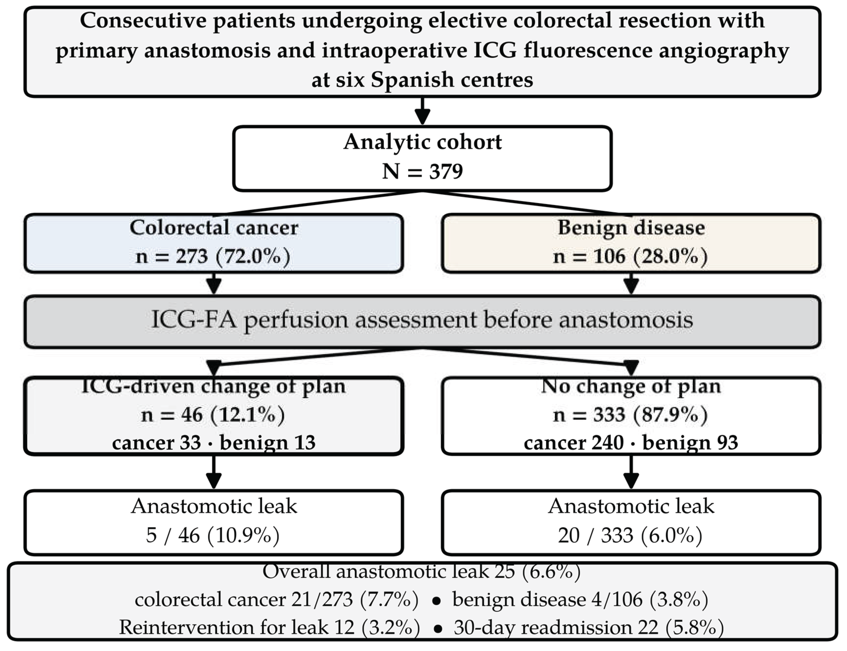
Figure 2.
Univariable association of patient-level, operative and organisational factors with ICG-driven change of the operative plan. Odds ratios (squares) are shown with 95% confidence intervals (horizontal lines) on a logarithmic scale; the dashed line marks OR = 1. None of the ten patient-level characteristics (upper panel, grey) was associated with decision change, and the estimates for malignant diagnosis, rectal location and neoadjuvant therapy lie within 0.03 of unity. Anatomical segment and treating centre (lower panel, blue) were both associated. ASA, American Society of Anesthesiologists; ICG, indocyanine green.
Figure 2.
Univariable association of patient-level, operative and organisational factors with ICG-driven change of the operative plan. Odds ratios (squares) are shown with 95% confidence intervals (horizontal lines) on a logarithmic scale; the dashed line marks OR = 1. None of the ten patient-level characteristics (upper panel, grey) was associated with decision change, and the estimates for malignant diagnosis, rectal location and neoadjuvant therapy lie within 0.03 of unity. Anatomical segment and treating centre (lower panel, blue) were both associated. ASA, American Society of Anesthesiologists; ICG, indocyanine green.
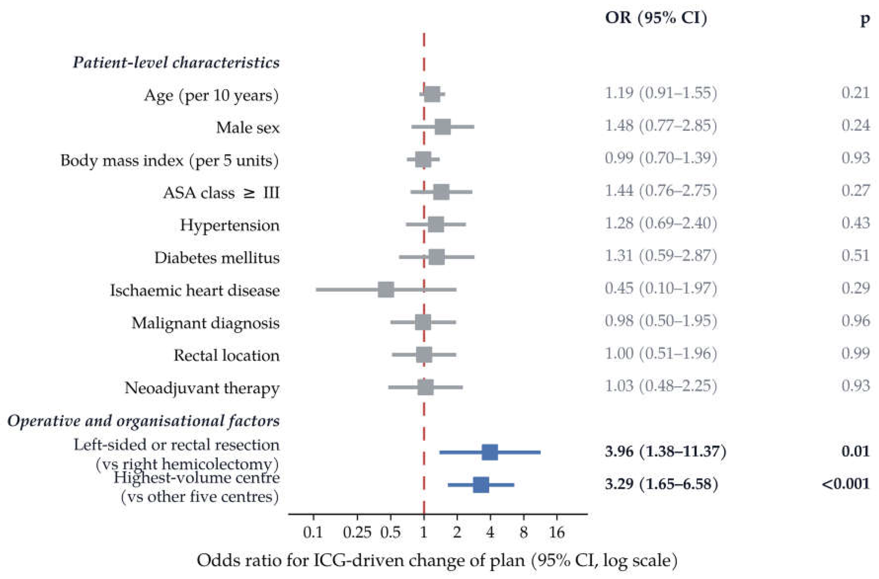
Figure 3.
Rate of ICG-driven change of the operative plan by centre. Points show the observed rate with exact binomial 95% confidence intervals; point size is proportional to centre volume, and the numerator/denominator is printed beside each interval. The dashed line marks the pooled rate. Centres are de-identified and ordered by rate. Heterogeneity across the six centres, chi-square = 13.3, p = 0.021. Adjusted for procedure family, neoadjuvant therapy, age and body mass index, the odds ratio for the highest-volume centre versus the other five was 5.75 (95% CI 2.13-15.57; p < 0.001). CI, confidence interval; ICG, indocyanine green.
Figure 3.
Rate of ICG-driven change of the operative plan by centre. Points show the observed rate with exact binomial 95% confidence intervals; point size is proportional to centre volume, and the numerator/denominator is printed beside each interval. The dashed line marks the pooled rate. Centres are de-identified and ordered by rate. Heterogeneity across the six centres, chi-square = 13.3, p = 0.021. Adjusted for procedure family, neoadjuvant therapy, age and body mass index, the odds ratio for the highest-volume centre versus the other five was 5.75 (95% CI 2.13-15.57; p < 0.001). CI, confidence interval; ICG, indocyanine green.
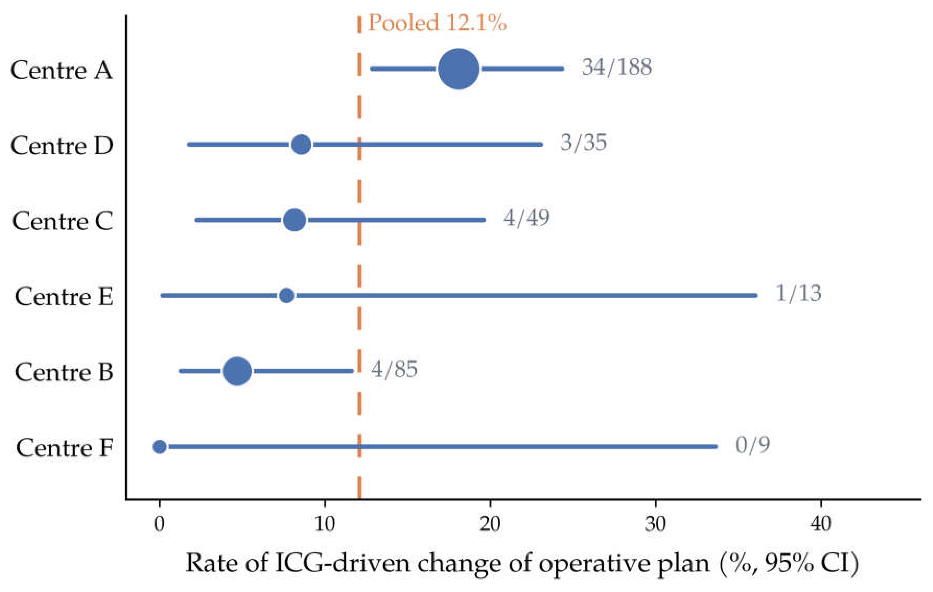
Figure 4.
Anastomotic leak rate by ICG-driven decision change, stratified by neoadjuvant therapy. Bars show the observed leak rate (%) within each stratum; fractions in parentheses are events/patients. Leak risk tracked with neoadjuvant status rather than with whether ICG-FA altered the operative plan. The higher crude rate in the revised stratum reflects confounding by indication (see Discussion). ICG, indocyanine green.
Figure 4.
Anastomotic leak rate by ICG-driven decision change, stratified by neoadjuvant therapy. Bars show the observed leak rate (%) within each stratum; fractions in parentheses are events/patients. Leak risk tracked with neoadjuvant status rather than with whether ICG-FA altered the operative plan. The higher crude rate in the revised stratum reflects confounding by indication (see Discussion). ICG, indocyanine green.
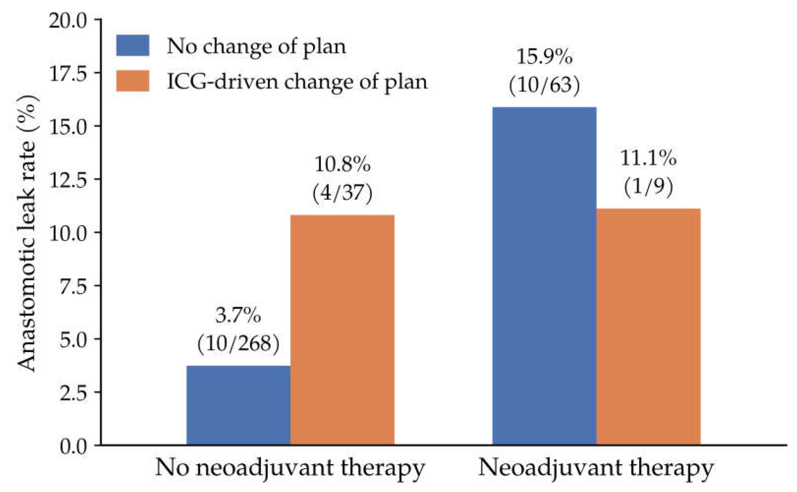

Table 1.
Baseline and operative characteristics of the cohort, stratified by anastomotic leak.
| Characteristic | Overall (n = 379) | No leak (n = 354) | Leak (n = 25) | p |
|---|---|---|---|---|
| Age, years | 67 (57–74) | 67 (57–74) | 68 (61–74) | 0.594 |
| Male sex | 225 (59.4) | 207 (58.5) | 18 (72.0) | 0.263 |
| Body mass index, kg/m² | 26.8 (24.4–29.7) | 26.7 (24.2–29.5) | 28.8 (26.0–30.3) | 0.078 |
| ASA class ≥ III | 111 (29.9) | 103 (29.8) | 8 (32.0) | 0.993 |
| Hypertension | 144 (38.2) | 132 (37.5) | 12 (48.0) | 0.406 |
| Diabetes mellitus | 61 (16.2) | 57 (16.2) | 4 (16.0) | 1.000 |
| Ischaemic heart disease | 32 (8.5) | 31 (8.8) | 1 (4.0) | 0.710 |
| Malignant diagnosis | 273 (72.0) | 252 (71.2) | 21 (84.0) | 0.248 |
| Rectal location | 115 (30.3) | 102 (28.8) | 13 (52.0) | 0.027 |
| Neoadjuvant therapy | 72 (19.1) | 61 (17.3) | 11 (44.0) | 0.003 |
| Open approach | 15 (4.0) | 14 (4.0) | 1 (4.0) | 1.000 |
| Conversion to open | 6 (1.6) | 4 (1.1) | 2 (8.0) | 0.053 |
| Protective ileostomy | 77 (21.0) | 66 (19.3) | 11 (45.8) | 0.005 |
| ICG-driven change of plan | 46 (12.1) | 41 (11.6) | 5 (20.0) | 0.353 |
Data are n (%) or median (IQR). Percentages are calculated on non-missing observations: age, n = 378; body mass index, n = 370; ASA class, n = 371; hypertension and diabetes mellitus, n = 377; ischaemic heart disease, n = 376; neoadjuvant therapy, n = 377; protective ileostomy, n = 366. ASA, American Society of Anesthesiologists; ICG, indocyanine green; IQR, interquartile range.
Table 3.
Distribution of patients, ICG-driven change of plan and anastomotic leak by participating centre.
Table 3.
Distribution of patients, ICG-driven change of plan and anastomotic leak by participating centre.
| Centre | Patients, n | Change of plan, n (%) | 95% CI | Anastomotic leak, n (%) | Change of plan, colorectal cancer subgroup |
|---|---|---|---|---|---|
| Centre A | 188 | 34 (18.1) | 12.9–24.3 | 4 (2.1) | 22/118 (18.6) |
| Centre B | 85 | 4 (4.7) | 1.3–11.6 | 12 (14.1) | 4/68 (5.9) |
| Centre C | 49 | 4 (8.2) | 2.3–19.6 | 6 (12.2) | 3/32 (9.4) |
| Centre D | 35 | 3 (8.6) | 1.8–23.1 | 3 (8.6) | 3/34 (8.8) |
| Centre E | 13 | 1 (7.7) | 0.2–36.0 | 0 (0.0) | 1/12 (8.3) |
| Centre F | 9 | 0 (0.0) | 0.0–33.6 | 0 (0.0) | 0/9 (0.0) |
| All centres | 379 | 46 (12.1) | 9.0–15.8 | 25 (6.6) | 33/273 (12.1) |
Centres are de-identified and ordered by volume. Exact binomial 95% confidence intervals are shown for the change-of-plan rate. Heterogeneity across centres for change of plan: chi-square = 13.3, asymptotic p = 0.021, Monte-Carlo permutation p = 0.024; intraclass correlation coefficient 0.032. Between-centre differences in leak rate are not interpreted (see Discussion). CI, confidence interval; ICG, indocyanine green.
Table 2.
Univariable and multivariable logistic regression for anastomotic leak.
| Variable | Univariable OR (95% CI) | p | Adjusted OR (95% CI) | p |
|---|---|---|---|---|
| Age (per year) | 1.01 (0.97–1.04) | 0.773 | — | — |
| Male sex | 1.83 (0.74–4.48) | 0.189 | — | — |
| Body mass index (per unit) | 1.06 (0.97–1.15) | 0.183 | 1.06 (0.97–1.15) | 0.199 |
| ASA class ≥ III | 1.11 (0.46–2.65) | 0.814 | — | — |
| Hypertension | 1.54 (0.68–3.47) | 0.299 | — | — |
| Diabetes mellitus | 0.99 (0.33–2.98) | 0.980 | — | — |
| Rectal location | 2.68 (1.18–6.06) | 0.018 | 2.64 (1.16–6.01) * | 0.021 |
| Neoadjuvant therapy | 3.75 (1.62–8.65) | 0.002 | 3.73 (1.60–8.66) | 0.002 |
| Conversion to open | 7.61 (1.32–43.74) | 0.023 | — | — |
| Protective ileostomy | 3.54 (1.52–8.25) | 0.003 | — | — |
| ICG-driven change of plan | 1.91 (0.68–5.36) | 0.220 | 1.88 (0.65–5.41) | 0.242 |
OR, odds ratio; CI, confidence interval. The multivariable model included neoadjuvant therapy, body mass index, and ICG-driven change of plan (n = 369, 25 events). * Rectal location was entered in a separate sensitivity model in place of neoadjuvant therapy owing to collinearity (n = 370, 25 events).
Disclaimer/Publisher’s Note: The statements, opinions and data contained in all publications are solely those of the individual author(s) and contributor(s) and not of MDPI and/or the editor(s). MDPI and/or the editor(s) disclaim responsibility for any injury to people or property resulting from any ideas, methods, instructions or products referred to in the content. |
© 2026 by the authors. Licensee MDPI, Basel, Switzerland. This article is an open access article distributed under the terms and conditions of the Creative Commons Attribution (CC BY) license.
Copyright: This open access article is published under a Creative Commons CC BY 4.0 license, which permit the free download, distribution, and reuse, provided that the author and preprint are cited in any reuse.