
Submitted:
12 September 2026
Posted:
14 September 2026
You are already at the latest version
Abstract
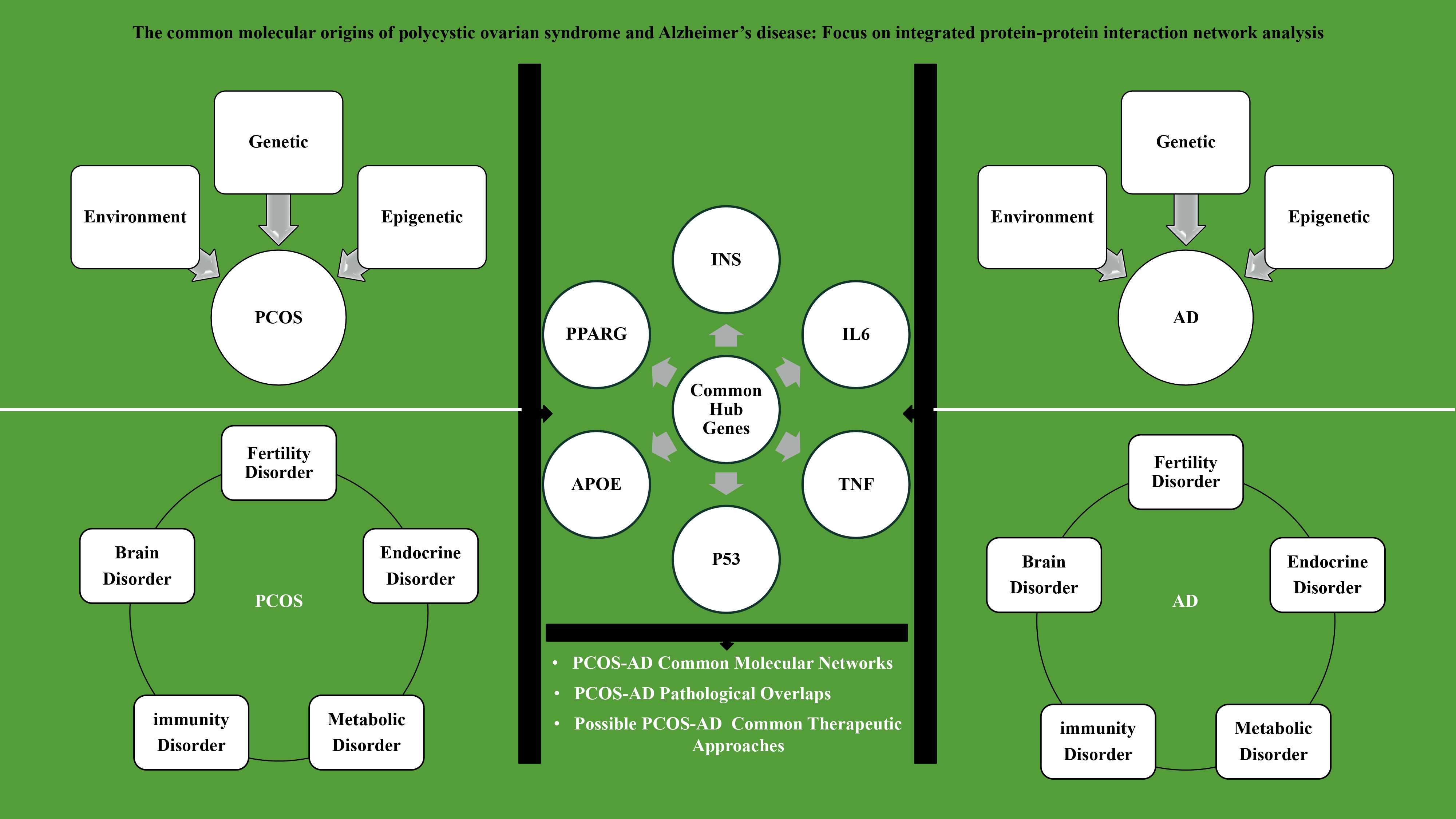
Introduction/Objective: Polycystic ovary syndrome (PCOS) and Alzheimer's disease (AD) are multifactorial disorders among the major global health concerns with an increasing rate. The common molecular, metabolic, and inflammatory pathways between PCOS and AD were reported without integration. Methods: Here, the hub and common interacting proteins between these two disorders were highlighted to propose novel targets and therapies. After curating gene data related to PCOS and AD from the Public Health Genomics and Precision Health Knowledge Base (Phenopedia) and submitting them to the STRING database, protein-protein interaction (PPI) networks were constructed and analyzed. The examined network statistics included nodes, edges, node degree, clustering coefficients, and enrichment p-values. To identify molecular pathways and hub genes, overlapping proteins between PCOS and AD networks were identified. Results: The PPI networks of AD and PCOS consisted of 734 and 370 nodes, respectively, and 83 overlapping proteins were identified in their intersection network. The high clustering coefficient, average node degree of 23, and 956 edges in the common network indicate strong modularity. Protein interaction enrichment was significant (p < 1.0e-16), and common hub genes included insulin, interleukin 6, tumor necrosis factor, tumor protein p53, apolipoprotein E, and peroxisome proliferator-activated receptor gamma. Discussion: There were likely to be common interconnected molecular networks between AD and PCOS, especially in the discussion of metabolic disorders, insulin resistance, chronic inflammation, and immunity. Conclusion: Given the pathological overlap of these two disorders, shared and more effective targeted therapeutic strategies can be developed.
Keywords:
polycystic ovary syndrome
; Alzheimer’s disease
; protein–protein interaction network
; hub genes
; molecular pathways
1. Introduction
There is a common cause-and-effect relation among modern-era diseases such as diabetes mellitus, obesity, cancer, neurodegenerative diseases, infertility, and polycystic ovarian syndrome (PCOS) [1]. PCOS is the most common cause of subfertility and infertility among women [2]. This modern syndrome was mainly known as a fertility disorder, however it affects the endocrine, metabolic, immune, and even nervous systems [3,4]. Therefore, PCOS is a complex disorder with multifactorial etiology and chronic consequences [5]. This syndrome does not involve only ovaries, and has various clinical manifestations [6,7,8,9,10]. Therefore, its initial name, Stein-Leventhal syndrome, may be a better option for PCOS as a disorder of ovary-organ axes. Accordingly, the treatment of PCOS are based on its clinical manifestations and must be investigated as a multi-organ disorder that leads to many neurological diseases.
In this continuum, Alzheimer’s disease (AD) is known as a complex, multifactorial, currently incurable, and eventually fatal disease [11,12,13,14,15,16]. AD is a neurodegenerative disease that includes multistage from preclinical, behavioral, mild cognitive impairment, and finally dementia [17,18,19]. There are 55 million individuals suffering from AD worldwide, and its prevalence is estimated to be doubled every five years [14,20]. It is the main cause of dementia, and 60-80% of dementia cases have AD [18,21,22,23,24], and its incidence is increased throughout aging [25]. It is reported that in 85 years old and over-65-year-old cases, there are 50% and 10% AD possibility, respectively [22]. The therapeutic, care, and social costs of AD will be high and noticeable regards to an estimated 152 million AD individuals and involving the patient’s family and health care systems; it is estimated to be about over $1 trillion by 2050 [14]. Therefore, AD is a major global health concern with a rising prevalence and substantial future burden.
Different hypotheses have been put forth about less-known pathogenesis of AD include tauopathy, amyloidogenic cascade, mitochondrial cascade, OS, neuroinflammation, cholinergic theory, glutamate dysfunction, germ theory, and vascular theory [12,13]. The whole gene set involved in human AD is exceled in the Supplementary file S1. In summary, neuronal cell bodies, axonal structure death and microglia-astrocyte activation (microgliosis and astrocytosis), altered brain neuronal apoptosis and autophagy, neuroinflammation, cerebrovascular changes, altered expression, transmission and function of acetylcholine, brain-derived neurotrophic factor (BDFN), and glutamate, as well as metabolic disorders, are among factors governing the pathogenesis of AD [18,21,25,26,27,28,29,30,31,32,33,34,35]. In this context, pathological aging causes neuroinflammation, synaptic damage, and eventually cognitive impairment; however, it should be kept in mind that AD is not a normal aging process [24,32].
The modulating role of metabolic disorders on neurons and organs was accepted, and the brain works with high metabolic rate, glucose and oxygen consumption, and lipid content [14,20,32]. Altered insulin signaling has been reported in AD, and its receptors are widely distributed in the hippocampus, the memory center [20]. The altered signaling in IR causes metabolic disorders of glucose, OS, a decline in neuronal energy production, amyloid-β production, tau protein phosphorylation, and leads to AD by impairing in mitochondrial function, cognition, and memory [19,36]. Therefore, managing metabolic status has a pivotal role in the AD monitoring strategy.
There are no effective treatments for AD and its therapy may be more effective through improving temporary memory than managing AD progression [15,24]. Nevertheless, there are several common and emerging novel therapies for AD. In general, higher education level, rich social network, regular physical activities, and more intellectually stimulating work have improving effects in AD treatment, as well [16]. The effectiveness of AD therapy relies upon the disease stage, genetic factors, general health, and age [13]. However, the novel therapeutic approaches are focused on delaying brain aging, CNS modulation, and slowing AD progression via neurotrophins [15,37]. An array of drugs that were prescribed for AD is found in the market (https://go.drugbank.com/categories/DBCAT001745; https://go.drugbank.com/indications/DBCOND0049114). Therefore, AD therapies currently aim to slow progression and support cognitive function rather than provide a strict cure.
Despite an extensive history of AD detection, many facets of its etiology remain unclear; as with PCOS, its multifactorial nature involves interactions of genes, environment, and epigenetics [26]. This synaptic disorder occurs along with insulin resistance (IR), type-2 diabetes mellitus (T2DM), obesity, hypercholesterolemia, cardiovascular disease, thrombosis, cortisol and hormonal disturbances, dysphagia, malnutrition, pneumonia, Coronavirus disease 2019 (COVID-19), depression, anxiety, and higher suicide risk which increase the fatality in AD patients [19,20,21,22,23,24,25,26,27,28,29,30,31,32,33,34,35,36,37,38,39,40,41,42,43,44,45,46,47,48,49]. The early and accurate diagnosis can be a decisive step in its management. Its current diagnostic methods include positron emission tomography scan, cerebrospinal fluid analysis, AD confirmatory-prognostic biomarkers (p-tau-181), and blood neurofilament light chain. Clinically, the amyloid and tau protein deposition and neurodegeneration were proposed for AD detection [18], however, there is an urgent need for more accurate and novel diagnostic approaches [22,25].
Regarding different involvement of biological disorders, specifically metabolic and inflammatory ones, and a preview of proteins involved in the pathogenesis of PCOS and AD, it seems that common proteins expressed in both diseases. In this review, among 83 proteins that are involved in the pathogenesis of both PCOS and AD, we sought to highlight the shared biomolecular landscape in these two disorders to define a novel strategic approach for the simultaneous prevention, management, and treatment of AD and PCOS.
2. Results
2.1. Text-Mining of Polycystic Ovarian Syndrome
2.1.1. Current Status of Polycystic Ovarian Syndrome in Clinics
PCOS was reported with bilateral polycystic ovaries, amenorrhea or oligomenorrhea, obesity, acne, and hirsutism [4,43,44,45,46,47]. There are four major PCOS phenotypes, which include classic phenotypes [10,48,49,50]. The three criteria of phenotype A are hyperandrogenism, oligoanovulation, and polycystic ovarian morphology (PCOM), while in phenotype B, ovarian morphology is normal and the other two criteria are present. Phenotype C is characterized by hyperandrogenism, normal menstruation, and PCOM, and eventually phenotype D is manifested by normal androgen level besides the presence of PCOM and oligoanovulation [36,51,52]. The phenotype A and D are the most and least severe forms, respectively [50]. The most frequent PCOS phenotype is a classic one presented with hyperinsulinemia, menstrual dysfunction, IR, atherogenic dyslipidemia, increased anti-Müllerian hormone (AMH) level, risk of obesity, hepatosteatosis, and metabolic syndrome (MetS) [49,51]. The mild elevation of insulin, androgen, and lipid profile levels in phenotype C brings more metabolic consequences (e.g., MetS) among high socioeconomic individuals [49,51]. The minimum metabolic dysfunction, lower levels of thyroid hormones, and lower luteinizing hormone (LH) to follicular stimulating hormone (FSH) ( ratio, and higher sex hormone binding globulin (SHBG) was reported for phenotype D [49,51].
The definitive diagnosis of PCOS in the contemporary era is usually based on Rotterdam criteria, which were discussed (vide supra), and its diagnosis is confirmed if there are two out of the three mentioned criteria [2,46,47,49,53,54,55,56,57,58,59,60]. The volume of more than 10 ml and the presence of 12 or more follicles with 2-9 mm diameter can be mentioned as the pelvic ultrasonic diagnostic criteria of PCOS [61]. The differential diagnosis to other diseases with similar symptoms, such as adrenal congenital hyperplasia, Cushing’s syndrome, androgen-secreting tumors, raised prolactin, insufficient LH, thyroid gland dysfunction, and 21-hydroxylase deficiency is to be considered [62,63]. It should be noted that the early PCOS diagnosis is a tough task due to overlapping symptoms [49]. The diagnostic value is depending on the individual’s age. It can encompass various aspects of psycho-metabolo-gynecological health and social status [49]. PCOS can impose a heavy economic burden on human society. For instance, an estimation in the United States of America has revealed an 8-billion-dollar economic burden of PCOS in 2020 [9,64]. Therefore, early PCOS detection can be effective in estimating therapeutic costs and even their decrement.
2.1.2. Epidemiology of Polycystic Ovarian Syndrome
PCOS has accrual incidence and prevalence with serious psychophysiological consequences, fertility issues, socioeconomic costs, and accompanying morbidity and even mortality [70,71]. The incidence of PCOS showed a 30.4% increase of incidence between 1990 and 2019 [9,10,64,72,73,74]. A 37.9% increase in PCOS incidence in North Africa and the Middle East was reported [75]. Furthermore, the number of PCOS cases in the Asian continent has risen during the last 30 years [10]. The prevalence of PCOS is dissimilar because of the demographic differences between population, study methodology, and employed diagnostic criteria [60,71,76,77,78]. This geographical variation are results of hormonal, metabolic, and neural makeup and environmental conditions, lifestyles, and individuals' genetics [79]. Therefore, this difference can dictate the existence of various PCOS phenotypes. In this context, the PCOS prevalence in Black, Caucasian, Spanish Caucasian, Chinese, and Indian was 4.0, 6.5, 5.6, and 9.13, respectively [49,51]. The prevalence of PCOS was higher in South Asia in comparison to the Caucasus (e.g., 40-50% for Pakistan) [49,51]. In contrast, another study indicated a higher PCOS prevalence in Caucasian and mixed ethnicities in comparison to Asians [80]. The higher hirsutism rate was reported in Middle Eastern, Mediterranean, Indian, and South Asian women in comparison to Eastern Asian and Caucasian women, while higher prevalence of hirsutism, acne, IR, T2DM, metabolic disorders, and elevated fasting insulin levels was reported in Hispanic women than in non-Hispanic white ones [61]. Another study indicated a higher PCOS prevalence in Black and Middle Eastern populations in comparison to the Chinese and White populations [70]. In an ethnical survey, Middle Eastern and Chinese women have the highest PCOS prevalence, and among Middle Eastern women, the PCOS prevalence is high in the Persian Gulf region [81]. Another interesting point is that the PCOS incidence in female Caucasians who live in America or Europe is lower than that in women resident in the Middle East; of course, the maximum PCOS incidence in these regions belongs to African American black or Afro-Brazilian [79]. There are different reports on PCOS symptoms and phenotypes between geographical regions, human races, and diagnostic methods, which one can mention to higher body mass index and waist circumference in African American and Hispanic women than in Northern European and Counterpart Asian [72]. The global incidence and prevalence of PCOS in different studies regarding diagnosis type have been reported as 6-20% and 2.2-26.7%, respectively [7,58]. Therefore, geography will be a determinant for the prevalence of PCOS.
PCOS is considered the main global cause of female infertility (30-60%), especially the anovulatory type (70-80%), while 90-95% of anovulatory infertile clients referred to infertility clinics have PCOS [49,51,69,82,83,84]. The high PCOS prevalence (55-60%) in first-degree relatives indicates its possibility of genetic inheritance [51,84,85,86]. The endocrine disruption is the dominant component of PCOS, in which there are increased LH pulse frequency, hyperandrogenism, and ratio in 70-75%, 60%, and over 90% of PCOS cases, respectively [87,88]. Indeed, another study reported high adrenal and ovarian androgens such as dehydroepiandrosterone (DHEA) and dehydroepiandrosterone sulfate (DHEAS) in 20-30% of PCOS cases; furthermore, 80% of females with hyperandrogenic symptoms suffered from PCOS [49,85]. Surprisingly, 71% of PCOS hyperandrogenic daughters are born to PCOS mothers [87]. Therefore, the hormonal milieu will be a determinant for PCOS, and future studies are acknowledged to study an integrated hormone network of PCOS.
Obesity is another PCOS-related issue both as an etiological factor and as a consequence, with a prevalence of 50-70% among individuals with PCOS [4,10,89]. Accordingly, 25-70% of PCOS patients are obese, and 40-88% of PCOS cases have obesity or overweight [54,74,84,86]. Moreover, diverse mechanisms involved in PCOS pathogenesis which are glucose regulation disturbances (31.1%) [10], higher fasting glucose (10%) [90], glucose intolerance (40%) [54], IR (65-70%, 75%, 50-70%) [54,85,91,92], T2DM (7.5%, 8-10%, 15.4%, 82%) [10,54,71,85], TD1M (40.5%) [85], arterial hypertension (40%, 19.8%) [44,71], MetS (43.9%, 30%, 28.6%) [71,93], autoimmune thyroid disease [94], hirsutism (70%) [85], diminished ovarian reserve (16.9%) [95], acne (15-30%) [85], early pregnancy loss (30-50%) [96], abortion (42-73%) [85], depression (16-55%, 27-50%, 18.7%) [54,58,71], anxiety (38.6%, 17.6%) [54,71], and more attempt for suicide (40%) [97]. There are variations in the menstrual cycle of PCOS patients, in which 85-90% and 30-40% of oligomenorrhea and amenorrhea cases suffer from PCOS, respectively [85]. In this line, obesity and adipose tissue dysfunction can alter PCOS development [98]. Therefore, PCOS can be explained as a multisystem disorder with various epidemiological patterns, and more comprehensive treatments are needed to be managed.
2.1.3. Pathobiology and Possible Therapy of Polycystic Ovarian Syndrome
In essence, different organs and functions are affected in PCOS, however the dysfunction of ovarian granulosa cells is the crystal-clear sign of PCOS [3,56,66]. PCOS has a polygenic inheritance pattern that encompasses individual genes, gene interaction, and altered gene-environment interplay [2,10,49]. Accordingly, the whole gene set involved in human PCOS is included in Supplementary file S1 to give a big picture for genes governing this phenotype (vide infra).
In this context, H19 is overexpressed in PCOS and manifests fluctuation in gene expression of connective tissue growth factor and microRNAs (miRNA) miR-19b that affect germ cells and kartogenin proliferation besides apoptosis, ovarian fibrosis and PCOS [3]. Another PCOS-gene related finding is altered expressions of neurokinin B, kisspeptin, and neurokinin 3 receptor, which are expressed in the central nervous system (CNS) and ovary and alter gonadotropin secretion, regulation of hypothalamic-pituitary-gonadal axis and follicle development, and ovulation timing [53,54,66,67,71,99,100,101]. PCOS-gonadotropin hormone-related genes include gonadotropin-releasing hormone (GnRH), LH, LH receptor (LHR), LH/chorionic gonadotropin receptor (LHCGR), FSH receptor (FSHR), FSH subunit beta (FSHβ), AMH, androgen receptor (AR), SHBG, and prolactin receptor [6,7,10,49,53,55,57,61,82,102,103,104,105,106,107]. The changes in expression of cytochromes P450 (CYP) genes (CYP11a, CYP17, CYP19, and CYP21) participated in adrenal-ovarian steroidogenesis, are another part of PCOS pathogenesis and are active in the production of estrogen, hydroxyprogesterone, 11-deoxycortisol, and progesterone, respectively [6,49,51,91,102,106].
The insulin gene expression and glycoxidative pathway are altered in PCOS. These genes include insulin (INS), insulin receptor substrate (IRS)-2, IRS-1, calpain 10 (CAPN10), insulin receptor (INSR), CAPN2, gastric inhibitory polypeptide receptor (GIPR), PPARG, fat mass obesity (FTO), high-mobility group (HMGA) 1-2, thyroid adenoma-associated (THADA), RAS oncogene family member 5B (RAB5B), and adenylyl cyclases (ACY5) [7,10,49,51,56,77,91,102,103]. In this context, INS was a hub gene in the PPI network of PCOS (Table 1).
The inflammatory adipokines and immune pathway related genes are inseparable compartments of PCOS including IL1β, IL8, leukemia inhibitory factor (LIF), nitric oxide synthase (NOS2), prostaglandin-endoperoxide synthase 2 (PTGS2), metastasis-associated lung adenocarcinoma transcript 1 (MALT1), matrix metallopeptidase 2 (MMP2), adenosine deaminase RNA specific (ADAR), transforming growth factor beta-1 (TGF-β1), tumor necrosis factor alpha (TNFα), and IL6 [47,102,107,108,109,110,111]. The sepia translocation (SET) is another gene involved in PCOS, inhibiting protein phosphatase 2A (PP2A), however the remarkable point about this gene is that the site of SET overexpression can bring different outcomes; in other words, its overexpression in the brain and ovary causes tau hyperphosphorylation in AD, hyperandrogenism in PCOS, and ovarian carcinoma [111,112]. The changes in controlling genes of serotonin (5HT) formation, appetite, mood, and circadian rhythm, such as basic helix-loop-helix ARNT-like 1 (BMAL1) are associated with eating, sleep, and metabolic disorders [64,113,114].
Briefly, the genes involved in PCOS were reported in the main literature [7,10,57,61,66,78,91,92,102,103,110,115]. In summary, tachykinin precursor 3 (TAC3), tachykinin receptor 3 (TACR3), steroid 5β-reductase (SRD5B), SRD5A, DENN domain containing 1A (DENND1A), paraoxonase 1 (PON1), extracellular signal-regulated kinases (ERK)1/2, FK506 binding protein 52 (FKBP52), AR, patatin-like phospholipase domain containing 3 (PNPLA3), maestro , fatty acid desaturase 2 (FADS2), triggering receptor expressed on myeloid cells 1 (TREM1), TOX3, small ubiquitin-like modifier (SUMO) 1P1, melanocortin 4 receptor (MC4R), prospero homeobox 1 (PROX1), oxytocin receptor gene (OXTR), GATA binding protein 4 (GATA4), RAD50, Erb-B2 receptor tyrosine kinase 4 (ERBB4)/HER4, Nei like deoxyribonucleic acid (DNA) glycosylase 2 (NEIL2), vitamin D receptor (VDR), and leptin (LEP) are major players in PCOS (see Supplementary file S1).
The hypothalamic-pituitary-ovarian axis is one of the major player in PCOS [77,116]. The GnRH cells regulate pituitary gonadotropins, the synthesis of gonadal sex hormones, and the release of adrenocorticotropic hormone and adrenal hormones [53]. The outcomes of increase GnRH neurons stimulation, GnRH secretion and neuronal pathway hyperactivation by different bioactive molecules such as glucagon-like peptide-1 (GLP-1), kisspeptin, AMH, and gamma-aminobutyric acid (GABA) innervation amplified LH amplitude and pulse frequency, decreased or normal FSH level, increased ratio [2,53,60,61,67,87,88,95,99,105,108,117]. The increased LH level leads to increased AMH, ovarian theca cell hyperplasia, excess estrogen and androgen, ovarian dysfunction, metabolic disorders mapped as increased leptin, adiponectin, and insulin, and decreased white fat browning, and IR, which decreases SHBG [2,53,61,117,118,119]. FSH fluctuations also cause follicular maturation disorder and cyst formation, terminate to anovulation [2]. The LH level is higher than FSH in PCOS, which disturbs follicular growth and leads to hirsutism through higher conversion of androstenedione to testosterone [108]. Androgens (DHEA and DHEAS) are produced in the ovaries and adrenal glands [49,60], and their increased levels give rise to hirsutism, increased LH levels, hyperinsulinemia, and insulin receptor desensitization, which cause a decrease in hepatic SHBG and, in turn, a higher free testosterone level [49,59]. Notably, there is adrenal hyperandrogenism in 20-30% of PCOS cases, which causes ovarian premature atresia, ovarian cyst, anovulation, and persistent estrogen that are risk factors for endometrium carcinoma [44,49]. The lower hepatic SHBG causes higher free estradiol, testosterone, and insulin levels. The insulin increment stimulates androgen production from the ovary and subsequently PCOS [5,51].
The impaired follicular growth, hyperandrogenism, and increased hepatic free fatty acid (FFA) flux can upturn insulin levels [90,108,116,119]. This increment boosts LH and androgen level in a vicious cycle, IR, cessation of follicular development, and higher cyst formation [49,108,116,120]. Following IR, the hepatic SHBG and insulin-like growth factor (IGF1) binding protein (IGFBP)-1 levels decrease, while androgens increase, which stimulates visceral adipose tissue and causes FFA production, thereby aggravating IR [103]. Insulin alters steroidogenesis by PPAR-γ activation, suppressing hepatic SHBG, increasing blood free steroid hormone, and being a gonadotrophic factor that is a direct stimulator of steroidogenic enzymes, synergy with LH and FSH, and increased pituitary responsiveness to GnRH [116]. One of the dermatological manifestations of PCOS is acanthosis nigricans, skin fold hyperpigmentation, which is considered an IR manifestation [92,116]. Due to the pivotal roles of insulin in metabolism and energy regulation of the brain, any disturbances in this case can lead to neurological disorders as well [121,122]. Therefore, regulation of insulin biosynthesis can be an effective strategy for managing this multisystem disorder.
There is also a bidirectional relationship between obesity-induced inflammation and PCOS, i.e., one is the other’s cause. On one hand, IR, increased insulin, androgens, and dyslipidemic lipid profile presented with an increment of triglycerides (TGs), low-density lipoprotein (LDL), very-low-density lipoprotein (VLDL), and a decrement of high-density lipoproteins (HDL) are all behind reasons for obesity; on the other hand, obesity aggravates this scenario and induces PCOS [120,123]. Although obesity might be necessary in PCOS development, it is not sufficient as there are PCOS cases among many anorexic and thin cases [43,124]. Leptin, a biomolecule involved in obesity, affects the hypothalamic-pituitary-ovarian axis and is a key regulator of ovulation; therefore, its possible involvement in PCOS is evident [43,113,120]. Consequently, not only does obesity cause inflammatory cytokine release such as TNFα, adipokinin activation, mitochondrial dysfunction, IR, and metabolic and hormonal disorders, but also IR ends in obesity, compensatory increased insulin release, and inflammation, and the result of both pathways is a vicious cycle [5,50,68]. Vitamin D deficiency, oxidative stress (OS), immunological disturbances, and chronic inflammation are occurred in PCOS, which can lead to autoimmune diseases and higher susceptibility to infections [47,68,94,95,123,125,126,127,128]. The more detailed immunological research will be acknowledged in elucidating PCOS pathogenesis, deeply. In conclusion, PCOS has genetic, epigenetic, neuroendocrine, metabolic, hormonal, and inflammatory clinical correlates that affect not only the reproductive system but also general health status through various pathobiological pathways. Consequently, there is an urgent prerequisite for a more comprehensive diagnostic and management approach for this syndrome.
Due to complexity, various unknown issues of PCOS, and absence of a comprehensive understanding of molecular mechanisms involved in this syndrome, there are not a determined therapy up to now, and this issue can be extended to the prevention, control, and early diagnosis [129]. Therefore, the pharmacology of PCOS, including metformin, hormone therapy, 3 hydroxy-3-methylglutaryl-coenzyme A reductase inhibitors, and lifestyle interventions focused on management of the symptoms regard to patient's referral because of one of the signs, not solving the main problem [2,9].
2.2. Text-Mining of Alzheimer’s Disease
Causes of Alzheimer’s Disease
Oxidative stress in the AD pathogenesis gives rise to overproduction of reactive oxygen species (ROS) and advanced glycoxidation end products (AGEs) which are neurotoxic and brings the inflammatory responses by releasing IL1 and TNFα. The outcomes of these inflammatory pathways are Aβ deposition, lipid oxidation, cellular apoptosis, and finally AD manifestation [14,24]. High lipid peroxidation and increased protein carbonyl content, indicate an OS condition in AD [24]. The point is a close relationship between OS and mitochondrial disorders. Following the mitochondrial dysfunctions, the consequences include decreased cytochrome C oxidase activity in the hippocampus and increased ROS production, which causes neuronal apoptosis, cortical atrophy, and AD [14,25]. This is a confirmation of mitochondrial hypometabolism that is reported in AD [20]. Therefore, OS can also alter metabolic pathways, which may cause overproduction of deteriorating biomolecules in the brain.
The common outcomes of all pathways occurred in AD are extracellular Aβ deposition and intra-neuronal tau tangles in the cerebral cortex and subcortical gray matter [13,17,18,32,36] which lead to neuronal damages and destruction [32], neuronal hypo-connectivity [22,25], neuronal apoptosis and brain atrophy [22,25], impaired synaptic plasticity [25], cognitive decline, and neuroinflammation [22]. Aβ, the hydrolytic product of amyloid-beta precursor protein, leads to synaptic dysfunction, cognitive impairment, microglial immune responses, cytokine release, cell death, and neurodegeneration [13,14,15,25,33,36,37,39,50]. In conclusion, Aβ is one of the most reliable targets for AD prevention and therapy.
2.3. The Protein-Protein Interaction Network Analysis
According to the PPI network results, there are 83 overlapping proteins interacted with 956 edges in the common PCOS-AD network, while the number of nodes (proteins) and edges (interactions) in the PCOS and AD networks are 370-5812 and 734-7817, respectively (Figure 1 and Table 1). Therefore, AD has a bigger network than PCOS and the common PCOS-AD network. The average node degree, an indicator of the number of connections per protein, is highest in PCOS (31.4) compared with AD (21.3) and their common network (23.0). These finding highlight their connectivity and the importance of shared proteins in functionality. There is a modular and interconnected structure, especially in related biological processes regarding the local clustering coefficient (0.674), the average node degree (23.0), and the presence of 956 edges in the common PCOS-AD network. The expected random interactions are lower than the observed ones (p < 1.0e-16), which indicate the possible biological relevance of these networks and not by chance. The highly interactive hub genes include insulin (INS), interleukin 6 (IL6), tumor necrosis factor (TNF), tumor protein p53 (TP53), apolipoprotein E (APOE), peroxisome proliferator-activated receptor gamma (PPARG), estrogen receptor 1 (ESR1), and interleukin 1 beta (IL1B). In conclusion, the presence of common hub genes in AD and PCOS specifies possible common molecular elements and pathophysiological overlap of these two disorders.
3. Discussion
PCOS and AD have many common features including their complexity and unknowns, being multifactorial entity, absence of effective therapy, catalytic role of modern lifestyle on their prevalence because of dietary habits, sedentary life style, stress, chronic chemical exposure, altered working hours (i.e., night shifts), inflammation, OS, metabolic makeup and disorders, gut-brain axis contribution, brain architectural changes, altered NTs, sleep disorders, common comorbidities including IR, T2DM, obesity, vascular injuries, CVDs, thyroid diseases, immunity disorders, and higher susceptibility to COVID-19, depression, anxiety, and suicide [62,68,131,132,133,134,135,136,137,138,139,140,141,142,143,144,145,146,147,148,149,150,151,152,153,154,155,156] which outlined in Figure 2.
With the expansion of molecular and cellular knowledge, the relationship between many diseases in terms of the existence of common genetic and molecular factors has received more attention, because it may be possible to manage diseases more effectively. It seems that PCOS and AD are also included in this topic, because they have common biological pathways, including metabolic processes (INS, PPRG), lipid metabolism (APOE), cell cycle control-programmed cell death (TP53), and inflammation (IL6, IL1B, and TNF; vide supra).
The complications of disruption in any of the aforementioned pathways, namely IR, metabolic dysfunction, and chronic inflammation, can result from the coordinated effect of these shared proteins, which is also indicated by the high clustering coefficient in the common network of AD and PCOS. APOE, INS, and IL6 are not only involved in metabolic disorders but also play critical roles in neurodegenerative diseases, especially AD. The systemic inflammation and impaired neuronal insulin signaling are two examples of pathologic consequences of altered levels of TNF, IL6, and insulin.
The identification of INS, IL6, TNF, APOE, IL1B, TP53, PPARG, CCL2, and ESR1 as common hub proteins in the PPI networks of PCOS and AD suggests that these disorders share interconnected molecular mechanisms despite their distinct clinical manifestations. The clustering of these proteins highlights four major biological processes: IR, chronic inflammation, hormonal dysregulation, and cellular stress responses. Collectively, these findings support the growing evidence that PCOS is not only a reproductive disorder but also a systemic metabolic condition that may predispose affected women to neurodegenerative diseases later in life [108]. To the best of our knowledge, there is currently no large prospective cohort study that conclusively demonstrates PCOS as an independent predictor of AD, although a proteomic study confirmed the interconnected nature of these disorders [128]. Therefore, the design of epidemiological investigations may uncover common aspects of these disorder.
Among the identified hub proteins, INS occupies a central role, emphasizing IR as a critical molecular link between PCOS and AD. IR is a hallmark feature of PCOS and contributes to hyperinsulinemia, obesity, and MetS. In CNS, impaired insulin signaling has been associated with reduced neuronal glucose utilization, increased OS, amyloid-β accumulation, and tau hyperphosphorylation, leading some researchers to characterize AD as a form of "type 3 diabetes" [108]. Thus, disturbances in insulin signaling may provide a mechanistic bridge between metabolic dysfunction in PCOS and neurodegeneration in AD.
The inflammatory mediators including IL6, TNF, IL1B, and CCL2 identified as shared hubs further highlight the significance of chronic low-grade inflammation in both disorders. Women with PCOS exhibit elevated circulating levels of pro-inflammatory cytokines, which contribute to IR and ovarian dysfunction. Similarly, in AD, activated microglia release IL6, TNF-α, and IL-1β, promoting neuroinflammation, neuronal injury, and progression of amyloid pathology. CCL2 regulates immune cell recruitment and sustains inflammatory signaling within both peripheral tissues and the brain [157]. Persistent systemic inflammation may therefore represent a common pathogenic mechanism linking PCOS to increased susceptibility to AD.
The identification of APOE as a shared hub protein is particularly noteworthy because APOE represents the strongest genetic risk factor for sporadic AD. APOE influences cholesterol transport, neuronal repair, amyloid-β aggregation, and neuroinflammation. The APOE ε4 allele significantly increases AD risk and accelerates disease onset. In parallel, dyslipidemia is a common metabolic abnormality in PCOS, characterized by elevated TGs and LDL-C levels [158]. Therefore, APOE-mediated disturbances in lipid metabolism may constitute an important molecular intersection between PCOS-associated metabolic dysfunction and AD pathogenesis.
Another key hub identified in the network is PPARG, a transcription factor involved in adipogenesis, insulin sensitivity, glucose homeostasis, and inflammatory regulation. PPARG polymorphisms have been associated with PCOS susceptibility, whereas activation of PPARG signaling has demonstrated neuroprotective effects in experimental models of AD through modulation of inflammatory pathways and improvement of metabolic function [159]. The presence of PPARG among the common hub proteins suggests that metabolic dysregulation and inflammation converge through shared regulatory mechanisms in both diseases.
The identification of ESR1 highlights the contribution of endocrine signaling to the molecular overlap between PCOS and AD. Estrogen signaling is essential for glucose metabolism, mitochondrial function, synaptic plasticity, and neuronal survival. Women with PCOS often exhibit hormonal imbalances involving altered estrogen-androgen signaling, while declining estrogen levels during aging and menopause have been associated with increased AD risk. Consequently, ESR1 may represent an important molecular mediator linking reproductive endocrine dysfunction to neurodegenerative processes [108].
The shared presence of TP53 further suggests that OS and cellular senescence contribute to the pathophysiology of both disorders. TP53 regulates DNA repair, apoptosis, and responses to oxidative damage [160]. Increased OS has been extensively reported in PCOS and is also a major driver of neuronal dysfunction and cell death in AD [24,59]. Therefore, TP53-mediated stress responses may represent another common pathway underlying disease progression.
Overall, the shared hub proteins identified through PPI network analysis indicate that IR (INS), chronic inflammation (IL6, TNF, IL1B, and CCL2), lipid dysregulation (APOE), endocrine imbalance (ESR1), metabolic regulation (PPARG), and cellular stress responses (TP53) constitute a highly interconnected molecular network linking PCOS and AD. These findings support the hypothesis that long-term metabolic and inflammatory disturbances in PCOS may contribute to an increased risk of neurodegeneration and cognitive decline. Furthermore, they suggest that therapeutic interventions targeting IR, inflammation, and hormonal dysregulation may have potential benefits for preventing or delaying AD-related pathology in women with PCOS.
4. Materials and Methods
Firstly, literature search using related keywords including AD, PCOS, their association, and related bioinformatics analyses was conducted across PubMed, ScienceDirect, and Google Scholar databases without any filtrations during the search process to distillate the common pathogenesis of two disorders through text mining. Secondly, the entire gene set involved in human PCOS and AD was curated (from Phenopedia, a knowledge-based component of the Public Health Genomics and Precision Health Knowledge Base (version 10.0) launched at https://phgkb.cdc.gov/PHGKB/startPagePhenoPedia.action. The retrieved gene lists were converted to their corresponding proteins reported in the UniProt launched at https://www.uniprot.org/ using the SynGO gene set analysis tool (https://www.syngoportal.org/) and submitted to the STRING (version 12) launched at https://string-db.org/ for constructing their protein-protein interaction (PPI) networks. To improve the reliability of the network, interactions were filtered using a STRING confidence score threshold of 0.4, and only interactions meeting or exceeding this threshold were retained for subsequent analysis. Hub genes were identified from the resulting PPI networks based on node degree of proteins in network. The STRING’s built-in functional enrichment analysis included without exhausted statistical analyses.
5. Conclusions
This study highlights a significant molecular overlap between AD and PCOS ontologically involved in metabolism, cell cycle, and inflammation. These common biological pathways highlight a potential intertwining between AD and PCOS. This study provides a rationale for investigating the likelihood of PCOS patients developing AD, and offers therapeutic approaches to target the shared molecules and mediators specifically in inflammation and insulin signaling, with dual advantages. To achieve this goal, more integrated and accurate laboratory and clinical studies will be appreciated at future.
Supplementary Materials
The following supporting information can be downloaded at the website of this paper posted on Preprints.org.
Funding
None.
Consent for Publication
Not applicable.
Conflicts of Interest
The authors declare no conflict of interest, financial or otherwise.
The Authors Confirm Contribution to the Paper as Follows
The authors confirm their contributions to the paper as follows: study conception and design, IK, HBS, ShM; data collection, ShM; analysis and interpretation of results, IK, ShM; drafting of the manuscript, ShM, IK, HBS. All authors reviewed the results and approved the final version of the manuscript.
Acknowledgments
Declared none.
Abbreviations
PCOS = Polycystic Ovarian Syndrome
AD = Alzheimer’s disease
IR = Insulin Resistance
T2DM = Type-2 Diabetes Mellitus
CVDs = Cardiovascular Disease
COVID-19 = Coronavirus disease 2019
PPI = Protein-Protein Interaction
INS = Insulin
IL6 = Interleukin 6
TNF = Tumor Necrosis Factor
TP53 = Tumor Protein p53
APOE = Apolipoprotein E
PPARG = Peroxisome proliferator-activated receptor gamma
ESR1= Estrogen Receptor 1
IL1B = Interleukin 1 beta
PCOM = Polycystic Ovarian Morphology
AMH = Anti-Müllerian Hormone
MetS = Metabolic Syndrome
LH = Luteinizing Hormone
FSH = Follicular Stimulating Hormone
SHBG =Sex Hormone Binding Globulin
DHEA = Dehydroepiandrosterone
DHEAS= Dehydroepiandrosterone Sulfate
CNS= Central Nervous System
GNRH = Gonadotropin-releasing hormone
LHR = LH receptor
LH/chorionic gonadotropin receptor = LHCGR
FSHR = FSH Receptor
FSHβ = FSH Subunit beta
AR = Androgen Receptor
PRLR = Prolactin Receptor
CYP= Cytochromes P450
IRS= Insulin Receptor Substrate
CAPN10 = Calpain 10
INSR = Insulin Receptor
GIPR = Gastric Inhibitory Polypeptide Receptor
FTO = Fat Mass Obesity
HMGA = High-Mobility Group
THADA = Thyroid Adenoma-Associated
RAB5B = RAS Oncogene Family Member 5B
ACY5 = Adenylyl Cyclases
LIF = Leukemia Inhibitory Factor
NOS2= Nitric Oxide Synthase
PTGS2 = Prostaglandin-Endoperoxide Synthase 2
MALT1 = Metastasis-Associated Lung Adenocarcinoma Transcript 1
MMP2 = Matrix Metallopeptidase 2
ADAR = Adenosine Deaminase RNA Specific
TGF-β1 = Transforming Growth Factor Beta-1
SET = Sepia (SE) Translocation
PP2A = Protein Phosphatase 2A
5HT = Serotonin
BMAL 1= Basic Helix-Loop-Helix ARNT Like 1
TAC3= Tachykinin Precursor 3
RACR3 = Tachykinin Receptor 3
SRD5B = Steroid 5β-Reductase
DENND1A = DENN Domain Containing 1A
PON 1 = Paraoxonase 1
ERK = Extracellular Signal-Regulated Kinases
FKBP52= FK506 Binding Protein 52
PNPLA3 = Patatin Like Phospholipase Domain Containing 3
FADS2 = Fatty Acid Desaturase 2
TREM1 = Triggering Receptor Expressed on Myeloid Cells 1
SUMO = Small Ubiquitin-Like Modifier
MC4R = Melanocortin 4 Receptor
PROX1= Prospero Homeobox 1
OXTR = Oxytocin Receptor Gene
GATA 4 = GATA Binding Protein 4
ERBB4 = Erb-B2 receptor tyrosine kinase 4
DNA = Deoxyribonucleic Acid
NEIL2 = Nei Like DNA Glycosylase 2
VDR = Vitamin D Receptor
GLP-1 = Glucagon-Like Peptide-1
GABA = Gamma-Aminobutyric Acid
FFA = Free Fatty Acid
IGFBP = Insulin-Like Growth Factor Binding Protein
TGs = Triglycerides
LDL = Low-density lipoprotein
VLDL = Very-low-density lipoprotein
HDL = High-density lipoprotein
OS = Oxidative Stress
NTs = Neurotransmitters
BDNF = Brain-Derived Neurotrophic Factor
ROS = Reactive Oxygen Species
AGEs = Glycoxidation End Products
References
- Gill, V., Kumar, V., Singh, K., Kumar, A., and Kim J.J. (2019). Advanced glycation end products (AGEs) may be a striking link between modern diet and health. Biomolecules. 9:888. [CrossRef]
- Sati, A., Desroziers, E., and Campbell, R.E. (2023). Microglia: Sculptors of the polycystic ovary syndrome (PCOS)-like brain? Reproduction. 166, R51–R61. [CrossRef]
- Sun, X.; Yan, X.; Liu, K.; Wu, M.; Li, Zh.; Wang, Y., et al. (2020). lncRNA H19 acts as a ceRNA to regulate the expression of CTGF by targeting miR-19b in polycystic ovary syndrome. Braz. J. Med. Biol. Res. 53:e9266. [CrossRef]
- Gunn, H.M. (2022). Proteomic and clinical insights into polycystic ovary syndrome in adolescents. Doctoral thesis (Ph.D), University College London.
- Khadilkar, S.S. (2016). Polycystic Ovarian Syndrome: Is it time to rename PCOS to HA-PODS? J. Obstet. Gynecol. India. 66, 81–87. [CrossRef]
- Pan, J.X., Tan, Y.J., Wang, F.F., Hou, N.N., Xiang, Y.Q., Zhang, J.Y., et al. (2018). Aberrant expression and DNA methylation of lipid metabolism genes in PCOS: a new insight into its pathogenesis. Clin. Epigenetics. 10:6. [CrossRef]
- Sanchez-Garrido, M.A., and Tena-Sempere, M. (2020). Metabolic dysfunction in polycystic ovary syndrome: Pathogenic role of androgen excess and potential therapeutic strategies. Mol. Metab. 35:100937. [CrossRef]
- Xu, H., Mao, L., Huang, W., Huang, Q., Li, L., & Liu, Y. (2025). Gene association study between polycystic ovary syndrome and metabolic syndrome: a transcriptomic analysis and machine learning approach. J Ovarian Res. 18(1), 220. [CrossRef]
- Saei Ghare Naz, M., Rahnemaei, F., Ramezani, F., Sayehmiri, F., Ghasemi, V., Banaei, M., and Ozgoli, G. (2023). Possible cognition changes in women with polycystic ovary syndrome: a narrative review. Obstet. Gynecol. Sci. 66, 347-363. [CrossRef]
- Che, Y., Yu, J., Li, Y.Sh., Zhu, Y.C., and Tao, T. (2023). Polycystic ovary syndrome: Challenges and possible solutions. J. Clin. Med. 12:1500. [CrossRef]
- Du, Q., Zhu, X., and Si, J. (2020). Angelica polysaccharide ameliorates memory impairment in Alzheimer’s disease rat through activating BDNF/TrkB/CREB pathway. Exp. Biol. Med (Maywood). 245, 1–10. [CrossRef]
- Gao, Q., Ma, C., Meng, S., Wang, G., Xing, Q., Xu, Y., et al. (2022a). Exploration of molecular features of PCOS with different androgen levels and immune-related prognostic biomarkers associated with implantation failure. Front. Endocrinol (Lausanne). 13:946504. [CrossRef]
- Andrade-Guerrero, J., Santiago-Balmaseda, A., JeronimoAguilar, P., Vargas-Rodríguez, I., Cadena-Suárez, A.R., Sánchez Garibay, C., et al. (2023). Alzheimer’s disease: An updated overview of its genetics. Int. J. Mol. Sci. 24:3754. [CrossRef]
- Twarowski, B., and Herbet, M. (2023). Inflammatory processes in Alzheimer’s disease— Pathomechanism, diagnosis and treatment: A review. Int. J. Mol. Sci. 24:6518. [CrossRef]
- Wei, M., Wu, T., and Chen, N. (2024). Bridging neurotrophic factors and bioactive peptides to Alzheimer's disease. Ageing. Res. Rev. 94:102177. [CrossRef]
- Zeng, B., Tanga, Ch., Wang, J., Yang, Q., Ren, Q., and Liu, X. (2024). Pharmacologic and nutritional interventions for early Alzheimer’s disease: A systematic review and network meta-analysis of randomized controlled trials. J. Alzheimers. Dis. 99, 1173–1186. [CrossRef]
- Choi, S.H., Bylykbashi, E., Chatila, Z.K., Leem S.W., Pullim B., Clemenson, G.D., et al. (2018). Induced adult neurogenesis plus BDNF mimicks the effects of exercise on cognition in an Alzheimer’s mouse model. Science. 361:eaan8821. [CrossRef]
- Scheltens, Ph., Strooper, B.D., Kivipelto, M., Holstege, H., Chételat, G., Teunissen, Ch.E., et al. (2021). Alzheimer’s disease. Lancet. 397, 1577–1590. [CrossRef]
- Chen, Y., Al-Nusaif, M., Li, S., Tan, X., Yang, H., Cai, H., and Le, W. (2024a). Progress on early diagnosing Alzheimer’s disease. Front. Med. 18, 446–464. [CrossRef]
- Zhang, J., Zhang, Y., Wang, J., Xia, Y., Zhang, J., and Chen, L. (2024). Recent advances in Alzheimer’s disease: mechanisms, clinical trials and new drug development strategies. Signal Transduct Target Ther. 9 (1), 211. [CrossRef]
- Orgeta, V., Tabet, N., Nilforooshan, R., and Howard, R. (2017). Efficacy of antidepressants for depression in Alzheimer’s disease: Systematic review and meta-Analysis. J. Alzheimers. Dis. 58, 725–733. [CrossRef]
- Lane, C.A., Hardy, J., and Schott, J.M. (2018). Alzheimer’s disease. Eur. J. Neurol. 25, 59–70. [CrossRef]
- Pentkowski, N.S., Rogge-Obando, K.K., Donaldson, T.N., Bouquin, S.J., and Clark, B.J. (2021). Anxiety and Alzheimer’s disease: Behavioral analysis and neural basis in rodent models of Alzheimer’s-related neuropathology. Neurosci. Biobehav. Rev. 127, 647–658. [CrossRef]
- Botto, R., Callai, N., Cermelli, A., Causarano, L., and Rainero, I. (2022). Anxiety and depression in Alzheimer’s disease: a systematic review of pathogenetic mechanisms and relation to cognitive decline. Neurol. Sci. 43, 4107–4124. [CrossRef]
- Ferrer-Cairols, I., Montoliu, T., Crespo-Sanmiguel, I., Pulopulos, M. M., Hidalgo, V., et al. (2022). Depression and suicide risk in mild cognitive impairment: The role of Alzheimer’s disease biomarkers. Psicothema 34, 553-561. [CrossRef]
- Gao, L., Zhang, Y., Sterling, K., and Song, W. (2022b). Brain-derived neurotrophic factor in Alzheimer’s disease and its pharmaceutical potential. Transl. Neurodegener. 11:4. [CrossRef]
- Rostagno, A.A. (2023). Pathogenesis of Alzheimer’s disease. Int. J. Mol. Sci. 24:107. [CrossRef]
- Khan, S., Barve, K.H., and Kumar, M.S. (2020). Recent Advancements in Pathogenesis, Diagnostics and Treatment of Alzheimer’s Disease. Curr. Neuropharmacol. 18, 1106-1125. [CrossRef]
- Perneczky, R., Dom, G., Chan, A., Falkai, P., and Bassetti, C. (2024). Anti-amyloid antibody treatments for Alzheimer's disease. Eur. J. Neurol. 31:e16049. [CrossRef]
- Legro, R.S. (2012). Obesity and PCOS: Implications for diagnosis and treatment. Semin. Reprod. Med. 30, 496–506. [CrossRef]
- Andrade, V.H.L.D., Mata, A.M.O.F.D., Borges, R.S., Costa-Silva, D.R., Martins, L.M., Paulo Ferreira, P.M.P., et al. (2016). Current aspects of polycystic ovary syndrome: A literature review. Rev. Assoc. Med. Bras. 62, 867-871. [CrossRef]
- Siddiqui, S., Mateen, S., Ahmad, R., and Moin, Sh. (2022). A brief insight into the etiology, genetics, and immunology of polycystic ovarian syndrome (PCOS). J. Assist. Reprod. Genet. 39, 2439–2473. [CrossRef]
- Emanuel, R.H.K., Docherty, P.D., Lunt, H., and Campbell, R.E. (2023) Comparing literature- and subreddit-derived laboratory values in polycystic ovary syndrome (PCOS): Validation of clinical data posted on PCOS Reddit Forums. JMIR. Form. Res. 7:e44810. [CrossRef]
- Huang, M.C., Chen, P.L., and Hsu, C.L. (2024a). Transcriptome analysis of mesenchymal progenitor cells revealed molecular insights into metabolic dysfunction and inflammation in polycystic ovary syndrome. Int. J. Mol. Sci. 25:7948. [CrossRef]
- Gleicher, N., Darmon, S., Patrizio, P., and Barad, D.H. (2022). Reconsidering the polycystic ovary syndrome (PCOS). Biomedicines. 10:1505. [CrossRef]
- Nautiyal, H., Imam, S.S., Alshehri, S., Ghoneim, M.M., Afzal, M., Alzarea, S.I., et al. (2022). Polycystic ovarian syndrome: A complex disease with a genetics approach. Biomedicines. 10:540. [CrossRef]
- Butler, A.E., Moin, A.S.M., Sathyapalan, T., and Atkin, S.L. (2024). A cross-sectional study of Alzheimer-related proteins in women with polycystic ovary syndrome. Int. J. Mol. Sci. 25:1158. [CrossRef]
- Khan, M.J., Ullah, A., and Basit, S. (2019). Genetic basis of polycystic ovary syndrome (PCOS): Current perspectives. App. Clin. Genet. 12, 249–260. [CrossRef]
- Carmina, E., and Lobo, R.A. (2022). Comparing lean and obese PCOS in different PCOS phenotypes: Evidence that the body weight is more important than the Rotterdam phenotype in influencing the metabolic status. Diagnostics, 12:2313. [CrossRef]
- Coutinho, E.A., and Kauffman, A.S. (2019). The Role of the Brain in the Pathogenesis and Physiology of Polycystic Ovary Syndrome (PCOS). Med. Sci (Basel). 7:84. [CrossRef]
- Barlampa, D., Bompoula, M.S., Bargiota, A., Kalantaridou, S., Mastorakos, G., and Valsamakis, G. (2021). Hypothalamic inflammation as a potential pathophysiologic basis for the heterogeneity of clinical, hormonal, and metabolic presentation in PCOS. Nutrients. 13:520. [CrossRef]
- Racine, Ch., Genêt, C., Bourgneuf, C., Dupont, Ch., Plisson-Petit, F., Sarry, J., et al. (2021). New anti-müllerian hormone target genes involved in granulosa cell survival in women with polycystic ovary syndrome. J. Clin. Endocrinol. Metab., 106, e1271–e1289. [CrossRef]
- Bai, L., Gong, J., Guo, Y., Li, Y., Huang, H., and Liu X. (2022). Construction of a ceRNA network in polycystic ovary syndrome (PCOS) driven by exosomal lncRNA. Front. Genet. 13:979924. [CrossRef]
- Sun, Q., Gao, Y., Yang, J., Lu, J., Feng, W., and Yang, W. (2022). Mendelian randomization analysis identifed potential genes pleiotropically associated with polycystic ovary syndrome. Reprod. Sci. 29, 1028–1037. [CrossRef]
- Dybciak, P., Raczkiewicz, D., Humeniuk, E., Powrózek, T., Gujski, M., Małecka-Massalska, T., et al. (2023). Depression in polycystic ovary syndrome: A systematic review and meta-analysis. J. Clin. Med. 12:6446. [CrossRef]
- Nikrad, N., Abbasalizad Farhangi, M., Pourteymour Fard Tabrizi, F., Vaezi, M., Mahmoudpour, A., and Mesgari-Abbasi, M. (2023). The effect of calorie-restriction along with thylakoid membranes of spinach on the gut-brain axis pathway and oxidative stress biomarkers in obese women with polycystic ovary syndrome: A randomized, double-blind, placebo-controlled clinical trial. J. Ovarian. Res. 16:216. [CrossRef]
- Talebi, S., Zeraattalab-Motlagh, S., Jalilpiran, Y., Payandeh, N., Ansari, S., Mohammadi, H., et al. (2023). The effects of pro-, pre-, and synbiotics supplementation on polycystic ovary syndrome: an umbrella review of metaanalyses of randomized controlled trials. Front. Nutr. 10:1178842. [CrossRef]
- Bai, H., Ding, H., and Wang, M. (2024). Polycystic ovary syndrome (PCOS): Symptoms, causes, and treatment. Clin. Exp. Obstet. Gynecol. 51:126. [CrossRef]
- Russo, N., Russo, M., Daino, D., Bucci, F., Pluchino, N., Casarosa, E., et al. (2012). Polycystic ovary syndrome: brain-derived neurotrophic factor (BDNF) plasma and follicular fluid levels. Gynecol. Endocrinol. 28, 241–244. [CrossRef]
- Unfer, V., Kandaraki, E., Pkhaladze, L., Roseff, S., Vazquez-Levin, M.H., Laganà, A.S., et al. (2024). When one size does not fit all: Reconsidering PCOS etiology, diagnosis, clinical subgroups, and subgroup-specific treatments. Endocr. Metab. Sci. 14: 100159. [CrossRef]
- Vatier, C., and Christin-Maitre, S. (2024). Epigenetic/circadian clocks and PCOS. Hum. Reprod. 39, 1167–1175. [CrossRef]
- Behboudi-Gandevani, S., Amiri, M., Bidhendi Yarandi, R., Noroozzadeh, M., Farahmand, M., Rostami Dovom, M., and Ramezani Tehrani, F. (2018). The risk of metabolic syndrome in polycystic ovary syndrome: A systematic review and meta-analysis. Clin. Endocrinol. (Oxf). 88, 169–184. [CrossRef]
- Blasco, V., Pinto, F.M., Fernández-Atucha, A., Prados, N., Tena-Sempere, M., Fernández-Sánchez, M., and Candenas, L. (2019). Altered expression of the kisspeptin/KISS1R and neurokinin B/NK3R systems in mural granulosa and cumulus cells of patients with polycystic ovarian syndrome. J. Assist. Reprod. Genet. 36, 113–120. [CrossRef]
- Esparza, L.A., Schafer, D., Ho, B.S., Thackray, V.G., and Kauffman, A.S. (2020). Hyperactive LH pulses and elevated kisspeptin and NKB gene expression in the arcuate nucleus of a PCOS mouse model. Endocrinology. 161, 1–15. [CrossRef]
- Wang, J., Yin, T., and Liu, S. (2023). Dysregulation of immune response in PCOS organ system. Front. Immunol. 14:1169232. [CrossRef]
- Huang, J., Fan, H., Li, C., Yang, K., Xiong, C., Xiong, S., et al. (2024b) Dysregulation of ferroptosis-related genes in granulosa cells associates with impaired oocyte quality in polycystic ovary syndrome. Front Endocrinol (Lausanne). 15:1346842. [CrossRef]
- Dong, J., and Rees, D.A. (2023). Polycystic ovary syndrome: pathophysiology and therapeutic opportunities. BMJ. Med. 2:e000548. [CrossRef]
- Sikarwar, N.S., and Kazim, F. (2024). Assessing the prevalence and implications of PCOS in women: A comprehensive study. EJCM. 14, 487 – 494.
- AlHussain, F., AlRuthia, Y., Al-Mandeel, H., Bellahwal, A., Alharbi, F., Almogbel, Y., et al. (2020). Metformin improves the depression symptoms of women with polycystic ovary syndrome in a lifestyle modification program. Patient. Prefer. Adherence. 14, 737–746. [CrossRef]
- Salari, N., Nankali, A., Ghanbari, A.H., Jafarpour, S., Ghasemi, H., Dokaneheifard, S., and Mohammadi, M. (2024). Global prevalence of polycystic ovary syndrome in women worldwide: a comprehensive systematic review and meta-analysis. Arch. Gynecol. Obstet. 310, 1303-13014. [CrossRef]
- Hameed, L., Khan, M.N., Dar Farooq, A., and Muhammad Siddiqui, Sh.M. (2024). Knowledge and prevalence of polycystic ovarian syndrome among undergraduate female students in Karachi. JPTCP. 31, 588-594. [CrossRef]
- Begum, G.S., Almashaikhi, N.A.T., Albalushi, M.Y., Alsalehi, H.M., Alazawi, R.S., Goud, B.K.M., and Dube, R. (2024). Prevalence of polycystic ovary syndrome (PCOS) and its associated risk factors among medical students in two countries. Int. J. Environ. Res. Public. Health. 21:1165. [CrossRef]
- Sutaji, Z., Elias, M.H., Ahmad, M.F., Karim, A.K.A., and Abu, M.A. (2022). A systematic review and integrated bioinformatic analysis of candidate genes and pathways in the endometrium of patients with polycystic ovary syndrome during the implantation window. Front Endocrinol (Lausanne), 13, 900767. [CrossRef]
- Luo, X., Dong, Y., Zheng, H., Zhou, X., Rong, L., Liu, X., et al. (2024). CAPN2 correlates with insulin resistance states in PCOS as evidenced by multi-dataset analysis. J. Ovarian. Res. 17:79. [CrossRef]
- Pinto, J., Cera, N., and Pignatelli, D. (2024). Psychological symptoms and brain activity alterations in women with PCOS and their relation to the reduced quality of life: A narrative review. J Endocrinol Invest. 47(7):1599–1620. [CrossRef]
- Ding, T., Hardiman, P.J., Petersen, I., Wang, F.F., Qu, F., and Baio, G. (2017). The prevalence of polycystic ovary syndrome in reproductive aged women of different ethnicity: a systematic review and meta-analysis. Oncotarget. 8, 96351-96358. [CrossRef]
- Suturina, L., Lizneva, D., Lazareva, L., Danusevich, I., Nadeliaeva, I., Belenkaya, L., et al. (2024). Ethnicity and the prevalence of polycystic ovary syndrome: The Eastern Siberia PCOS epidemiology and phenotype study. J. Clin. Endocrinol. Metab. 110, e32-e43. [CrossRef]
- Alkhezi, F., AlNemash, N., AlMutairi, J., Saleh, S., AlMutairi, M., Saleh, Sh., and Akhtar, S. (2024). Prevalence of and risk factors associated with polycystic ovary syndrome among female university students of health sciences in a Middle Eastern country. Womens. Health. Rep (New Rochelle). 5, 579-587. [CrossRef]
- Hossain, M., Cao, M., Wang, Q., Kim, J.Y., Schellander, K., Tesfaye, D., and Tsang, B.K. (2013). Altered expression of miRNAs in a dihydrotestosterone-induced rat PCOS model. J. Ovarian. Res. 6:36. [CrossRef]
- Guan, H.R., Li, B., Zhang, Z.H., Wu, H.S., He, X.L., Dong, Y.J., et al. (2023). Integrated bioinformatics and network pharmacology to explore the therapeutic target and molecular mechanisms of bailing capsule on polycystic ovary syndrome. BMC. Complement. Med. Ther. 23:458. [CrossRef]
- Elnashar, A. (2024). Lean polycystic ovary syndrome: A narrative review. Clin Exp Obstet Gynecol. 51 (6), 142. [CrossRef]
- Sirmans, S.M., and Pate, K.A. (2014). Epidemiology, diagnosis, and management of polycystic ovary syndrome. Clin. Epidemiol. 6, 1–13. [CrossRef]
- Parker, J., O’Brien, C, Hawrelak, J., and Gersh, F.L. (2022). Polycystic Ovary Syndrome: An evolutionary adaptation to lifestyle and the environment. Int. J. Environ. Res. Public. Health. 19:1336. [CrossRef]
- Paris, V.R., Edwards, M.C., Aflatounian, A., Bertoldo, M.J., Ledger, W.L., Handelsman, D.J., et al. (2021). Pathogenesis of reproductive and metabolic PCOS traits in a mouse model. J. Endocr. Soc. 5, 1–15. [CrossRef]
- Silva, M.S.B., Decoster, L., Delpouve, G., Lhomme, T., Ternier, G., Prevot, V., and Giacobini, P. (2023). Overactivation of GnRH neurons is sufficient to trigger polycystic ovary syndrome-like traits in female mice. E.Bio.Medicine 97:104850. [CrossRef]
- Cardinale, V., Lepore, E., Basciani, S., Artale, S., Nordio, M., Bizzarri, M., and Unfer, V. (2022). Positive effects of α-lactalbumin in the management of symptoms of polycystic ovary syndrome. Nutrients. 14:3220. [CrossRef]
- Castellano, Ch.A., Baillargeon, J.P., Nugent, S., Tremblay, S., Fortier, M., Imbeault, H., et al. (2015). Regional brain glucose hypometabolism in young women with polycystic ovary syndrome: Possible link to mild insulin resistance. PLoS. One. 10:e0144116. [CrossRef]
- Bruni, V., Capozzi, A., and Lello, S. (2021). The role of genetics, epigenetics and lifestyle in polycystic ovary syndrome development: The state of the art. Reprod. Sci. 29, 668-679. [CrossRef]
- Herbert, S., and Woolf, K. (2023). Moving beyond weight: A narrative review of the dietary and lifestyle management for reducing cardiometabolic risk in polycystic ovary syndrome (PCOS). Nutrients. 15:5069. [CrossRef]
- Guo, H., Luo, J., and Lin, H. (2023). Exploration of the pathogenesis of polycystic ovary syndrome based on gut microbiota A review. Medicine (Baltimore). 102:e36075. [CrossRef]
- Romitti, M., Fabris, V.C., Ziegelmann, P.K., Maia, A.L., and Spritzer, P.M. (2018). Association between PCOS and autoimmune thyroid disease: a systematic review and meta-analysis. Endocr. Connect. 7, 1158–1167. [CrossRef]
- Geng, X., He, Z., Bao, Z., Di, W., and Gu, Z. (2023). Aberrant HPO axis alterations and autoimmune abnormalities in PCOS patients with DOR: A retrospective analysis. J.Clin. Med. 12:5212. [CrossRef]
- Liu, X., Wang, L., Zuo, X., Li, Ch., and Teng, Y. (2021). Women with PCOS with a history of early pregnancy loss show a higher risk of gestational diabetes mellitus. Int. J. Gen. Med. 14, 6409–6416. [CrossRef]
- Williams, S., Fido, D., and Sheffield, D. (2022). Polycystic ovary syndrome (PCOS) and non-suicidal self-injury (NSSI): A community-based study. Healthcare (Basel). 10:1118. [CrossRef]
- Spremović Rađenović, S., Pupovac, M., Andjić, M., Bila, J., Srećković, S., Gudović, A., et al. (2022). Prevalence, risk factors, and pathophysiology of nonalcoholic fatty liver disease (NAFLD) in women with polycystic ovary syndrome (PCOS). Biomedicines. 10:131. [CrossRef]
- Saydam, BO., and Yildiz, B.O. (2021). Polycystic Ovary Syndrome and Brain: An Update on Structural and Functional Studies. J. Clin. Endocrinol. Metab. 106, e430–e441. [CrossRef]
- McCarthy, E.A., Dischino, D., Maguire, C., Leon, S., Talbi, R., et al. (2022). Inhibiting KissPanda 1 neurons with Kappa opioid receptor agonists to treat polycystic ovary syndrome and vasomotor symptoms. J. Clin. Endocrinol. Metab. 107, e328–e347. [CrossRef]
- Panda, S.P., Kesharwani, A., Singh, G.D., Prasanth, D., Vatchavai, B.R., Kumari, P.V.K., et al. (2023). Impose of KNDy/GnRH neural circuit in PCOS, ageing, cancer and Alzheimer's disease: StAR actions in prevention of neuroendocrine dysfunction. Ageing. Res. Rev. 92:102086. [CrossRef]
- Hiam, D., Moreno-Asso, A., Teede, H.J., Laven, J.S.E., Stepto, N.K., Moran, L.J., and Gibson-Helm, M. (2019). The genetics of polycystic ovary syndrome: An overview of candidate gene systematic reviews and genome-wide association studies. J. Clin. Med. 8:1606. [CrossRef]
- Laven, J.S.E. (2019). Follicle Stimulating Hormone Receptor (FSHR) Polymorphisms and Polycystic Ovary Syndrome (PCOS). Front. Endocrinol (Lausanne). 10:23. [CrossRef]
- Witchel, S.F., Oberfield, Sh.O., and Peña, A.S. (2019). Polycystic ovary syndrome: Pathophysiology, presentation, and treatment with emphasis on adolescent girls. J. Endocr. Soc. 3Sanchez, 1545-1573. [CrossRef]
- Piri-Gharaghie, T. (2021). Polycystic ovary syndrome and genetic factors influencing its development: A review article. J. Pers. Med. 6, 25-29. [CrossRef]
- Li, Sh., Li, Y., Yan, X., Zhao, Sh., Yang, Z., Wang, Y., et al. (2024a). MALAT1 expression in granulosa cells in PCOS patients with different phenotypes. Sc.i Rep. 14:5019. [CrossRef]
- Ajmal, N., Khan, S.Z., and Shaikh, R. (2019). Polycystic ovary syndrome (PCOS) and genetic predisposition: A review article. Eur. J. Obstet. Gynecol. Reprod. Biol X. 3:100060. [CrossRef]
- Sarahian, N., Sarvazad, H., Sajadi, E., Rahnejat, N., and Eskandari Roozbahani, N. (2021). Investigation of common risk factors between polycystic ovary syndrome and Alzheimer’s disease: A narrative review. Reprod. Health. 18:156. [CrossRef]
- Amjadi, F., Zandieh, Z., Mehdizadeh, M., Ajdary, M., Aghamajidi, A., Raoufi, E., and Aflatoonian, R. (2022). Molecular signature of immunological mechanism behind impaired endometrial receptivity in polycystic ovarian syndrome. Arch. Endocrinol. Metab. 66, 303-311. [CrossRef]
- Li, Q., Sang, Y., Chen, Q., Ye, B., Zhou, X., and Zhu, Y. (2023a). Integrated bioinformatics analysis elucidates granulosa cell whole-transcriptome landscape of PCOS in China. J. Ovarian. Res. 16 (1), 154. [CrossRef]
- Chen, H., Li, T., Gao, R., Cheng, M., Zhang, Q., Liu, X., et al. (2024a). RNA editing landscape of adipose tissue in polycystic ovary syndrome provides insight into the obesity-related immune responses. Front. Endocrinol (Lausanne). 15:1379293. [CrossRef]
- Xu, S., Duan, P., Li, J., Senkowski, T., Guo, F., Chen H, et al. (2016) Zinc finger and X-linked Factor (ZFX) binds to human set transcript 2 promoter and transactivates SET expression. Int. J. Mol. Sci. 17:1737. [CrossRef]
- Steegers-Theunissen, R.P.M., Wiegel, R.E., Jansen, P.W., Laven, J.S.E., and Sinclair, K.D. (2020). Polycystic ovary syndrome: A brain disorder characterized by eating problems originating during puberty and adolescence. Int. J. Mol. Sci. 21:8211. [CrossRef]
- Zhai, J., Li, Sh., Hu, M., Di, F., Liu, J., and Du, Y. (2020). Decreased brain and muscle ARNT-like protein 1 expression mediated the contribution of hyperandrogenism to insulin resistance in polycystic ovary syndrome. Reprod. Biol. Endocrinol. 18:32. [CrossRef]
- Song, Sh., and Tan, Y. (2019). Expression of FKBP52 in the ovaries of PCOS rats. Int. J. Mol. Med. 43, 868-878. [CrossRef]
- Liu, M., Murthi, S., and Poretsky, L. (2020). Polycystic ovary syndrome and gender identity. Yale. J. Biol. Med. 93, 529-537.
- Barbotin, A.L., Mimouni, N.A.H., Kuchcinski, G., Lopes, R., Viard, R., Rasika, S., et al. (2023). Hypothalamic neuroglial plasticity is regulated by antiMüllerian hormone and disrupted in polycystic ovary syndrome. EBio. Medicine. 90:104535. [CrossRef]
- Ruddenklau, A., and Campbell, R.E. (2019). Neuroendocrine impairments of polycystic ovary syndrome. Endocrinology. 160, 2230–2242. [CrossRef]
- Liao, B., Qiao, J., and Pang, Y. (2021). Central regulation of PCOS: Abnormal neuronal-reproductive-metabolic circuits in PCOS pathophysiology. Front. Endocrinol (Lausanne). 12:667422. [CrossRef]
- Walker, O.L.S., Holloway, A.C., and Raha, S. (2019). The role of the endocannabinoid system in female reproductive tissues. J. Ovarian. Res. 12:3. [CrossRef]
- Halmos, T., and Suba, I. (2011). Role of the brain in the regulation of metabolism and energy expenditure: the central role of insulin, and insulin resistance of the brain. Orv. Hetil. 152, 83-91. [CrossRef]
- Kleinridders, A., Ferris, H.A., Cai, W., and Kahn, C.R. (2014). Insulin Action in Brain Regulates Systemic Metabolism and Brain Function. Diabetes. 63, 2232–2243. [CrossRef]
- Mukherjee, A.G., Wanjari, U.R., Kannampuzha, S., Murali, R., Namachivayam, A., Ganesan, R., et al. (2023). The implication of mechanistic approaches and the role of the microbiome in polycystic ovary syndrome (PCOS): A review. Metabolites. 13:129. [CrossRef]
- Misra, M. (2014). Neuroendocrine mechanisms in athletes. Handb. Clin. Neurol. 124, 373–386. [CrossRef]
- Huffman, A.M., Rezq, S., Cardozo, B.J.C., and Romero, D.G. (2021). SARS-CoV-2 viral entry proteins in hyperandrogenemic female mice: Implications for women with PCOS and COVID-19. Int. J. Mol. Sci. 22:4472. [CrossRef]
- Juber, N.F., Abdulle, A., Ahmad, A., AlAnouti, F., Loney, T., Idaghdour, Y., et al. (2024). Associations between polycystic ovary syndrome (PCOS) and antibiotic use: Results from the UAEHFS. Antibiotics (Basel).13:397. [CrossRef]
- Li, X., Gao, B., Gao, B., Li, X., and Xia, X. (2024b). Transcriptome profiling reveals dysregulation of inflammatory and protein synthesis genes in PCOS. Sci. Rep. 14 (1), 16596. [CrossRef]
- Sobczuk, J., Paczkowska, K., Andrusiów, S., Bolanowski, M., and Daroszewski, J. (2024). Are women with polycystic ovary syndrome at increased risk of Alzheimer disease? Lessons from insulin resistance, tryptophan and gonadotropin disturbances and their link with Amyloid-Beta aggregation. Biomolecules. 14:918. [CrossRef]
- Liu, D., Xue, Gao., Xiong-Fei, Pan., Tao, Zhou., Cairong, Zhu., Fei, Li., et al. (2023). The hepato-ovarian axis: genetic evidence for a causal association between non-alcoholic fatty liver disease and polycystic ovary syndrome. BMC. Med. 21:62. [CrossRef]
- Liu, Y., Tan, Y., Zhang, Zh., Yi, M., Zhu, L., and Peng, W. (2024). The interaction between ageing and Alzheimer’s disease: insights from the hallmarks of ageing. Transl. Neurodegener. 13:7. [CrossRef]
- Narayanan, M., Huynh, J.L., Wang, K., Yang, X., Yoo, S., McElwee, J., et al. (2014). Common dysregulation network in the human prefrontal cortex underlies two neurodegenerative diseases. Mol. Syst. Biol. 10:743. [CrossRef]
- Hsu, Y.C., Huang, Y.Y., Tsai, S.Y., Kuo, Y.W., Lin, J.H., Ho, H.H., et al. (2024). Efficacy of probiotic supplements on brain-derived neurotrophic factor, inflammatory biomarkers, oxidative stress and cognitive function in patients with Alzheimer’s Dementia: A 12-week randomized, double-blind active-controlled study. Nutrients. 16:16. [CrossRef]
- Hubka, P. (2006). Neural network plasticity, BDNF and behavioral interventions in Alzheimer's disease. Bratisl. Lek. Listy. 107, 395-401.
- Tapia-Arancibia, L., Aliaga, E., Silhol, M., and Arancibia, S. (2008). New insights into brain BDNF function in normal aging and Alzheimer disease. Brain. Res. Rev. 59, 201-220. [CrossRef]
- Conti, E., Grana, D., Stefanoni, G., Corsini, A., Botta, M., Magni, P., et al. (2019). Irisin and BDNF serum levels and behavioral disturbances in Alzheimer's disease. Neurol. Sci. 40, 1145-1150. [CrossRef]
- Xie, B., Zhou, H., Liu, W., Yu, W., Liu, Z., Jiang, L., et al. (2020). Evaluation of the diagnostic value of peripheral BDNF levels for Alzheimer's disease and mild cognitive impairment: results of a meta-analysis. Int. J. Neurosci. 130, 218-230. [CrossRef]
- Qi, H., Duan, Sh., Xu, Y., and Zhang, H. (2024). Frontiers and future perspectives of neuroimmunology. Fundam. Res. 4, 206-217. [CrossRef]
- Alzheimer’s association report. (2024). 2024 Alzheimer’s disease facts and figures. Alzheimers. Dement. 20, 3708-3821. [CrossRef]
- Amidfar. M., Garcez, M.L., Askari, Gh., Bagherniya, M., Khorvash, F., Golpour-Hamedani, S., and Oliveira, J.D. (2024). Role of BDNF Signaling in the Neuroprotective and Memory-enhancing Effects of Flavonoids in Alzheimer's Disease. CNS. Neurol. Disord. Drug. Targets. 23, 984-995. [CrossRef]
- Veitch, D.P., Weiner, M.W., Miller, M., Aisen, P.S., Ashford, M.A., Beckett, L.A., et al. (2024). The Alzheimer’s disease neuroimaging initiative in the era of Alzheimer’s disease treatment: A review of ADNI studies from 2021 to 2022. Alzheimers. Dement. 20, 652–694. [CrossRef]
- Yin, Y., Yang, H., Li, R., Wu, G., Qin, Q., and Tang, Y. (2024). A systematic review of the role of TREM2 in Alzheimer’s disease. Chin. Med. J (Engl). 137, 1684-1694. [CrossRef]
- Eggert, S., Kins, S., Endres, K., and Brigadski, T. (2021). Brothers in arms: proBDNF/BDNF and sAPPα/Aβ-signaling and their common interplay with ADAM10, TrkB, p75NTR, sortilin, and sorLA in the progression of Alzheimer's disease. Biol. Chem. 403, 43-71. [CrossRef]
- Piancatelli, D., Aureli, A., Sebastiani, P., Colanardi, A., Del Beato, T., Del Cane, L., Sucapane, P., Marini, C., and Di Loreto, S. (2022). Gene- and gender-related decrease in serum BDNF levels in Alzheimer’s disease. Int. J. Mol. Sci. 23:14599. [CrossRef]
- Sadeu, J.C., Doede´, A.M.C.M., Neal, M.S., Hughes, E.G., and Foster, W.G. (2012). Neurotrophins (BDNF and NGF) in follicular fluid of women with different infertility diagnoses. Reprod. BioMed. Online. 24, 174–179. Epub 2011 Nov 27. [CrossRef]
- Lim, A.J.R, Huang, Zh., Chua, S.E., Kramer, M.S., and Yong, E.L. (2016). Sleep Duration, Exercise, Shift Work and Polycystic Ovarian Syndrome-Related Outcomes in a Healthy Population: A Cross-Sectional Study. PLoS. One. 11:e0167048. [CrossRef]
- Serafini, G., Calcagno, P., Lester, D., Girardi, P., Amore, M., Pompili, M. (2016). Suicide risk in Alzheimer's disease: A systematic review. Curr. Alzheimer. Res. 13, 1083-1099. [CrossRef]
- Cooney, L.G., Lee, I., Sammel, M.D., and Dokras, A. (2017). High prevalence of moderate and severe depressive and anxiety symptoms in polycystic ovary syndrome: a systematic review and meta-analysis. Hum. Reprod. 32, 1075-1091. [CrossRef]
- Mendez, M.F. (2021). The relationship between anxiety and Alzheimer’s disease. J. Alzheimers. Dis. Rep. 5, 171–177. [CrossRef]
- Subramanian, A., Anand, A., Adderley, N.J., Okoth, K., Toulis, K.A., Gokhale, K., et al. (2021). Increased COVID-19 infections in women with polycystic ovary syndrome: a population-based study. Eur. J. Endocrinol, 184, 637–645. [CrossRef]
- Banerjee, S., Cooney, L.G., and Stanic, A.K. (2023). Immune dysfunction in polycystic ovary syndrome. Immunohorizons. 7, 323–332. [CrossRef]
- Li, W., Sun, L., Yue, L., and Xiao, Sh. (2023b). Alzheimer’s disease and COVID-19: Interactions, intrinsic linkages, and the role of immunoinflammatory responses in this process. Front Immunol, 14, 1120495. [CrossRef]
- Ali, N.H., Al-Kuraishy, H.M., Al-Gareeb, A., Alexiou, A., Papadakis, M., Bahaa, M.M., et al. (2024). New insight on the potential detrimental effect of metabolic syndrome on the Alzheimer disease neuropathology: Mechanistic role. J. Cell. Mol. Med. 28:e70118. [CrossRef]
- Moore, A., and Ritchie, M.D. (2024). Is the Relationship Between Cardiovascular Disease and Alzheimer's Disease Genetic? A Scoping Review. Genes (Basel). 15:1509. [CrossRef]
- Nakhal. M.M., Yassin, L.K., Alyaqoubi, R., Saeed, S., Alderei, A., Alhammadi, A., et al. (2024). The Microbiota– Gut–Brain Axis and Neurological Disorders: A Comprehensive Review. Life (Basel) 14:1234. [CrossRef]
- Kadam, R., Gupta, M., Lazarov, O., and Prabhakar, B.S. (2024). Brain-immune interactions: implication for cognitive impairments in Alzheimer's disease and autoimmune disorders. J. Leukoc. Biol. 116, 1269-1290. [CrossRef]
- P, S., and Vellapandian, C. (2024). Hypothalamic-Pituitary-Adrenal (HPA) Axis: Unveiling the Potential Mechanisms Involved in Stress-Induced Alzheimer's disease and Depression. Cureus. 16:e67595. [CrossRef]
- Seddon, A.R., MacArthur, C.P., Hampton, M.B., and Stevens, A.J. (2024). Inflammation and DNA methylation in Alzheimer's disease: mechanisms of epigenetic remodelling by immune cell oxidants in the ageing brain. Redox. Rep. 29:2428152. [CrossRef]
- Stańczak, N.A., Grywalska, E., and Dudzińska, E. (2024). The latest reports and treatment methods on polycystic ovary syndrome. Ann. Med, 56:2357737. [CrossRef]
- Ye, J., Huang, Zh., Liang, Ch., Yun, Zh., Huang, L., Liu, Y., and Luo, Z. (2024). Thyroid dysfunction and risk of different types of dementia: A systematic review and meta-analysis. Medicine (Baltimore). 103:e39394. [CrossRef]
- Jones, A., Ali, M.U., Mayhew, A., Aryal, K., Correia, R.H., et al. (2025). Environmental risk factors for all-cause dementia, Alzheimer's disease dementia, vascular dementia, and mild cognitive impairment: An umbrella review and meta-analysis. Environ. Res. 270:121007. [CrossRef]
- Land, K.L., Ghuneim, S.M., Williams, B.A., and Hannon, P.R. (2025). Impact of real-life environmental exposures on reproduction: Phthalates disrupt female reproductive health: a call for enhanced investigation into mixtures. Reproduction. 169:e240117. [CrossRef]
- Mirzohreh, ST., Ansari, A., Piroti., H., Khademi, R., Jahanbani, Y., and Alamdari, PB. (2025). Risk of cardiovascular and cerebrovascular events in polycystic ovarian syndrome women: An updated meta-analysis of cohort studies. Turk. J. Obstet. Gynecol, 22, 170-85. [CrossRef]
- Pathmanathan, J., Westover, M.B., Sivakumaran, S., Donoghue, J., and Puryear, C.B. (2025). The role of sleep in Alzheimer's disease: a mini review. Front. Neurosci. 19:1428733. [CrossRef]
- Senthilkumar, H., and Arumugam, M. (2025). Gut microbiota: a hidden player in polycystic ovary syndrome. J. Transl. Med, 23:443. [CrossRef]
- Shaikh, A., Ahmad, F., Lin Teoh, S., Kumar, J., and Yahaya, M.F. (2025). Neurotrophic factor alpha 1 gene therapy in Alzheimer's disease: scope and advancements. Front. Mol. Neurosci. 18:1518868. [CrossRef]
- Su, P., Chen, Ch., and Sun, Y. (2025). Physiopathology of polycystic ovary syndrome in endocrinology, metabolism and inflammation. J. Ovarian. Res, 18:34. [CrossRef]
- Tharayil, S.P., and Shukla, P. (2025). Connecting the dots: Mitochondrial dysfunction, PCOS, and insulin resistance—Insights and therapeutic advances. Int. J. Mol. Sci., 26:6233. [CrossRef]
- Wong, S., Le, G.H., Ying Lo, H.K., Cao, B., Lim, P., Rhee, T.G., Ho, R., et al. (2025). Suicide risk in persons with polycystic ovarian syndrome: a systematic review. Ann. Gen. Psychiatry, 24:38. [CrossRef]
- Yavari, M., Kalupahana, N.S., Harris, B.N.; Ramalingam, L., Zu, Y., Kahathuduwa, C.N., and Moustaid-Moussa, N. (2025). Mechanisms linking obesity, insulin resistance, and Alzheimer’s disease: Effects of polyphenols and omega-3 polyunsaturated fatty acids. Nutrients: 1203. [CrossRef]
- Jannot, L., Gazeu, A., Bendriss-Vermare, N., Pochon, C., & Sartelet, H. (2026). CCL2: A double-edged sword in neuroblastoma, with a critical role in MYCN-amplified tumors. Translational oncology, 67, 102746. [CrossRef]
- Jackson, R. J., Hyman, B. T., & Serrano-Pozo, A. (2024). Multifaceted roles of APOE in Alzheimer disease. Nature reviews. Neurology, 20(8), 457–474. [CrossRef]
- Unluturk, U., Harmanci, A., Kocaefe, C., & Yildiz, B. O. (2007). The Genetic Basis of the Polycystic Ovary Syndrome: A Literature Review Including Discussion of PPAR-gamma. PPAR research, 2007, 49109. [CrossRef]
- Soelter, T. M., Howton, T. C., Clark, A. D., Oza, V. H., & Lasseigne, B. N. (2024). Altered glia-neuron communication in Alzheimer's Disease affects WNT, p53, and NFkB Signaling determined by snRNA-seq. Cell communication and signaling: CCS, 22(1), 317. [CrossRef]
Figure 1.
The comparison of protein-protein interaction (PPI) networks. Here, the PPI network of A: polycystic ovarian syndrome (PCOS), B: Alzheimer’s disease (AD), and C: common proteins of PCOS and AD. The PPI networks were built and analyzed using STRING ver. 12 launched at https://string-db.org/. Networks were displayed by presenting protein names on nodes at the center of colorful bubbles and their interactions (edges).
Figure 1.
The comparison of protein-protein interaction (PPI) networks. Here, the PPI network of A: polycystic ovarian syndrome (PCOS), B: Alzheimer’s disease (AD), and C: common proteins of PCOS and AD. The PPI networks were built and analyzed using STRING ver. 12 launched at https://string-db.org/. Networks were displayed by presenting protein names on nodes at the center of colorful bubbles and their interactions (edges).
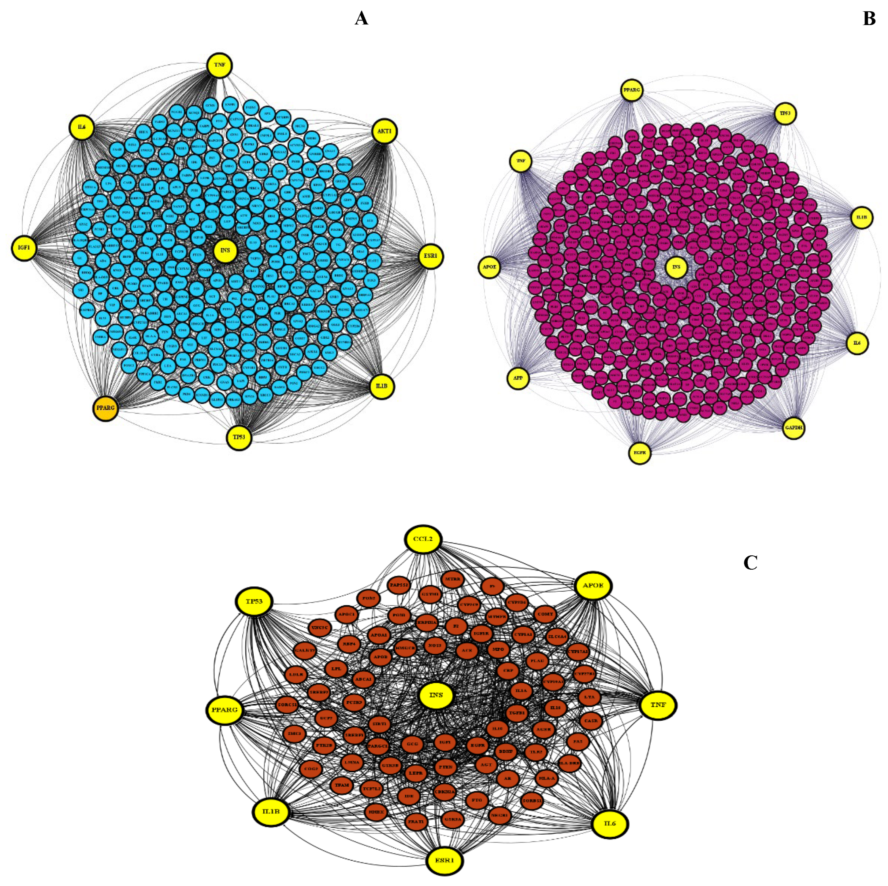
Figure 2.
Common features of polycystic ovarian syndrome (PCOS) and Alzheimer’s disease (AD). Abbreviations: BDNF: Brain-derived neurotrophic factor; CVDs: cardiovascular diseases; COVID-19: Coronavirus disease 2019; IR: insulin resistance; T2DM: type 2 diabetes mellitus. .
Figure 2.
Common features of polycystic ovarian syndrome (PCOS) and Alzheimer’s disease (AD). Abbreviations: BDNF: Brain-derived neurotrophic factor; CVDs: cardiovascular diseases; COVID-19: Coronavirus disease 2019; IR: insulin resistance; T2DM: type 2 diabetes mellitus. .
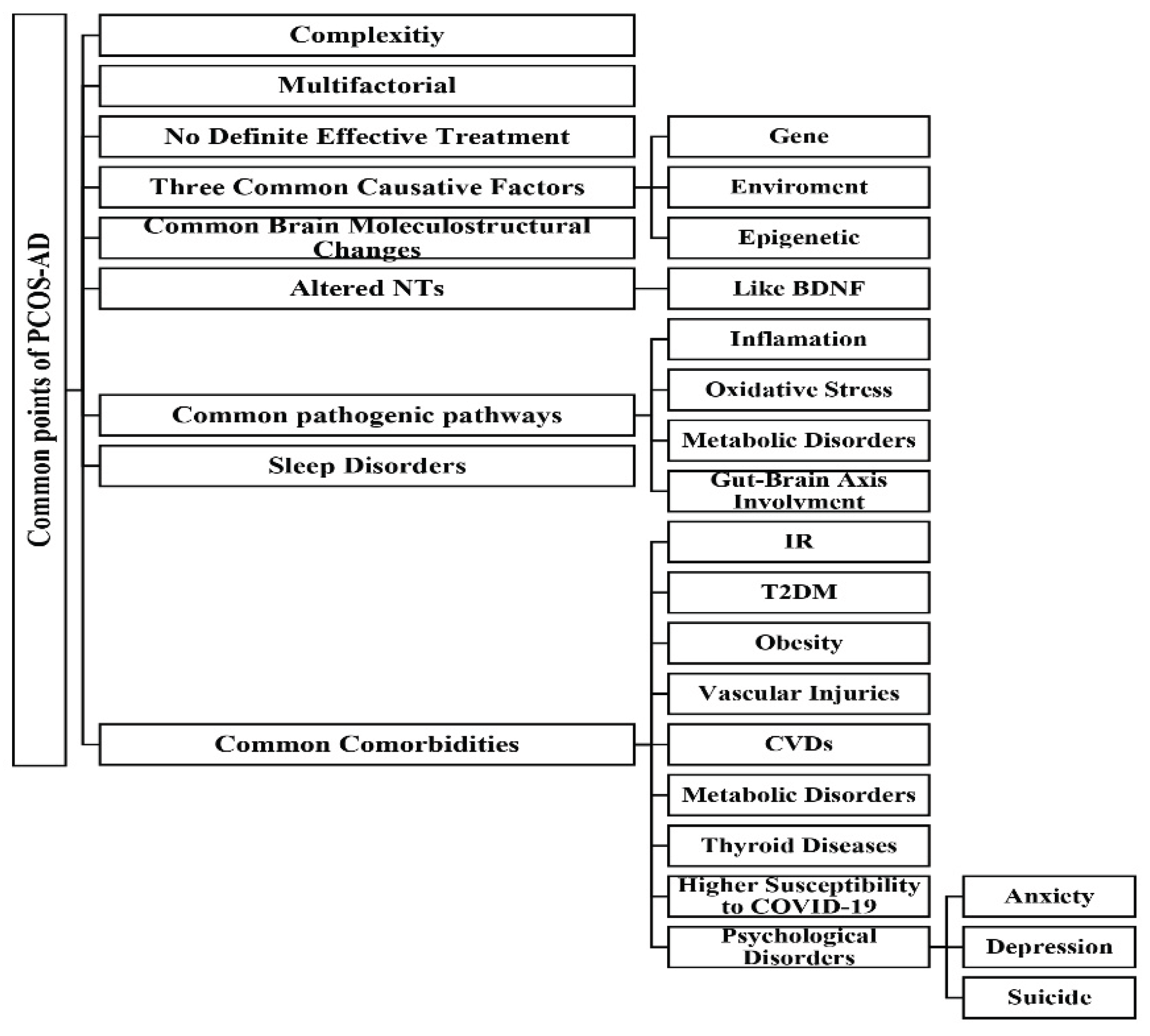

Table 1.
The statistics of protein-protein interaction network of polycystic ovarian syndrome (PCOS), Alzheimer’s disease (AD), and common PCOS-AD networks analyzed using STRING ver. 12 launched at https://string-db.org/.
Table 1.
The statistics of protein-protein interaction network of polycystic ovarian syndrome (PCOS), Alzheimer’s disease (AD), and common PCOS-AD networks analyzed using STRING ver. 12 launched at https://string-db.org/.
| PCOS | AD | PCOS Ո AD | |
|---|---|---|---|
| Number of nodes | 370 | 734 | 83 |
| Number of edges | 5812 | 7817 | 956 |
| Average node degree | 31.4 | 21.3 | 23.0 |
| Average local clustering coefficient | 0.506 | 0.407 | 0.674 |
| Expected number of edges | 2253 | 4431 | 324 |
| Protein-protein interaction enrichment p-value | < 1.0e-16 | < 1.0e-16 | < 1.0e-16 |
| Hub proteins | INS, AKT1, IL6, TP53, TNF, ESR1, PPARG, IL1B, IGF1 | GAPDH, APOE, TNF, TP53, IL6, APP, INS, IL1B, EGFR, PPARG | INS, IL6, TNF, APOE, IL1B, TP53, PPARG, CCL2, ESR1 |
Note: Abbreviations: INS: Insulin; IL6: Interleukin 6; TP53: Tumor protein p53; TNF: Tumor necrosis factor; ESR1: Estrogen receptor 1; PPARG: Peroxisome proliferator-activated receptor gamma; IL1B: Interleukin 1 beta; IGF1: Insulin-like growth factor 1; GAPDH: Glyceraldehyde-3-phosphate dehydrogenase; APOE: Apolipoprotein E; APP: Amyloid-Beta Precursor Protein; EGFR: Epidermal growth factor receptor; CCL2: C-C motif chemokine ligand 2.
Disclaimer/Publisher’s Note: The statements, opinions and data contained in all publications are solely those of the individual author(s) and contributor(s) and not of MDPI and/or the editor(s). MDPI and/or the editor(s) disclaim responsibility for any injury to people or property resulting from any ideas, methods, instructions or products referred to in the content. |
© 2026 by the authors. Licensee MDPI, Basel, Switzerland. This article is an open access article distributed under the terms and conditions of the Creative Commons Attribution (CC BY) license.
Copyright: This open access article is published under a Creative Commons CC BY 4.0 license, which permit the free download, distribution, and reuse, provided that the author and preprint are cited in any reuse.