

Submitted:
12 September 2026
Posted:
14 September 2026
You are already at the latest version
Abstract
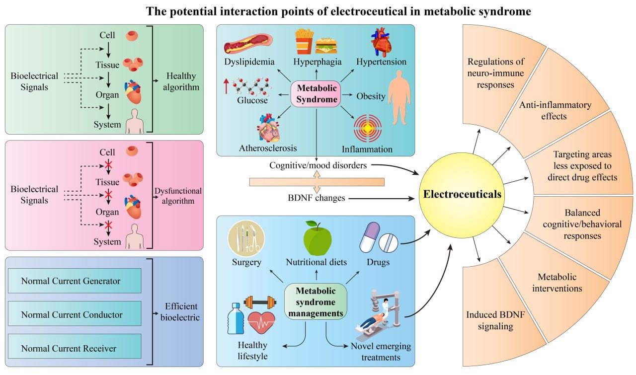
Bioelectricity is a fundamental characteristic of biological systems, affecting cells, tissues, organs, and the organism as a whole. Therefore, monitoring and modulating bioelectric states through electroceuticals (ECs) may offer new solutions for many modern diseases that lack specific therapies or are resistant to current pharmaceuticals. Today, metabolic syndrome (MetS) is a significant health challenge, with many of its underlying processes and molecular factors (e.g., brain-derived neurotrophic factor) that rely on a physiological bioelectric system. This narrative review aims to highlight the opportunities and challenges of ECs in managing MetS and to explore potential future approaches in this therapeutic discipline by searching for related words and phrases in PubMed, Embase, Scopus, Web of Science, Google Scholar, and ClinicalTrials.gov. ECs can modulate neuro-metabolic and autonomic circuits, which may influence multiple components of MetS. The most important limitations of this section include the lack of standardized and well-defined safe therapeutic parameters, lack of precise targeted dosing parameters, risk of potential adverse effects of autonomic stimulation, and lack of comprehensive and long-term clinical data on the effect of ECs on MetS. Therefore, it can be concluded that ECs currently could represent a supportive complementary therapy in the field of MetS, which requires more comprehensive and detailed research to create a possible strong therapeutic approach in the future.
Keywords:
metabolic syndrome
; electroceutical
; bioelectricity
; obesity
; brain‐derived neurotrophic factor
1. Introduction
Electroceuticals (ECs) and bioelectronic medicine (BEM) are medical disciplines that employ electrical stimulation of organs to treat diseases and improve health [1,2]. Bioelectric signaling influences many metabolic processes, mainly ion and metabolite transport and electrochemical synapses, and its modulation can modify pathogenesis [3,4]. The EC modalities can be classified according to the energy type, namely electrical, magnetic, electromagnetic, optogenetic/photostimulatory, and focused ultrasound, or based on delivery/target including invasive and noninvasive tools for nervous systems [1,2,3].
Electrical stimulation using electrodes includes implanted deep brain stimulation (DBS), vagus nerve stimulation (VNS), transcutaneous vagus nerve stimulation (tVNS), transcranial direct current stimulation (tDCS), transcranial alternating current stimulation (tACS), cranial electrotherapy stimulation (CES), electroconvulsive therapy (ECT) [1,2,3,5,6]. Single-pulse transcranial magnetic stimulation (TMS), paired-pulse TMS, and repetitive transcranial magnetic stimulation (rTMS) belong to magnetic stimulation. Photobiomodulation using infrared or near-infrared light, repetitive electromagnetic field stimulation (REMFS), and other electromagnetic fields (EMF) are all approaches employed for BEM [1,2,7,8]. External nerve stimulation alters cellular membrane potential and triggers sequential changes at the cellular and systemic levels [1]. In fact, neural stimulation can alter cellular bioelectric signaling presented with changes in excitability, function, production, and survival of cells [1,2].
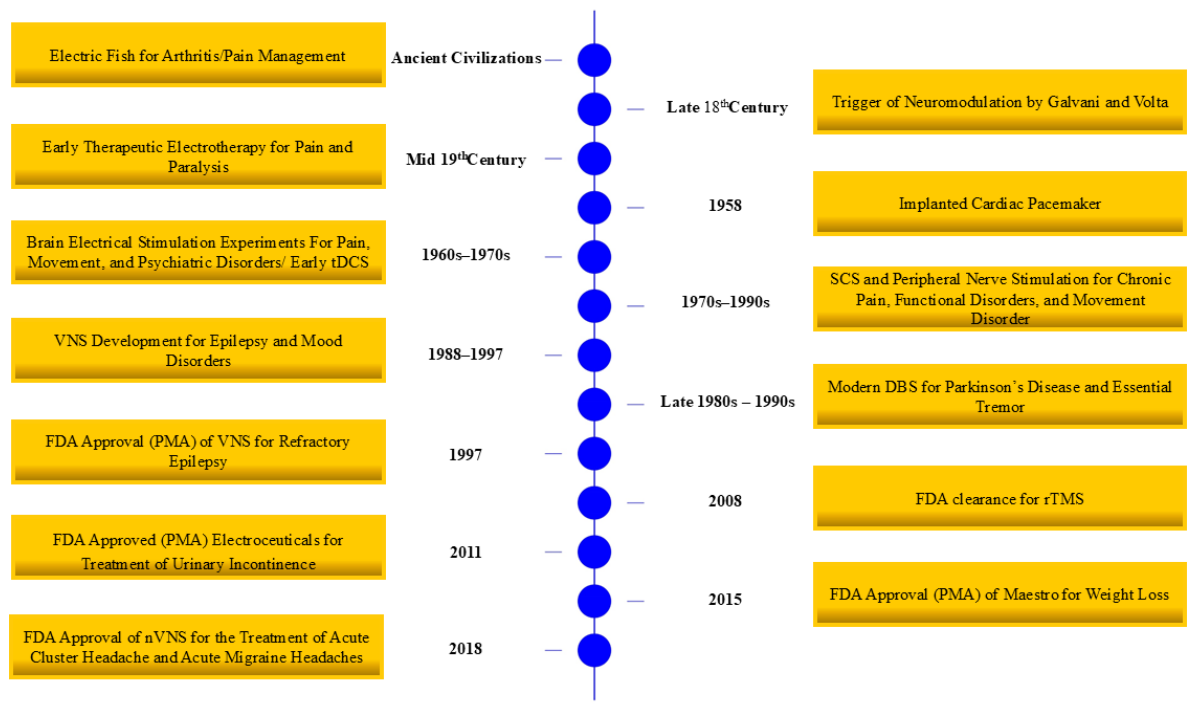
Bioelectronic medicine has a long history and dates back to ancient civilizations, particularly Greece and Egypt, when electric fish were used therapeutically for pain and arthritis [9] (Figure 1). In the 18th and 19th centuries, electrical devices replaced these living sources of electricity. During this period, experiment dubbed as Galvani demonstrated the effects of electricity on animals, and the foundations of electrical priming and neuromodulation were established [10]. The 19th century saw a major expansion of electrotherapy for psychiatric disorders and pain management [11,12,13]. The mid-nineteenth century saw the use of artificially generated electrical currents to control pain and paralysis [13]. In the 1930s, the development of external cardiac pacing concepts began [14], which in the 1950s led to the fabrication of original implantable human pacemaker [15]. This marked the beginning of modern research in this field, although this type of research was initially developed in other branches. It was not until 1967 that the first clinical trials of DBS and spinal cord stimulation for chronic pain and movement disorders were conducted, and the therapeutic effectiveness of DBS began to be discussed [16,17]. In the 1960s and 1970s, experiments were conducted on electrical brain stimulation to treat psychiatric, motor, and pain disorders, besides early tDCS [18,19]. VNS was first used in 1974 for epilepsy, and later DBS became a therapeutic option for Parkinson's disease and tremors between 1987 and 1990 [16,20,21]. In the 1970s to 1990s, the impact of spinal cord stimulation (SCS) on pain control was further developed, and the need for improved implant hardware became more apparent [22,23]. The 1980s-1990s saw an increasing emphasis on hardware engineering and optimizing implantation techniques. During this period, peripheral nerve stimulation and SCS made significant advances in pain management, besides advances in DBS programming, especially for Parkinson’s disease and essential tremor [21,24,25,26]. Collectively, these basic and empirical studies contributed to the rapid expansion and advancement of ECs and BEM from 2000 onward (vide infra).
In 1997, VNS received premarket approval (PMA) from the Food and Drug Administration (FDA) for epilepsy; it was the first peripheral EC device approved for neuromodulation of a central nervous system disorder [3,27,28]. In the 2000s, there was increasing attention was directed toward functional neuromodulation for the treatment of depression, cognitive impairment, and post-stroke recovery using closed-loop neuromodulation and responsive devices (e.g., responsive neurostimulation) were introduced during this period to improve therapeutic precision and efficacy [29,30]. Received FDA clearance and noninvasive EC therapies for treatment-resistant depression was rTMS in 2008 [31]. Another device that received FDA clearance in 2013 for drug-resistant major depressive disorder is BrainsWay Deep TMS [32]. In the 2020s, the field has increasingly focused on improving functionality and general usability, including customized miniaturized design, more precise targeting, repurposing, biomarker-guided treatment, and new therapeutic indications for cognitive, immune, and metabolic disorders have been proposed.
Despite the FDA-PMA history for VNS in 1997, the applications of electroconvulsive therapy are not limited to direct neurological diseases and include more recent applications (vide infra). The FDA approved VNS under PMA in 1997 and 2005 for treatment-resistant epilepsy and depression, respectively [3]. Intermittent theta-burst stimulation (iTBS) received FDA-clearance for major depressive disorder in individuals with no significant improvement from their previous antidepressant medication [3], DBS received FDA approval under PMA for Parkinson's disease [3], the InterStim system was FDA-approved (PMA) for the treatment of urinary incontinence [27]. Deep transcranial magnetic stimulation (dTMS) received FDA clearance for depression and obsessive-compulsive disorder [33]. Pain management through neuromodulation via spinal cord stimulation (SCS) is another FDA-approved application under PMA [33]. Noninvasive VNS (nVNS) received FDA clearance for the treatment of acute cluster headache and acute migraine headaches in 2018 [34]. The Maestro rechargeable system, was approved by the FDA in 2015 under the PMA pathway for weight loss in obese patients with specified BMI and clinical criteria; its mechanism is vagal block therapy [27]. The Medtronic Enterra device received FDA authorization under a Humanitarian Device Exemption (HDE) for gastroparesis-associated refractory nausea/vomiting by gastric electrical stimulation [27].
Recent advances have substantially transformed peripheral ECs. These developments include (1) high-density, multi-contact nerve interfaces that enable fascicle-level selectivity and reduce off-target activation [35]; (2) chronically stable, soft conductive coatings and hydrogels that lower impedance and attenuate foreign-body responses to maintain signal quality long-term [36]; (3) miniaturization and wireless, battery-free power-delivery strategies (magnetoelectric or ultrasonic powering of millimetric implants) that allow less invasive placement near peripheral targets [37]; (4) closed-loop sensing and control algorithms that use neural or physiological biomarkers to adapt stimulation in real time, enhancing efficacy and reducing side effects [38]; and (5) new stimulation modalities and waveform strategies, notably Kilohertz-frequency conduction block and optimized multi-contact waveforms, along with emerging optical/optogenetic methods for fiber-type specificity, which together enable selective excitation or reversible blockade of peripheral fibers [39]. These innovations in neural interfaces, materials, power/packaging, algorithms, and waveforms form the current technical foundation for precise, durable peripheral EC therapies. The potential application of these technologies to MetS has attracted increasing attention in recent years. Therefore, this review aims to comprehensively examine the development of ECs for metabolic syndrome (MetS), covering the neuronal pathways, challenges, and technological advances.
Figure 1.
Timeline of key electroceuticals (EC) milestones. The timeline summarizes the most important periods of time in the advancement of ECs. DBS: deep brain stimulation; FDA: food and drug administration; rTMS: repetitive transcranial magnetic stimulation; SCS: spinal cord stimulation; nVNS: noninvasive VNS; tDCS: transcranial direct current stimulation; VNS: vagus nerve stimulation.
Figure 1.
Timeline of key electroceuticals (EC) milestones. The timeline summarizes the most important periods of time in the advancement of ECs. DBS: deep brain stimulation; FDA: food and drug administration; rTMS: repetitive transcranial magnetic stimulation; SCS: spinal cord stimulation; nVNS: noninvasive VNS; tDCS: transcranial direct current stimulation; VNS: vagus nerve stimulation.
2. Search Strategy and Scope of the Review
This article was conducted as a narrative review and was not designed or reported as a systematic review or meta-analysis. A structured literature search was performed in PubMed, Embase, Scopus, Web of Science, Google Scholar, and ClinicalTrials.gov to identify relevant studies. The search strategy combined Medical Subject Headings (MeSH), where applicable, and free-text keywords using Boolean operators. Representative search terms included: ("bioelectricity" OR "bioelectronic medicine" OR "electroceuticals" OR "electrophysiology" OR "neuromodulation" OR "electrical stimulation" OR "vagus nerve stimulation") AND ("metabolic syndrome" OR "brain-derived neurotrophic factor" OR "BDNF"). Additional relevant articles were identified through manual screening of the reference lists of eligible publications.
Only full-text articles published in English in peer-reviewed journals were considered. Original research articles, clinical studies, randomized and non-randomized trials, observational studies, systematic reviews, and high-quality narrative reviews were eligible for inclusion. Relevant human, animal, and in vitro studies were considered when they provided mechanistic or translational insights into the role of bioelectricity and electroceutical interventions in metabolic disorders. Publications not directly related to the review topic, conference abstracts, editorials, letters, and duplicate reports were excluded.
The review focused on studies involving populations with MetS and direct evidence in MetS was prioritized, whereas evidence from individual metabolic components including obesity, insulin resistance, type 2 diabetes mellitus, and metabolic dysfunction-associated steatotic liver disease (MASLD), preclinical studies, and unrelated clinical applications was considered separately as indirect, mechanistic, or technological evidence. Interventions of interest included bioelectronic and EC approaches, such as vagus nerve stimulation, neuromodulation, and other therapeutic electrical stimulation modalities. Where applicable, comparator groups included sham stimulation, standard medical therapy, placebo, or untreated controls. Outcomes of interest included metabolic, physiological, neurohumoral, and clinical measures related to metabolic regulation and disease progression. Studies were selected based on their relevance to the review objectives, scientific quality, mechanistic insight, and translational significance. Because this was a narrative review, no formal risk-of-bias assessment or quantitative evidence synthesis was performed. The evidence was synthesized narratively to provide an integrated overview of current knowledge, emerging therapeutic applications, and future research directions in bioelectronic medicine for MetS.
3. Overview of Metabolic Syndrome and Its Electroceuticals
An Overview of Metabolic Syndrome
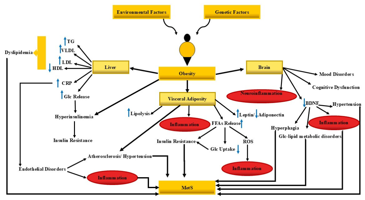
The development of MetS, syndrome X is influenced by genetics, modern hypokinetic and sedentary lifestyle, high-calorie diet, sleep disorders, noise pollution, aging, and sociocultural conflicts [40]. Epidemiological studies presented a prevalence rate of 14 to 39% for this disease [41,42]. Major agencies have defined coherent diagnostic criteria for MetS in different way [42,43]. For instance, according to the National Cholesterol Education Program and Adult Treatment Panel III, the presence of 3 out of 5 factors in an individual is essential as a diagnostic criterion for this syndrome: 1. Waist circumference: men ≥102 cm (40 in) and women ≥88 cm (35 in), 2. Triglycerides: ≥150 mg/dL (1.7 mmol/L), 3. High-density lipoprotein cholesterol: men <40 mg/dL (1.04 mmol/L) and women <50 mg/dL (1.29 mmol/L), 4. Blood pressure: systolic ≥130 mmHg and/or diastolic ≥85 mmHg, 5. Fasting plasma glucose: ≥110 mg/dL; however, the corrected value has updated to ≥100 mg/dL MetS is a multisystem disorder involving multi-organs and physiological pathways, but its major complications include central obesity [44], ectopic fat accumulation [45], insulin resistance (IR) [46], and diabetes mellitus [47], cardiovascular diseases [48], non-alcoholic fatty liver [44], neurohormonal regulatory disorders [49] (Figure 2). Inflammatory conditions occurring in MetS [50], and other molecular changes including BDNF and cerebral/blood supply [51], polycystic ovary syndrome [52], and even Alzheimer's disease (AD) [53], are not far from being expected. Collectively, MetS is a polysystemic disorder needs to be treated with novel tools of BEM.
After Kyler identified the relationship between hypertension, hyperglycemia, and gout; Vague emphasized the component of obesity and metabolic disorders in 1947, and about 40 years later, Reaven proposed ‘Syndrome X’ for a set of metabolic disorders centered on IR, not obesity [42]. In 1989, Kaplan added the obesity factor to Kyler's proposal and named this syndrome the ‘Deadly Quartet’[42]. The naming of MetS as ‘IR syndrome’ finally happened in 1992 [42,54]. Routine treatment for MetS is currently mainly symptomatic, involves lifestyle improvement, and Pharmacological interventions are directed toward specific components of MetS, such as antihypertensive drugs [55], beta blockers [40], angiotensin converting enzyme inhibitors [55], diuretics [40], statins [55], hypoglycemic agents like thiazolidinediones [55], fibrates [55], bile acid sequestrants [40], niacin [40], rapamycin [40], metabolic/bariatric surgical interventions [56], device-based therapies, molecular/gene therapy, and EC [40]. Therefore, it is necessary to explore etiology and novel therapy that addresses the causes of this syndrome.
Figure 2.
Pathological mechanisms involved in metabolic syndrome (MetS). This figure illustrates factors contributing to the development of MetS criteria. Obesity-driven MetS arises from complex interactions among genetic, environmental, inflammatory, and neuroendocrine factors. Visceral adiposity promotes free fatty acid release, chronic inflammation, and insulin resistance, leading to dyslipidemia, hyperglycemia, endothelial dysfunction, and hypertension. Metabolic and inflammatory signals also affect the brain, contributing to neuroinflammation, altered BDNF signaling, cognitive dysfunction and mood-related disorders. The bidirectional communication between peripheral organs and the central nervous system highlights key neuromodulatory targets for electroceutical therapies aimed at restoring metabolic homeostasis and reducing systemic inflammation. BDNF, brain-derived neurotrophic factor; CRP, C-reactive protein; FFAs, free fatty acids; Glc, glucose; HDL, high-density lipoprotein; LDL, low density lipoprotein; ROS, reactive oxygen species; TG, triglyceride; VLDL, very low-density lipoprotein.
Figure 2.
Pathological mechanisms involved in metabolic syndrome (MetS). This figure illustrates factors contributing to the development of MetS criteria. Obesity-driven MetS arises from complex interactions among genetic, environmental, inflammatory, and neuroendocrine factors. Visceral adiposity promotes free fatty acid release, chronic inflammation, and insulin resistance, leading to dyslipidemia, hyperglycemia, endothelial dysfunction, and hypertension. Metabolic and inflammatory signals also affect the brain, contributing to neuroinflammation, altered BDNF signaling, cognitive dysfunction and mood-related disorders. The bidirectional communication between peripheral organs and the central nervous system highlights key neuromodulatory targets for electroceutical therapies aimed at restoring metabolic homeostasis and reducing systemic inflammation. BDNF, brain-derived neurotrophic factor; CRP, C-reactive protein; FFAs, free fatty acids; Glc, glucose; HDL, high-density lipoprotein; LDL, low density lipoprotein; ROS, reactive oxygen species; TG, triglyceride; VLDL, very low-density lipoprotein.
4. Electroceuticals: From Neurobioelectronic Regulation of Intermediary Metabolism to Metabolic Syndrome
Glucose metabolism and blood sugar regulation are closely linked to bioelectric signaling, as glucose metabolism in beta cells increases the ratio of ATP to adenosine diphosphate (ADP), which causes the closure of ATP-sensitive potassium channels (KATP) and membrane depolarization [57]; voltage-gated calcium channels (Cav) then open, which causes Ca2+ entry and insulin granule exocytosis [57]. Insulin sensitivity and glucose uptake by muscle cells, which are essential for maintaining glucose homeostasis, are regulated by membrane excitability and Ca2+ handling [58]. Normal lipid metabolism is dependent on the proper functioning of bioelectricity-dependent events, for example, lipolysis, adipokine (leptin and adiponectin) secretion, activities of metabolic enzymes in adipocytes and hepatocytes are controlled by intracellular Ca2+ signaling regulated by ion channels such as transient receptor potential (TRP) channels and Cav [59]. Therefore, appropriate regulation of bioelectric signaling is important for basic cell function.
The electrical and chemical synapses are dependent on bioelectricity, and this is crucial in the transmission of central metabolic regulatory messages to organs using peripheral nerves. Sensory vagus afferents induce satiety, prevent overeating, and regulate glucose homeostasis [60]. These signals arise from mechanical signals from gastric distension, chemical signals from gut hormones, and inflammatory signals from the gut, liver, and adipose tissue which are conveyed to the brainstem [61]. Hepatic glucose production, insulin secretion, and gut motility are under the control of efferent vagal activity, and dysfunctions in this pathway may contribute to pro-inflammatory conditions, IR, or glucose intolerance [61]. Thus, these afferent and efferent nervous systems, by transmitting adipose and enteric messages including cholecystokinin, glucagon-like peptide-1 (GLP-1) to the hypothalamus and brainstem, can regulate glucose production, hepatic gluconeogenesis, adipose tissue lipolysis, brown adipose tissue thermogenesis, insulin secretion, gastric emptying rate, and even satiety duration [62,63,64]. Notably, neuroimmune cholinergic anti-inflammatory processes can reduce inflammation and its complications, such as atherosclerosis and IR [65,66].
The sympathetic nervous system, through its innervation of the vasculature, liver, pancreas, and adipose tissue, plays a regulatory role in blood pressure, hepatic glucose output, and lipolysis [67,68]. In stressful conditions and sympathetic hyperactivity, an increase in blood pressure, stimulation of lipolysis, and blood sugar disorders may occur [69,70]. In conclusion, maintaining an appropriate balance of autonomic peripheral nerve signaling is vital for the health of metabolic processes, and any malfunction of this system can lead to secondary metabolic disorders.
In a normal and efficient metabolism, not only the health of the endocrine glands and nerves individually, but also their proper relationship and the neuroendocrine axes (e.g., hypothalamic-pituitary-adrenal and hypothalamic-pituitary-thyroid axes) are coordinated interactions [71]. Therefore, correcting the dysfunction within these neuroendocrine pathways may also have a significant impact on improving the metabolic status of the body. The bioelectricity may regulate metabolic processes and energy supply since it contains central neural circuits located in the hypothalamus, brainstem centers, mesolimbic dopamine system, and limbic structures [56,72,73]. These centers integrate afferent inputs with hormonal signals of GLP-1, leptin, ghrelin, and insulin [72,73]. The encoding of hormonal signals and nutrients, appetite, ingestive motivation, regulation of glucose-gut-brain signaling, and signaling of peripheral nerves can be governed by the central centers [72,73]; therefore, dysregulation of these centers can lead to hyperphagia, weight gain, and behavioral disorders related to metabolism. Neurotransmitters and neuromodulators, including serotonin, dopamine, acetylcholine, norepinephrine, and neuropeptides such as orexin, neuropeptide Y (NPY), alpha-melanocyte-stimulating hormone, and activation of agouti-related peptide (AgRP), can influence neuronal activity and downstream peripheral metabolic organs [56,72,73]. Neural firing rate, burst patterns, and oscillatory synchronization encode metabolic states [74]. All neurotransmitter reuptake [75,76], cellular ionic homeostasis [77], besides cellular reactions to metabolic processes, inflammation, and toxic substances are regulated by astrocytes and other glial cells [56]. Therefore, EC interventions on the brain and spinal cord may regulate metabolism and influence MetS through modifying both neuronal and non-neural circuits.
Interestingly, EC interventions have been shown to modulate BDNF, an important regulator of neuro-metabolic functions. This neurotrophin supports neuronal survival and function of neurons [78], and controls metabolic processes [79]. BDNF transcription and secretion are influenced by neuronal depolarization and calcium influx through voltage-gated Ca2+ channels and N-methyl-D-aspartate receptors [80]. In addition, BDNF and its specific receptor, tropomyosin receptor kinase B, “participate in the regulation of ion channels and metabolism [79]. Centrally, BDNF modulate the activity of anti-inflammatory pro-opiomelanocortin and appetitive AgRP/NPY neurons, and its consequences include reduced food intake [81], and increased energy expenditure, enhanced satiety signaling, and altered motivational aspects of ingesting behavior [82]. The peripheral effects of BDNF are related to energy expenditure [82], thermogenesis [81], anti-inflammatory effects [83], peripheral insulin sensitizing [82], and reduced hepatic glucose production [84]. BDNF improves mitochondrial ATP synthesis for cellular ion pumps, membrane potentials, and cellular excitability [85]. Evidently, a reduced BDNF levels lead to hyperphagia, obesity, reduced energy expenditure, and impaired glucose homeostasis, which reflects its lower levels in patients with MetS, diabetes mellitus, and obesity [86]. Exercise or intake of some antidepressants increases BDNF levels, which highlights its lower levels in depressed individuals [87]. The contribution of BDNF in many neurometabolic diseases, such as MetS and AD, is clear. Some literature pointed to various ECs that enhance BDNF signaling following DBS [88], and rTMS [89]. In sum, BDNF may be a potential therapeutic target in MetS, which can be modulated by appropriately designed EC interventions.
The manipulation of bioelectricity for reprogramming of metabolism seems reasonable, and it can trigger a cascade of therapeutic effects. For instance, electrical stimulation of carotid baroreceptors, hypothalamus, vagus nerve, gastric areas, and muscles can trigger a cascade of neurometabolic events, such as regulation of appetite, brain reward system and emotional behavior, hepatic glucose production, insulin secretion, lipolysis, thermogenesis, and inflammation [90]. This electrical manipulation may have therapeutic potential for MetS through controlling blood pressure, improving blood sugar and lipid profile, reducing inflammation, and decreasing appetite [28]. The ECs are prescribed as anti-inflammatory and anti-apoptotic agents, and changes in metabolism especially brain glucose access through DBS or tDCS techniques [91,92,93]. Taken together, ECs can become a tool for controlling inflammatory and metabolic pathways.
To summarize, ECs are being investigated as a potential new therapeutic approach in MetS in several ways, including multi-axis modulation, balanced energy intake, glucose and lipid metabolism, autonomic system related responses, systemic inflammation, induced neurotrophic signaling specifically BDNF [2,94], programmability, temporal precision, and access to deep neural targets (Figure 3). Noteworthy, ECs could act as supplements to pharmacotherapy for MetS, and their potential effects on modifying the individual's behavioral responses, such as greater interest and motivation for exercise or dietary choices can be the subject of future studies.
5. Technical Development and Challenges in Electroceutical Design: Overview
EC-based therapies modulate physiology; however, they carry specific risks and technical challenges that must be addressed to ensure safe, effective, and meaningful clinical use. In this context, radiofrequency and some electromagnetic modalities can produce tissue heating, and tissue properties; electrome, anatomy, electrode and device geometry, electromagnetic exposure parameters must be carefully evaluated in any conceptual design and optimization of EC systems [2]. Adverse effects of ECs across modalities include infection, hematoma, pulmonary embolism, respiratory issues, transient cognitive decline, headaches, voice changes, and neuropsychiatric outcomes [2,3,34]. Technically, device designing and reliability are also important. Implants and stimulators face hardware failure modes (lead breakage, battery depletion, encapsulation) and software issues (bugs, connectivity loss) [2,34]. High treatment and device costs ar a general consideration in nearly all types of EC design [3]. More biocompatible, resilient, and recyclable materials are desirable to reduce foreign-body reactions, biofouling, and long-term failure.
Technical progress in ECs has moved from large, open-loop stimulators toward compact, target-specific systems that integrate sensing, stimulation, and communication [95]. Early devices focused on delivering safe, reproducible pulses to relatively accessible nerves; present-day efforts prioritize precision targeting, programmable waveforms, and compatibility with ambulatory use [96]. The engineering challenge is to deliver therapeutically effective electrical doses while minimizing off-target effects, power consumption, and device footprint. Therefore, progress in microfabrication, low-power electronics, and materials science strengthens advancement.
The electrode–tissue interface is a crucial hardware element that determines selectivity, efficacy, and longevity of stimulation, and designs range from cuff and penetrating microelectrodes to conformable surface arrays [97]. Key technical goals are to maximize charge transfer efficiency, reduce impedance and inflammatory encapsulation, and maintain stable contact despite tissue motion [98]. Novel geometries (e.g., multi-contact cuffs, thin-film arrays) and surface coatings (e.g., conductive polymers, porosity-enhancing treatments) improve stimulation selectivity and long-term performance [99]. Device developers must balance invasiveness against precision as more selective interfaces often require more complex implantation techniques and surgical approaches.
Beyond simple pulse amplitude and frequency, modern ECs exploit a broad parameter space including waveform shape, pulse width, burst patterns, and inter-pulse timing to tune neural recruitment and therapeutic outcomes [100]. Optimization requires understanding the electrophysiology of the target (fiber types, conduction velocities, and refractory dynamics), and may employ computational models to predict which parameters preferentially activate desired pathways while minimizing activation of neighboring structures [101]. Energy-efficient waveforms and staggered multi-site stimulation strategies can reduce battery drain and side effects [102]. Standardizing parameter reporting and dose metrics is essential for comparing studies and translating protocols between systems.
The precise targeting of neural structures is essential for reproducible metabolic effects, but autonomic nerves are often small anatomical entities intertwined with other fibers [103]. Advances in intraoperative mapping (electrophysiologic recording, evoked potentials), imaging co-registration (ultrasound, magnetic resonance imaging), and neuronavigation tools have improved localization and reduced implantation variability [104]. For noninvasive approaches, biomarker-guided positioning (e.g., heart-rate variability, reflex responses) helps identify effective stimulation sites [105]. Robust surgical workflows and training protocols are equally important to ensure consistent outcomes across centers.
Power autonomy and secure wireless telemetry govern device usability and patient comfort; rechargeable batteries, inductive charging, and energy harvesting are all active strategies to extend runtime without enlarging implants [106]. Low-power system-on-chip designs, duty-cycling of stimulation, and local processing to reduce wireless traffic improve energy efficiency [107]. Telemetry protocols must ensure reliable, low-latency data transfer for programming and diagnostics while maintaining patient security and privacy [108]. Miniaturization of electronics and packaging enables less-invasive implants and broader adoption among ambulatory populations [96].
The next generation of ECs moves to closed-loop control by integrating sensors (biopotentials, biochemical sensors, motion/respiratory monitors) that provide real-time feedback to adapt stimulation [109]. Closed-loop systems promise better efficacy and fewer side effects by delivering stimulation when physiological biomarkers indicate a therapeutic need, and by dynamically adjusting stimulation parameters [110]. Implementing robust control algorithms requires reliable biomarkers, artifact-resilient sensing, and safety-constrained controllers that avoid runaway stimulation [110]. Demonstrating that sensor-driven adaptation improves clinical outcomes remains a central translational milestone [111].
Chronic implantation exposes devices to biofouling, fibrosis, corrosion, and mechanical fatigue; therefore, appropriate material selection and hermetic packaging are crucial for long-term reliability [112]. Biocompatible polymers, thin ceramic coatings, and silicone encapsulation are commonly used, and each impacts stiffness, conformability, and immune response [113]. Designing devices with predictable failure modes (e.g., predictable end-of-life behavior) also helps with regulatory approval and patient safety [114].
Software ecosystems, including firmware, clinician programming tools, and cloud analytics, are essential components that affect usability, reproducibility, and regulatory classification [115]. Data pipelines that convert raw sensor streams into actionable biomarkers need validated algorithms, secure storage, and transparent performance metrics; regulatory agencies are also increasingly evaluating software as a medical device [116]. From a manufacturing standpoint, reproducible microfabrication, quality control of electrode properties, and scalable assembly processes determine commercial viability. Regulatory pathways, clinical trial design, and reimbursement strategies must be planned in parallel to technical development to ensure timely translation.
6. Discussion: Evidence Synthesis
EC interventions encompass neuromodulatory approaches including transcranial magnetic stimulation [117], low-frequency electrostatic fields (LFEF) [117], electrical muscle stimulation (EMS) [118] and whole-body electromyostimulation (WB-EMS) [119], mild electrical stimulation combined with heat shock (MES+HS) [120], neuromuscular electrical stimulation (NMES) [121], and pulsed electromagnetic field therapy (PEMF) [122].
To facilitate a structured interpretation of the evidence, the effects of ECs were examined across three levels including, category 1 (direct clinical evidence on the effect of electrostimulation intervention on metabolic syndrome), category 2 (clinical evidence on individual components of MetS), and finally category 3 (preclinical mechanistic evidence, which includes studies of animal or laboratory models), are discussed below. A summary of the studies reviewed in categories 1 and 2 is presented in Table 1, while category 3 are discussed separately.
6.1. Category 1: Direct Clinical Evidence
Studies investigating the effects of ECs across the full range of diagnostic parameters of MetS are limited, making it difficult to draw definitive conclusions about their overall therapeutic efficacy. The study by de Moraes et al. [2023] on 30 MetS patients was conducted an open-label, randomized (2:1), two-arm, parallel-group controlled trial that investigated the effect of transcutaneous auricular vagus nerve stimulation (taVNS) [123]. This study resulted in reductions in systolic and diastolic blood pressure and heart rate after this electrical intervention, but the study did not evaluate or report the effects of taVNS on other parameters, especially metabolic parameters of this syndrome [123]. Notably, another point of this study was a shift toward a less inflammatory immune profile in patients following taVNS, which can be interpreted beyond the usual expectations of the effect of modulation of vagal activity [123]. It appears that clinical studies specifically designed to simultaneously evaluate all diagnostic components of MetS while adequately controlling for relevant confounding factors and establishing the therapeutic efficacy of ECs are currently lacking. However, several studies have investigated the effects of different EC modalities on one or more components of MetS, as discussed in the following section.
6.2. Category 2: Clinical Evidence on Individual Components of MetS
6.2.1. Adiposity and Body Composition
Studies evaluating electrical stimulation for visceral adiposity and obesity have yielded mixed results. Kondo et al. [2014] reported that mild electrical stimulation combined with heat shock (MES+HS) reduced visceral adiposity in patients with MetS and type 2 diabetes mellitus (T2DM) [120]. In contrast, Choi et al. [2018] found that electrical muscle stimulation (EMS) did not significantly reduce visceral or total abdominal fat in adults with abdominal obesity, despite a significant reduction in waist circumference [124]. Similarly, whole-body electromyostimulation (WB-EMS) has been associated with reductions in body weight and body fat in obese women with MetS [125].
Vagal nerve blockade (vBloc) produced significant weight loss in a double-blind, randomized controlled trial involving individuals with moderate obesity [126]. However, the intervention did not meet the prespecified co-primary efficacy endpoints reported by Ikramuddin et al., despite producing greater weight loss among participants with severe obesity than in the sham-control group [127]. Another neuromodulatory approach, vagus nerve stimulation (VNS), was evaluated in a small study involving 15 participants and was associated with a significant increase in energy expenditure and changes in physical activity. These findings may provide a potential explanation for the secondary weight-loss effects reported with this approach [128]. In addition, Lam et al. [2024] reported that electroacupuncture (EA) may reduce waist circumference, hip circumference, body weight, body mass index (BMI), and waist-to-hip ratio [129].
6.2.2. Dyslipidemia
Studies investigating the effect of ECs on lipid profile have been less extensive, and it seems that most of the possible changes in blood lipids may be secondary to factors such as body weight loss. An example of this can be seen in the study by Apovian et al. [2017] on the effect of vBloc on obesity, which reported a decrease in low density lipoprotein (LDL), triglycerides, and an increase in high-density lipoprotein (HDL) following the use of this technology in individuals, but given the significant weight loss, these lipid profile changes may be more related to this weight change [130]. Another example of the effect of ECs on lipid profile is the reduction in cholesterol following the combination of LFEF+ТМS in the study by Benkov et al. [2022] on patients with MetS [117], although given the limited 10-session treatment intervention, the certainty of the effectiveness of this technique may still require further investigation.
Based on a randomized controlled trial conducted by Rerksuppaphol & Rerksuppaphol [2014] on the effect of electroacupuncture on regulating blood lipid levels in dyslipidemic patients, a significant reduction in serum lipid levels was reported after the completion of the aforementioned treatment period, and even a reduction in LDL cholesterol and total cholesterol was reported even at the follow-up visit [131]. Of course, it should be remembered that perhaps, based on the scope and specificity of the studies conducted in this field, these changes should be considered more as secondary metabolic outcomes. Therefore, although different electrical and neuromodulatory approaches have demonstrated potential benefits for reducing body weight, adiposity, and anthropometric measures, the available evidence remains heterogeneous, and further well-designed clinical studies are required to establish their efficacy and clinical relevance in the management of MetS.
6.2.3. Glycemic Regulation and Insulin Resistance
Existing reports indicate that different EC modalities may influence another important component of MetS, namely hyperglycemia and insulin resistance, although the magnitude and statistical significance of these effects have varied across studies. NMES has been reported to significantly reduce fasting blood glucose [121]. However, in the study by Miyamoto et al., despite a significant change in fasting glucose concentration, no significant change was observed in hemoglobin A1c (HbA1c) levels [132]. Another notable finding of the same study was a change in plasma BDNF levels [132]. EMS has also been reported to reduce fasting blood glucose and HbA1c according to van Buuren et al. [2015]; however, mean body weight and body-fat percentage did not change significantly following the intervention [133].
In a study by Shikora et al. [2013] involving 28 patients with obesity and type 2 diabetes, implantation of an intermittent vBLOC device was associated with reductions in HbA1c, body weight, and mean arterial pressure [134]. However, because weight loss and improved glycemic control occurred concurrently following vBLOC treatment, a definitive causal conclusion regarding the independent effect of the intervention on blood glucose cannot be drawn. In individuals with impaired glucose tolerance (IGT), taVNS has been reported to produce significant changes in glycosylated hemoglobin (HbA1c), 2-h plasma glucose (2hPG), and fasting plasma glucose (FPG), suggesting its potential utility in the management of IGT and prediabetes. However, further well-designed and specialized clinical studies are required to confirm these findings and establish the therapeutic efficacy of taVNS in this population [135]. However, these findings do not indicate that electrical stimulation is consistently effective in improving glycemic control. Kufaishi et al. [2025], for example, reported no significant benefit of tVNS for glycemic control in the studied diabetic population [136]. Similarly, Lu et al. [2023] reported no significant reduction in HbA1c following transcutaneous electrical nerve stimulation (TENS) in patients with diabetes [137].
Overall, although several studies have reported beneficial effects of electrical interventions on blood glucose and related metabolic parameters, the available evidence remains inconsistent. Therefore, it is premature to draw general conclusions regarding the efficacy of ECs for glycemic control in MetS. Further well-designed clinical and mechanistic studies are needed to clarify the effects of different electrical stimulation modalities and to elucidate the underlying mechanisms responsible for their effects on glucose metabolism.
6.2.4. Blood Pressure and Vascular Function
Blood pressure can also be modulated by EC interventions. For example, baroreflex activation therapy (BAT) has been reported to reduce systolic blood pressure in patients with resistant hypertension [138]. Similarly, respiratory-gated auricular vagus afferent nerve stimulation has been shown to decrease heart rate, mean arterial pressure, and diastolic blood pressure. However, Garcia et al. (2022) reported that these rapid, frequency-dependent effects may vary according to sex and race, highlighting the potential influence of individual characteristics on responses to neuromodulation [139].
Pulsed electromagnetic field therapy (PEMF) represents another emerging approach being investigated for blood pressure control. Kim et al. [2020] reported that PEMF increased nitric oxide levels and reduced systolic blood pressure, diastolic blood pressure, and mean arterial pressure in hypertensive patients with MetS at rest, while also improving blood pressure responses during exercise [122]. Therefore, although these findings suggest that different EC modalities may have beneficial effects on blood pressure through distinct neurovascular and physiological mechanisms, the available evidence remains limited and heterogeneous. Further controlled clinical studies are needed to determine the efficacy, durability, and patient-specific responses to EC interventions for blood pressure management in MetS.
6.3. Category 3: Preclinical Mechanistic Evidence
Preclinical studies are an important step toward understanding the mechanisms, efficacy, and safety of proposed therapeutic approaches, and several such studies have investigated ECs. Evidence from preclinical models suggests that electrical stimulation can influence several metabolic parameters relevant to MetS, including glucagon-like peptide-1 (GLP-1) signaling, insulin sensitivity, insulin and glucagon secretion, and glucose tolerance following VNS in animal models [140,141,142,143,144,145,146]. Direct electrical muscle stimulation may also enhance glucose uptake [147,148,149].
Electrical neuromodulation can also influence adipose tissue and metabolic processes, resulting in changes in appetite, body-weight gain, mitochondrial function, and thermogenesis [150,151,152,153]. Inflammation, another important component of MetS, has also been modulated by electrical interventions such as vagal stimulation and electroacupuncture. These approaches have been associated with changes in inflammatory cytokines, macrophage activity, and nuclear factor kappa-light-chain-enhancer of activated B cells (NF-κB) signaling [154,155,156,157]. Reduction of blood pressure through suppression of sympathetic activity by electrically mediated baroreceptor stimulation represents another investigated effect of ECs on cardiovascular parameters relevant to MetS [158,159].
Collectively, these findings indicate that EC approaches can influence multiple components of MetS. However, the optimal EC modality, stimulation parameters, and treatment protocols required to produce reliable and reproducible therapeutic effects remain unclear. In addition, some preclinical studies have reported contradictory findings. For example, Yin et al. [2019] and Payne et al. [2020] reported increases in glucose and glucagon following VNS [141,142]. Similarly, vagal afferent stimulation has been reported to inhibit brown adipose tissue thermogenesis in some experimental models [150,160]. These discrepancies highlight the complexity of neuro-metabolic responses to electrical stimulation and the importance of considering stimulation modality, anatomical target, stimulation parameters, and experimental model when interpreting preclinical findings.
Several considerations should be addressed in future preclinical studies of ECs. First, greater diversity in animal models should be encouraged, while selecting species that adequately reflect relevant human physiological mechanisms. Reliance on a single animal species may limit the generalizability of findings and may fail to account for interspecies differences that influence the efficacy and safety of EC interventions. Second, preclinical study designs should incorporate translational strategies that facilitate the subsequent application of experimental findings to human clinical research. Ideally, potential differences between animal and human responses should be anticipated during the initial study-design stage rather than recognized only after the completion of preclinical and clinical studies. Therefore, careful consideration of species-specific physiology, stimulation parameters, target anatomy, and clinically relevant outcome measures is essential for improving the translation of preclinical EC research into safe and effective human therapies.
7. Limitations and Future Perspectives
BEM is an interdisciplinary field that integrates medicine, physics, engineering, and related biomedical sciences. Further development of this field requires new perspectives on the opportunities, limitations, and challenges associated with EC technologies. The modulation of bioelectric signaling involved in intermediary metabolism may provide novel and currently underexplored therapeutic opportunities for MetS. However, increasing therapeutic efficacy while minimizing off-target autonomic effects remains a major challenge in the development of EC interventions. Another critical consideration is the determination and standardization of appropriate stimulation parameters and therapeutic dosing. In addition, battery life and power-efficiency constraints may limit the long-term use of implantable and wearable EC devices; therefore, improved power-management strategies, rechargeable or wireless energy solutions, and energy-efficient device architectures should remain priorities for future development.
Sustained progress in EC technology will require close collaboration among researchers from multiple disciplines, including medicine, neuroscience, bioengineering, materials science, and computational sciences. Overall, larger, well-designed, and adequately controlled preclinical and clinical studies are needed to establish the efficacy, safety, optimal dosing parameters, long-term durability, and patient-specific responses to EC interventions. Such multidisciplinary and translational research will be essential for determining whether ECs can evolve from an emerging therapeutic concept into a reliable and clinically meaningful approach for the management of MetS.
8. Conclusions
The EC therapeutic field is likely to play an increasing role in future clinical practice. ECs represent a promising but still emerging therapeutic strategy for MetS because they can modulate neural circuits involved in appetite regulation, glucose homeostasis, lipid metabolism, autonomic balance, inflammation, and energy expenditure. Among the potential therapeutic targets, vagal, sympathetic, hypothalamic, gastric, and neuroimmune pathways appear particularly relevant to MetS because they connect central metabolic regulation with peripheral organs, including the liver, pancreas, gut, adipose tissue, and skeletal muscle. However, the field has not yet reached the level of mechanistic precision or clinical evidence required for routine application in metabolic disorders. Current evidence is insufficient to establish the efficacy, reproducibility, and safety of ECs for the clinical management of MetS, representing an important gap in this field.
The key unresolved challenges extend beyond the general feasibility of electrical stimulation and include target specificity, biomarker selection, dose definition, treatment durability, device reliability, and clinical validation. First, the autonomic and metabolic circuits relevant to MetS are anatomically and functionally complex, and current stimulation approaches often lack sufficient selectivity to distinguish beneficial metabolic effects from off-target autonomic, cardiovascular, gastrointestinal, or neuropsychiatric responses. Second, there is no standardized EC “dose” for MetS. The absence of standardized parameters for stimulation site, frequency, amplitude, pulse width, timing, and treatment duration, together with the limited comprehensive assessment of patients’ metabolic status during EC therapy, makes it difficult to compare and reproduce findings across studies. Third, reliable biomarkers for closed-loop control remain underdeveloped. Measures such as glucose excursions, heart-rate variability, inflammatory markers, appetite-related hormones, BDNF, vagal activity, and neural activity are promising candidates; however, their causal relationships with clinical improvement require more rigorous investigation. Fourth, long-term safety and device durability remain unresolved, particularly for implanted systems exposed to fibrosis, biofouling, battery limitations, hardware failure, and changes in the tissue–device interface. Moreover, most available evidence is fragmented across individual conditions or components, including obesity, diabetes, hypertension, inflammation, and gastrointestinal dysfunction, rather than being evaluated directly in well-characterized populations with MetS. Most studies have also focused on one or a few diagnostic components of MetS, while comprehensive studies simultaneously evaluating the effects of ECs across all diagnostic components remain lacking. Therefore, the absence of adequately designed studies in well-characterized MetS populations represents another important challenge for the development of this therapeutic approach.
Therefore, an important direction for future research is to integrate translational neuro-metabolic phenotyping with closed-loop device development. Progress will depend on identifying which patient subgroups respond to specific neural targets, which biomarkers most reliably reflect therapeutic benefit, and how stimulation parameters should be adapted over time. Future studies should aim to identify neural targets and stimulation parameters capable of producing reproducible and clinically meaningful metabolic effects. Large, controlled, and long-term clinical studies are needed to evaluate clinically relevant outcomes, including waist circumference, insulin sensitivity, postprandial glucose control, blood pressure, lipid profiles, inflammatory status, body weight, quality of life, and medication requirements. Although such studies may be costly, they are essential for establishing the clinical value of ECs. In parallel, engineering efforts should focus on selective peripheral nerve interfaces, stable biocompatible materials, low-power systems, secure telemetry, and adaptive algorithms capable of personalizing stimulation while minimizing adverse effects.
In summary, ECs should currently be regarded as a complementary and investigational approach rather than a replacement for lifestyle modification, pharmacological therapy, or surgical management of MetS. Their greatest potential may lie in the precision modulation of neuro-metabolic circuits, particularly when stimulation can be guided by validated biomarkers and adapted to individual physiological responses. Future studies should progressively move from broad proof-of-concept stimulation experiments toward mechanistically grounded, biomarker-driven, and clinically rigorous trials capable of determining where ECs provide meaningful additional value in the management of MetS. Ultimately, the clinical value of ECs will depend on whether they can deliver sufficiently precise and mechanistically informed stimulation to produce effective, durable, reproducible, and clinically meaningful metabolic benefits in appropriately selected populations with MetS.
Author Contributions
ShM: Conceptualization, Writing – original draft, Data curation, Formal Analysis, Investigation. IK: Conceptualization, Writing – original draft, Writing – review & editing, Investigation, Formal Analysis, Validation, Visualization, Project administration, Supervision. LJM: Writing – original draft, Data curation, Investigation, Visualization, Validation, Methodology. AMA: Writing – original draft, Data curation, Investigation, Resources. NS: Writing – original draft, Data curation, Investigation, Methodology, Validation. HBS: Conceptualization, Writing – original draft, Writing – review & editing, Data curation, Supervision, Validation, Funding acquisition, Methodology.
Funding
The authors declare that financial support was received for the research, authorship, and/or publication of this article. Razi University and Babylon University provided the facilities and institutional support necessary to conduct this work. Uppsala University provided scientific supervision and financial support for the publication fee. The funders had no role in the study conception, literature selection, data analysis, interpretation of the findings, or the decision to submit the manuscript for publication.
Institutional Review Board Statement
Not applicable.
Informed Consent Statement
Not applicable.
Data Availability Statement
No new data were generated.
Acknowledgments
We acknowledge and thank those who support and participate in the study.
Conflicts of interest
The authors declare no conflict of interest, financial or otherwise.
Generative AI statement
We consulted with DeepSeek available at https://chat.deepseek.com/, and ChatGPT launched at https://chatgpt.com/, to construct the initial framework and to edit this review.
References
- Bok J, Ha J, Ahn BJ, Jang Y. Disease-modifying effects of non-invasive electroceuticals on β-amyloid plaques and tau tangles for Alzheimer’s disease. Int J Mol Sci. (2022) 24(1):679. [CrossRef]
- Perez FP, Walker B, Morisaki J, Kanakri H, Rizkalla M. Neurostimulation devices to treat Alzheimer’s disease. Explor Neurosci. (2025) 4:100674. [CrossRef]
- Jung B, Yang C, Lee SH. Electroceutical and bioelectric therapy: its advantages and limitations. Clin Psychopharmacol Neurosci. (2023) 21(1):19-31. [CrossRef]
- Zhang G, Levin M. Bioelectricity is a universal multifaced signaling cue in living organisms. Mol Biol Cell. (2025) 36(2):pe2. [CrossRef]
- Brunyé TT, Patterson JE, Wooten T, Hussey EK. A critical review of cranial electrotherapy stimulation for neuromodulation in clinical and non-clinical samples. Front Hum Neurosci. (2021) 15:625321. [CrossRef]
- Davidson B, Bhattacharya A, Sarica C, Darmani G, Raies N, Chen R, et al. Neuromodulation techniques–From non-invasive brain stimulation to deep brain stimulation. Neurotherapeutics. (2024) 21(3):e00330. [CrossRef]
- Tsai SR, Hamblin MR. Biological effects and medical applications of infrared radiation. J Photochem Photobiol B. (2017) 170:197-207. [CrossRef]
- Perez FP, Maloney B, Chopra N, Morisaki JJ, Lahiri DK. Repeated electromagnetic field stimulation lowers amyloid-beta peptide levels in primary human mixed brain tissue cultures. Sci Rep. (2021) 11(1):621. [CrossRef]
- Tsoucalas G, Karamanou M, Lymperi M, Gennimata V, Androutsos G. The “torpedo” effect in medicine. Int Marit Health. (2014) 65(2):65-7. [CrossRef]
- Parent A. Giovanni Aldini: from animal electricity to human brain stimulation. Can J Neurol Sci. (2004) 31(4):576-84. [CrossRef]
- Beveridge AW, Renvoize EB. Electricity: a history of its use in the treatment of mental illness in Britain during the second half of the 19th century. Br J Psychiatry. (1988) 153:157-62. [CrossRef]
- Bryan BA. Wilhelm Erb's electrotherapeutics and scientific medicine in the 19th century Germany. [dissertation]. London, University College London; 1997.
- Gilman SL. Electrotherapy and mental illness: then and now. Hist Psychiatry. (2008) 19 (75 Pt 3):339-57. [CrossRef]
- Defaye P, Biffi M, El-Chami M, Boveda S, Glikson M, Piccini J, et al. Cardiac pacing and lead devices management: 25 years of research at EP Europace journal. Europace. (2023) 25(8):euad202. [CrossRef]
- Van Hemel NM, Van Der Wall EE. 8 October 1958, D Day for the implantable pacemaker. Neth Heart J. (2008) 16(Suppl 1):S3-4.
- Gardner J. A history of deep brain stimulation: technological innovation and the role of clinical assessment tools. Soc Stud Sci. (2013) 43(5):707-728. [CrossRef]
- Pycroft L, Stein J, Aziz T. Deep brain stimulation: an overview of history, methods, and future developments. Brain Neurosci Adv. (2018) 2:2398212818816017. [CrossRef]
- Schwalb JM, Hamani C. The history and future of deep brain stimulation. Neurotherapeutics. (2008) 5(1):3-13. [CrossRef]
- Rothwell J. Transcranial brain stimulation: past and future. Brain Neurosci Adv. (2018) 2:2398212818818070. [CrossRef]
- Hariz MI, Blomstedt P, Zrinzo L. Deep brain stimulation between 1947 and 1987: the untold story. Neurosurg Focus. (2010) 29(2):E1. [CrossRef]
- Hariz M, Blomstedt P. Deep brain stimulation for Parkinson's disease. J Intern Med. (2022) 292(5):764-778. [CrossRef]
- Stojanovic MP, Abdi S. Spinal cord stimulation. Pain Physician. (2002) 5(2):156-166.
- Huang Q, Duan W, Sivanesan E, Liu S, Yang F, Chen Z, et al. Spinal cord stimulation for pain treatment after spinal cord injury. Neurosci Bull. (2019) 35(3):527-539. [CrossRef]
- Frey J, Cagle J, Johnson KA, Wong JK, Hilliard JD, Butson CR, et al. Past, present, and future of deep brain stimulation: hardware, software, imaging, physiology and novel approaches. Front Neurol. (2022) 13:825178. [CrossRef]
- Harmsen IE, Wolff Fernandes F, Krauss JK, Lozano AM. Where are we with deep brain stimulation? A review of scientific publications and ongoing research. Stereotact Funct Neurosurg. (2022) 100(3):184-197. [CrossRef]
- Mangano N, Torpey A, Devitt C, Wen GA, Doh C, Gupta A. Closed-loop spinal cord stimulation in chronic pain management: mechanisms, clinical evidence, and emerging perspectives. Biomedicines. (2025) 13(5):1091. [CrossRef]
- Horn CC, Ardell JL, Fisher LE. Electroceutical targeting of the autonomic nervous system. Physiology (Bethesda). (2019) 34(2):150-162. [CrossRef]
- Chen Z, Liu K. Mechanism and applications of vagus nerve stimulation. Curr Issues Mol Biol. (2025) 47(2):122. [CrossRef]
- Saway BF, Palmer C, Hughes C, Triano M, Suresh RE, Gilmore J, et al. The evolution of neuromodulation for chronic stroke: from neuroplasticity mechanisms to brain-computer interfaces. Neurotherapeutics. (2024) 21(3):e00337. [CrossRef]
- Rotenberg A, Lavelle K, Nadler RM, Gunning M, Kiss ZH, Illes J. Infringement of neuromodulation patents (2000–2024). Neuromodulation. 29(3):449-453. [CrossRef]
- Miron JP, Jodoin VD, Lesperance P, Blumberger DM. Repetitive transcranial magnetic stimulation for major depressive disorder: basic principles and future directions. Ther Adv Psychopharmacol. (2021) 11:20451253211042696. [CrossRef]
- Cohen SL, Bikson M, Badran BW, George MS. A visual and narrative timeline of US FDA milestones for Transcranial Magnetic Stimulation (TMS) devices. Brain Stimul. (2022) 15(1):73-75. [CrossRef]
- Ramadi KB, Srinivasan SS, Traverso G. Electroceuticals in the gastrointestinal tract. Trends Pharmacol Sci. (2020) 41(12):960-976. [CrossRef]
- Magisetty R, Park SM. New era of electroceuticals: clinically driven smart implantable electronic devices moving towards precision therapy. Micromachines (Basel). (2022) 13(2):161. [CrossRef]
- Brill NA, Naufel SN, Polasek K, Ethier C, Cheesborough J, Agnew S, et al. Evaluation of high-density, multi-contact nerve cuffs for activation of grasp muscles in monkeys. J Neural Eng. (2018) 15(3):036003. [CrossRef]
- Rivnay J, Inal S, Collins BA, Sessolo M, Stavrinidou E, Strakosas X, et al. Structural control of mixed ionic and electronic transport in conducting polymers. Nat Commun. (2016) 7:11287. [CrossRef]
- Chen JC, Kan P, Yu Z, Alrashdan F, Garcia R, Singer A, et al. A wireless millimetric magnetoelectric implant for the endovascular stimulation of peripheral nerves. Nat Biomed Eng. (2022) 6(6):706-716. [CrossRef]
- Gonzalez-Gonzalez MA, Conde SV, Latorre R, Thebault SC, Pratelli M, Spitzer NC, et al. Bioelectronic medicine: a multidisciplinary roadmap from biophysics to precision therapies. Front Integr Neurosci. (2024) 18:1321872. [CrossRef]
- Kilgore KL, Bhadra N. Reversible nerve conduction block using kilohertz frequency alternating current. Neuromodulation. (2014) 17(3):242-254; discussion 254-5. [CrossRef]
- Hamooya BM, Siame L, Muchaili L, Masenga SK, Kirabo A. Metabolic syndrome: epidemiology, mechanisms, and current therapeutic approaches. Front Nutr. (2025) 12:1661603. [CrossRef]
- Kim JE, Kim JS, Jo MJ, Cho E, Ahn SY, Kwon YJ, et al. The roles and associated mechanisms of adipokines in development of metabolic syndrome. Molecules. (2022) 27(2):334. [CrossRef]
- Obeidat AA, Ahmad MN, Ghabashi MA, Alazzeh AY, Habib SM, Abu Al-Haijaa D, et al. Developmental trends of metabolic syndrome in the past two decades: a narrative review. J Clin Med. (2025) 14(7):2402. [CrossRef]
- Pigeot I, Ahrens W. Epidemiology of metabolic syndrome. Pflugers Arch. (2025) 477(5):669-680. [CrossRef]
- Mohamed SM, Shalaby MA, El-Shiekh RA, El-Banna HA, Emam SR, Bakr AF. Metabolic syndrome: risk factors, diagnosis, pathogenesis, and management with natural approaches. Food Chem Adv. (2023) 3:100335. [CrossRef]
- Rasouli N, Molavi B, Elbein SC, Kern PA. Ectopic fat accumulation and metabolic syndrome. Diabetes Obes Metab. (2007) 9(1):1-10. [CrossRef]
- Prosperi S, Chiarelli F. Insulin resistance, metabolic syndrome and polycystic ovaries: an intriguing conundrum. Front Endocrinol (Lausanne). (2025) 16:1669716. [CrossRef]
- Charkos TG, Lemi H, Arero G, Getnet M. Metabolic syndrome among type 2 diabetes mellitus patients in Ethiopia: a systematic review and meta-analysis. Front Clin Diabetes Healthc. (2024) 5:1437288. [CrossRef]
- Tune JD, Goodwill AG, Sassoon DJ, Mather KJ. Cardiovascular consequences of metabolic syndrome. Transl Res. (2017) 183:57-70. [CrossRef]
- Purkayastha S, Cai D. Neuroinflammatory basis of metabolic syndrome. Mol Metab. (2013) 2(4):356-363. [CrossRef]
- Reddy P, Lent-Schochet D, Ramakrishnan N, McLaughlin M, Jialal I. Metabolic syndrome is an inflammatory disorder: a conspiracy between adipose tissue and phagocytes. Clinica chimica acta. (2019) 496:35-44. [CrossRef]
- Davarpanah M, Shokri-Mashhadi N, Ziaei R, Saneei P. A systematic review and meta-analysis of association between brain-derived neurotrophic factor and type 2 diabetes and glycemic profile. Sci Rep. (2021) 11(1):13773. [CrossRef]
- Chen W, Pang Y. Metabolic syndrome and PCOS: pathogenesis and the role of metabolites. Metabolites. (2021) 11(12):869. [CrossRef]
- Azizan Z, Zali H, Mirmotalebisohi SA, Bazrgar M, Ahmadiani A. Deciphering molecular bridges: unveiling the interplay between metabolic syndrome and Alzheimer’s disease through a systems biology approach and drug repurposing. PLoS One. (2024) 19(5):e0304410. [CrossRef]
- Jha BK, Sherpa ML, Imran M, Mohammed Y, Jha LA, Paudel KR, et al. Progress in understanding metabolic syndrome and knowledge of its complex pathophysiology. Diabetology. (2023) 4(2):134-159. [CrossRef]
- Lim S, Eckel RH. Pharmacological treatment and therapeutic perspectives of metabolic syndrome. Rev Endocr Metab Disord. (2014) 15(4):329-341. [CrossRef]
- Zhang H, Zhang X, Chai Y, Wang Y, Zhang J, Chen X. Astrocyte-mediated inflammatory responses in traumatic brain injury: mechanisms and potential interventions. Front Immunol. (2025) 16:1584577. [CrossRef]
- Röder PV, Wu B, Liu Y, Han W. Pancreatic regulation of glucose homeostasis. Exp mol med. (2016) 48(3):e219-e. [CrossRef]
- Uryash A, Mijares A, Adams JA, Lopez JR. Impairment of glucose uptake induced by elevated intracellular Ca(2+) in hippocampal neurons of malignant hyperthermia-susceptible mice. Cells. (2024) 13(22):1888. [CrossRef]
- Moccia F, Fiorio Pla A, Lim D, Lodola F, Gerbino A. Intracellular Ca(2+) signalling: unexpected new roles for the usual suspect. Front Physiol. (2023) 14:1210085. [CrossRef]
- Aydin MD, Aydin A, Caglar O, Aydin ME, Karadeniz E, Nalci KA, et al. New description of vagal nerve commanted intrapancreatic taste buds and blood glucose level: An experimental analysis. Bioimpacts. (2021) 11(3):181-185. [CrossRef]
- Grabauskas G, Owyang C. Plasticity of vagal afferent signaling in the gut. Medicina (Kaunas). (2017) 53(2):73-84. [CrossRef]
- Jarrah M, Tasabehji D, Fraer A, Mokadem M. Spinal afferent neurons: emerging regulators of energy balance and metabolism. Front Mol Neurosci. (2024) 17:1479876. [CrossRef]
- Nowacka A, Śniegocki M, Ziółkowska EA. Vagal oxytocin receptors as molecular targets in gut–brain signaling: implications for appetite, satiety, obesity, and esophageal motility—a narrative review. Int J Mol Sci. (2025) 26(16):7812. [CrossRef]
- Lorsignol A, Rabiller L, Labit E, Casteilla L, Penicaud L. The nervous system and adipose tissues: a tale of dialogues. Am J Physiol Endocrinol Metab. (2023) 325(5):E480-E90. [CrossRef]
- Qian Z, Yang H, Li H, Liu C, Yang L, Qu Z, et al. The cholinergic anti-inflammatory pathway attenuates the development of atherosclerosis in Apoe-/-mice through modulating macrophage functions. Biomedicines. (2021) 9(9):1150. [CrossRef]
- Magagnoli J, Cummings TH, Hardin JW, Ambati J, Sutton SS. Targeting the cholinergic anti-inflammatory pathway for type 2 diabetes prevention: a retrospective cohort study. Diabetes Obes Metab. (2025) 27(2):675-682. [CrossRef]
- Ren W, Hua M, Cao F, Zeng W. The sympathetic-immune milieu in metabolic health and diseases: insights from pancreas, liver, intestine, and adipose tissues. Adv Sci (Weinh). (2024) 11(8):e2306128. [CrossRef]
- Lambert GW, Patel M, Lambert EA. The influence of the sympathetic nervous system on cardiometabolic health in response to weight gain or weight loss. Metabolites. (2025) 15(5):286. [CrossRef]
- Moreira MC, Pinto IS, Mourão AA, Fajemiroye JO, Colombari E, Reis ÂAdS, et al. Does the sympathetic nervous system contribute to the pathophysiology of metabolic syndrome? Front Physiol. (2015) 6:234. [CrossRef]
- López-Cano C, Gutiérrez-Carrasquilla L, Sánchez E, González J, Yeramian A, Martí R, et al. Sympathetic hyperactivity and sleep disorders in individuals with type 2 diabetes. Front Endocrinol (Lausanne). (2019) 10:752. [CrossRef]
- Vargas-Beltran AM, Armendariz-Pineda SM, Martínez-Sánchez FD, Martinez-Perez C, Torre A, Cordova-Gallardo J. Interplay between endocrine disorders and liver dysfunction: mechanisms of damage and therapeutic approaches. World J Gastroenterol. (2025) 31(32):108827. [CrossRef]
- Basu R, Flak JN. Hypothalamic neural circuits regulating energy expenditure. Vitam Horm. (2025) 127:79-124. [CrossRef]
- Espinoza D, Seibold F, Stanley S. Central and peripheral neural circuits regulating glucose homeostasis. NPJ Biomed Innov. (2025) 2(1):34. [CrossRef]
- Madrer N, Perera ND, Uccelli NA, Abbondanza A, Andersen JV, Carsana EV, et al. Neural metabolic networks: key elements of healthy brain function. J Neurochem. (2025) 169(6):e70084. [CrossRef]
- Adermark L, Lagstrom O, Loften A, Licheri V, Havenang A, Loi EA, et al. Astrocytes modulate extracellular neurotransmitter levels and excitatory neurotransmission in dorsolateral striatum via dopamine D2 receptor signaling. Neuropsychopharmacology. (2022) 47(8):1493-1502. [CrossRef]
- Andersen JV, Schousboe A, Wellendorph P. Astrocytes regulate inhibitory neurotransmission through GABA uptake, metabolism, and recycling. Essays Biochem. (2023) 67(1):77-91. [CrossRef]
- Nowacka A, Sniegocki M, Ziolkowska EA. Calcium dynamics in astrocyte-neuron communication from intracellular to extracellular signaling. Cells. (2025) 14(21):1709. [CrossRef]
- Numakawa T, Kajihara R. The role of brain-derived neurotrophic factor as an essential mediator in neuronal functions and the therapeutic potential of its mimetics for neuroprotection in neurologic and psychiatric disorders. Molecules. (2025) 30(4):848. [CrossRef]
- Grzelak N, Kaczmarek D, Mrowczynski W. Comparison of the effects of BDNF/TRKB signalling on metabolic biomarkers in the liver of sedentary and trained rats with normal and knockout BDNF genotypes. Front Physiol. (2023) 14:1268648. [CrossRef]
- Colucci-D’Amato L, Speranza L, Volpicelli F. Neurotrophic factor BDNF, physiological functions and therapeutic potential in depression, neurodegeneration and brain cancer. Int J Mol Sci. (2020) 21(20):7777. [CrossRef]
- Wang C, Bomberg E, Billington C, Levine A, Kotz CM. Brain-derived neurotrophic factor in the hypothalamic paraventricular nucleus reduces energy intake. Am J Physiol Regul Integr Comp Physiol. (2007) 293(3):R1003-1012. [CrossRef]
- Noble EE, Billington CJ, Kotz CM, Wang C. The lighter side of BDNF. Am J Physiol Regul Integr Comp Physiol. (2011) 300(5):R1053-1069. [CrossRef]
- Charlton T, Prowse N, McFee A, Heiratifar N, Fortin T, Paquette C, et al. Brain-derived neurotrophic factor (BDNF) has direct anti-inflammatory effects on microglia. Front Cell Neurosci. (2023) 17:1188672. [CrossRef]
- Jo Y-H, Chua Jr SC. The brain–liver connection between BDNF and glucose control. Diabetes. (2013) 62(5):1367-1368. [CrossRef]
- Swain M, K. Soman S, Tapia K, Dagda RY, Dagda RK. Brain-derived neurotrophic factor protects neurons by stimulating mitochondrial function through protein kinase a. J Neurochem. (2023) 167(1):104-125. [CrossRef]
- Harvey T, Rios M. The role of BDNF and TrkB in the central control of energy and glucose balance: an update. Biomolecules. (2024) 14(4):424. [CrossRef]
- Romero Garavito A, Diaz Martinez V, Juarez Cortes E, Negrete Diaz JV, Montilla Rodriguez LM. Impact of physical exercise on the regulation of brain-derived neurotrophic factor in people with neurodegenerative diseases. Front Neurol. (2024) 15:1505879. [CrossRef]
- Sun Z, Jia L, Shi D, He Y, Ren Y, Yang J, et al. Deep brain stimulation improved depressive-like behaviors and hippocampal synapse deficits by activating the BDNF/mTOR signaling pathway. Behav Brain Res. (2022) 419:113709. [CrossRef]
- Ginelli E, Sanna L, Paribello P, Isayeva U, Corona G, Zai CC, et al. Impact of repetitive transcranial magnetic stimulation on clinical and cognitive outcomes, and brain-derived neurotrophic factor levels in treatment-resistant depression. Front Psychiatry. (2025) 16:1584673. [CrossRef]
- Conde SV, Sacramento JF, Zinno C, Mazzoni A, Micera S, Guarino MP. Bioelectronic modulation of carotid sinus nerve to treat type 2 diabetes: current knowledge and future perspectives. Front Neurosci. (2024) 18:1378473. [CrossRef]
- Luo G, Shi X, Jiang L, Wu L, Yi C, Xian W, et al. Effects of STN-DBS surgery on cerebral glucose metabolism and distribution of DAT in Parkinson's disease. Brain Behav. (2023) 13(8):e3172. [CrossRef]
- Eser P, Kocabicak E, Bekar A, Temel Y. Insights into neuroinflammatory mechanisms of deep brain stimulation in Parkinson's disease. Exp Neurol. (2024) 374:114684. [CrossRef]
- Muccio M, Pilloni G, Walton Masters L, He P, Krupp L, Datta A, et al. Simultaneous and cumulative effects of tDCS on cerebral metabolic rate of oxygen in multiple sclerosis. Front Hum Neurosci. (2024) 18:1418647. [CrossRef]
- Olsen LK, Solis E, Jr., McIntire LK, Hatcher-Solis CN. Vagus nerve stimulation: mechanisms and factors involved in memory enhancement. Front Hum Neurosci. (2023) 17:1152064. [CrossRef]
- Kassiri H, Muneeb A, Salahi R, Dabbaghian A. Closed-loop implantable neurostimulators for individualized treatment of intractable epilepsy: a review of recent developments, ongoing challenges, and future opportunities. IEEE Trans Biomed Circuits Syst. (2024) 18(6):1268-1295. [CrossRef]
- Badnjević A, Spahić L, Bego T, Deumić S. Medical devices and instrumentation. In: Biomedical instrumentation: concepts, technologies, and future innovations. Cham: Springer. (2026) p. 73-339. [CrossRef]
- Zhang B, You X, Liu Y, Xu J, Xu S. Multi-level perception systems in fusion of lifeforms: classification, challenges and future conceptions. Sensors (Basel). (2026) 26(2):576. [CrossRef]
- Choi K, Park J, Song WJ, An S, Choe S, Keum BR, et al. Soft ionic and electronic triboelectric nanogenerators: toward attachable and implantable biomedical applications. Adv Mater. (2026) 38(32):e17321. [CrossRef]
- Wang S, Xu H, Song X, Gu Y, Yu L. The Evolution and Integrated Systems of Microbatteries. Adv Funct Mater. (2026) 36(33):e24957. [CrossRef]
- Katebi M, Erfanian A, Karami MA, Sawan M. Challenges and trends of implantable functional electrical neural stimulators: System architecture and parameters. IEEE Access. (2024) 12:103203-103236. [CrossRef]
- Tirgar P, Callaghan NI, Wong ESL, Lee JH, Simmons CA, Ehrlicher AJ. Mechanical factors in cardiac function: Stiffness and dynamic mechanotransduction pathways regulating contractility. Biophys Rev. (2026) 7(1). [CrossRef]
- Oh S, Ruiz JRL, Kim K, Slager N, Ko E, Voroslakos M, et al. Fiber-Less, Large-Scale Opto-Electrophysiology Interface for Micro-Scale Interaction of Multiple Brain Regions. IEEE Trans Biomed Eng. (2025). [CrossRef]
- Paton JFR, Zera T, Vadigepalli R, Herring N, Paterson DJ. Multimodal, device-based therapeutic targeting of the cardiovascular autonomic nervous system. Nat Rev Cardiol. (2026) 23(4):255-278. [CrossRef]
- Gupta A, Boissenin A, Vardalakis N, Taillade M, Orignac H, Biendon N, et al. High-accuracy electrode implantation in deep brain structures using multi-camera neuronavigation in non-human primates. J Neural Eng. (2025) 22(5):056039. [CrossRef]
- Wang F, Li Y, Deng Y, Li X, Zhao C, Zhang L, et al. External trigeminal nerve stimulation for neuropsychiatric disorders: mechanisms, efficacy, and future directions. Front Neurol. (2025) 16:1737506. [CrossRef]
- Khanna VK. Wireless communications and powering of implants. In: Implantable medical electronics: prosthetics, drug delivery, and health monitoring. Cham: Springer. 2016. p. 185-207. [CrossRef]
- Song Y, Li Y, Wang Y, Ye C, Zhu C, Yang R. Artificial intelligence powered smart multimodal hand-wearable systems for healthcare and human–machine interaction. Adv Funct Mater. (2026) 36(36):e32014. [CrossRef]
- Vashishth TK, Sharma V, Sharma KK, Vashishth A. Challenges and considerations in implementing internet of things and fog computing for healthcare. In: Fundamentals of fog computing and the internet of things for smart healthcare. Elsevier; 2026. p. 321-348. [CrossRef]
- Xuan X, Wu B, Chen Y, Cen W, Zhou Y, Zheng S, et al. Cyber metasurface system for electromagnetic field closed-loop sensing and manipulation. Commun Eng. (2026) 5(1):41. [CrossRef]
- Wahezi S, Kaye AD, Yener U, Hunter C, George TK, Bikson M, et al. Selecting Neuromodulation Devices For Chronic Pain Conditions: A Narrative Review. Pain Physician. (2026) 29(1):17-36.
- Alzahrani A, Aljohany M, Alsirhani H. Real-time wearable biomechanics framework for sports injury prevention and rehabilitation optimization. Sci Rep. (2026) 16(1):4436. [CrossRef]
- Movva AK, Sohn MO, McCloskey CP, Tennyson JM, Mitra K, Adams SB, et al. Next-generation biomaterials: advanced coatings and smart interfaces for implant technology: a narrative review. Coatings. (2026) 16(1):87. [CrossRef]
- Xu M, Zhu Z, Ma H, Zhou G, Artem I, Chen Z, et al. Extreme manufacturing of minimally invasive intracortical neural electrodes. Int J Extrem Manuf. (2026) 8(2):022009. [CrossRef]
- Ali KA, Shaw TK, Sarkar S, Bishal A, Mukherjee R, Hossain CM. Physical and mechanical evaluation of medical devices. In: Medical devices in modern healthcare: Current research on new materials and engineering, device evaluation and regulatory implications. 1st ed. Academic Press; (2026) p. 395-436. [CrossRef]
- Pathri BP, Ganduri KV. Applications of smart robotics in healthcare. In: Emerging technologies in the healthcare system for sustainable development. 1st ed. Academic Press. (2026) p. 41-80. [CrossRef]
- Raut KG, Chabukswar AR, Jagdale SC, Giri PT, Patil Y. AI-driven biomarker discovery and validation. In: Artificial intelligence-driven precision medicine for triple negative breast cancer. 1st ed. Academic Press. (2026) p. 209-26. [CrossRef]
- Benkov AA, Nagornev SN, Frolkov VK, Reps VF, Edelev DA, Fesyun AD, Maxim Yu. Yakovlev MY, et al. Simultaneous application of transcranial magnetic stimulation and low-frequency electrostatic field as treatment of carbohydrate and lipid disorders in patients with metabolic syndrome. Eur J Transl Myol. (2022) 32(1):10351. [CrossRef]
- Poole RB, Harrold CP, Burridge JH, Byrne CD, Holt RIG. Electrical muscle stimulation acutely mimics exercise in neurologically intact individuals but has limited clinical benefits in patients with type 2 diabetes. Diabetes Obes Metab. (2005) 7(4):344-351. [CrossRef]
- Mendel L, Kast S, von Stengel S, Kohl M, Roemer FW, Uder M, Kemmler W. The effect of whole-body electromyostimulation on metabolic syndrome in affected adults: a subanalysis of a randomized controlled trial. Front Sports Act Living. (2025) 7:1585579. [CrossRef]
- Kondo T, Ono K, Kitano S, Matsuyama R, Goto R, Suico MA, et al. Mild electrical stimulation with heat shock reduces visceral adiposity and improves metabolic abnormalities in subjects with metabolic syndrome or type 2 diabetes: randomized crossover trials. EBioMedicine. (2014) 1(1):80-89. [CrossRef]
- Sanchez MJ, Mossayebi A, Sigaroodi S, Apaflo JN, Galvan MJ, Min K, et al. Effects of neuromuscular electrical stimulation on glycemic control: a systematic review and meta-analysis. Meta-Analysis Front Endocrinol (Lausanne) (2023) 14:1222532. [CrossRef]
- Kim CH, Wheatley-Guy CM, Stewart, GM, Yeo D, Shen WK, Johnson BD. The impact of pulsed electromagnetic field therapy on blood pressure and circulating nitric oxide levels: a double blind, randomized study in subjects with metabolic syndrome. Blood Press. (2020) 29(1):47-54. [CrossRef]
- de Moraes TL, Costa FO, Cabral DG, Fernandes DM, Sangaleti CT, Dalboni MA, et al. Brief periods of transcutaneous auricular vagus nerve stimulation improve autonomic balance and alter circulating monocytes and endothelial cells in patients with metabolic syndrome: a pilot study. Bioelectron Med. (2023) 9(1):7. [CrossRef]
- Choi EJ, Kim YJ, Lee SY. Effects of electrical muscle stimulation on waist circumstance in adults with adbominal obesity: a randomized, double-blind, sham-controlled trial. JNMA J Nepal Med Assoc. (2018) 56(214):904-911. [CrossRef]
- Reljic D, Konturek PC, Herrmann HJ, Neurath MF, Zopf Y. Effects of whole-body electromyostimulation exercise and caloric restriction on cardiometabolic risk profile and muscle strength in obese women with the metabolic syndrome: a pilot study. J Physiol Pharmacol. (2020) 71(1). [CrossRef]
- Morton JM, Shah SN, Wolfe BM, Apovian CM, Miller CJ, Tweden KS, et al. Effect of vagal nerve blockade on moderate obesity with an obesity-related comorbid condition: the ReCharge study. Obes Surg. (2016). 26(5):983-989. [CrossRef]
- Ikramuddin FS, Blackstone R, Brancatisano A, Toouli J, Shah SN, Wolf BM, et al. Effect of reversible intermittent intra-abdominal vagal nerve blockade on morbid obesity: the ReCharge randomized clinical trial. JAMA. (2014) 312(9):915-922. [CrossRef]
- Vijgen GHEJ, Bouvy ND, Leenen L, Rijkers K, Cornips E, Majoie M, et al. Vagus nerve stimulation increases energy expenditure: relation to brown adipose tissue activity. PLoS One. (2013) 8(10):e77221. [CrossRef]
- Lam TF, Lyu Z, Wu X, Wong YP, Cao P, Wong EY, et al. Electro-acupuncture for central obesity: a patient-assessor blinded, randomized sham-controlled clinical trial. BMC Complement Med Ther. (2024) 24(1):62. [CrossRef]
- Apovian CM, Shah SN, Wolfe BM, Ikramuddin S, Miller CJ, Katherine S Tweden, KS, et al. Two-year outcomes of vagal nerve blocking (vBloc) for the treatment of obesity in the ReCharge trial. Obes Surg. (2017) 27(1):169-176. [CrossRef]
- Rerksuppaphol L, Rerksuppaphol S. A randomized controlled trial of electroacupuncture at body acupoints and Fenglong for regulating serum lipids in dyslipidemic patients in Thailand. Complement Ther Clin Pract. (2014) 20(1):26-31. [CrossRef]
- Miyamoto T, Iwakura T, Matsuoka N, Iwamoto M, Takenaka M, Akamatsu Y, et al. Impact of prolonged neuromuscular electrical stimulation on metabolic profile and cognition-related blood parameters in type 2 diabetes: a randomized controlled cross-over trial. Diabetes Res Clin Pract. (2018) 142:37-45. [CrossRef]
- van Buuren F, Horstkotte D, Mellwig KP, Fründ A, Vlachojannis M, Bogunovic N, et al. Electrical myostimulation (EMS) improves glucose metabolism and oxygen uptake in type 2 diabetes mellitus patients--results from the EMS study. Diabetes Technol Ther. (2015) 17(6):413-419. [CrossRef]
- Shikora S, Toouli J, Herrera MF, Kulseng B, Zulewski H, Brancatisano R, et al. Vagal blocking improves glycemic control and elevated blood pressure in obese subjects with type 2 diabetes mellitus. J Obes. (2013):2013:245683. [CrossRef]
- Huang F, Dong J, Kong J, Wang H, Meng H, Spaeth RB, et al. Effect of transcutaneous auricular vagus nerve stimulation on impaired glucose tolerance: a pilot randomized study. BMC Complement Altern Med. (2014) 14:203. [CrossRef]
- Kufaishi H, Bertoli D, Kornum DS, Ranjan AG, Nørgaard K, Klaus Krogh K, et al. Possible glycemic effects of vagus nerve stimulation evaluated by continuous glucose monitoring in people with diabetes and autonomic neuropathy: a randomized, sham-controlled trial. Diabetes Technol Ther. (2025) 27(1):52-59. [CrossRef]
- Lu JY, Ou HY, Wu CZ, Yang CY, Jiang JY, Lu CH, et al. A randomized trial on the effect of transcutaneous electrical nerve stimulator on glycemic control in patients with type 2 diabetes. Sci Rep. (2023) 13(1):2662. [CrossRef]
- Bisognano JD, Bakris G, Nadim MK, Sanchez L, Kroon AA, Schafer J, et al. Baroreflex activation therapy lowers blood pressure in patients with resistant hypertension: results from the double-blind, randomized, placebo-controlled rheos pivotal trial. J Am Coll Cardiol. (2011) 58(7):765-773. [CrossRef]
- Garcia RG, Staley R, Aroner S, Stowell J, Sclocco R, Napadow V, et al. Optimization of respiratory-gated auricular vagus afferent nerve stimulation for the modulation of blood pressure in hypertension. Front Neurosci. (2022) 16:1038339. [CrossRef]
- Ionescu E, Jeanrenaud B. Effect of electrical stimulation of the vagus nerve on insulinemia and glycemia in Acomys cahirinus mice. Endocrinology. (1988) 123(2):885–890. [CrossRef]
- Yin J, Ji F, Gharibani P, Chen JDZ. Vagal nerve stimulation for glycemic control in a rodent model of type 2 diabetes. Obesity Surgery. (2019) 29(9):2869–2877. [CrossRef]
- Payne SC, Ward G, MacIsaac RJ, Hyakumura T, Fallon JB, Villalobos J. Differential effects of vagus nerve stimulation strategies on glycemia and pancreatic secretions. Physiol Rep. (2020) 8(11):e14479. [CrossRef]
- Dirr EW, Patel Y, Johnson RD, Otto KJ. The effects of targeted vagus nerve stimulation on glucose homeostasis in STZ-induced diabetic rodents. Front Neurosci. (2023) 17:1179276. [CrossRef]
- Kawana Y, Imai J, Morizawa YM, Ikoma Y, Kohata M, Komamura H, et al. Optogenetic stimulation of vagal nerves for enhanced glucose-stimulated insulin secretion and β cell proliferation. Nat Biomed Eng. (2024) 8(7):808-822. [CrossRef]
- Tahiri A, Youssef A, Inoue R, Moon S, Alsarkhi L, Berroug L, et al. Vagal sensory neuron-derived FGF3 controls insulin secretion. Dev Cell. (2025) 60(1):51-61.e4. [CrossRef]
- Malbert C.H, Picq C, Divoux JL, Henry C, Michael Horowitz. Obesity-associated alterations in glucose metabolism are reversed by chronic bilateral stimulation of the abdominal vagus nerve. Diabetes. (2017) 66(4):848-857. [CrossRef]
- Roy D, Jóhannsson E, Bonen A, Marette A. Electrical stimulation induces fiber type-specific translocation of GLUT-4 to T tubules in skeletal muscle. Am J Physiol. (1997) 273(4):E688-94. [CrossRef]
- Osorio-Fuentealba C, Contreras-Ferrat AE, Altamirano F, Espinosa A, Li Q, Niu W, et al. Electrical stimuli release ATP to increase GLUT4 translocation and glucose uptake via PI3Kγ-Akt-AS160 in skeletal muscle cells. Diabetes. (2013) 62(5):1519-1526. [CrossRef]
- Li Z, Yue Y, Hu F, Zhang C, Ma X, Li N, et al. Electrical pulse stimulation induces GLUT4 translocation in C2C12 myotubes that depends on Rab8A, Rab13, and Rab14. Am J Physiol Endocrinol Metab. (2018) 314(5):E478-E493. [CrossRef]
- Li Z, de Jonge WJ, Wang Y, Rensen PCN, Kooijman S. Electrical neurostimulation promotes brown adipose tissue thermogenesis. Front Endocrinol (Lausanne). (2020) 11:567545. [CrossRef]
- Chen SH, Chen HC, Hsieh CL, Chao PM. Electric stimulation of ears accelerates body weight loss mediated by high-fat to low-fat diet switch accompanied by increased white adipose tissue browning in C57BL/6 J mice. BMC Complement Altern Med. (2018) 8(1):323. [CrossRef]
- Zhang Y, Song D, Liu R, Gong H, Wang C, Zhao MG, et al. Transcutaneous auricular vagus nerve stimulation inhibits food intake and body weight gain through the orexin dependent pathway in high fat diet mice. Sci Rep. (2025) 15(1):19286. [CrossRef]
- Xin C, Li S, Feng B, Sun L, Wang Y, Zhang J, et al. Transcutaneous auricular vagus nerve stimulation promotes adipose tissue browning and mitochondrial integrity by regulating BCAA metabolism in obese rats: an experimental study. Int J Surg. (2025) 111(7):4866-4871. [CrossRef]
- Wang H, Yu M, Ochani M, Amella CA, Tanovic M, Susarla S, et al. Nicotinic acetylcholine receptor alpha7 subunit is an essential regulator of inflammation. Nature. (2003) 421(6921):384-388. [CrossRef]
- Torres-Rosas R, Yehia G, Peña G, Mishra P, del Rocio Thompson-Bonilla M, Moreno-Eutimio MA, et al. Dopamine mediates vagal modulation of the immune system by electroacupuncture. Nat Med. (2014) 20(3):291-295. [CrossRef]
- Wen CK, Lee TY. Electroacupuncture prevents white adipose tissue inflammation through modulation of hypoxia-inducible factors-1α-dependent pathway in obese mice. BMC Complement Altern Med. (2015) 15:452. [CrossRef]
- Uemura M, Maeshige N, Yamaguchi A, Ma X, Matsuda M, Nishimura Y, et al. Electrical stimulation facilitates NADPH production in pentose phosphate pathway and exerts an anti-inflammatory effect in macrophages. Sci Rep. (2023) 13(1):17819. [CrossRef]
- Domingos-Souza G, Santos-Almeida FM, Meschiari CA, Ferreira NS, Pereira CA, Pestana-Oliveira N, et al. The ability of baroreflex activation to improve blood pressure and resistance vessel function in spontaneously hypertensive rats is dependent on stimulation parameters. Hypertens Res. (2021) 44(8):932-940. [CrossRef]
- Katayama PL, Castania JA, Dias DPM, Patel KP, Fazan R Jr, Salgado HC. Role of chemoreceptor activation in hemodynamic responses to electrical stimulation of the carotid sinus in conscious rats. Hypertension. (2015) 66(3):598-603. [CrossRef]
- Madden CJ, Santos da Conceicao EP, Morrison SF. Vagal afferent activation decreases brown adipose tissue (BAT) sympathetic nerve activity and BAT thermogenesis. Temperature (Austin). (2016) 4(1):89-96. [CrossRef]
Figure 3.
Multimodal mechanisms and therapeutic benefits of electroceuticals (ECs) in metabolic syndrome (MetS). Schematic overview of the principal mechanisms through which electroceutical therapies may improve MetS. ECs modulate neural circuits involved in metabolic regulation, promote brain-derived neurotrophic factor signaling, regulate neuroimmune responses, and exert anti-inflammatory effects. By targeting neural pathways that are often inaccessible to conventional pharmacological interventions, electroceuticals can simultaneously influence multiple physiological processes, including metabolism, thermogenesis, appetite regulation, and insulin secretion. BDNF, brain-derived neurotrophic factor.
Figure 3.
Multimodal mechanisms and therapeutic benefits of electroceuticals (ECs) in metabolic syndrome (MetS). Schematic overview of the principal mechanisms through which electroceutical therapies may improve MetS. ECs modulate neural circuits involved in metabolic regulation, promote brain-derived neurotrophic factor signaling, regulate neuroimmune responses, and exert anti-inflammatory effects. By targeting neural pathways that are often inaccessible to conventional pharmacological interventions, electroceuticals can simultaneously influence multiple physiological processes, including metabolism, thermogenesis, appetite regulation, and insulin secretion. BDNF, brain-derived neurotrophic factor.
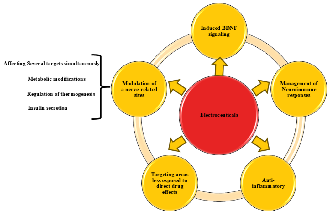

Table 1.
Electroceuticals and neuromodulation relevant to metabolic syndrome.
| Study Design | Subjects (n) | Intervention | Comparator | Duration | Key Findings | Citation |
|---|---|---|---|---|---|---|
| Open label, randomized (2:1), two-arm, parallel-group controlled trial | MetS patients (n = 30) | taVNS on the cymba conchae of the left ear |
No treatment | 8 Weeks | -Reduced HR,BP -Improved sym pathovagal balance -Antiinflammatory effect |
De Moraes et al.(2023) |
| Open-label Randomized Crossover Trials | 40 Subjects with MetS or T2DM | MES + HS | No treatment | 12 Weeks with MES + HS 12 Weeks (No treatment) |
-Decreased visceral adiposity -Improve Fasting plasma glucose, insulin levels, HbA1c < 7.0%, inflammatory cytokines or adipokines -Increased HSP72 expression in monocytes -Lower glucose excursions on meal tolerance test |
Kondo et al. (2014) |
| Randomized, Double-blind, Sham-Controlled Trial | 60 Patients with abdominal obesity | EMS | TENS/Sham stimulation | 12-Week | -Reduced waist circumference in EMS>TENS -Fasting free fasting acid levels in EMS> TENS |
Choi et al. (2018) |
| Randomized Controlled Trial (RCT) – Pilot Study | 29 Obese women diagnosed with MetS | WB-EMS + Caloric restriction | Inactive control + Caloric restriction/nutritional counseling | 12-Week | -Decreased body weight, body fat percentage, and total cholesterol levels -Improved overall cardiometabolic risk score and body composition |
Reljic et al. 2020 |
| Double-blind, randomized controlled clinical trial (ReCharge Trial) | 84 obese with defined BMI with at least one obesity-related comorbid condition | vBloc + Weight management counseling | Sham device + Weight management counseling | 12 Months | Weight loss in vBloc> sham control | Morton et al.,(2016) |
| Randomized, double-blind, sham-controlled clinical trial |
239 Participants with defined BMI and 1 or more obesity-related condition |
vBloc + Weight management education & program | Sham device + Weight management education & program | 12 Months | Weight loss in vBloc> sham control But failed to meet the coprimary prespecified efficacy objectives |
Ikramuddin et al., (2014) |
| Clinical trial |
15 patients with stable VNS therapy for refractory epilepsy | VNS (on) | VNS (off) | 2 Weeks | -Higher BMR -Increased energy expenditure |
Vijgen et al., (2016) |
| Patient-assessor blinded, randomized, sham-controlled clinical trial | 168 Obese participants | Electracupuncture | Sham acupuncture | 8 Weeks | -Improved waist circumference and body fat percentage -Reduced body weight & BMI -Reduced hip circumference and waist-to-hip circumference ratio |
Lam et al. (2024) |
| Double-blind, randomized controlled trial | 239 Obese participants with defined BMI and | vBloc | Sham | 24 Months | -Significant weight loss -Improved lipid profile, HbA1c level, and systolic and diastolic BPs |
Apovian et al. 2017 |
| Randomized trial | 90 MetS patients | LFEF + TMS + Diet, antihypertensive drugs, physiotherapy exercises |
Two active comparator groups: TMS alone and LFEF alone, +Diet, antihypertensive drugs, physiotherapy exercises | 10 Days | -Reduction in cholesterol -Effectiveness of LFEF + TMS > LFEF alone/ TMS alone |
Benkov et al. (2022) |
| Randomized, controlled, open-label Trial | 60 Dyslipidemic patients | Electroacupuncture | No acupuncture intervention | 4 Weeks | -Improved lipid profile -Sustained after the treatment effect on regulation of serum lipids |
Rerksuppaphol & Rerksuppaphol (2014) |
| Meta-analysis of randomized controlled trials | 180 participants in 9 randomized controlled selected studies (healthy weight, obese, population with T2DM or SCI) | NMES | Control/Placebo group | Different | -Reduced fasting blood glucose -Improved glycemic control |
Sanchez et al. (2023) |
| Randomized controlled cross-over trial | Fourteen patients with T2DM | NMES | Control | 8 Weeks | -Improved the fasting glucose concentration and percent body fat -Significant BDNF changes in NMES>Control -No significant changes in HbA1c and blood lipid profile |
Miyamoto et al. (2018) |
| Prospective, single-arm, before–after interventional study | 15 Obese Individuals with T2DM | EMS | Baseline/pre-intervention values; no separate control group | 10 Weeks | -Decreased fasting blood glucose and HbA(1c) -No statistical change in total weight and proportion of body fat |
van Buuren et al. (2015) |
| Open-label multicenter Clinical Trial | 28 Obese Individuals with T2DM | vBloc | Baseline/pre-intervention values; no separate control group | 12 Months | -Reduced mean %EWL, HbA1c, and mean arterial pressure | Shikora et al. (2013) |
| Pilot randomized clinical trial | 102 Participants with IGT | taVNS | Sham taVNS | 12 Weeks | -Reduced the two-hour glucose tolerance -Reduced systolic BP |
Huang et al. (2014) |
| Randomized controlled trial | 145 Individuals with T1DM, T2DM & DAN | taVNS | Sham taVNS | 12 Weeks | -No significant effectiveness in improvement of glycemic variability or HbA1c | Kufaishi et al. (2025) |
| Randomized Controlled Trial | 160 Patients with type 2 diabetes on oral antidiabetic drugs 1:1 | TENS | Placebo | 20 Weeks | -No statistically significant reduction of HbA1c | Lu et al. (2023) |
| Double-blind, randomized, prospective, multicenter, placebo-controlled Phase III clinical trial | 265 Subjects with resistant hypertension | Baroreflex activation therapy | Placebo / Control | 6 Months | -Safe reduction in systolic BP | Bisognano et al. (2011) |
| Pilot study (within-person experimental design) | 20 Hypertensive subjects | RAVANS | Sham | 50 min/session (10-min baseline + 30-min stimulation + 10-min post-stimulation); 5 sessions, ≥24 h apart | -Reduction of heart rate, mean arterial BP, and diastolic BP -These rapid frequency-dependent effects may vary depending on race and sex |
Garcia et al. (2022) |
| Randomized controlled trial | 24 mild to moderate MeS subjects | PEMF | Sham | 12 Weeks | -NO increment -Reduced systolic BP, diastolic BP, and mean arterial pressure in resting hypertensive MetS patients -Improved BP even during exercise |
Kim et al. (2020) |
Note: BMI, body mass index; BMR, basal metabolic rate; BP, blood pressure; %EWL, mean excess weight loss percentages; DAN, diabetic autonomic neuropathy; EMS, electrical muscle stimulation; HbA1c, hemoglobin A1c; HDL, high-density lipoprotein; HR, heart rate; LDL, low-density lipoprotein; LFEF, low-frequency electrostatic field; IGT, impaired glucose tolerance; MES + HS, mild electrical stimulation with heat shock; MetS, metabolic syndrome; NMES, neuromuscular electrical stimulation; NO, nitric oxide; PEMF, pulsed electromagnetic field therapy; RAVANS, respiratory-gated auricular vagus afferent nerve stimulation; SCI, spinal cord injury; VNS, vagus nerve stimulation; taVNS, transcutaneous auricular vagus nerve stimulation; TENS, transcutaneous electrical nerve stimulation; TG, triglyceride; ТМS, transcranial magnetic stimulation; T2DM, type 2 diabetes mellitus; vBloc, vagal nerve blockade; WB-EMS, whole-body electromyostimulation.
Disclaimer/Publisher’s Note: The statements, opinions and data contained in all publications are solely those of the individual author(s) and contributor(s) and not of MDPI and/or the editor(s). MDPI and/or the editor(s) disclaim responsibility for any injury to people or property resulting from any ideas, methods, instructions or products referred to in the content. |
© 2026 by the authors. Licensee MDPI, Basel, Switzerland. This article is an open access article distributed under the terms and conditions of the Creative Commons Attribution (CC BY) license (http://creativecommons.org/licenses/by/4.0/).
Copyright: This open access article is published under a Creative Commons CC BY 4.0 license, which permit the free download, distribution, and reuse, provided that the author and preprint are cited in any reuse.