

Submitted:
11 September 2026
Posted:
14 September 2026
You are already at the latest version
Abstract
Traumatic brain injury (TBI), chronic traumatic encephalopathy (CTE), and Alzheimer's disease (AD) share several neuropathophysiological mechanisms, including axonal injury, neuroinflammation, and abnormal protein aggregation, which may contribute to overlapping clinical manifestations. This review compares the clinical phenotypes of TBI, CTE, and AD across somatic and sensory, motor, cognitive, and neuropsychiatric domains to identify shared and distinguishing characteristics. Common manifestations include memory and attention deficits, executive dysfunction, slowed information processing, gait and balance impairment, depression, anxiety, irritability, apathy, and personality changes. Despite these similarities, important differences were identified. TBI presents acutely following injury and is associated with headache, vestibular disturbances, loss of consciousness, and post-traumatic amnesia. CTE typically develops years after repetitive head trauma and initially manifests with mood and behavioural disturbances, followed by progressive cognitive decline and dementia in advanced stages. AD is characterized by progressive episodic memory impairment, followed by language, visuospatial, executive, and functional decline. The overlap in clinical presentation, particularly between CTE and AD, may complicate differential diagnosis. Careful assessment of symptom onset, disease progression, injury history, neuropsychological findings, neuroimaging, and biomarkers is essential for accurate diagnosis and may improve early recognition and clinical management.
Keywords:
traumatic brain injury (TBI)
; chronic traumatic encephalopathy (CTE)
; Alzheimer’s disease (AD)
; neurodegeneration
; phenotypic overlap
; disease progression
; differential diagnosis
1. Introduction
1.1. Traumatic Brain Injury (TBI): Definition, Epidemiology, Etiology, Pathophysiology/Pathology, Classification
Traumatic brain injury (TBI) is an injury to the head or body caused by an external force or penetrating injury that disrupts the normal brain function [1], possibly resulting in temporary or permanent physical, cognitive and psychological impairment [2]. It is a global public health concern and a major contributor to death and disability [3], as TBIs account for 30-40% of all injuries globally [4]. Annually 50-60 million people worldwide experience a TBI with projections suggesting it will remain one of the top three causes of trauma related death and disability by 2030 [5]. Certain populations are at a higher risk of sustaining TBIs. Young children (0-4 years), adolescents (15-24 years) and older adults (>75 years) exhibit the highest incidence of TBIs per 100’000 individuals [1], with a greater occurrence among men than women [3]. Additionally, military personnel and athletes participating in contact sports, are particularly vulnerable to mild traumatic brain injuries (mTBIs) and repetitive mild traumatic brain injuries (r-mTBIs) [1].
The most common causes of TBI are accidental falls, assaults, motor vehicle-, construction-, and sports-related accidents [3]. These incidents can damage the brain through mechanisms including blunt trauma, penetrating injury, acceleration-deceleration forces, and blast injuries, disrupting the normal brain function and initiating a cascade of pathophysiological processes [3]. The pathophysiology of TBI consists of two phases: primary and secondary brain injury [6]. The primary injury occurs at the moment of impact and results in immediate cellular damage including cell lysis, axonal disconnection and the release of intracellular ions, neurotransmitters, and proteins [7]. Diffuse axonal injury (DAI) is a common form of primary traumatic brain injury characterized by widespread axonal damage resulting from shearing forces during rapid acceleration and deceleration forces of the brain within the skull [6,8]. It is associated with cytoskeletal disruption, calcium influx, impaired axonal transport, and the accumulation of amyloid precursor protein (APP) and amyloid-beta (Aβ) in injured axons within hours after the trauma [7,9]. The secondary brain injury is a progressive process that develops after the initial trauma and can persist for hours to years, contributing significantly to long-term neurological deficits [6]. Key mechanisms include oxidative stress, excitotoxicity, mitochondrial dysfunction, apoptosis, and neuroinflammation, all of which contribute to progressive neuronal and synaptic loss [7].
TBI can be classified according to injury severity [1], anatomical distribution of brain damage [3], and temporal progression [10]. Depending on the severity, TBIs can be classified as mild, moderate, and severe using the Glasgow Coma Scale (GCS), which tests eye, verbal, and motor stimuli and is useful in the initial assessment of TBI [11]. The GCS assigns a score on the 15-point scale and classifies a TBI as mild (13-15), moderate (9-12) and severe (<8) [11,12]. In addition to the GCS, TBI severity classification also considers the duration of loss of consciousness (LOC), alteration of consciousness (AOC), post-traumatic amnesia (PTA), and structural neuroimaging findings (Table 1) [1,13]. The term mild traumatic brain injury (mTBI) is often used interchangeably with concussion; although concussions technically are a subset of mTBIs [14].
Classification of traumatic brain injury severity into mild TBI (mTBI), moderate TBI, and severe TBI according to duration of loss of consciousness (LOC), alteration of consciousness (AOC), post-traumatic amnesia (PTA), Glasgow Coma Scale (GCS) score, and structural imaging findings. Adapted from reviewed literature [1,13].
TBI can also be classified according to the extent and distribution of brain tissue involvement as a focal or diffuse injury. Focal injuries are localized lesions due to direct impact to the head, resulting in contusions, haemorrhages, and tissue damage at the site of injury (coup) or opposite side (contrecoup). Diffuse injuries involve widespread brain damage and are commonly caused by acceleration–deceleration and rotational forces that stretch and disrupt axons, leading to cerebral oedema, and widespread neurological impairment [6]. Linear forces are more commonly associated with focal injuries, whereas rotational forces are more frequently linked to diffuse injuries, particularly DAI. Both injury patterns may coexist following a traumatic event [3].
Based on temporal progression, TBI can be classified as acute or chronic [10]. Acute TBI refers to the immediate injury and its early sensory, motor, neurocognitive and psychological manifestations, which may persist up to months in some individuals [15]. Chronic TBI refers to the long-term neurological, cognitive, behavioural, and functional consequences that develop or persist months to years after injury. Chronic manifestations of TBI encompass a spectrum of disorders including chronic traumatic encephalopathy [10].
1.2. Chronic Traumatic Encephalopathy (CTE): Definition, Epidemiology, Etiology, Pathophysiology/ Pathology, Classification
Chronic traumatic encephalopathy (CTE) formerly known as “punch-drunk” or “dementia pugilistica,” is a progressive neurodegenerative tauopathy that results from repetitive brain trauma [16,17]. It presents years to decades after repetitive brain trauma, leading to progressive cognitive decline, motor deficits, psychological symptoms, and eventually dementia, resembling Alzheimer’s disease (AD) [18]. CTE has been linked to contact sports such as boxing, American football, wrestling, and soccer. Given the millions of athletes participating in contact sports, CTE represents a major public health issue [18]. In recent years, post-mortem findings, especially in American football players, have significantly increased the public and scientific awareness of CTE [19]. Although predominantly reported in contact sports, CTE has also been reported in non-athlete population such as military veterans, individuals exposed to repetitive physical violence, epilepsy patients, those with self-injury behaviours, and alcoholics with repeated falls [17,19,20]. Nonetheless, the epidemiology of CTE remains unclear, with its incidence and prevalence among retired athletes, military personnel, and the general population unknown [11,17].
The pathophysiology of CTE following repetitive TBI remains incompletely understood [4]. Key mechanisms include the accumulation of hyperphosphorylated tau (p-tau) in neurons and glial cells, driven by calcium influx and glutamate-mediated excitotoxicity, resulting in tau aggregation and neurofibrillary tangles (NFTs) formation, leading to neuronal toxicity and disruption of neural circuits [4,21]. Neuroinflammation, microglial dysfunction, impaired reparative mechanisms, and disruption of the glymphatic system further contribute to progressive neurodegeneration, toxic protein accumulation and worsening of the brain injury [4].
The hallmark neuropathological feature of CTE is the accumulation of p-tau NFTs in a perivascular distribution at the depths of the cortical sulci, a pattern considered pathognomonic for the disease [22]. Additional microscopic findings include neuronal loss, gliosis, axonal degeneration, and TAR DNA-binding protein 43 (TDP-43) accumulation, and less commonly Aβ deposits [21,22]. Macroscopic changes in early CTE include mild enlargement of the lateral ventricles and/or third ventricle and/ or mild septal abnormalities. Advanced CTE is characterized by enlargement of the lateral and third ventricles, cavum septum pellucidum, septal perforations, and pallor of the substantia nigra and locus coeruleus. In severe cases, marked medial temporal lobe atrophy and profound global cerebral atrophy may also be present [22].
CTE classification comprises two complementary approaches: the neuropathological staging system proposed by McKee et al. and the symptom-based Traumatic Encephalopathy Syndrome (TES) clinical criteria [23]. McKee et al. (2013) classified CTE into four progressive neuropathological stages (I-IV) based on the extent and distribution of p-tau pathology [10,16,24]. In Stage I, p-tau pathology is confined to isolated perivascular foci at the depths of the cortical sulci, primarily within the frontal cortex. Stage II is characterized by multiple p-tau lesions with limited spread into adjacent superficial cortical layers, while the medial temporal lobe remains spared. In Stage III, p-tau pathology becomes widespread, involving the frontal, temporal, insular, and parietal cortices, as well as medial temporal structures including the amygdala, hippocampus, and entorhinal cortex. Stage IV represents the most advanced stage, with severe and extensive p-tau deposition throughout most cortical and medial temporal regions, sparing the calcarine cortex except in the most severe cases [16].
In contrast, the Traumatic Encephalopathy Syndrome (TES) consensus criteria provide a symptom-based clinical framework for identifying individuals with suspected CTE during life [23,25]. Classification is based on a history of repetitive head impacts, persistent and progressive cognitive impairment, and the exclusion of alternative neurological disorders, with supportive behavioural, mood, or motor symptoms strengthening the diagnosis [26]. Based on these criteria, individuals are classified as probable TES, possible TES, or unlikely TES, while a definitive CTE classification is only possible through postmortem neuropathological confirmation [27].
1.3. Alzheimer’s Disease: Definition, Epidemiology, Etiology, Pathophysiology/Pathology, Classification
Alzheimer’s disease (AD) is a neurodegenerative disorder associated with progressive neuronal loss, cognitive impairment including memory decline, impaired thinking and changes in behaviour [28,29]. AD is the most common form of dementia and accounts for approximately 60–80% of all dementia cases [29]. It poses a major health challenge, with an estimated 55 million cases worldwide in 2019 and projections indicating a rise to nearly 139 million cases by 2050 [30]. AD predominantly affects individuals over 65 years, with a prevalence of 10–30% in this age group and an incidence that doubles every 10 years after the age of 60. It is more common in women, particularly in those over the age of 80 [31].
The pathophysiology of AD is complex and multifactorial, involving genetic, environmental, and age-related factors [29,32]. According to the amyloid beta hypothesis, proposed by John Hardy and David Allsop, Aβ accumulation is an early event that triggers downstream processes including tau pathology, cell death, vascular damage and ultimately dementia. In parallel, tau proteins become hyperphosphorylated and aggregate into NFTs, disrupting microtubule stability and axonal transport [29]. Mutations in the APP, presenilin 1 (PSEN1), and presenilin 2 (PSEN2) genes were identified to increase Aβ aggregation, while comorbidities such as cardiovascular diseases, obesity and diabetes further elevate the risk of AD [29]. In addition to amyloid and tau pathology, several other mechanisms contribute to AD progression, including mitochondrial dysfunction, oxidative stress, insulin resistance, and neuroinflammation [29,32].
The neuropathological hallmarks of AD are the accumulation of extracellular Aβ plaques and intracellular NFTs composed of p-tau, which define the disease at autopsy [29,32,33,34]. These pathological changes primarily affect memory-related regions, such as the hippocampus. However, they are not restricted to these regions, as their spatiotemporal progression extends to other anatomical structures, including the cerebellum, basal ganglia, and motor cortex [35]. Additional microscopic characteristics include cerebral amyloid angiopathy (CAA), resulting from Aβ deposition within the walls of cerebral blood vessels, progressive neuronal and synaptic loss, and reactive gliosis [29,32,33,34]. Macroscopically, AD is characterized by symmetrical cortical atrophy, predominantly involving the medial temporal lobes, with relative preservation of the primary motor, sensory, and visual cortices. The resulting loss of brain parenchyma leads to secondary enlargement of the lateral ventricles (ex vacuo hydrocephalus) [34].
AD can be classified according to its neuropathological characteristics, etiology, age of onset, and disease progression. To standardize neuropathological assessment, the 2012 National Institute on Aging–Alzheimer’s Association (NIA–AA) guidelines introduced the ABC score, which integrates the Thal Aβ phase (A), Braak neurofibrillary tangle stage (B), and CERAD neuritic plaque score (C) to classify AD neuropathological change as not, low, intermediate, or high [36].
The Thal classification (Thal Aβ phase) stages the progressive neuroanatomical spread of Aβ pathology throughout the brain. Aβ deposition begins in the neocortex (phase 1), extends to the hippocampus and cingulate gyrus (phase 2), progresses to the striatum and basal forebrain (phase 3), then to the midbrain central gray matter and brainstem (phase 4), and finally reaches the cerebellum (phase 5) [37,38].
The Braak staging system describes the progression of tau pathology. NFTs initially appear in the transentorhinal and entorhinal cortices (stages I–II), spread to limbic structures including the hippocampal formation, amygdala, thalamus, and claustrum (stages III–IV), and ultimately involve the associative neocortex, followed by the primary sensory, motor, and visual cortices (stages V–VI) [34].
The CERAD (Consortium to Establish a Registry for Alzheimer’s Disease) neuropathological classification is based on a semiquantitative assessment of neuritic plaques, diffuse plaques, NFTs, and other pathological changes. An age-related neuritic plaque score combined with the clinical history determines the diagnostic certainty of AD (definite, probable, possible, or no evidence of AD) [39].
Based on etiology, AD is classified as familial (fAD) or sporadic (sAD). Familial AD accounts for fewer than 5% of cases and is caused by mutations in APP, PSEN1, or PSEN2, whereas sporadic AD results from a multifactorial combination of aging, genetic susceptibility such as Apolipoprotein E ε4 (APOE ε4), and environmental factors [28,40]. AD is further categorized by age of onset: late-onset AD (LO-AD) develops after 65 years, while early-onset AD (EO-AD), accounting for approximately 5.5% of cases, occurs before 65 years [41].
Clinically, AD progresses through five stages: preclinical AD, mild cognitive impairment (MCI), mild dementia, moderate dementia, and severe dementia, characterized by progressive cognitive and functional decline [40].
1.4. Rationale, Objectives, and Scope of the Review
Traumatic brain injury (TBI), chronic traumatic encephalopathy (CTE), and Alzheimer’s disease (AD) share several pathophysiological mechanisms, including axonal injury, neuroinflammation, and abnormal protein aggregation [7,28,40]. Increasing evidence suggests that TBI contributes to long-term neurodegenerative processes. Epidemiological studies report a 58–82% higher risk for developing AD, particularly following moderate-to-severe injuries and among APOE ε4 carriers [7,42,43,44,45,46]. Although the mechanisms linking TBI to AD remain incompletely understood, both conditions share several pathophysiological features, including axonal injury, neuroinflammation, and abnormal Aβ and p-tau accumulation [3,7,47] . Experimental and postmortem studies further demonstrate that TBI can induce AD-like Aβ and tau pathology, which may persist long after the initial injury [3,7,47,48]. Additionally, CTE is a latent consequence of TBI and individuals with one or two copies of the APOɛ4 allele have a higher risk for developing CTE [49]. Both CTE and AD exhibit neuropathological features that overlap with those observed following brain injury, including persistent tau and Aβ pathology [16,50,51].
Similar pathophysiological mechanisms in TBI, CTE, and AD, raise the question of whether these conditions also share clinical features and how this overlap may complicate clinical recognition, differential diagnosis, and disease classification. Recognizing and distinguishing these conditions may be challenging, as all three can present with sensory, cognitive, motor and neuropsychiatric and behavioural symptoms [25,52,53,54,55].
The aim of this review is to compare the clinical phenotypes of TBI, CTE, and AD by examining their shared and distinguishing clinical manifestations, as well as differences in symptom onset, progression, and clinical course. For the purposes of this review, clinical manifestations are organized into four domains: sensory and somatic, motor, cognitive, and neuropsychiatric and behavioural. Pathophysiology and diagnostics are discussed where they provide context for the clinical presentation, whereas therapeutic interventions are beyond the scope of this review.
2. Traumatic Brain Injury vs. Chronic Traumatic Encephalopathy vs. Alzheimer’s Disease
2.1. Traumatic Brain Injury (TBI)
Acute traumatic brain injury (TBI) occurs immediately at the time of the traumatic event and is followed by a spectrum of clinical signs and symptoms [10]. The severity and presentation of symptoms depend on whether the injury is classified as mild, moderate, or severe and what part of the brain the injury affects. While signs and symptoms often appear immediately after the injury, they may also manifest hours to days later [12,56]. In the acute phase, patients may experience a period of LOC, PTA, or AOC [13,57].
2.1.1. Somatic and Sensory Manifestations
The clinical presentation of TBI includes a wide range of symptoms such as headache, dizziness, vertigo, nausea and vomiting [1,56,58]. Headache is the most common reported symptom after TBI, which may begin immediately after injury and can last for months after the injury event [3]. Post-TBI headache is categorized as acute or chronic, with the acute type resolving within 2 months and the chronic type persisting for longer [56]. Headache usually coexists with dizziness, which is reported in ~24% to 74% of TBI patients [3]. Sensory changes are especially common in moderate to severe TBI patients and can involve hypersensitivity to a stimulus or a diminished/loss of function [59]. These changes include blurry vision, hypersensitivity to light (photophobia), hypersensitivity to sound (hyperacusis), tinnitus, and a subjective sense of feeling dazed or stunned [1,56,58]. Patients with moderate to severe TBI may also experience a loss of vision/ hearing in one or both eyes/ ears, mydriasis in one or both eyes and numbness or tingling of arms and legs [1]. While some changes like hearing loss can be objectively measured, others, such as noise hypersensitivity, are inherently subjective and depend on self-report [59].
2.1.2. Motor Manifestations
Acutely, TBI causes neuromotor deficits that vary according to injury severity. mTBI is commonly associated with balance and coordination impairments, whereas severe TBI can result in spastic paralysis, postural instability, gait abnormalities, and reduced fine motor control [53]. An injury to the basal ganglia (or dorsolateral frontal region) can lead to tremor, bradykinesia and cogwheeling [56]. Other motor manifestations include weakness slurred speech (dysarthria), and convulsions or seizures [1]. Motor impairments are rather common, with nearly 78% of moderate to severe TBI patients reporting some degree of gross neuromotor impairment during rehabilitation. Motor impairments may persist beyond the acute phase reducing mobility and potentially contributing to poorer quality of life following TBI [53].
2.1.3. Cognitive Manifestations
TBI-related cognitive manifestations vary according to injury location and severity [60]. Common cognitive consequences include deficits in attention, memory, and executive functioning [61]. Patients may also experience difficulties with concentration, reduced information processing speed, and impaired learning capacity [60,61].
Attention impairments may arise from damage to the neural network responsible for attention processes. It includes the reticular formation, thalamus, hippocampal and entorhinal areas, as well as the frontal and right parietal lobes and their axonal connections [61].
Memory impairment is also frequently observed and may affect studying, planning, recalling information, and navigating daily life [62]. Acutely after a TBI, patients may experience anterograde amnesia and retrograde amnesia together with disorientation, attentional deficits and agitation as part of PTA or post-TBI syndrome [63]. Additionally, delirium is a common consequence of TBI. It constitutes an acute alteration in mental status characterized by confusion, inattention and fluctuating levels of arousal [64].
Executive dysfunction, often associated with frontal lobe injury [61], may result in difficulties with problem-solving, planning, reasoning, multitasking and judgement [47]. Patients may also experience difficulties with organizing and sequencing of tasks, making it more difficult to perform preinjury jobs or follow instructions that would ordinarily be routine. As a result, patients may become irritable, anxious, apathic or depressed [56].
2.1.4. Neuropsychiatric and Behavioural Manifestations
TBI is associated with a wide range of psychiatric manifestations that contribute significantly to disability and reduced quality of life in chronic TBI. The development of these manifestations is influenced by both the location of brain damage and acute alterations in neurotransmitter systems, including acetylcholine, norepinephrine, dopamine, and serotonin [65,66]. Damage to specific frontal and fronto-subcortical networks can result in distinct neuropsychiatric syndromes. Orbitofrontal lesions are associated with disinhibition, dorsolateral frontal lesions with executive dysfunction, anterior cingulate lesions with apathy, and broader frontal circuit disruption may lead to emotional, behavioural, and personality changes [65].
Common psychiatric manifestations following TBI include depression, anxiety, post-traumatic stress disorder (PTSD), suicidality, aggression, impulsivity, irritability, emotional lability, apathy, and personality changes [56,65,66].
Depression is the most frequently reported psychiatric disorder, affecting approximately 25–50% of individuals within the first year after injury and up to 64% during their lifetime [65]. Anxiety disorders and PTSD are also highly prevalent, while bipolar disorder, psychosis, and substance-use disorders may occur less frequently but remain important clinical sequelae [66]. Furthermore, behavioural dyscontrol, characterized by aggression, agitation, disinhibition, withdrawal, and poor impulse control, is common after TBI and is often associated with frontal lobe damage and poorer social and rehabilitation outcomes [67]. The severity and presentation of psychiatric symptoms are further influenced by injury characteristics, premorbid psychiatric history, social support, substance use, and other individual risk factors [66].
2.1.5. Sleep Disturbances and Fatigue
Sleep disturbances are common after a TBI, affecting 30–70% of individuals [68]. Insomnia, fatigue and sleepiness are the most frequent post-TBI sleep complaints. Narcolepsy (with or without cataplexy), sleep apnea (obstructive and/or central), periodic limb movement disorder, and parasomnias occur less commonly [68]. Other post-TBI sleep complaints include maintaining sleep, hypersomnia and day-time somnolence [3,66].
Although often associated with sleep disturbances, fatigue represents a distinct and highly prevalent symptom following TBI. Precise estimates of post-TBI fatigue vary greatly between 21–73% but are consistently exceeding the prevalence of fatigue in the general population of 10–20% [69]. Post-TBI fatigue can become persistent and debilitating. It is associated with poor social activity and isolation affecting the patient’s quality of life [3].
2.1.6. Traumatic Brain Injury Diagnosis and Biomarkers
TBI diagnosis is clinical and relies on patient history, GCS scoring, and symptom assessment tools, neuroimaging and fluid biomarkers. No single definitive diagnostic test exists, particularly for mTBI [1,70,71,72,73]. Assessment should include a detailed patient history, mental status examination, neurological examination, and evaluation of neuropsychiatric symptoms using standardized instruments (e.g., Neurobehavioural Rating Scale, Neuropsychiatric Inventory) [74]. Neuropsychological testing can identify cognitive impairments that may not be evident in routine clinical examination, particularly valuable for concussion [75]. In acute sport-related concussions, neuropsychological assessment can be conducted using the Sport Concussion Assessment Tool, 6th Edition (SCAT6), a standardized multidomain tool designed to assist clinicians in the evaluation of concussion [76].
Computed Tomography (CT) and Structural Magnetic Resonance Imaging (MRI) are used to rule out acute haemorrhagic events or severe structural injuries requiring immediate neurosurgical intervention [12,70]. In an uncomplicated mTBI without structural brain injury such as intracranial hematomas or cranial haemorrhages, a conventional CT and MRI are normal, and a diagnosis is based on clinical presentation [18,70,71].
Advanced MRI techniques, including diffusion tensor imaging (DTI), functional MRI (fMRI), and magnetic resonance spectroscopy (MRS), can detect subtle white matter and metabolic abnormalities not visible on conventional imaging and have shown potential to support diagnosis and prognosis after brain injury [12,70,77,78]. fMRI and DTI are useful tools for identifying ultrastructural and functional brain alterations associated with concussions [75].
Although inconsistently integrated into clinical practice, blood (serum or plasma) and cerebrospinal fluid (CSF) biomarkers can support TBI assessment, management, and prognosis while reducing unnecessary interventions [1]. Key biomarkers include glial fibrillary acidic protein (GFAP), ubiquitin C-terminal hydrolase-L1 (UCH-L1), S100 calcium-binding protein B (S100B), neurofilament light chain (NFL) and total tau (t-tau) [1,79].
2.1.7. Progression and Prognosis
The outcome following TBI ranges from complete recovery to permanent disability and death [2]. Consequences may include neurological deficits, cognitive impairment, reduced quality of life, and persistent post-traumatic symptoms [80]. mTBI is generally considered a self-limiting condition, with most individuals recovering within 1–2 weeks and symptoms returning to pre-injury levels within 7–14 days [1,58]. However, a subset of patients experiences persistent post-concussive symptoms, and approximately 10–20% develop post-concussion syndrome (PCS), with symptoms lasting months or years after the injury [81,82,83]. Although post-concussive symptoms are widely recognized as common during the first month after injury, and may persist longer following moderate-to-severe TBI, the prevalence and duration of symptoms beyond three months in mTBI remain debated [80,84]. Studies indicate that symptoms following mTBI may persist for up to one year in a substantial proportion of patients [85,86]. It has been observed that across mild, moderate, and severe TBI, cognitive symptoms, particularly memory difficulties, slowed thinking, and concentration problems, are among the most commonly reported long-term complaints, while emotional symptoms such as irritability and frustration may also persist [80]. Long-term deficits have been documented following severe TBI, with impairments in memory, attention, language, and functional independence persisting for up to seven years after injury [87].
Several factors have been associated with poorer outcomes following TBI, including older age, lower Glasgow Coma Scale (GCS) scores, absent pupillary reactivity, major extracranial injuries, and more severe CT abnormalities. Greater injury severity has been associated with an increased risk of mortality and severe disability [88]. Long-term complications may include chronic traumatic encephalopathy (CTE), dementia, depression, post-traumatic stress disorder (PTSD), endocrine dysfunction, and other long-term complications that contribute to increased morbidity and mortality [89].
2.2. Chronic Traumatic Encephalopathy (CTE)
Chronic traumatic encephalopathy (CTE) follows an insidious course, with a mean latent interval of 8 years between the last trauma and the development of symptoms. Initial symptoms have been documented to first appear between the ages 35 to 45, but range in onset between ages 24 to 65 [46,70]. CTE progresses through several stages, with symptoms becoming progressively more severe over time [16]. Data on clinical manifestations of CTE have only recently been accumulating via post-mortem medical record reviews and interviews of friends or family members of individuals with neuropathologically documented CTE [18].
2.2.1. Somatic Manifestation
The literature primarily described persistent and chronic headache as a somatic manifestation of CTE [90,91,92,93]. Headache is an early clinical manifestation of CTE, characteristic of Stage I disease [21]. In a retrospective autopsy cohort study, McKee et al. (2013) reported that 13 of 51 (25.5%) clinically characterized individuals with neuropathologically confirmed CTE had headaches. Headache was documented in 4 Stage I cases, 5 Stage II cases, and 4 Stage III cases, with no Stage IV cases reporting headache [16]. Further supporting these findings, Stern et al. (2013) reported in their retrospective cohort study that 34.4% of symptomatic individuals with neuropathologically confirmed CTE had a history of significant headaches [94].
2.2.2. Motor Manifestations
Motor manifestations were among the earliest and most consistently reported clinical features of CTE, particularly among former boxers [26]. Motor features of CTE may include parkinsonism, ataxia, tremor, rigidity, gait, balance problems, weakness, coordination problems, spasticity and dysarthria [16,26,46,58,92]. Motor neuron disease (MND) has been noted in a subset of athletes with CTE, but whether this phenotype is a subtype of CTE or an overlap of two disease processes remains unclear [10]. In more recent descriptions of CTE, published after 2005, motor symptoms have played a less prominent role in the clinical presentation, with greater emphasis placed on mood, behavioural, and cognitive symptoms [26]. It has been hypothesized that sport-specific injury biomechanics influence the development of motor manifestations in CTE. In boxing, angular acceleration and torsional injury involving the brainstem and cerebellum may contribute to earlier degeneration of brainstem structures such as the substantia nigra, leading to parkinsonism. Transverse and linear acceleration and deceleration injuries are more characteristic of football dynamics. They may lead to substantia nigra degeneration in the later disease course, alongside widespread cortical and basal ganglionic degeneration that may mask CTE motor symptoms [94].
2.2.3. Cognitive Manifestations
Cognitive symptoms in CTE include impairments in new learning, memory, language, attention, concentration, information processing speed, visuospatial abilities and executive functioning such as impaired sequencing abilities, judgement, abstraction, reasoning, planning and organization [91,93].
As CTE advances, the neurodegenerative process may progress to dementia [21]. Approximately 45% of individuals with CTE develop dementia [95], although reported prevalence estimates vary across cohorts and study populations. Layden et al. (2026) found that, among 366 individuals with autopsy-confirmed CTE, 147 (40.2%) were retrospectively determined to have had dementia before death based on a consensus clinical diagnosis informed by proxy reports and medical-record review. Dementia was observed in 52.6% of individuals with Stage III CTE and 82.5% of those with Stage IV CTE. Relative to individuals without CTE, Stage III and Stage IV pathology were associated with 2.12-fold and 4.48-fold increased odds of dementia, respectively [96]. Stern et al. (2013) reported that among 33 symptomatic athletes with neuropathologically confirmed CTE, 30.3% had been diagnosed with dementia during life, and all dementia cases were associated with Stage IV CTE pathology. Individuals whose initial presentation involved cognitive impairment were more likely to develop dementia (54.5%) than those presenting with behavioural or mood symptoms (18.2%) [94].
2.2.4. Neuropsychiatric and Behavioural Manifestations
Behavioural disturbances are often the earliest symptoms of CTE and can include depression, mood swings, apathy, impulsivity, aggression, and suicidality [3,10,97,98]. In the mildest stage, affective symptoms can manifest as emotional lability, euphoria and hypomania [91,99]. Other behavioural manifestations include explosivity [25], disinhibition [100], agitation [25], irritability [25,97], social withdrawal [101,102], reduced motivation [103] and violence [100]. Late neurobehavioural features include exacerbation of aggression, suspiciousness, childishness, loquacity and restlessness [91]. Behavioural symptoms are accompanied by neuropsychiatric symptoms such as personality changes [46], anxiety [25], paranoia [25], delusions and hallucinations [98]. Drug and alcohol abuse is common among athletes with CTE [100].
Depressive symptoms are prevalent in CTE and include not only depressed mood but also hopelessness, suicidal ideation, and vegetative symptoms [25]. A high proportion of pathologically validated CTE cases has died by suicide, with reported rates ranging from 10% to 29% across cohorts [25].
CTE also demonstrates an overlap with other neuropsychiatric conditions [90]. Many psychiatric symptoms attributed to CTE are also prevalent in men with depression in the general population [104]. Notably, mood and behavioural symptoms associated with CTE are reported to be the most concerning features for family members and colleagues [18].
2.2.5. Chronic Traumatic Encephalopathy Diagnosis and Biomarkers
A purely clinical diagnosis of CTE is difficult because its disease course and clinical presentation are not unique [105]. The definitive diagnosis of CTE currently requires post-mortem neuropathological examination [20,94,106,107,108]. The pathognomonic neuropathological finding is the accumulation of p-tau in a perivascular distribution at the depths of the cortical sulci, while additional NFTs in cortical and subcortical regions, including the hippocampus, amygdala, and thalamus, represent supportive findings [79].
Structural MRI, diffusion MRI, PET, and SPECT have demonstrated structural, microstructural, and molecular abnormalities in individuals with suspected CTE. Among these modalities, tau-binding PET showed the greatest diagnostic potential, distinguishing suspected CTE from healthy controls and AD through the detection of characteristic tau pathology. However, current neuroimaging biomarkers have not yet been sufficiently validated for a definitive in vivo diagnosis of CTE and are therefore considered supportive rather than confirmatory [109].
To date, no reliable in vivo biomarker for CTE has been established [110]. However, exosomal tau has emerged as a promising candidate, with elevated plasma exosomal tau levels reported in individuals with a history of repetitive head impacts and symptoms consistent with CTE [79,111]. Higher exosomal tau concentrations have also been associated with poorer performance in processing speed, working memory, and episodic memory, suggesting a potential relationship between exosomal tau levels and cognitive impairment in CTE [111].
2.2.6. Progression and Prognosis
Based on McKee et al. four-stage pathological classification, the progression of CTE can be described as a gradual worsening of clinical symptoms that correlates with increasing brain pathology [21]. Stage I is often asymptomatic or characterized by nonspecific symptoms such as headache, irritability, mild depressive symptoms, mild aggression, mild short term memory deficits and reduced concentration. Stage II is associated with cognitive and behavioural symptoms, including short-term memory impairment, disorganization, difficulty planning, aggression, explosivity, more severe depression and suicidality. By Stage III, patients typically exhibit significant cognitive impairment with memory loss, executive dysfunction, attention deficits, as well as apathy alongside symptoms from earlier stages. Stage IV represents advanced disease with severe executive dysfunction, profound memory loss, dementia, and additional features such as language impairment, paranoia, depression, visuospatial deficits, aggression, explosivity, and motor deficits including gait disturbances and parkinsonism [16,21,70,112,113].
2.3. Clinical Manifestations of Alzheimer’s Disease (AD)
Alzheimer’s disease (AD) is characterized clinically by insidious onset and slowly progressive cognitive dysfunction, particularly impaired memory [114]. Symptoms onset most often develops after 65 years of age (late-onset AD), with a subset developing symptoms earlier, before the age of 65 years (early-onset AD) [41]. AD does not only affect cognitive functions leading to functional and executive disabilities, but is also associated with neuropsychiatric symptoms, behavioural disorders and loss of motor functions [28,114].
2.3.1. Somatic and Sensory Manifestations
Somatic manifestations in AD include sensory deficits such as hearing and visual impairments, gustatory and olfactory dysfunctions, and tactile disturbances [40,115,116,117]. Sensory impairments are increasingly recognized as important non-cognitive manifestations of AD and may precede the onset of cognitive decline, particularly olfactory and gustatory deficits. Sensory dysfunction is associated with AD-related neuropathological changes, including Aβ accumulation, tau pathology, and neuronal loss within primary and sensory association cortices, which are thought to contribute to sensory deficits. Olfactory dysfunction is linked to early neuropathological changes in the olfactory bulb and entorhinal cortex. Hearing impairment is strongly associated with AD, although its contribution to disease progression remains uncertain. In addition, visual abnormalities, including retinal thinning, together with altered tactile perception, further demonstrate that AD affects multiple sensory systems beyond cognition [116].
2.3.2. Motor Manifestations
Motor manifestations in AD become more pronounced during the intermediate and late stages, although current evidence suggests they can also emerge in the early stages [35]. Motor manifestations in AD may include gait, bradykinesia, slowness, rigidity, motor denervation, muscle atrophy and decreased strength [35,118,119]. Difficulties with coordination, manual dexterity, and both dynamic and static balance have also been documented [35].
One of the most frequently reported motor manifestations in AD is gait impairment. Studies have shown that gait abnormalities become more prevalent and severe as the disease progresses, leading to reduced mobility and an increased risk of falls, which may result in severe injuries and fractures [35,120].
Furthermore, muscle atrophy and reduced muscle strength are among the most common motor signs, occurring even in the early stages of the disease and progressing as the disease advances. Studies have correlated decreased muscle strength with reduced brain volume and cognitive decline, as well as with abnormal weight loss and cachexia, which impair patient functionality and increase the risk of fractures [35]. Motor impairments in AD are thought to be associated with the progressive spread of AD pathology into brain regions involved in motor control, including the motor cortex, cerebellum, and basal ganglia [35].
2.3.3. Cognitive Manifestations
Cognitive deficits in AD include progressive decline in memory, attention, difficulties learning new information, problem-solving deficits, confusion, language, and visuospatial impairments, as well as impaired executive function, aphasia, apraxia, and agnosia [28,31,121,122].
Episodic memory, a type of long-term declarative memory, is defined as the ability to acquire and recollect personally experienced events within their emotional, spatial, and temporal context. Episodic memory impairment is among the earliest cognitive manifestations of AD and generally declines before executive functions and other cognitive functions [123]. Working memory is also impaired early on in the disease course [124]. Memory loss is closely linked to damage in the hippocampus, as it is essential for consolidating short-term memory into long-term memory. AD pathology begins in the entorhinal cortex and hippocampus, where progressive neuronal loss and atrophy disrupt the formation of new memories. Consequently, reduced hippocampal volume is strongly associated with poorer memory performance in AD [124].
In addition to memory deficits, attentional impairments are an important feature of AD and contribute to cognitive symptoms. Because attention is essential for encoding and processing information, deficits in attention can worsen cognitive impairment. Evidence suggests that early degeneration of the locus coeruleus and disruption of the noradrenergic system contribute to these attentional deficits [125].
Executive functions, defined as the ability to abstract, plan organize, shift and adapt current to future behaviour, are commonly impaired in AD [122], leading to difficulties with problem-solving, decision-making, multitasking, and household management [31]. Patients may require more time to complete daily tasks or find it harder to complete multistep tasks such as bathing and dressing [40]. In later stages, patients may struggle to recognize loved ones or may become incontinent [40]. As AD pathology progresses beyond the medial temporal lobe, additional cognitive deficits emerge and the full dementia syndrome becomes apparent [126].
2.3.4. Neuropsychiatric and Behavioural Manifestations
Neuropsychiatric symptoms (NPS) are a hallmark of Alzheimer’s disease, affecting almost all individuals with dementia over the course of the disease and may emerge early, even during the preclinical or MCI stages [127]. NPS include apathy, depression, aggression, anxiety, irritability and psychosis, including delusions and hallucinations. Other manifestations are emotional distress, personality changes, euphoria, disinhibition, agitation, and appetite changes [28,121,128,129,130,131,132,133]. Apathy, depression, anxiety, and irritability may emerge during the early or even preclinical stages of AD [121]. Agitation, delusions, disinhibition, and hallucinations are more commonly associated with the moderate-to-late stages of the disease. In addition, aberrant motor behaviours, characterized by excessive purposeless or repetitive activities such as wandering, fidgeting, rummaging, and repeated dressing or undressing, are more commonly observed during the moderate stages of the disease [121]. A meta-analysis of 62 studies reported that women were more likely to exhibit depressive and psychotic symptoms, whereas men demonstrated greater levels of apathy [134]. The presence of NPS has been associated with accelerated cognitive decline, progression to severe dementia, increased mortality, and reduced quality of life [128,129].
2.3.5. Sleep Disturbances
Sleep disturbances are common in AD and may occur years before the onset of clinical symptoms, making them a potential early indicator of disease development [121,135,136]. Common manifestations include insomnia, disrupted sleep–wake cycles, difficulty initiating and maintaining sleep, early-morning awakening, excessive daytime sleepiness, daytime napping, and sleep fragmentation [135,136]. In addition, reduced slow-wave and REM sleep can be observed in AD, together with sleep disorders such as obstructive sleep apnea, sleep-disordered breathing, restless legs syndrome, and periodic limb movements during sleep [135]. The relationship between sleep and AD appears to be bidirectional, whereby AD pathology disrupts normal sleep, while poor sleep may contribute to disease progression and neurodegeneration, highlighting sleep as a possible target for therapeutic intervention [135,136].
2.3.6. Alzheimer’s Disease Diagnosis and Biomarkers
Historically, a definitive diagnosis of AD required post-mortem histopathological confirmation of amyloid plaques and NFTs, while living patients were diagnosed with probable AD based on clinical presentation [137]. Advances in biomarker research have since enabled the detection of AD pathology in vivo using CSF biomarkers and molecular neuroimaging, allowing diagnosis at earlier stages of the disease before dementia develops [137].
Diagnosis of AD is based on a combination of clinical assessment, cognitive testing, laboratory investigations, and neuroimaging. A detailed medical history, including information from family members or caregivers, is essential to evaluate cognitive decline and functional impairment. Cognitive screening tools such as the Montreal Cognitive Assessment (MoCA) and Mini-Mental State Examination (MMSE), and Alzheimer’s Disease Assessment Scale-Cognitive Subscale (ADAS-Cog) are commonly used to assess cognitive function [29,54]. Laboratory tests, including complete blood count, metabolic panel, thyroid function tests, and vitamin B12 levels, are performed to exclude reversible causes of cognitive impairment [54]. Structural neuroimaging, particularly, MRI, may reveal characteristic brain atrophy in AD, especially within medial temporal lobe structures such as the hippocampus and entorhinal cortex [138]. PET imaging and CSF biomarkers, including Aβ42, phosphorylated tau, and total tau, can support the diagnosis and staging of AD and are valuable for detecting disease progression from presymptomatic and mild cognitive impairment stages before dementia develops [139]. Current research is focused on developing minimally invasive biomarkers, particularly blood-based biomarkers, and integrating artificial intelligence (AI) and machine learning to analyse biomarker, imaging, and clinical data, with the aim of improving early diagnosis, diagnostic accuracy, and personalised risk prediction [139].
2.3.7. Progression and Prognosis
AD is characterized by a progressive decline in episodic memory that gradually extends to other cognitive domains and increasingly interferes with activities of daily living. As pathology spreads to additional cortical regions, deficits in executive function, language, and visuospatial abilities emerge, ultimately resulting in dementia [40,126].
Disease progression is preceded by a preclinical phase, during which AD pathology is detectable by biomarkers despite the absence of cognitive symptoms. This asymptomatic phase may last 10–20 years before subtle impairments in memory, cognition, and language develop, consistent with mild cognitive impairment (MCI), while dementia is not yet present [40,140,141,142,143]. Continued neurodegeneration leads to progressive loss of functional independence. Early dementia is associated with largely preserved independence but an increasing need for assistance, whereas advancing disease results in greater confusion, difficulty performing multistep tasks, personality changes, incontinence, and eventually profound cognitive impairment, markedly reduced verbal communication, immobility, and complete dependence on caregivers [40,144]. AD is relentlessly progressive, with an average life expectancy of 4–8 years after diagnosis in individuals aged 65 years or older, although some patients may survive up to 20 years after symptom onset. Progressive physical decline, reduced mobility, and dysphagia increase the risk of infections, particularly pneumonia, which is the leading cause of death. Other complications include depression, agitation, delirium, wandering, dehydration, malnutrition, falls, and bladder or bowel dysfunction, all of which further reduce quality of life [54].
Summary of the major clinical manifestations reported in traumatic brain injury (TBI), Chronic traumatic encephalopathy (CTE), and Alzheimer’s disease (AD) across somatic/sensory, cognitive, motor, neuropsychiatric/behavioural, and sleep/fatigue domains.
Compiled by the authors based on the reviewed literature. References supporting each clinical domain are as follows:
3. Discussion
3.1. Comparative Overview of Clinical Manifestations
3.1.1. Somatic and Sensory Manifestations: Comparative Overview
TBIs are characterised by acute sensory–vestibular symptoms, including headache, dizziness, vertigo, nausea, vomiting, photophobia, hyperacusis, tinnitus, and a subjective feeling of being dazed or stunned [1,56,58]. Vertigo and dizziness may occur as the result of an injury to vestibular structures [11]. In CTE, the primary somatic symptom is headache [90,91,92,93], which is often one of the earliest manifestations and associated with Stage I of the disease [16,21]. Early CTE pathology is characterized by the accumulation of perivascular p-tau at the depths of cortical sulci, primarily in the frontal cortex, before the pathology spreads further [16,21,22]. In contrast, AD is characterised by progressive sensory deficits rather than hypersensitivity. These include impairments in hearing, vision, olfaction, gustation, and tactile perception [40,115,116,117], and are associated with Aβ accumulation, tau pathology, and neuronal loss affecting the olfactory bulb, entorhinal cortex, and sensory association cortices [116].
Overall, somatic and sensory manifestations differ across TBI, CTE, and AD. TBI is characterised by acute sensory–vestibular disturbances and headache, CTE shares the manifestation of headache but lacks the acute vestibular and gastrointestinal features of TBI, whereas AD is characterised by progressive sensory deficits resulting from neurodegeneration.
3.1.2. Motor Manifestations: Comparative Overview
The reviewed literature identified several overlapping motor manifestations across TBI, CTE, and AD. TBI may result in balance and coordination impairments, gait abnormalities, reduced fine motor control, weakness, dysarthria, tremor, bradykinesia, and spastic paralysis, with the severity largely depending on the extent and location of the injury [1,53,56]. Similarly, CTE has been associated with gait and balance impairment, coordination deficits, tremor, weakness, spasticity, and dysarthria but also parkinsonism, rigidity and ataxia [16,26,46,58,92]. AD shares several motor manifestations with TBI and CTE, including gait impairments, bradykinesia, rigidity, impaired coordination, and balance deficits. AD can additionally present with muscle atrophy and reduced muscle strength, with both reported from the early stages of the disease and worsening over time [35,118,119]. Despite these similarities, the timing and underlying mechanisms differ considerably. In TBI, motor deficits occur immediately following injury due to acute disruption of motor pathways involving the corticospinal tracts, basal ganglia, cerebellum, or dorsolateral frontal cortex [53,56].
In contrast, CTE and AD are progressive neurodegenerative disorders in which motor impairment develops gradually. In CTE the degeneration of brainstem structures, including the substantia nigra may lead to parkinsonism [94]. In AD, motor impairments are thought to be associated with the progressive spread of AD pathology into brain regions involved in motor control, including the motor cortex, cerebellum, and basal ganglia [35].
Overall, TBI, CTE, and AD share several motor manifestations, including gait disturbance, balance impairment, coordination deficits, and bradykinesia, which may complicate clinical differentiation. Nevertheless, the temporal profiles differ: TBI produces acute motor dysfunction secondary to traumatic injury, whereas CTE and AD demonstrate progressive motor decline associated with neurodegeneration.
3.1.3. Cognitive Manifestations: Comparative Overview
The reviewed literature identified several shared cognitive manifestations across TBI, CTE, and AD. TBI is associated with impairments in attention, concentration, memory, information-processing speed, learning capacity, and executive functioning [60,61]. Similarly, CTE is characterized by deficits in memory, new learning, attention, concentration, processing speed and executive functioning [91,93]. AD likewise presents with impairments in episodic and working memory, attention, new learning, executive functioning, language, and visuospatial abilities [28,31,121,122,123,124,125]. However, the literature also identified differences in the cognitive profiles of TBI, CTE, and AD. Acute TBI may be accompanied by anterograde and retrograde amnesia, disorientation, agitation, and delirium [63,64]. In contrast, CTE is associated with gradually worsening cognitive impairment that may progress to dementia in advanced pathological stages [21,94,95,96]. AD typically begins with episodic memory impairment before extending to language, visuospatial, executive, and functional deficits, with aphasia, apraxia, agnosia, confusion, and impaired recognition of loved ones in advanced disease [28,31,40,121,126].
These differences may reflect distinct pathological processes and patterns of neural network involvement. In TBI, cognitive deficits reflect the acute disruption on attention, memory, and executive networks, including the reticular formation, thalamus, hippocampal and entorhinal regions, frontal and parietal cortices, and their axonal connections, with executive dysfunction commonly associated with frontal lobe injury [61]. In contrast, cognitive impairment in CTE and AD reflects progressive neurodegenerative processes in which pathological changes gradually spread to anatomical regions and interconnected cognitive networks, resulting in progressive cognitive deterioration that may ultimately culminate in dementia [21,28,31,94,95,96,121,145].
Taken together, TBI, CTE, and AD share impairments in memory, attention, information-processing speed, new learning and executive functioning. However, TBI is characterized by acute post-traumatic cognitive disturbances, such as amnesia, delirium and disorientation. CTE and AD exhibit progressive cognitive deterioration that may ultimately culminate in dementia.
3.1.4. Neuropsychiatric and Behavioural Manifestations: Comparative Overview
Neuropsychiatric and behavioural manifestations are common across TBI, CTE, and AD. Following TBI, patients may experience depression, suicidality, anxiety, aggression, impulsivity, irritability, emotional lability, apathy, personality changes, and PTSD [56,65,66]. Similarly, CTE is associated with depression, suicidality, anxiety, aggression, impulsivity, irritability, apathy, social withdrawal, personality changes, paranoia, delusions, and hallucinations [3,10,25,46,97,98,101,102]. In AD, common neuropsychiatric manifestations include depression, anxiety, aggression, irritability, agitation, apathy, personality changes, psychosis, delusions, and hallucinations [28,121,128,129,130,131,132,133].
Although behavioural disturbances occur in both TBI and CTE, TBI is particularly associated with post-traumatic psychiatric sequelae such as PTSD [66,67]. In contrast, mood and behavioural disturbances often represent the earliest and most prominent clinical manifestations of CTE, with later stages characterized by worsening aggression, paranoia, delusions, hallucinations, prominent depressive symptoms, and suicidality [3,10,25,91,97,98,99]. In AD, neuropsychiatric symptoms may emerge during the preclinical or MCI stages, whereas agitation, aberrant motor behaviours, delusions, disinhibition, and hallucinations are more commonly observed during the moderate-to-late stages of the disease [121,124].
Overall, substantial overlap exists in depression, anxiety, apathy, aggression, irritability, and personality changes across TBI, CTE, and AD. However, important differences exist in the temporal profile of these manifestations. TBI is distinguished by trauma-related psychiatric sequelae such as PTSD, CTE by the early prominence of mood and behavioural disturbances, and AD by neuropsychiatric manifestations that emerge from the preclinical stage and evolve throughout disease progression.
3.1.5. Clinical Overlap, Mimicry and Temporal Distinction Among TBI, CTE, and AD
Although TBI, CTE, and AD share several clinical manifestations, their clinical phenotypes differ because of important differences in symptom onset, progression, and overall disease trajectory. TBI occurs following a mechanical insult to the brain and is characterized by the acute onset of symptoms, often including LOC, PTA, cognitive impairment, mood disturbances, and sleep disruption [1,11,56,58,65]. Additionally, men, athletes, military personal, young children, adolescents and older adults are at high risk for TBI [1,3,11]. While persistent symptoms may occur in a subset of individuals, particularly following moderate-to-severe injury or in post-concussion syndrome, most patients demonstrate partial or complete recovery rather than progressive neurological decline [1,58,80,81]. Symptoms in TBI may overlap with symptoms in CTE and AD. However, overall phenotypes differ as TBI is an acute injury which may present with a LOC, amnesia, vestibular and gastrointestinal symptoms also affecting children and adolescents. TBI symptoms often resolve and do not follow the progressive neurodegenerative course observed in CTE or AD.
In contrast, CTE and AD show greater clinical overlap. Both conditions are characterized by progressive cognitive decline, neuropsychiatric disturbances, and eventual dementia [16,21,28,40]. Cognitive manifestations such as memory impairment, executive dysfunction, language difficulties, and visuospatial deficits have been reported in both disorders, alongside overlapping neuropsychiatric symptoms including depression, apathy, irritability, aggression, and personality changes [16,25,28,46,91,93,121]. Dementia in CTE has been described to resemble AD dementia [19]. The symptom overlap may complicate clinical differentiation, particularly because no validated in vivo biomarker currently exists for the definitive diagnosis of CTE [110].
Despite the similarities, temporal patterns provide an important point of distinction. CTE typically develops after repetitive head trauma, often in contact sport athletes, and is characterized by a prolonged latent interval, with symptoms commonly starting between 35 and 45 years of age, although wider age ranges have been reported [18,46]. In contrast, sporadic AD most commonly manifests after the age of 65 years and demonstrates a steadily progressive neurodegenerative course [31,40]. Additionally, AD affects women more often, especially over the age of 80 years [31]. Consequently, sex, history of repetitive head trauma, age of symptom onset, and symptom progression may provide useful clinical clues when differentiating CTE from AD (Figure 1).
Based on the evidence reviewed, TBI, CTE, and AD have distinct etiologies but share several pathophysiological mechanisms, including neuroinflammation, axonal injury, and tau-related pathology. Nonetheless, clinical manifestations and their different temporal progression differ among the conditions. We therefore propose the hypothesis that these differences may not only be explained by the different etiologies but also the neuroanatomical structures and brain networks affected by the similar underlying pathophysiology. For example, the early involvement of frontal cortical regions in CTE may contribute to the early mood and behavioural symptoms whereas the early involvement of the entorhinal cortex and hippocampus in AD may explain why episodic memory impairment is the characteristic initial symptom. In contrast, the clinical manifestations of TBI occur immediately after injury and largely depend on the location and severity of the mechanical brain injury.
Temporal progression and clinical differentiation of traumatic brain injury (TBI), chronic traumatic encephalopathy (CTE), and Alzheimer’s disease (AD). The figure summarizes the temporal progression and key clinical features of TBI, CTE, and AD across four stages: (1) initial event or risk factors, (2) acute or latent phase, (3) clinical manifestations, and (4) advanced disease or outcome. TBI is characterized by an acute injury followed by variable recovery, whereas CTE develops after repetitive head trauma with a prolonged latency before progressive neuropsychiatric and cognitive decline. In contrast, AD follows a gradual neurodegenerative course from a preclinical stage to severe dementia. The lower panel summarizes the principal clinical features that distinguish these conditions, including injury history, symptom onset, disease progression, cognitive profile, and typical age of presentation. Created by the authors based on the reviewed literature (TBI: [1,3,10,11,12,13,15,47,56,58,60,61,62,63,64,65,66,68,70,80,81,82,83,84,85,86,89]; CTE: [3,10,16,18,21,25,26,46,58,70,90,91,92,93,94,96,97,98,112,113]; AD: [28,29,31,35,40,54,114,121,123,124,125,126,135,140,141,142,143,144,145]).
3.2. Research Gaps and Future Directions
Despite advances in understanding TBI, CTE, and AD, important knowledge gaps remain. In TBI, the translation of findings from experimental models to human pathology remains incomplete, and the clinical utility of neuroimaging and fluid biomarkers requires further validation [1,47,79]. In CTE, research is limited by the unclear mechanisms underlying tau pathology and neuroinflammation, the lack of validated in vivo biomarkers, and overlapping clinical phenotypes with other disorders. Furthermore, much of the available evidence is derived from postmortem cohorts, in which clinical histories are reconstructed retrospectively. This limits reliable epidemiological estimates and complicates therapeutic development [4,24,26,79,99,110,146,147]. Additionally, the lack of prospective longitudinal clinicopathological studies continues to hinder understanding of CTE progression and validation of diagnostic criteria [148]. Gender effects also remain understudied, as the vast majority of reported CTE cases have occurred in males, highlighting the need for research examining potential sex-specific differences in susceptibility, clinical presentation, and disease progression [149]. Despite growing evidence that TBI increases the risk of AD and AD related dementias, important research gaps remain. These include limited understanding of the biological mechanisms linking TBI to neurodegeneration, the lack of validated biomarkers and diagnostic criteria for post-TBI dementia, and a shortage of long-term longitudinal studies capable of tracking disease progression. Addressing these gaps is essential for improving diagnosis, monitoring, and treatment of post-traumatic neurodegeneration [3,150,151].
4. Conclusion
TBI, CTE, and AD share several symptoms and pathophysiological mechanisms but differ in their temporal course and neuropathological features. Their overall phenotypes differ. The reviewed evidence suggests that TBI, CTE, and AD share a broad spectrum of clinical manifestations, with the greatest overlap observed in the cognitive, motor, and neuropsychiatric domains. Common features include memory and attention deficits, executive dysfunction, gait and balance impairments, depression, anxiety, irritability, apathy, and personality changes. Despite these similarities, important distinctions exist that could possibly be reflected by differing anatomical distribution of pathophysiology, as hypothesized in this review. TBI is characterized by the acute onset of symptoms following injury and is frequently accompanied by headache, vestibular disturbances, LOC, and PTA. In contrast, CTE and AD are progressive neurodegenerative conditions. CTE is particularly associated with prominent mood and behavioural disturbances, whereas AD is characterized by progressive episodic memory impairment and broader cognitive decline involving language, visuospatial abilities, and functional independence.
The findings highlight that symptoms alone are insufficient to reliably distinguish these conditions. Accurate diagnosis requires consideration of the patient’s sex, injury history, age of symptom onset, symptom progression, neuropsychological assessment, neuroimaging, and available biomarkers. Improved understanding of shared and distinguishing features may facilitate earlier recognition and more accurate diagnosis of the conditions. However, further longitudinal studies and validated in vivo biomarkers, particularly for CTE, are needed to improve diagnostic accuracy.
Author Contributions
Conceptualization, D.K.; methodology, E.S.B. and A.K.; formal analysis, E.S.B. and A.K.; data curation, E.S.B. and A.K.; writing—original draft preparation, E.S.B.; writing—review and editing, E.S.B., A.K. and D.K.; supervision, D.K.; project administration, D.K.; funding acquisition, D.K. All authors have read and agreed to the published version of the manuscript.
Funding
This research received no external funding.
Data Availability Statement
No new data were created or analyzed in this study.
Acknowledgments
During the preparation of this manuscript, the authors used Perplexity AI for the purpose of identifying a limited number of relevant sources and Rayyan Artificial Intelligence Screening Tool (Rayyan Systems, Inc.) for the purpose of supporting the study selection process. During the preparation of this manuscript, the authors used ChatGPT (OpenAI, GPT-5.5) for the purpose of assisting with paraphrasing, language refinement, and drafting of selected passages for the purpose of improving clarity and coherence of the manuscript. During the preparation of this manuscript, the authors used ChatGPT (OpenAI, GPT-5.5) for the purpose of assisting with the further development and organization of Figure 1, based on an initial version created by the authors. The authors have critically reviewed and edited the output and take full responsibility for the content of this publication.
Conflicts of Interest
The authors declare no conflicts of interest.
Abbreviations
The following abbreviations are used in this manuscript:
| Aβ | Amyloid-beta |
| ABC | Amyloid (A), Braak stage (B), CERAD score (C) classification |
| AD | Alzheimer’s disease |
| ADAS-Cog | Alzheimer’s Disease Assessment Scale–Cognitive Subscale |
| AI | Artificial intelligence |
| AOC | Alteration of consciousness |
| APOE ε4 | Apolipoprotein E epsilon 4 |
| APP | Amyloid precursor protein |
| CAA | Cerebral amyloid angiopathy |
| CERAD | Consortium to Establish a Registry for Alzheimer’s Disease |
| CSF | Cerebrospinal fluid |
| CT | Computed tomography |
| CTE | Chronic traumatic encephalopathy |
| DAI | Diffuse axonal injury |
| DTI | Diffusion tensor imaging |
| EO-AD | Early-onset Alzheimer’s disease |
| fAD | Familial Alzheimer’s disease |
| fMRI | Functional magnetic resonance imaging |
| GCS | Glasgow Coma Scale |
| GFAP | Glial fibrillary acidic protein |
| LOC | Loss of consciousness |
| LO-AD | Late-onset Alzheimer’s disease |
| MCI | Mild cognitive impairment |
| MMSE | Mini-Mental State Examination |
| MND | Motor neuron disease |
| MoCA | Montreal Cognitive Assessment |
| MRI | Magnetic resonance imaging |
| MRS | Magnetic resonance spectroscopy |
| mTBI | Mild traumatic brain injury |
| NFL | Neurofilament light chain |
| NFT (NFTs in text) | Neurofibrillary tangle(s) |
| NIA–AA | National Institute on Aging–Alzheimer’s Association |
| NPS | Neuropsychiatric symptoms |
| PCS | Post-concussion syndrome |
| PET | Positron emission tomography |
| PSEN1 | Presenilin 1 |
| PSEN2 | Presenilin 2 |
| PTA | Post-traumatic amnesia |
| PTSD | Post-traumatic stress disorder |
| p-tau | Hyperphosphorylated tau |
| REM | Rapid eye movement |
| r-mTBI | Repetitive mild traumatic brain injury |
| S100B | S100 calcium-binding protein B |
| sAD | Sporadic Alzheimer’s disease |
| SCAT6 | Sport Concussion Assessment Tool, 6th Edition |
| SPECT | Single-photon emission computed tomography |
| TBI | Traumatic brain injury |
| TDP-43 | TAR DNA-binding protein 43 |
| TES | Traumatic Encephalopathy Syndrome |
| t-tau | Total tau |
| UCH-L1 | Ubiquitin C-terminal hydrolase-L1 |
References
- Bielanin, J.P.; Metwally, S.A.H.; Paruchuri, S.S.; Sun, D. An Overview of Mild Traumatic Brain Injuries and Emerging Therapeutic Targets. Neurochem. Int. 2024, 172, 105655. [Google Scholar] [CrossRef] [PubMed]
- Mofatteh, M. Examining the Association between Traumatic Brain Injury and Headache. J. Integr. Neurosci. 2021, 20, 1079. [Google Scholar] [CrossRef] [PubMed]
- Chauhan, N.B. Chronic Neurodegenerative Consequences of Traumatic Brain Injury. Restor. Neurol. Neurosci. 2014, 32, 337–365. [Google Scholar] [CrossRef] [PubMed]
- Duve, K.; Shkrobot, S.; Petakh, P.; Oksenych, V.; Kamyshnyi, O. Clinical, Neuroimaging, and Genetic Markers Associated with Cognitive and Functional Outcomes After Traumatic Brain Injury. J. Clin. Med. 2025, 14, 2796. [Google Scholar] [CrossRef] [PubMed]
- Maas, A.I.R.; Menon, D.K.; Manley, G.T.; Abrams, M.; Åkerlund, C.; Andelic, N.; Aries, M.; Bashford, T.; Bell, M.J.; Bodien, Y.G.; et al. Traumatic Brain Injury: Progress and Challenges in Prevention, Clinical Care, and Research. Lancet Neurol. 2022, 21, 1004–1060. [Google Scholar] [CrossRef] [PubMed]
- Ng, S.Y.; Lee, A.Y.W. Traumatic Brain Injuries: Pathophysiology and Potential Therapeutic Targets. Front Cell Neurosci. 2019, 13, 528. [Google Scholar] [CrossRef] [PubMed]
- Zedde, M.; Piazza, F.; Pascarella, R. Traumatic Brain Injury and Chronic Traumatic Encephalopathy: Not Only Trigger for Neurodegeneration but Also for Cerebral Amyloid Angiopathy? Biomedicines 2025, 13, 881. [Google Scholar] [CrossRef] [PubMed]
- Smith, D.H.; Chen, X.; Iwata, A.; Graham, D.I. Amyloid β Accumulation in Axons after Traumatic Brain Injury in Humans. J. Neurosurg. 2003, 98, 1072–1077. [Google Scholar] [CrossRef] [PubMed]
- Uryu, K.; Chen, X.-H.; Martinez, D.; Browne, K.D.; Johnson, V.E.; Graham, D.I.; Lee, V.M.-Y.; Q.Trojanowski, J.; Smith, D.H. Multiple Proteins Implicated in Neurodegenerative Diseases Accumulate in Axons After Brain Trauma in Humans. Exp. Neurol. 2007, 208, 185–192. [Google Scholar] [CrossRef] [PubMed]
- Jordan, B.D. The Clinical Spectrum of Sport-Related Traumatic Brain Injury. Nat. Rev. Neurol. 2013, 9, 222–230. [Google Scholar] [CrossRef] [PubMed]
- Mullally, W.J. Concussion. Am. J. Med. 2017, 130, 885–892. [Google Scholar] [CrossRef] [PubMed]
- Ramalho, J.; Castillo, M. Dementia Resulting from Traumatic Brain Injury. Dement. Neuropsychol. 2015, 9, 356–368. [Google Scholar] [CrossRef] [PubMed]
- Marino, A.N.; Mui, M.; Dobryakova, E.; Sandry, J. Classification Systems for Moderate-Severe Traumatic Brain Injury: Review and Recommendations. Curr. Phys. Med. Rehabil. Rep. 2025, 13, 32. [Google Scholar] [CrossRef]
- Ferry, B.; DeCastro, A. Concussion. In StatPearls; StatPearls Publishing: Treasure Island (FL), 2026. [Google Scholar]
- DeKosky, S.T.; Blennow, K.; Ikonomovic, M.D.; Gandy, S. Acute and Chronic Traumatic Encephalopathies: Pathogenesis and Biomarkers. Nat. Rev. Neurol. 2013, 9, 192–200. [Google Scholar] [CrossRef] [PubMed]
- McKee, A.C.; Stein, T.D.; Nowinski, C.J.; Stern, R.A.; Daneshvar, D.H.; Alvarez, V.E.; Lee, H.-S.; Hall, G.; Wojtowicz, S.M.; Baugh, C.M.; et al. The Spectrum of Disease in Chronic Traumatic Encephalopathy. Brain 2013, 136, 43–64. [Google Scholar] [CrossRef] [PubMed]
- Sivak, S.; Nosal, V.; Bittsansky, M.; Dluha, J.; Dobrota, D.; Kurca, E. Type and Occurrence of Serious Complications in Patients after Mild Traumatic Brain Injury. Bratisl. Lek. Listy 2016, 116, 22–25. [Google Scholar] [CrossRef] [PubMed]
- Lakhan, S.E.; Kirchgessner, A. Chronic Traumatic Encephalopathy: The Dangers of Getting “Dinged.”. SpringerPlus 2012, 1, 2. [Google Scholar] [CrossRef] [PubMed]
- Ling, H.; Hardy, J.; Zetterberg, H. Neurological Consequences of Traumatic Brain Injuries in Sports. Mol. Cell. Neurosci. 2015, 66, 114–122. [Google Scholar] [CrossRef] [PubMed]
- Kelly, J.P.; Priemer, D.S.; Perl, D.P.; Filley, C.M. Sports Concussion and Chronic Traumatic Encephalopathy: Finding a Path Forward. Ann. Neurol. 2023, 93, 222–225. [Google Scholar] [CrossRef] [PubMed]
- Munakomi, S.; Puckett, Y. Chronic Traumatic Encephalopathy. In StatPearls; StatPearls Publishing: Treasure Island (FL), 2026. [Google Scholar]
- Baugh, C.M.; Robbins, C.A.; Stern, R.A.; McKee, A.C. Current Understanding of Chronic Traumatic Encephalopathy. Curr. Treat. Options Neurol. 2014, 16, 306. [Google Scholar] [CrossRef] [PubMed]
- Jun, S.; Arrasmith, C.M.; Liaudanskaya, V. Chronic Traumatic Encephalopathy: A Devastating Legacy of Repetitive Concussion. Neurobiol. Dis. 2026, 227, 107508. [Google Scholar] [CrossRef] [PubMed]
- Perrine, K.; Helcer, J.; Tsiouris, A.J.; Pisapia, D.J.; Stieg, P. The Current Status of Research on Chronic Traumatic Encephalopathy. World Neurosurg. 2017, 102, 533–544. [Google Scholar] [CrossRef] [PubMed]
- Mahar, I.; Alosco, M.L.; McKee, A.C. Psychiatric Phenotypes in Chronic Traumatic Encephalopathy. Neurosci. Biobehav. Rev. 2017, 83, 622–630. [Google Scholar] [CrossRef] [PubMed]
- Montenigro, P.H.; Baugh, C.M.; Daneshvar, D.H.; Mez, J.; Budson, A.E.; Au, R.; Katz, D.I.; Cantu, R.C.; Stern, R.A. Clinical Subtypes of Chronic Traumatic Encephalopathy: Literature Review and Proposed Research Diagnostic Criteria for Traumatic Encephalopathy Syndrome. Alzheimers Res. Ther. 2014, 6, 68. [Google Scholar] [CrossRef] [PubMed]
- Reams, N.; Eckner, J.T.; Almeida, A.A.; Aagesen, A.L.; Giordani, B.; Paulson, H.; Lorincz, M.T.; Kutcher, J.S. A Clinical Approach to the Diagnosis of Traumatic Encephalopathy Syndrome: A Review. JAMA Neurol. 2016, 73, 743. [Google Scholar] [CrossRef] [PubMed]
- Cortés, N.; Andrade, V.; Maccioni, R.B. Behavioral and Neuropsychiatric Disorders in Alzheimer’s Disease. J. Alzheimers Dis. 2018, 63, 899–910. [Google Scholar] [CrossRef] [PubMed]
- Kamatham, P.T.; Shukla, R.; Khatri, D.K.; Vora, L.K. Pathogenesis, Diagnostics, and Therapeutics for Alzheimer’s Disease: Breaking the Memory Barrier. Ageing Res. Rev. 2024, 101, 102481. [Google Scholar] [CrossRef] [PubMed]
- Riffo-Lepe, N.; González-Sanmiguel, J.; Armijo-Weingart, L.; Saavedra-Sieyes, P.; Hernandez, D.; Ramos, G.; San Martín, L.S.; Aguayo, L.G. Synaptic and Synchronic Impairments in Subcortical Brain Regions Associated with Early Non-Cognitive Dysfunction in Alzheimer’s Disease. Neural Regen. Ther. 2026, 21, 248–264. [Google Scholar] [CrossRef] [PubMed]
- Eratne, D.; Loi, S.M.; Farrand, S.; Kelso, W.; Velakoulis, D.; Looi, J.C. Alzheimer’s Disease: Clinical Update on Epidemiology, Pathophysiology and Diagnosis. Australas. Psychiatry 2018, 26, 347–357. [Google Scholar] [CrossRef] [PubMed]
- Sheppard, O.; Coleman, M. Alzheimer’s Disease: Etiology, Neuropathology and Pathogenesis. In Alzheimer’s Disease: Drug Discovery; Huang, X., Ed.; Exon Publications: Brisbane (AU), 2020; ISBN 978-0-6450017-0-9. [Google Scholar]
- Trejo-Lopez, J.A.; Yachnis, A.T.; Prokop, S. Neuropathology of Alzheimer’s Disease. Neurotherapeutics 2022, 19, 173–185. [Google Scholar] [CrossRef] [PubMed]
- Serrano-Pozo, A.; Frosch, M.P.; Masliah, E.; Hyman, B.T. Neuropathological Alterations in Alzheimer Disease. Cold Spring Harb. Perspect. Med. 2011, 1, a006189. [Google Scholar] [CrossRef] [PubMed]
- Andrade-Guerrero, J.; Martínez-Orozco, H.; Villegas-Rojas, M.M.; Santiago-Balmaseda, A.; Delgado-Minjares, K.M.; Pérez-Segura, I.; Baéz-Cortés, M.T.; Del Toro-Colin, M.A.; Guerra-Crespo, M.; Arias-Carrión, O.; et al. Alzheimer’s Disease: Understanding Motor Impairments. Brain Sci. 2024, 14, 1054. [Google Scholar] [CrossRef] [PubMed]
- Serrano-Pozo, A.; Qian, J.; Muzikansky, A.; Monsell, S.E.; Montine, T.J.; Frosch, M.P.; Betensky, R.A.; Hyman, B.T. Thal Amyloid Stages Do Not Significantly Impact the Correlation Between Neuropathological Change and Cognition in the Alzheimer Disease Continuum. J. Neuropathol. Exp. Neurol. 2016, 75, 516–526. [Google Scholar] [CrossRef] [PubMed]
- Wharton, S.B.; Wang, D.; Parikh, C.; Matthews, F.E.; Brayne, C.; Ince, P.G. on behalf of the Cognitive Function and Ageing Neuropathology Study Group Epidemiological Pathology of Aβ Deposition in the Ageing Brain in CFAS: Addition of Multiple Aβ-Derived Measures Does Not Improve Dementia Assessment Using Logistic Regression and Machine Learning Approaches. Acta Neuropathol. Commun. 2019, 7, 198. [Google Scholar] [CrossRef] [PubMed]
- Joshi, A.; Giorgi, F.M.; Sanna, P.P. Transcriptional Patterns in Stages of Alzheimer’s Disease Are Cell-Type–Specific and Partially Converge with the Effects of Alcohol Use Disorder in Humans. eNeuro 2024, 11, ENEURO.0118–24. [Google Scholar] [CrossRef] [PubMed]
- Fillenbaum, G.G.; van Belle, G.; Morris, J.C.; Mohs, R.C.; Mirra, S.S.; Davis, P.C.; Tariot, P.N.; Silverman, J.M.; Clark, C.M.; Welsh-Bohmer, K.A.; et al. CERAD (Consortium to Establish a Registry for Alzheimer’s Disease) The First 20 Years. Alzheimers Dement 2008, 4, 96–109. [Google Scholar] [CrossRef] [PubMed]
- Ferrer, I. Alzheimer’s Disease Is an Inherent, Natural Part of Human Brain Aging: An Integrated Perspective. Free Neuropathol. 2022, 17. [Google Scholar] [CrossRef] [PubMed]
- Seath, P.; Macedo-Orrego, L.E.; Velayudhan, L. Clinical Characteristics of Early-Onset versus Late-Onset Alzheimer’s Disease: A Systematic Review and Meta-Analysis. Int. Psychogeriatr. 2024, 36, 1093–1109. [Google Scholar] [CrossRef] [PubMed]
- Mortimer, J.A.; Van Duijn, C.M.; Chandra, V.; Fratiglioni, L.; Graves, A.B.; Heyman, A.; Jorm, A.F.; Kokmen, E.; Kondo, K.; Rocca, W.A.; et al. Head Trauma as a Risk Factor for Alzheimer’s Disease: A Collaborative Re-Analysis of Case-Control Studies. Int. J. Epidemiol. 1991, 20, S28–S35. [Google Scholar] [CrossRef] [PubMed]
- Fleminger, S. Head Injury as a Risk Factor for Alzheimer’s Disease: The Evidence 10 Years on; a Partial Replication. J. Neurol. Neurosurg. Psychiatry 2003, 74, 857–862. [Google Scholar] [CrossRef] [PubMed]
- Guo, Z.; Cupples, L.A.; Kurz, A.; Auerbach, S.H.; Volicer, L.; Chui, H.; Green, R.C.; Sadovnick, A.D.; Duara, R.; DeCarli, C.; et al. Head Injury and the Risk of AD in the MIRAGE Study. Neurology 2000, 54, 1316–1323. [Google Scholar] [CrossRef] [PubMed]
- Plassman, B.L.; Havlik, R.J.; Steffens, D.C.; Helms, M.J.; Newman, T.N.; Drosdick, D.; Phillips, C.; Gau, B.A.; Welsh–Bohmer, K.A.; Burke, J.R.; et al. Documented Head Injury in Early Adulthood and Risk of Alzheimer’s Disease and Other Dementias. Neurology 2000, 55, 1158–1166. [Google Scholar] [CrossRef] [PubMed]
- Daneshvar, D.H.; Goldstein, L.E.; Kiernan, P.T.; Stein, T.D.; McKee, A.C. Post-Traumatic Neurodegeneration and Chronic Traumatic Encephalopathy. Mol. Cell. Neurosci. 2015, 66, 81–90. [Google Scholar] [CrossRef] [PubMed]
- Gupta, R.; Sen, N. Traumatic Brain Injury: A Risk Factor for Neurodegenerative Diseases. Rev. Neurosci. 2016, 27, 93–100. [Google Scholar] [CrossRef] [PubMed]
- Van Amerongen, S.; Caton, D.K.; Pijnenburg, Y.A.L.; Scheltens, P.; Vijverberg, E.G.B. Clinical Features of Patients with Alzheimer’s Disease and a History of Traumatic Brain Injury. Dement Geriatr. Cogn. Dis. Extra 2022, 12, 122–130. [Google Scholar] [CrossRef] [PubMed]
- Lucke-Wold, B.P.; Turner, R.C.; Logsdon, A.F.; Bailes, J.E.; Huber, J.D.; Rosen, C.L. Linking Traumatic Brain Injury to Chronic Traumatic Encephalopathy: Identification of Potential Mechanisms Leading to Neurofibrillary Tangle Development. J. Neurotrauma 2014, 31, 1129–1138. [Google Scholar] [CrossRef] [PubMed]
- Johnson, V.E.; Stewart, W.; Smith, D.H. Traumatic Brain Injury and Amyloid-β Pathology: A Link to Alzheimer’s Disease? Nat. Rev. Neurosci. 2010, 11, 361–370. [Google Scholar] [CrossRef] [PubMed]
- Johnson, V.E.; Stewart, W.; Smith, D.H. Widespread τ and Amyloid-β Pathology Many Years after a Single Traumatic Brain Injury in Humans. Brain Pathol. 2012, 22, 142–149. [Google Scholar] [CrossRef] [PubMed]
- Cole, W.R.; Bailie, J.M. Neurocognitive and Psychiatric Symptoms Following Mild Traumatic Brain Injury. In Translational Research in Traumatic Brain Injury; Laskowitz, D., Grant, G., Eds.; Frontiers in Neuroscience: Boca Raton (FL); CRC Press/Taylor and Francis Group, 2016; ISBN 978-1-4665-8491-4. [Google Scholar]
- Corrigan, F.; Wee, I.C.; Collins-Praino, L.E. Chronic Motor Performance Following Different Traumatic Brain Injury Severity—A Systematic Review. Front Neurol. 2023, 14, 1180353. [Google Scholar] [CrossRef] [PubMed]
- Lui, F.; Tsao, J.W. Alzheimer Disease. In StatPearls; StatPearls Publishing: Treasure Island (FL), 2026. [Google Scholar]
- Zhang, N.K.; Zhang, S.K.; Zhang, L.I.; Tao, H.W.; Zhang, G.-W. Sensory Processing Deficits and Related Cortical Pathological Changes in Alzheimer’s Disease. Front Aging Neurosci. 2023, 15, 1213379. [Google Scholar] [CrossRef] [PubMed]
- Riggio, S.; Wong, M. Neurobehavioral Sequelae of Traumatic Brain Injury. Mt. Sinai J. Med. 2009, 76, 163–172. [Google Scholar] [CrossRef] [PubMed]
- Menon, D.K.; Silverberg, N.D.; Ferguson, A.R.; Bayuk, T.J.; Bhattacharyay, S.; Brody, D.L.; Cota, S.A.; Ercole, A.; Figaji, A.; Gao, G.; et al. Clinical Assessment on Days 1–14 for the Characterization of Traumatic Brain Injury: Recommendations from the 2024 NINDS Traumatic Brain Injury Classification and Nomenclature Initiative Clinical/Symptoms Working Group. J. Neurotrauma 2025, 42, 1038–1055. [Google Scholar] [CrossRef] [PubMed]
- Simma, B.; Lütschg, J.; Callahan, J.M. Mild Head Injury in Pediatrics: Algorithms for Management in the ED and in Young Athletes. Am. J. Emerg. Med. 2013, 31, 1133–1138. [Google Scholar] [CrossRef] [PubMed]
- Carmichael, J.; Spitz, G.; Marzolla, M.C.; Gould, K.R.; Olver, J.; van Heugten, C.M.; Ponsford, J. Prevalence and Clinical Significance of Sensory Changes after Moderate-Severe Traumatic Brain Injury: A Cross-Sectional Study. Ann. Phys. Rehabil. Med. 2025, 68, 101993. [Google Scholar] [CrossRef] [PubMed]
- Wender, C.L.A.; Sandroff, B.M.; Krch, D. Cardiorespiratory Fitness and Working Memory in Persons with Traumatic Brain Injury: A Cross-Sectional Analysis. J. Clin. Exp. Neuropsychol. 2024, 46, 765–775. [Google Scholar] [CrossRef] [PubMed]
- Arciniegas, D.B.; Held, K.; Wagner, P. Cognitive Impairment Following Traumatic Brain Injury. Curr. Treat. Options Neurol. 2002, 4, 43–57. [Google Scholar] [CrossRef] [PubMed]
- Abdul Razak, L.H.; Denis, T.; Murugiah, Y.; Yoong, W.K.; Idris, Z.; Senik, M.H. The Effect of Traumatic Brain Injury on Memory. Malays. J. Med. Sci. 2024, 31, 52–74. [Google Scholar] [CrossRef] [PubMed]
- Vile, A.R.; Jang, K.; Gourlay, D.; Marshman, L.A.G. Posttraumatic Amnesia: A Systematic Review and Meta-Analysis. Proposal for a New Severity Classification. World Neurosurg. 2022, 162, e369–e393. [Google Scholar] [CrossRef] [PubMed]
- Roberson, S.W.; Patel, M.B.; Dabrowski, W.; Ely, E.W.; Pakulski, C.; Kotfis, K. Challenges of Delirium Management in Patients with Traumatic Brain Injury: From Pathophysiology to Clinical Practice. Curr. Neuropharmacol. 2021, 19, 1519–1544. [Google Scholar] [CrossRef] [PubMed]
- Koliatsos, V.E.; Rao, V. The Behavioral Neuroscience of Traumatic Brain Injury. Psychiatr. Clin. North Am. 2020, 43, 305–330. [Google Scholar] [CrossRef] [PubMed]
- Ahmed, S.; Venigalla, H.; Mekala, H.M.; Dar, S.; Hassan, M.; Ayub, S. Traumatic Brain Injury and Neuropsychiatric Complications. Indian J. Psychol. Med. 2017, 39, 114–121. [Google Scholar] [CrossRef] [PubMed]
- Torregrossa, W.; Raciti, L.; Rifici, C.; Rizzo, G.; Raciti, G.; Casella, C.; Naro, A.; Calabrò, R.S. Behavioral and Psychiatric Symptoms in Patients with Severe Traumatic Brain Injury: A Comprehensive Overview. Biomedicines 2023, 11, 1449. [Google Scholar] [CrossRef] [PubMed]
- Viola-Saltzman, M.; Watson, N.F. Traumatic Brain Injury and Sleep Disorders. Neurol. Clin. 2012, 30, 1299–1312. [Google Scholar] [CrossRef] [PubMed]
- Andelic, N.; Røe, C.; Brunborg, C.; Zeldovich, M.; Løvstad, M.; Løke, D.; Borgen, I.M.; Voormolen, D.C.; Howe, E.I.; Forslund, M.V.; et al. Frequency of Fatigue and Its Changes in the First 6 Months after Traumatic Brain Injury: Results from the CENTER-TBI Study. J. Neurol. 2021, 268, 61–73. [Google Scholar] [CrossRef] [PubMed]
- Hobbs, J.G.; Young, J.S.; Bailes, J.E. Sports-Related Concussions: Diagnosis, Complications, and Current Management Strategies. Neurosurg. Focus 2016, 40, E5. [Google Scholar] [CrossRef] [PubMed]
- Rao, V.; Syeda, A.; Roy, D.; Peters, M.E.; Vaishnavi, S. Neuropsychiatric Aspects of Concussion: Acute and Chronic Sequelae. Concussion 2017, 2, CNC29. [Google Scholar] [CrossRef] [PubMed]
- Giza, C.C.; Prins, M.L.; Hovda, D.A. It’s Not All Fun and Games: Sports, Concussions, and Neuroscience. Neuron 2017, 94, 1051–1055. [Google Scholar] [CrossRef] [PubMed]
- Key, V.H.; Noland, J.B. Concussion in Head Trauma. Facial Plast. Surg. Clin. North Am. 2022, 30, 11–14. [Google Scholar] [CrossRef] [PubMed]
- Arciniegas, D.B.; Topkoff, J.; Silver, J.M. Neuropsychiatric Aspects of Traumatic Brain Injury. Curr. Treat. Options Neurol. 2000, 2, 169–186. [Google Scholar] [CrossRef] [PubMed]
- Musumeci, G.; Ravalli, S.; Amorini, A.M.; Lazzarino, G. Concussion in Sports. J. Funct. Morphol. Kinesiol. 2019, 4, 37. [Google Scholar] [CrossRef] [PubMed]
- Echemendia, R.J.; Burma, J.S.; Bruce, J.M.; Davis, G.A.; Giza, C.C.; Guskiewicz, K.M.; Naidu, D.; Black, A.M.; Broglio, S.; Kemp, S.; et al. Acute Evaluation of Sport-Related Concussion and Implications for the Sport Concussion Assessment Tool (SCAT6) for Adults, Adolescents and Children: A Systematic Review. Br. J. Sports Med. 2023, 57, 722–735. [Google Scholar] [CrossRef] [PubMed]
- Dashnaw, M.L.; Petraglia, A.L.; Bailes, J.E. An Overview of the Basic Science of Concussion and Subconcussion: Where We Are and Where We Are Going. Neurosurg. Focus 2012, 33, E5. [Google Scholar] [CrossRef] [PubMed]
- Galgano, M.A.; Cantu, R.; Chin, L.S. Chronic Traumatic Encephalopathy: The Impact on Athletes. Cureus 2016. [Google Scholar] [CrossRef] [PubMed]
- Mavroudis, I.; Balmus, I.-M.; Ciobica, A.; Luca, A.-C.; Gorgan, D.L.; Dobrin, I.; Gurzu, I.L. A Review of the Most Recent Clinical and Neuropathological Criteria for Chronic Traumatic Encephalopathy. Healthcare 2023, 11, 1689. [Google Scholar] [CrossRef] [PubMed]
- Machamer, J.; Temkin, N.; Dikmen, S.; Nelson, L.D.; Barber, J.; Hwang, P.; Boase, K.; Stein, M.B.; Sun, X.; Giacino, J.; et al. Symptom Frequency and Persistence in the First Year after Traumatic Brain Injury: A TRACK-TBI Study. J. Neurotrauma 2022, 39, 358–370. [Google Scholar] [CrossRef] [PubMed]
- Tator, C.H.; Davis, H.S.; Dufort, P.A.; Tartaglia, M.C.; Davis, K.D.; Ebraheem, A.; Hiploylee, C. Postconcussion Syndrome: Demographics and Predictors in 221 Patients. J. Neurosurg. 2016, 125, 1206–1216. [Google Scholar] [CrossRef] [PubMed]
- Heslot, C.; Cogné, M.; Guillouët, E.; Perdrieau, V.; Lefevre-Dognin, C.; Glize, B.; Bonan, I.; Azouvi, P. Management of Unfavorable Outcome after Mild Traumatic Brain Injury: Review of Physical and Cognitive Rehabilitation and of Psychological Care in Post-Concussive Syndrome. Neurochirurgie 2021, 67, 283–289. [Google Scholar] [CrossRef] [PubMed]
- Suhr, J.A.; Gunstad, J. Postconcussive Symptom Report: The Relative Influence of Head Injury and Depression. occup. Med. (Lond.) 2002, 24, 981–993. [Google Scholar] [CrossRef] [PubMed]
- Carroll, L.J.; Cassidy, J.D.; Peloso, P.M.; Borg, J.; von Holst, H.; Holm, L.; Paniak, C.; Pépin, M. WHO Collaborating Centre Task Force on Mild Traumatic Brain Injury Prognosis for Mild Traumatic Brain Injury: Results of the WHO Collaborating Centre Task Force on Mild Traumatic Brain Injury. J. Rehabil. Med. 2004, 84–105. [Google Scholar] [CrossRef] [PubMed]
- Theadom, A.; Parag, V.; Dowell, T.; McPherson, K.; Starkey, N.; Barker-Collo, S.; Jones, K.; Ameratunga, S.; Feigin, V.L. BIONIC Research Group Persistent Problems 1 Year after Mild Traumatic Brain Injury: A Longitudinal Population Study in New Zealand. Br. J. Gen. Pract. 2016, 66, e16-23. [Google Scholar] [CrossRef] [PubMed]
- Nelson, L.D.; Temkin, N.R.; Dikmen, S.; Barber, J.; Giacino, J.T.; Yuh, E.; Levin, H.S.; McCrea, M.A.; Stein, M.B.; Mukherjee, P.; et al. Recovery After Mild Traumatic Brain Injury in Patients Presenting to US Level I Trauma Centers: A Transforming Research and Clinical Knowledge in Traumatic Brain Injury (TRACK-TBI) Study. JAMA Neurol. 2019, 76, 1049–1059. [Google Scholar] [CrossRef] [PubMed]
- Stålnacke, B.-M.; Saveman, B.-I.; Stenberg, M. Long-Term Follow-Up of Disability, Cognitive, and Emotional Impairments after Severe Traumatic Brain Injury. Behav. Neurol. 2019, 2019, 9216931. [Google Scholar] [CrossRef] [PubMed]
- Collaborators, M.C.T. Predicting Outcome after Traumatic Brain Injury: Practical Prognostic Models Based on Large Cohort of International Patients. BMJ 2008, 336, 425–429. [Google Scholar] [CrossRef] [PubMed]
- Ginsburg, J.; Smith, T. Traumatic Brain Injury. In StatPearls; StatPearls Publishing: Treasure Island (FL), 2026. [Google Scholar]
- Filley, C.M.; Arciniegas, D.B.; Brenner, L.A.; Anderson, C.A.; Kelly, J.P. Chronic Traumatic Encephalopathy: A Clinical Perspective. J. Neuropsychiatry Clin. Neurosci. 2019, 31, 170–172. [Google Scholar] [CrossRef] [PubMed]
- McCrory, P.; Zazryn, T.; Cameron, P. The Evidence for Chronic Traumatic Encephalopathy in Boxing. 2007, 37, 467–476. [Google Scholar] [CrossRef] [PubMed]
- Iverson, G.L.; Kissinger-Knox, A.; Huebschmann, N.A.; Castellani, R.J.; Gardner, A.J. A Narrative Review of Psychiatric Features of Traumatic Encephalopathy Syndrome as Conceptualized in the 20th Century. Front. Neurol. 2023, 14, 1214814. [Google Scholar] [CrossRef] [PubMed]
- Karantzoulis, S.; Randolph, C. Modern Chronic Traumatic Encephalopathy in Retired Athletes: What Is the Evidence? Neuropsychol. Rev. 2013, 23, 350–360. [Google Scholar] [CrossRef] [PubMed]
- Stern, R.A.; Daneshvar, D.H.; Baugh, C.M.; Seichepine, D.R.; Montenigro, P.H.; Riley, D.O.; Fritts, N.G.; Stamm, J.M.; Robbins, C.A.; McHale, L.; et al. Clinical Presentation of Chronic Traumatic Encephalopathy. Neurology 2013, 81, 1122–1129. [Google Scholar] [CrossRef] [PubMed]
- McKee, A.C.; Alosco, M.; Huber, B.R. Repetitive Head Impacts and Chronic Traumatic Encephalopathy. Neurosurg. Clin. N Am. 2016, 27, 529–535. [Google Scholar] [CrossRef] [PubMed]
- Layden, R.M.; Groh, J.R.; Miner, A.E.; Kidd, A.; Nosek, S.B.; Gil, S.G.; Abdolmohammadi, B.; Lenio, S.; Nowinski, C.J.; Tripodis, Y.; et al. CTE Neuropathology Alone Is Associated with Dementia and Cognitive Symptoms. Alzheimers Dement 2026, 22, e71032. [Google Scholar] [CrossRef] [PubMed]
- Lindsley, C.W. Chronic Traumatic Encephalopathy (CTE): A Brief Historical Overview and Recent Focus on NFL Players. ACS Chem. Neurosci. 2017, 8, 1629–1631. [Google Scholar] [CrossRef] [PubMed]
- Hazrati, L.-N.; Tartaglia, M.C.; Diamandis, P.; Davis, K.D.; Green, R.E.; Wennberg, R.; Wong, J.C.; Ezerins, L.; Tator, C.H. Absence of Chronic Traumatic Encephalopathy in Retired Football Players with Multiple Concussions and Neurological Symptomatology. Front. Hum. Neurosci. 2013, 7. [Google Scholar] [CrossRef] [PubMed]
- Iverson, G.L.; Gardner, A.J. Symptoms of Traumatic Encephalopathy Syndrome Are Common in the US General Population. Brain Commun. 2021, 3, fcab001. [Google Scholar] [CrossRef] [PubMed]
- Concannon, L.G.; Kaufman, M.S.; Herring, S.A. Counseling Athletes on the Risk of Chronic Traumatic Encephalopathy. Sports Health 2014, 6, 396–401. [Google Scholar] [CrossRef] [PubMed]
- McCrory, P.; Meeuwisse, W.H.; Kutcher, J.S.; Jordan, B.D.; Gardner, A. What Is the Evidence for Chronic Concussion-Related Changes in Retired Athletes: Behavioural, Pathological and Clinical Outcomes? Br. J. Sports Med. 2013, 47, 327–330. [Google Scholar] [CrossRef] [PubMed]
- Asken, B.M.; Rabinovici, G.D. Identifying Degenerative Effects of Repetitive Head Trauma with Neuroimaging: A Clinically-Oriented Review. Acta Neuropathol. Commun. 2021, 9, 96. [Google Scholar] [CrossRef] [PubMed]
- Albayram, O.; Albayram, S.; Mannix, R. Chronic Traumatic Encephalopathy—a Blueprint for the Bridge between Neurological and Psychiatric Disorders. Transl. Psychiatry 2020, 10, 424. [Google Scholar] [CrossRef] [PubMed]
- Iverson, G.L.; Gardner, A.J. Risk of Misdiagnosing Chronic Traumatic Encephalopathy in Men With Depression. J. Neuropsychiatry Clin. Neurosci. 2020, 32, 139–146. [Google Scholar] [CrossRef] [PubMed]
- Stern, R.A.; Tripodis, Y.; Baugh, C.M.; Fritts, N.G.; Martin, B.M.; Chaisson, C.; Cantu, R.C.; Joyce, J.A.; Shah, S.; Ikezu, T.; et al. Preliminary Study of Plasma Exosomal Tau as a Potential Biomarker for Chronic Traumatic Encephalopathy. J. Alzheimers Dis. 2016, 51, 1099–1109. [Google Scholar] [CrossRef] [PubMed]
- Eaton, R.G.; Lonser, R.R. History of Biological, Mechanistic, and Clinical Understanding of Concussion. Neurosurg. Focus 2024, 57, E2. [Google Scholar] [CrossRef] [PubMed]
- Gouttebarge, V.; Aoki, H.; Lambert, M.; Stewart, W.; Kerkhoffs, G. A History of Concussions Is Associated with Symptoms of Common Mental Disorders in Former Male Professional Athletes across a Range of Sports. Phys. Sportsmed. 2017, 45, 443–449. [Google Scholar] [CrossRef] [PubMed]
- Tharmaratnam, T.; Iskandar, M.A.; Tabobondung, T.C.; Tobbia, I.; Gopee-Ramanan, P.; Tabobondung, T.A. Chronic Traumatic Encephalopathy in Professional American Football Players: Where Are We Now? Front Neurol. 2018, 9, 445. [Google Scholar] [CrossRef] [PubMed]
- Sparks, P.; Lawrence, T.; Hinze, S. Neuroimaging in the Diagnosis of Chronic Traumatic Encephalopathy: A Systematic Review. Clin. J. Sport Med. 2020, 30 Suppl 1, S1–S10. [Google Scholar] [CrossRef] [PubMed]
- Hanlon, F.M.; McGrew, C.A.; Mayer, A.R. Does a Unique Neuropsychiatric Profile Currently Exist for Chronic Traumatic Encephalopathy? Curr. Sports Med. Rep. 2017, 16, 30–35. [Google Scholar] [CrossRef] [PubMed]
- Bergauer, A.; van Osch, R.; van Elferen, S.; Gyllvik, S.; Venkatesh, H.; Schreiber, R. The Diagnostic Potential of Fluid and Imaging Biomarkers in Chronic Traumatic Encephalopathy (CTE). Biomed. Pharmacother. 2022, 146, 112602. [Google Scholar] [CrossRef] [PubMed]
- Turk, K.W.; Budson, A.E. Chronic Traumatic Encephalopathy. Continuum (Minneap Minn) 2019, 25, 187–207. [Google Scholar] [CrossRef] [PubMed]
- Fesharaki-Zadeh, A. Chronic Traumatic Encephalopathy: A Brief Overview. Front Neurol. 2019, 10, 713. [Google Scholar] [CrossRef] [PubMed]
- Buchman, A.S.; Bennett, D.A. Loss of Motor Function in Preclinical Alzheimer’s Disease. Expert Rev. Neurother. 2011, 11, 665–676. [Google Scholar] [CrossRef] [PubMed]
- Marin, C.; Vilas, D.; Langdon, C.; Alobid, I.; López-Chacón, M.; Haehner, A.; Hummel, T.; Mullol, J. Olfactory Dysfunction in Neurodegenerative Diseases. Curr. Allergy Asthma Rep. 2018, 18, 42. [Google Scholar] [CrossRef] [PubMed]
- Hong, S.; Baek, S.-H.; Lai, M.K.P.; Arumugam, T.V.; Jo, D.-G. Aging-Associated Sensory Decline and Alzheimer’s Disease. Mol. Neurodegener. 2024, 19, 93. [Google Scholar] [CrossRef] [PubMed]
- De Cleene, N.; Schwarzová, K.; Labrecque, S.; Cerejo, C.; Djamshidian, A.; Seppi, K.; Heim, B. Olfactory Dysfunction as Potential Biomarker in Neurodegenerative Diseases: A Narrative Review. Front. Neurosci. 2025, 18, 1505029. [Google Scholar] [CrossRef] [PubMed]
- Bologna, M.; Guerra, A.; Colella, D.; Cioffi, E.; Paparella, G.; Di Vita, A.; D’Antonio, F.; Trebbastoni, A.; Berardelli, A. Bradykinesia in Alzheimer’s Disease and Its Neurophysiological Substrates. Clin. Neurophysiol. 2020, 131, 850–858. [Google Scholar] [CrossRef] [PubMed]
- Kurlan, R.; Richard, I.H.; Papka, M.; Marshall, F. Movement Disorders in Alzheimer’s Disease: More Rigidity of Definitions Is Needed. Mov. Disord. 2000, 15, 24–29. [Google Scholar] [CrossRef]
- Castrillo, A.; Olmos, L.M.G.; Rodríguez, F.; Duarte, J. Gait Disorder in a Cohort of Patients With Mild and Moderate Alzheimer’s Disease. Am. J. Alzheimers Dis. Other Demen 2016, 31, 257–262. [Google Scholar] [CrossRef] [PubMed]
- Argyriou, S.; Fullard, J.F.; Krivinko, J.M.; Lee, D.; Wingo, T.S.; Wingo, A.P.; Sweet, R.A.; Roussos, P. Beyond Memory Impairment: The Complex Phenotypic Landscape of Alzheimer’s Disease. Trends Mol. Med. 2024, 30, 713–722. [Google Scholar] [CrossRef] [PubMed]
- Swanberg, M.M.; Tractenberg, R.E.; Mohs, R.; Thal, L.J.; Cummings, J.L. Executive Dysfunction in Alzheimer Disease. Arch. Neurol. 2004, 61, 556–560. [Google Scholar] [CrossRef] [PubMed]
- Gagliardi, G.; Epelbaum, S.; Houot, M.; Bakardjian, H.; Boukadida, L.; Revillon, M.; Dubois, B.; Dalla Barba, G.; La Corte, V. INSIGHT-preAD study group Which Episodic Memory Performance Is Associated with Alzheimer’s Disease Biomarkers in Elderly Cognitive Complainers? Evidence from a Longitudinal Observational Study with Four Episodic Memory Tests (Insight-PreAD). J. Alzheimers Dis. 2019, 70, 811–824. [Google Scholar] [CrossRef] [PubMed]
- Jahn, H. Memory Loss in Alzheimer’s Disease. Dialogues Clin. Neurosci. 2013, 15, 445–454. [Google Scholar] [CrossRef] [PubMed]
- Malhotra, P.A. Impairments of Attention in Alzheimer’s Disease. Curr. Opin. Psychol. 2019, 29, 41–48. [Google Scholar] [CrossRef] [PubMed]
- Weintraub, S.; Wicklund, A.H.; Salmon, D.P. The Neuropsychological Profile of Alzheimer Disease. Cold Spring Harb. Perspect. Med. 2012, 2, a006171–a006171. [Google Scholar] [CrossRef] [PubMed]
- Lanctôt, K.L.; Amatniek, J.; Ancoli-Israel, S.; Arnold, S.E.; Ballard, C.; Cohen-Mansfield, J.; Ismail, Z.; Lyketsos, C.; Miller, D.S.; Musiek, E.; et al. Neuropsychiatric Signs and Symptoms of Alzheimer’s Disease: New Treatment Paradigms. Alzheimer’s Dement. Transl. Res. Clin. Interv. 2017, 3, 440–449. [Google Scholar] [CrossRef] [PubMed]
- Hiu, S.K.W.; Bigirumurame, T.; Kunonga, P.; Bryant, A.; Pillai, M. Neuropsychiatric Inventory Domains Cluster into Neuropsychiatric Syndromes in Alzheimer’s Disease: A Systematic Review and Meta-analysis. Brain Behav. 2022, 12, e2734. [Google Scholar] [CrossRef] [PubMed]
- Zhang, W.; Wang, X.; Lü, Y.; Yu, W. Relations of Neuropsychiatric Symptoms with Disease Stage, Sex, and Daily Function in Mild Cognitive Impairment and Dementia Due to Alzheimer’s Disease: A Cross-Sectional Study. J. Psychosom. Res. 2022, 161, 110994. [Google Scholar] [CrossRef] [PubMed]
- Zhao, Q.-F.; Tan, L.; Wang, H.-F.; Jiang, T.; Tan, M.-S.; Tan, L.; Xu, W.; Li, J.-Q.; Wang, J.; Lai, T.-J.; et al. The Prevalence of Neuropsychiatric Symptoms in Alzheimer’s Disease: Systematic Review and Meta-Analysis. J. Affect Disord. 2016, 190, 264–271. [Google Scholar] [CrossRef] [PubMed]
- Collins, J.D.; Henley, S.M.D.; Suárez-González, A. A Systematic Review of the Prevalence of Depression, Anxiety, and Apathy in Frontotemporal Dementia, Atypical and Young-Onset Alzheimer’s Disease, and Inherited Dementia. Int. Psychogeriatr. 2023, 35, 457–476. [Google Scholar] [CrossRef] [PubMed]
- Sayed, N.; Culver, C.; Dams-O’Connor, K.; Hammond, F.; Diaz-Arrastia, R. Clinical Phenotype of Dementia after Traumatic Brain Injury. J. Neurotrauma 2013, 30, 1117–1122. [Google Scholar] [CrossRef] [PubMed]
- Holmgren, S.; Andersson, T.; Berglund, A.; Aarsland, D.; Cummings, J.; Freund-Levi, Y. Neuropsychiatric Symptoms in Dementia: Considering a Clinical Role for Electroencephalography. J. Neuropsychiatry Clin. Neurosci. 2022, 34, 214–223. [Google Scholar] [CrossRef] [PubMed]
- Eikelboom, W.S.; Pan, M.; Ossenkoppele, R.; Coesmans, M.; Gatchel, J.R.; Ismail, Z.; Lanctôt, K.L.; Fischer, C.E.; Mortby, M.E.; Van Den Berg, E.; et al. Sex Differences in Neuropsychiatric Symptoms in Alzheimer’s Disease Dementia: A Meta-Analysis. Alz Res. Ther. 2022, 14, 48. [Google Scholar] [CrossRef] [PubMed]
- Brzecka, A.; Leszek, J.; Ashraf, G.M.; Ejma, M.; Ávila-Rodriguez, M.F.; Yarla, N.S.; Tarasov, V.V.; Chubarev, V.N.; Samsonova, A.N.; Barreto, G.E.; et al. Sleep Disorders Associated With Alzheimer’s Disease: A Perspective. Front Neurosci. 2018, 12, 330. [Google Scholar] [CrossRef] [PubMed]
- Rothman, S.M.; Mattson, M.P. Sleep Disturbances in Alzheimer’s and Parkinson’s Diseases. Neuromolecular Med. 2012, 14, 194–204. [Google Scholar] [CrossRef] [PubMed]
- Petersen, R.C. How Early Can We Diagnose Alzheimer Disease (and Is It Sufficient)? Neurology 2018, 91, 395–402. [Google Scholar] [CrossRef] [PubMed]
- Chandra, A.; Dervenoulas, G.; Politis, M. for the Alzheimer’s Disease Neuroimaging Initiative Magnetic Resonance Imaging in Alzheimer’s Disease and Mild Cognitive Impairment. J. Neurol. 2019, 266, 1293–1302. [Google Scholar] [CrossRef] [PubMed]
- Gunes, S.; Aizawa, Y.; Sugashi, T.; Sugimoto, M.; Rodrigues, P.P. Biomarkers for Alzheimer’s Disease in the Current State: A Narrative Review. Int. J. Mol. Sci. 2022, 23, 4962. [Google Scholar] [CrossRef] [PubMed]
- Dubois, B.; Hampel, H.; Feldman, H.H.; Scheltens, P.; Aisen, P.; Andrieu, S.; Bakardjian, H.; Benali, H.; Bertram, L.; Blennow, K.; et al. Preclinical Alzheimer’s Disease: Definition, Natural History, and Diagnostic Criteria. Alzheimers Dement 2016, 12, 292–323. [Google Scholar] [CrossRef] [PubMed]
- Tahami Monfared, A.A.; Byrnes, M.J.; White, L.A.; Zhang, Q. Alzheimer’s Disease: Epidemiology and Clinical Progression. Neurol. Ther. 2022, 11, 553–569. [Google Scholar] [CrossRef] [PubMed]
- Mark, R.E.; Brehmer, Y. Preclinical Alzheimer’s Dementia: A Useful Concept or Another Dead End? Eur. J. Ageing 2022, 19, 997–1004. [Google Scholar] [CrossRef] [PubMed]
- Bradfield, N.I. Mild Cognitive Impairment: Diagnosis and Subtypes. Clin. EEG Neurosci. 2023, 54, 4–11. [Google Scholar] [CrossRef] [PubMed]
- Zvěřová, M. Clinical Aspects of Alzheimer’s Disease. Clin. Biochem. 2019, 72, 3–6. [Google Scholar] [CrossRef] [PubMed]
- Johnstone, D.M.; Mitrofanis, J.; Stone, J. The Brain’s Weakness in the Face of Trauma: How Head Trauma Causes the Destruction of the Brain. Front. Neurosci. 2023, 17, 1141568. [Google Scholar] [CrossRef] [PubMed]
- Levin, B.; Bhardwaj, A. Chronic Traumatic Encephalopathy: A Critical Appraisal. Neurocrit Care 2014, 20, 334–344. [Google Scholar] [CrossRef] [PubMed]
- Schwab, N.; Hazrati, L.-N. Assessing the Limitations and Biases in the Current Understanding of Chronic Traumatic Encephalopathy. J. Alzheimers Dis. 2018, 64, 1067–1076. [Google Scholar] [CrossRef] [PubMed]
- Schaffert, J.; Didehbani, N.; LoBue, C.; Hart, J.; Rossetti, H.; Lacritz, L.; Cullum, C.M. Frequency and Predictors of Traumatic Encephalopathy Syndrome in a Prospective Cohort of Retired Professional Athletes. Front. Neurol. 2021, 12, 617526. [Google Scholar] [CrossRef] [PubMed]
- Gardner, R.C.; Yaffe, K. Epidemiology of Mild Traumatic Brain Injury and Neurodegenerative Disease. Mol. Cell Neurosci. 2015, 66, 75–80. [Google Scholar] [CrossRef] [PubMed]
- Dams-O’Connor, K.; Awwad, H.O.; Hoffman, S.; Pugh, M.J.; Johnson, V.E.; Keene, C.D.; McGavern, L.; Mukherjee, P.; Opanashuk, L.; Umoh, N.; et al. Alzheimer’s Disease-Related Dementias Summit 2022: National Research Priorities for the Investigation of Post-Traumatic Brain Injury Alzheimer’s Disease and Related Dementias. J. Neurotrauma. 2023, 40, 1512–1523. [Google Scholar] [CrossRef] [PubMed]
- Smolen, P.; Dash, P.K.; Redell, J.B. Traumatic Brain Injury-Associated Epigenetic Changes and the Risk for Neurodegenerative Diseases. Front. Neurosci. 2023, 17, 1259405. [Google Scholar] [CrossRef] [PubMed]
Figure 1.
Temporal progression and clinical differentiation of traumatic brain injury (TBI), chronic traumatic encephalopathy (CTE), and Alzheimer’s disease (AD).
Figure 1.
Temporal progression and clinical differentiation of traumatic brain injury (TBI), chronic traumatic encephalopathy (CTE), and Alzheimer’s disease (AD).
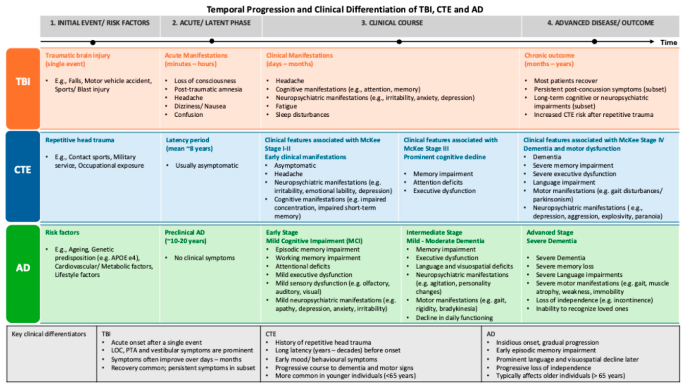

Table 1.
Classification of Traumatic Brain Injury Severity Based Clinical Indicators.
| Severity | Loss Of Consciousness (LOC) | Alterations Of Consciousness (AOC) | Post-Traumatic Amnesia (PTA) | Glasgow Coma Scale (GCS) | Structural Imaging |
|---|---|---|---|---|---|
| Mild TBI (mTBI) | < 30 min | A few moments to ≤ 24 h | 0–1 day | 13–15 | Generally normal |
| Moderate TBI | > 30 min and < 24 h | > 24 h | 1–7 days | 9–12 | Normal or abnormal |
| Severe TBI | > 24 h |
> 24 h | > 7 days |
< 9 |
Frequently abnormal |
Table 2.
Major clinical manifestations reported in traumatic brain injury (TBI), Chronic traumatic encephalopathy (CTE), and Alzheimer’s disease (AD) across somatic/sensory, cognitive, motor, neuropsychiatric/behavioural, and sleep/fatigue domains.
Table 2.
Major clinical manifestations reported in traumatic brain injury (TBI), Chronic traumatic encephalopathy (CTE), and Alzheimer’s disease (AD) across somatic/sensory, cognitive, motor, neuropsychiatric/behavioural, and sleep/fatigue domains.
| Clinical Domain | Traumatic Brain Injury (TBI) | Chronic Traumatic Encephalopathy (CTE) | Alzheimer’s Disease (AD) |
|---|---|---|---|
| Somatic & Sensory | Headache; vestibular symptoms (dizziness, vertigo); nausea/vomiting; sensory disturbances (photophobia, tinnitus, hyperacusis, visual changes, numbness and tingling) | Persistent headache |
Sensory impairment (olfactory, visual, hearing, gustatory, and tactile deficits) |
| Motor | Gait and balance impairment; coordination deficits; tremor; bradykinesia; dysarthria; weakness | Gait and balance impairment; coordination deficits; parkinsonism; tremor; bradykinesia; rigidity; ataxia; dysarthria; weakness | Gait and balance impairment; coordination deficits; bradykinesia; rigidity; reduced strength; muscle atrophy |
| Cognitive | Attention and concentration deficits; memory impairment; impaired new learning; slowed information processing; executive dysfunction; amnesia; disorientation; confusion | Attention and concentration deficits; memory impairment; impaired new learning; slowed information processing; executive dysfunction; language and visuospatial impairment; dementia | Attention deficits; memory impairment; impaired new learning; executive dysfunction; language and visuospatial impairment; aphasia, apraxia and agnosia; confusion; dementia |
| Behavioural / Neuropsychiatric | Depression; anxiety; irritability; aggression; impulsivity; behavioural dyscontrol; apathy; emotional lability; personality changes; PTSD; suicidality |
Depression; anxiety; irritability; aggression; impulsivity; explosivity; apathy; emotional lability; personality changes; paranoia; psychosis (delusions and hallucinations); suicidality | Depression; anxiety; irritability; aggression; disinhibition; apathy; personality changes; psychosis (delusions and hallucinations); wandering and repetitive behaviours |
| Sleep & Fatigue | Insomnia; hypersomnia; excessive daytime sleepiness; sleep-disordered breathing; fatigue | Not well characterized | Insomnia; excessive daytime sleepiness; sleep fragmentation; sleep-disordered breathing |
Disclaimer/Publisher’s Note: The statements, opinions and data contained in all publications are solely those of the individual author(s) and contributor(s) and not of MDPI and/or the editor(s). MDPI and/or the editor(s) disclaim responsibility for any injury to people or property resulting from any ideas, methods, instructions or products referred to in the content. |
© 2026 by the authors. Licensee MDPI, Basel, Switzerland. This article is an open access article distributed under the terms and conditions of the Creative Commons Attribution (CC BY) license (http://creativecommons.org/licenses/by/4.0/).
Copyright: This open access article is published under a Creative Commons CC BY 4.0 license, which permit the free download, distribution, and reuse, provided that the author and preprint are cited in any reuse.