

Submitted:
10 September 2026
Posted:
14 September 2026
You are already at the latest version
Abstract
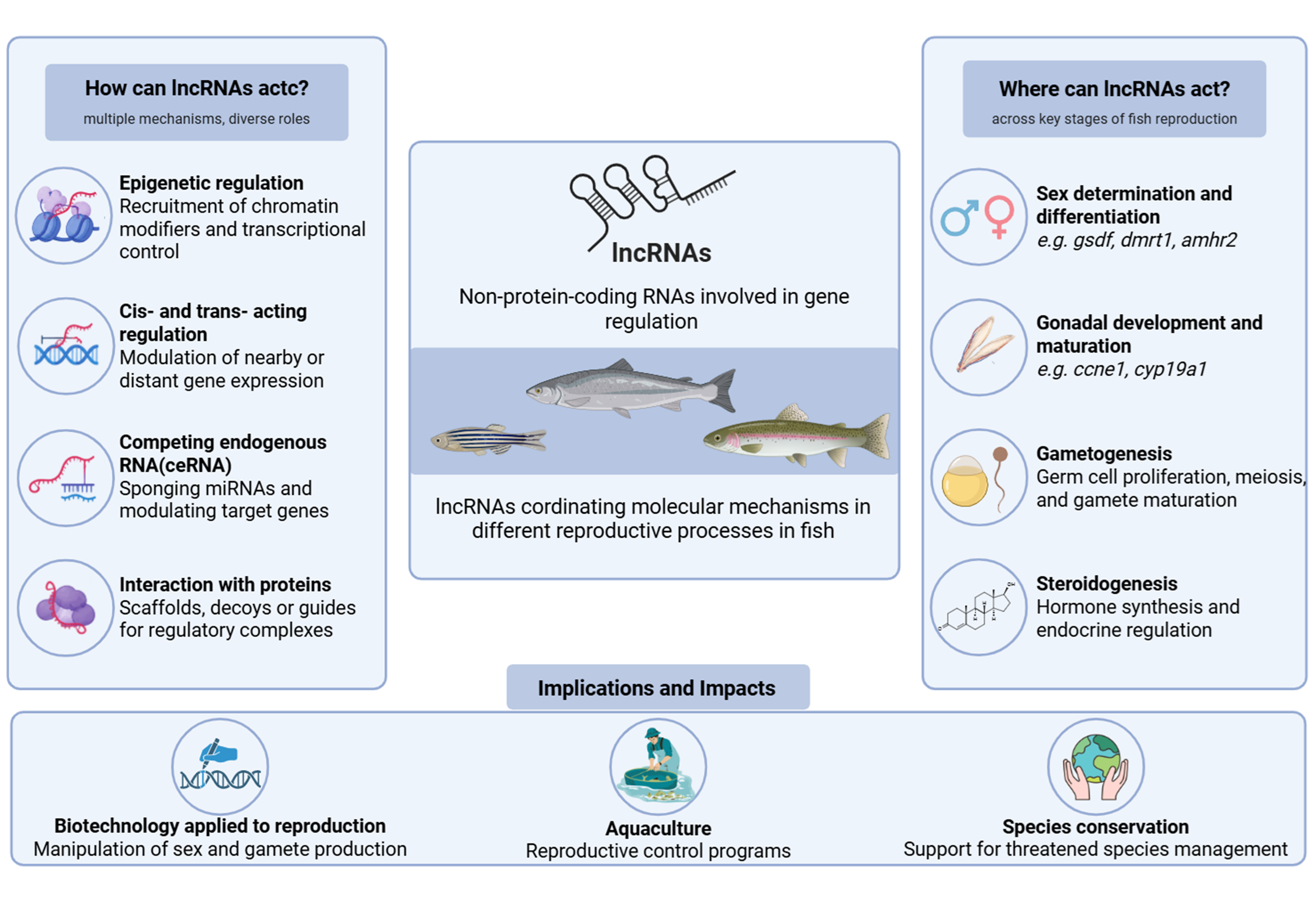
Long non-coding RNAs (lncRNAs) have emerged as important regulators of gene expression acting through transcriptional, post-transcriptional, and epigenetic mechanisms. In teleosts, high-throughput sequencing has identified a growing number of lncRNAs associated with gonadal development and reproductive function, yet current knowledge remains fragmented across species and reproductive contexts. This review summarizes recent progress in understanding lncRNA-mediated regulation of fish reproduction, with emphasis on gametogenesis and germ cell development, gonadal maturation, steroidogenesis, sex determination and differentiation, sex reversal, sexual dimorphism, and fertility. Evidence from transcriptomic and functional studies indicates that lncRNAs are involved in complex regulatory networks through cis- and trans-acting effects, competing endogenous RNA (ceRNA) interactions, and hormone-responsive pathways. Several lncRNAs have been associated with key reproductive genes, including gsdf, dmrt1, amhr2, ccne1, and cyp19a1, underscoring their potential roles in gonadal development and reproductive performance. Despite these advances, most reported lncRNAs remain functionally uncharacterized, limiting a full understanding of their biological significance. Future studies that integrate transcriptomics, genome editing, and mechanistic validation will be essential to define the precise functions of those lncRNAs. Overall, current evidence supports lncRNAs as emerging regulatory components of teleost reproductive biology with potential relevance to aquaculture, reproductive biotechnology, and environmental research.
Keywords:
gene expression
; reproductive biology
; reproductive genes
; teleost reproduction
; gonadal development
; reproductive lncRNAs
1. Introduction
Long noncoding RNAs (lncRNAs) are traditionally defined as transcripts longer than 200 nucleotides that lack protein-coding potential. For a long time, these RNAs were considered “junk transcripts,” however, it is now well established that they play important roles in regulating gene expression in eukaryotic organisms. LncRNAs influence multiple cellular processes, including chromatin organization, RNA translation, processing and stability, and protein regulation – thereby contributing to the fine-tuning of molecular programs at several levels [1,2,3].
Research on lncRNAs began in the early 1990s with the characterization of Xist (X-inactive specific transcript) and H19 (maternally expressed imprinted transcript), the first lncRNAs for which biological functions were described. Xist is a key regulatory lncRNA responsible for X-chromosome inactivation in female mammals. It is transcribed from the X chromosome and acts in cis by spreading across the chromosome from which it is expressed and recruiting chromatin-remodeling complexes that establish transcriptional silencing [4]. H19, another developmentally regulated lncRNA, is highly expressed during embryogenesis and has been implicated in the modulation of genes associated with growth, proliferation, and cell differentiation. Dysregulated H19 expression has also been reported in several types of cancer, where it promotes epithelial–mesenchymal transition and contributes to tumor progression and aggressiveness [5,6].
Despite their significance, these pioneering discoveries initially attracted limited scientific attention because they were regarded as isolated observations. Interest in lncRNAs increased substantially with the emergence of high-throughput sequencing technologies in the early 2010s. The ENCODE (Encyclopedia of DNA Elements) project revealed that more than 80% of the human genome is transcribed, whereas less than 2% encodes proteins. These findings led to the identification of thousands of long transcripts that exhibited tissue-specific, developmental stage-specific, and disease-associated expression patterns. Importantly, many of these transcripts were found to possess regulatory functions, challenging the traditional protein-centric view of genome activity [7,8,9].
The biogenesis of lncRNAs closely resembles that of canonical messenger RNAs (mRNAs). Most lncRNAs are transcribed by RNA polymerase II and undergo post-transcriptional processing, including 5′ capping, 3′ polyadenylation, and, in many cases, splicing. However, some lncRNAs are transcribed by RNA polymerase III and may not undergo extensive processing, highlighting the diversity of lncRNA biogenesis pathways [2,10,11,12].
Despite some exceptions, most lncRNAs are expressed at relatively low levels compared with mRNAs and often display highly restricted, cell type-specific expression patterns. Their expression is frequently linked to cellular identity, developmental stage, and physiological state [2,11]. Although lncRNAs are predominantly localized in the nucleus, many are also found in the cytoplasm and mitochondria. Importantly, their low abundance does not indicate limited functional significance – on the contrary, accumulating evidence suggests that low expression levels may enhance the specificity of lncRNA-mediated regulatory interactions and constitute an intrinsic feature of their mechanisms of action [13].
LncRNAs are usually categorized according to their genomic position and orientation relative to protein-coding genes [14,15] (Figure 1A). Within this classification, sense lncRNAs are transcribed from the same strand as a protein-coding gene, and may partially or completely overlap its exons or introns. In contrast, antisense lncRNAs originate from the opposite strand of a coding locus. Bidirectional lncRNAs are transcribed in the opposite direction from promoters located adjacent to protein-coding genes. Intronic lncRNAs originate entirely from the intronic region of a protein-coding gene, without overlapping exons, whereas intergenic lncRNAs (lincRNAs) are located between protein-coding genes and do not overlap any annotated coding regions.
LncRNA in Gene Regulation: Mechanisms and Functions
LncRNAs interact with DNA, RNA, and proteins to coordinate chromatin organization and transcriptional activity at both local and distant genomic loci. Depending on their mode of action, lncRNAs can regulate neighboring genes (cis-acting lncRNAs) or influence the expression of genes located elsewhere in the genome (trans-acting lncRNAs). Beyond transcriptional regulation, lncRNAs modulate multiple aspects of RNA metabolism, including splicing, stability, localization, and translation. They have also been implicated in the organization and function of organelles and cellular compartments, highlighting their broad regulatory potential across diverse biological processes [2,16].
The regulatory functions of lncRNAs are mediated through a variety of molecular mechanisms (Figure 1B). One important mechanism involves direct interactions with DNA through the formation of RNA-DNA-DNA triplexes or R-loops, which can alter chromatin accessibility and thereby activate or repress gene expression. A well-characterized example is the lncRNA TARID, which forms an R-loop at CG-rich sequences near the transcription start site of the tumor suppressor gene TCF21. This structure recruits TET1 (ten-eleven translocation 1), leading to promoter demethylation and a chromatin state permissive for gene expression [17].
LncRNAs also regulate chromatin organization through interactions with chromatin-modifying and -remodeling complexes that are recruited to specific genomic regions, either promoting or inhibiting gene expression. For example, the lncRNA ANRIL recruits Polycomb group protein complexes to the promoters of its neighbour genes CDKN2A and CDKN2B, thereby contributing to the regulation of cellular senescence [18]. Conversely, some lncRNAs function as molecular decoys by preventing histone or chromatin modifiers from binding to their target genomic regions. The lncRNA lncPRESS1, for instance, maintains the pluripotent state in human embryonic stem cells by sequestering the chromatin-bound histone deacetylase SIRT6. This interaction prevents the deacetylation of the promoters of several pluripotency genes, thereby inhibiting differentiation and preserving the undifferentiated cellular state [19].
Beyond local chromatin regulation, certain lncRNAs exert chromosome-scale effects on genome organization. The archetypal example is XIST, the lncRNA responsible for X-chromosome inactivation in female mammals. XIST spreads along one X chromosome, coating it and recruiting chromatin-remodeling complexes that promote extensive heterochromatin formation and transcriptional silencing across the chromosome. Through this mechanism, XIST establishes dosage compensation between males and females by ensuring that only one X chromosome remains transcriptionally active [20].
Another major class of lncRNA functions involves their role as molecular scaffolds. In this capacity, lncRNAs facilitate the assembly of multiprotein complexes by bringing together proteins that may not otherwise interact efficiently. A well-characterized example is the long isoform of the lncRNA NEAT1, which serves as a structural scaffold for the formation and maintenance of paraspeckles – membraneless subnuclear bodies that participate in RNA processing, nuclear retention of transcripts, and cellular responses to stress. Through the recruitment and spatial organization of multiple nuclear proteins, NEAT1 plays an essential role in paraspeckle assembly and function [21].
LncRNAs can also regulate gene expression at the post-transcriptional level by functioning as competing endogenous RNAs (ceRNAs), commonly referred to as molecular sponges. In this mechanism, lncRNAs bind microRNAs (miRNAs) through complementary sequence interactions, thereby reducing the availability of these miRNAs to bind their target messenger RNAs. As a result, repression of the target transcripts is alleviated, leading to increased gene expression. A representative example is LncND, a primate-specific lncRNA expressed in neural progenitor cells. LncND sequesters miR-143-3p, a microRNA that negatively regulates the neurodevelopmental genes NOTCH1 and NOTCH2. By limiting miR-143-3p availability, LncND promotes Notch signaling and contributes to neuronal differentiation and development [22].
Altogether, these examples illustrate the remarkable versatility of lncRNAs as regulators of gene expression. Through their ability to interact with DNA, RNA, and proteins, lncRNAs influence transcriptional, epigenetic, and post-transcriptional processes across multiple biological contexts. Their diverse mechanisms of action underscore their importance as key components of gene regulatory networks and provide a foundation for understanding their roles in development, physiology, and disease.
2. LncRNAs in Fish Reproduction
LncRNAs as Regulators of Gametogenesis and Spermatogenesis
Among the diverse biological processes influenced by lncRNAs, mammalian gametogenesis represents a highly regulated and extensively studied example (Robles et al., 2019). In fish, RNA-Seq studies have identified thousands of lncRNAs with reproductive organ–specific expression, strongly associated with reproductive processes and their maintenance [23,24]. In the flounder Paralichthys olivaceus, differentially expressed lncRNAs (DElncRNAs) have been implicated in both cis and trans regulation of reproduction-related transcripts, including genes such as foxo1, foxm1, smad3, foxr1, camk4, ar, and tgfb3, which participate in classical gonadal development pathways such as insulin, MAPK, and calcium signaling [25]. Enrichment analyses revealed that positively regulated cis targets were associated with fertilization and steroid biosynthesis, whereas negatively regulated ones were linked to steroid binding, microtubule binding, sexual differentiation, gonadal development, and MAPK signaling. Conversely, in trans regulation – based on co-expression with mRNAs – DElncRNAs were associated with hormonal regulation, dynein binding, calcium signaling, and ovarian steroidogenesis, as well as MAPK, Wnt, and TGF-β pathways. Expression profiles varied among ovarian, intersex, and testicular stages, often showing higher abundance in ovaries and intersex gonads, suggesting roles in sexual differentiation and gametogenesis. Moreover, experimental work involving the administration of follicle-stimulating hormone (Fsh) and human chorionic gonadotropin (hCG) confirmed the hormone-responsive expression of specific lncRNAs [26], reinforcing their participation in regulatory networks controlling both gamete formation and sex reversal. Taken together, these findings demonstrate that lncRNAs are key regulators of gametogenesis in fish, acting through epigenetic, transcriptional, and post-transcriptional mechanisms to modulate major developmental pathways during gonadal differentiation [25].
Functional studies in model teleost species have provided additional evidence for the regulatory potential of lncRNAs in reproduction. In medaka (Oryzias latipes), lnc4c was identified as a gonad-specific lncRNA and proposed as the primary microRNA transcript (pri-miRNA) for ola-miR-202 [24]. Using single-molecule fluorescence in situ hybridization, the authors demonstrated that pre-miR-202 is highly expressed in the nuclei of somatic testicular cells, including Sertoli and Leydig cells, whereas lnc4c is expressed at relatively low levels. Co-expression of lnc4c and pre-miR-202 was observed exclusively in Sertoli cells and involved only a limited number of transcript molecules. Importantly, deletion of the lnc4c locus resulted in reduced testis size, decreased germ cell abundance, and impaired sperm viability. These defects were accompanied by increased production of hormones and signaling molecules by somatic testicular cells, as well as accelerated differentiation of male germ cells. Therefore, these findings indicate that lnc4c plays a critical role in maintaining normal testicular development and spermatogenesis in medaka [24].
Additional evidence for the involvement of lncRNAs in germ cell development has been reported in zebrafish (Danio rerio) [27]. The lncRNA lnc172 is highly expressed in the cytoplasm of spermatogonia, spermatocytes, and myoid cells in the testes, while in the ovaries, it was detected in ovarian somatic cells [27]. In contrast, lnc304 was localized to both the nucleus and cytoplasm of spermatogonia and spermatocytes, while in females, it was localized in the nucleus of the previtellogenic oocyte. Both lncRNAs exhibited co-localization with the germ cell marker ddx4 (aka vasa) in spermatogonia. Functional analyses involving knockdown and overexpression experiments demonstrated that elevated lnc172 expression disrupted normal primordial germ cells (PGC) migration, causing ectopic localization of germ cells in the tail, somites, eyes, and brain. These results highlight a crucial role for lnc172 in guiding PGCs toward the genital ridge during early development [27].
Further insights into lncRNA-mediated regulation of fertility have been obtained from studies of rainbow trout (Oncorhynchus mykiss). Comprehensive analyses of the interactions among miRNAs, mRNAs, and lncRNAs during gonadal development in triploid fish at different developmental stages (65 days post-fertilization (dpf), 180 dpf, and 600 dpf) highlight the importance of lncRNAs involved in the regulation of gonadal fertility [28]. Their study identified 22 differentially expressed genes (DEGs) associated with meiosis and fertility, including sycp2, sycp3, dmc1, and ccne1. In zebrafish, sycp2 has been shown to be essential for homologous recombination and pairing during the early stages of meiosis [29]. Similarly, dmc1, which is associated with homologous recombination and expressed exclusively during meiosis, plays key roles in gametogenesis and gonadal development across several species [30,31,32]. In addition, ccne1, a member of the cyclin family, is important for homologous pairing and DNA repair during meiosis [33]. The authors reported reduced ccne1 expression in triploid trout (180 and 600 dpf), while dre-miR-15a-5p_R+1, a predicted negative regulator of ccne1, was increased. Moreover, three novel lncRNAs (MSTRG.74687.3, MSTRG.76205.1, and MSTRG.30919.1) showed inversed correlation with this miRNA. These findings suggest a potential lncRNA–miRNA–mRNA regulatory network in which these lncRNAs may modulate ccne1 expression indirectly through miRNA-mediated regulation, thereby potentially influencing meiotic progression and gonadal development in trout [28].
A subsequent study conducted by the same research group further explored the roles of ccne1, ccne2, and the associated lncRNAs during oogenesis and spermatogenesis in rainbow trout [34]. The authors demonstrated that ccne1 positively regulated the expression of lncRNA MSTRG.74687, whereas silencing ccne2 did not significantly alter its expression. These findings suggest that MSTRG.74687 may participate in regulatory pathways controlling gonadal development. Functional analyses further supported distinct biological roles for ccne1 and ccne2 in coordinating cell proliferation and reproductive gene expression. Together, these studies revealed a regulatory axis involving lncRNAs, omy-miR-15a-5p, and ccne1 that contributes to the control of meiosis and reproductive development in rainbow trout [34].
Gene expression analyses also identified miR-210 as an additional regulator of reproductive processes, influencing the prioritization and expression of key meiotic genes [35,36]. The resulting lncRNA–miRNA–mRNA interaction networks appear to play central roles in either promoting or repressing genes required for meiosis and gonadal development. These findings contribute to a growing understanding of the epigenetic mechanisms underlying fertility and sterility in fish and may ultimately provide valuable targets for improving reproductive management and selective breeding programs in aquaculture.
Therefore, these studies demonstrate that lncRNAs are integral components of the regulatory networks governing gametogenesis in fish. Through interactions with protein-coding genes, miRNAs, and endocrine signaling pathways, lncRNAs contribute to germ cell development, meiotic progression, gonadal differentiation, and reproductive function. Despite these advances, the specific molecular mechanisms through which many lncRNAs exert their effects remain poorly understood. Future studies integrating functional genomics, gene-editing approaches, and single-cell technologies will be essential for elucidating the precise biological roles of lncRNAs during fish reproduction.
LncRNAs in Response to Sex Hormones
Advances in RNA sequencing technologies and the discovery of lncRNAs in recent decades have demonstrated that gametogenesis and fertility do not depend exclusively on protein-coding genes. This group of transcripts has been associated with crucial roles in various well-documented biological processes [14,37,38,39,40]. In the reproductive system, lncRNAs have been implicated in sex hormone responses [41], oocyte meiosis [42], ovulation [43], and sexual differentiation [44].
In mammals, lncRNAs regulate reproduction mainly by modulating sex steroid signaling and steroidogenesis pathways. In males, the lncRNAs PRNCR1 and PCGEM1 interact with androgen receptor signaling in males [41]. In females, several lncRNAs contribute to ovarian physiology by regulating steroidogenesis, follicular development, and hormone production. SRA and Neat1 are involved in steroid hormone synthesis and corpus luteum function [45,46], while H19 modulates steroidogenic pathways and follicular development through post-transcriptional mechanisms [47,48]. Together, these findings demonstrate that lncRNAs act at multiple levels in reproductive endocrine pathways. However, compared with mammals, the functional roles of lncRNAs in hormonal regulation and gonadal development remain poorly understood in non-mammalian vertebrates.
In vertebrates, reproduction is regulated by a complex network of interactions involving multiple organs, signaling pathways, and gene regulatory mechanisms [49,50]. Successful gamete production requires precise coordination among transcription factors, growth factors, and sex steroids – including estrogens, progesterone, and androgens – which govern gonadal development, gametogenesis, and reproductive function [51,52]. At the systemic level, this process is regulated by the hypothalamic-pituitary-gonadal (HPG) axis, which integrates endocrine signals to regulate gonadal activity throughout the reproductive cycle [49,53].
Within the HPG axis, gonadotropin-releasing hormone (GnRH) secreted by the hypothalamus stimulates the pituitary gland to release follicle-stimulating hormone (Fsh) and luteinizing hormone (Lh) [54]. In females, these gonadotropins promote ovarian steroidogenesis through activation of their respective receptors, leading to the production of estrogens and progestogens that regulate oogenesis, follicular growth, meiotic progression, and ovulation [53]. In males, Fsh and Lh act primarily on Sertoli and Leydig cells, stimulating androgen and growth factor production and supporting spermatogenesis [49].
In teleosts, oogenesis is orchestrated by coordinated endocrine and paracrine signaling events that drive the progression of oogonia into fully mature oocytes. This dynamic process encompasses oogonia proliferation, primary growth, vitellogenesis, final maturation, and ovulation [53]. Conversely, the testicular functions center on androgen biosynthesis and the continuous maintenance of spermatogenesis [49]. Male germ cell development proceeds through successive phases of mitosis, meiosis, and spermiogenesis within a specialized cystic organization in which Sertoli cells provide structural, nutritional, and endocrine support to developing germ cells [49,55]. The interstitial compartment contains blood vessels, immune cells, and steroidogenic Leydig cells that contribute to endocrine regulation of reproduction.
Unlike mammals, the interaction between gonadotropins and their receptors does not appear to be specific in fish [56]. Notably, Fsh is considered more potent than Lh in driving androgen synthesis due to the expression of Fsh receptors on Leydig cells [57], a mechanism well documented in zebrafish [58]. Furthermore, Fsh promotes spermatogonial proliferation and differentiation by stimulating Sertoli cells to produce insulin-like growth factor 3 (Igf3), while concurrently downregulating the expression of anti-Müllerian hormone (Amh), a recognized inhibitor of spermatogonial differentiation [59,60]. Therefore, Fsh acts as a master regulator of the spermatogenic process, finely orchestrating the balance of stimulatory and inhibitory molecular pathways required for proper germ cell progression [61].
While the systemic control exerted by this classical network that governs gametogenesis, particularly in non-mammalian vertebrates, remains poorly understood, any disruption of these highly coordinated dynamics severely compromises reproductive success. Interest in the role of lncRNAs in fish reproductive endocrinology has increased considerably in recent years [25]. Studies in this area are promising for understanding, at the molecular level, the dynamics of the regulation of gametogenesis and sex hormones involved in gonadal development, as well as topics related to maturation, sexual differentiation, reproductive health, and fertility. These studies also aim to identify interventional targets and biomarkers essential for advancing reproductive biotechnologies and improving the management of species of ecological and economic interest. This relationship between hormonal regulation of gonadal development and lncRNAs has already been demonstrated in fish [25,40,62].
Evidence linking lncRNAs to hormonal regulation in fish has already emerged from several species. In the rice-field eel (Monopterus albus), Fsh promotes oocyte growth and facilitates the transition from primary to secondary follicles, a critical step during oocyte maturation [63]. In the Japanese eel (Anguilla japonica), human chorionic gonadotropin (hCG) stimulates gonadal maturation and regulates the expression of the lncRNA HAS2-AS1, which in turn promotes the expression of HAS2, a gene associated with cumulus expansion and cellular migration [64]. Further evidence for hormone-responsive lncRNA expression was provided by He and colleagues (2022) [26] , who investigated ovarian lncRNAs in M. albus. The authors demonstrated that Fsh and hCG induced the expression of the lncRNA LOC109960696, associated with Smad2, in both time- and dose-dependent manners. In addition, five differentially expressed lncRNAs (LOC109952131, LOC109953466, LOC109954337, LOC109954360, and LOC109958454) exhibited significantly higher expression levels in ovaries and intersex gonads than in testes. Exposure to Fsh or hCG further increased the expression of these transcripts, indicating that gonadotropin signaling directly influences lncRNA expression profiles during gonadal development.
The role of lncRNAs in hormone-dependent reproductive regulation has also been investigated in the gynogenetic Japanese flounder (Paralichthys olivaceus) [25]. Transcriptomic analyses identified 2,284 lncRNAs differentially expressed between testes and ovaries, including 1,870 testis-biased and 414 ovary-biased transcripts. Interestingly, the proportion of sex-biased lncRNAs exceeded that observed for both mRNAs and miRNAs, suggesting an enhanced contribution of non-coding RNAs to gonadal regulation. Functional analyses revealed that these DElncRNAs participate in steroidogenesis, gonadal development, fertilization, and sex differentiation through both cis- and trans-regulatory mechanisms. Moreover, several lncRNAs functioned as competing endogenous RNAs within complex lncRNA–miRNA–mRNA interaction networks. Through interactions with miRNAs such as let-7 and miR-20a, these lncRNAs influenced the expression of dnah1, a gene encoding an axonemal dynein heavy chain required for flagellar assembly and sperm motility. Given that mutations in dynein-related genes are associated with male infertility in multiple vertebrate species, these findings provide compelling evidence that lncRNAs contribute directly to reproductive performance and sexual development [25,65].
Collectively, current evidence indicates that lncRNAs represent important regulatory components of endocrine pathways controlling fish reproduction. By integrating hormonal signals with transcriptional, post-transcriptional, and epigenetic regulatory mechanisms, lncRNAs influence steroidogenesis, gametogenesis, gonadal development, and fertility. Nevertheless, the majority of available studies remain descriptive, and the precise molecular mechanisms underlying lncRNA-mediated hormonal regulation are still poorly understood. Future research combining functional genomics, endocrine manipulation, and gene-editing approaches will be essential for elucidating how lncRNAs coordinate hormonal signaling networks and reproductive physiology in teleost fish.
3. Regulatory Roles of lncRNAs in Sex Determination, Sexual Dimorphism and Sex Differentiation
LncRNAs in Sexual Size Dimorphism
Sexual size dimorphism is defined as a pronounced difference in body size between males and females within a species [66,67,68]. Evolutionary studies have shown that this characteristic is commonly observed in many teleost fish species [68,69,70,71,72,73,74]. In fish, sexual size dimorphism is associated with sexual selection and is linked to territoriality in males and fecundity in females [66]. Notably, this phenomenon is observed in several fish species of commercial importance to aquaculture, such as the olive flounder (Paralichthys olivaceus) and the Nile tilapia (Oreochromis niloticus) [75,76].
Two interesting studies in fish have identified potential lncRNAs involved in the establishment of female-biased sexual size dimorphism in fish. In Chinese tongue sole (Cynoglossus semilaevis), highlighted genes were found together with their respective lncRNAs and miRNAs, forming regulatory networks [77]. Analysis of these networks suggested that miR-460-x may regulate two genes, cdk2 and bub3, both of which play critical roles in the cell cycle and cell proliferation [46,78,79]. Interestingly, miR-460-x was downregulated in females, whereas several associated lncRNAs (MSTRG.24810.1, MSTRG.3094.1, MSTRG.5569.1, MSTRG.9796.1, MSTRG.23232.1, and MSTRG.7061.1) were upregulated. This pattern suggests that these lncRNAs may be involved in competition with miRNAs for binding to target genes (consistent with the competing endogenous RNA – ceRNA – hypothesis), thereby regulating sexual size dimorphism. However, the precise molecular mechanisms through which these ncRNA influence growth-related pathways remain to be elucidated.
A similar pattern was reported in olive flounder (Paralichthys olivaceus), a marine fish with high commercial value in East Asia due to its flesh quality and rapid growth [80,81]. In this species, females exhibit faster growth rates than males, resulting in female-biased sexual size dimorphism [81]. By integrating miRNA–target anticorrelation analyses, lncRNA–mRNA co-expression data, and statistical testing, the authors constructed a ceRNA regulatory network, with the strongest signals detected in gonadal and muscle tissues. Functional enrichment analyses revealed pathways associated with growth and cell proliferation, including those involving miR-460-x and its target genes, as well as a novel miRNA (novel-m0387-3p) linked to apoptosis and other regulatory processes. Additionally, several lncRNAs were predicted to exert trans-regulatory effects on genes located at distant genomic loci, further supporting their potential role in growth regulation.
Furthermore, transcriptomic analyses of brain and pituitary tissues revealed significant sex-specific differences in gene expression and identified several enriched KEGG pathways, including ovarian steroidogenesis, prolactin signaling pathway, arachidonic acid metabolism, Hippo signaling pathway, and cytokine–cytokine receptor interaction, among others. When integrated with mRNA–miRNA–lncRNA regulatory networks, these findings suggest that non-coding RNA-mediated regulatory mechanisms play crucial roles in the establishment and maintenance of female-biased sexual size dimorphism in olive flounder.
LncRNAs in Sex Determination Mechanisms
Fish are considered one of the most species-diverse groups of vertebrates [82], and this diversity may be associated with the wide range of reproductive strategies observed within the group [83]. Sex determination in fish refers to the mechanisms that direct sexual differentiation, whereas sexual differentiation is the process through which testes or ovaries develop from an initially undifferentiated gonad [84]. These mechanisms may be genetic or environmental in nature and play a fundamental role in reproductive biology [83,85].
The turbot (Scophthalmus maximus) is widely cultivated in Europe and China [86,87](. In this species, females exhibit higher growth rates than males [87,88], making the production of all-female stocks highly desirable for aquaculture. Transcriptome analysis of males and females turbot were performed to identify and better understand the regulatory mechanisms associated with the species’ primary sex-determining gene. A lncRNA of 78,309 bp was identified within the genomic region containing the sex-determining gene sox2 (2,557 bp). This lncRNA, named sox2ot, spans the sex-determination region and contains the sox2 gene within one of its introns. Furthermore, it exhibits complete complementarity to the 3′ untranslated region (3′UTR) of sox2. These findings suggest that sox2ot may participate in post-transcriptional regulatory mechanisms controlling the activity of this sex-determining gene [87]. Expression analyses revealed female-specific upregulation of sox2 during the critical period of gonadal differentiation (50–55 dpf). In contrast, the lncRNA sox2ot did not exhibit differential expression between sexes, supporting the hypothesis that its role may be limited to the regulation of sox2 rather than acting as a sex-specific transcript itself. Together, these findings suggest that sox2ot may contribute to the post-transcriptional regulation of sox2 during sex determination in this species.
LncRNA-Mediated Regulation of Sex Differentiation and Sex Reversal
In teleosts, endocrine sex reversal has been demonstrated in both gonochoristic species and hermaphrodites. Hormonal sex reversal provides a valuable tool for producing monosex populations in aquaculture and for advancing our understanding of the mechanisms underlying sexual differentiation [89,90].
Previous studies in teleost fish have shown that treatment with mifepristone (RU485), a progesterone antagonist, altered the expression of steroid hormone receptor genes, thereby influencing the reproductive process and sexual differentiation in these animals [91,92]. During the process of induced masculinization (RU486-500 µg/g diet) in Takifugu rubripes, thereabout hundreds of DElncRNAs were identified across sex and treatment. A key finding was that the predicted targets of such differentially expressed lncRNAs are enriched in canonical regulators of sex determination/differentiation, as well as genes involved in steroid hormone pathways. Specifically, lncRNAs targeting genes of the steroidogenic axis, including dmrt1, dmrt3, gsdf, amhr2, and nanos2, as well as ovarian pathway genes such as foxl2, cyp19a1a, and hsd17b1, were significantly enriched [93]. Additionally, enrichment of RU486-associated DElncRNAs was observed in pathways related to ovarian steroidogenesis, steroid hormone biosynthesis, calcium signaling, and cortisol synthesis and secretion. These findings suggest that RU486 promotes masculinization through stress- and endocrine-mediated mechanisms, involving extensive regulatory reprogramming by non-coding RNAs. In this context, lncRNAs appear to participate in both co-expression interactions and competitive endogenous RNA (ceRNA) networks [93].
In Takifugu rubripes, masculinization induced by the aromatase inhibitor (AI) letrozole was also evaluated [94]. Transcriptomic analysis revealed differential expression of lncRNAs between genetic sexes and between control XX gonads and AI-treated XX gonads. The results indicate that letrozole treatment triggers substantial ncRNA remodeling during masculinization. Notably, the authors identified DElncRNAs and a ceRNA regulatory network comprising 180 mRNAs, 30 miRNAs, 314 lncRNAs, and 6 circRNAs, which were implicated in the regulation of cell fate determination, cell motility, and hormonal signaling during induced masculinization. Functional enrichment analysis of targets associated with this network highlighted pathways such as TGF-β signaling and ion transport-related processes, which may contribute to the cellular signaling transitions underlying steroid-induced sex reversal [94]. The authors also identified gsdf (a member of the TGF-β superfamily essential for sex determination/differentiation in teleosts) as a predicted target of lncRNA000338. Moreover, gsdf expression was elevated in undifferentiated male gonads, providing a compelling example of a potential lncRNA–sex gene regulatory pair acting during the earliest stages of sexual differentiation [94].
Also in T. rubripes, early sexual differentiation was assessed by analyzing undifferentiated (bipotential) gonads approximately 40 days post-hatching. Sex-biased lncRNA expression is already detectable at this developmental stage. This finding supports the hypothesis that lncRNAs contribute to the early divergence of male and female developmental pathways prior to overt gonadal differentiation [95]. Together, these studies indicate that lncRNAs in T. rubripes are associated with key genes involved in sex determination/differentiation, such as dmrt1, gsdf, amhr2, and nanos2, as well as with endocrine pathways governing estrogen production and broader steroid/stress physiology (e.g., cyp19a1a and hsd17b1 for estrogen synthesis; cyp11c1 for cortisol synthesis and secretion). These pathways correspond precisely to the biological mechanisms targeted in models of sex reversal induced by steroidal/endocrine disruptors.
Another interesting hermaphroditic fish is Monopterus albus, which undergoes sex reversal from ovary to testis through an ovotestis stage, characterized by the presence of both male and female gonadal tissue within the same individual. Gonadal tissue development occurs through the processes of sex determination and differentiation [96,97,98,99]. This process is known to occur cyclically: during the initial developmental stage, the gonadal arrangement is ovarian; after spawning, the development of male germ cells begins; upon reaching sexual maturity, both ovarian and testicular structures can be observed; and finally, exclusively testicular tissue is formed [97,98,99,100].
Qiaomu Hu et al. (2023) [101] investigated the epigenetic and regulatory mechanisms of lncRNAs during gonadal differentiation and sex reversal in Monopterus albus. Differential expression of lncRNAs was observed when comparing the different gonadal stages (ovary, ovotestis, and testis). Following an integrated analysis of lncRNA and mRNA localization and co-expression data, two relevant lncRNAs and their respective target genes were selected: MSTRG.38036/psmβ8 and MSTRG.12998/ptk2β. Expression of both transcript pairs increased from the ovary to the ovotestis stage and subsequently declined during the transition from ovotestis to testis, suggesting dynamic regulation throughout gonadal development. Based on the expression profiles of genes previously implicated in sexual differentiation [102,103], the authors proposed that these transcripts may contribute to the sex-reversal process.
To further investigate this hypothesis, DNA methylation patterns were examined during sex reversal induced by 17α-methyltestosterone treatment in M. albus larvae. DNA methylation levels were negatively correlated with the expression of MSTRG.38036 and MSTRG.12998, suggesting that methylation may repress the activity of these lncRNAs. The authors related these findings to previous studies demonstrating the role of DNA methylation in regulating gene expression during sex reprogramming [104,105]. Furthermore, dual-luciferase reporter assays revealed that ptk2β regulates cyp19a1 expression through the Ptk2β/EGFR/STAT3 signaling pathway, providing evidence that lncRNAs participate in central molecular pathways controlling gonadal determination and sexual differentiation.
Overall, these studies demonstrate that lncRNAs and their associated mRNAs may contribute to the gonadal remodeling processes underlying sex reversal. This conclusion is supported by the association of these transcripts with genes previously implicated in teleost sexual differentiation and by their integration into regulatory networks involved in gonadal plasticity through the modulation of cellular pathways governing proliferation, differentiation, and tissue reorganization. The findings of Hu and colleagues (2023) [101] broaden our understanding of lncRNA function in fish and suggest that these molecules serve as important molecular mediators of sexual differentiation and gonadal sex reversal, potentially through the integration of epigenetic regulation and transcriptional control.
4. LncRNAs in Gonadal Maturation and Reproductive Regulation
Gonadal maturation refers to the morphological changes through which the male and female gonads undergo to achieve full growth and maturity, leading to the production of viable gametes (sperm or oocytes) during one or more reproductive periods [106]. In teleosts, gonadal maturation is closely linked to the process of puberty, which marks the transition from juvenile status to reproductive capability. According to Okuzawa (2002) [107], the onset and progression of puberty are governed by a complex interplay of neuroendocrine signals, particularly those operating through the HPG axis. The onset of this process occurs with the increased release of GnRH, which stimulates the pituitary to secrete the gonadotropic hormones Fsh and Lh. These hormones promote gonadal growth, germ cell differentiation, and the synthesis of sex steroids, such as estradiol and 11-ketotestosterone, which are crucial for follicular maturation and spermatogenesis, respectively. Environmental factors such as photoperiod and temperature also play crucial roles in regulating puberty and gonadal maturation, with their effects varying among species and reproductive strategies. Furthermore, nutritional status and overall physiological condition can influence HPG-axis sensitivity, thereby affecting the timing of reproductive maturation.
LncRNA-Mediated Regulation of Ovarian Maturation
Ovarian development is a complex biological process involving multiple signaling pathways and gene expression mechanisms [108,109,110]. Successful oogenesis requires the coordinated action of hormones, growth factors, transcription factors, and other molecular regulators [111]. Dong and collaborators (2021) [50] investigated the involvement of lncRNAs in oocyte growth, maturation, and consequently ovulation in Chinese tongue sole (C. semilaevis). High-throughput sequencing was performed on ovaries collected at three developmental stages: stage IV (late vitellogenesis), stage V (maturation), and stage VI (post-ovulation), according to the classification proposed by [112]. Transcriptome sequencing identified 33,562 lncRNAs, including 10,201 previously annotated transcripts and 23,361 novel lncRNAs. The transition from stage V to stage VI exhibited substantially greater transcriptional remodeling than the transition from stage IV to stage V. Specifically, 58 DElncRNAs were identified between stages IV and V, whereas 187 DElncRNAs were detected between stages V and VI, with 13 transcripts shared between both comparisons.
Previous studies have shown that lncRNAs regulate gene expression through antisense, cis-, and trans-regulatory mechanisms and may also influence nearby genes through positional effects [10,113,114]. Consistent with these observations, target-prediction analyses identified numerous putative lncRNA–mRNA regulatory relationships. For example, mmp14 was identified as a cis target of lncRNAs XR_522498.1 and XR_522499.1, whereas col4a1 and crem were predicted cis targets of XR_523405.2 and XR_001761307.1, respectively. Functional enrichment analyses revealed that antisense and cis target genes were associated with pathways including the cell cycle, apoptosis, TGF-β signaling, and Wnt signaling. These pathways regulate a broad range of cellular processes, including proliferation, migration, differentiation, apoptosis, and embryonic development [115,116]. In avian species, TGF-β signaling promotes granulosa-cell proliferation [117], whereas in mice it contributes to the maintenance of oocyte meiotic arrest through mechanisms regulated by Fsh and Lh [118]. Similarly, inhibition of Wnt signaling has been reported to promote oocyte maturation in pigs [119], while in mice it is associated with granulosa-cell cycle arrest [120]. These findings support the hypothesis that lncRNAs may influence ovarian differentiation and maturation by modulating genes involved in these signaling pathways.
Dong and collaborators (2021) [50] further demonstrated that they identified 31 co-expressed lncRNA–mRNA pairs, including 25 positively correlated and 6 negatively correlated interactions in the ovary of Chinese tongue sole (C. semilaevis). Among these, igfbp1, a gene encoding insulin-like growth factor-binding proteins that are highly expressed during the pre-ovulatory period, showed a positive correlation with lncRNA XR_522957. In contrast, cyp17a1, a key steroidogenic enzyme, displayed a negative correlation with lncRNA XR_522278.2. Dual-fluorescence in situ hybridization demonstrated the co-localization of XR_522278.2 and XR_522171.2 with their respective target genes, cyp17a1 and cyp19a1, within follicular cells. To further explore potential reproductive functions, the authors constructed a competing endogenous RNA (ceRNA) network. Several genes directly involved in reproduction, including mmp14, pgr, cyp17a1, cyp19a1, and hsd17b1, were predicted to be regulated by 30 lncRNAs through interactions with 35 miRNAs. Collectively, these findings provide strong evidence that lncRNAs participate in the regulation of gene-expression programs governing ovarian differentiation, maturation, and ovulation.
LncRNAs in the Regulation of IGF-Induced Ovarian Maturation
Insulin-like growth factors (IGF-1 and IGF-2) constitute an evolutionarily conserved system primarily involved in cellular proliferation across different biological contexts, including growth, proliferation, survival, migration, and differentiation [121,122,123,124,125,126]. In fish, IGFs are primarily synthesized in the liver under growth hormone (GH) stimulation and subsequently released into circulation, where they regulate multiple physiological systems and tissues, including the ovaries, testes, and pituitary gland [127,128,129,130,131,132,133,134,135]. Both igf1 and igf2 are expressed in fish ovaries and have been implicated in ovarian growth and maturation across several species [82,136,137,138]. Their ovarian expression promotes oocyte growth and maturation, while igf1 also plays an important role in regulating ovarian steroidogenesis [139,140,141,142,143].
Ndandala et al. (2025) [23] investigated the involvement of lncRNAs in ovarian growth, development, and maturation in golden pompano (Trachinotus ovatus) by examining stage III ovaries exposed in vitro to IGF1 or IGF2, with the former eliciting a substantially stronger response than the latter. Relative to controls, IGF1 treatment induced the differential expression of 1,494 lncRNAs and 8,728 mRNAs, whereas IGF2 treatment resulted in only 101 DElncRNAs and 373 DEGs. Co-expression and target analyses indicated that candidate lncRNAs and their associated genes were enriched in pathways related to the cell cycle, oocyte meiosis, focal adhesion, and multiple reproductive signaling pathways, including MAPK, PI3K–Akt, GnRH, Wnt, FOXO, and estrogen signaling. Among the identified transcripts, four lncRNAs responsive to IGF1 stimulation (MSTRG.66521.1, MSTRG.49969.1, MSTRG.59923.1, and MSTRG.13767.1) and two responsive to IGF2 stimulation (MSTRG.20896.2 and MSTRG.58123.2) were co-regulated with genes implicated in ovarian development and maturation, including adam23, cyp19a1, cyp17a, slc26a6, htr2b, h2ax, nanos3, krt18, pgr, and inhbb.
Particularly noteworthy was the association with inhbb, whose expression increases sharply in the ovary prior to spawning [144,145]. Furthermore, the trans-target genes identified in the study were enriched in TGF-β signaling pathways involving inhibins and activins, which are known regulators of follicular development and steroidogenesis [145]. Expression profiling revealed that all candidate lncRNAs were highly expressed in the brain, pituitary, liver, and especially in the gonads, suggesting potential roles in coordinating growth and reproductive axis activity. Nevertheless, functional validation through gene knockdown or overexpression experiments remains necessary. Altogether, these findings support the involvement of lncRNAs in IGF-mediated molecular pathways governing ovarian development and oocyte maturation, expanding our understanding of non-coding RNA functions in fish reproduction.
A similar role for lncRNAs has been reported in IGF3-mediated ovarian maturation. The article [77] investigated lncRNA–mRNA regulatory interactions associated with IGF3-induced oocyte maturation in spotted scat (Scatophagus argus). Transcriptomic analyses identified 55 DElncRNAs and 153 DEGs following IGF3 treatment, with particular emphasis placed on two lncRNAs, MSTRG.30211.1 and MSTRG.3260.6.
Co-expression analyses demonstrated that MSTRG.30211.1 was associated with multiple genes involved in key processes required for oocyte maturation. These included ccnd2 and ska2, which regulate cell-cycle progression and meiotic entry; ctsl and kcnma1, involved in oocyte hydration; cdh13, associated with extracellular matrix remodeling; leprot, previously linked to reproductive function in fish; and fyna, which encodes a tyrosine kinase involved in cellular growth, survival, adhesion, motility, and immune responses [146,147,148,149,150,151,152,153,154]. Additional correlated genes included notch2, etnk1, and rsu1. The second candidate lncRNA, MSTRG.3260.6, was associated with several genes involved in signaling, cellular proliferation, and genome maintenance, including pik3ca, styk1b, ndrg3, rap1gap1, adam13, and rpa1. Notably, rpa1 is essential for DNA replication, telomere maintenance, and DNA damage responses during cell division [155,156].
The lncRNA–mRNA network constructed by Wang et al. (2022) [77] indicated that both candidate lncRNAs were connected to a substantial proportion of genes involved in oocyte maturation, suggesting that they may act as central regulators of ovarian developmental processes. Expression analyses further showed that MSTRG.30211.1 was predominantly expressed in ovaries, whereas MSTRG.3260.6 exhibited high expression in both gonads but was more abundant in testes. Together, these findings provide additional evidence that lncRNAs participate in the regulation of IGF-dependent ovarian maturation pathways in teleost fish.
Transcriptomic Regulation of Gonadal Maturation in Males
Studies investigating lncRNAs involved in the regulation of gonadal maturation in male fish remain limited. A recent study byHuang and colleagues (2025) [34] expanded current knowledge regarding the genetic mechanisms underlying sexual maturation in golden pompano (Trachinotus ovatus). Similar to other vertebrates, sexual maturation in salmon is regulated by the HPG axis [157,158]. Salmon has high economic importance for aquaculture; however, when a large proportion of individuals undergo early puberty, somatic growth is reduced due to energy reallocation toward reproduction, compromising production efficiency [159,160,161].
To characterize transcriptomic changes associated with maturation, Huang et al. (2025) analyzed pituitary samples from late-maturing four-year-old males together with 90 gonadal samples from one-year-old fish exhibiting early maturation potential. These gonadal samples included ovaries, immature testes, and mature testes, allowing the identification of maturation-associated transcriptional signatures. Mature testes displayed the highest degree of transcriptomic specialization among the analyzed tissues. Elevated expression of tissue-specific mRNAs, lncRNAs, and lincRNAs was observed, a pattern consistent with previous findings in mammals and birds [98,162]. In contrast, ovaries exhibited comparatively lower transcriptomic specificity. The authors proposed that the extensive transcriptional activity observed in testes reflects the complex gene-expression programs required to support spermatogenesis [163].
Co-expression analyses identified tissue-specific modules whose enriched pathways reflected the biological functions of the corresponding tissues. In immature testes, for example, genes within the module were significantly enriched in the Wnt signaling pathway, which is widely recognized for its role in spermatogonial maintenance and cell fate determination [164,165]. Notably, dysregulation of this pathway has previously been associated with early puberty in Atlantic salmon [166,167], further supporting its potential involvement in male sexual maturation. The same module was also enriched for immune-related processes, which may reflect the establishment of testicular immune privilege, a specialized mechanism that protects developing germ cells from autoimmune responses [168]. Such enrichment may therefore represent an adaptive feature required to maintain germ cell integrity throughout gonadal development. Importantly, the co-expression modules contained both lncRNAs and protein-coding genes, with many candidate interactions involving previously identified lincRNAs. A substantial proportion of these pairs exhibited close genomic proximity and similar expression profiles, supporting the possibility of coordinated regulation through cis-acting mechanisms.
Several candidate lincRNA–gene pairs were highlighted. Among them, tnfrsf11b, which was preferentially expressed in immature testes, was associated with lincRNA MSTRG.73942 – located only 82 bp upstream in the opposite orientation. This gene has previously been linked to increased sperm production and male fertility [169]. Similarly, fgl1, specifically expressed in mature testes, was associated with lincRNA MSTRG.73143 located 45 bp upstream. FGL1 has been implicated in protecting viable spermatozoa from degenerative processes within seminiferous tubules [170].
Another notable example was lincRNA MSTRG.78700, located downstream of Fam46a, a gene previously reported to be downregulated in Sertoli cells lacking endogenous androgen receptor signaling [171]. In that study, Fam46a exhibited testis-specific expression, particularly in immature testes. The authors also acknowledged methodological limitations, particularly the use of poly(A)-enriched RNA libraries, which restricted the detection of non-polyadenylated lncRNAs. Consequently, they suggested that future studies should employ alternative approaches, such as ribosomal RNA depletion or total RNA sequencing, to improve the identification and characterization of non-coding transcripts. Overall, this study considerably expanded the reproductive transcriptomic landscape of Atlantic salmon by identifying novel protein-coding genes, lincRNAs, and tissue-specific expression patterns. The co-expression networks revealed candidate regulatory pathways and lncRNA–gene interactions potentially involved in sexual maturation. Taken together, these findings highlight the highly specialized transcriptomic profile of mature testes and reinforce the potential regulatory role of lincRNAs in male reproductive development and function.
5. Overview of lncRNAs Associated with Reproductive Processes in Fish
Given the wide range of reproductive processes associated with lncRNA activity in fish, Table 1 provides a comprehensive summary of the principal findings discussed throughout this review. The table highlights the reported involvement of lncRNAs in gonadal development, germ cell maintenance and differentiation, gametogenesis, gonadal maturation, sex determination and differentiation, sex reversal, and sex-specific reproductive regulation, facilitating comparisons across species and reproductive contexts.
6. Conclusions and Future Directions
In this review, we presented the results from multiple transcriptomic datasets focusing on lncRNAs identified as potential regulators of several important aspects of fish reproduction, as well as the potential application of these transcripts in future studies aimed at further elucidating their biological functions and exploring their use in genetic improvement programs for aquaculture productivity. Accumulating evidence indicates that these molecules are tightly regulated during reproduction, exhibiting sex-, tissue-, cell type-, and developmental stage-specific expression patterns across diverse teleost species. From a mechanistic perspective, the field is shifting from simple cataloging approaches toward more regulatory and integrative studies. For example, increasing attention has been directed toward interactions involving competitive endogenous RNAs (ceRNAs), emerging as a related hypothesis explaining how lncRNAs, circRNAs, and miRNAs shape sex determination and sexual differentiation in fish [173,174], as well as hormone-induced sex reversal [101], particularly in hermaphroditic models exposed to gonadotropins (Fsh/hCG) and modulation of the hypothalamic–pituitary–gonadal axis [26].
Despite this progress, important limitations remain. Much of the literature in fish is still dominated by bulk RNA sequencing, differential expression analyses, enrichment approaches, and, more recently, target prediction and ceRNA network analyses, often relying on relatively simple and limited validation strategies. In numerous systems, validation is largely restricted to qPCR and localization assays, creating uncertainty regarding directionality, cellular autonomy, and whether predicted networks actually operate in vivo under physiological conditions [50,95]. There are also technical and bioinformatic limitations that may influence conclusions, such as annotation and mapping success being strongly dependent on reference genome quality and phylogenetic proximity. Many differentially expressed non-coding RNAs (ncRNAs) identified in teleosts remain unannotated, reinforcing the need for more refined, species-adapted approaches, particularly for non-model species [175]. These limitations explain why many studies, despite identifying large numbers of “novel” lncRNAs, cannot generalize their conservation, mechanisms, or biological relevance across taxa [95,176].
Future progress in the field will depend on the adoption of more mechanistically oriented approaches. First, functional validation should become a standard component of lncRNA studies, particularly in tractable model species such as zebrafish, where loss-of-function and gain-of-function experiments have already begun to reveal direct roles for lncRNAs in germ-cell development and reproductive processes [27]. Second, experimental investigation of ceRNA-mediated regulation should be expanded through approaches such as miRNA-binding site mutagenesis, reporter assays, RNA pull-down experiments, and in vivo functional analyses [93,174]. Third, the development of high-quality transcriptomic resources – including improved reference genomes and transcriptomes, spatial transcriptomics, and single-cell sequencing technologies – will be essential for resolving cell-specific lncRNA functions and improving annotation accuracy across fish species.
Several datasets explicitly position ncRNAs as valuable resources for the control of sex differentiation, reproductive performance, and productivity in aquaculture. Furthermore, studies involving endocrine disruption models and hormonal manipulation have demonstrated that ncRNA regulatory networks are highly responsive to exogenous perturbations, highlighting their relevance for both environmental monitoring and reproductive management [93,177]. In Monopterus, the proposed application of gene editing to generate females with enhanced fecundity provides a realistic example of how lncRNA-mediated mechanisms may be translated into practical interventions. However, this prospect also underscores the need for rigorous functional genetic validation before lncRNA-informed biomarkers or molecular targets can be reliably implemented on a large scale [177].
In conclusion, fish reproductive biology is entering a new phase in which lncRNAs can no longer be regarded as passive transcriptional by-products. Instead, they are increasingly recognized as key regulatory components associated with fundamental processes such as gametogenesis, steroidogenesis, and sexual differentiation. Emerging mechanistic evidence further supports competing endogenous RNA (ceRNA) interactions and epigenetic–endocrine coupling as actionable regulatory frameworks underlying reproductive function. Future advances will depend on the integration of robust cross-species annotations and functional validation approaches, ultimately enabling both fundamental insights into the evolution of vertebrate reproduction and practical applications in aquaculture, conservation, and environmental risk assessment.
Author Contributions
Conceptualization, B.M.S; writing – original draft preparation, B.M.S.; A.O.R.; M.S.R.; A.C.N.M. and L.M.F.P.; visualization, L.M.F.P. and B.M.S; writing – review and editing, all authors; supervision, R.H.N.; funding acquisition, R.H.N. and B.M.S. All authors have read and agreed to the published version of the manuscript.
Funding
This work was funded by grants from FAPESP (FAPESP, Grants No. 25/27954-1 to MSR; 25/17760-5 to ACNM; 23/13211-1 to BMS; 22/10389-1 to AOR and 21/06742-5 to RHN). RHN was awarded a productivity scholarship from the Brazilian National Council for Scientific and Technological Development (CNPq) (proc. no. 305808/2020-6). This study was financed in part by the Coordenação de Aperfeiçoamento de Pessoal de Nível Superior – Brasil (CAPES) – Finance Code 001.
Research Ethics Committee Statement
Not applicable.
Acknowledgments
The authors gratefully acknowledge the financial support provided by the São Paulo Research Foundation (FAPESP, Grants No. 25/27954-1 to MSR; 25/17760-5 to ACNM; 23/13211-1 to BMS; 22/10389-1 to AOR and 21/06742-5 to RHN). RHN was awarded a productivity scholarship from the Brazilian National Council for Scientific and Technological Development (CNPq) (proc. no. 305808/2020-6). This study was financed in part by the Coordenação de Aperfeiçoamento de Pessoal de Nível Superior – Brasil (CAPES) – Finance Code 001. The authors also thank colleagues and collaborators for valuable insights that contributed to the development of this review.
Conflicts of Interest
The authors declare that they have no conflict of interest.
References
- Chen, L.-L.; Kim, V.N. Small and Long Non-Coding RNAs: Past, Present, and Future. Cell 2024, 187, 6451–6485. [Google Scholar] [CrossRef] [PubMed]
- Mattick, J.S.; Amaral, P.P.; Carninci, P.; Carpenter, S.; Chang, H.Y.; Chen, L.-L.; Chen, R.; Dean, C.; Dinger, M.E.; Fitzgerald, K.A.; et al. Long Non-Coding RNAs: Definitions, Functions, Challenges and Recommendations. Nat. Rev. Mol. Cell Biol. 2023, 24, 430–447. [Google Scholar] [CrossRef] [PubMed]
- Xing, W.; Gao, W.; Mao, G.; Zhang, J.; Lv, X.; Wang, G.; Yan, J. Long Non-coding RNA s in Aging Organs and Tissues. Clin. Exp. Pharmacol. Physiol. 2017, 44, 30–37. [Google Scholar] [CrossRef] [PubMed]
- Broockdorff, N.; Ashworth, A.; Kay, G.; McCabe, V.; Norris, D.; Cooper, P.; Swift, S. The Product of the Mouse Xist Gene Is a 15 Kb Inactive X-Specific Transcript Containing No Conserved ORF and Located in the Nucleus. Cell 1992, 71, 515–526. [Google Scholar] [CrossRef] [PubMed]
- Brannan, C.I.; Dees, E.C.; Ingram, R.S.; Tilghman, S.M. The Product of the H19 Gene May Function as an RNA. Mol. Cell. Biol. 1990, 10, 28–36. [Google Scholar] [CrossRef] [PubMed]
- Lv, M.; Zhong, Z.; Huang, M.; Tian, Q.; Jiang, R.; Chen, J. lncRNA H19 Regulates Epithelial–Mesenchymal Transition and Metastasis of Bladder Cancer by miR-29b-3p as Competing Endogenous RNA. Biochim. Biophys. Acta BBA-Mol. Cell Res. 2017, 1864, 1887–1899. [Google Scholar] [CrossRef] [PubMed]
- Derrien, T.; Johnson, R.; Bussotti, G.; Tanzer, A.; Djebali, S.; Tilgner, H.; Guernec, G.; Martin, D.; Merkel, A.; Knowles, D.G.; et al. The GENCODE v7 Catalog of Human Long Noncoding RNAs: Analysis of Their Gene Structure, Evolution, and Expression. Genome Res. 2012, 22, 1775–1789. [Google Scholar] [CrossRef] [PubMed]
- The ENCODE Project Consortium An Integrated Encyclopedia of DNA Elements in the Human Genome. Nature 2012, 489, 57–74. [CrossRef] [PubMed]
- Rinn, J.L.; Chang, H.Y. Genome Regulation by Long Noncoding RNAs. Annu. Rev. Biochem. 2012, 81, 145–166. [Google Scholar] [CrossRef] [PubMed]
- Quinn, J.J.; Chang, H.Y. Unique Features of Long Non-Coding RNA Biogenesis and Function. Nat. Rev. Genet. 2016, 17, 47–62. [Google Scholar] [CrossRef] [PubMed]
- Schlackow, M.; Nojima, T.; Gomes, T.; Dhir, A.; Carmo-Fonseca, M.; Proudfoot, N.J. Distinctive Patterns of Transcription and RNA Processing for Human lincRNAs. Mol. Cell 2017, 65, 25–38. [Google Scholar] [CrossRef] [PubMed]
- Stergachis, A.B.; Neph, S.; Reynolds, A.; Humbert, R.; Miller, B.; Paige, S.L.; Vernot, B.; Cheng, J.B.; Thurman, R.E.; Sandstrom, R.; et al. Developmental Fate and Cellular Maturity Encoded in Human Regulatory DNA Landscapes. Cell 2013, 154, 888–903. [Google Scholar] [CrossRef] [PubMed]
- Jachowicz, J.W.; Strehle, M.; Banerjee, A.K.; Blanco, M.R.; Thai, J.; Guttman, M. Xist Spatially Amplifies SHARP/SPEN Recruitment to Balance Chromosome-Wide Silencing and Specificity to the X Chromosome. Nat. Struct. Mol. Biol. 2022, 29, 239–249. [Google Scholar] [CrossRef] [PubMed]
- Mercer, T.R.; Mattick, J.S. Structure and Function of Long Noncoding RNAs in Epigenetic Regulation. Nat. Struct. Mol. Biol. 2013, 20, 300–307. [Google Scholar] [CrossRef] [PubMed]
- Peng, Z.; Liu, C.; Wu, M. New Insights into Long Noncoding RNAs and Their Roles in Glioma. Mol. Cancer 2018, 17, 61. [Google Scholar] [CrossRef] [PubMed]
- Statello, L.; Guo, C.-J.; Chen, L.-L.; Huarte, M. Gene Regulation by Long Non-Coding RNAs and Its Biological Functions. Nat. Rev. Mol. Cell Biol. 2021, 22, 96–118. [Google Scholar] [CrossRef] [PubMed]
- Arab, K.; Karaulanov, E.; Musheev, M.; Trnka, P.; Schäfer, A.; Grummt, I.; Niehrs, C. GADD45A Binds R-Loops and Recruits TET1 to CpG Island Promoters. Nat. Genet. 2019, 51, 217–223. [Google Scholar] [CrossRef] [PubMed]
- Yap, K.L.; Li, S.; Muñoz-Cabello, A.M.; Raguz, S.; Zeng, L.; Mujtaba, S.; Gil, J.; Walsh, M.J.; Zhou, M.-M. Molecular Interplay of the Noncoding RNA ANRIL and Methylated Histone H3 Lysine 27 by Polycomb CBX7 in Transcriptional Silencing of INK4a. Mol. Cell 2010, 38, 662–674. [Google Scholar] [CrossRef] [PubMed]
- Jain, A.K.; Xi, Y.; McCarthy, R.; Allton, K.; Akdemir, K.C.; Patel, L.R.; Aronow, B.; Lin, C.; Li, W.; Yang, L.; et al. LncPRESS1 Is a P53-Regulated LncRNA That Safeguards Pluripotency by Disrupting SIRT6-Mediated De-Acetylation of Histone H3K56. Mol. Cell 2016, 64, 967–981. [Google Scholar] [CrossRef] [PubMed]
- Engreitz, J.M.; Pandya-Jones, A.; McDonel, P.; Shishkin, A.; Sirokman, K.; Surka, C.; Kadri, S.; Xing, J.; Goren, A.; Lander, E.S.; et al. The Xist lncRNA Exploits Three-Dimensional Genome Architecture to Spread Across the X Chromosome. Science 2013, 341, 1237973. [Google Scholar] [CrossRef] [PubMed]
- Clemson, C.M.; Hutchinson, J.N.; Sara, S.A.; Ensminger, A.W.; Fox, A.H.; Chess, A.; Lawrence, J.B. An Architectural Role for a Nuclear Noncoding RNA: NEAT1 RNA Is Essential for the Structure of Paraspeckles. Mol. Cell 2009, 33, 717–726. [Google Scholar] [CrossRef] [PubMed]
- Rani, N.; Nowakowski, T.J.; Zhou, H.; Godshalk, S.E.; Lisi, V.; Kriegstein, A.R.; Kosik, K.S. A Primate lncRNA Mediates Notch Signaling during Neuronal Development by Sequestering miRNA. Neuron 2016, 90, 1174–1188. [Google Scholar] [CrossRef] [PubMed]
- Ndandala, C.B.; Guo, Y.; Ju, Z.; Fachri, M.; Mwemi, H.M.; Chen, H. Integrated lncRNA and mRNA Transcriptome Analyses of IGF1 and IGF2 Stimulated Ovaries Reveal Genes and Pathways Potentially Associated with Ovarian Development and Oocyte Maturation in Golden Pompano (Trachinotus Ovatus). Animals 2025, 15, 1134. [Google Scholar] [CrossRef] [PubMed]
- Wang, Q.; Deng, J.; Jiang, Y.; Wang, Z.; Xia, B.; Chen, T. A Medaka Gonad-Specific lncRNA May Act as Pri-miR-202 to Regulate Testicular Endocrine Homeostasis and Spermatogenesis. Theriogenology 2024, 214, 273–285. [Google Scholar] [CrossRef] [PubMed]
- Cheng, J.; Yang, F.; Liu, S.; Zhao, H.; Lu, W.; Zhang, Q. Transcriptomic Analysis Reveals Functional Interaction of mRNA–lncRNA–miRNA in Steroidogenesis and Spermatogenesis of Gynogenetic Japanese Flounder (Paralichthys Olivaceus). Biology 2022, 11, 213. [Google Scholar] [CrossRef] [PubMed]
- He, Z.; Ye, L.; Yang, D.; Ma, Z.; Deng, F.; He, Z.; Hu, J.; Chen, H.; Zheng, L.; Pu, Y.; et al. Identification, Characterization and Functional Analysis of Gonadal Long Noncoding RNAs in a Protogynous Hermaphroditic Teleost Fish, the Ricefield Eel (Monopterus Albus). BMC Genom. 2022, 23, 450. [Google Scholar] [CrossRef] [PubMed]
- Li, W.; Liu, W.; Mo, C.; Yi, M.; Gui, J. Two Novel lncRNAs Regulate Primordial Germ Cell Development in Zebrafish. Cells 2023, 12, 672. [Google Scholar] [CrossRef] [PubMed]
- Huang, T.; Gu, W.; Liu, E.; Shi, X.; Wang, B.; Wu, W.; Dong, F.; Xu, G. Comprehensive Analysis of miRNA-mRNA/lncRNA during Gonadal Development of Triploid Female Rainbow Trout (Oncorhynchus Mykiss). Genomics 2021, 113, 3533–3543. [Google Scholar] [CrossRef] [PubMed]
- Takemoto, K.; Imai, Y.; Saito, K.; Kawasaki, T.; Carlton, P.M.; Ishiguro, K.; Sakai, N. Sycp2 Is Essential for Synaptonemal Complex Assembly, Early Meiotic Recombination and Homologous Pairing in Zebrafish Spermatocytes. PLoS Genet. 2020, 16, e1008640. [Google Scholar] [CrossRef] [PubMed]
- Hinch, A.G.; Becker, P.W.; Li, T.; Moralli, D.; Zhang, G.; Bycroft, C.; Green, C.; Keeney, S.; Shi, Q.; Davies, B.; et al. The Configuration of RPA, RAD51, and DMC1 Binding in Meiosis Reveals the Nature of Critical Recombination Intermediates. Mol. Cell 2020, 79, 689–701.e10. [Google Scholar] [CrossRef] [PubMed]
- Pittman, D.L.; Cobb, J.; Schimenti, K.J.; Wilson, L.A.; Cooper, D.M.; Brignull, E.; Handel, M.A.; Schimenti, J.C. Meiotic Prophase Arrest with Failure of Chromosome Synapsis in Mice Deficient for Dmc1, a Germline-Specific RecA Homolog. Mol. Cell 1988, 1, 697–705. [Google Scholar] [CrossRef] [PubMed]
- Chen, J.; Cui, X.; Jia, S.; Luo, D.; Cao, M.; Zhang, Y.; Hu, H.; Huang, K.; Zhu, Z.; Hu, W. Disruption of Dmc1 Produces Abnormal Sperm in Medaka (Oryzias Latipes). Sci. Rep. 2016, 6, 30912. [Google Scholar] [CrossRef] [PubMed]
- Sauer, K.; Lehner, C.F. The Role of Cyclin E in the Regulation of Entry into S Phase. Prog. Cell Cycle Res. 1995, Volume 1, 125–139. [Google Scholar] [CrossRef] [PubMed]
- Huang, T.; Cao, B.; Liu, E.; Gu, W.; Sun, Y.; Ge, K.; Wang, G.; Li, D.; Fan, P.; Xing, R.; et al. Long Non-Coding RNA 74687 Regulates Meiotic Progression and Gonadal Development in Rainbow Trout (Oncorhynchus Mykiss) via the miR-15a-5p–Ccne1 Regulatory Axis. Int. J. Mol. Sci. 2025, 26, 8036. [Google Scholar] [CrossRef] [PubMed]
- Papadaki, M.; Le, N.-S.; Mylonas, C.C.; Sarropoulou, E. Exploring the Fanconi Anemia Gene Expression and Regulation by MicroRNAs in Gilthead Seabream (Sparus Aurata) at Different Gonadal Development Stages. Mar. Biotechnol. 2025, 27, 74. [Google Scholar] [CrossRef] [PubMed]
- Van Gelderen, T.A.; Ribas, L. Downstream Effects of Dre-miR-210-5p in Zebrafish Primary Ovarian Cell Culture 2023. [CrossRef]
- Faghihi, M.A.; Kocerha, J.; Modarresi, F.; Engström, P.G.; Chalk, A.M.; Brothers, S.P.; Koesema, E.; St. Laurent, G.; Wahlestedt, C. RNAi Screen Indicates Widespread Biological Function for Human Natural Antisense Transcripts. PLoS ONE 2010, 5, e13177. [Google Scholar] [CrossRef] [PubMed]
- Arun, G.; Akhade, V.S.; Donakonda, S.; Rao, M.R.S. Mrhl RNA, a Long Noncoding RNA, Negatively Regulates Wnt Signaling through Its Protein Partner Ddx5/P68 in Mouse Spermatogonial Cells. Mol. Cell. Biol. 2012, 32, 3140–3152. [Google Scholar] [CrossRef] [PubMed]
- Fatica, A.; Bozzoni, I. Long Non-Coding RNAs: New Players in Cell Differentiation and Development. Nat. Rev. Genet. 2014, 15, 7–21. [Google Scholar] [CrossRef] [PubMed]
- Zhou, Z.; Leng, C.; Wang, Z.; Long, L.; Lv, Y.; Gao, Z.; Wang, Y.; Wang, S.; Li, P. The Potential Regulatory Role of the lncRNA-miRNA-mRNA Axis in Teleost Fish. Front. Immunol. 2023, 14, 1065357. [Google Scholar] [CrossRef] [PubMed]
- Yang, L.; Lin, C.; Jin, C.; Yang, J.C.; Tanasa, B.; Li, W.; Merkurjev, D.; Ohgi, K.A.; Meng, D.; Zhang, J.; et al. lncRNA-Dependent Mechanisms of Androgen-Receptor-Regulated Gene Activation Programs. Nature 2013, 500, 598–602. [Google Scholar] [CrossRef] [PubMed]
- Yang, C.; Wang, P.; Liu, S.; Miao, J.; Liu, X.; Miao, Y.; Du, Z. Long Noncoding RNA 2193 Regulates Meiosis through Global Epigenetic Modification and Cytoskeleton Organization in Pig Oocytes. J. Cell. Physiol. 2020, 235, 8304–8318. [Google Scholar] [CrossRef] [PubMed]
- Lian, Z.; Zou, X.; Han, Y.; Deng, M.; Sun, B.; Guo, Y.; Zhou, L.; Liu, G.; Liu, D.; Li, Y. Role of mRNAs and Long Non-coding RNAs in Regulating the Litter Size Trait in Chuanzhong Black Goats. Reprod. Domest. Anim. 2020, 55, 486–495. [Google Scholar] [CrossRef] [PubMed]
- Feng, B.; Li, S.; Wang, Q.; Tang, L.; Huang, F.; Zhang, Z.; Mahboobe, S.; Shao, C. lncRNA DMRT2-AS Acts as a Transcriptional Regulator of Dmrt2 Involving in Sex Differentiation in the Chinese Tongue Sole (Cynoglossus Semilaevis). Comp. Biochem. Physiol. B Biochem. Mol. Biol. 2021, 253, 110542. [Google Scholar] [CrossRef] [PubMed]
- Nakagawa, S.; Shimada, M.; Yanaka, K.; Mito, M.; Arai, T.; Takahashi, E.; Fujita, Y.; Fujimori, T.; Standaert, L.; Marine, J.-C.; et al. The lncRNA Neat1 Is Required for Corpus Luteum Formation and the Establishment of Pregnancy in a Subpopulation of Mice. Development 2014, 141, 4618–4627. [Google Scholar] [CrossRef] [PubMed]
- Li, F.; Kim, H.; Ji, Z.; Zhang, T.; Chen, B.; Ge, Y.; Hu, Y.; Feng, X.; Han, X.; Xu, H.; et al. The BUB3-BUB1 Complex Promotes Telomere DNA Replication. Mol. Cell 2018, 70, 395–407.e4. [Google Scholar] [CrossRef] [PubMed]
- Men, Y.; Fan, Y.; Shen, Y.; Lu, L.; Kallen, A.N. The Steroidogenic Acute Regulatory Protein (StAR) Is Regulated by the H19/Let-7 Axis. Endocrinology 2017, 158, 402–409. [Google Scholar] [CrossRef] [PubMed]
- Zhang, R.; Wesevich, V.; Chen, Z.; Zhang, D.; Kallen, A.N. Emerging Roles for Noncoding RNAs in Female Sex Steroids and Reproductive Disease. Mol. Cell. Endocrinol. 2020, 518, 110875. [Google Scholar] [CrossRef] [PubMed]
- Schulz, R.W.; De França, L.R.; Lareyre, J.-J.; LeGac, F.; Chiarini-Garcia, H.; Nobrega, R.H.; Miura, T. Spermatogenesis in Fish. Gen. Comp. Endocrinol. 2010, 165, 390–411. [Google Scholar] [CrossRef] [PubMed]
- Dong, Y.; Lyu, L.; Zhang, D.; Li, J.; Wen, H.; Shi, B. Integrated lncRNA and mRNA Transcriptome Analyses in the Ovary of Cynoglossus Semilaevis Reveal Genes and Pathways Potentially Involved in Reproduction. Front. Genet. 2021, 12, 671729. [Google Scholar] [CrossRef] [PubMed]
- Guedes-Alonso, R.; Montesdeoca-Esponda, S.; Sosa-Ferrera, Z.; Santana-Rodríguez, J.J. Liquid Chromatography Methodologies for the Determination of Steroid Hormones in Aquatic Environmental Systems. Trends Environ. Anal. Chem. 2014, 3–4, 14–27. [Google Scholar] [CrossRef]
- Islam, P.; Hossain, Md.I.; Khatun, P.; Masud, R.I.; Tasnim, S.; Anjum, M.; Islam, Md.Z.; Nibir, S.S.; Rafiq, K.; Islam, Md.A. Steroid Hormones in Fish, Caution for Present and Future: A Review. Toxicol. Rep. 2024, 13, 101733. [Google Scholar] [CrossRef] [PubMed]
- Lubzens, E.; Young, G.; Bobe, J.; Cerdà, J. Oogenesis in Teleosts: How Fish Eggs Are Formed. Gen. Comp. Endocrinol. 2010, 165, 367–389. [Google Scholar] [CrossRef] [PubMed]
- Nóbrega, R.H.; Lareyre, J.-J. Testicular Function and Hormonal Regulation in Fishes. In Hormones and Reproduction of Vertebrates; Elsevier, 2024; Volume 1, pp. 63–90. ISBN 978-0-443-16009-7. [Google Scholar]
- França, L.R.; Nóbrega, R.H.; Morais, R.D.V.S.; De Castro Assis, L.H.; Schulz, R.W. Sertoli Cell Structure and Function in Anamniote Vertebrates. In Sertoli Cell Biology; Elsevier, 2015; pp. 385–407. ISBN 978-0-12-417047-6. [Google Scholar]
- Sambroni, E.; Le Gac, F.; Breton, B.; Lareyre, J.-J. Functional Specificity of the Rainbow Trout (Oncorhynchus Mykiss) Gonadotropin Receptors as Assayed in a Mammalian Cell Line. J. Endocrinol. 2007, 195, 213–228. [Google Scholar] [CrossRef] [PubMed]
- Planas, J.V.; Swanson, P.; Dickhoff, W.W. Regulation of Testicular Steroid Production in Vitro by Gonadotropins (GTH I and GTH II) and Cyclic AMP in Coho Salmon (Oncorhynchus Kisutch). Gen. Comp. Endocrinol. 1993, 91, 8–24. [Google Scholar] [CrossRef] [PubMed]
- García-López, Á.; De Jonge, H.; Nóbrega, R.H.; De Waal, P.P.; Van Dijk, W.; Hemrika, W.; Taranger, G.L.; Bogerd, J.; Schulz, R.W. Studies in Zebrafish Reveal Unusual Cellular Expression Patterns of Gonadotropin Receptor Messenger Ribonucleic Acids in the Testis and Unexpected Functional Differentiation of the Gonadotropins. Endocrinology 2010, 151, 2349–2360. [Google Scholar] [CrossRef] [PubMed]
- Skaar, K.S.; Nóbrega, R.H.; Magaraki, A.; Olsen, L.C.; Schulz, R.W.; Male, R. Proteolytically Activated, Recombinant Anti-Müllerian Hormone Inhibits Androgen Secretion, Proliferation, and Differentiation of Spermatogonia in Adult Zebrafish Testis Organ Cultures. Endocrinology 2011, 152, 3527–3540. [Google Scholar] [CrossRef] [PubMed]
- Nóbrega, R.H.; Morais, R.D.V.D.S.; Crespo, D.; De Waal, P.P.; De França, L.R.; Schulz, R.W.; Bogerd, J. Fsh Stimulates Spermatogonial Proliferation and Differentiation in Zebrafish via Igf3. Endocrinology 2015, 156, 3804–3817. [Google Scholar] [CrossRef] [PubMed]
- Crespo, D.; Assis, L.H.C.; Furmanek, T.; Bogerd, J.; Schulz, R.W. Expression Profiling Identifies Sertoli and Leydig Cell Genes as Fsh Targets in Adult Zebrafish Testis. Mol. Cell. Endocrinol. 2016, 437, 237–251. [Google Scholar] [CrossRef] [PubMed]
- Baumann, L.; Holbech, H.; Keiter, S.; Kinnberg, K.L.; Knörr, S.; Nagel, T.; Braunbeck, T. The Maturity Index as a Tool to Facilitate the Interpretation of Changes in Vitellogenin Production and Sex Ratio in the Fish Sexual Development Test. Aquat. Toxicol. 2013, 128–129, 34–42. [Google Scholar] [CrossRef] [PubMed]
- Wu, Y.; He, Z.; Zhang, L.; Jiang, H.; Zhang, W. Ontogeny of Immunoreactive Lh and Fsh Cells in Relation to Early Ovarian Differentiation and Development in Protogynous Hermaphroditic Ricefield Eel Monopterus Albus1. Biol. Reprod. 2012, 86. [Google Scholar] [CrossRef] [PubMed]
- Yung, Y.; Ophir, L.; Yerushalmi, G.M.; Baum, M.; Hourvitz, A.; Maman, E. HAS2-AS1 Is a Novel LH/hCG Target Gene Regulating HAS2 Expression and Enhancing Cumulus Cells Migration. J. Ovarian Res. 2019, 12, 21. [Google Scholar] [CrossRef] [PubMed]
- Neesen, J. Disruption of an Inner Arm Dynein Heavy Chain Gene Results in Asthenozoospermia and Reduced Ciliary Beat Frequency. Hum. Mol. Genet. 2001, 10, 1117–1128. [Google Scholar] [CrossRef] [PubMed]
- Horne, C.R.; Hirst, A.G.; Atkinson, D. Selection for Increased Male Size Predicts Variation in Sexual Size Dimorphism among Fish Species. Proc. R. Soc. B Biol. Sci. 2020, 287, 20192640. [Google Scholar] [CrossRef] [PubMed]
- Shine, R. Proximate Determinants of Sexual Differences in Adult Body Size. Am. Nat. 1990, 135, 278–283. [Google Scholar] [CrossRef]
- Parker, G.A. The Evolution of Sexual Size Dimorphism in Fish*. J. Fish. Biol. 1992, 41, 1–20. [Google Scholar] [CrossRef]
- Chen, I.-P.; Stuart-Fox, D.; Hugall, A.F.; Symonds, M.R.E. SEXUAL SELECTION AND THE EVOLUTION OF COMPLEX COLOR PATTERNS IN DRAGON LIZARDS. Evolution 2012, 66, 3605–3614. [Google Scholar] [CrossRef] [PubMed]
- O’Mara, M.T.; Gordon, A.D.; Catlett, K.K.; Terranova, C.J.; Schwartz, G.T. Growth and the Development of Sexual Size Dimorphism in Lorises and Galagos. Am. J. Phys. Anthropol. 2012, 147, 11–20. [Google Scholar] [CrossRef] [PubMed]
- Mei, J.; Gui, J.-F. Genetic Basis and Biotechnological Manipulation of Sexual Dimorphism and Sex Determination in Fish. Sci. China Life Sci. 2015, 58, 124–136. [Google Scholar] [CrossRef] [PubMed]
- Belhaoues, F.; Breit, S.; Forstenpointner, G.; Gardeisen, A. Sexual Dimorphism in Limb Long Bones of the German Shepherd Dog. Anat. Histol. Embryol. 2020, 49, 464–477. [Google Scholar] [CrossRef] [PubMed]
- Kuntner, M.; Coddington, J.A. Sexual Size Dimorphism: Evolution and Perils of Extreme Phenotypes in Spiders. Annu. Rev. Entomol. 2020, 65, 57–80. [Google Scholar] [CrossRef] [PubMed]
- Tracy, C.B.; Nguyen, J.; Abraham, R.; Shirangi, T.R. Evolution of Sexual Size Dimorphism in the Wing Musculature of Drosophila. PeerJ 2020, 8, e8360. [Google Scholar] [CrossRef] [PubMed]
- Zhang, Q.; Sun, X.; Qi, J.; Wang, Z.; Wang, X.; Wang, X.; Zhai, T. Sex Determination Mechanisms in Fish. J. Ocean Univ. China 2009, 8, 155–160. [Google Scholar] [CrossRef]
- Baroiller, J.; D’Cotta, H. Sex Control in Tilapias. In Sex Control in Aquaculture; Wang, H., Piferrer, F., Chen, S., Shen, Z., Eds.; Wiley, 2018; pp. 189–234. ISBN 978-1-119-12726-0. [Google Scholar]
- Wang, Y.; Li, Z.; Guo, Y.; Nong, C.; Ndandala, C.B.; Yang, H.; Huang, H.; Li, G.; Chen, H. Analysis on lncRNA and mRNA Expression Profiles of IGF3-Induced Ovarian Maturation in Spotted Scat (Scatophagus Argus). Aquac. Rep. 2022, 27, 101367. [Google Scholar] [CrossRef]
- Choi, Y.J.; Anders, L. Signaling through Cyclin D-Dependent Kinases. Oncogene 2014, 33, 1890–1903. [Google Scholar] [CrossRef] [PubMed]
- Poon, R.Y.C. Cell Cycle Control: A System of Interlinking Oscillators. In Cell Cycle Oscillators; Methods in Molecular Biology; Coutts, A.S., Weston, L., Eds.; Springer US: New York, NY, 2021; Vol. 2329, pp. 1–18. ISBN 978-1-0716-1537-9. [Google Scholar]
- Stieglitz, J.D.; Hoenig, R.H.; Baggett, J.K.; Tudela, C.E.; Mathur, S.K.; Benetti, D.D. Advancing Production of Marine Fish in the United States: Olive Flounder, Paralichthys olivaceus . Aquac. J. World Aquac. Soc. 2021, 52, 566–581. [Google Scholar] [CrossRef]
- Zhang, Y.; Zhang, W.; Li, A.; Jian, Y.; Zhang, S.; Liu, D.; Tan, X.; You, F.; Pang, Q.; Jiao, S. Identification of miRNAs, lncRNAs, circRNAs, and Their ceRNA Networks in the Establishment of Sexual Size Dimorphism in the Olive Flounder (Paralichthys Olivaceus). Aquaculture 2025, 605, 742509. [Google Scholar] [CrossRef]
- Nelson, J.S.; Grande, T.C.; Wilson, M.V.H. Fishes of the World, 1st ed.; Wiley, 2016; ISBN 978-1-118-34233-6. [Google Scholar]
- Penman, D.J.; Piferrer, F. Fish Gonadogenesis. Part I: Genetic and Environmental Mechanisms of Sex Determination. Rev. Fish. Sci. 2008, 16, 16–34. [Google Scholar] [CrossRef]
- Hayes, T.B. Sex Determination and Primary Sex Differentiation in Amphibians: Genetic and Developmental Mechanisms. J. Exp. Zool. 1998, 281, 373–399. [Google Scholar] [CrossRef]
- Zeng, S.; Zhang, C.; Lin, H.; Yang, P.; Hong, P.; Jiang, Z. Isolation and Characterisation of Acid-Solubilised Collagen from the Skin of Nile Tilapia (Oreochromis Niloticus). Food Chem. 2009, 116, 879–883. [Google Scholar] [CrossRef]
- Robledo, D.; Palaiokostas, C.; Bargelloni, L.; Martínez, P.; Houston, R. Applications of Genotyping by Sequencing in Aquaculture Breeding and Genetics. Rev. Aquac. 2018, 10, 670–682. [Google Scholar] [CrossRef] [PubMed]
- Martínez, P.; Robledo, D.; Taboada, X.; Blanco, A.; Gómez-Tato, A.; Álvarez-Blázquez, B.; Cabaleiro, S.; Piferrer, F.; Bouza, C.; Viñas, A.M. Multiple Evidences Suggest Sox2 as the Main Driver of a Young and Complex Sex Determining ZW/ZZ System in Turbot. Scophthalmus Maximus ) 2019. [CrossRef]
- Piferrer, F.; Cal, R.M.; Gómez, C.; Álvarez-Blázquez, B.; Castro, J.; Martı́nez, P. Induction of Gynogenesis in the Turbot (Scophthalmus Maximus). Aquaculture 2004, 238, 403–419. [Google Scholar] [CrossRef]
- Hunter, G.A.; Donaldson, E.M. 5 Hormonal Sex Control and Its Application to Fish Culture. In Fish Physiology; Elsevier, 1983; Vol. 9, pp. 223–303. ISBN 978-0-12-350429-6. [Google Scholar]
- Pandian, T.J.; Sheela, S.G. Hormonal Induction of Sex Reversal in Fish. Aquaculture 1995, 138, 1–22. [Google Scholar] [CrossRef]
- Blüthgen, N.; Castiglioni, S.; Sumpter, J.P.; Fent, K. Effects of Low Concentrations of the Antiprogestin Mifepristone (RU486) in Adults and Embryos of Zebrafish (Danio Rerio): 1. Reproductive and Early Developmental Effects. Aquat. Toxicol. 2013, 144–145, 83–95. [Google Scholar] [CrossRef] [PubMed]
- Zhou, L.; Luo, F.; Fang, X.; Charkraborty, T.; Wu, L.; Wei, J.; Wang, D. Blockage of Progestin Physiology Disrupts Ovarian Differentiation in XX Nile Tilapia ( Oreochromis Niloticus ). Biochem. Biophys. Res. Commun. 2016, 473, 29–34. [Google Scholar] [CrossRef] [PubMed]
- Gao, R.; Yan, H.; Zhou, H.; Hu, M.; Wang, J.; Shen, X.; Li, M.; Chen, J.; Liu, Q.; Liu, Y.; et al. The Potential Regulatory Role of Non-Coding RNAs in Mifepristone-Induced Masculinization in Takifugu Rubripes Gonads. BMC Genom. 2025, 26, 471. [Google Scholar] [CrossRef] [PubMed]
- Shen, X.; Yan, H.; Hu, M.; Zhou, H.; Zhang, Q.; Gao, R.; Liu, Q.; Sun, Q. A Detailed Transcriptome Study Uncovers the Epigenetic Characteristics Associated with Aromatase Inhibitor-Induced Masculinization in Takifugu Rubripes Larvae Gonads. BMC Genom. 2025, 26, 380. [Google Scholar] [CrossRef] [PubMed]
- Yan, H.; Liu, Q.; Jiang, J.; Shen, X.; Zhang, L.; Yuan, Z.; Wu, Y.; Liu, Y. Identification of Sex Differentiation-Related microRNA and Long Non-Coding RNA in Takifugu Rubripes Gonads. Sci. Rep. 2021, 11, 7459. [Google Scholar] [CrossRef] [PubMed]
- Qu, X.C.; Jiang, J.Y.; Cheng, C.; Feng, L.; Liu, Q.G. Cloning and Transcriptional Expression of a Novel Gene during Sex Inversion of the Rice Field Eel (Monopterus Albus). SpringerPlus 2015, 4, 745. [Google Scholar] [CrossRef] [PubMed]
- Liu, C. Rudimentary Hermaphroditism in the Symbranchoid Eel Monopterus Javanensis. Sinensia 1944, 15, 1–8. [Google Scholar]
- Chen, H.; Liu, H.; Li, R.; Lin, X.; Luo, H.; Ji, S.; Hu, W.; Luo, D. Blood Cell Identification and Hematological Analysis during Natural Sex Reversal in Rice Field Eel (Monopterus Albus). Aquaculture 2021, 538, 736543. [Google Scholar] [CrossRef]
- Tao, Y.-X.; Lin, H.-R.; Van Der Kraak, G.; Peter, R.E. Hormonal Induction of Precocious Sex Reversal in the Ricefield Eel, Monopterus Albus. Aquaculture 1993, 118, 131–140. [Google Scholar] [CrossRef]
- Xiao, Q.; Sun, Y.; Liang, X.; Zhang, L.; Onxayvieng, K.; Li, Z.; Li, D. Visualizing Primordial Germ Cell Migration in Embryos of Rice Field Eel (Monopterus Albus) Using Fluorescent Protein Tagged 3′ Untranslated Regions of Nanos3, Dead End and Vasa. Comp. Biochem. Physiol. B Biochem. Mol. Biol. 2019, 235, 62–69. [Google Scholar] [CrossRef] [PubMed]
- Hu, Q.; Xia, X.; Lian, Z.; Tian, H.; Li, Z. Regulatory Mechanism of LncRNAs in Gonadal Differentiation of Hermaphroditic Fish, Monopterus Albus. Biol. Sex. Differ. 2023, 14, 74. [Google Scholar] [CrossRef] [PubMed]
- Ge, C.; Ye, J.; Weber, C.; Sun, W.; Zhang, H.; Zhou, Y.; Cai, C.; Qian, G.; Capel, B. The Histone Demethylase KDM6B Regulates Temperature-Dependent Sex Determination in a Turtle Species. Science 2018, 360, 645–648. [Google Scholar] [CrossRef] [PubMed]
- Chen, S.; Zhang, G.; Shao, C.; Huang, Q.; Liu, G.; Zhang, P.; Song, W.; An, N.; Chalopin, D.; Volff, J.-N.; et al. Whole-Genome Sequence of a Flatfish Provides Insights into ZW Sex Chromosome Evolution and Adaptation to a Benthic Lifestyle. Nat. Genet. 2014, 46, 253–260. [Google Scholar] [CrossRef] [PubMed]
- Navarro-Martín, L.; Viñas, J.; Ribas, L.; Díaz, N.; Gutiérrez, A.; Di Croce, L.; Piferrer, F. DNA Methylation of the Gonadal Aromatase (Cyp19a) Promoter Is Involved in Temperature-Dependent Sex Ratio Shifts in the European Sea Bass. PLoS Genet. 2011, 7, e1002447. [Google Scholar] [CrossRef] [PubMed]
- Heard, E.; Rougeulle, C.; Arnaud, D.; Avner, P.; Allis, C.D.; Spector, D.L. Methylation of Histone H3 at Lys-9 Is an Early Mark on the X Chromosome during X Inactivation. Cell 2001, 107, 727–738. [Google Scholar] [CrossRef] [PubMed]
- Munro, A.D.; Lam, T.J. Control of Gonad Growth, Maturation and Spawning in Teleost Fish: A Review. In Proceedings of the Proceedings of the Seminar-Workshop on Breeding and Seed Production of Cultured Finfishes in the Philippines, 1993. [Google Scholar]
- Okuzawa, K. Puberty in Teleosts. Fish. Physiol. Biochem. 2002, 26, 31–41. [Google Scholar] [CrossRef]
- Jia, Y.; Nan, P.; Zhang, W.; Wang, F.; Zhang, R.; Liang, T.; Ji, X.; Du, Q.; Chang, Z. Transcriptome Analysis of Three Critical Periods of Ovarian Development in Yellow River Carp ( Cyprinus Carpio ). Theriogenology 2018, 105, 15–26. [Google Scholar] [CrossRef] [PubMed]
- Li, Z.; Ren, X.; Guo, Y.; Ru, X.; Tian, C.; Shi, H.; Jiang, M.; Zhu, C.; Li, G.; Chen, H. Identification and Ovarian Developmental Regulation of Insulin-like Growth Factor 3 in Spotted Scat (Scatophagus Argus). Aquac. Rep. 2021, 21, 100866. [Google Scholar] [CrossRef]
- Meijide, F.J.; Rey Vázquez, G.; Grier, H.J.; Lo Nostro, F.L.; Guerrero, G.A. Development of the Germinal Epithelium and Early Folliculogenesis during Ovarian Morphogenesis in the Cichlid Fish Cichlasoma Dimerus (Teleostei, Perciformes). Acta Zool. 2016, 97, 18–33. [Google Scholar] [CrossRef]
- Mehdi, Y.; Seyed, E.M. The Mechanism of Reproduction and Hormonal Function in Finfish Species: A Review. Sci. Res. Essays 2011, 6, 3561–3570. [Google Scholar] [CrossRef]
- Shi, B.; Liu, X.; Thomas, P.; Pang, Y.; Xu, Y.; Li, X.; Li, X. Identification and Characterization of a Progestin and adipoQ Receptor (PAQR) Structurally Related to Paqr7 in the Ovary of Cynoglossus Semilaevis and Its Potential Role in Regulating Oocyte Maturation. Gen. Comp. Endocrinol. 2016, 237, 109–120. [Google Scholar] [CrossRef] [PubMed]
- Tan-Wong, S.M.; Dhir, S.; Proudfoot, N.J. R-Loops Promote Antisense Transcription across the Mammalian Genome. Mol. Cell 2019, 76, 600–616.e6. [Google Scholar] [CrossRef] [PubMed]
- Elcheva, I.A.; Spiegelman, V.S. The Role of Cis- and Trans-Acting RNA Regulatory Elements in Leukemia. Cancers 2020, 12, 3854. [Google Scholar] [CrossRef] [PubMed]
- Motomura, E.; Narita, T.; Nasu, Y.; Kato, H.; Sedohara, A.; Nishimatsu, S.; Sakai, M. Cell-autonomous Signal Transduction in the Xenopus Egg Wnt/ β -catenin Pathway. Dev. Growth Differ. 2014, 56, 640–652. [Google Scholar] [CrossRef] [PubMed]
- Yu, H.; Wang, Y.; Jin, C.; Liu, Y.; He, Y.; Zhang, Q. The Functional Differentiation of Four Smad4 Paralogs in TGF-β Signaling Pathway of Japanese Flounder (Paralichthys Olivaceus). Cell. Signal. 2020, 71, 109601. [Google Scholar] [CrossRef] [PubMed]
- Schmierer, B.; Schuster, M.K.; Shkumatava, A.; Kuchler, K. Activin A Signaling Induces Smad2, but Not Smad3, Requiring Protein Kinase A Activity in Granulosa Cells from the Avian Ovary. J. Biol. Chem. 2003, 278, 21197–21203. [Google Scholar] [CrossRef]
- Yang, J.; Zhang, Y.; Xu, X.; Li, J.; Yuan, F.; Bo, S.; Qiao, J.; Xia, G.; Su, Y.; Zhang, M. Transforming Growth Factor-β Is Involved in Maintaining Oocyte Meiotic Arrest by Promoting Natriuretic Peptide Type C Expression in Mouse Granulosa Cells. Cell Death Dis. 2019, 10, 558. [Google Scholar] [CrossRef] [PubMed]
- Shi, M.; Cheng, J.; He, Y.; Jiang, Z.; Bodinga, B.M.; Liu, B.; Chen, H.; Li, Q. Effect of FH 535 on in Vitro Maturation of Porcine Oocytes by Inhibiting WNT Signaling Pathway. Anim. Sci. J. 2018, 89, 631–639. [Google Scholar] [CrossRef] [PubMed]
- De Cian, M.-C.; Gregoire, E.P.; Le Rolle, M.; Lachambre, S.; Mondin, M.; Bell, S.; Guigon, C.J.; Chassot, A.-A.; Chaboissier, M.-C. R-Spondin2 Signaling Is Required for Oocyte-Driven Intercellular Communication and Follicular Growth. Cell Death Differ. 2020, 27, 2856–2871. [Google Scholar] [CrossRef] [PubMed]
- Wang, D.-S.; Jiao, B.; Hu, C.; Huang, X.; Liu, Z.; Cheng, C.H.K. Discovery of a Gonad-Specific IGF Subtype in Teleost. Biochem. Biophys. Res. Commun. 2008, 367, 336–341. [Google Scholar] [CrossRef] [PubMed]
- Li, M.; Wu, F.; Gu, Y.; Wang, T.; Wang, H.; Yang, S.; Sun, Y.; Zhou, L.; Huang, X.; Jiao, B.; et al. Insulin-Like Growth Factor 3 Regulates Expression of Genes Encoding Steroidogenic Enzymes and Key Transcription Factors in the Nile Tilapia Gonad1. Biol. Reprod. 2012, 86. [Google Scholar] [CrossRef] [PubMed]
- Elabd, H.; Soror, E.; El-Asely, A.; El-Gawad, E.A.; Abbass, A. Dietary Supplementation of Moringa Leaf Meal for Nile Tilapia Oreochromis Niloticus: Effect on Growth and Stress Indices. Egypt. J. Aquat. Res. 2019, 45, 265–271. [Google Scholar] [CrossRef]
- Sparkman, A.M.; Schwartz, T.S.; Madden, J.A.; Boyken, S.E.; Ford, N.B.; Serb, J.M.; Bronikowski, A.M. Rates of Molecular Evolution Vary in Vertebrates for Insulin-like Growth Factor-1 (IGF-1), a Pleiotropic Locus That Regulates Life History Traits. Gen. Comp. Endocrinol. 2012, 178, 164–173. [Google Scholar] [CrossRef] [PubMed]
- Özcan Gökçek, E.; Işık, R. Associations between Genetic Variants of the Insulin-like Growth Factor I (IGF-I) Gene and Growth Traits in European Sea Bass (Dicentrarchus Labrax, L.). Fish. Physiol. Biochem. 2020, 46, 1131–1138. [Google Scholar] [CrossRef] [PubMed]
- Ipsa, E.; Cruzat, V.F.; Kagize, J.N.; Yovich, J.L.; Keane, K.N. Growth Hormone and Insulin-Like Growth Factor Action in Reproductive Tissues. Front. Endocrinol. 2019, 10, 777. [Google Scholar] [CrossRef] [PubMed]
- Jones, J.I.; Clemmons, D.R. Insulin-Like Growth Factors and Their Binding Proteins: Biological Actions*. Endocr. Rev. 1995, 16, 3–34. [Google Scholar] [CrossRef] [PubMed]
- Reinecke, M.; Collet, C. The Phylogeny of the Insulin-like Growth Factors. In International Review of Cytology; Elsevier, 1998; Vol. 183, pp. 1–94. ISBN 978-0-12-364587-6. [Google Scholar]
- Wang, B.; Xu, Y.; Liu, X.; Liu, Q.; Liu, Y.; Zhang, Y.; Shi, B. Molecular Characterization and Expression Profiles of Insulin-like Growth Factors in Yellowtail Kingfish (Seriola Lalandi) during Embryonic Development. Fish. Physiol. Biochem. 2019, 45, 375–390. [Google Scholar] [CrossRef] [PubMed]
- Song, F.; Wang, L.; Zhu, W.; Fu, J.; Dong, J.; Dong, Z. A Novel Igf3 Gene in Common Carp (Cyprinus Carpio): Evidence for Its Role in Regulating Gonadal Development. PLoS ONE 2016, 11, e0168874. [Google Scholar] [CrossRef] [PubMed]
- Prado, P.S.; Pinheiro, A.P.B.; Weber, A.A.; Bazzoli, N.; Rizzo, E. Expression Patterns and Immunolocalisation of IGF-I and IGF-II in Male and Female Gonads of the Neotropical Characid Fish Astyanax Fasciatus. Fish. Physiol. Biochem. 2019, 45, 167–176. [Google Scholar] [CrossRef] [PubMed]
- Reinecke, M.; Björnsson, B.T.; Dickhoff, W.W.; McCormick, S.D.; Navarro, I.; Power, D.M.; Gutiérrez, J. Growth Hormone and Insulin-like Growth Factors in Fish: Where We Are and Where to Go. Gen. Comp. Endocrinol. 2005, 142, 20–24. [Google Scholar] [CrossRef] [PubMed]
- Mohammed-Geba, K.; Martos-Sitcha, J.A.; Galal-Khallaf, A.; Mancera, J.M.; Martínez-Rodríguez, G. Insulin-like Growth Factor 1 (IGF-1) Regulates Prolactin, Growth Hormone, and IGF-1 Receptor Expression in the Pituitary Gland of the Gilthead Sea Bream Sparus Aurata. Fish. Physiol. Biochem. 2016, 42, 365–377. [Google Scholar] [CrossRef] [PubMed]
- Picha, M.E.; Shi, B.; Thomas, P. Dual Role of IGF-II in Oocyte Maturation in Southern Flounder Paralichthys Lethostigma: Up-Regulation of mPRα and Resumption of Meiosis. Gen. Comp. Endocrinol. 2012, 177, 220–230. [Google Scholar] [CrossRef] [PubMed]
- Higuchi, K.; Gen, K.; Izumida, D.; Kazeto, Y.; Hotta, T.; Takashi, T.; Aono, H.; Soyano, K. Changes in Gene Expression and Cellular Localization of Insulin-like Growth Factors 1 and 2 in the Ovaries during Ovary Development of the Yellowtail, Seriola Quinqueradiata. Gen. Comp. Endocrinol. 2016, 232, 86–95. [Google Scholar] [CrossRef] [PubMed]
- Srivastava, R.K.; Van Der Kraak, G. Regulation of DNA Synthesis in Goldfish Vitellogenic Ovarian Follicles by Hormones and Growth Factors. J. Exp. Zool. 1994, 270, 263–272. [Google Scholar] [CrossRef]
- Lokman, P.M.; George, K.A.N.; Divers, S.L.; Algie, M.; Young, G. 11-Ketotestosterone and IGF-I Increase the Size of Previtellogenic Oocytes from Shortfinned Eel, Anguilla Australis, in Vitro. Reproduction 2007, 133, 955–967. [Google Scholar] [CrossRef] [PubMed]
- Weber, G.M.; Sullivan, C.V. Insulin-Like Growth Factor-I Induces Oocyte Maturational Competence but Not Meiotic Resumption in White Bass (Morone Chrysops) Follicles In Vitro: Evidence for Rapid Evolution of Insulin-Like Growth Factor Action1. Biol. Reprod. 2005, 72, 1177–1186. [Google Scholar] [CrossRef] [PubMed]
- Kagawa, H.; Kobayashi, M.; Hasegawa, Y.; Aida, K. Insulin and Insulin-like Growth Factors I and II Induce Final Maturation of Oocytes of Red Seabream, Pagrus Major, in Vitro. Gen. Comp. Endocrinol. 1994, 95, 293–300. [Google Scholar] [CrossRef] [PubMed]
- Maestro, M.A.; Planas, J.V.; Moriyama, S.; Gutiérrez, J.; Planas, J.; Swanson, P. Ovarian Receptors for Insulin and Insulin-like Growth Factor I (IGF-I) and Effects of IGF-I on Steroid Production by Isolated Follicular Layers of the Preovulatory Coho Salmon Ovarian Follicle. Gen. Comp. Endocrinol. 1997, 106, 189–201. [Google Scholar] [CrossRef] [PubMed]
- Weber, G.M.; Sullivan, C.V. Effects of Insulin-Like Growth Factor-I on In Vitro Final Oocyte Maturation and Ovarian Steroidogenesis in Striped Bass, Morone Saxatilis1. Biol. Reprod. 2000, 63, 1049–1057. [Google Scholar] [CrossRef] [PubMed]
- Bobe, J.; Maugars, G.; Nguyen, T.; Rime, H.; Jalabert, B. Rainbow Trout Follicular Maturational Competence Acquisition Is Associated with an Increased Expression of Follicle Stimulating Hormone Receptor and Insulin-like Growth Factor 2 Messenger RNAs. Mol. Reprod. Dev. 2003, 66, 46–53. [Google Scholar] [CrossRef] [PubMed]
- Campbell, B.; Dickey, J.; Beckman, B.; Young, G.; Pierce, A.; Fukada, H.; Swanson, P. Previtellogenic Oocyte Growth in Salmon: Relationships among Body Growth, Plasma Insulin-Like Growth Factor-1, Estradiol-17beta, Follicle-Stimulating Hormone and Expression of Ovarian Genes for Insulin-Like Growth Factors, Steroidogenic-Acute Regulatory Protein and Receptors for Gonadotropins, Growth Hormone, and Somatolactin1. Biol. Reprod. 2006, 75, 34–44. [Google Scholar] [CrossRef] [PubMed]
- Lu, H.; Zhao, C.; Zhu, B.; Zhang, Z.; Ge, W. Loss of Inhibin Advances Follicle Activation and Female Puberty Onset but Blocks Oocyte Maturation in Zebrafish. Endocrinology 2020, 161, bqaa184. [Google Scholar] [CrossRef] [PubMed]
- Zhao, C.; Zhai, Y.; Geng, R.; Wu, K.; Song, W.; Ai, N.; Ge, W. Genetic Analysis of Activin/Inhibin β Subunits in Zebrafish Development and Reproduction. PLoS Genet. 2022, 18, e1010523. [Google Scholar] [CrossRef] [PubMed]
- Hanisch, A.; Silljé, H.H.; Nigg, E.A. Timely Anaphase Onset Requires a Novel Spindle and Kinetochore Complex Comprising Ska1 and Ska2. EMBO J. 2006, 25, 5504–5515. [Google Scholar] [CrossRef] [PubMed]
- Hernandez-Gonzalez, I.; Gonzalez-Robayna, I.; Shimada, M.; Wayne, C.M.; Ochsner, S.A.; White, L.; Richards, J.S. Gene Expression Profiles of Cumulus Cell Oocyte Complexes during Ovulation Reveal Cumulus Cells Express Neuronal and Immune-Related Genes: Does This Expand Their Role in the Ovulation Process? Mol. Endocrinol. 2006, 20, 1300–1321. [Google Scholar] [CrossRef] [PubMed]
- Shi, X.; Xiong, X.; Lan, D.; Chen, W.; Hu, J.; Cai, W.; Li, J. Cloning and Expression Analysis of CCND2 Gene in Yak Ovaries during Different Periods of Estrus. Sci. Agric. Sin. 2017, 50, 2604–2613. [Google Scholar] [CrossRef]
- Carnevali, O.; Cionna, C.; Tosti, L.; Cerdà, J.; Gioacchini, G. Changes in Cathepsin Gene Expression and Relative Enzymatic Activity during Gilthead Sea Bream Oogenesis. Mol. Reprod. Dev. 2008, 75, 97–104. [Google Scholar] [CrossRef] [PubMed]
- LaFleur, G.J.; Thomas, P. Evidence for a Role of Na+,K+ -ATPase in the Hydration of Atlantic Croaker and Spotted Seatrout Oocytes during Final Maturation. J. Exp. Zool. 1991, 258, 126–136. [Google Scholar] [CrossRef] [PubMed]
- Toyooka, K.O.; Toyooka, S.; Virmani, A.K.; Sathyanarayana, U.G.; Euhus, D.M.; Gilcrease, M.; Minna, J.D.; Gazdar, A.F. Loss of Expression and Aberrant Methylation of the CDH13 (H-Cadherin) Gene in Breast and Lung Carcinomas. Cancer Res. 2001, 61, 4556–4560. [Google Scholar] [PubMed]
- Couturier, C.; Sarkis, C.; Séron, K.; Belouzard, S.; Chen, P.; Lenain, A.; Corset, L.; Dam, J.; Vauthier, V.; Dubart, A.; et al. Silencing of OB-RGRP in Mouse Hypothalamic Arcuate Nucleus Increases Leptin Receptor Signaling and Prevents Diet-Induced Obesity. Proc. Natl. Acad. Sci. 2007, 104, 19476–19481. [Google Scholar] [CrossRef] [PubMed]
- Touvier, T.; Conte-Auriol, F.; Briand, O.; Cudejko, C.; Paumelle, R.; Caron, S.; Baugé, E.; Rouillé, Y.; Salles, J.-P.; Staels, B.; et al. LEPROT and LEPROTL1 Cooperatively Decrease Hepatic Growth Hormone Action in Mice. J. Clin. Invest. 2009, 119, 3830–3838. [Google Scholar] [CrossRef] [PubMed]
- Wary, K.K.; Mariotti, A.; Zurzolo, C.; Giancotti, F.G. A Requirement for Caveolin-1 and Associated Kinase Fyn in Integrin Signaling and Anchorage-Dependent Cell Growth. Cell 1998, 94, 625–634. [Google Scholar] [CrossRef] [PubMed]
- Grudic, A.; Jul-Larsen, Å.; Haring, S.J.; Wold, M.S.; Lønning, P.E.; Bjerkvig, R.; Bøe, S.O. Replication Protein A Prevents Accumulation of Single-Stranded Telomeric DNA in Cells That Use Alternative Lengthening of Telomeres. Nucleic Acids Res. 2007, 35, 7267–7278. [Google Scholar] [CrossRef] [PubMed]
- Lin, Y.-L.; Shivji, M.K.K.; Chen, C.; Kolodner, R.; Wood, R.D.; Dutta, A. The Evolutionarily Conserved Zinc Finger Motif in the Largest Subunit of Human Replication Protein A Is Required for DNA Replication and Mismatch Repair but Not for Nucleotide Excision Repair. J. Biol. Chem. 1998, 273, 1453–1461. [Google Scholar] [CrossRef] [PubMed]
- Mobley, K.B.; Aykanat, T.; Czorlich, Y.; House, A.; Kurko, J.; Miettinen, A.; Moustakas-Verho, J.; Salgado, A.; Sinclair-Waters, M.; Verta, J.-P.; et al. Maturation in Atlantic Salmon (Salmo Salar, Salmonidae): A Synthesis of Ecological, Genetic, and Molecular Processes. Rev. Fish. Biol. Fish. 2021, 31, 523–571. [Google Scholar] [CrossRef]
- Ellis, B.J. The Hypothalamic–Pituitary–Gonadal Axis: A Switch-Controlled, Condition-Sensitive System in the Regulation of Life History Strategies. Horm. Behav. 2013, 64, 215–225. [Google Scholar] [CrossRef] [PubMed]
- Good, C.; Davidson, J. A Review of Factors Influencing Maturation of Atlantic Salmon, Salmo Salar, with Focus on Water Recirculation Aquaculture System Environments. J. World Aquac. Soc. 2016, 47, 605–632. [Google Scholar] [CrossRef]
- Iversen, M.; Myhr, A.I.; Wargelius, A. Approaches for Delaying Sexual Maturation in Salmon and Their Possible Ecological and Ethical Implications. J. Appl. Aquac. 2016, 28, 330–369. [Google Scholar] [CrossRef]
- Taranger, G.L.; Carrillo, M.; Schulz, R.W.; Fontaine, P.; Zanuy, S.; Felip, A.; Weltzien, F.-A.; Dufour, S.; Karlsen, Ø.; Norberg, B.; et al. Control of Puberty in Farmed Fish. Gen. Comp. Endocrinol. 2010, 165, 483–515. [Google Scholar] [CrossRef] [PubMed]
- Yang, R.Y.; Quan, J.; Sodaei, R.; Aguet, F.; Segrè, A.V.; Allen, J.A.; Lanz, T.A.; Reinhart, V.; Crawford, M.; Hasson, S.; et al. A Systematic Survey of Human Tissue-Specific Gene Expression and Splicing Reveals New Opportunities for Therapeutic Target Identification and Evaluation 2018. [CrossRef]
- Johnston, D.S.; Wright, W.W.; DiCandeloro, P.; Wilson, E.; Kopf, G.S.; Jelinsky, S.A. Stage-Specific Gene Expression Is a Fundamental Characteristic of Rat Spermatogenic Cells and Sertoli Cells. Proc. Natl. Acad. Sci. 2008, 105, 8315–8320. [Google Scholar] [CrossRef] [PubMed]
- Dong, W.-L.; Tan, F.-Q.; Yang, W.-X. Wnt Signaling in Testis Development: Unnecessary or Essential? Gene 2015, 565, 155–165. [Google Scholar] [CrossRef] [PubMed]
- Young, J.C.; Kerr, G.; Micati, D.; Nielsen, J.E.; Rajpert-De Meyts, E.; Abud, H.E.; Loveland, K.L. WNT Signalling in the Normal Human Adult Testis and in Male Germ Cell Neoplasms. Hum. Reprod. 2020, 35, 1991–2003. [Google Scholar] [CrossRef] [PubMed]
- Verta, J.-P.; Moustakas-Verho, J.E.; Donner, I.; Frapin, M.; Ruokolainen, A.; Debes, P.V.; Erkinaro, J.; Primmer, C.R. A Complex Mechanism Translating Variation of a Simple Genetic Architecture into Alternative Life Histories. Proc. Natl. Acad. Sci. 2024, 121, e2402386121. [Google Scholar] [CrossRef] [PubMed]
- Crespo, D.; Bogerd, J.; Sambroni, E.; LeGac, F.; Andersson, E.; Edvardsen, R.B.; Bergman, E.J.; Björnsson, B.T.; Taranger, G.L.; Schulz, R.W. The Initiation of Puberty in Atlantic Salmon Brings about Large Changes in Testicular Gene Expression That Are Modulated by the Energy Status. BMC Genom. 2019, 20, 475. [Google Scholar] [CrossRef] [PubMed]
- Hedger, M.P. Immunophysiology and Pathology of Inflammation in the Testis and Epididymis. J. Androl. 2011, 32, 625–640. [Google Scholar] [CrossRef] [PubMed]
- Blomberg Jensen, M.; Andreassen, C.H.; Jørgensen, A.; Nielsen, J.E.; Juel Mortensen, L.; Boisen, I.M.; Schwarz, P.; Toppari, J.; Baron, R.; Lanske, B.; et al. RANKL Regulates Male Reproductive Function. Nat. Commun. 2021, 12, 2450. [Google Scholar] [CrossRef] [PubMed]
- Nagdas, S.K.; Wallace, S.; Eaford, D.; Baker, R.; Carr, K.; Raychoudhuri, S.S. Fibrinogen-related Protein, FGL2, of Hamster Cauda Epididymal Fluid: Purification, Kinetic Analysis of Its Prothrombinase Activity, and Its Role in Segregation of Nonviable Spermatozoa. Mol. Reprod. Dev. 2020, 87, 1206–1218. [Google Scholar] [CrossRef] [PubMed]
- Fietz, D.; Markmann, M.; Lang, D.; Konrad, L.; Geyer, J.; Kliesch, S.; Chakraborty, T.; Hossain, H.; Bergmann, M. Transfection of Sertoli Cells with Androgen Receptor Alters Gene Expression without Androgen Stimulation. BMC Mol. Biol. 2015, 16, 23. [Google Scholar] [CrossRef] [PubMed]
- He, Z.; Deng, F.; Xiong, S.; Cai, Y.; He, Z.; Wang, X.; Li, S.; Yang, D.; Yan, T. Expression and Regulation of Smad2 by Gonadotropins in the Protogynous Hermaphroditic Ricefield Eel (Monopterus Albus). Fish. Physiol. Biochem. 2020, 46, 1155–1165. [Google Scholar] [CrossRef] [PubMed]
- Tang, L.; Huang, F.; You, W.; Poetsch, A.; Nóbrega, R.H.; Power, D.M.; Zhu, T.; Liu, K.; Wang, H.-Y.; Wang, Q.; et al. ceRNA Crosstalk Mediated by ncRNAs Is a Novel Regulatory Mechanism in Fish Sex Determination and Differentiation. Genome Res. 2022, 32, 1502–1515. [Google Scholar] [CrossRef] [PubMed]
- Yang, C.; Zhao, Y.; Zhao, W.; Huang, H.; Zhang, Q.; Liu, J. CeRNA Profiling and the Role in Regulating Gonadal Development in Gold Pompano. BMC Genom. 2025, 26, 43. [Google Scholar] [CrossRef] [PubMed]
- Papadaki, M.; Kaitetzidou, E.; Mylonas, C.C.; Sarropoulou, E. Non-Coding RNA Expression Patterns of Two Different Teleost Gonad Maturation Stages. Mar. Biotechnol. 2020, 22, 683–695. [Google Scholar] [CrossRef] [PubMed]
- Song, F.; Gu, Y.; Chen, Y.; Zhang, K.; Shi, L.; Sun, J.; Zhang, Z.; Luo, J. Transcriptome Analysis Provides Insights into Differentially Expressed Long Noncoding RNAs between the Testis and Ovary in Golden Pompano (Trachinotus Blochii). Aquac. Rep. 2022, 22, 100971. [Google Scholar] [CrossRef]
- Hou, M.; Wang, Q.; Zhang, J.; Zhao, R.; Cao, Y.; Yu, S.; Wang, K.; Chen, Y.; Ma, Z.; Sun, X.; et al. Differential Expression of miRNAs, lncRNAs, and circRNAs between Ovaries and Testes in Common Carp (Cyprinus Carpio). Cells 2023, 12, 2631. [Google Scholar] [CrossRef] [PubMed]
Figure 1.
Genomic classification and molecular mechanisms of action associated with long non-coding RNAs (lncRNAs). (A) Schematic illustration of the main classes of lncRNAs according to their genomic localization relative to protein-coding genes. An lncRNA can be classified as sense, when it overlaps a protein-coding gene; antisense when it is transcribed from the opposite strand of a protein-coding gene; intergenic when it is located between protein-coding genes; intronic when it is derived from an intron; and bidirectional when it is transcribed in the opposite direction from promoters shared with neighboring genes. (B) Overview of the major molecular mechanisms and modes of action of lncRNAs. In the nucleus, lncRNAs regulate gene expression in cis or trans, modulating chromatin organization, transcriptional activity, and/or the recruitment of regulatory factors to target loci. Within cytoplasm, lncRNAs can function as microRNA sponges, interact with proteins to form multiprotein complexes, and regulate mRNA stability or translation. In both processes, lncRNAs ultimately contribute to the regulation of gene expression. Illustration by the authors.
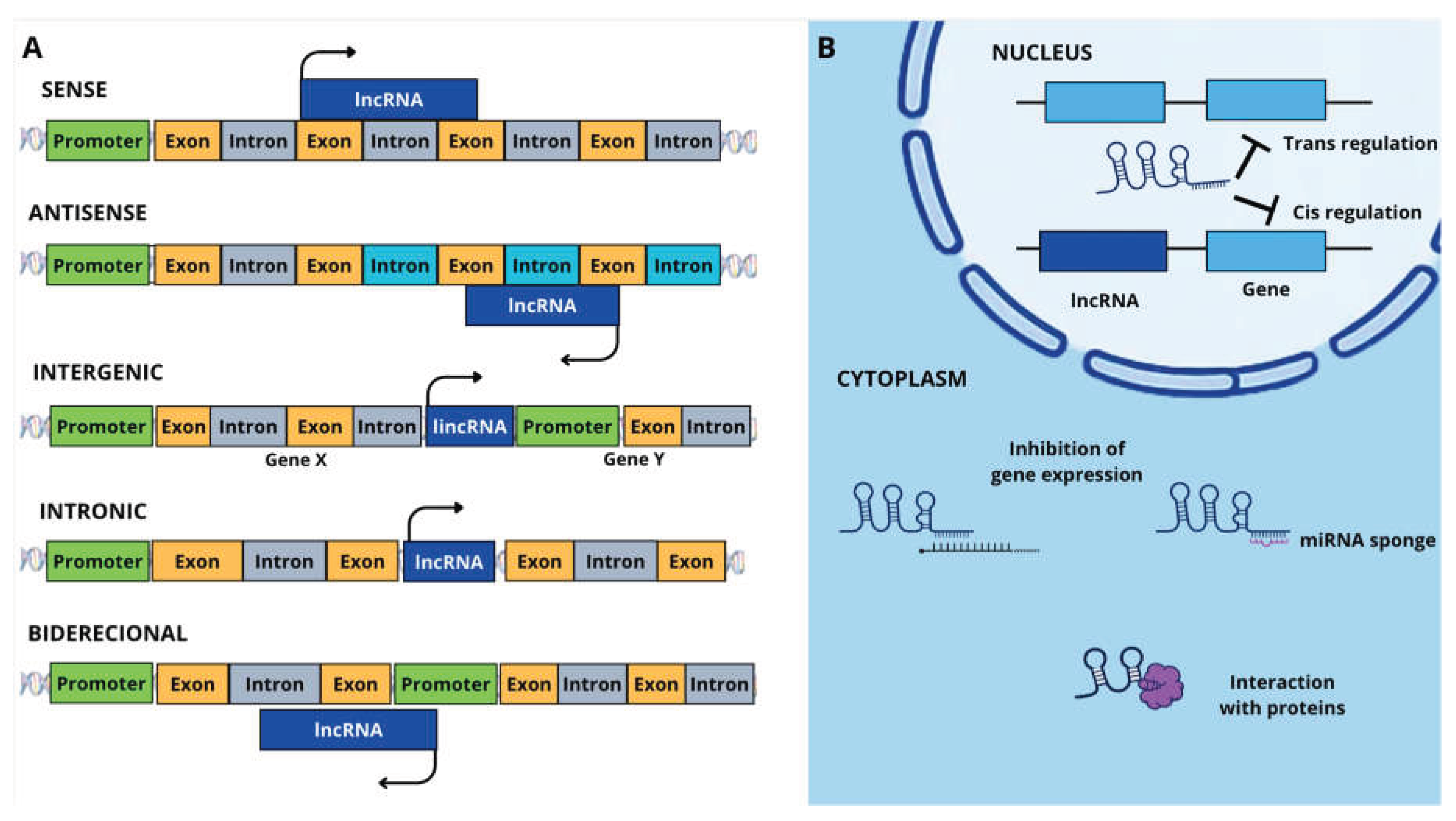

Table 1.
Summary of the main reported lncRNAs and their associated roles across reproductive processes in fish.
Table 1.
Summary of the main reported lncRNAs and their associated roles across reproductive processes in fish.
| Species | Biological Process | lncRNA | Main Finding | Reference |
|---|---|---|---|---|
|
Danio rerio (Zebrafish) |
Primordial germ cell migration | lnc172 | Overexpression caused abnormal dispersion of PGCs, indicating role in genital ridge migration | [27] |
|
Danio rerio (Zebrafish) |
Germ cell development | lnc304 | Localized in germ cells and associated with gonadal germline development |
[27] |
|
Oryzias latipes (Medaka) |
Spermatogenesis / sperm production | lnc4c | Functions as pri-miRNA of miR-202; deletion reduced sperm viability and germ cell number | [24] |
|
Oncorhynchus mykiss (Rainbow trout) |
Meiotic regulation | MSTRG.74687.3 MSTRG.76205.1 MSTRG.30919.1 | Indirect regulation of ccne1 affecting meiosis and G2/M transition | [28] |
|
Oncorhynchus mykiss (Rainbow trout) |
Gonadal cell proliferation | MSTRG.74687 | Associated with cyclin regulation during gametogenesis | [34] |
|
Cynoglossus semilaevis (Chinese tongue sole) |
Oocyte maturation | XR_522278.2 XR_522171.2 | Co-localized with steroidogenic genes involved in maturation pathways | [50] |
|
Cynoglossus semilaevis (Chinese tongue sole) |
Ovulation / follicular remodeling | XR_522498.1 XR_522499.1 XR_523405.2 | Associated with ECM remodeling, differentiation and ovulatory pathways | [50] |
|
Trachinotus ovatus (Golden pompano) |
Oocyte meiosis | MSTRG.66521.1 MSTRG.49969.1 MSTRG.59923.1 MSTRG.13767.1 |
Associated with cell cycle and meiotic pathways after IGF stimulation | [23] |
|
Trachinotus ovatus (Golden pompano) |
Steroidogenesis | MSTRG.20896.2 MSTRG.58123.2 |
Co-regulated with steroidogenic genes including cyp19a1 and cyp17a | [23] |
|
Scatophagus argus (Spotted scat) |
Meiotic entry / cell cycle | MSTRG.30211.1 | Associated with ccnd2 and ska2 regulating meiotic progression | [77] |
|
Scatophagus argus (Spotted scat) |
Oocyte hydration / ECM remodeling | MSTRG.30211.1 | Correlated with hydration genes and extracellular matrix remodeling | [77] |
|
Scatophagus argus (Spotted scat) |
DNA replication / cell division | MSTRG.3260.6 | Associated with DNA replication and cell proliferation pathways | [77] |
|
Monopterus albus (Ricefield eel) |
Hormonal regulation of ovarian growth | LOC109960696 | Fsh/hCG responsive lncRNA associated with Smad2 regulation | [172] |
|
Monopterus albus (Ricefield eel) |
Gonadal sex reversal | MSTRG.38036 MSTRG.12998 | Expression changes during ovary→ovotestis→testis transition | [101] |
|
Monopterus albus (Ricefield eel) |
Epigenetic regulation during sex reversal | MSTRG.38036 MSTRG.12998 | DNA methylation negatively correlated with lncRNA expression | [101] |
|
Takifugu rubripes (Japanese pufferfish) |
Steroid-induced masculinization | lncRNA000338 | [94] | |
|
Takifugu rubripes (Japanese pufferfish) |
Steroidogenesis during sex differentiation | lnc_000338 | Predicted target association with gsdf, in male undifferentiated gonads | [93] |
|
Takifugu rubripes (Japanese pufferfish) |
Masculinization / male pathway activation | MSTRG.13468.1 MSTRG.17819.1 XR_003886055.1 XR_003886586.1 XR_003887080.1 XR_003887947.1 |
Predicted targets and regulators associated with dmrt1 | [93] |
|
Takifugu rubripes (Japanese pufferfish) |
Testicular differentiation | MSTRG.3717.2 XR_003885862.1 XR_003888314.1 XR_003890080.1 | Predicted regulator associated with gsdf/amhr2 pathways | [93] |
|
Takifugu rubripes (Japanese pufferfish) |
Sex differentiation | lnc_000690 lnc_000370 XLOC_021951 XR_965485.1 | Predicted regulator associated with gsdf network | [95] |
|
Takifugu rubripes (Japanese pufferfish) |
Female pathway maintenance | XR_003885863.1 XR_003888756.1 XR_003888884.1 XR_003889260.1 XR_003889849.1 XR_003890064.1 XR_003890751.1 XR_003890915.1 XR_003891384.1 | Associated with female-related genes including steroidogenesis pathways | [93] |
|
Cynoglossus semilaevis (Chinese tongue sole) |
Ovarian development, maturation and ovulation | MSTRG.24810.1 MSTRG.3094.1 MSTRG.5569.1 MSTRG.9796.1 MSTRG.23232.1 MSTRG.7061.1 |
Potential ceRNA regulation of female-biased growth | [77] |
|
Cynoglossus semilaevis (Chinese tongue sole) |
Ovarian differentiation / reproductive regulation | XR_522278.2, XR_522171.2 XR_522957 XR_522498.1 XR_522499.1 XR_523405.2 | lncRNAs associated with steroidogenesis, follicular differentiation, ovulation and reproductive pathways through cis/trans/ceRNA regulation | [50] |
|
Salmo salar (Atlantic salmon) |
Spermatogonial maintenance | MSTRG.73942 | Associated with Wnt pathway and immature testes | [34] |
|
Salmo salar (Atlantic salmon) |
Male fertility / sperm maintenance | MSTRG.73143 | Associated with FGL1 and mature testicular function | [34] |
Disclaimer/Publisher’s Note: The statements, opinions and data contained in all publications are solely those of the individual author(s) and contributor(s) and not of MDPI and/or the editor(s). MDPI and/or the editor(s) disclaim responsibility for any injury to people or property resulting from any ideas, methods, instructions or products referred to in the content. |
© 2026 by the authors. Licensee MDPI, Basel, Switzerland. This article is an open access article distributed under the terms and conditions of the Creative Commons Attribution (CC BY) license (http://creativecommons.org/licenses/by/4.0/).
Copyright: This open access article is published under a Creative Commons CC BY 4.0 license, which permit the free download, distribution, and reuse, provided that the author and preprint are cited in any reuse.