

Submitted:
10 September 2026
Posted:
11 September 2026
You are already at the latest version
Abstract
Background and Aim: HDV infection represents one of the most severe forms of chronic viral hepatitis, characterized by rapid fibrosis progression and poor long-term outcomes, and is associated with a markedly increased likelihood of developing liver cirrhosis, hepatocellular carcinoma (HCC), and hepatic decompensation. Metabolic dysfunction-associated steatotic liver disease (MASLD), now recognized as the most common chronic liver disorder globally, has been proposed to amplify hepatic injury through overlapping metabolic and inflammatory mechanisms when concurrent with viral hepatitis. To date, however, systematic data on the co-occurrence of MASLD and metabolic dysfunction-associated steatohepatitis (MASH) in the setting of chronic HDV infection are remarkably scarce. Accordingly, the present study aimed to characterize the prevalence and severity of MASLD and MASH, as assessed by transient elastography (FibroScan), in a cohort of patients with chronic HDV infection. Methods: A total of 125 anti-HDV seropositive patients were enrolled in this cross-sectional study . HDV-RNA quantification was performed in all participants. Liver stiffness measurement (LSM, kPa) and controlled attenuation parameter (CAP, dB/m) were obtained via FibroScan. Results: The mean patient age was 39.0 years; 74.4% of participants were male (n=93). Of all the patients, 68% (n=85) had received pegylated interferon-alpha (peg-IFN-alpha) therapy for at least 12 months; however, sustained virological response (SVR) was achieved in only 14.1% (n=12). The overall mean LSM was 9.1 kPa, while the mean CAP was 228 dB/m. Liver cirrhosis was identified in 36.8% of patients (n=46; mean LSM 16.2 kPa), with 17.4% of cirrhotic patients (n=8) classified as decompensated and referred for liver transplantation evaluation. HCC was detected in 1.6% (n=2). MASLD was present in 24.0% of patients (n=30; mean CAP 289 dB/m) and MASH in 8.8% (n=11). LSM values were significantly higher in the cirrhotic group compared with non-cirrhotic patients (p< 0.001). No statistically significant difference in CAP was observed between cirrhotic and non-cirrhotic groups (p=0.121). Conclusion: Chronic HDV infection carries a heavy burden of cirrhosis and HCC, further compounded by markedly limited therapeutic efficacy. The presence of MASLD in approximately one in four patients, and MASH in nearly one in ten, underscores the necessity of routine FibroScan-based metabolic liver assessment in this high-risk population.
Keywords:
hepatitis delta virus
; FibroScan
; MASLD
; MASH
; liver cirrhosis
; liver stiffness measurement
1. Introduction
Hepatitis delta virus (HDV) is a defective single-stranded RNA agent that requires the hepatitis B virus (HBV) surface antigen (HBsAg) as its outer envelope and therefore exclusively infects HBV-carrying individuals, either as simultaneous co-infection or subsequent superinfection [1]. Blach et al. estimated that approximately 4.5% of HBsAg-positive individuals globally carry anti-HDV antibodies, translating to an estimated 70 million individuals with anti-HDV seropositivity, of whom a substantial proportion are estimated to harbour active viral replication [2,3,4]. A Polaris Observatory modelling study spanning 25 countries confirmed substantial geographic heterogeneity in HDV prevalence, with Mongolia recording the highest national estimates [4].
From a clinical standpoint, HDV co-infection accelerates the progression to liver cirrhosis by approximately threefold and doubles the risk of HCC when compared with HBV monoinfection [5]. HDV-RNA positivity has been independently associated with advanced fibrosis and cirrhosis in multiple cohort studies [6]. Turkey is recognized as an endemic region for both HBV and HDV infection; a meta-analysis published in 2024 reported an anti-HDV seroprevalence of 3.37% among HBsAg-positive blood donors and considerably higher rates among patients with established cirrhosis [7]. The southeastern Anatolian region has historically demonstrated among the highest rates of HBV/HDV co-infection in Turkey [7,8], positioning Diyarbakir as a clinically relevant study site.
Metabolic dysfunction-associated steatotic liver disease (MASLD) currently affects an estimated 30-32% of the global adult population [9,10], making it the single most common cause of chronic liver disease worldwide. The more advanced form, metabolic dysfunction-associated steatohepatitis (MASH), independently drives fibrosis progression, cirrhosis, and HCC development. The MASLD nomenclature was formally established in 2023 through a multisociety Delphi consensus statement, replacing the older non-alcoholic fatty liver disease (NAFLD) terminology [11]. The co-existence of MASLD and chronic viral hepatitis is thought to potentiate hepatic fibrogenesis through overlapping metabolic and immune-mediated injury pathways [12].
Vibration-controlled transient elastography (VCTE; FibroScan, Echosens, Paris, France) provides simultaneous non-invasive assessment of hepatic fibrosis via . Liver stiffness measurement (LSM, kPa) and steatosis via controlled attenuation parameter (CAP, dB/m), and is endorsed as a first-line evaluation tool by current major international guidelines [13,14]. Despite this, studies simultaneously evaluating both fibrosis and steatosis in HDV-infected patients using FibroScan are exceptionally limited. The present investigation, therefore, aimed to fill this gap by systematically characterizing FibroScan-derived LSM and CAP parameters in a well-defined cohort of patients with serologically confirmed chronic HDV infection.
2. Materials and Methods
2.1. Study Design and Patient Selection
This cross-sectional observational study was conducted at the Infectious Diseases Department of the Faculty of Medicine Hospital, Dicle University, Diyarbakir, Turkey. The study received approval from the Dicle University Faculty of Medicine Ethics Committee (Approval No: TIP.23.022) and was carried out in accordance with the principles of the Declaration of Helsinki. Written informed consent was obtained from all participants. A total of 125 anti-HDV seropositive patients presenting between March 2020 and June 2023 were enrolled.
Inclusion criteria were: age 18 years or older, documented anti-HDV seropositivity of at least six months duration, and volunteer to participate.
Exclusion criteria were: hepatitis C virus (HCV) co-infection, HIV co-infection, prior liver transplantation, excessive alcohol use (>140 g/week in women; >210 g/week in men), active malignancy, and conditions precluding reliable elastography (severe obesity, ascites).
2.2. Clinical and Laboratory Assessment
Quantitative HDV-RNA was measured by a validated commercial real-time RT-PCR assay in all patients. Cardiometabolic risk factors were defined according to the 2023 Delphi consensus: type 2 diabetes (FBG >6.9 mmol/L or HbA1c >6.4% or antidiabetic therapy), prediabetes, increased waist circumference (≥102 cm in men, ≥88 cm in women), hypertension (BP ≥130/85 mmHg or antihypertensive therapy), and dyslipidaemia (TG ≥1.70 mmol/L or low HDL or lipid-lowering therapy).
2.3. FibroScan Protocol
All FibroScan examinations were performed by a certified operator following a minimum two-hour fast. The M probe was used when the skin-to-capsule distance was <2.5 cm, and the XL probe when ≥2.5 cm. A minimum of ten valid measurements were obtained per patient; a success rate ≥60% and IQR/median ratio ≤0.30 were required for a result to be considered reliable [13]. LSM was reported in kPa and CAP in dB/m. Fibrosis staging followed established LSM thresholds: F0-F1 (<7 kPa), F2 (7.0-9.4 kPa), F3 (9.5-12.4 kPa), and F4/cirrhosis (≥12.5 kPa) [13,15]. Hepatic steatosis was defined as CAP ≥248 dB/m; MASLD diagnosis additionally required the presence of at least one cardiometabolic risk factor as per the 2023 multisociety criteria [11]. Given the absence of liver biopsy data, MASH was operationally defined as MASLD with LSM ≥7.2 kPa and/or elevated alanine aminotransferase (ALT), consistent with published surrogate definitions [11,16].
2.4. Statistical Analysis
All analyses were performed using R statistical software (version 4.6.0; R Foundation for Statistical Computing, Vienna, Austria). Figures were produced with the ggplot2 package (version 3.5.1). Continuous variables are expressed as mean ± standard deviation (SD) or median (IQR) as appropriate; categorical variables are reported as number and percentage (%). After confirming distributional assumptions with the Shapiro-Wilk test, between-group comparisons were made using the Mann-Whitney U test. Categorical variables were examined with the chi-square test or, where appropriate, Fisher’s exact test. A two-tailed p-value <0.05 was considered statistically significant.
3. Results
3.1. Demographic Characteristics
One hundred and twenty-five patients were enrolled (93 male, 32 female; 74.4% male predominance); the mean age was 39.0 years. Anti-HDV positivity was confirmed in all participants, and HDV-RNA was requested for each. Detectable HDV-RNA was found in 113 patients (90.4%), while 12 patients (9.6%) had undetectable HDV-RNA—all of whom had previously received peg-IFN therapy and achieved sustained virological clearance (Figure 1).
3.2. Treatment Status and SVR
Sixty-eight percent of patients (n=85) had received peg-IFN-alpha-2a for at least 12 months; the remaining 32% (n=40) had not received any anti-HDV treatment. Of the 85 treated patients, only 14.1% (n=12) achieved sustained HDV-RNA negativity(Figure 1).
3.3. FibroScan Findings
Across all patients, mean LSM was 9.1 kPa (SD ±5.8) and mean CAP was 228 dB/m (SD ±42). The wide SD for LSM reflects the bimodal distribution of the cohort, comprising both cirrhotic and non-cirrhotic patients. By LSM-based staging, 34.4% of patients were classified as F0-F1, 14.4% as F2, 14.4% as F3, and 36.8% as F4 (cirrhosis). Mean LSM in the cirrhotic group was 16.2 kPa versus 6.0 kPa in the non-cirrhotic group, a difference that was statistically significant (p<0.001). In contrast, CAP values did not differ significantly between cirrhotic and non-cirrhotic patients (p=0.121) (Figure 2, Figure 3, Figure 4 and Figure 5).
3.4. Cirrhosis and Clinical Complications.
Cirrhosis was identified in 36.8% of patients (n=46). Among cirrhotic patients, 17.4% (n=8) met criteria for clinical decompensation and were referred for liver transplantation evaluation. HCC was confirmed in 1.6% (n=2) of the total cohort; both HCC cases arose in patients with established cirrhosis and were diagnosed by radiological and biochemical criteria (Figure 1).
3.5. MASLD and MASH Prevalence
MASLD criteria were fulfilled in 24.0% of patients (n=30; mean CAP 289 dB/m). This was markedly higher than the mean CAP recorded in MASLD-negative patients (200 dB/m). MASH was identified in 8.8% of the cohort (n=11). A significant difference in CAP between cirrhotic and non-cirrhotic patients (p=0.121) raises the possibility that hepatic steatosis in this cohort may not be driven solely by cirrhosis-related changes, though this warrants prospective confirmation (Figure 6, Figure 7 and Figure 8).
3.6. Summary of Clinical Findings (Table 1)
| Parameter | Value | Rate / Mean | p-value |
| Total Patients | n = 125 | — | — |
| Male Sex | n = 93 | 74.4% | — |
| Mean Age | 39.0 years | — | — |
| Mean LSM | 9.1 kPa | — | — |
| Mean CAP | 228 dB/m | — | — |
| Liver Cirrhosis | n = 46 | 36.8% | — |
| Mean LSM in Cirrhotic Group | 16.2 kPa | — | <0.001 |
| Decompensated Cirrhosis | n = 8 | 17.4%* | — |
| HCC | n = 2 | 1.6% | — |
| Transplant Waitlist | n = 8 | 6.4% | — |
| MASLD | n = 30 | 24.0% | — |
| Mean CAP in MASLD Group | 289 dB/m | — | — |
| MASH | n = 11 | 8.8% | — |
| Peg-IFN Treatment | n = 85 | 68.0% | — |
| SVR (HDV-RNA undetectable) | n = 12 | 14.1%** | — |
| CAP: Cirrhotic vs Non-cirrhotic | — | — | 0.121 (NS) |
4. Discussion
The present study offers one of the few Turkish cohort analyses simultaneously evaluating liver fibrosis and metabolic steatosis using FibroScan in a sizeable group of HDV-infected patients. Our results converge on three main observations: the substantial burden of cirrhosis and HCC, the persistently poor SVR rates with peg-IFN therapy, and the clinically meaningful prevalence of MASLD/MASH as a concurrent metabolic comorbidity.
4.1. SVR, Cirrhosis and HCC Burden
Of the 85 treated patients, only 14.1% (n=12) achieved sustained HDV-RNA negativity. This rate is at the lower boundary of the SVR range reported for standard peg-IFN in the published literature [17,18] (Figure 1).
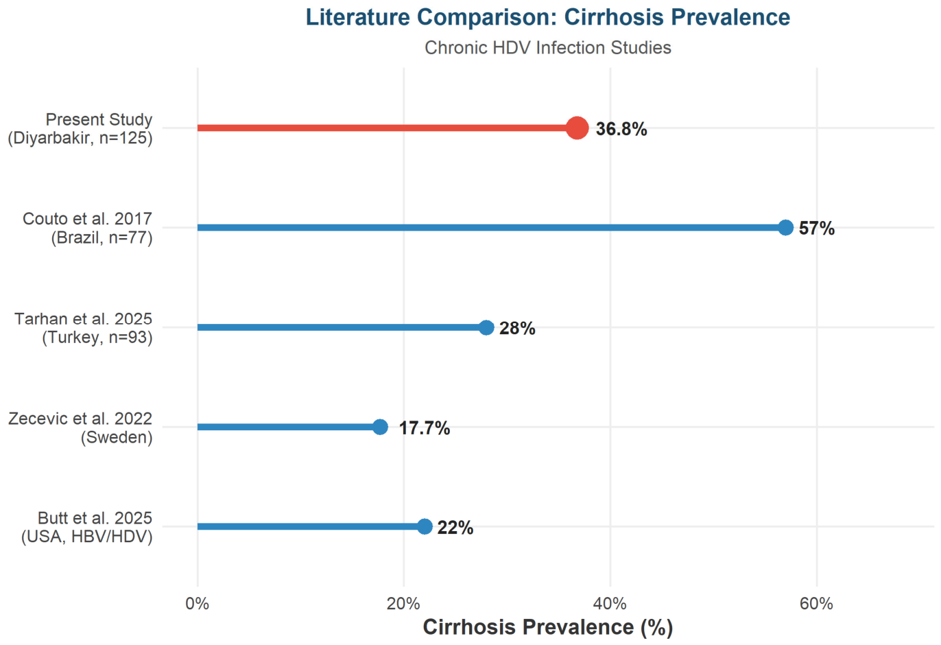
A cirrhosis prevalence of 36.8% in our series is consistent with the known severity of HDV-related liver disease. Couto and colleagues reported compensated advanced chronic liver disease (c-ACLD, defined as LSM ≥15 kPa) in 57% of 77 HDV-infected patients in Brazil [19]; the somewhat lower cirrhosis rate in our cohort likely reflects our use of the conventional LSM ≥12.5 kPa threshold. Dietz-Fricke et al. documented that HCC developed exclusively in patients with pre-existing cirrhosis within a longitudinal German cohort [20]. Tarhan and colleagues, in a 2025 retrospective Turkish cohort of 93 patients, reported a cirrhosis rate of 28% and similarly noted that peg-IFN therapy failed to prevent cirrhosis or HCC in a sizeable proportion of patients [21]. In a large retrospective analysis of US Veterans Health Administration data, Butt et al. reported a cirrhosis incidence rate of 1.03 per 100 person-years in HBV/HDV co-infected individuals, significantly exceeding that of HBV monoinfection [22]. The finding of decompensated cirrhosis in 17.4% of cirrhotic patients in our series, with eight patients already listed for transplantation, points to the need for more effective therapeutic options (Figure 6 and Figure 8).
4.2. Limitations of Pegylated Interferon and Emerging Therapies
The SVR rate of 14.1% observed with peg-IFN in our study aligns with the lower end of rates summarised in published meta-analyses [17]. The LIMT-1 Phase II trial with pegylated interferon lambda demonstrated a somewhat higher virological response of 36% at the doses tested [24], indicating that interferon type and dosing may influence outcomes. A notable recent advance in HDV therapeutics has been bulevirtide (BLV), an entry inhibitor targeting the NTCP receptor, which received full European Medicines Agency approval in May 2023. In the pivotal MYR301 Phase III trial, BLV significantly outperformed tenofovir monotherapy in terms of combined virological and biochemical response at 96 weeks [25,26]. Real-world data from the ARISTOTLE pilot observational study were consistent with these findings, with 54.6% of 108 patients achieving virological response at six months [27]. The limited availability of BLV in Turkey at the time of the present study likely contributed to the low SVR rates observed [28].
4.3. MASLD and MASH as Concurrent Metabolic Comorbidities
A MASLD prevalence of 24.0% in our HDV cohort is clinically significant when considered alongside the global adult prevalence of approximately 30% [9]. The observation that CAP did not differ significantly between cirrhotic and non-cirrhotic patients (p=0.121) is particularly noteworthy: the absence of such a difference raises the possibility that hepatic steatosis may not be driven solely by cirrhosis-related changes in this population. A contemporary review examining the intersection of MASLD and viral hepatitis highlighted that converging metabolic and viral fibrogenic pathways can synergistically accelerate disease [12]. Analogous findings in HCV-related liver disease have shown that MASLD co-existence confers an adjusted odds ratio of 2.29 for clinically significant fibrosis compared with steatosis alone [29]; whether similar risk amplification applies to HDV co-infection with MASLD remains to be established in prospective studies (Figure 5, Figure 6 and Figure 7).
The detection of MASH in 8.8% of patients indicates that active necroinflammation beyond simple steatosis is present in a notable proportion of this cohort. As HDV itself induces marked intrahepatic inflammation, the synergistic interaction between HDV-driven hepatitis and MASH-associated necroinflammation may represent a particularly aggressive phenotype that merits dedicated prospective study [12,30].
4.4. Strengths and Limitations
Strengths of this study include a comparatively large, geographically homogeneous cohort from a high-endemic region, uniform FibroScan assessment of both LSM and CAP in all patients, and application of the current 2023 Delphi MASLD consensus criteria for diagnosis. Limitations include the absence of individual-level raw data precluding multivariable regression and ROC analyses, the cross-sectional design, which prevents causal inference, the operational definition of MASH in the absence of liver biopsy data, and the heterogeneity of treatment history across patients. Future prospective studies incorporating patient-level data should aim to identify independent predictors of MASLD in HDV-infected patients and evaluate the impact of bulevirtide on both fibrosis and steatosis trajectories.
5. Conclusion
Our findings document that chronic HDV infection carries a substantial burden of cirrhosis and HCC alongside persistently low therapeutic success rates. The concurrent presence of MASLD in nearly one quarter and MASH in nearly one tenth of patients draws attention to the metabolic dimension of liver disease in this vulnerable population. Although no significant difference in CAP was observed between cirrhotic and non-cirrhotic patients, the high rates of cirrhosis (36.8%) and the presence of MASLD in one in four patients collectively support the value of periodic FibroScan monitoring—primarily for fibrosis surveillance, and secondarily for metabolic steatosis detection—in all patients with chronic HDV infection. Expanding access to bulevirtide and other novel agents in endemic regions such as southeastern Turkey should be considered a clinical and public health imperative.
References
- Rizzetto, M.; Canese, M.G.; Arico, S.; et al. Immunofluorescence detection of a new antigen-antibody system (delta/anti-delta) associated to hepatitis B virus in liver and serum of HBsAg carriers. Gut 1977, 18(12), 997–1003. [Google Scholar] [CrossRef] [PubMed]
- Chen, H.Y.; Shen, D.T.; Ji, D.Z.; et al. Prevalence and burden of hepatitis D virus infection in the global population: a systematic review and meta-analysis. Gut 2019, 68(3), 512–521. [Google Scholar] [CrossRef] [PubMed]
- Stockdale, A.J.; Kreuels, B.; Henrion, M.Y.R.; et al. The global prevalence of hepatitis D virus infection: systematic review and meta-analysis. J. Hepatol. 2020, 73(3), 523–532. [Google Scholar] [CrossRef] [PubMed]
- Blach, S.; Kondili, L.A.; Aghemo, A.; et al. Adjusted estimate of the prevalence of hepatitis delta virus in 25 countries and territories. J. Hepatol. 2024, 80(3), 432–441. [Google Scholar] [CrossRef] [PubMed]
- Yurdaydin, C.; Abbas, Z.; Basaranoglu, M.; et al. EASL clinical practice guidelines: management of hepatitis D. J. Hepatol. 2023, 79(3), 674–699. [Google Scholar] [CrossRef] [PubMed]
- Dietz-Fricke, C.; Versiani Matos, N.; Zecevic, M.; et al. High rates of cirrhosis and severe clinical events in patients with HBV/HDV co-infection: longitudinal analysis of a German cohort. BMC Infect. Dis. 2020, 20(1), 45. [Google Scholar] [CrossRef] [PubMed]
- Toy, M.; Guler, B.; Somay, K.; Gencdal, G.; Yurdaydin, C. Hepatitis delta virus infection in Turkey: a meta-analysis of prevalence. Int. J. Infect. Dis. Reg. 2024. [Google Scholar] [CrossRef] [PubMed]
- Celen, M.K.; Ayaz, C.; Hosoglu, S.; et al. Prevalence of hepatitis delta virus (HDV) infection in chronic hepatitis B patients in eastern Turkey. Eur. J. Intern Med. 2011, 22(1), 98–101. [Google Scholar]
- Riazi, K.; Azhari, H.; Charette, J.H.; et al. The prevalence and incidence of NAFLD worldwide: a systematic review and meta-analysis. Lancet Gastroenterol. Hepatol. 2022, 7(9), 851–861. [Google Scholar] [CrossRef] [PubMed]
- Le, M.H.; Yeo, Y.H.; Zou, B.; et al. Forecasted 2040 global prevalence of nonalcoholic fatty liver disease using hierarchical Bayesian approach. Clin. Mol. Hepatol. 2022, 28(4), 841–850. [Google Scholar] [CrossRef] [PubMed]
- Rinella, M.E.; Lazarus, J.V.; Ratziu, V.; et al. A multisociety Delphi consensus statement on new fatty liver disease nomenclature. Ann. Hepatol. 2023, 28(4), 101133. [Google Scholar] [CrossRef] [PubMed]
- Higuera-de-la-Tijera, F.; Castro-Narro, G.E.; Velarde-Ruiz Velasco, J.A.; et al. When metabolic dysfunction-associated steatotic liver disease meets viral hepatitis. J. Clin. Med. 2025, 14(10), 3422. [Google Scholar] [CrossRef] [PubMed]
- Castera, L.; Friedrich-Rust, M.; Loomba, R. Noninvasive assessment of liver disease in patients with nonalcoholic fatty liver disease. Gastroenterology 2019, 156(5), 1264–1281. [Google Scholar] [CrossRef] [PubMed]
- EASL-EASD-EASO Clinical Practice Guidelines on the management of metabolic dysfunction-associated steatotic liver disease (MASLD). J. Hepatol. 2024, 81(3), 492–542. [CrossRef] [PubMed]
- Castera, L.; Foucher, J.; Bernard, P.H.; et al. Pitfalls of liver stiffness measurement: a 5-year prospective study of 13,369 examinations. Hepatology 2010, 51(3), 828–835. [Google Scholar] [CrossRef] [PubMed]
- Newsome, P.N.; Sasso, M.; Deeks, J.J.; et al. FibroScan-AST (FAST) score for the non-invasive identification of patients with non-alcoholic steatohepatitis with significant activity and fibrosis: a prospective derivation and global validation study. Lancet Gastroenterol. Hepatol. 2020, 5(4), 362–373. [Google Scholar] [CrossRef] [PubMed]
- Ozer, B.; Serin, E.; Gumurdulu, Y.; et al. Standard and pegylated interferon therapy of HDV infection: a systematic review and meta-analysis. J. Gastroenterol. Hepatol. 2013, 28(5), 804–809. [Google Scholar]
- Anastasiou, O.E.; Carunufu, F.A.; Curescas, M.G.; et al. Five-year follow-up of 96 weeks peginterferon plus tenofovir disoproxil fumarate in hepatitis D. Liver Int. 2024, 44(1), 139–147. [Google Scholar] [CrossRef] [PubMed]
- Couto, I.; Victoria, M.; Veloso, V.G.; et al. Prevalence and predictors for compensated advanced chronic liver disease (c-ACLD) in patients with chronic hepatitis delta virus (HDV) infection. PLoS ONE 2017, 12(3), e0174453. [Google Scholar] [CrossRef] [PubMed]
- Dietz-Fricke, C.; Versiani Matos, N.; Zecevic, M.; et al. High rates of cirrhosis and severe clinical events in patients with HBV/HDV co-infection: longitudinal analysis of a German cohort. BMC Infect. Dis. 2020, 20(1), 45. [Google Scholar] [CrossRef] [PubMed]
- Tarhan, F.; Karatayli, E.; Yalcin, K.; et al. Long-term outcomes of hepatitis delta patients including cirrhosis and hepatocellular carcinoma. J. Infect. Dev. Ctries. 2025, 19(1), 146–154. [Google Scholar] [PubMed]
- Butt, A.A.; Yan, P.; Shaikh, O.S.; Bedimo, R. Screening rates and clinical outcomes for hepatitis delta in individuals with HBV coinfection. Open Forum Infect. Dis. 2025, 12(1), ofae631. [Google Scholar] [CrossRef]
- Zecevic, M.; Dietz-Fricke, C.; Kaita, K.; et al. Shear wave elastography: how well does it perform in chronic hepatitis D virus infection? Dig. Liver Dis. 2022, 55(1), 78–84. [Google Scholar]
- Etzion, O.; Hamid, S.; Lurie, Y.; et al. End of study results from LIMT HDV study: 36 weeks peg-interferon lambda monotherapy leads to durable virologic suppression in patients with chronic hepatitis delta virus infection. Liver Int. 2023, 43(1), 90–100. [Google Scholar]
- Wedemeyer, H.; Aleman, S.; Brunetto, M.R.; et al. A phase 3, randomized trial of bulevirtide in chronic hepatitis D. N Engl. J. Med. 2023, 389(1), 22–32. [Google Scholar] [CrossRef] [PubMed]
- Lampertico, P.; Brunetto, M.R.; Aleman, S.; et al. Bulevirtide monotherapy or in combination for chronic hepatitis delta: 2025 update. Liver Int. 2025, 45(6), e16068. [Google Scholar] [CrossRef] [PubMed]
- Facchetti, F.; Binda, M.; Guerini, F.R.; et al. Assessment of response and safety of bulevirtide treatment in patients with chronic delta virus infection: the ARISTOTLE pilot observational study. Int. J. Mol. Sci. 2025, 26(4), 1498. [Google Scholar]
- Lampertico, P.; Brunetto, M.R.; Buti, M.; et al. Bulevirtide monotherapy in patients with chronic HDV: efficacy and safety results through week 96 from a phase III randomized trial. J. Hepatol. 2024, 82(2), 283–291. [Google Scholar] [CrossRef] [PubMed]
- Elgretli, W.; Chen, T.; Sebastiani, G. Association of MASLD phenotypes with liver fibrosis in hepatitis C: the role of cardiometabolic risk factors. Aliment Pharmacol. Ther. 2025, 61(7), 1189–1199. [Google Scholar]
- Stasi, C.; Silvestri, C.; Voller, F. MASLD in the context of viral hepatitis: emerging evidence and clinical implications. Biomedicines 2024, 12(11), 2534. [Google Scholar]
- Lacombe, K.; Nicand, E.; Velay, A.; et al. Regular screening for hepatitis delta virus among chronic hepatitis B carriers improves the diagnostic of this infection and subsequent cirrhosis development. J. Viral Hepat. 2024, 31(5), 285–293. [Google Scholar]
Figure 1.
Clinical complications in patients with chronic HDV infection (n=125). The proportions of cirrhosis, decompensated cirrhosis, hepatocellular carcinoma (HCC), and transplant waitlist listing are presented.
Figure 1.
Clinical complications in patients with chronic HDV infection (n=125). The proportions of cirrhosis, decompensated cirrhosis, hepatocellular carcinoma (HCC), and transplant waitlist listing are presented.
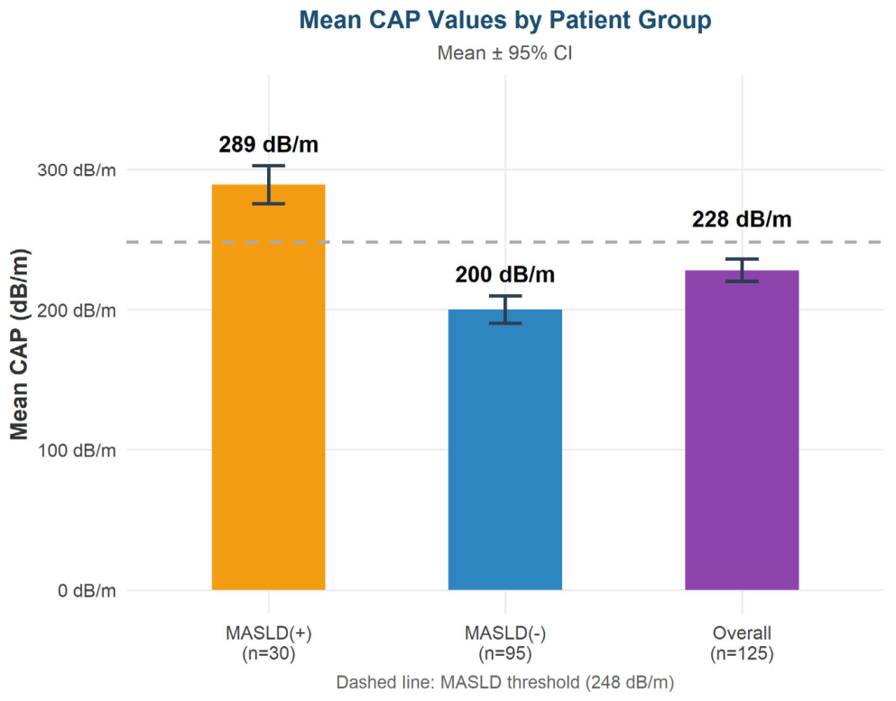
Figure 2.
Mean liver stiffness measurement (LSM) values by patient subgroup. Error bars represent 95% confidence intervals. The dashed red line indicates the cirrhosis threshold of 12.5 kPa.
Figure 2.
Mean liver stiffness measurement (LSM) values by patient subgroup. Error bars represent 95% confidence intervals. The dashed red line indicates the cirrhosis threshold of 12.5 kPa.
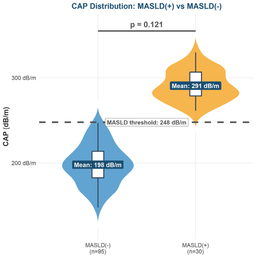
Figure 3.
LSM-based hepatic fibrosis stage distribution according to METAVIR scoring. F0-F1: <7 kPa;F2: 7-9.5 kPa; F3: 9.5-12.5 kPa; F4/Cirrhosis: ≥12.5 kPa.
Figure 3.
LSM-based hepatic fibrosis stage distribution according to METAVIR scoring. F0-F1: <7 kPa;F2: 7-9.5 kPa; F3: 9.5-12.5 kPa; F4/Cirrhosis: ≥12.5 kPa.
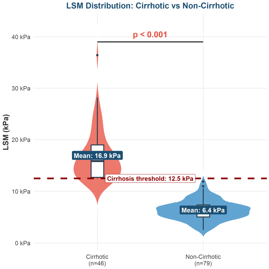
Figure 4.
Violin plot of LSM distribution stratified by cirrhosis status. The embedded box plot displays the interquartile range and median. The dark red dashed line marks the cirrhosis threshold (12.5 kPa). Labelled boxes indicate group means. Mann-Whitney U test: p<0.001.
Figure 4.
Violin plot of LSM distribution stratified by cirrhosis status. The embedded box plot displays the interquartile range and median. The dark red dashed line marks the cirrhosis threshold (12.5 kPa). Labelled boxes indicate group means. Mann-Whitney U test: p<0.001.
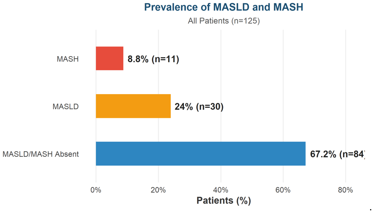
Figure 5.
Violin plot of CAP distribution stratified by MASLD status. The embedded box plot displays the interquartile range and median. The dashed line marks the MASLD threshold (248 dB/m). Labelled boxes indicate group means. Mann-Whitney U test: p=0.121 (not significant).
Figure 5.
Violin plot of CAP distribution stratified by MASLD status. The embedded box plot displays the interquartile range and median. The dashed line marks the MASLD threshold (248 dB/m). Labelled boxes indicate group means. Mann-Whitney U test: p=0.121 (not significant).
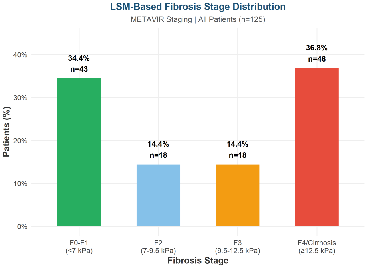
Figure 6.
Mean controlled attenuation parameter (CAP) values by patient subgroup. Error bars represent 95% confidence intervals. The dashed line indicates the MASLD steatosis threshold of 248 dB/m.
Figure 6.
Mean controlled attenuation parameter (CAP) values by patient subgroup. Error bars represent 95% confidence intervals. The dashed line indicates the MASLD steatosis threshold of 248 dB/m.
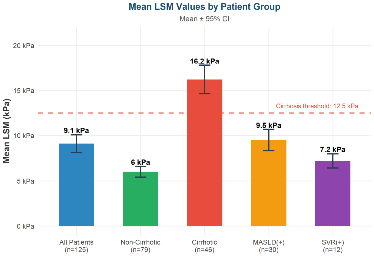
Figure 7.
Prevalence of MASLD and MASH among all patients (n=125), displayed as a horizontal bar chart.
Figure 7.
Prevalence of MASLD and MASH among all patients (n=125), displayed as a horizontal bar chart.
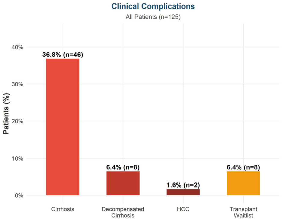
Figure 8.
Lollipop chart comparing cirrhosis prevalence rates across selected chronic HDV studies in the literature. The present study (red) is highlighted for direct comparison.
Figure 8.
Lollipop chart comparing cirrhosis prevalence rates across selected chronic HDV studies in the literature. The present study (red) is highlighted for direct comparison.
Disclaimer/Publisher’s Note: The statements, opinions and data contained in all publications are solely those of the individual author(s) and contributor(s) and not of MDPI and/or the editor(s). MDPI and/or the editor(s) disclaim responsibility for any injury to people or property resulting from any ideas, methods, instructions or products referred to in the content. |
© 2026 by the authors. Licensee MDPI, Basel, Switzerland. This article is an open access article distributed under the terms and conditions of the Creative Commons Attribution (CC BY) license.
Copyright: This open access article is published under a Creative Commons CC BY 4.0 license, which permit the free download, distribution, and reuse, provided that the author and preprint are cited in any reuse.