

Submitted:
09 September 2026
Posted:
11 September 2026
You are already at the latest version
Abstract
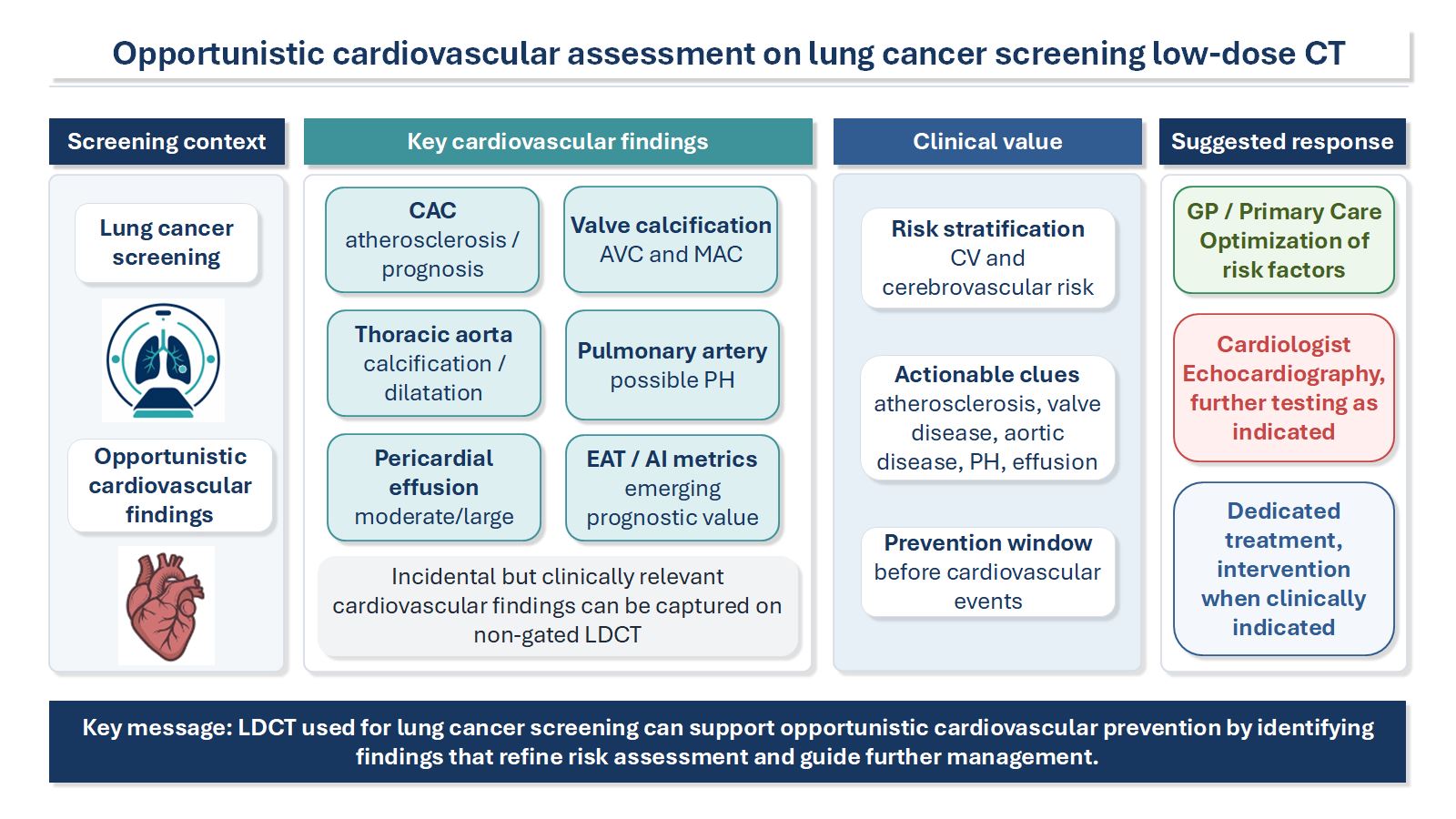
Population-based lung cancer screening with low-dose computed tomography (LDCT) is expanding across Europe, including a publicly funded Polish programme entering the guaranteed-benefits basket in October 2026. Because screening-eligible individuals share substantial tobacco-related cardiovascular risk, LDCT also provides an opportunity for cardiovascular risk assessment without additional radiation. We conducted a structured narrative review of PubMed literature, prioritising guidelines, major cohort studies, systematic reviews and cardiovascular outcome data, with targeted updates for recent regulatory and clinical developments. We summarise clinically actionable findings on non-contrast, non-ECG-gated LDCT: coronary artery calcification, aortic valve and mitral annular calcification, thoracic aortic disease, pericardial effusion, and pulmonary artery enlargement, as well as investigational cardiac volumetry, epicardial adipose tissue and AI-assisted quantification. Screening LDCT can provide robust risk-informative markers and pragmatic visual grading, but it cannot replace dedicated cardiac imaging or establish haemodynamic diagnoses. Standardised reporting, proportionate referral thresholds, and coordinated pathways linking radiology, pulmonology, primary care and cardiology are needed to translate incidental cardiovascular findings into scalable prevention while avoiding unnecessary downstream testing.
Keywords:
lung cancer screening
; low-dose computed tomography
; incidental cardiovascular findings
; coronary artery calcification
; cardiovascular risk assessment
; opportunistic screening
; atherosclerosis
; aortic calcification
; Poland
Introduction
Population-based lung cancer screening with low-dose computed tomography (LDCT) is expanding across Europe and globally, supported by robust evidence showing a reduction in lung cancer mortality among high-risk individuals. Europe’s Beating Cancer Plan and the subsequent EU Council Recommendation [1] on cancer screening have further encouraged Member States to evaluate and implement organised LDCT-based lung cancer screening programmes. Several European countries are therefore launching, piloting, or expanding national and regional screening initiatives. Poland is entering a decisive phase of this process. A regulation of the Minister of Health amending the regulation on guaranteed healthcare services, published on 22 July 2026, introduces a publicly funded lung cancer screening programme based on low-dose computed tomography (LDCT) into the national basket of guaranteed health services, with contracts for its delivery permitted from 1 October 2026, following a positive recommendation of the President of the Agency for Health Technology Assessment and Tariff System (AOTMiT, Recommendation No. 98/2025) [2,3]. The programme will primarily target adults aged 55–74 years with a smoking history of at least 20 pack-years who currently smoke or quit within the preceding 15 years, with extended eligibility for individuals aged 50–54 years who have additional risk factors [2]. The screening-eligible population is characterised by a high burden of tobacco exposure, which constitutes the dominant shared driver of both lung cancer and cardiovascular disease: the individuals most likely to attend lung cancer screening are also among those at highest cardiovascular risk.
The timing is pertinent in view of Poland’s disease burden. The Ministry of Health’s regulatory impact assessment accompanying the above regulation cites over 20,700 new bronchus and lung cancer cases in 2022 and over 26,000 deaths attributed to these cancers [4]. In parallel, cardiovascular disease remains the leading cause of death in Poland, with diseases of the circulatory system accounting for nearly 37% of all deaths in 2023 [5]. Smoking also amplifies cardiometabolic risk, including a higher likelihood of type 2 diabetes, further increasing overall cardiovascular risk in screening participants [6,7].
A crucial and often underutilised consequence of this overlap is that LDCT performed for lung cancer screening provides a pragmatic platform for opportunistic cardiovascular assessment without additional radiation or a separate imaging pathway. Cardiovascular abnormalities are among the most clinically consequential incidental findings encountered in lung cancer screening cohorts. In the NLST, coronary artery calcifications (CAC) were among the most frequently detected significant incidental findings [8,9], and existing reporting frameworks already anticipate this interface. American and European guidance supports a structured approach to incidental findings on low-dose CT. Commonly reported cardiovascular findings include coronary artery calcification, aortic valve calcification, ascending thoracic aortic dilatation, pericardial effusion, and pulmonary artery enlargement, with predefined thresholds used to guide referral and follow-up [10,11]. It should be noted that non-contrast, non-ECG-gated LDCT is not a substitute for dedicated cardiac imaging; cardiovascular information derived from it is mainly risk-informative rather than diagnostic.
With nationwide LDCT screening programmes entering implementation in multiple European countries, pulmonologists leading these programmes, alongside cardiologists, and primary care physicians, will increasingly encounter structured cardiovascular reports and need to understand their clinical implications. Translating incidental cardiovascular information into measurable benefit requires a shared understanding of what can and cannot be inferred from LDCT, pragmatic referral and follow-up thresholds, and clear pathways linking radiology reports to primary care and cardiovascular prevention. If designed thoughtfully, oncology-led screening programmes can serve as a vehicle for earlier cardiovascular risk identification and prevention at scale.
Aim and Scope of this Review
This narrative review aims to equip pulmonologists, and other clinicians involved in lung cancer screening programmes with a practical framework for opportunistic cardiovascular assessment on non-contrast, non-ECG-gated LDCT. We address the most robust and clinically actionable findings: coronary artery calcification, aortic valve calcification, mitral annular calcification, thoracic aortic disease, pericardial effusion and pulmonary artery enlargement, alongside investigational metrics and the emerging role of AI-assisted quantification. We discuss implications of the expanding landscape of population-based LDCT screening for translating opportunistic imaging findings into scalable cardiovascular prevention.
Literature Search and Methodology
This narrative review was conducted through a structured search of PubMed, supplemented by AI-assisted literature identification (Claude, Anthropic; ChatGPT, OpenAI) for predefined topical queries. Priority was given to clinical practice guidelines and multi-society position statements from major societies, randomised controlled trials and large prospective cohort studies, systematic reviews and meta-analyses, and large population-based datasets with cardiovascular outcomes data. The core structured search was conducted up to January 2026; the reference list was subsequently updated through September 2026 to incorporate key Polish regulatory documents relevant to implementation of the national screening programme. The final selection was limited to English-language sources, included to provide national context. AI tools were used only to assist literature identification and language editing; all cited references and substantive claims were independently checked by the authors against the original sources, and final inclusion decisions were made by the authors.
Technical Scope and Cardiovascular Findings of Screening LDCT
Before addressing individual findings, it is important to define what screening LDCT can and cannot reliably show. Low-dose CT in lung cancer screening is performed without intravenous contrast and without ECG gating, using protocols optimised for pulmonary parenchyma rather than cardiac anatomy [12,13,14]. Cardiovascular information from screening LDCT is therefore opportunistic and risk-informative rather than diagnostic [10,11,15]. It can identify macroscopic calcifications (e.g., coronary and valvular calcium) and approximate large-vessel calibre (e.g., ascending aorta, main pulmonary artery) [10,11,15,16], but it cannot reliably grade coronary stenosis, characterise non-calcified plaque, or determine aortic valve stenosis severity [12]. Accordingly, findings should be reported within structured frameworks and used to prompt cardiovascular risk assessment and targeted follow-up when appropriate, while avoiding overinterpretation and unnecessary downstream testing [10,11,15] (Table 1). The following sections address each major finding category in turn.
Coronary Artery Calcification on Screening LDCT
Coronary heart disease (CHD) may manifest catastrophically. In the Framingham cohort, 13-20% of coronary syndromes presented as sudden death depending on age, and approximately 60% of sudden CHD deaths occurred in individuals without previously recognized clinical heart disease [17]. Likewise, in Atherosclerosis Risk in Communities (ARIC) community surveillance, 32.6% of CHD deaths were sudden, and among those, 63.5% occurred in individuals without a prior CHD diagnosis, underscoring the potential public health value of opportunistic cardiovascular risk stratification in high-risk screening settings [18]. These observations provide the epidemiological rationale for opportunistic CAC detection in lung cancer screening populations, who share many of the same risk characteristics.
In lung cancer screening, coronary arteries are not the primary target of image optimisation. Because LDCT is non-contrast and non-ECG-gated, coronary segments may be partially blurred by cardiac motion, and image noise may be higher than in dedicated cardiac protocols [19,20]. These factors can reduce conspicuity of small or focal calcifications and, conversely, can make dense calcium appear more extensive because of partial-volume and blooming effects. As a result, agreement with dedicated ECG-gated calcium-scoring CT, while generally good, is not exact at the individual-patient level [20].
Visual CAC Assessment on LDCT: Methodology and Limitations
Current screening pathways favour visual, semi-quantitative CAC grading rather than formal Agatston quantification [10,11,15]. A widely adopted approach is a visual grade of none, mild, moderate, and severe, recommended for routine reporting on non-gated thoracic CT to communicate cardiovascular risk in a way that is feasible at scale [10,11,15]. An alternative method is ordinal vessel-based scoring, in which calcification is graded from 0 to 3 in each major coronary artery: the left main coronary artery, left anterior descending artery, right coronary artery, and left circumflex artery. The scores are summed to give a total score from 0 to 12, categorized as absent (0), mild (1–3), moderate (4–6), or severe (7–12) [21] (Figure 1).
From a clinical perspective, visual scoring systems may yield inconsistent results in individual patients. They are observer-dependent [20], and performance is weakest at the low end of disease burden, where partial-volume effects are most pronounced [22]. They can also be misleading in specific anatomic situations (e.g., hypoplastic vessels, anatomical variants), where some vessels may be systematically over- or under-represented in an ordinal framework. Qualitative categories do not directly translate into established treatment thresholds unless the clinical pathway defines how each grade should modify prevention decisions and downstream management [23,24].
Agatston Scoring on Non-Gated LDCT: Feasibility and Caveats
Agatston scoring can be performed on non-gated LDCT in research settings and in some advanced clinical workflows, and correlations with ECG-gated calcium scoring are typically high [13,23]. However, variability between techniques can be substantial [25]. Agatston values derived from non-gated chest CT should not be treated as interchangeable with standardized ECG-gated scoring [26]. Systematic differences may also arise from acquisition and reconstruction parameters, including slice thickness and volume averaging [27]. Meta-analytic comparisons show excellent overall agreement with ECG-gated CT but also demonstrate misclassification and underestimation of higher CAC categories in a subset of scans, with agreement influenced by reconstruction parameters such as slice thickness and kernel [28].
Clinical Interpretation of CAC on Screening LDCT
LDCT-derived CAC (visual or quantitative) is a marker of atherosclerotic disease presence and burden, and its detection on screening LDCT carries independent prognostic value for long-term mortality [29]. In NLST participants, CAC assessed on LDCT using both visual and Agatston-based methods was strongly associated with coronary heart disease death and all-cause mortality, with simple visual approaches performing comparably to quantitative methods for risk stratification [19]. At the same time, LDCT provides no information on non-calcified plaque and cannot rule out obstructive coronary artery disease [30]. Absence of visible CAC on a non-gated, low-dose scan should therefore not be used to exclude coronary artery disease in symptomatic patients [31]. The appropriate clinical framing is: LDCT identifies CAD risk when CAC is present, but does not characterize stenosis severity or plaque composition, and symptom-driven diagnostic pathways remain unchanged.
An additional consideration in screened populations is the so-called statin paradox: statin therapy may increase measured coronary calcification while reducing cardiovascular risk and events. Consequently, CAC severity on LDCT should be interpreted in clinical context (including lipid-lowering therapy) and should not be used in isolation to infer disease progression or to escalate care inappropriately in treated patients [32].
A further practical pitfall is the presence of coronary stents. On non-contrast datasets, metallic stents and dense calcification can be difficult to distinguish without clinical history, potentially leading to overestimation of CAC severity. Noteworthy, CAC scoring is generally not indicated in patients with prior revascularization, as stent material renders the assessment unreliable and clinically uninformative [33] (Figure 1).
Opportunistic CAC assessment performed on screening LDCT provides clinically meaningful prognostic information at a population level, but its interpretation requires awareness of technical limitations, concomitant therapies, and prior interventional history.
Aortic Valve Calcification on Screening LDCT
Degenerative calcific aortic stenosis (AS) is common in older adults and increases with age: in the population-based Tromso Study, echocardiographic AS prevalence rose from 0.2% in individuals in their 50s to approximately 10% in those aged 80-89 years [34]. A meta-analysis of individuals older than 75 years reported a pooled prevalence of 12.4% for any AS and 3.4% for severe AS, indicating a substantial reservoir of potentially actionable disease in the age group targeted by lung cancer screening programs [35]. Although AS may remain asymptomatic for years, prognosis becomes poor once symptoms develop in severe AS without valve intervention [36]. Sudden death can occur even without preceding symptoms, with contemporary registry data indicating risk in conservatively managed severe AS of up to 7.2% [37].
On non-contrast, non-ECG-gated LDCT performed for lung cancer screening, aortic valve calcification (AVC) is usually conspicuous and can be reported using a visual grade (e.g., none, mild, moderate, or severe) [11,15] (Figure 2).
This approach is inherently semi-quantitative and observer-dependent, with better reproducibility at higher calcification burdens [38]. In lung screening cohorts, visual AVC grading on LDCT shows clinically useful association with echocardiographic AS severity and may help identify individuals with previously unrecognized, asymptomatic or minimally symptomatic clinically significant AS who warrant targeted transthoracic echocardiography [39]. Although echocardiography remains the gold standard for assessment of AS, quantitative CT aortic valve calcium scoring (CT-AVC; Agatston units) can be derived from non-contrast CT and may be useful in patients with discordant echocardiographic findings, with validated diagnostic thresholds established for dedicated ECG-gated protocols [40]. CT-AVC values obtained from screening LDCT should not be treated as interchangeable with standard gated measurements, as motion artifacts, reconstruction parameters, and partial-volume effects may bias absolute values [41]. On non-gated LDCT, fine anatomic separation of leaflet from peri-annular calcification may also be less reliable, impairing diagnostic specificity [42,43]. Recent ESC/EACTS recommendations have adopted lower sex-specific calcium-burden thresholds for severe AS, reinforcing the need for heightened vigilance when AVC is present [44]. In practical terms, incidental moderate-to-severe AVC on screening LDCT should prompt symptom screening and referral for transthoracic echocardiography [11,45]. AVC on screening LDCT should therefore be regarded not as an incidental finding to be dismissed, but as a potential entry point for timely echocardiographic evaluation in a population where significant aortic stenosis may otherwise go undetected.
Mitral Annular Calcification
Mitral annular calcification (MAC) is typically conspicuous on non-contrast, non-ECG-gated LDCT and, in screening practice, is best reported qualitatively (e.g., none, mild, moderate, and severe) [15] (Figure 2). Although LDCT cannot assess hemodynamic severity (mitral stenosis and/or regurgitation), MAC is recognized as a marker of degenerative and atherosclerotic disease burden: population studies link MAC to higher cardiovascular morbidity and mortality and to cerebrovascular events [46,47], and MAC is also associated with incident atrial fibrillation [48,49]. Current European guidance on incidental findings in lung cancer screening LDCT (ERS/ESTI 2023) does not recommend any specific clinical follow-up action based on MAC alone, reflecting the limited evidence for MAC-directed intervention in asymptomatic screening populations. MAC identified on screening LDCT should therefore be regarded as a marker of overall cardiovascular risk burden rather than an independent trigger for downstream investigation or referral. In clinical practice, its presence may reinforce the importance of cardiovascular risk factor optimization, though no specific management action is currently mandated by screening guidelines.
Thoracic Aortic Calcification
Standard non-contrast, non-ECG-gated LDCT for lung cancer screening routinely covers the whole thoracic aorta (ascending aorta, arch, and descending aorta) and often the proximal origins of supra-aortic vessels, depending on scan range [50]. The risk associated with thoracic aortic calcification (TAC) has been assessed in the context of both screening LDCT and coronary CT angiography (CCTA) [51]. There is no clear consensus regarding the methodology for assessing TAC; approaches to quantification vary, including binary assessment and modified Agatston-based methods [52] (Figure 2). In one lung cancer screening study, TAC scores provided additional prognostic information beyond CAC and established risk factors, particularly for predicting non-CHD cardiovascular mortality and all-cause mortality [53]. In a large cohort study, TAC of 500 (Agatston score) or more, compared with TAC of 0, was significantly associated with a higher risk of cardiovascular disease and all-cause mortality, with the strongest association observed among individuals with a negative CAC score [54]. These observations support using TAC on screening LDCT primarily for cardiovascular risk stratification and targeted prevention, rather than as a stand-alone diagnostic test.
Ascending Aortic Dilatation
Ascending aortic dilatation may be detected incidentally and can remain asymptomatic for years; age and smoking exposure represent relevant risk contexts, and longitudinal data in screened smokers suggest that average thoracic aortic growth is small but measurable over time [55]. Key limitations of LDCT for proximal aortic assessment include motion artifacts in the aortic root and ascending aorta due to the absence of ECG gating, and non-contrast diameter measurement, where the lumen-wall interface is less distinct and diameters may be overestimated if wall thickness is inadvertently included. For triage in screening programs, multidisciplinary statements propose referral for significant ascending aortic dilatation at diameters greater than 40 or 42 mm [10,11,56]; however, recent evidence suggests that a higher referral threshold of greater than 45 mm may be more appropriate, while follow-up appears to be most cost-effective when the aortic diameter exceeds 50 mm [11,57] (Figure 2).
Pericardial Effusion on Screening LDCT
Pericardial effusion may be seen on non-contrast LDCT as a fluid collection within the pericardial sac. In the lung cancer screening setting, reporting should remain pragmatic. The 2023 ERS/ESTI statement recommends that trivial or small amounts of pericardial fluid should not be reported, whereas moderate or large effusions should be reported and referred for clinical assessment and echocardiography [11]. On non-ECG-gated LDCT, effusion size can only be assessed approximately; therefore, echocardiographic thresholds should not be directly applied to CT. Reports should describe moderate or large effusions qualitatively. Substantial effusions, particularly when new or associated with cardiac decompensation, should be communicated promptly. In screening participants, malignant pericardial involvement should remain part of the differential diagnosis, although other causes are also frequent.
Pulmonary Artery Enlargement on Screening LDCT
On non-contrast, non-ECG-gated LDCT, the main pulmonary artery (MPA) is usually well visualized and can be measured on axial images at the level of its bifurcation [58]. An MPA diameter greater than 31 mm or a PA-to-aorta (PA:Ao) ratio greater than 0.9 may flag possible pulmonary hypertension (PH) [10,59] (Figure 3). LDCT may also support aetiological assessment by revealing parenchymal disease consistent with WHO group 3 PH (lung disease or hypoxia), and may occasionally show indirect signs of CTEPH (WHO group 4), such as mosaic attenuation or peripheral wedge-shaped opacities, though dedicated contrast-enhanced imaging is required for definitive work-up [59,60]. Ancillary signs of right-sided pressure overload, including right-sided chamber enlargement and inferior vena cava dilatation, may strengthen clinical suspicion [59,60] (Figure 3).
It must be emphasized that PH cannot be diagnosed on LDCT; definitive diagnosis requires hemodynamic confirmation with right heart catheterization as part of a structured diagnostic algorithm [59]. MPA enlargement on screening LDCT should therefore prompt clinical assessment, followed by echocardiography, with onward referral to a specialist centre if PH is suspected. In the context of a lung cancer screening programme, pulmonologists should consider pulmonary vascular disease within the broader differential of dyspnoea or functional impairment in screening participants.
Beyond Routine Reporting: Investigational Cardiovascular Metrics on Screening LDCT
Cardiac Chamber Volumetry and LV Myocardial Mass
On non-contrast, non-ECG-gated LDCT, delineation of the endocardial border is constrained by the similar attenuation of blood and myocardium, image noise, and cardiac motion. Accordingly, chamber volumes and left ventricular (LV) myocardial mass, when reported, generally rely on automated AI-based segmentation rather than manual or visual assessment [61,62]. Despite the limitations related to the absence of ECG gating, LDCT-derived measures such as LV and left atrial volumetry and LV mass have shown prognostic value, particularly within multi-parameter predictive models, having been associated with higher all-cause and cardiovascular mortality and an increased risk of heart failure hospitalization [63]. At present, the absence of universally accepted cutoffs and clearly defined downstream pathways in lung cancer screening limits these measures largely to research use.
Epicardial Adipose Tissue (EAT) Quantification
EAT quantification on non-contrast chest CT, most commonly volume and, in some studies, attenuation as a surrogate of fat quality, is an actively investigated opportunistic biomarker [64]. Mechanistically, EAT is thought to exert adverse cardiovascular effects through paracrine and vasocrine secretion of pro-inflammatory and profibrotic mediators, and has been implicated in the pathogenesis of coronary artery disease, atrial fibrillation, and heart failure [65,66]. Across observational studies, automated EAT volume and attenuation have shown prognostic value beyond traditional risk factors and CAC, including in individuals without detectable CAC, and longitudinal EAT changes on serial LDCT have been associated with cardiovascular outcomes with reported sex-specific differences [64,67,68]. However, translation to routine clinical decision-making remains constrained by the non-ECG-gated nature of screening LDCT: motion artifacts, partial-volume effects at fat-soft tissue interfaces, and variability introduced by acquisition and reconstruction parameters (including tube voltage) can bias EAT estimates, and no validated thresholds or management pathways currently exist for EAT derived from non-gated protocols [69].
Artificial Intelligence and Automated Quantification in LDCT Screening
Lung cancer screening LDCT is particularly well suited for automation because it is performed at scale with broadly standardized acquisition parameters and consistent anatomical coverage, generating high-volume datasets for algorithm development and external validation [12]. Deep-learning pipelines have already demonstrated accurate, rapid automated quantification of calcifications on LDCT, most notably coronary artery calcium (including Agatston-like outputs) and, in some approaches, multi-site calcification assessments encompassing the coronary arteries, thoracic aorta, and cardiac valves, potentially converting current largely visual reporting into reproducible quantitative metrics [70,71,72]. Beyond calcium, multi-parameter models derived from non-contrast chest CT (including CAC together with chamber volumetry and LV mass index) have shown incremental prognostic value and improved risk stratification compared with routine qualitative reporting [61]. For safe clinical deployment, automated pipelines should incorporate quality-control layers that flag motion artifacts or low-confidence segmentations for targeted human verification, and deliver results as structured outputs that can be integrated into radiology reports and interoperable data standards [73]. Clinical decision thresholds and downstream pathways are not yet established for AI-derived metrics obtained from LDCT, and outputs will still require clinical contextualization and confirmation [74].
Discussion
Lung cancer screening LDCT programmes create a scalable and repeated opportunity to improve cardiovascular prevention by leveraging opportunistic risk markers visible on non-contrast, non-ECG-gated scans, without additional radiation or a separate imaging pathway. As nationwide programmes enter implementation across Europe, the clinical value of cardiovascular information embedded in LDCT reports will depend critically on systematic reporting, acknowledgement, and clinical follow-up.
Pulmonologists, as the primary clinical leads of lung cancer screening programmes, are uniquely positioned at the interface between oncological and cardiovascular care in this population. Understanding the meaning and limitations of cardiovascular incidental findings, and knowing when to escalate versus when to simply intensify preventive care, is becoming an essential competency for screening programme coordinators. This highlights the need for predefined collaboration and referral pathways between lung cancer screening programmes and cardiology services for clinically relevant cardiovascular incidental findings.
In parallel with the broader European roll-out, Poland is already generating local evidence for integrated, multi-disease prevention based on screening LDCT. The MULTIPREVENT cohort study in Gdansk, funded by the Medical Research Agency (ABM), is evaluating multimorbidity in high-risk smokers by combining multiparametric analysis of non-contrast chest LDCT (including pulmonary and cardiac findings, body composition, and upper-abdominal findings) with spirometry, blood pressure and anthropometric measurements, selected genetic and laboratory testing (including Lp(a) and HbA1c), and structured clinical and epidemiological data collection. Recruitment is ongoing, and the aim is to develop predictive models tailored to the Polish population and enable coordinated preventive interventions [75,76]. Such initiatives illustrate the broader potential for lung cancer screening infrastructure to serve as a platform for integrated disease prevention.
At a minimum, structured reporting should deliver a concise, clinically interpretable core set of findings paired with proportionate, pathway-based downstream suggestions. The downstream impact, however, depends not only on reporting consistency but also on whether report recipients understand the meaning and limitations of these markers. Cross-specialty education, particularly in pulmonology and primary care, is essential to avoid both missed prevention opportunities and unnecessary diagnostic cascades. Standardised minimum datasets, agreed upon across radiology, pulmonology, primary care and cardiology, provide the foundation for this communication.
In many asymptomatic individuals with isolated calcification markers, the most effective and scalable response will be intensification of preventive care: lipid lowering, blood pressure control, diabetes management, and smoking cessation, without automatic escalation to advanced imaging. Specialist pathways should be reserved for patients who are symptomatic, have high-risk features, or show rapid progression (Figure 4).
This principle of proportionate response is central to sustainable programme design and avoids the risk of generating uncoordinated downstream testing and service overload.
AI-assisted quantification is likely to increase both the volume and granularity of cardiovascular information extracted from screening LDCT, converting predominantly visual assessments into reproducible quantitative metrics. However, a major implementation gap remains: decision thresholds and downstream pathways are not yet uniformly established for AI-derived metrics obtained from non-gated LDCT. Health systems planning LDCT roll-out should anticipate and prepare for this influx of quantitative findings, establishing governance frameworks that ensure outputs are clinically contextualised, quality-assured, and connected to evidence-based care pathways before wide-scale deployment.
Limitations
Several limitations of this review should be acknowledged. The narrative design allowed integration of clinically relevant evidence and current perspectives across a broad field, but did not include formal systematic review methods or meta-analysis. The scope is focused on findings with the most established evidence base and clinical relevance in the context of population-based LDCT screening; it does not address all cardiovascular findings potentially visible on LDCT. The evidence base for some findings (e.g., MAC, thoracic aortic calcification) remains less robust than for CAC, and recommendations must therefore be interpreted as expert-informed guidance rather than evidence-based mandates. Finally, the regulatory and reimbursement landscape for AI-assisted cardiovascular reporting on LDCT is evolving rapidly, and specific recommendations in this area may require updating as the field matures.
Conclusion
Population-based LDCT lung cancer screening creates a scalable opportunity to improve cardiovascular prevention by leveraging opportunistic risk markers, including coronary and valvular calcification, aortic dilatation, pericardial effusion, and pulmonary artery enlargement, visible on non-contrast, non-ECG-gated scans. Real-world benefit will require standardised minimum core datasets in structured reports, proportionate follow-up recommendations, and education across pulmonology, radiology, cardiology, and primary care so that imaging markers translate into appropriate preventive action rather than unstructured testing. Pulmonologists leading these programmes have a critical role in ensuring that cardiovascular findings are embedded within coordinated, multidisciplinary follow-up pathways. AI is likely to amplify the availability of quantitative metrics, but its clinical utility will depend on establishing validated thresholds and integrated pathways that reliably connect findings with evidence-based care. Future implementation research should determine how such approaches can be adapted across different healthcare systems and whether they improve preventive therapy uptake, risk-factor control, and cardiovascular outcomes.
Author Contributions
Conceptualization, K.S. and W.R.; methodology, K.S. and M.J.; investigation/literature review, K.S., J.B. and M.K.; writing—original draft preparation, K.S.; writing—review and editing, J.F., M.J., J.B., M.K., E.S. and W.R.; visualization, K.S.; supervision, E.S. and W.R. All authors have read and agreed to the published version of the manuscript.
Funding
This research received no external funding.
Institutional Review Board Statement
Not applicable to the narrative review methodology.
Informed Consent Statement
Not applicable.
Data Availability Statement
No new data were created or analysed in this study. Data sharing is not applicable to this article.
Acknowledgments
During preparation of this manuscript, the authors used Claude (Anthropic) and ChatGPT (OpenAI) for language editing and to assist in identifying potentially relevant literature for predefined topical queries. All references suggested through AI-assisted searches were independently verified against the original sources by the authors. The authors reviewed and edited all content and take full responsibility for the final manuscript.
Conflicts of Interest
The authors are investigators in the ongoing MULTIPREVENT project, which is discussed in this review. This involvement is disclosed for transparency. The authors declare no other conflicts of interest.
References
- Council of the European Union. Council Recommendation of 9 December 2022 on strengthening prevention through early detection: A new EU approach on cancer screening replacing Council Recommendation 2003/878/EC. Official Journal of the European Union. 2022;C473:1-10. Available from: https://eur-lex.europa.eu/legal-content/EN/TXT/?uri=CELEX:32022H1213(01).
- Minister of Health. Rozporządzenie Ministra Zdrowia z dnia 14 lipca 2026 r. zmieniające rozporządzenie w sprawie świadczeń gwarantowanych z zakresu programów zdrowotnych (Dz.U. z 2026 r. poz. 976) [Regulation of the Minister of Health of 14 July 2026 amending the regulation on guaranteed healthcare services in the field of health programmes]. Warsaw: Dziennik Ustaw Rzeczypospolitej Polskiej; 2026. Available from: https://isap.sejm.gov.pl.
- Agencja Oceny Technologii Medycznych i Taryfikacji (AOTMiT). Rekomendacja nr 98/2025 Prezesa Agencji Oceny Technologii Medycznych i Taryfikacji w sprawie objęcia refundacją świadczenia opieki zdrowotnej: Program badań przesiewowych raka płuca z zastosowaniem niskodawkowej tomografii komputerowej [Recommendation No. 98/2025 of the President of the Agency for Health Technology Assessment and Tariff System]. Warsaw: AOTMiT; 2025. Available from: https://www.aotm.gov.pl.
- Minister of Health. Regulatory Impact Assessment (Ocena Skutków Regulacji) for the draft regulation of the Minister of Health amending the regulation on guaranteed healthcare services in the field of health programmes. List number: MZ1850. Warsaw: Government Legislation Centre; 2025. Available from: https://legislacja.rcl.gov.pl.
- Statistics Poland (GUS). Trwanie życia w 2024 r. [Life expectancy in Poland, 2024]. Warsaw: Statistics Poland; 2025. Available from: https://stat.gov.pl/.
- World Health Organization. Tobacco and diabetes. WHO tobacco knowledge summaries. Geneva: World Health Organization; 2023. Available from: https://iris.who.int/bitstreams/757905a4-be8a-4669-8a6d-94570e1a9588/download.
- Maddatu J, Anderson-Baucum E, Evans-Molina C. Smoking and the risk of type 2 diabetes. Translational Research. 2017 June;184:101-7.
- Gareen IF, Gutman R, Sicks J, Tailor TD, Hoffman RM, Trivedi AN, et al. Significant Incidental Findings in the National Lung Screening Trial. JAMA Intern Med. 2023 July 1;183(7):677.
- Lin Y, Khurelsukh K, Li IG, Wu CT, Wu YM, Lin G, et al. Incidental Findings in Lung Cancer Screening. Cancers. 2024 July 20;16(14):2600.
- Dyer DS, White C, Conley Thomson C, Gieske MR, Kanne JP, Chiles C, et al. A Quick Reference Guide for Incidental Findings on Lung Cancer Screening CT Examinations. Journal of the American College of Radiology. 2023 Feb;20(2):162-72.
- O’Dowd EL, Tietzova I, Bartlett E, Devaraj A, Biederer J, Brambilla M, et al. ERS/ESTS/ESTRO/ESR/ESTI/EFOMP statement on management of incidental findings from low dose CT screening for lung cancer. Eur Respir J. 2023 Oct;62(4):2300533.
- Revel MP, Biederer J, Nair A, Silva M, Jacobs C, Snoeckx A, et al. ESR Essentials: lung cancer screening with low-dose CT-practice recommendations by the European Society of Thoracic Imaging. Eur Radiol [Internet]. 2025 Aug 23 [cited 2026 Jan 6]; Available from: https://link.springer.com/10.1007/s00330-025-11910-9.
- Budoff MJ, Nasir K, Kinney GL, Hokanson JE, Barr RG, Steiner R, et al. Coronary artery and thoracic calcium on noncontrast thoracic CT scans: Comparison of ungated and gated examinations in patients from the COPD Gene cohort. Journal of Cardiovascular Computed Tomography. 2011 Mar;5(2):113-8.
- Gupta A, Bera K, Kikano E, Pierce JD, Gan J, Rajdev M, et al. Coronary Artery Calcium Scoring: Current Status and Future Directions. RadioGraphics. 2022 July;42(4):947-67.
- Williams MC, Abbas A, Tirr E, Alam S, Nicol E, Shambrook J, et al. Reporting incidental coronary, aortic valve and cardiac calcification on non-gated thoracic computed tomography, a consensus statement from the BSCI/BSCCT and BSTI. The British Journal of Radiology. 2021 Jan 1;94(1117):20200894.
- Munden RF, Carter BW, Chiles C, MacMahon H, Black WC, Ko JP, et al. Managing Incidental Findings on Thoracic CT: Mediastinal and Cardiovascular Findings. A White Paper of the ACR Incidental Findings Committee. Journal of the American College of Radiology. 2018 Aug;15(8):1087-96.
- Gordon T, Kannel WB. Premature mortality from coronary heart disease. The Framingham Study. JAMA. 1971;215(10):1617-25. [CrossRef]
- Ni H, Coady S, Rosamond W, et al. Trends from 1987 to 2004 in sudden death due to coronary heart disease: The Atherosclerosis Risk in Communities (ARIC) study. American Heart Journal. 2009;157(1):46-52. [CrossRef]
- Chiles C, Duan F, Gladish GW, et al. Association of Coronary Artery Calcification and Mortality in the National Lung Screening Trial: A Comparison of Three Scoring Methods. Radiology. 2015;276(1):82-90. [CrossRef]
- Kim YK, Sung YM, Cho SH, Park YN, Choi HY. Reliability analysis of visual ranking of coronary artery calcification on low-dose CT of the thorax for lung cancer screening: comparison with ECG-gated calcium scoring CT. Int J Cardiovasc Imaging. 2014;30(S2):81-87. [CrossRef]
- Shemesh J, Henschke CI, Shaham D, et al. Ordinal Scoring of Coronary Artery Calcifications on Low-Dose CT Scans of the Chest is Predictive of Death from Cardiovascular Disease. Radiology. 2010;257(2):541-548. [CrossRef]
- Pack JD, Xu M, Wang G, Baskaran L, Min J, De Man B. Cardiac CT blooming artifacts: clinical significance, root causes and potential solutions. Vis Comput Ind Biomed Art. 2022;5(1):29. [CrossRef]
- Yu J, Qian L, Sun W, et al. Automated total and vessel-specific coronary artery calcium (CAC) quantification on chest CT: direct comparison with CAC scoring on non-contrast cardiac CT. BMC Med Imaging. 2022;22(1):177. [CrossRef]
- Visseren FLJ, Mach F, Smulders YM, et al. 2021 ESC Guidelines on cardiovascular disease prevention in clinical practice. European Heart Journal. 2021;42(34):3227-3337. [CrossRef]
- Takx RAP, De Jong PA, Leiner T, et al. Automated Coronary Artery Calcification Scoring in Non-Gated Chest CT: Agreement and Reliability. PLoS ONE. 2014;9(3):e91239. [CrossRef]
- Hecht HS, Cronin P, Blaha MJ, et al. 2016 SCCT/STR guidelines for coronary artery calcium scoring of noncontrast noncardiac chest CT scans. Journal of Cardiovascular Computed Tomography. 2017;11(1):74-84. [CrossRef]
- Groen JM, Greuter MJ, Schmidt B, Suess C, Vliegenthart R, Oudkerk M. The Influence of Heart Rate, Slice Thickness, and Calcification Density on Calcium Scores Using 64-Slice Multidetector Computed Tomography: A Systematic Phantom Study. Investigative Radiology. 2007;42(12):848-855. [CrossRef]
- Kim JY, Suh YJ, Han K, Choi BW. Reliability of Coronary Artery Calcium Severity Assessment on Non-Electrocardiogram-Gated CT: A Meta-Analysis. Korean J Radiol. 2021;22(7):1034-1043. [CrossRef]
- Pisiak S, Gilis-Malinowska N, Dziadziuszko K, Jaguszewski MJ, Rzyman W, Hellmann M. Incidental coronary artery calcifications found in low-dose computed tomography screening for lung cancer are an independent predictor of long-term mortality. Polish Archives of Internal Medicine. Published online January 27, 2025. [CrossRef]
- Mokhtar J, Albaree M, Battistin V, et al. Inadequacy of coronary calcium scoring in evaluating coronary artery disease: A call to shifting to high-resolution CT coronary imaging. International Journal of Cardiology Cardiovascular Risk and Prevention. 2025;26:200476. [CrossRef]
- Gottlieb I, Miller JM, Arbab-Zadeh A, et al. The Absence of Coronary Calcification Does Not Exclude Obstructive Coronary Artery Disease or the Need for Revascularization in Patients Referred for Conventional Coronary Angiography. Journal of the American College of Cardiology. 2010;55(7):627-634. [CrossRef]
- Jin HY, Weir-McCall JR, Leipsic JA, et al. The Relationship Between Coronary Calcification and the Natural History of Coronary Artery Disease. JACC: Cardiovascular Imaging. 2021;14(1):233-242. [CrossRef]
- Ahn Y, Jeong GJ, Lee D, Kim C, Lee JG, Yang DH. Automatic identification of coronary stent in coronary calcium scoring CT using deep learning. Sci Rep. 2024;14(1):25730. [CrossRef]
- Eveborn GW, Schirmer H, Heggelund G, Lunde P, Rasmussen K. The evolving epidemiology of valvular aortic stenosis. The Tromso Study. Heart. 2013;99(6):396-400. [CrossRef]
- Osnabrugge RLJ, Mylotte D, Head SJ, et al. Aortic Stenosis in the Elderly. Journal of the American College of Cardiology. 2013;62(11):1002-1012. [CrossRef]
- Horstkotte D, Loogen F. The natural history of aortic valve stenosis. European Heart Journal. 1988;9(suppl E):57-64. [CrossRef]
- Taniguchi T, Morimoto T, Shiomi H, et al. Sudden Death in Patients With Severe Aortic Stenosis: Observations From the CURRENT AS Registry. JAHA. 2018;7(11):e008397. [CrossRef]
- Zhu Y, Wang Y, Gioia WE, et al. Visual scoring of aortic valve calcifications on low-dose CT in lung cancer screening. Eur Radiol. 2020;30(5):2658-2668. [CrossRef]
- Gunning SGS, Graby J, Mody Y, et al. Visual ordinal grading of aortic valve calcification on routine non-gated chest CT predicts prognosis and alters management. Eur Radiol. 2025;35(10):6291-6301. [CrossRef]
- Pawade T, Clavel MA, Tribouilloy C, et al. Computed Tomography Aortic Valve Calcium Scoring in Patients With Aortic Stenosis. Circ: Cardiovascular Imaging. 2018;11(3):e007146. [CrossRef]
- Lee HY, Kim SM, Lee KS, et al. Quantification of Aortic Valve Calcifications Detected During Lung Cancer-Screening CT Helps Stratify Subjects Necessitating Echocardiography for Aortic Stenosis Diagnosis. Medicine. 2016;95(19):e3710. [CrossRef]
- Francone M, Budde RPJ, Bremerich J, et al. CT and MR imaging prior to transcatheter aortic valve implantation: standardisation of scanning protocols, measurements and reporting--a consensus document by the European Society of Cardiovascular Radiology (ESCR). Eur Radiol. 2020;30(5):2627-2650. [CrossRef]
- Pawade T, Sheth T, Guzzetti E, Dweck MR, Clavel MA. Why and How to Measure Aortic Valve Calcification in Patients With Aortic Stenosis. JACC: Cardiovascular Imaging. 2019;12(9):1835-1848. [CrossRef]
- Praz F, Borger MA, Lanz J, et al. 2025 ESC/EACTS Guidelines for the management of valvular heart disease. European Heart Journal. 2025;46(44):4635-4736. [CrossRef]
- Klein-Awerjanow K, Rzyman W, Ostrowski M, Fijalkowska J, Szurowska E, Fijalkowski M. Aortic Stenosis as an Additional Finding in Low-Dose Computed Tomography Lung Cancer Screening: A Cross-Sectional Study. Ann Intern Med. 2021 Oct;174(10):1482–3. [CrossRef]
- Fox CS, Vasan RS, Parise H, Levy D, O’Donnell CJ, D’Agostino RB, et al. Mitral Annular Calcification Predicts Cardiovascular Morbidity and Mortality: The Framingham Heart Study. Circulation. 2003 Mar 25;107(11):1492-6.
- Benjamin EJ, Plehn JF, D’Agostino RB, Belanger AJ, Comai K, Fuller DL, et al. Mitral Annular Calcification and the Risk of Stroke in an Elderly Cohort. N Engl J Med. 1992 Aug 6;327(6):374-9.
- Fox CS, Parise H, Vasan RS, Levy D, O’Donnell CJ, D’Agostino RB, et al. Mitral annular calcification is a predictor for incident atrial fibrillation. Atherosclerosis. 2004 Apr;173(2):291-4.
- O’Neal WT, Efird JT, Nazarian S, Alonso A, Heckbert SR, Soliman EZ. Mitral annular calcification and incident atrial fibrillation in the Multi-Ethnic Study of Atherosclerosis. Europace. 2015 Mar 1;17(3):358-63.
- European Society of Thoracic Imaging. Chest CT for Lung Cancer Screening: ESTI Lung Cancer Screening Certification Project -- Technical Standards. Published June 14, 2019. Accessed January 18, 2026. https://www.myesti.org/content-esti/uploads/ESTI-LCS-technical-standards_2019-06-14.pdf.
- Alam YSJ, Nafakhi H, Jumaah AS, Yasseen AA, Alam ZAS, Abdulraheem AA. Thoracic Aorta Calcification and Risk of All-Cause Mortality and Cardiovascular Outcomes: A systematic review and meta-analysis. Sultan Qaboos University Medical Journal. 2026;26(1):92-103. [CrossRef]
- Desai MY, Cremer PC, Schoenhagen P. Thoracic Aortic Calcification. JACC: Cardiovascular Imaging. 2018;11(7):1012-1026. [CrossRef]
- Lessmann N, De Jong PA, Celeng C, et al. Sex Differences in Coronary Artery and Thoracic Aorta Calcification and Their Association With Cardiovascular Mortality in Heavy Smokers. JACC: Cardiovascular Imaging. 2019;12(9):1808-1817. [CrossRef]
- Razavi AC, Dzaye O, Cainzos-Achirica M, et al. Association of thoracic aortic calcium with incident cardiovascular disease and all-cause mortality across the spectrum of coronary artery calcium burden. American Journal of Preventive Cardiology. 2025;21:100916. [CrossRef]
- Boczar KE, Cheung K, Boodhwani M, et al. Sex Differences in Thoracic Aortic Aneurysm Growth: Role of Aortic Stiffness. Hypertension. 2019;73(1):190-196. [CrossRef]
- Adriaans BP, Ramaekers MJFG, Heuts S, et al. Determining the optimal interval for imaging surveillance of ascending aortic aneurysms. Neth Heart J. 2021;29(12):623-631. [CrossRef]
- Hammer MM, Kong CY. Cost-effectiveness of Follow-up CT for Incidental Ascending Aortic Dilatation. Radiology: Cardiothoracic Imaging. 2023;5(2):e220169. [CrossRef]
- Truong QA, Massaro JM, Rogers IS, Mahabadi AA, Kriegel MF, Fox CS, et al. Reference Values for Normal Pulmonary Artery Dimensions by Noncontrast Cardiac Computed Tomography: The Framingham Heart Study. Circ: Cardiovascular Imaging. 2012 Jan;5(1):147-54.
- Humbert M, Kovacs G, Hoeper MM, Badagliacca R, Berger RMF, Brida M, et al. 2022 ESC/ERS Guidelines for the diagnosis and treatment of pulmonary hypertension. European Heart Journal. 2022 Oct 11;43(38):3618-731.
- Rajaram S, Swift AJ, Condliffe R, Johns C, Elliot CA, Hill C, et al. CT features of pulmonary arterial hypertension and its major subtypes: a systematic CT evaluation of 292 patients from the ASPIRE Registry. Thorax. 2015 Apr 1;70(4):382-7.
- Miller RJH, Killekar A, Shanbhag A, Bednarski B, Michalowska AM, Ruddy TD, et al. Predicting mortality from AI cardiac volumes mass and coronary calcium on chest computed tomography. Nat Commun. 2024 Mar 29;15(1):2747.
- Hota P, Simpson S. Going beyond Cardiomegaly: Evaluation of Cardiac Chamber Enlargement at Non-Electrocardiographically Gated Multidetector CT: Current Techniques, Limitations, and Clinical Implications. Radiology: Cardiothoracic Imaging. 2019 Apr;1(1):e180024.
- Hijazi W, Shanbhag A, Miller RJH, Kavanagh P, Killekar A, Lemley M, et al. Deep Learning-Derived Cardiac Chamber Volumes and Mass From PET/CT Attenuation Scans: Associations With Myocardial Flow Reserve and Heart Failure. Circ: Cardiovascular Imaging [Internet]. 2025 July [cited 2026 Jan 19];18(7).
- Foldyna B, Hadzic I, Zeleznik R, Langenbach MC, Raghu VK, Mayrhofer T, et al. Deep learning analysis of epicardial adipose tissue to predict cardiovascular risk in heavy smokers. Commun Med. 2024 Mar 13;4(1):44.
- Iacobellis G. Epicardial adipose tissue in contemporary cardiology. Nat Rev Cardiol. 2022 Sept;19(9):593-606.
- Packer M. Epicardial Adipose Tissue May Mediate Deleterious Effects of Obesity and Inflammation on the Myocardium. Journal of the American College of Cardiology. 2018 May;71(20):2360-72.
- Brendel JM, Mayrhofer T, Hadzic I, Norton E, Langenbach IL, Langenbach MC, et al. Sex-specific prognostic value of automated epicardial adipose tissue quantification on serial lung cancer screening chest computed tomography. European Heart Journal - Cardiovascular Imaging. 2025 Oct 30;26(11):1782-92.
- Langenbach IL, Hadzic I, Zeleznik R, Langenbach MC, Maintz D, Mayrhofer T, et al. Association of Epicardial Adipose Tissue Changes on Serial Chest CT Scans with Mortality: Insights from the National Lung Screening Trial. Radiology. 2025 Feb 1;314(2):e240473.
- Xu Y, Hrybouski S, Paterson DI, Li Z, Lan Y, Luo L, et al. Comparison of epicardial adipose tissue volume quantification between ECG-gated cardiac and non-ECG-gated chest computed tomography scans. BMC Cardiovasc Disord. 2022 Dec 13;22(1):545.
- Chao H, Shan H, Homayounieh F, Singh R, Khera RD, Guo H, et al. Deep learning predicts cardiovascular disease risks from lung cancer screening low dose computed tomography. Nat Commun. 2021 May 20;12(1):2963.
- De Vos BD, Lessmann N, De Jong PA, Isgum I. Deep Learning-Quantified Calcium Scores for Automatic Cardiovascular Mortality Prediction at Lung Screening Low-Dose CT. Radiology: Cardiothoracic Imaging. 2021 Apr 1;3(2):e190219.
- Rathore S, Gautam A, Raghav P, Subramaniam V, Gupta V, Rathore M, et al. Fully automated coronary artery calcium score and risk categorization from chest CT using deep learning and multiorgan segmentation: A validation study from National Lung Screening Trial (NLST). IJC Heart & Vasculature. 2025 Feb;56:101593.
- Hwang EJ, Goo JM, Park CM. AI Applications for Thoracic Imaging: Considerations for Best Practice. Radiology. 2025 Feb 1;314(2):e240650.
- Parsa S, Saleh A, Raygor V, Hoeting N, Rao A, Navar AM, et al. Measurement and Application of Incidentally Detected Coronary Calcium. Journal of the American College of Cardiology. 2024 Apr;83(16):1557-67.
- Widłak P, Mieczkowski J, Tomasik B, Sienkiewicz K, Bidzińska J, Sitkiewicz M, et al. MULTIPREVENT: Integrated screening for smoking-related multimorbidity using low-dose chest computed tomography. Public Health. 2026 Sep;258:106423. [CrossRef]
- Medical Research Agency. MULTIPREVENT: multimorbidity -- personalized health prevention for adults. Project no. 2023/ABM/03/00036. Warsaw: Medical Research Agency; 2024. Available from: https://abm.gov.pl/pl/wyszukiwarka-badan.
Figure 1.
Visual grading of coronary artery calcification (CAC) on lung cancer screening low-dose computed tomography (LDCT). Axial non-contrast, non–ECG-gated images. (A) No CAC. (B) Mild CAC: focal calcification in the left anterior descending artery (arrow). (C) Severe CAC: extensive calcifications in the left anterior descending and right coronary arteries (arrows). (D) Coronary stents in the right coronary artery and left anterior descending artery (arrows).
Figure 1.
Visual grading of coronary artery calcification (CAC) on lung cancer screening low-dose computed tomography (LDCT). Axial non-contrast, non–ECG-gated images. (A) No CAC. (B) Mild CAC: focal calcification in the left anterior descending artery (arrow). (C) Severe CAC: extensive calcifications in the left anterior descending and right coronary arteries (arrows). (D) Coronary stents in the right coronary artery and left anterior descending artery (arrows).
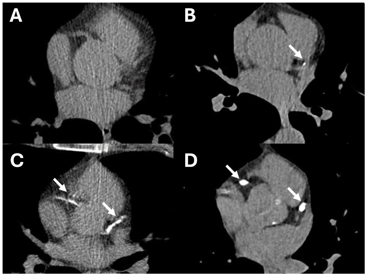
Figure 2.
Opportunistic cardiovascular findings on lung cancer screening low-dose computed tomography (LDCT). Axial non-contrast, non–ECG-gated images. (A) Aortic valve calcification (AVC): dense calcification at the level of the aortic valve (arrow). (B) Mitral annular calcification (MAC): high-attenuation calcification at the mitral annulus (arrow). (C) Thoracic aortic calcification (TAC): calcification visible in the ascending and descending thoracic aorta (arrows). (D) Ascending aortic dilatation: markedly enlarged ascending aorta measuring 4.66 cm on axial non-contrast image (arrow).
Figure 2.
Opportunistic cardiovascular findings on lung cancer screening low-dose computed tomography (LDCT). Axial non-contrast, non–ECG-gated images. (A) Aortic valve calcification (AVC): dense calcification at the level of the aortic valve (arrow). (B) Mitral annular calcification (MAC): high-attenuation calcification at the mitral annulus (arrow). (C) Thoracic aortic calcification (TAC): calcification visible in the ascending and descending thoracic aorta (arrows). (D) Ascending aortic dilatation: markedly enlarged ascending aorta measuring 4.66 cm on axial non-contrast image (arrow).
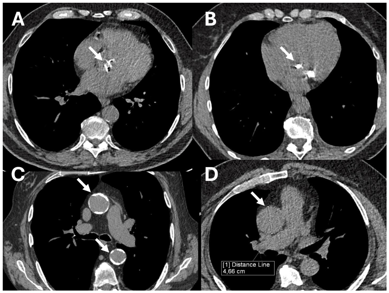
Figure 3.
Pulmonary artery enlargement and ancillary signs of pulmonary hypertension (PH) on computed tomography (CT). (A) Non-contrast LDCT: enlarged main pulmonary artery (MPA) measuring 3.66 cm (arrow); PA-to-aorta ratio 1.21 (B) Non-contrast LDCT: right ventricular predominance (arrow). (C) Contrast-enhanced CT pulmonary angiography: confirmed right ventricular dilatation in the same patient (arrow). (D) Contrast-enhanced CT: anomalous drainage of the right upper pulmonary vein into the superior vena cava (arrow), a congenital left-to-right shunt.
Figure 3.
Pulmonary artery enlargement and ancillary signs of pulmonary hypertension (PH) on computed tomography (CT). (A) Non-contrast LDCT: enlarged main pulmonary artery (MPA) measuring 3.66 cm (arrow); PA-to-aorta ratio 1.21 (B) Non-contrast LDCT: right ventricular predominance (arrow). (C) Contrast-enhanced CT pulmonary angiography: confirmed right ventricular dilatation in the same patient (arrow). (D) Contrast-enhanced CT: anomalous drainage of the right upper pulmonary vein into the superior vena cava (arrow), a congenital left-to-right shunt.
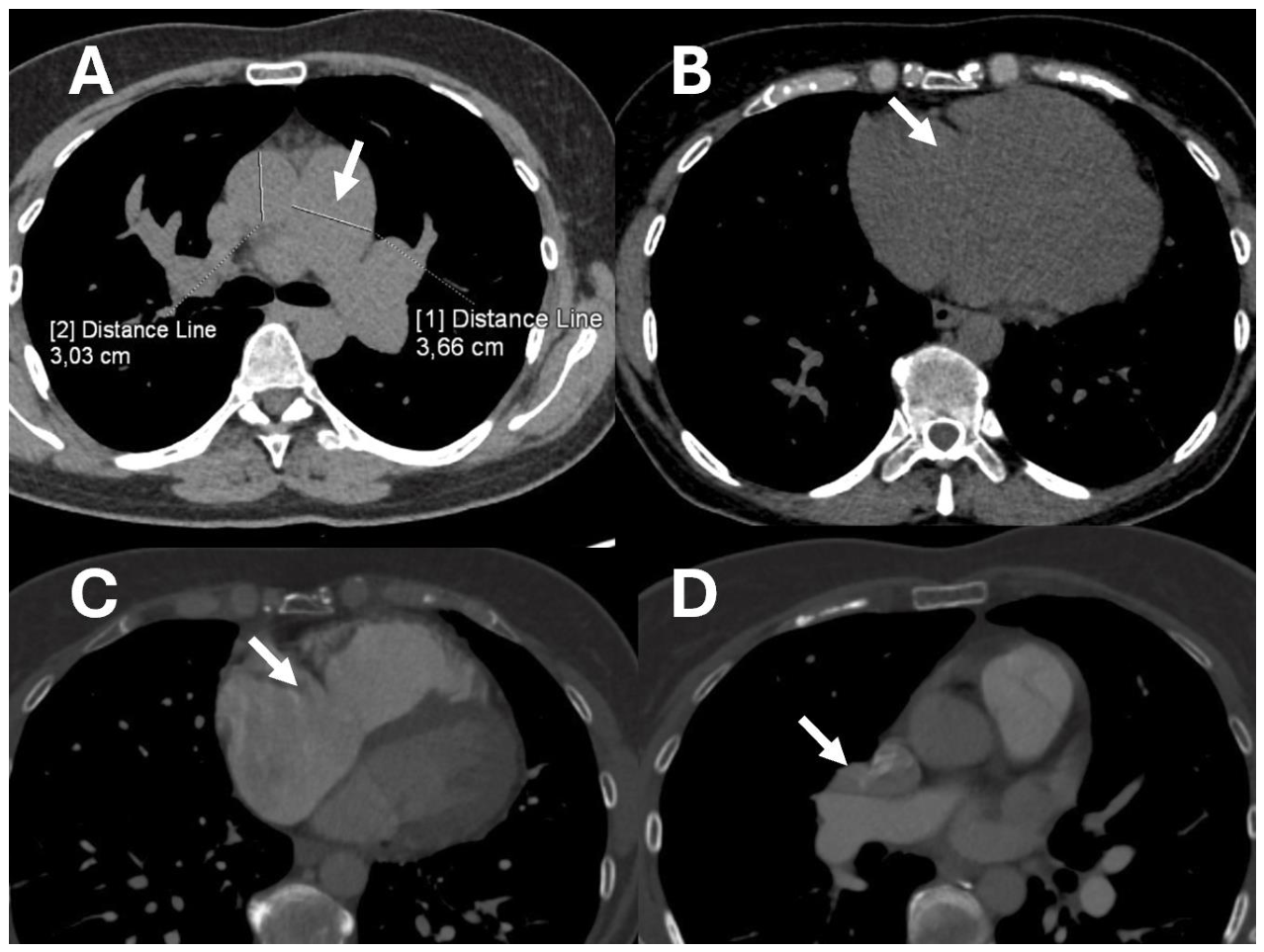
Figure 4.
Management pathway for cardiovascular findings detected on screening LDCT. The flow chart outlines symptom-based referral and finding-specific triage for asymptomatic screening participants, distinguishing core reportable findings from risk-informative findings. Abbreviations: AF, atrial fibrillation; AS, aortic stenosis; AVC, aortic valve calcification; CAC, coronary artery calcification; CAD, coronary artery disease; CT, computed tomography; CV, cardiovascular; ECG, electrocardiography; GP, general practitioner; LDCT, low-dose computed tomography; MAC, mitral annular calcification; MPA, main pulmonary artery; MRI, magnetic resonance imaging; PA, pulmonary artery; PA:Ao, pulmonary artery-to-aorta ratio; PH, pulmonary hypertension; RHC, right heart catheterisation; TAC, thoracic aortic calcification; TTE, transthoracic echocardiography.
Figure 4.
Management pathway for cardiovascular findings detected on screening LDCT. The flow chart outlines symptom-based referral and finding-specific triage for asymptomatic screening participants, distinguishing core reportable findings from risk-informative findings. Abbreviations: AF, atrial fibrillation; AS, aortic stenosis; AVC, aortic valve calcification; CAC, coronary artery calcification; CAD, coronary artery disease; CT, computed tomography; CV, cardiovascular; ECG, electrocardiography; GP, general practitioner; LDCT, low-dose computed tomography; MAC, mitral annular calcification; MPA, main pulmonary artery; MRI, magnetic resonance imaging; PA, pulmonary artery; PA:Ao, pulmonary artery-to-aorta ratio; PH, pulmonary hypertension; RHC, right heart catheterisation; TAC, thoracic aortic calcification; TTE, transthoracic echocardiography.
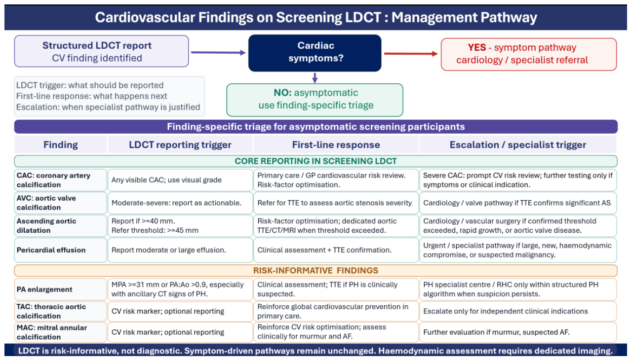

Table 1.
Opportunistic cardiovascular findings on lung cancer screening LDCT: reporting, clinical significance, and key limitations.
Table 1.
Opportunistic cardiovascular findings on lung cancer screening LDCT: reporting, clinical significance, and key limitations.
| Finding | Reporting method | Clinical significance | Key limitations of LDCT |
| Coronary artery calcification (CAC) | Visual grading: none / mild / moderate / severe; Agatston scoring feasible in research settings | Marker of atherosclerotic burden and prognosis; strongly associated with CHD death and all-cause mortality; absence of CAC does not exclude obstructive coronary disease | Non-ECG-gating causes motion blur; partial-volume and blooming effects may underestimate burden; stent artefacts may mimic dense CAC; Agatston values not interchangeable with gated CT |
| Aortic valve calcification (AVC) | Visual grading: none / mild / moderate / severe; quantitative CT-AVC (Agatston) feasible but thresholds validated for ECG-gated protocols only | Correlates with echocardiographic AS severity; may identify previously unrecognized, minimally symptomatic clinically significant aortic stenosis | Cannot determine haemodynamic severity; separation of leaflet from peri-annular calcification less reliable on non-gated scans; CT-AVC values not interchangeable with gated protocols |
| Mitral annular calcification (MAC) | No mandatory reporting format; Visual grading: none / mild / moderate / severe | Marker of degenerative and atherosclerotic burden; associated with higher cardiovascular morbidity, mortality, cerebrovascular events, and incident atrial fibrillation | Cannot assess haemodynamic severity; no validated MAC-specific management algorithms; primarily risk-informative rather than diagnostic |
| Thoracic aortic calcification (TAC) | No mandatory reporting format; | Marker of systemic atherosclerosis and cardiovascular risk | No established TAC-specific referral algorithm |
| Ascending aortic dilatation | Axial diameter measurement; reported if ≥40 (45) mm | May remain asymptomatic for years; actionable if diameter exceeds referral threshold; longitudinal data show small but measurable growth in screened smokers | Absence of ECG gating causes motion artefacts in aortic root; non-contrast measurement may overestimate diameter if wall included; thresholds differ between screening and dedicated aortic imaging protocols |
| Pericardial effusion | Trivial or small pericardial fluid should not be reported. Moderate or large pericardial effusion should be reported. Assessment on non-ECG-gated LDCT should be qualitative rather than based on echocardiographic size thresholds. | Moderate or large pericardial effusion may indicate clinically relevant cardiovascular disease and, in the lung cancer screening population, should also raise consideration of malignant pericardial involvement. Large effusions or those associated with CT signs of haemodynamic compromise require prompt clinical communication. | Non-ECG-gated LDCT allows only approximate qualitative assessment of pericardial effusion. Echocardiographic size thresholds should not be directly applied, and reported moderate or large effusions require clinical assessment and echocardiography to evaluate haemodynamic significance. |
| Pulmonary artery (PA) enlargement | Axial MPA diameter (threshold >31 mm); PA-to-aorta ratio (PA:Ao >0.9); ancillary signs: right-sided cardiac chamber enlargement, IVC dilatation, mosaic attenuation | Non-specific marker of possible pulmonary hypertension (PH); may support WHO (lung disease, group 3) or suggest CTEPH (group 4) based on ancillary findings | Cannot diagnose PH or determine PH aetiology; non-gated acquisition precludes reliable chamber volumetry |
AS, aortic stenosis; AVC, aortic valve calcification; CAC, coronary artery calcification; CHD, coronary heart disease; CT-AVC, CT aortic valve calcium score; CTEPH, chronic thromboembolic pulmonary hypertension; ECG, electrocardiography; IVC, inferior vena cava; LDCT, low-dose computed tomography; MAC, mitral annular calcification; MPA, main pulmonary artery; PA, pulmonary artery; PA:Ao, pulmonary artery-to-aorta ratio; PH, pulmonary hypertension; TAC, thoracic aortic calcification; TTE, transthoracic echocardiography; WHO, World Health Organization.
Disclaimer/Publisher’s Note: The statements, opinions and data contained in all publications are solely those of the individual author(s) and contributor(s) and not of MDPI and/or the editor(s). MDPI and/or the editor(s) disclaim responsibility for any injury to people or property resulting from any ideas, methods, instructions or products referred to in the content. |
© 2026 by the authors. Licensee MDPI, Basel, Switzerland. This article is an open access article distributed under the terms and conditions of the Creative Commons Attribution (CC BY) license (http://creativecommons.org/licenses/by/4.0/).
Copyright: This open access article is published under a Creative Commons CC BY 4.0 license, which permit the free download, distribution, and reuse, provided that the author and preprint are cited in any reuse.