

Submitted:
09 September 2026
Posted:
09 September 2026
You are already at the latest version
Abstract
Background: We have efficiently combined arthroscopic lateral retinacular release with medial reefing for proximal realignment of the patella in patients with patellar dislocation. We therefore conducted this study to assess long-term outcomes of our technique. Methods: The current study was conducted in a total of 67 patients who were surgically treated at our medical institution between February 2012 and March 2024. The patients were evaluated for whether there were significant differences in the patellar tilt angle, congruence angle, tibial tubercle-trochlear groove distance (TT-TG), Insall-Salvati index, Lysholm knee scores and Kujala score between preoperatively and postoperatively. Results: A total of 47 patients (n = 47; 25 men and 22 women) (48 knees) were finally included in the current study; their mean age was 30.7 ± 11.3 (range, 15-63) years old and they were followed up during a mean period of 36.9 ± 25.5 (range, 24-120) months. Preoperatively, the Q-angle and sulcus angle were measured as 14.0° ± 2.7° and 134.4° ± 1.5°, respectively. Preoperatively, the patellar tilt angle, congruence angle, Insall-Salvati index, TT-TG distance, Lysholm knee scores and Kujala scores were measured as 21.6° ± 4.7°, 16.9° ± 14.2°, 0.9 ± 0.1, 17.5 ± 1.9 mm, 45.7 ± 6.4 points and 58.7 ± 8.7, respectively. Postoperatively, these measurements were 10.9° ± 1.4°, 1.0° ± 3.7°, 1.1 ± 0.1, 12.9 ± 1.6 mm, 87.9 ± 4.3 points and 89.7 ± 3.7 points in the corresponding order. These difference reached statistical significance (p < 0.05). Postoperatively, there was only one case (2.1%) of recurrence at postoperative 2 months, but this was corrected by revision arthroscopic repair. Moreover, there was one case of hematoma, but this was managed with aspiration. Furthermore, there were two cases of a loss of flexion of < 135°. Two months later, this was treated with manipulation. But there were no other postoperative complications such as patellar fracture or infection. Conclusion: Arthroscopic lateral retinacular release combined with medial reefing for proximal realignment of the patella is an effective, safe modality in patients with acute or subacute patellar dislocation.
Keywords:
knee
; patellar dislocation
; arthroscopy
; Lysholm knee score
; recurrence
1. Introduction
Patellar dislocation accounts for 2-3% of total cases of knee joint injuries [1,2]. It commonly occurs on the lateral side, which subsequently causes a rupture of the medial patellofemoral ligament (MPFL) in about 90% of total cases [1,3]. Moreover, it is frequently seen in young, active individuals aged between 10 and 20 years old, showing no male or female predilection [2,4,5]. Most cases of the first-time patellar dislocation occur during sport activities at a reported incidence of 55-72% [2,4]. The multifactorial pathophysiology of patellar dislocation has been documented; patients with patella alta, trochlear dysplasia, increased tibial tuberosity-trochlear groove [TT-TG] distance, femoral torsion or genu varum deformity are vulnerable to it [6,7,8].
To date, diverse surgical procedures have been performed for patients with patellar dislocation; these include medial MPFL reconstruction and medial soft tissue realignment surgery [9,10]. According to a systematic review, medial soft tissue realignment surgery produces less favorable outcomes as compared with MPFL reconstruction [11]. Considering that considerable efforts have been made to surgically treat patients with patellar dislocation, however, medial soft tissue realignment surgery deserves special attention.
It has been shown that patients undergoing isolated lateral release are at increased risks of recurrent patellar dislocation and poor clinical outcomes [12,13]. When combined with medial soft tissue reefing, however, lateral release was reported to be effective in lowering the rate of recurrent instability and raising the degree of patients’ satisfaction with surgical outcomes [14]. Moreover, full-arthroscopic or arthroscopically-assisted stabilization of the patella has been reported to show good treatment outcomes according to short-to-mid-term follow-up studies [15,16].
To date, an arthroscopically-assisted proximal extensor mechanism realignment procedure, involving both the lateral retinacular release and reefing of the vastus medialis, has been mainly performed for patients with recurrent patellar dislocation of subluxation. Of note, however, the acute patellar dislocation is commonly accompanied by medial retinacular tears and MPFL tears [17]. We have therefore devised a surgical technique for patients with acute patellar dislocation as we have done for those with recurrent patellar dislocation.
Given the above background, we have efficiently combined lateral retinacular release with medial reefing for proximal realignment of the patella in patients with patellar dislocation. We therefore conducted this study to assess long-term outcomes of our technique in a retrospective, single-institution setting.
2. Materials and Methods
2.1. Study Patients and Setting
The current single-center, retrospective, observational study was conducted in a total of 67 patients (n = 67) who were surgically treated at our medical institution between February 2012 and March 2024.
Inclusion criteria for the current study are as follows:
- (1)
- Chief complaint of symptoms in the patellofemoral joint
- (2)
- Diagnosis of acute or subacute first-time patellar dislocation
- (3)
- Failed conservative treatments
- (4)
- Osteochondral injury or loose body
- (5)
- Treatment with arthroscopic lateral retinacular release combined with medial reefing
- (6)
- The patients who had been followed up during a mean period of ≥ 24 months.
Exclusion criteria for the current study are as follows:
- (1)
- Chronic patellar dislocation (n = 1)
- (2)
- Trochlear dysplasia with crossing sign (n = 3)
- (3)
- Dislocation after total knee arthroplasty (n = 1)
- (4)
- Treatment with lateral retinacular release without medial reefing (n = 3)
- (5)
- Patellofemoral osteoarthritis of ≥ Grade 3 based on the Kellgren-Lawrence classification system (n = 10)
- (6)
- Fulkerson’s operation (n = 1)
- (7)
- The need for distal realignment of the extensor mechanism (e.g., Q-angle ≥ 20°, deformity of the lateral femoral condyle or rotational malalignment of the lower extremity) (n = 1).
We therefore included a total of 47 patients (n = 47) in the current study; it was conducted in compliance with the relevant ethics guidelines following the approval by the Institutional Review Board of our medical institution (DMC-2024-05-013). All procedures described herein were performed in accordance with the 1964 Declaration of Helsinki and its later amendments or comparable ethical standards. But informed consent was waived due to its retrospective nature.
1.1. Treatment Protocol
On physical examination, the patients were evaluated for patellar gliding test and patellar tilt. Then, they received a plain radiography and a magnetic resonance imaging (MRI), based on which they were diagnosed with acute or subacute patellar dislocation. Moreover, they were also examined for whether they had concurrent injuries. Preoperatively, they received conservative therapies such as long leg splint and brace. The patients presenting with instability and pain at the patellofemoral joint despite the above therapies underwent surgery. Thus, they underwent arthroscopic lateral retinacular release and medial joint capsule plication.
We first performed an arthroscopy for concurrent injuries in the knee joint, particularly including osteochondral fracture, injuries to the loose body or the meniscus. Then, we performed arthroscopic repair of these lesions. On arthroscopic diagnosis, there were 15 cases of osteochondral fracture and loose body, for which we performed both loose body removal and chondroplasty. We also examined whether the patients had a tear of the medial retinaculum and confirmed the patellar tilt and the degree of lateral dislocation during full extension of the knee joint. Finally, we measured the patellar tracking during the movement of the knee joint. Following this, an arthroscope was placed in the anterolateral portal and an electrocauter was placed in the anteromedial portal. Approximately 1 cm vertical to the lateral border of the patella, the fatty layer and the surface of lateral quadriceps femoris could be exposed from the proximal pole of the patella to the lower fibers of the vastus lateralis, lateral patellar retinacula and joint capsule were released. Thereafter, the location and tracking of patella were evaluated again. A spinal needle with a non-cutting edge was inserted in the medical capsular membrane from both directions. The suture was performed using arthroscopic all-inside suture technique. In the same manner, 4-5 sutures were made using PDS #1-0 (Figure 1).
There are no established criteria for assessing the degree of plication. At a gap distance of approximately 2-2.5 cm, however, the articular membrane was overlapped using approximately three threads. Then, the patellar tilt, the degree of lateral dislocation and patellar tracking were assessed. If insufficient, the plication was strengthened by 1-2 overlaps. Finally, we confirmed that there were improvements in the patellar tilt and dislocation (Figure 2).
Postoperatively, the patients were recommended to wear long leg brace for three weeks and to perform active movement of the knee joint. Only the patients with satisfactory results of quadriceps setting test were recommended to wear patellar supporting knee brace.
1.1. Outcome Measures
- (1)
- Patellar tilt angle: The patellar tilt angle is a measure of the medial/lateral tilt of the patellar plane relative to the femur. The normal patellofemoral joint is characterized by the lateral opening of the angle between the lateral facet of the patella and any horizontal line. In the abnormal patellofemoral joint, however, the lines forming the patellar tilt angle are parallel or open medially. The patellar tilt angle measuring as 0°-5°, 5°-10° and > 10° is rated as normal, borderline and abnormal, respectively [18].
- (2)
- Congruence angle: The congruence angle is an indicator of medial/lateral subluxuation of the patella within the trochlear groove, which is similar to physical examination of the patellar glide. Its mean values are measured as -6° ± 11° in individuals with no patellar dislocation [18].
- (3)
- Insall-Salvati index: The Insall-Salvati index is the ratio of length of distal part of tendon of quadriceps femoris extending from distal pole of patella to tibial tuberosity to diagonal length of the patella on lateral radiographic view of knee joint taken at 30°-angle of flexion [19].
- (4)
- TT-TG distance: TT-TG distance is a measure of lateralization of the tibial tuberosity relative to the femoral trochlea. It is considered a key factor that is used to assess patellofemoral disorders, which is based on published studies showing that lateralization of the tibial tuberosity is closely associated with patellofemoral instability [20].
- (5)
- Lysholm knee score: The Lysholm score is a 100-point scoring system for examining a patient’s knee-specific symptoms; it is composed of eight items, such as limp, support, locking, pain, instability, swelling, stair climbing and squatting, whose total scores are rated as “Excellent” (95-100 points), “Good” (84-94 points), “Fair” (65-83 points) or “Poor” (< 65 points) [21].
- (6)
- Kujala scores: The Kujala scoring system is composed of a 13-item screening instrument, comprising 13 multiple choice Questions (the presence of a limp, the need for support, an ability to walk, to climb stairs, to squat, to run and to jump, prolonged sitting with knees in the flexed position, the presence of knee pain, knee swelling and presence of abnormal painful patellar movement, atrophy of the thigh muscles and deficiency of knee flexion). It was originally designed to assess patellofemoral pain in adolescents and young adults, with a variable ordinal response format [22].
1.1. Patient Evaluation and Criteria
The patients underwent plain radiography and MRI both preoperatively and postoperatively. Preoperatively, they were evaluated for the measurement of the Q-angle and sulcus angle, as indices of malalignment of the extensor mechanism of the knee [23,24]. Then, they were evaluated for the measurement of the patellar tilt angle, congruence angle, TT-TG distance, Insall-Salvati index, Lysholm knee scores and Kujala score both preoperatively and postoperatively. We analyzed whether there were significant differences in these measurements between preoperatively and postoperatively.
Through a retrospective analysis of the patients’ medical records, we analyzed their age, sex, height, weight, body mass index (BMI), the side of the affected knee, the mode of anesthesia and length of follow-up period. Postoperative outcomes were assessed as proposed by Crosby and Insair [25].
1.1. Statistical Analysis
All data was expressed as mean ± standard deviation or the number of the subjects with percentage, where appropriate. Statistical analysis was done using the SPSS ver. 18.0 for windows (SPSS Inc., Chicago, IL). The patients’ characteristics were analyzed using the χ2-test. Moreover, clinical and radiological outcomes were compared between preoperatively and postoperatively using a paired t-test. A p-value of < 0.05 was considered statistically significant.
1. Results
1.1. Baseline Characteristics of the Patients
A total of 47 patients (n = 47; 25 men and 22 women) (48 knees) were included in the current study; their mean age was 30.7 ± 11.3 (range, 15-63) years old and they were followed up during a mean period of 36.9 ± 25.5 (range, 24-120) months. Preoperatively, the Q-angle and sulcus angle were measured as 14.0° ± 2.7° and 134.4° ± 1.5°, respectively.
1.1. Clinical and Radiological Outcomes
Clinical and radiological outcomes are summarized in Table 2.
Preoperatively, the patellar tilt angle, congruence angle, Insall-Salvati index, TT-TG distance, Lysholm knee scores and Kujala scores were measured as 21.6° ± 4.7°, 16.9° ± 14.2°, 0.9 ± 0.1, 17.5 ± 1.9 mm, 45.7 ± 6.4 points and 58.7 ± 8.7, respectively. Postoperatively, these measurements were 10.9° ± 1.4°, 1.0° ± 3.7°, 1.1 ± 0.1, 12.9 ± 1.6 mm, 87.9 ± 4.3 points and 89.7 ± 3.7 points in the corresponding order. These difference reached statistical significance (p < 0.05).
Postoperatively, there was only one case (2.1%) of recurrence at postoperative 2 months, but this was corrected by revision arthroscopic repair. Moreover, there was one case of hematoma, but this was managed with aspiration. Furthermore, there were two cases of a loss of flexion of < 135°. Two months later, this was treated with manipulation. But there were no other postoperative complications such as patellar fracture or infection.
1.1. Illustrative Case
A 24-year-old woman with patellar dislocation received arthroscopic lateral retinacular release combined with medial reefing for proximal realignment of the patella (Figure 4, Figure 5 and Figure 6).
The patellar tilt angle was preoperatively measured as 19.6° but it was postoperatively corrected to 10.2°.
The highest point of the medial (B) and lateral (C) condyles and the lowest point of the intercondylar sulcus (A) were identified. Then, the sulcus angle (BAC) was bisected to establish the 0 reference line (AO). This was followed by identification of the lowest point on the articular ridge of the patella (D). A line extending from A to D was drawn. The angle DAO corresponded to the congruence angle. All values medial to the 0 reference line AO were designated as negative and those lateral as positive. The congruence angle was preoperatively measured as -16.0° but it was postoperatively corrected to -1.5°.
Lines perpendicular to the posterior condylar axis are placed through the deepest part of the trochlear groove and the center of the tibial tubercle. The TT-TG distance is referred to as that between these two lines. It was preoperatively measured as 17.5 mm but was postoperatively corrected to 11.3 mm.
1. Discussion
Patellar dislocation is classified into traumatic or recurrent cases depending on its etiology. It is also classified into acute, subacute or chronic cases depending on the stage of its onset [25]. Patients with patellar dislocation usually present with pain, malaise, crepitus, a sensation of locking and anxiety [26]. Patellar dislocation is characterized by lateral soft tissue tightness accompanied by medial soft tissue relaxation, resulting in a weakening of the extensor mechanism of the knee joint [27]. This should be completely understood for the appropriate management of patients with patellar dislocation. In the treatment of patellar dislocation, it is mandatory to analyze causes, the alignment of lower extremities and the degree of bone maturation, based on which orthopedic surgeons should decide on single or combined use of proximal and distal realignment. But selection of the treatment of choice poses a challenge for them [25].
Acute or subacute patellar dislocation is commonly accompanied by rupture of medial patellofemoral ligament and synovial membrane as well as osteochondral fracture [27]. Patients with acute patellar dislocation undergoing non-operative treatments may be vulnerable to its recurrence, thus being at a risk of persistent patellofemoral instability [28]. This strongly indicates that an early diagnosis and surgical treatment of acute or subacute patellar dislocation would be mandatory.
Patients with acute patellar dislocation may be suspected of having both MPFL tears and medial retinacular tears when they present with patellar subluxation and lateral tilting on plain radiography after manual reduction. They should be managed with medial retinacular repair [29]. In our series, a diagnosis of MPFL tears was confirmed based on findings, such as bleeding in the femoral origin of the MPFL, synovial tears and MPFL tears on MRI scans. In such cases, a non-absorbable thread was passed through the medial part of MPFL. Then, a pull-out suture was performed towards the adductor tubercle of the femur. Our surgical technique includes the MPFL reefing by involving the entire medial retinaculum.
To date, diverse surgical procedures have been developed to treat patellofemoral instability, most of which have shown inconsistent treatment outcomes, with no established gold standard [12,14]. Recently, however, lateral retinacular release combined with medial reefing has been frequently performed to treat patients with patellar dislocation [12].
According to a review of literatures, patients with TT-TG distance < 20 mm and minimal degenerative changes in the medial patellofemoral joint are indicated in lateral retinacular release combined with medial placation [30]. A combination of lateral retinacular release with medial plication can be supported by previous published studies. Lattermann et al. analyzed treatment outcomes of lateral retinacular release by reviewing a total of 14 published articles about the surgical management of patellar instability [13]. These authors reported a mean rate of patient satisfaction of 80% based on short term results. After a long-term follow-up of > 4 years, however, the rate of patient satisfaction was decreased to 63.5% [13]. Thus, these authors concluded that isolated use of lateral retinacular release of the patella has not proven to be of long-term benefit for the treatment of patellar instability [13].
We found significant improvements in clinical and radiological outcomes postoperatively as compared with preoperatively. Preoperatively, the patellar tilt angle, congruence angle, Insall-Salvati index, TT-TG distance, Lysholm knee scores and Kujala scores were measured as 21.6° ± 4.7°, 16.9° ± 14.2°, 0.9 ± 0.1, 17.5 ± 1.9 mm, 45.7 ± 6.4 points and 58.7 ± 8.7, respectively. Postoperatively, these measurements were 10.9° ± 1.4°, 1.0° ± 3.7°, 1.1 ± 0.1, 12.9 ± 1.6 mm, 87.9 ± 4.3 points and 89.7 ± 3.7 points in the corresponding order. These differences reached statistical significance (p < 0.05). These results indicate that our technique is an effective surgical modality.
According to a review of previous published studies about the safety of lateral retinacular release combined with medial reefing in patients with patellar dislocation, rates of postoperative complications were 0%-14.8% [15,17,31]. In our series, however, there was only one case (2.1%) of recurrence after surgery, but this was corrected by revision arthroscopic repair. Taken together, our results suggest the excellent safety profile of lateral retinacular release combined with medial reefing in patients with patellar dislocation.
But our results cannot be generalized not only because we evaluated a small series of the patients under the retrospective design but also because we conducted the current study at a single institution located in Korea. Therefore, the possibility of selection bias could not be completely ruled out.
5. Conclusions
Based on our results, it can be concluded that arthroscopic lateral retinacular release combined with medial reefing for proximal realignment of the patella is an effective, safe modality in patients with acute or subacute first-time patellar dislocation. But further large-scale, multi-center studies with long periods of follow-up are warranted to corroborate our results.
Author Contributions
Conceptualization, T.R.; data curation, T.R.; formal analysis, T.R.; investigation, T.R.; methodology, T.R.; supervision, T.R.; writing—original draft, T.R.; writing—review and editing, T.R. All authors have read and agreed to the published version of the manuscript.
Funding
The author received no external funding.
Institutional Review Board Statement
The current study was conducted in compliance with the relevant ethics guidelines following the approval by the Institutional Review Board of our medical institution (DMC-2024-05-013). All procedures described herein were performed in accordance with the 1964 Declaration of Helsinki and its later amendments or comparable ethical standards. But informed consent was waived due to its retrospective nature.
Acknowledgments
The author thanks Laon Medi Solution Inc. (Seoul, Republic of Korea) and KDH Medical Inc. (Gwangmyeong, Gyeonggi, Republic of Korea) for additional support of this study.
Conflicts of Interest
The author has nothing to declare in relation to this work.
References
- Arendt, E.A.; Fithian, D.C.; Cohen, E. Current concepts of lateral patella dislocation. Clin. Sports Med. 2002, 21, 499–519. [Google Scholar] [CrossRef] [PubMed]
- Balcarek, P.; Walde, T.A.; Frosch, S.; et al. Patellar dislocations in children, adolescents and adults: A comparative MRI study of medial patellofemoral ligament injury patterns and trochlear groove anatomy. Eur. J. Radiol. 2011, 79, 415–420. [Google Scholar] [CrossRef] [PubMed]
- Burks, R.T.; Desio, S.M.; Bachus, K.N.; Tyson, L.; Springer, K. Biomechanical evaluation of lateral patellar dislocations. Am. J. Knee Surg. 1998, 11, 24–31. [Google Scholar] [PubMed]
- Atkin, D.M.; Fithian, D.C.; Marangi, K.S.; Stone, M.L.; Dobson, B.E.; Mendelsohn, C. Characteristics of patients with primary acute lateral patellar dislocation and their recovery within the first 6 months of injury. Am. J. Sports Med. 2000, 28, 472–479. [Google Scholar] [CrossRef] [PubMed]
- Ostermeier, S.; Holst, M.; Bohnsack, M.; et al. In vitro measurement of patellar kinematics following reconstruction of the medial patellofemoral ligament. Knee Surg. Sports Traumatol. Arthrosc. 2007, 15, 276–285. [Google Scholar] [CrossRef] [PubMed]
- Askenberger, M.; Janarv, P.M.; Finnbogason, T.; Arendt, E.A. Morphology and anatomic patellar instability risk factors in first-time traumatic lateral patellar dislocations: A prospective MRI study in skeletally immature children. Am. J. Sports Med. 2017, 45, 50–58. [Google Scholar] [CrossRef] [PubMed]
- Nha, K.W.; Ha, Y.; Oh, S.; et al. Surgical treatment with closing-wedge distal femoral osteotomy for recurrent patellar dislocation with genu valgum. Am. J. Sports Med. 2018, 46, 1632–1640. [Google Scholar] [CrossRef] [PubMed]
- Christensen, T.C.; Sanders, T.L.; Pareek, A.; Mohan, R.; Dahm, D.L.; Krych, A.J. Risk factors and time to recurrent ipsilateral and contralateral patellar dislocations. Am. J. Sports Med. 2017, 45, 2105–2110. [Google Scholar] [CrossRef] [PubMed]
- Yoon, K.H.; Kim, E.J.; Kwon, Y.B.; Hwang, I.U.; Kim, S.G. Comparison of clinical and radiological outcomes between transosseous tunnel and suture anchor patellar fixation for medial patellofemoral ligament reconstruction: A cohort study with 2-year follow-up. Orthop. J. Sports Med. 2020, 8, 2325967120917660. [Google Scholar] [CrossRef] [PubMed]
- Schorn, D.; Yang-Strathoff, S.; Gosheger, G.; et al. Long-term outcomes after combined arthroscopic medial reefing and lateral release in patients with recurrent patellar instability: A retrospective analysis. BMC Musculoskelet. Disord. 2017, 18, 277. [Google Scholar] [CrossRef] [PubMed]
- Lee, D.Y.; Park, Y.J.; Song, S.Y.; Hwang, S.C.; Park, J.S.; Kang, D.G. Which technique is better for treating patellar dislocation? A systematic review and meta-analysis. Arthroscopy 2018, 34, 3082–3093.e1. [Google Scholar] [CrossRef] [PubMed]
- Colvin, A.C.; West, R.V. Patellar instability. J. Bone Jt. Surg. Am. 2008, 90, 2751–2762. [Google Scholar] [CrossRef] [PubMed]
- Lattermann, C.; Toth, J.; Bach, B.R., Jr. The role of lateral retinacular release in the treatment of patellar instability. Sports Med. Arthrosc. Rev. 2007, 15, 57–60. [Google Scholar] [CrossRef] [PubMed]
- Ricchetti, E.T.; Mehta, S.; Sennett, B.J.; Huffman, G.R. Comparison of lateral release versus lateral release with medial soft-tissue realignment for the treatment of recurrent patellar instability: A systematic review. Arthroscopy 2007, 23, 463–468. [Google Scholar] [CrossRef] [PubMed]
- Ali, S.; Bhatti, A. Arthroscopic proximal realignment of the patella for recurrent instability: Report of a new surgical technique with 1 to 7 years of follow-up. Arthroscopy 2007, 23, 305–311. [Google Scholar] [CrossRef] [PubMed]
- Schöttle, P.B.; Scheffler, S.U.; Schwarck, A.; Weiler, A. Arthroscopic medial retinacular repair after patellar dislocation with and without underlying trochlear dysplasia: A preliminary report. Arthroscopy 2006, 22, 1192–1198. [Google Scholar] [CrossRef] [PubMed]
- Nha, K.W.; Kim, H.S.; Cho, S.T.; Bae, J.H.; Jang, K.M.; Kim, S.G. Arthroscopy-controlled medial reefing and lateral release for recurrent patellar dislocation: Clinical, radiologic outcomes and complications. BMC Musculoskelet. Disord. 2021, 22, 430. [Google Scholar] [CrossRef] [PubMed]
- Collado, H.; Fredericson, M. Patellofemoral pain syndrome. Clin. Sports Med. 2010, 29, 379–398. [Google Scholar] [CrossRef] [PubMed]
- Insall, J.; Salvati, E. Patella position in the normal knee joint. Radiology 1971, 101, 101–104. [Google Scholar] [CrossRef] [PubMed]
- Wilcox, J.J.; Snow, B.J.; Aoki, S.K.; Hung, M.; Burks, R.T. Does landmark selection affect the reliability of tibial tubercle–trochlear groove measurements using MRI? Clin. Orthop. Relat. Res. 2012, 470, 2253–2260. [Google Scholar] [CrossRef] [PubMed]
- Wright, R.W. Knee injury outcome measures. J. Am. Acad. Orthop. Surg. 2009, 17, 31–39. [Google Scholar] [CrossRef] [PubMed]
- Haddad, B.I.; Hamdan, M.; Isleem, U.; Al-Saber, M.G.; Al-Hadidi, F.A.; AlRyalat, S.A.; Alnaimat, F. Validation of the cultural adaptation of the Kujala score in Arabic. J. Orthop. Surg. Res. 2021, 16, 323. [Google Scholar] [CrossRef] [PubMed]
- Smith, T.O.; Hunt, N.J.; Donell, S.T. The reliability and validity of the Q-angle: A systematic review. Knee Surg. Sports Traumatol. Arthrosc. 2008, 16, 1068–1079. [Google Scholar] [CrossRef] [PubMed]
- Davies, A.P.; Costa, M.L.; Shepstone, L.; Glasgow, M.M.; Donell, S. The sulcus angle and malalignment of the extensor mechanism of the knee. J. Bone Jt. Surg. Br. 2000, 82, 1162–1166. [Google Scholar] [CrossRef] [PubMed]
- Song, I.S.; Kim, S.K.; Song, C. Arthroscopic treatment for acute or subacute patellar dislocation. Knee Surg. Relat. Res. 2007, 17, 186–192. [Google Scholar]
- Hong, E.; Kraft, M.C. Evaluating anterior knee pain. Med. Clin. North Am. 2014, 98, 697–717. [Google Scholar] [CrossRef] [PubMed]
- Dodson, C.C.; Shindle, M.K.; Dines, J.S.; Altchek, D.W. Arthroscopic suture anchor repair for lateral patellar instability. Knee Surg. Sports Traumatol. Arthrosc. 2010, 18, 143–146. [Google Scholar] [CrossRef] [PubMed]
- Yang, F.; Chen, C.; Chen, R.; Yang, C.; Xiao, H.; Feng, Z.; Geng, B.; Xia, Y. Recurrent patellar dislocation: treatments and challenges. Front. Surg. 2025, 12, 1507362. [Google Scholar] [CrossRef] [PubMed]
- Tsai, C.H.; Hsu, C.J.; Hung, C.H.; Hsu, H.C. Primary traumatic patellar dislocation. J. Orthop. Surg. Res. 2012, 7, 21. [Google Scholar] [CrossRef] [PubMed]
- Mulford, J.S.; Wakeley, C.J.; Eldridge, J.D. Assessment and management of chronic patellofemoral instability. J. Bone Jt. Surg. Br. 2007, 89, 709–716. [Google Scholar] [CrossRef] [PubMed]
- Lee, J.J.; Lee, S.J.; Won, Y.G.; Choi, C.H. Lateral release and medial plication for recurrent patella dislocation. Knee Surg. Sports Traumatol. Arthrosc. 2012, 20, 2438–2444. [Google Scholar] [CrossRef] [PubMed]
Figure 1.
Five-knot tying of the medial capsule after arthroscopic lateral retinacular release.
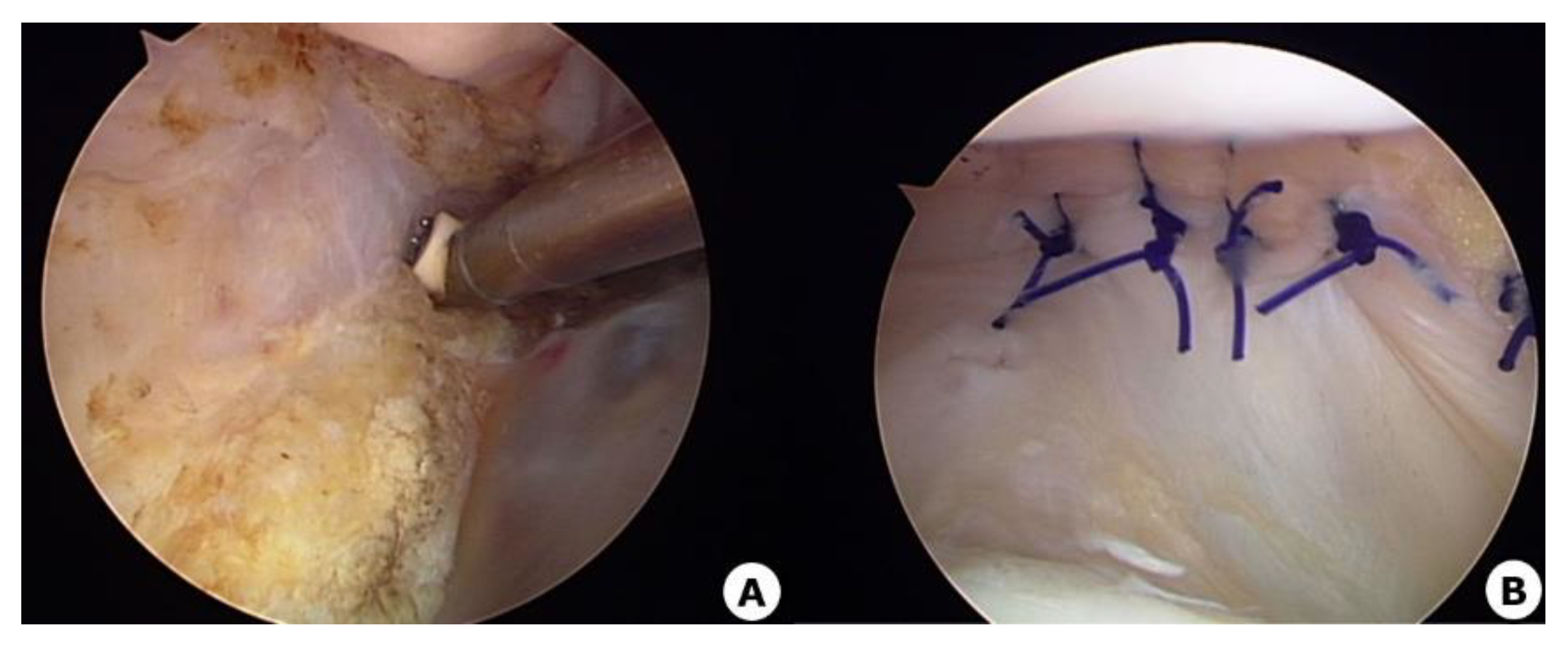
Figure 2.
Postoperative improvements in the patellar tilt and dislocation.
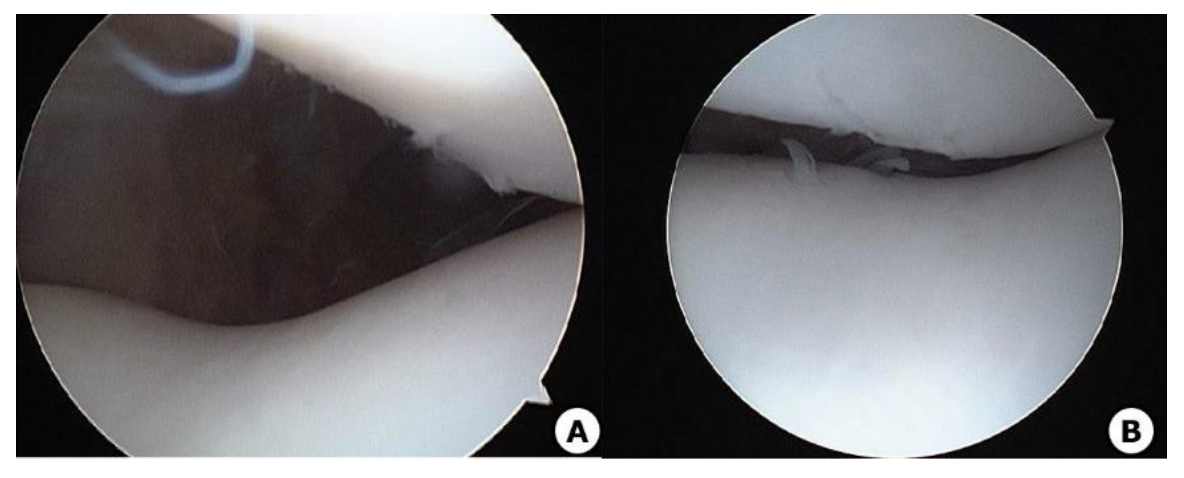
Figure 3.
Study flow chart.
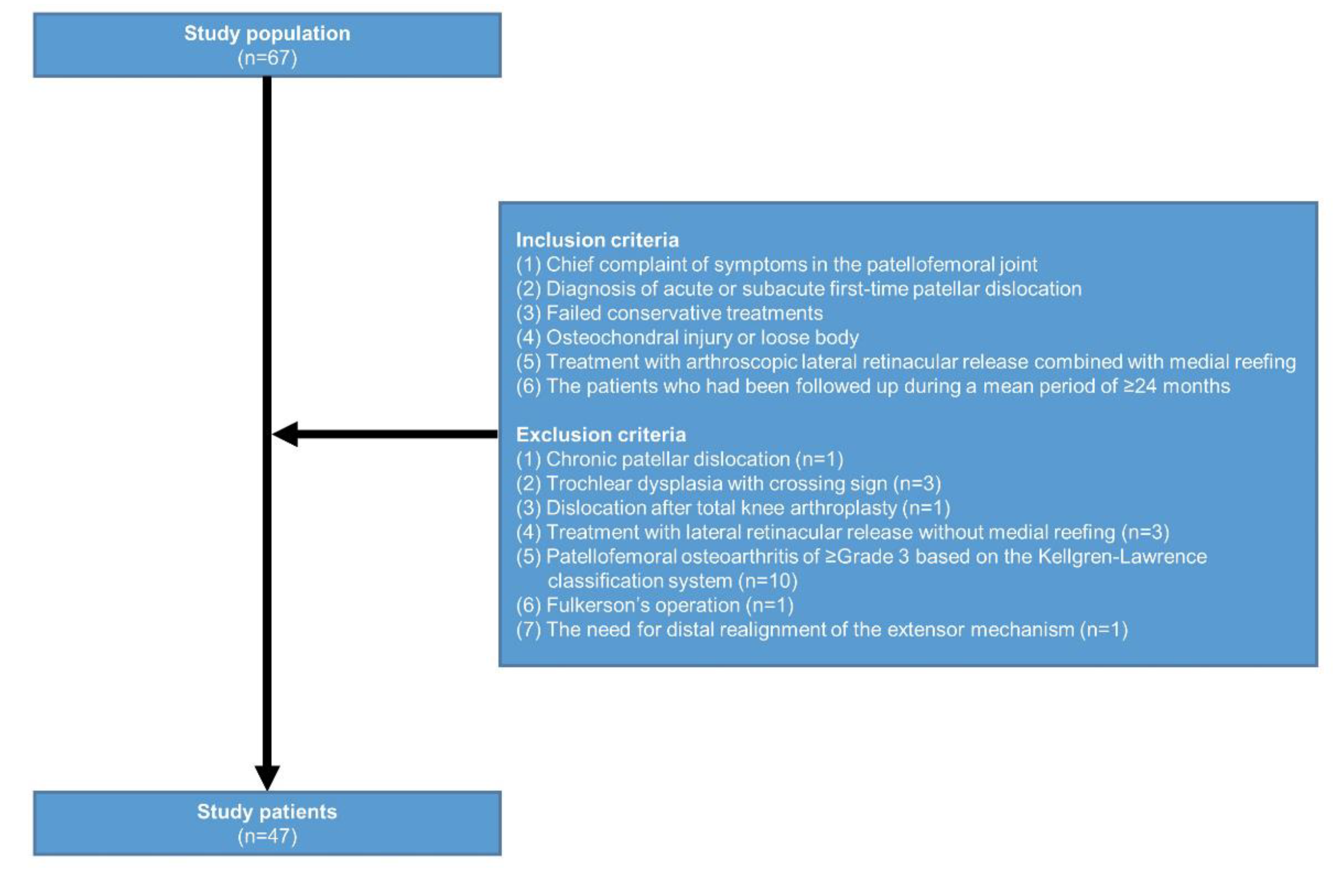
Figure 4.
Patellar tilt angle.
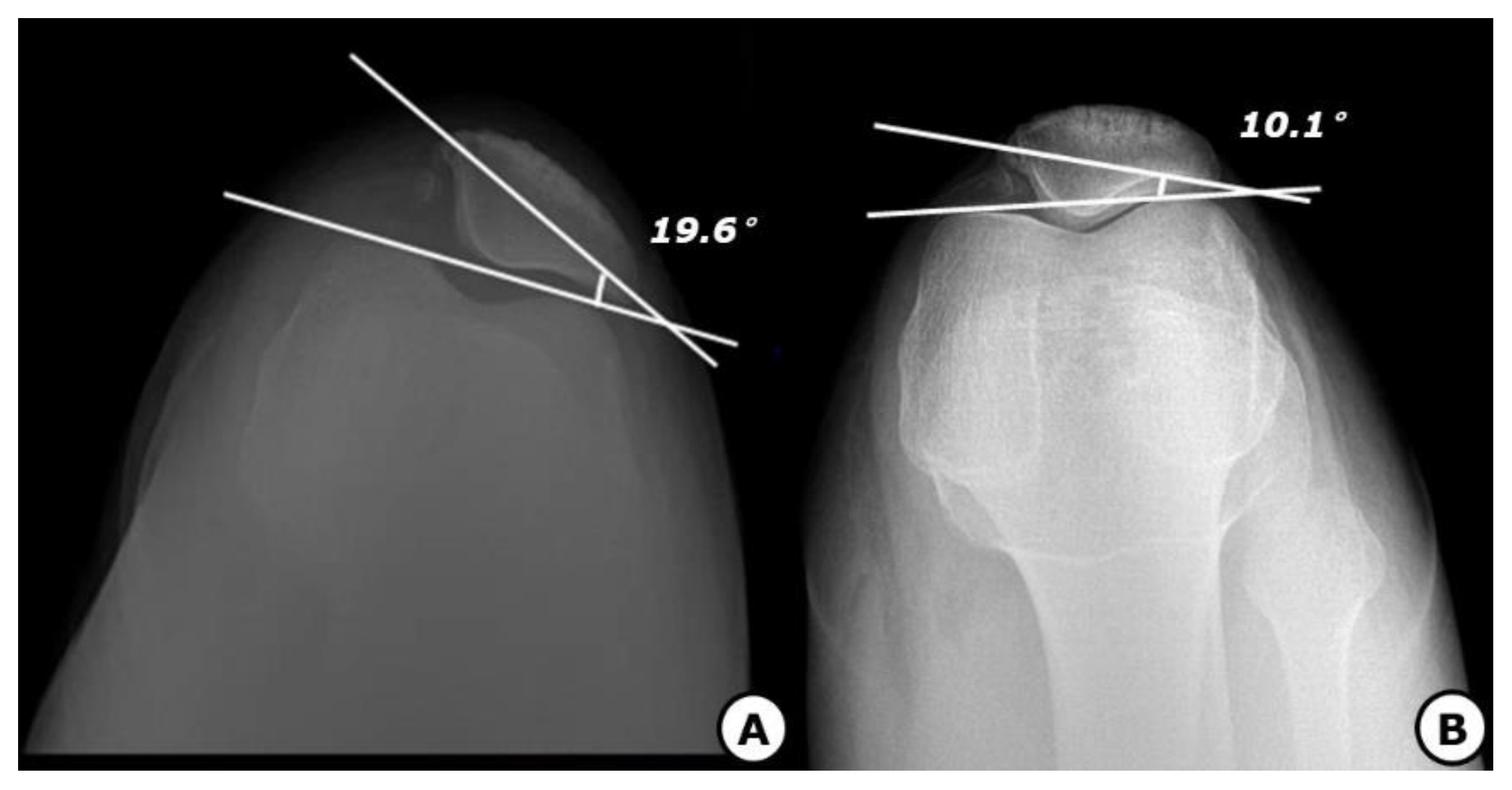
Figure 5.
Congruence angle.
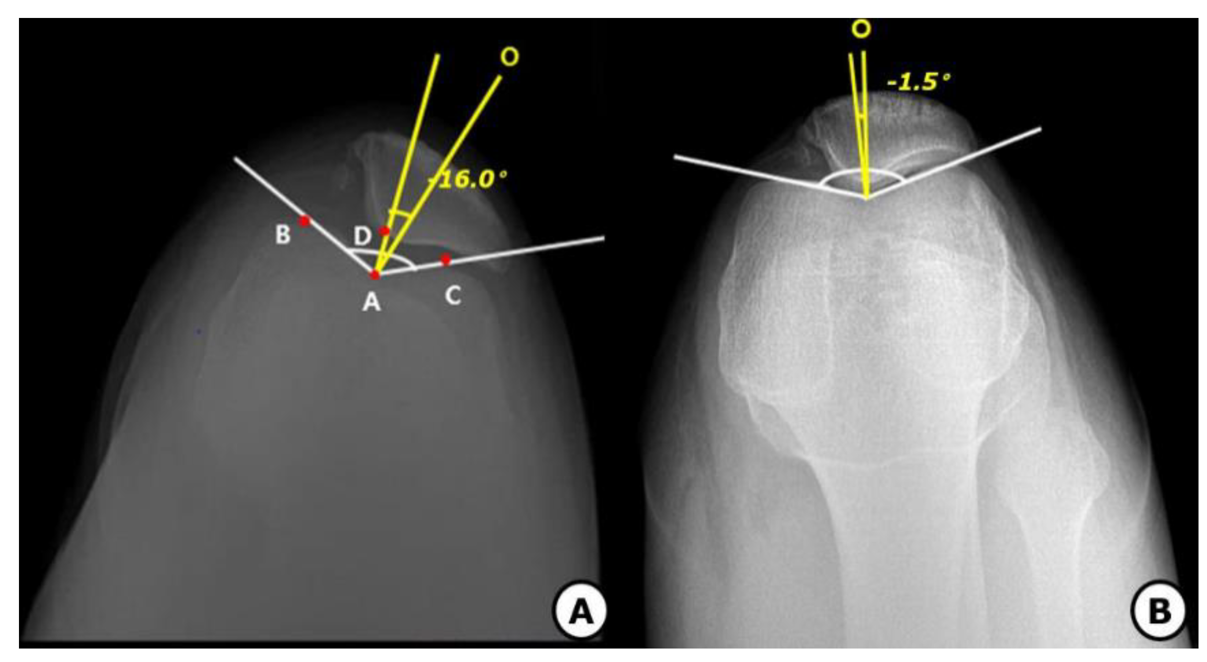
Figure 6.
The tibial tubercle-trochlear groove (TT-TG) distance.
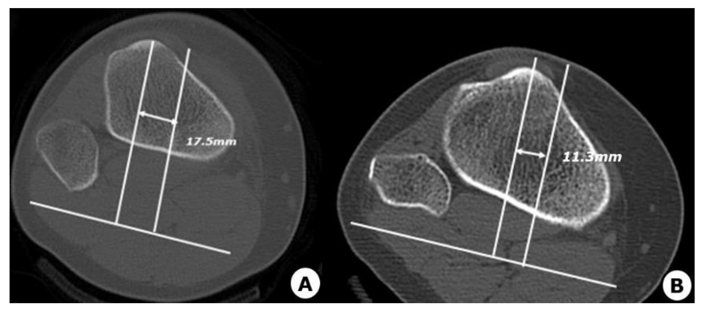

Table 1.
Baseline characteristics of the patients (n = 47; 48 knees).
| Variables | Values | ||||
| Age (years old) | 30.7 ± 11.3 (15-63) | ||||
| Sex | |||||
| Men | 25 (53.2%) | ||||
| Women | 22 (46.9%) | ||||
| Laterality of the affected knee | |||||
| Unilateral cases | 47 (97.9%) | ||||
| Bilateral cases | 1 (2.1%) | ||||
| Height (cm) | 168.9 ± 9.7 | ||||
| Weight (kg) | 64.1 ± 13.1 | ||||
| BMI (kg/m2) | 22.3 ± 3.3 | ||||
| FU period (months) | 36.9 ± 25.5 (24-120) | ||||
| Mode of anesthesia | |||||
| General anesthesia | 9 (18.7%) | ||||
| Spinal anesthesia | 39 (81.3%) | ||||
| The side of the affected knee | |||||
| Left | 21 (43.7%) | ||||
| Right | 27 (56.3%) | ||||
| Preoperative measurements | |||||
| Q-angle (°) | 14.0 ± 2.7 | ||||
| Sulcus angle (°) | 134.4 ± 1.5 | ||||
Abbreviations: BMI, body mass index; FU, follow-up. Values are mean ± standard deviation with range or the number of the patients with percentage, where appropriate.
Table 2.
Clinical and radiological outcomes.
| Variables | Values | p-value | |
| Preoperative measurements (n = 48) |
Postoperative measurements (n = 48) |
||
| Patellar tilt angle (°) | 21.6 ± 4.7 | 10.9 ± 1.4 | 0.024* |
| Congruence angle (°) | 16.9 ± 14.2 | 1.0 ± 3.7 | 0.037* |
| Insall-Salvati index | 0.9 ± 0.1 | 1.1 ± 0.1 | <0.001* |
| TT-TG distance (mm) | 17.5 ± 1.9 | 12.9 ± 1.6 | 0.016* |
| Lysholm knee scores | 45.7 ± 6.4 | 87.9 ± 4.3 | <0.001* |
| Kujala scores | 58.7 ± 8.7 | 89.7 ± 3.7 | < 0.001* |
Abbreviations: TT-TG, tibial tubercle-trochlear groove; VAS, visual analogue scale. *Statistical significance at p < 0.05.
Disclaimer/Publisher’s Note: The statements, opinions and data contained in all publications are solely those of the individual author(s) and contributor(s) and not of MDPI and/or the editor(s). MDPI and/or the editor(s) disclaim responsibility for any injury to people or property resulting from any ideas, methods, instructions or products referred to in the content. |
© 2026 by the authors. Licensee MDPI, Basel, Switzerland. This article is an open access article distributed under the terms and conditions of the Creative Commons Attribution (CC BY) license (http://creativecommons.org/licenses/by/4.0/).
Copyright: This open access article is published under a Creative Commons CC BY 4.0 license, which permit the free download, distribution, and reuse, provided that the author and preprint are cited in any reuse.