

Submitted:
08 September 2026
Posted:
09 September 2026
You are already at the latest version
Abstract
Ferroptosis is a novel form of regulated cell death that plays a key role in the pathogenesis of a wide range of multifactorial diseases. Its uniqueness lies in the iron-dependent accumulation of lipid peroxides, which distinguishes it from apoptosis, necroptosis, and autophagy. Understanding the molecular mechanisms of ferroptosis opens up new therapeutic prospects, but the dual role of this process – both as a factor of malignant cell death and a mediator of tissue damage – creates significant challenges for clinical application. The paper systematizes current ideas about the molecular mechanisms of ferroptosis regulation, including iron metabolism, lipid peroxidation, and antioxidant defense systems. Signaling pathways that modulate sensitivity to ferroptosis are analyzed. Particular attention is paid to the dual role of ferroptosis in the pathogenesis of diseases: induction of ferroptosis is considered a promising strategy for overcoming the resistance of malignant neoplasms to chemotherapy, immunotherapy and radiotherapy, while its inhibition is critical for protecting tissues from ischemia-reperfusion injury, neurodegeneration and inflammation. Key counterarguments are highlighted, in particular the pro-inflammatory and immunosuppressive effects of ferroptosis, which may contribute to tumor progression. Ferroptosis is a universal mechanism of cell death with powerful therapeutic potential, however, its clinical implementation requires a differentiated approach depending on the pathological context. Promising directions are the development of highly selective modulators, identification of specific biomarkers of ferroptosis in vivo and conducting well-designed clinical trials. In-depth study of molecular regulatory networks will allow translating fundamental knowledge into personalized treatment strategies for multifactorial diseases.
Keywords:
ferroptosis
; regulated cell death
; iron
; lipid peroxidation
; glutathione peroxidase 4
; iron metabolism
; ferroptosis inducers
; ferroptosis inhibitors
; cancer
; neurodegenerative diseases
; ischemia-reperfusion injury
; therapeutic targets
; ferritinophagy
; pro-tumorigenic role
; immunotherapy
; radiotherapy
1. Introduction
Regulated cell death (RCD) is a fundamental biological process required for tissue homeostasis, embryonic development, and immune defense [1]. For decades, apoptosis was considered the main form of programmed cell death, but the discovery of alternative mechanisms such as necroptosis, pyroptosis, and ferroptosis has significantly expanded our understanding of the pathophysiological processes [2,3]. Ferroptosis, first characterized by Dixon et al. in 2012, is an iron-dependent form of non-apoptotic cell death induced by the accumulation of lipid peroxides in membranes and characterized by unique morphological, biochemical, and genetic features [4,5]. Unlike apoptosis, ferroptosis is not accompanied by caspase activation, and its morphological marker is the decrease in mitochondrial volume with increased membrane density and the disappearance of cristae [4,6].
Over the past decade, compelling evidence has accumulated that ferroptosis plays a key role in the pathogenesis of a wide range of diseases, including malignancies, neurodegenerative disorders, cardiovascular pathology, metabolic syndromes, and inflammatory bowel diseases [7,8,9,10]. Of particular interest is the dual role of this process: induction of ferroptosis is considered a promising strategy to overcome tumor resistance to chemotherapy and immunotherapy, while its inhibition may protect tissues from ischemia-reperfusion injury and neurodegeneration [11,12,13].
Ferroptosis is a universal, complexly regulated mechanism of cell death, the understanding of which opens up new therapeutic horizons. The key challenge remains the search for specific modulators capable of differentially influencing this process depending on the pathological context, which requires in-depth study of molecular regulatory networks, in particular iron metabolism, lipids and antioxidant systems.
2. Main Text
Molecular mechanisms of ferroptosis regulation. Iron metabolism as a driving force of ferroptosis. Iron is a critical trace element required for numerous physiological processes, including oxygen transport, DNA synthesis, and mitochondrial respiration [14]. However, excess iron, especially in the form of Fe2+, can catalyze the Fenton reaction, generating highly reactive hydroxyl radicals that initiate lipid peroxidation [15]. It is this mechanism that underlies the iron-dependent nature of ferroptosis. Intracellular iron homeostasis is regulated by a complex network of transport proteins: transferrin (TF) binds Fe3+ and delivers it to the cell via transferrin receptor 1 (TFR1), after which Fe3+ is reduced to Fe2+ by STEAP3 and released into the cytoplasm via DMT1 [16,17]. Excess iron is stored in ferritin (FTH1/FTL), and its excretion is ensured by a single exporter – ferroportin (FPN) [18]. An important regulatory mechanism is NCOA4-mediated ferritinophagy – a selective autophagic process that ensures the degradation of ferritin in lysosomes with the release of free iron, which increases the sensitivity of cells to ferroptosis [19,20]. Inhibition of NCOA4 or blocking its interaction with FTH1, as shown in the study by Fang et al., significantly reduces the level of intracellular iron and prevents ferroptosis [21].
Lipid peroxidation: a central event in ferroptosis. The accumulation of lipid hydroperoxides is a key event determining the development of ferroptosis. Of particular note are polyunsaturated fatty acids (PUFA), in particular arachidonic (AA) and adrenic (AdA) acids, which are highly sensitive to oxidation due to the presence of bis-allyl hydrogen atoms [22,23]. The key enzymes in this process are ACSL4 (acyl-CoA synthetase of long-chain fatty acids) and LPCAT3 (lysophosphatidylcholine acyltransferase). ACSL4 catalyzes the esterification of free PUFA to form PUFA-CoA, which is then incorporated into membrane phosphatidylethanolamines (PE) by LPCAT3, forming PUFA-PE [24,25]. Further oxidation of PUFA-PE occurs by iron-containing lipoxygenases (LOX) or by nonenzymatic autoxidation initiated by the Fenton reaction [26,27]. Doll et al. demonstrated that ACSL4 knockout renders cells resistant to ferroptosis induced by the GPX4 inhibitor RSL3, confirming the central role of this enzyme [24]. Interestingly, recent studies have identified an alternative peroxidation pathway mediated by POR (cytochrome P450 oxidoreductase), which acts independently of LOX [28].
Antioxidant defense systems against ferroptosis. GPX4/GSH-dependent system. The primary defense mechanism against ferroptosis is the selenium-containing enzyme glutathione peroxidase 4 (GPX4), which reduces toxic lipid hydroperoxides to harmless lipid alcohols using glutathione (GSH) as a cofactor [29,30]. GSH synthesis is limited by the availability of cysteine, which enters the cell through the System Xc- antiporter (SLC7A11/SLC3A2) in exchange for glutamate [31]. Inhibition of System Xc- with erastin or sulfasalazine, or direct inhibition of GPX4 RSL3, leads to GSH depletion, accumulation of lipid peroxides, and ferroptosis [4,32]. Importantly, GPX4 activity depends on the incorporation of selenium in the form of selenocysteine, therefore selenium deficiency increases susceptibility to ferroptosis (Figure 1) [33].
GPX4-independent systems. In 2019, two independent groups of researchers identified FSP1 (ferroptosis suppressor protein 1, also known as AIFM2) as an alternative defense mechanism. FSP1, localized at the plasma membrane, reduces coenzyme Q10 (CoQ10) to ubiquinol (CoQ10H2) using NADPH, which acts as a lipophilic radical-scavenging antioxidant, inhibiting lipid peroxidation independently of GPX4 and GSH [34,35]. Doll et al. showed that expression of FSP1 protects cells from ferroptosis even in the absence of GPX4, making this pathway a promising target for therapeutic intervention [34]. Another GPX4-independent system is the GCH1-BH4-DHFR pathway. GTP cyclohydrolase 1 (GCH1) is a key enzyme in the synthesis of tetrahydrobiopterin (BH4), which has been shown to act as a potent antioxidant that prevents lipid peroxidation [36]. Soula et al. demonstrated that activation of GCH1 significantly increases the resistance of cells to ferroptosis [37]. In addition, the mitochondrial enzyme DHODH (dihydroorotate dehydrogenase) is able to reduce CoQ10 to CoQ10H2 in the inner mitochondrial membrane, providing additional protection against ferroptosis at the level of this organelle (Figure 2) [38].
The role of ferroptosis in the pathogenesis of diseases. Ferroptosis in cancer therapy: opportunities and challenges. Resistance to apoptosis is one of the key characteristics of malignant cells, therefore, the induction of alternative death pathways, in particular ferroptosis, is considered a promising strategy to overcome chemoresistance [11,39]. Mesenchymal and undifferentiated cancer cells, which usually exhibit resistance to apoptosis, have been shown to be extremely sensitive to ferroptosis, making it a particularly attractive target for the therapy of resistant tumors (Figure 3) [40,41].
Synergy with immunotherapy. An important discovery was that activated CD8+ T lymphocytes secrete interferon-γ (IFNγ), which suppresses the expression of SLC7A11 and SLC3A2 in tumor cells through activation of the JAK/STAT1 pathway, thereby sensitizing them to ferroptosis [12]. Wang et al. showed that the combination of immunotherapy with immune checkpoint inhibitors and ferroptosis inducers (e.g., cysteinase) leads to synergistic inhibition of tumor growth in vivo [12]. Moreover, Liao et al. found that IFNγ, together with arachidonic acid, induces ferroptosis through transcriptional activation of ACSL4, opening new possibilities for combination therapy [42]. However, ferroptosis can also contribute to immunosuppression. For example, ferroptotic tumor cells release the oncogenic protein KRASG12D, which is taken up by macrophages and induces their polarization towards a pro-tumorigenic M2 phenotype through an AGER-dependent mechanism [43]. This highlights the need for a balanced approach to the modulation of ferroptosis in immunotherapy.
Synergy with radiotherapy. Radiotherapy induces ferroptosis through two main mechanisms: first, the generation of reactive oxygen species (ROS) from water radiolysis, which directly initiates lipid peroxidation; and second, the inhibition of SLC7A11 through the ATM-dependent pathway, which leads to GSH depletion [13,44]. Lei et al. demonstrated that the combination of SLC7A11 inhibitors (e.g., sulfasalazine) with radiotherapy significantly enhanced ferroptotic cell death in non-small cell lung cancer and inhibited the growth of xenografts [13]. Interestingly, hypoxia, which often accompanies solid tumors, can increase SLC7A11 expression through HIF-1α, contributing to radioresistance, making inhibition of this pathway critical for improving the efficacy of radiotherapy [45].
Controversies: The pro-tumorigenic role of ferroptosis. Despite its antitumor potential, ferroptosis may also contribute to carcinogenesis. In models of pancreatic cancer and hepatocellular carcinoma, chronic ferroptotic injury induces inflammation mediated by the release of DAMP molecules (such as HMGB1 and 8-hydroxyguanosine), which contributes to immunosuppression and tumor progression [46,47]. Dai et al. have shown that ferroptotic injury in pancreatic cells activates a STING-dependent pathway, leading to the recruitment of M2-polarized macrophages and stimulation of tumor growth [46]. Thus, the temporal and localized nature of ferroptosis induction is critical to avoid unwanted pro-inflammatory effects.
Ferroptosis in neurodegenerative and ischemic diseases. In contrast to its role in cancer therapy, the inhibition of ferroptosis serves as a protective strategy in neurodegenerative diseases and ischemic-reperfusion injuries. Iron accumulation and increased lipid peroxidation are common pathogenetic features of Alzheimer’s, Parkinson’s and Huntington’s diseases [48]. In Alzheimer’s disease, FPN expression is reduced in brain tissues. Its reduction prevents the excretion of Fe2+/Fe3+, which causes intracellular accumulation and iron overload. This leads to the induction of ferroptosis, which can be inhibited by Ferrostatin-1 [49]. In models of Parkinson’s disease, α-synuclein increases the sensitivity to ferroptosis by modulating the composition of membrane ether phospholipids [50]. Importantly, iron-chelating agents (deferoxamine, deferiprone) and lipophilic antioxidants have shown efficacy in preclinical models of neurodegeneration, and clinical trials of deferiprone in Parkinson’s disease have shown a slowing of disease progression [51,52]. In ischemic stroke, the hypoxia-inducible factor HIF-1α upregulates TFR1 expression, promoting neuronal iron accumulation and ferroptosis [53]. Inhibition of ACSL4 or activation of Nrf2 has been shown to reduce infarct size and improve neurological deficits in animal models [54,55].
Ferroptosis in cardiovascular disease. Cardiomyocytes are particularly sensitive to iron-dependent oxidative stress. Doxorubicin-induced cardiomyopathy is a classic example of ferroptosis-mediated damage: the drug forms a complex with Fe2+ in mitochondria, triggering lipid peroxidation and ferroptosis [56]. Tadokoro et al. demonstrated that overexpression of GPX4 in cardiomyocytes prevents this effect [56].
Atherosclerosis is also closely linked to ferroptosis: oxidized low-density lipoproteins (oxLDL) induce ferroptosis of macrophages, which contributes to the destabilization of atherosclerotic plaques [57,58]. Bai et al. showed that inhibition of ferroptosis by Ferrostatin-1 reduced lipid peroxidation and endothelial dysfunction in aortic endothelial cells of ApoE-/- (Figure 4) [58].
Ferroptosis in reproductive and lung diseases. In the respiratory system, ferroptosis is a key trigger of acute lung injury (ALI), where lipopolysaccharide (LPS) induces iron-dependent death of alveolar epithelium, and is also involved in the pathogenesis of bronchial asthma through ALOX15-mediated peroxidation of membrane lipids and maintenance of chronic inflammation [5,23,26]. In the reproductive system, labile iron accumulation and ferroptosis of ovarian granulosa cells are critical links in the disruption of folliculogenesis and steroidogenesis in polycystic ovary syndrome (PCOS) and premature ovarian insufficiency (POI) [14,15]. Furthermore, in endometriosis, peritoneal iron overload induces ferroptosis of stromal cells with subsequent release of pro-inflammatory DAMP molecules, confirming the importance of pharmacological control of lipid peroxidation in respiratory and reproductive medicine [6,46].
Therapeutic strategies for ferroptosis modulation. Inducers of ferroptosis. Classical inducers of ferroptosis are erastin and RSL3, which inhibit System Xc- and GPX4, respectively [4,32].
However, the clinical application of these compounds is limited by their low solubility and metabolic instability. More stable analogs have been developed, including the imidazole ketone erastin (IKE), which has shown antitumor activity in mouse models of lymphoma [59]. In addition, some FDA-approved drugs, such as sulfasalazine (an anti-inflammatory) and sorafenib (a kinase inhibitor), also induce ferroptosis in certain tumor types [60,61]. Artesunate, an artemisinin derivative, induces ferroptosis by enhancing ferritinophagy and increasing free iron levels, and has been shown to be effective against pancreatic tumors [62]. Simvastatin and other statins induce ferroptosis in triple-negative breast cancer by inhibiting the mevalonate pathway and reducing CoQ10 levels [ 63].
Inhibitors of ferroptosis. The most studied inhibitors are Ferrostatin-1 and Liproxstatin-1, which act as lipid peroxyl radical scavengers, interrupting the lipid peroxidation chain reaction [4,64]. These compounds have shown efficacy in models of ischemia-reperfusion injury in the kidney, liver, and heart [65,66]. In addition, iron chelators such as deferoxamine, deferiprone, and deferasirox are widely used to treat iron overload and are being studied as potential agents for inhibiting ferroptosis in neurodegenerative diseases [67,68]. Another promising direction is the use of antioxidants such as N-acetylcysteine (NAC) and vitamin E, which increase GSH levels or directly neutralize free radicals [69].
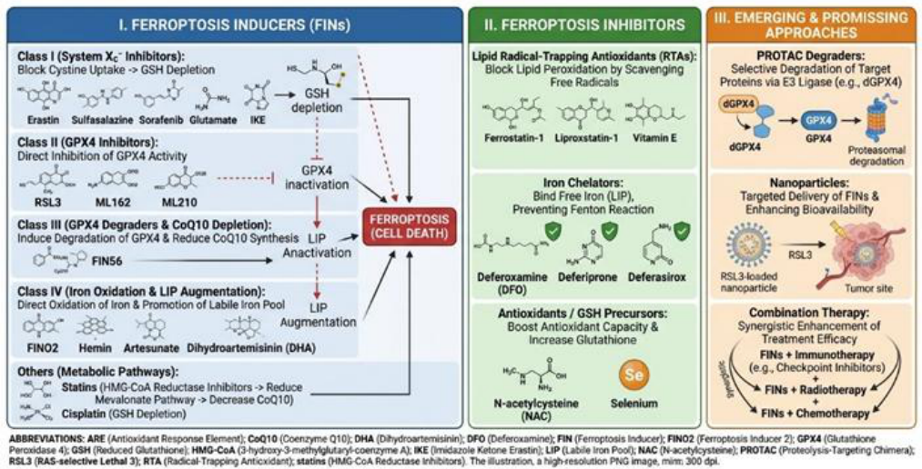
Promising approaches: PROTACs and nanotechnology. In recent years, the development of proteolysis-targeting chimeras (PROTACs) for GPX4 degradation has received considerable attention. Luo et al. created a PROTAC-degrader of dGPX4 that selectively induces ferroptosis in cancer cells without systemic toxicity (Figure 5) [70].
3. Conclusions
Ferroptosis is a unique form of regulated cell death that plays a dual role in the pathogenesis of multifactorial diseases. On the one hand, induction of ferroptosis is a promising strategy to overcome the resistance of cancer cells to conventional therapies, especially in combination with immunotherapy and radiotherapy. On the other hand, inhibition of ferroptosis is crucial for protecting tissues from ischemia-reperfusion injury, neurodegeneration, and inflammation.
Key regulatory networks involving iron metabolism (TFR1, FPN, NCOA4-mediated ferritinophagy), lipid metabolism (ACSL4, LPCAT3, LOX), and antioxidant systems (GPX4/GSH, FSP1-CoQ10, GCH1-BH4, DHODH) offer numerous potential targets for therapeutic intervention. Despite significant progress, unresolved issues remain: specific biomarkers of ferroptosis in vivo are lacking, the interaction between ferroptosis and other types of cell death is poorly understood, and most studies are limited to preclinical models. Future research should focus on identifying clinically relevant biomarkers, developing highly selective ferroptosis modulators with optimal pharmacokinetic properties, and conducting well-designed clinical trials to evaluate the efficacy and safety of ferroptosis-targeted therapies in patients. Understanding the context-dependent role of ferroptosis and its fine regulation is the key to successfully translating this knowledge into clinical practice.
Funding
This research received no external funding.
Data Availability Statement
No data were generated as a result of this study.
Acknowledgments
Not applicable.
Conflicts of Interest
The author declares that he has no competing interests.
Declaration on the Use of Artificial Intelligence: During the preparation of this work, the author used Google Gemini and Google Search tools solely to assist with literature search, data analysis, and language editing to improve readability. The tool was not used to generate scientific conclusions or original text. Following the use of these tools, the author reviewed, edited, and evaluated the content as needed and takes full responsibility for the final contents of the publication.
Abbreviations
The following abbreviations are used in this manuscript:
| 4-HNE | 4-Hydroxynonenal |
| AA | Arachidonic acid |
| ACSL4 | Acyl-CoA synthetase long-chain family member 4 |
| AD | Alzheimer’s disease |
| AdA | Adrenic acid |
| AGER | Advanced glycation end products receptor |
| AIFM2 | Mitochondria-associated apoptosis-inducing factor 2 |
| AKI | Acute kidney injury |
| ALI | Acute lung injury |
| ALOX15 | Arachidonate 15-lipoxygenase |
| APAP | Acetaminophen (N-acetyl-p-aminophenol / paracetamol) |
| ARE | Antioxidant response element |
| ATM | Ataxia telangiectasia mutated kinase |
| BH2 | Dihydrobiopterin |
| BH4 | Tetrahydrobiopterin |
| CD | Crohn’s disease |
| CoQ10 | Coenzyme Q10 (ubiquinone) |
| CoQ10H2 | Ubiquinol (reduced coenzyme Q10) |
| DAMP | Damage-associated molecular pattern |
| DFO | Deferoxamine |
| DHA | Dihydroartemisinin |
| DHFR | Dihydrofolate reductase |
| DHODH | Dihydroorotate dehydrogenase |
| DMT1 | Divalent metal transporter 1 |
| DNA | Deoxyribonucleic acid |
| DOX | Doxorubicin |
| FDA | Food and drug administration |
| FIN / FINs | Ferroptosis inducer(s) |
| FIN56 | Ferroptosis inducer 56 |
| FINO2 | Ferroptosis inducer endoperoxide 2 |
| FPN | Ferroportin |
| FSP1 | Ferroptosis suppressor protein 1 |
| FTH1 | Ferritin heavy chain 1 |
| FTL | Ferritin light chain |
| GCH1 | GTP cyclohydrolase 1 |
| GPX4 | Glutathione peroxidase 4 |
| GSH | Reduced glutathione |
| GSSG | Oxidized glutathione (glutathione disulfide) |
| GTP | Guanosine triphosphate |
| HD | Huntington’s disease |
| HIF-1α | Hypoxia-inducible factor 1-alpha |
| HMGB1 | High mobility group box 1 |
| HMG-CoA | 3-Hydroxy-3-methylglutaryl-coenzyme A |
| IFNγ / IFN-γ | Interferon-gamma |
| IKE | Imidazole ketone erastin |
| I/R | Ischemia/reperfusion |
| JAK | Janus kinase |
| KRAS / KRASG12D | Kirsten rat sarcoma virus oncogene (KRAS proto-oncogene) |
| LIP | Labile iron pool |
| LOX | Lipoxygenase |
| LPCAT3 | Lysophosphatidylcholine acyltransferase 3 |
| LPS | Lipopolysaccharide |
| MDA | Malondialdehyde |
| ML162 | Molecular libraries 162 (selective GPX4 inhibitor) |
| ML210 | Molecular libraries 210 (selective GPX4 inhibitor) |
| NAC | N-Acetylcysteine |
| NADPH | Nicotinamide adenine dinucleotide phosphate |
| NCOA4 | Nuclear receptor coactivator 4 |
| NOX4 | NADPH oxidase 4 |
| Nrf2 | Nuclear factor erythroid 2-related factor 2 |
| oxLDL | Oxidized low-density lipoprotein |
| PCOS | Polycystic ovary syndrome |
| PD | Parkinson’s disease |
| PE | Phosphatidylethanolamine |
| POI | Premature ovarian insufficiency |
| POR | Cytochrome P450 oxidoreductase |
| PROTAC | Proteolysis-targeting chimera |
| PUFAs | Polyunsaturated fatty acids |
| RCD | Regulated cell death |
| ROS | Reactive oxygen species |
| RSL3 | RAS-selective lethal 3 |
| RTA | Radical-trapping antioxidant |
| SLC3A2 | Solute carrier family 3 member 2 |
| SLC7A11 | Solute carrier family 7 member 11 |
| STAT1 | Signal transducer and activator of transcription 1 |
| STEAP3 | Six-transmembrane epithelial antigen of the prostate 3 |
| STING | Stimulator of interferon genes |
| TBI | Traumatic brain injury |
| TF | Transferrin |
| TFR1 | Transferrin receptor 1 |
| UC | Ulcerative colitis |
References
- Galluzzi, L.; Vitale, I.; Aaronson, S.A.; Abrams, J.M.; Adam, D.; Agostinis, P.; et al. Molecular mechanisms of cell death: recommendations of the Nomenclature Committee on Cell Death 2018. Cell Death Differ. 2018, 25, 486–541. [Google Scholar] [CrossRef]
- Kerr, J.F.; Wyllie, A.H.; Currie, A.R. Apoptosis: a basic biological phenomenon with wide-ranging implications in tissue kinetics. Br. J. Cancer 1972, 26, 239–257. [Google Scholar] [CrossRef]
- Vandenabeele, P.; Galluzzi, L.; Vanden Berghe, T.; Kroemer, G. Molecular mechanisms of necroptosis: an ordered cellular explosion. Nat. Rev. Mol. Cell Biol. 2010, 11, 700–714. [Google Scholar] [CrossRef]
- Dixon, S.J.; Lemberg, K.M.; Lamprecht, M.R.; Skouta, R.; Zaitsev, E.M.; Gleason, C.E.; et al. Ferroptosis: an iron-dependent form of nonapoptotic cell death. Cell 2012, 149, 1060–1072. [Google Scholar] [CrossRef]
- Stockwell, B.R.; Friedmann Angeli, J.P.; Bayir, H.; Bush, A.I.; Conrad, M.; Dixon, S.J.; et al. Ferroptosis: A Regulated Cell Death Nexus Linking Metabolism, Redox Biology, and Disease. Cell 2017, 171, 273–285. [Google Scholar] [CrossRef]
- Xie, Y.; Hou, W.; Song, X.; Yu, Y.; Huang, J.; Sun, X.; et al. Ferroptosis: process and function. Cell Death Differ. 2016, 23, 369–379. [Google Scholar] [CrossRef]
- Chen, X.; Kang, R.; Kroemer, G.; Tang, D. Broadening horizons: the role of ferroptosis in cancer. Nat. Rev. Clin. Oncol. 2021, 18, 280–296. [Google Scholar] [CrossRef]
- Fang, X.; Ardehali, H.; Min, J.; Wang, F. The molecular and metabolic landscape of iron and ferroptosis in cardiovascular disease. Nat. Rev. Cardiol. 2023, 20, 7–23. [Google Scholar] [CrossRef]
- Masaldan, S.; Bush, A.I.; Devos, D.; Rolland, A.S.; Moreau, C. Striking while the iron is hot: iron metabolism and ferroptosis in neurodegeneration. Free Radic. Biol. Med. 2019, 133, 221–233. [Google Scholar] [CrossRef]
- Xu, M.; Tao, J.; Yang, Y.; Tan, S.; Liu, H.; Jiang, J.; et al. Ferroptosis involves in intestinal epithelial cell death in ulcerative colitis. Cell Death Dis. 2020, 11, 86. [Google Scholar] [CrossRef]
- Zhang, C.; Liu, X.; Jin, S.; Chen, Y.; Guo, R. Ferroptosis in cancer therapy: a novel approach to reversing drug resistance. Mol. Cancer 2022, 21, 47. [Google Scholar] [CrossRef]
- Wang, W.; Green, M.; Choi, J.E.; Gijon, M.; Kennedy, P.D.; Johnson, J.K.; et al. CD8(+) T cells regulate tumor ferroptosis during cancer immunotherapy. Nature 2019, 569, 270–274. [Google Scholar] [CrossRef]
- Lei, G.; Zhang, Y.; Koppula, P.; Liu, X.; Zhang, J.; Lin, S.H.; et al. The role of ferroptosis in ionizing radiation-induced cell death and tumor suppression. Cell Res. 2020, 30, 146–162. [Google Scholar] [CrossRef]
- Bogdan, A.R.; Miyazawa, M.; Hashimoto, K.; Tsuji, Y. Regulators of iron homeostasis: new players in metabolism, cell death, and disease. Trends Biochem. Sci. 2016, 41, 274–286. [Google Scholar] [CrossRef]
- Dixon, S.J.; Stockwell, B.R. The role of iron and reactive oxygen species in cell death. Nat. Chem. Biol. 2014, 10, 9–17. [Google Scholar] [CrossRef]
- Gao, M.; Monian, P.; Quadri, N.; Ramasamy, R.; Jiang, X. Glutaminolysis and transferrin regulate ferroptosis. Mol. Cell 2015, 59, 298–308. [Google Scholar] [CrossRef]
- Feng, H.; Schorpp, K.; Jin, J.; Yozwiak, C.E.; Hoffstrom, B.G.; Decker, A.M.; et al. Transferrin receptor is a specific ferroptosis marker. Cell Rep. 2020, 30, 3411–3423. [Google Scholar] [CrossRef]
- Donovan, A.; Lima, C.A.; Pinkus, J.L.; Pinkus, G.S.; Zon, L.I.; Robine, S.; et al. The iron exporter ferroportin/Slc40a1 is essential for iron homeostasis. Cell Metab. 2005, 1, 191–200. [Google Scholar] [CrossRef]
- Mancias, J.D.; Wang, X.; Gygi, S.P.; Harper, J.W.; Kimmelman, A.C. Quantitative proteomics identifies NCOA4 as the cargo receptor mediating ferritinophagy. Nature 2014, 509, 105–109. [Google Scholar] [CrossRef]
- Hou, W.; Xie, Y.; Song, X.; Sun, X.; Lotze, M.T.; Zeh, H.J.; et al. Autophagy promotes ferroptosis by degradation of ferritin. Autophagy 2016, 12, 1425–1428. [Google Scholar] [CrossRef]
- Fang, Y.; Chen, X.; Tan, Q.; Zhou, H.; Xu, J.; Gu, Q. Inhibiting Ferroptosis through Disrupting the NCOA4-FTH1 Interaction: A New Mechanism of Action. ACS Cent. Sci. 2021, 7, 980–989. [Google Scholar] [CrossRef]
- Kagan, V.E.; Mao, G.; Qu, F.; Angeli, J.P.; Doll, S.; St. Croix, C.S.; et al. Oxidized arachidonic and adrenic PEs navigate cells to ferroptosis. Nat. Chem. Biol. 2017, 13, 81–90. [Google Scholar] [CrossRef]
- Yang, W.S.; Kim, K.J.; Gaschler, M.M.; Patel, M.; Shchepinov, M.S.; Stockwell, B.R. Peroxidation of polyunsaturated fatty acids by lipoxygenases drives ferroptosis. Proc. Natl. Acad. Sci. USA 2016, 113, E4966–E4975. [Google Scholar] [CrossRef]
- Doll, S.; Proneth, B.; Tyurina, Y.Y.; Panzilius, E.; Kobayashi, S.; Ingold, I.; et al. ACSL4 dictates ferroptosis sensitivity by shaping cellular lipid composition. Nat. Chem. Biol. 2017, 13, 91–98. [Google Scholar] [CrossRef]
- Dixon, S.J.; Winter, G.E.; Mousavi, L.S.; Lee, E.D.; Snijder, B.; Rebsamen, M.; et al. Human haploid cell genetics reveals roles for lipid metabolism genes in nonapoptotic cell death. ACS Chem. Biol. 2015, 10, 1604–1609. [Google Scholar] [CrossRef]
- Shah, R.; Shchepinov, M.S.; Pratt, D.A. Resolving the role of lipoxygenases in the initiation and execution of ferroptosis. ACS Cent. Sci. 2018, 4, 387–396. [Google Scholar] [CrossRef]
- Cappelletti, M.M.; Manceau, H.; Puy, H.; Peoc’h, K. Ferroptosis in Liver Diseases: An Overview. Int. J. Mol. Sci. 2020, 21, 4908. [Google Scholar]
- Yan, B.; Ai, Y.; Sun, Q.; Ma, Y.; Cao, Y.; Wang, J.; et al. Membrane Damage during Ferroptosis Is Caused by Oxidation of Phospholipids Catalyzed by the Oxidoreductases POR and CYB5R1. Mol. Cell 2021, 81, 355–369. [Google Scholar] [CrossRef]
- Yang, W.S.; SriRamaratnam, R.; Welsch, M.E.; Shimada, K.; Skouta, R.; Viswanathan, V.S.; et al. Regulation of ferroptotic cancer cell death by GPX4. Cell 2014, 156, 317–331. [Google Scholar] [CrossRef]
- Friedmann Angeli, J.P.; Schneider, M.; Proneth, B.; Tyurina, Y.Y.; Tyurin, V.A.; Hammond, V.J.; et al. Inactivation of the ferroptosis regulator Gpx4 triggers acute renal failure in mice. Nat. Cell Biol. 2014, 16, 1180–1191. [Google Scholar] [CrossRef]
- Koppula, P.; Zhuang, L.; Gan, B. Cystine transporter SLC7A11/xCT in cancer: ferroptosis, nutrient dependency, and cancer therapy. Protein Cell 2021, 12, 599–620. [Google Scholar] [CrossRef]
- Dixon, S.J.; Patel, D.N.; Welsch, M.; Skouta, R.; Lee, E.D.; Hayano, M.; et al. Pharmacological inhibition of cystine-glutamate exchange induces endoplasmic reticulum stress and ferroptosis. eLife 2014, 3, e02523. [Google Scholar] [CrossRef]
- Ingold, I.; Berndt, C.; Schmitt, S.; Doll, S.; Poschmann, G.; Buday, K.; et al. Selenium Utilization by GPX4 Is Required to Prevent Hydroperoxide-Induced Ferroptosis. Cell 2018, 172, 409–422. [Google Scholar] [CrossRef]
- Doll, S.; Freitas, F.P.; Shah, R.; Aldrovandi, M.; da Silva, M.C.; Ingold, I.; et al. FSP1 is a glutathione-independent ferroptosis suppressor. Nature 2019, 575, 693–698. [Google Scholar] [CrossRef]
- Bersuker, K.; Hendricks, J.M.; Li, Z.; Magtanong, L.; Ford, B.; Tang, P.H.; et al. The CoQ oxidoreductase FSP1 acts parallel to GPX4 to inhibit ferroptosis. Nature 2019, 575, 688–692. [Google Scholar] [CrossRef]
- Kraft, V.A.N.; Bezjian, C.T.; Pfeiffer, S.; Ringelstetter, L.; Müller, C.; Zandkarimi, F.; et al. GTP Cyclohydrolase 1/Tetrahydrobiopterin Counteract Ferroptosis through Lipid Remodeling. ACS Cent. Sci. 2020, 6, 41–53. [Google Scholar] [CrossRef]
- Soula, M.; Weber, R.A.; Zilka, O.; Alwaseem, H.; La, K.; Yen, F.; et al. Metabolic determinants of cancer cell sensitivity to canonical ferroptosis inducers. Nat. Chem. Biol. 2020, 16, 1351–1360. [Google Scholar] [CrossRef]
- Mao, C.; Liu, X.; Zhang, Y.; Lei, G.; Yan, Y.; Lee, H.; et al. DHODH-mediated ferroptosis defense is a targetable vulnerability in cancer. Nature 2021, 593, 586–590. [Google Scholar] [CrossRef]
- Hangauer, M.J.; Viswanathan, V.S.; Ryan, M.J.; Bole, D.; Eaton, J.K.; Matov, A.; et al. Drug-tolerant persister cancer cells are vulnerable to GPX4 inhibition. Nature 2017, 551, 247–250. [Google Scholar] [CrossRef]
- Viswanathan, V.S.; Ryan, M.J.; Dhruv, H.D.; Gill, S.; Eichhoff, O.M.; Seashore-Ludlow, B.; et al. Dependence of a therapy-resistant state of cancer cells on a lipid peroxidase pathway. Nature 2017, 547, 453–457. [Google Scholar] [CrossRef]
- Tsoi, J.; Robert, L.; Paraiso, K.; Galvan, C.; Sheu, K.M.; Lay, J.; et al. Multi-stage Differentiation Defines Melanoma Subtypes with Differential Vulnerability to Drug-Induced Iron-Dependent Oxidative Stress. Cancer Cell 2018, 33, 890–904. [Google Scholar] [CrossRef]
- Liao, P.; Wang, W.; Wang, W.; Kryczek, I.; Li, X.; Bian, Y.; et al. CD8(+) T cells and fatty acids orchestrate tumor ferroptosis and immunity via ACSL4. Cancer Cell 2022, 40, 365–378. [Google Scholar] [CrossRef]
- Dai, E.; Han, L.; Liu, J.; Xie, Y.; Zeh, H.J.; Kang, R.; et al. Autophagy-dependent ferroptosis drives tumor-associated macrophage polarization via release and uptake of oncogenic KRAS protein. Autophagy 2020, 16, 2069–2083. [Google Scholar] [CrossRef]
- Lang, X.; Green, M.D.; Wang, W.; Yu, J.; Choi, J.E.; Jiang, L.; et al. Radiotherapy and Immunotherapy Promote Tumoral Lipid Oxidation and Ferroptosis via Synergistic Repression of SLC7A11. Cancer Discov. 2019, 9, 1673–1685. [Google Scholar] [CrossRef]
- Yang, Z.; Su, W.; Wei, X.; Qu, S.; Zhao, D.; Zhou, J.; et al. HIF-1α drives resistance to ferroptosis in solid tumors by promoting lactate production and activating SLC1A1. Cell Rep. 2023, 42, 112945. [Google Scholar] [CrossRef]
- Dai, E.; Han, L.; Liu, J.; Xie, Y.; Zeh, H.J.; Kang, R.; et al. Ferroptotic damage promotes pancreatic tumorigenesis through a TMEM173/STING-dependent DNA sensor pathway. Nat. Commun. 2020, 11, 6339. [Google Scholar] [CrossRef]
- Conche, C.; Finkelmeier, F.; Pesic, M.; Nicolas, A.M.; Böttger, T.W.; Kennel, K.B.; et al. Combining ferroptosis induction with MDSC blockade renders primary tumors and metastases in the liver sensitive to immune checkpoint blockade. Gut 2023, 72, 1774–1782. [Google Scholar] [CrossRef]
- Reichert, C.O.; de Freitas, F.A.; Sampaio-Silva, J.; Rokita-Rosa, L.; Barros, P.L.; Levy, D.; et al. Ferroptosis Mechanisms Involved in Neurodegenerative Diseases. Int. J. Mol. Sci. 2020, 21, 8765. [Google Scholar] [CrossRef]
- Bao, W.D.; Pang, P.; Zhou, X.T.; Hu, F.; Xiong, W.; Chen, K.; et al. Loss of ferroportin induces memory impairment by promoting ferroptosis in Alzheimer’s disease. Cell Death Differ. 2021, 28, 1548–1562. [Google Scholar] [CrossRef]
- Mahoney-Sanchez, L.; Bouchacoui, H.; Boussaad, I.; Jonneaux, A.; Timmerman, K.; Berdeaux, O.; et al. Alpha synuclein determines ferroptosis sensitivity in dopaminergic neurons via modulation of ether-phospholipid membrane composition. Cell Rep. 2022, 40, 111231. [Google Scholar] [CrossRef]
- Devos, D.; Moreau, C.; Devedjian, J.C.; Kluza, J.; Petrault, M.; Laloux, C.; et al. Targeting chelatable iron as a therapeutic modality in Parkinson’s disease. Antioxid. Redox Signal 2014, 21, 195–210. [Google Scholar] [CrossRef]
- Devos, D.; Labreuche, J.; Rascol, O.; Corvol, J.C.; Duhamel, A.; Guyon Delannoy, P.; et al. Trial of Deferiprone in Parkinson’s Disease. N. Engl. J. Med. 2022, 387, 2045–2055. [Google Scholar] [CrossRef]
- Tuo, Q.Z.; Lei, P.; Jackman, K.A.; Li, X.L.; Xiong, H.; Li, X.L.; et al. Tau-mediated iron export prevents ferroptotic damage after ischemic stroke. Mol. Psychiatry 2017, 22, 1520–1530. [Google Scholar] [CrossRef]
- Cui, Y.; Zhang, Y.; Zhao, X.; Shao, L.; Liu, G.; Sun, C.; et al. ACSL4 exacerbates ischemic stroke by promoting ferroptosis-induced brain injury and neuroinflammation. Brain Behav. Immun. 2021, 93, 312–321. [Google Scholar] [CrossRef]
- Guan, X.; Li, Z.; Zhu, S.; Yan, X.; Yu, H.; Chen, G. Galangin attenuates cerebral ischemia-reperfusion injury by inhibiting ferroptosis via the Nrf2/SLC7A11/GPX4 axis. Life Sci. 2021, 281, 119771. [Google Scholar] [CrossRef]
- Tadokoro, T.; Ikeda, M.; Ide, T.; Deguchi, H.; Ikeda, S.; Okabe, K.; et al. Mitochondria-dependent ferroptosis plays a pivotal role in doxorubicin cardiotoxicity. JCI Insight 2020, 5, e132747. [Google Scholar] [CrossRef]
- Meng, Z.; Liang, H.; Zhao, J.; Gao, J.; Liu, C.; Ma, X.; et al. HMOX1 upregulation promotes ferroptosis in diabetic atherosclerosis. Life Sci. 2021, 284, 119935. [Google Scholar] [CrossRef]
- Bai, T.; Li, M.; Liu, Y.; Qiao, Z.; Wang, Z. Inhibition of ferroptosis alleviates atherosclerosis through attenuating lipid peroxidation and endothelial dysfunction in mouse aortic endothelial cells. Free Radic. Biol. Med. 2020, 160, 92–102. [Google Scholar] [CrossRef]
- Zhang, Y.; Tan, H.; Daniels, J.D.; Zandkarimi, F.; Liu, H.; Brown, L.M.; et al. Imidazole Ketone Erastin Induces Ferroptosis and Slows Tumor Growth in a Mouse Lymphoma Model. Cell Chem. Biol. 2019, 26, 623–633. [Google Scholar] [CrossRef]
- Gout, P.W.; Buckley, A.R.; Simms, C.R.; Bruchovsky, N. Sulfasalazine, a potent suppressor of lymphoma growth by inhibition of the x(c)-cystine transporter: a new action for an old drug. Leukemia 2001, 15, 1633–1640. [Google Scholar] [CrossRef]
- Louandre, C.; Ezzoukhy, Z.; Godin, C.; Barbare, J.C.; Mazière, J.C.; Chauffert, B.; et al. Iron-dependent cell death of hepatocellular carcinoma cells exposed to sorafenib. Int. J. Cancer 2013, 133, 1732–1742. [Google Scholar] [CrossRef]
- Eling, N.; Reuter, L.; Hazin, J.; Hamacher-Brady, A.; Brady, N.R. Identification of artesunate as a specific activator of ferroptosis in pancreatic cancer cells. Oncoscience 2015, 2, 517–532. [Google Scholar] [CrossRef]
- Yao, X.; Xie, R.; Cao, Y.; Tang, J.; Men, Y.; Peng, H.; et al. Simvastatin induced ferroptosis for triple-negative breast cancer therapy. J. Nanobiotechnology 2021, 19, 311. [Google Scholar] [CrossRef]
- Zilka, O.; Shah, R.; Li, B.; Friedmann Angeli, J.P.; Griesser, M.; Conrad, M.; et al. On the Mechanism of Cytoprotection by Ferrostatin-1 and Liproxstatin-1 and the Role of Lipid Peroxidation in Ferroptotic Cell Death. ACS Cent. Sci. 2017, 3, 232–243. [Google Scholar] [CrossRef]
- Linkermann, A.; Skouta, R.; Himmerkus, N.; Mulay, S.R.; Dewitz, C.; De Zwaan, F.G.; Lubig, A.; Lintner, C.J.; Krautwald, S.; Ziessman, E.A.; et al. Synchronized renal tubular cell death involves ferroptosis. Proc. Natl. Acad. Sci. USA 2014, 111, 16836–16841. [Google Scholar] [CrossRef]
- Fang, X.; Wang, H.; Han, D.; Xie, E.; Yang, X.; Wei, J.; et al. Ferroptosis as a target for protection against cardiomyopathy. Proc. Natl. Acad. Sci. USA 2019, 116, 2672–2680. [Google Scholar] [CrossRef]
- Selim, M.; Foster, L.D.; Moy, C.S.; Xi, G.; Hill, M.D.; Morgenstern, L.B.; et al. Deferoxamine mesylate in patients with intracerebral haemorrhage (i-DEF): a multicentre, randomized, placebo-controlled, double-blind phase 2 trial. Lancet Neurol. 2019, 18, 428–438. [Google Scholar] [CrossRef]
- Nuñez, M.T.; Chana-Cuevas, P. New Perspectives in Iron Chelation Therapy for the Treatment of Neurodegenerative Diseases. Pharmaceuticals 2018, 11, 109. [Google Scholar] [CrossRef]
- Karuppagounder, S.S.; Alin, L.; Chen, Y.; Brand, D.; Bourassa, M.W.; Dietrich, K.; et al. N-acetylcysteine targets 5 lipoxygenase-derived, toxic lipids and can synergize with prostaglandin E(2) to inhibit ferroptosis and improve outcomes following hemorrhagic stroke in mice. Ann. Neurol. 2018, 84, 854–872. [Google Scholar] [CrossRef]
- Luo, T.; Zheng, Q.; Shao, L.; Ma, T.; Mao, L.; Wang, M. Intracellular Delivery of Glutathione Peroxidase Degrader Induces Ferroptosis in vivo. Angew. Chem. Int. Ed. Engl. 2022, 61, e202206277. [Google Scholar] [CrossRef]
- Song, R.; Li, T.; Ye, J.; Sun, F.; Hou, B.; Saeed, M.; et al. Acidity-Activatable Dynamic Nanoparticles Boosting Ferroptotic Cell Death for Immunotherapy of Cancer. Adv. Mater. 2021, 33, e2101155. [Google Scholar] [CrossRef]
- Gao, M.; Deng, J.; Liu, F.; Fan, A.; Wang, Y.; Wu, H.; et al. Triggered ferroptotic polymer micelles for reversing multidrug resistance to chemotherapy. Biomaterials 2019, 223, 119486. [Google Scholar] [CrossRef]
Figure 1.
Molecular mechanisms of ferroptosis regulation: iron metabolism, lipid peroxidation and antioxidant defense. The scheme shows the cascade of biochemical reactions that control ferroptotic cell death. Top panel (iron metabolism): extracellular Fe3+ in complex with TF binds to TFR1, is endosomized and reduced to Fe2+ by the protein STEAP3, being released into the cytoplasm (labile iron pool, LIP) via DMT1; excess iron is deposited in ferritin (FTH1/FTL) or excreted via FPN; NCOA4-mediated ferritinophagy releases additional Fe2+, which in the Fenton reaction generates reactive oxygen species (•OH). Middle panel (lipid peroxidation): ACSL4 and LPCAT3 enzymes incorporate polyunsaturated fatty acids (PUFA) into membrane phosphatidylethanolamines (PUFA-PE), which are oxidized enzymatically (LOX) or nonenzymatically to toxic peroxides (PUFA-PE-OOH) and their degradation products (MDA, 4-HNE). Lower panel: key antioxidant defense pathways (GPX4/GSH, FSP1-CoQ10, GCH1-BH4 and DHODH) that neutralize lipid peroxides. Abbreviations: 4-HNE – 4-Hydroxynonenal; AA – Arachidonic acid; ACSL4 – Acyl-CoA synthetase long-chain family member 4; BH4 – Tetrahydrobiopterin; CoQ10 – Coenzyme Q10; DHODH – Dihydroorotate dehydrogenase; DMT1 – Divalent metal transporter 1; FPN – Ferroportin; FSP1 – Ferroptosis suppressor protein 1; FTH1/FTL – Ferritin heavy/light chains; GCH1 – GTP cyclohydrolase 1; GPX4 – Glutathione peroxidase 4; GSH/GSSG – Reduced/oxidized glutathione; LIP – Labile iron pool; LOX – Lipoxygenases; LPCAT3 – Lysophosphatidylcholine acyltransferase 3; MDA – Malondialdehyde; NCOA4 – Nuclear receptor coactivator 4; PUFA – Polyunsaturated fatty acids; SLC7A11/SLC3A2 – System Xc- antiporter components; STEAP3 – Metalloreductase; TF – Transferrin; TFR1 – Transferrin receptor 1. Note: Image created using Gemini tool (Google, 2026) based on the directions provided by the author to illustrate the concept. The final image was curated and verified for technical accuracy by the author.
Figure 1.
Molecular mechanisms of ferroptosis regulation: iron metabolism, lipid peroxidation and antioxidant defense. The scheme shows the cascade of biochemical reactions that control ferroptotic cell death. Top panel (iron metabolism): extracellular Fe3+ in complex with TF binds to TFR1, is endosomized and reduced to Fe2+ by the protein STEAP3, being released into the cytoplasm (labile iron pool, LIP) via DMT1; excess iron is deposited in ferritin (FTH1/FTL) or excreted via FPN; NCOA4-mediated ferritinophagy releases additional Fe2+, which in the Fenton reaction generates reactive oxygen species (•OH). Middle panel (lipid peroxidation): ACSL4 and LPCAT3 enzymes incorporate polyunsaturated fatty acids (PUFA) into membrane phosphatidylethanolamines (PUFA-PE), which are oxidized enzymatically (LOX) or nonenzymatically to toxic peroxides (PUFA-PE-OOH) and their degradation products (MDA, 4-HNE). Lower panel: key antioxidant defense pathways (GPX4/GSH, FSP1-CoQ10, GCH1-BH4 and DHODH) that neutralize lipid peroxides. Abbreviations: 4-HNE – 4-Hydroxynonenal; AA – Arachidonic acid; ACSL4 – Acyl-CoA synthetase long-chain family member 4; BH4 – Tetrahydrobiopterin; CoQ10 – Coenzyme Q10; DHODH – Dihydroorotate dehydrogenase; DMT1 – Divalent metal transporter 1; FPN – Ferroportin; FSP1 – Ferroptosis suppressor protein 1; FTH1/FTL – Ferritin heavy/light chains; GCH1 – GTP cyclohydrolase 1; GPX4 – Glutathione peroxidase 4; GSH/GSSG – Reduced/oxidized glutathione; LIP – Labile iron pool; LOX – Lipoxygenases; LPCAT3 – Lysophosphatidylcholine acyltransferase 3; MDA – Malondialdehyde; NCOA4 – Nuclear receptor coactivator 4; PUFA – Polyunsaturated fatty acids; SLC7A11/SLC3A2 – System Xc- antiporter components; STEAP3 – Metalloreductase; TF – Transferrin; TFR1 – Transferrin receptor 1. Note: Image created using Gemini tool (Google, 2026) based on the directions provided by the author to illustrate the concept. The final image was curated and verified for technical accuracy by the author.
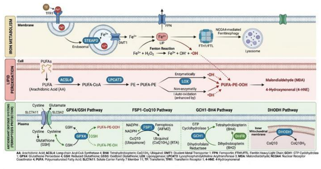
Figure 2.
Molecular organization and subcellular localization of antioxidant defense systems against ferroptosis. Detail of the four main GPX4-dependent and GPX4-independent systems for neutralizing lipid peroxidation in different cellular compartments. (A) GPX4/GSH pathway: the System Xc- transporter (SLC7A11/SLC3A2) ensures the uptake of cystine for the synthesis of GSH, which serves as a cofactor for GPX4 in the reduction of toxic lipid hydroperoxides (PUFA-PE-OOH) to safe lipid alcohols (PUFA-PE-OH); erastin and RSL3 act as specific inhibitors of these links. (B) FSP1-CoQ10 pathway at the plasma membrane: the myristoylated protein FSP1 reduces CoQ10 to ubiquinol (CoQ10H2), which acts as a lipophilic antioxidant-scavenging peroxyl radical independent of GSH. (C) Cytosolic GCH1-BH4-DHFR pathway: GCH1 synthesizes tetrahydrobiopterin (BH4), which inhibits PUFA-PE degradation and protects membranes from oxidation. (D) Mitochondrial DHODH pathway: the mitochondrial inner membrane enzyme DHODH reduces CoQ10 to CoQ10H2 in parallel with the oxidation of dihydroorotate to orotate, providing local protection of mitochondria. Abbreviations: BH2/BH4 – Dihydro/tetrahydrobiopterin; CoQ10/CoQ10H2 – Ubiquinone/ubiquinol; DHFR – Dihydrofolate reductase; DHODH – Dihydroorotate dehydrogenase; FSP1 – Ferroptosis suppressor protein 1; GCH1 – GTP cyclohydrolase 1; GPX4 – Glutathione peroxidase 4; GSH/GSSG – Reduced/oxidized glutathione; PUFA-PE-OOH – Phosphatidylethanolamine hydroperoxide. Note: Image created using Gemini tool (Google, 2026) based on the directions provided by the author to illustrate the concept. The final image was curated and verified for technical accuracy by the author.
Figure 2.
Molecular organization and subcellular localization of antioxidant defense systems against ferroptosis. Detail of the four main GPX4-dependent and GPX4-independent systems for neutralizing lipid peroxidation in different cellular compartments. (A) GPX4/GSH pathway: the System Xc- transporter (SLC7A11/SLC3A2) ensures the uptake of cystine for the synthesis of GSH, which serves as a cofactor for GPX4 in the reduction of toxic lipid hydroperoxides (PUFA-PE-OOH) to safe lipid alcohols (PUFA-PE-OH); erastin and RSL3 act as specific inhibitors of these links. (B) FSP1-CoQ10 pathway at the plasma membrane: the myristoylated protein FSP1 reduces CoQ10 to ubiquinol (CoQ10H2), which acts as a lipophilic antioxidant-scavenging peroxyl radical independent of GSH. (C) Cytosolic GCH1-BH4-DHFR pathway: GCH1 synthesizes tetrahydrobiopterin (BH4), which inhibits PUFA-PE degradation and protects membranes from oxidation. (D) Mitochondrial DHODH pathway: the mitochondrial inner membrane enzyme DHODH reduces CoQ10 to CoQ10H2 in parallel with the oxidation of dihydroorotate to orotate, providing local protection of mitochondria. Abbreviations: BH2/BH4 – Dihydro/tetrahydrobiopterin; CoQ10/CoQ10H2 – Ubiquinone/ubiquinol; DHFR – Dihydrofolate reductase; DHODH – Dihydroorotate dehydrogenase; FSP1 – Ferroptosis suppressor protein 1; GCH1 – GTP cyclohydrolase 1; GPX4 – Glutathione peroxidase 4; GSH/GSSG – Reduced/oxidized glutathione; PUFA-PE-OOH – Phosphatidylethanolamine hydroperoxide. Note: Image created using Gemini tool (Google, 2026) based on the directions provided by the author to illustrate the concept. The final image was curated and verified for technical accuracy by the author.
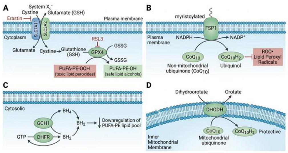
Figure 3.
The dual role of ferroptosis in disease pathogenesis and therapy: a comparative analysis of induction and inhibition strategies. The scheme illustrates the opposite therapeutic approaches to the modulation of ferroptosis depending on the pathological context. Left panel: Strategy for induction of ferroptosis in oncological diseases (in particular in resistant and mesenchymal tumors). The use of inducers (FINs: erastin, RSL3, sorafenib, etc.) or combination therapy with immunotherapy (IFNγ from CD8+ T cells) and radiotherapy causes massive accumulation of lipid ROS and death of cancer cells. At the same time, the risks are given: potential promotion of tumor progression through the release of DAMPs (HMGB1) and polarization of macrophages into the M2 phenotype (KRASG12D). Right panel: Strategy for inhibiting ferroptosis in neurodegeneration (Alzheimer’s, Parkinson’s disease), ischemic-reperfusion injury (stroke, infarction) and inflammation. The use of inhibitors (Ferrostatin-1, Liproxstatin-1), iron chelators (DFO, deferiprone) and antioxidants protects healthy tissues from lipid peroxidation. Abbreviations: AD – Alzheimer’s disease; AKI – Acute kidney injury; DFO – Deferoxamine; FINs – Ferroptosis inducers; HD – Huntington’s disease; HMGB1 – High mobility group B1 protein; IFNγ – Interferon-gamma; Lipid ROS – Lipid reactive oxygen species; NAC – N-Acetylcysteine; PD – Parkinson’s disease. Note: Image created using Gemini tool (Google, 2026) based on the directions provided by the author to illustrate the concept. The final image was curated and verified for technical accuracy by the author.
Figure 3.
The dual role of ferroptosis in disease pathogenesis and therapy: a comparative analysis of induction and inhibition strategies. The scheme illustrates the opposite therapeutic approaches to the modulation of ferroptosis depending on the pathological context. Left panel: Strategy for induction of ferroptosis in oncological diseases (in particular in resistant and mesenchymal tumors). The use of inducers (FINs: erastin, RSL3, sorafenib, etc.) or combination therapy with immunotherapy (IFNγ from CD8+ T cells) and radiotherapy causes massive accumulation of lipid ROS and death of cancer cells. At the same time, the risks are given: potential promotion of tumor progression through the release of DAMPs (HMGB1) and polarization of macrophages into the M2 phenotype (KRASG12D). Right panel: Strategy for inhibiting ferroptosis in neurodegeneration (Alzheimer’s, Parkinson’s disease), ischemic-reperfusion injury (stroke, infarction) and inflammation. The use of inhibitors (Ferrostatin-1, Liproxstatin-1), iron chelators (DFO, deferiprone) and antioxidants protects healthy tissues from lipid peroxidation. Abbreviations: AD – Alzheimer’s disease; AKI – Acute kidney injury; DFO – Deferoxamine; FINs – Ferroptosis inducers; HD – Huntington’s disease; HMGB1 – High mobility group B1 protein; IFNγ – Interferon-gamma; Lipid ROS – Lipid reactive oxygen species; NAC – N-Acetylcysteine; PD – Parkinson’s disease. Note: Image created using Gemini tool (Google, 2026) based on the directions provided by the author to illustrate the concept. The final image was curated and verified for technical accuracy by the author.
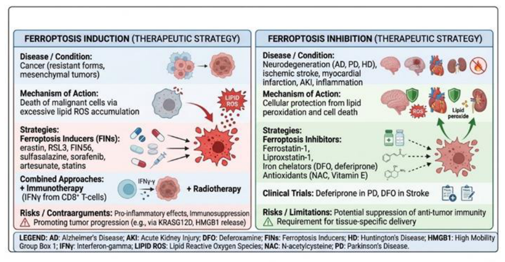
Figure 4.
Systematic map of the involvement of ferroptosis in the pathogenesis of major human diseases. Mapping the molecular mechanisms and pathophysiological consequences of ferroptosis in various organs and systems of the body. The central block reflects the basic intracellular axes (iron metabolism via TF/TFR1/FPN, lipid peroxidation involving ACSL4/LPCAT3/ALOX15 and antioxidant defense GPX4/GSH). The peripheral blocks detail specific pathological conditions: Brain and nervous system: Alzheimer’s disease (decreased FPN, increased peroxidation), Parkinson’s disease (α-synuclein-mediated sensitivity), Huntington’s disease, ischemic stroke (HIF-1α/TFR1 axis) and traumatic brain injury. Cardiovascular system: doxorubicin-induced cardiomyopathy (mitochondrial DOX-Fe2+), atherosclerosis (macrophage ferroptosis by oxLDL) and myocardial ischemia-reperfusion injury. Kidney: acute kidney injury (AKI) due to ischemia/reperfusion or inhibition of GPX4 in tubular epithelium. Lung: acute lung injury (ALI) and bronchial asthma (ALOX15-mediated ferroptosis). Liver: hepatocellular carcinoma, nonalcoholic steatohepatitis (NASH) and toxic injury (paracetamol/APAP). Gastrointestinal tract: ulcerative colitis and Crohn’s disease (ferroptosis of intestinal epithelial cells). Reproductive system: polycystic ovary syndrome (PCOS), premature ovarian failure and endometriosis. Abbreviations: ALOX15 – Arachidonate-15-lipoxygenase; APAP – Acetaminophen; DOX – Doxorubicin; FPN – Ferroportin; LIP – Labile iron pool; NOX4 – NADPH oxidase 4; oxLDL – Oxidized low-density lipoproteins; TFR1 – Transferrin receptor 1. Note: Image created using Gemini tool (Google, 2026) based on the directions provided by the author to illustrate the concept. The final image was curated and verified for technical accuracy by the author.
Figure 4.
Systematic map of the involvement of ferroptosis in the pathogenesis of major human diseases. Mapping the molecular mechanisms and pathophysiological consequences of ferroptosis in various organs and systems of the body. The central block reflects the basic intracellular axes (iron metabolism via TF/TFR1/FPN, lipid peroxidation involving ACSL4/LPCAT3/ALOX15 and antioxidant defense GPX4/GSH). The peripheral blocks detail specific pathological conditions: Brain and nervous system: Alzheimer’s disease (decreased FPN, increased peroxidation), Parkinson’s disease (α-synuclein-mediated sensitivity), Huntington’s disease, ischemic stroke (HIF-1α/TFR1 axis) and traumatic brain injury. Cardiovascular system: doxorubicin-induced cardiomyopathy (mitochondrial DOX-Fe2+), atherosclerosis (macrophage ferroptosis by oxLDL) and myocardial ischemia-reperfusion injury. Kidney: acute kidney injury (AKI) due to ischemia/reperfusion or inhibition of GPX4 in tubular epithelium. Lung: acute lung injury (ALI) and bronchial asthma (ALOX15-mediated ferroptosis). Liver: hepatocellular carcinoma, nonalcoholic steatohepatitis (NASH) and toxic injury (paracetamol/APAP). Gastrointestinal tract: ulcerative colitis and Crohn’s disease (ferroptosis of intestinal epithelial cells). Reproductive system: polycystic ovary syndrome (PCOS), premature ovarian failure and endometriosis. Abbreviations: ALOX15 – Arachidonate-15-lipoxygenase; APAP – Acetaminophen; DOX – Doxorubicin; FPN – Ferroportin; LIP – Labile iron pool; NOX4 – NADPH oxidase 4; oxLDL – Oxidized low-density lipoproteins; TFR1 – Transferrin receptor 1. Note: Image created using Gemini tool (Google, 2026) based on the directions provided by the author to illustrate the concept. The final image was curated and verified for technical accuracy by the author.
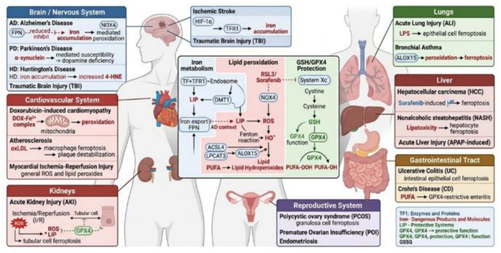
Figure 5.
Classification of pharmacological and innovative therapeutic strategies for ferroptosis modulation. Systematization of therapeutic agents by mechanism of action and technological approach. Block I (Ferroptosis inducers, FINs): Class I (System Xc- inhibitors: erastin, sulfasalazine, sorafenib, IKE), which deplete GSH; Class II (direct GPX4 inhibitors: RSL3, ML162, ML210); Class III (GPX4 degraders and CoQ10: FIN56); Class IV (iron oxidizers and LIP stimulators: FINO2, hemin, artesunate, DHA); and metabolic modulators (statins, cisplatin). Block II (Ferroptosis Inhibitors): Antioxidants-Radical Traps (RTAs: Ferrostatin-1, Liproxstatin-1, Vitamin E); Iron Chelators (deferoxamine, deferiprone, deferasirox); and GSH Precursors/Antioxidants (N-Acetylcysteine, Selenium). Block III (Innovative Approaches): Use of PROTAC-Degraders (dGPX4) for Selective Proteasome-mediated Degradation of GPX4; Nanoparticles for Targeted Delivery of FINs to the Tumor Microenvironment; and Combination Therapy (FINs Combined with Immunotherapy, Radiotherapy or Chemotherapy). Abbreviations: DHA – Dihydroartemisinin; DFO – Deferoxamine; FIN – Inducer of ferroptosis; IKE – Imidazole ketone erastin; LIP – Labile iron pool; NAC – N-Acetylcysteine; PROTAC – Proteolysis-targeting chimera; RTA – Radical-trapping antioxidant. Note: Image created using Gemini tool (Google, 2026) based on the directions provided by the author to illustrate the concept. The final image was curated and verified for technical accuracy by the author.
Figure 5.
Classification of pharmacological and innovative therapeutic strategies for ferroptosis modulation. Systematization of therapeutic agents by mechanism of action and technological approach. Block I (Ferroptosis inducers, FINs): Class I (System Xc- inhibitors: erastin, sulfasalazine, sorafenib, IKE), which deplete GSH; Class II (direct GPX4 inhibitors: RSL3, ML162, ML210); Class III (GPX4 degraders and CoQ10: FIN56); Class IV (iron oxidizers and LIP stimulators: FINO2, hemin, artesunate, DHA); and metabolic modulators (statins, cisplatin). Block II (Ferroptosis Inhibitors): Antioxidants-Radical Traps (RTAs: Ferrostatin-1, Liproxstatin-1, Vitamin E); Iron Chelators (deferoxamine, deferiprone, deferasirox); and GSH Precursors/Antioxidants (N-Acetylcysteine, Selenium). Block III (Innovative Approaches): Use of PROTAC-Degraders (dGPX4) for Selective Proteasome-mediated Degradation of GPX4; Nanoparticles for Targeted Delivery of FINs to the Tumor Microenvironment; and Combination Therapy (FINs Combined with Immunotherapy, Radiotherapy or Chemotherapy). Abbreviations: DHA – Dihydroartemisinin; DFO – Deferoxamine; FIN – Inducer of ferroptosis; IKE – Imidazole ketone erastin; LIP – Labile iron pool; NAC – N-Acetylcysteine; PROTAC – Proteolysis-targeting chimera; RTA – Radical-trapping antioxidant. Note: Image created using Gemini tool (Google, 2026) based on the directions provided by the author to illustrate the concept. The final image was curated and verified for technical accuracy by the author.
Disclaimer/Publisher’s Note: The statements, opinions and data contained in all publications are solely those of the individual author(s) and contributor(s) and not of MDPI and/or the editor(s). MDPI and/or the editor(s) disclaim responsibility for any injury to people or property resulting from any ideas, methods, instructions or products referred to in the content. |
© 2026 by the author. Licensee MDPI, Basel, Switzerland. This article is an open access article distributed under the terms and conditions of the Creative Commons Attribution (CC BY) license.
Copyright: This open access article is published under a Creative Commons CC BY 4.0 license, which permit the free download, distribution, and reuse, provided that the author and preprint are cited in any reuse.