
Submitted:
08 September 2026
Posted:
09 September 2026
You are already at the latest version
Abstract
Background: Severe varicella in previously healthy adults is uncommon, and underlying genetic susceptibility is increasingly recognized as a contributing factor in such cases. However, the coexistence of heterozygous variants affecting distinct immunological pathways has rarely been documented. Case presentation: We report a 39-year-old previously healthy Chinese male who presented with a 2-day history of fever and a 1-day history of a generalized vesicular rash after exposure to a patient with herpes zoster. The patient rapidly developed severe pneumonia, acute liver injury (ALT 617.9 U/L, AST 1055.2 U/L), acute myocardial injury (troponin T 17.91 pg/mL), acute kidney injury (creatinine 121.4 μmol/L), and severe thrombocytopenia (36×10⁹/L) with coagulopathy. Lymphocyte subset analysis obtained during acute illness revealed decreased CD4⁺ T cells (373.6/μL), elevated CD8⁺ T cells (1450.3/μL), and an inverted CD4⁺/CD8⁺ ratio, likely reflecting infection-induced perturbations. Whole-exome sequencing identified a heterozygous missense variant in FOXN1 (NM_003593.3) (c.163G>A, p.G55S) and a heterozygous missense variant in PIK3CD (NM_005026.4) (c.1487G>A, p.R496Q), both classified as variants of uncertain significance without functional validation. The patient received high-flow nasal cannula oxygen therapy, intravenous acyclovir, intravenous immunoglobulin, and multiorgan supportive care, and achieved full recovery without sequelae. Conclusions: This case represents the first report of coexisting FOXN1 and PIK3CD concurrent heterozygous variants in an adult presenting with severe varicella and multiorgan dysfunction. As both variants are VUS and lack functional characterization, any attribution of functional impact remains speculative. It is hypothesized that the FOXN1 variant, if pathogenic, might reduce thymic T-cell output, and the PIK3CD variant, if gain-of-function, could cause T- and B-cell dysregulation, potentially impairing antiviral responses. Causality has not been established, and functional studies are required to determine the significance of these variants. This case underscores the importance of genetic evaluation in previously healthy adults who develop unusually severe infections and suggests that concurrent heterozygous variants of uncertain significance may contribute to a "hidden immunodeficiency" phenotype, wherein a baseline vulnerability only becomes clinically apparent upon a potent viral trigger.
Keywords:
varicella-zoster virus
; severe varicella
; multiple organ dysfunction syndrome
; FOXN1 gene
; PIK3CD gene
; activated PI3Kδ syndrome
; inborn errors of immunity
; case report
1. Introduction
Varicella, caused by primary infection with varicella-zoster virus (VZV), typically follows a benign and self-limited course in children. In adults, however, varicella is less common but tends to be more severe, with pneumonia occurring in approximately 5%–15% of cases [1]. The disease may also be complicated by hepatitis, encephalitis, disseminated intravascular coagulation, and myocarditis. Severe varicella predominantly affects immunocompromised hosts, pregnant women, and smokers, and is relatively rare in individuals without a known history of immunodeficiency [2].
However, an increasing number of severe infections in patients previously deemed "immunocompetent" have been attributed to underlying inborn errors of immunity (IEI) or other genetic susceptibilities, suggesting that the absence of a clinically overt immunodeficiency history does not exclude significant genetic predisposition. With the widespread application of whole-exome and whole-genome sequencing, an increasing number of severe infections in patients previously deemed "immunocompetent" have been attributed to underlying genetic susceptibility. A systematic review by Ansari et al. highlighted the association between severe VZV infection and both inborn errors of immunity (IEI) and acquired immunodeficiencies, emphasizing that mutations affecting T- and B-cell function confer heightened susceptibility to viral infections [3]. Gain-of-function (GOF) variants in PIK3CD, which cause activated PI3Kδ syndrome type 1 (APDS1), exemplify this relationship. APDS1 is characterized by recurrent respiratory tract infections, lymphoproliferation, autoimmunity, and increased susceptibility to herpesvirus infections, with reported herpesvirus infection rates ranging from 37.5% to 49% [4,5].
FOXN1 encodes forkhead box protein N1, a transcription factor essential for thymic epithelial cell development and T-cell maturation. Biallelic pathogenic variants in FOXN1 cause T-B⁺NK⁺ severe combined immunodeficiency, accompanied by congenital alopecia and nail dystrophy. More recently, heterozygous FOXN1 variants resulting in haploinsufficiency have been recognized as a cause of non-severe combined immunodeficiency, manifesting as reduced T-cell counts and increased susceptibility to respiratory viral infections [6,7].
Herein, we report the first case of an adult patient carrying concurrent heterozygous variants in both FOXN1 and PIK3CD who presented with severe varicella and rapidly progressive multiorgan dysfunction. This case highlights the possible synergistic effect of concurrent genetic variants of uncertain significance in conferring susceptibility to severe viral infections in individuals without a prior history of immunodeficiency.
2. Case Presentation
2.1. Clinical History
A 39-year-old previously healthy Chinese male was admitted to our hospital in December 2024 with a 2-day history of fever and a 1-day history of a generalized rash. One week prior to symptom onset, the patient had documented exposure to a family member with herpes zoster. Two days before admission, he developed fever with a maximum temperature of 39.2°C, accompanied by fatigue and diffuse myalgia. One day before admission, erythematous maculopapules appeared initially on the face and scalp, rapidly progressing to vesicles and spreading to the trunk and extremities, with involvement of the oral mucosa.
The patient had no significant past medical history, including hypertension, diabetes mellitus, chronic hepatitis, or tuberculosis. He denied long-term use of glucocorticoids or other immunosuppressive agents and reported no history of recurrent infections. He was a non-smoker and denied any family history of genetic disorders or primary immunodeficiency.
2.2. Physical Examination
On admission, the patient's vital signs were as follows: temperature 39.0°C, heart rate 112 beats per minute, respiratory rate 26 breaths per minute, blood pressure 127/78 mmHg, and oxygen saturation 88% on room air. The patient was alert and oriented but appeared acutely ill with tachypnea. Physical examination revealed diffuse erythematous macules, papules, vesicles, and scattered pustules distributed predominantly on the face, scalp, trunk, and extremities in a centripetal pattern (Figure 1). Some vesicles had ruptured and crusted. Oral mucosal involvement was noted (Figure 2). Auscultation of the lungs revealed coarse breath sounds bilaterally without definite rales or rhonchi. Cardiac examination revealed a regular rate and rhythm without murmurs, rubs, or gallops. The abdomen was soft and nontender, without hepatosplenomegaly.
2.3. Laboratory and Imaging Findings
Admission laboratory findings are summarized in Table 1. Notable abnormalities included severe thrombocytopenia (platelet count 36×10⁹/L), markedly elevated hepatic transaminases (ALT 617.9 U/L, AST 1055.2 U/L), hyperbilirubinemia (total bilirubin 52.8 μmol/L), elevated cardiac biomarkers (troponin T 17.91 pg/mL, CK-MB 38.7 U/L, LDH 2993.6 U/L), coagulopathy (D-dimer 161.28 mg/L, PT 13.8 s), and acute kidney injury (creatinine 121.4 μmol/L). Inflammatory markers were elevated, with C-reactive protein of 98.64 mg/L and procalcitonin of 1.28 ng/mL. Lymphocyte subset analysis performed during the acute phase decreased CD4⁺ T-cell count (373.6/μL), increased CD8⁺ T-cell count (1450.3/μL), and an inverted CD4⁺/CD8⁺ ratio (reference ranges: CD4⁺ T cells 518.8–963.9/μL; CD8⁺ T cells 326.7–640.2/μL). Notably, this pattern reflects a profound CD8⁺ T-cell expansion driven by the acute viral infection. It should be noted that these values were obtained during severe acute infection and may primarily reflect viral-induced lymphopenia and CD8⁺ T-cell expansion rather than a baseline immunodeficiency.
Chest computed tomography (CT) revealed diffuse bilateral ground-glass opacities and areas of consolidation, predominantly affecting the lower lobes, consistent with severe varicella pneumonia (Figure 3 and Figure 4). Non-contrast cranial CT showed no abnormalities. Abdominal ultrasonography demonstrated coarsened hepatic echotexture without splenomegaly.
2.4. Genetic Findings
To investigate the potential genetic basis for severe varicella in this apparently healthy adult, whole-exome sequencing was performed on peripheral blood after obtaining informed consent. The analysis revealed two heterozygous missense variants, both of which are currently classified as variants of uncertain significance (VUS) and have not been functionally validated.
FOXN1 heterozygous variant (OMIM: 600838): The variant c.163G>A (p.G55S) was identified in the FOXN1 gene. This variant has an unknown allele frequency in population databases, and no homozygous individuals have been reported. In ClinVar, this variant is currently classified as a VUS (Variation ID: 934579). FOXN1 encodes forkhead box protein N1, a transcription factor essential for thymic epithelial cell development and T-cell maturation. While biallelic pathogenic variants cause T-B⁺NK⁺ severe combined immunodeficiency, heterozygous variants in some cases have been associated with non-severe combined immunodeficiency characterized by reduced T-cell counts and increased susceptibility to viral infections [6,7]. However, no functional studies have been performed on p.G55S, and it remains unknown whether this specific missense substitution results in haploinsufficiency or alters FOXN1 function.
PIK3CD heterozygous variant (OMIM: 602839): The variant c.1487G>A (p.R496Q) was identified in exon 12 (coding exon 10) of the PIK3CD gene, resulting in the substitution of arginine by glutamine at position 496 within the C2 domain [11]. This variant is classified as a VUS in ClinVar. PIK3CD encodes the catalytic subunit p110δ of phosphoinositide 3-kinase δ (PI3Kδ). Although GOF variants in PIK3CD have been shown to cause APDS1—characterized by combined immunodeficiency, recurrent respiratory infections, lymphoproliferation, and increased susceptibility to herpesvirus infections [8,9]—it has not been functionally demonstrated that p.R496Q acts as a gain-of-function allele, and its pathogenicity therefore remains speculative.
2.5. Clinical Course and Outcome
Following admission, the patient was started on high-flow nasal cannula oxygen therapy (FiO₂ 50%, flow rate 40 L/min) to maintain oxygen saturation >95%. Intravenous acyclovir (10 mg/kg every 8 h) was initiated for antiviral therapy, combined with intravenous immunoglobulin (400 mg/kg/day for 5 days) for immunomodulation. Supportive care included hepatoprotective therapy (magnesium isoglycyrrhizinate, glutathione), myocardial protection (creatine phosphate sodium), correction of coagulopathy (vitamin K₁, fresh frozen plasma), and other symptomatic treatments.
By day 3 of hospitalization, the patient's temperature normalized. By day 5, respiratory symptoms had markedly improved, and high-flow nasal cannula oxygen therapy was discontinued. The cutaneous lesions gradually dried and crusted. On hospital day 12, the rash had completely crusted, and repeat laboratory testing demonstrated normalization of hepatic function, cardiac biomarkers, coagulation parameters, and renal function. Follow-up chest CT showed near-complete resolution of bilateral pulmonary infiltrates. The patient was discharged in stable condition. At 3-month follow-up, he remained well without sequelae.
3. Discussion
3.1. Clinical Features and Prognosis of Severe Varicella in Adults
Adult varicella is typically more severe than childhood varicella. The reported incidence of pneumonia in adult varicella ranges from 5% to 15%, and affected patients may rapidly progress to acute respiratory distress syndrome (ARDS), with mortality rates reaching 10%–30% [2]. In addition to pneumonia, hepatitis, myocarditis, encephalitis, thrombocytopenia, and disseminated intravascular coagulation are common complications of severe adult varicella. In the present case, the patient developed multiorgan dysfunction syndrome characterized by severe pneumonia, acute liver injury, acute myocardial injury, acute kidney injury, and severe thrombocytopenia within a short period after symptom onset. The rapidity of disease progression and the breadth of organ involvement are unusual in previously healthy adults with varicella, suggesting the presence of a possible underlying host immunodeficiency.
3.2. PIK3CD Variants and Immunodeficiency
The PIK3CD gene encodes the catalytic subunit p110δ of PI3Kδ, which is predominantly expressed in leukocytes and plays a critical role in the PI3K/AKT/mTOR signaling pathway. GOF variants in PIK3CD lead to hyperactivation of PI3Kδ and sustained downstream signaling, resulting in immune dysregulation—B-cell hyperactivation with hypogammaglobulinemia or hyper-IgM syndrome, and T-cell senescence with functional impairment [10]. The c.1487G>A (p.R496Q) variant identified in our patient is located in the C2 domain and is currently classified as a VUS. It must be stressed that no functional studies have been performed on this specific variant; therefore, designating it as a GOF variant is purely conjectural. Takeda et al. previously reported that missense variants in the C2 domain (e.g., N334K and C416R) can cause the APDS1 phenotype, and GOF variants have been confirmed in multiple domains of PIK3CD [11]. Crank et al. described hyper-IgM syndrome cases associated with various PIK3CD pathogenic variants [8]. Thus, while the variant's location in a domain known to harbor APDS1-associated mutations raises suspicion, it does not establish pathogenicity for p.R496Q.
Large cohort studies have demonstrated that herpesvirus infection rates in patients with APDS range from 37.5% to 49%, including VZV, Epstein-Barr virus, and cytomegalovirus [4,5]. In the present case, the patient presented with severe varicella as the initial manifestation, without a history of recurrent infections, lymphoproliferation, or autoimmunity, representing a possible atypical or "mild" phenotype that could be related to the PIK3CD variant only if it is eventually proven to be a GOF allele. Nevertheless, the severe thrombocytopenia and multiorgan dysfunction that developed following VZV infection are compatible with the pathophysiological features of APDS, in which viral infections in the context of immune dysregulation can trigger excessive inflammatory responses.
3.3. Pathogenicity Analysis of the FOXN1 c.163G>A Variant
The FOXN1 c.163G>A (p.G55S) variant identified in our patient is classified as a VUS in ClinVar, and its allele frequency in population databases is unknown. In silico prediction tools indicate that this residue is conserved across some species. Although this variant has not been previously reported in patients with FOXN1-related disorders, several indirect lines of evidence might suggest potential pathogenicity. FOXN1 haploinsufficiency has been demonstrated to cause delayed thymic epithelial cell development and reduced T-cell output. Bosticardo et al. showed that individuals carrying heterozygous loss-of-function FOXN1 variants may present with low T-cell receptor excision circle (TREC) levels and T-cell lymphopenia at birth, and reduced CD8⁺ T-cell counts in adulthood [6]. Pasternak et al. further confirmed that heterozygous FOXN1 variants are associated with "mild and variable immunodeficiency" [12]. Brader et al. reported a case of delayed-onset immune dysfunction in a man carrying a heterozygous frameshift FOXN1 variant, noting that "previously healthy heterozygous mutation carriers may be at risk of developing immune dysfunction later in life"[13].
However, it is critical to emphasize that the T-cell subset analysis in this patient was performed during acute severe VZV infection, a condition known to cause transient CD4⁺ T-cell lymphopenia and CD8⁺ T-cell expansion. Consequently, the observed CD4⁺ T-cell count of 373.6/μL and inverted CD4⁺/CD8⁺ ratio may be predominantly, or entirely, attributable to the acute infection rather than to a pre-existing thymic defect caused by the FOXN1 variant. These immunological data cannot be used as reliable evidence of impaired thymic output. Definitive assessment of the FOXN1 variant's functional impact would require immunological studies after full recovery, including measurement of TREC levels, naïve/memory T-cell subset distribution, T-cell proliferation assays, and serum immunoglobulin levels.
Moses et al. proposed a systematic framework for assessing the functional consequences of FOXN1 variants, categorizing them as loss-of-function, gain-of-function, dominant-negative, or benign, and highlighted that different variants exert distinct effects on transcriptional activity, nuclear localization, and T-cell output [7]. The p.G55S variant resides in the N-terminal region of FOXN1; its specific functional impact requires experimental validation.
3.4. Potential Synergistic Effect of Concurrent Heterozygous FOXN1 and PIK3CD Variants
A notable aspect of this case is the simultaneous presence of heterozygous variants in both FOXN1 and PIK3CD. Notably, FOXN1 primarily affects thymic T-cell development and maturation, whereas PIK3CD GOF variants predominantly cause functional dysregulation of B and T cells. It is important to underline that the discussion below is entirely speculative, as neither variant has been functionally characterized.
We hypothesize a two-hit model for the severe phenotype observed in this patient. The FOXN1 variant, if pathogenic, might establish a baseline vulnerability by reducing the peripheral naïve T-cell pool through impaired thymic output. Upon VZV infection, the PIK3CD variant, if it were a gain-of-function allele, could further compromise the antiviral response by promoting T-cell exhaustion and impairing the generation of functional VZV-specific effector T cells. This combination of a quantitative defect (reduced T-cell reserves) and a qualitative defect (dysfunctional T-cell signaling) could synergistically predispose to uncontrolled viral dissemination and the subsequent hyperinflammatory multiorgan dysfunction syndrome.
These two genes act at different levels of the immune system, and their variants could, in this hypothetical framework, exert synergistic pathogenic effects. However, causality has not been established, and this model remains speculative without functional validation of the specific variants identified.
To our knowledge, no previous cases of coexisting FOXN1 and PIK3CD variants have been reported in the literature. Notably, Roifman-Chitayat syndrome (OMIM: 613328) is a digenic disorder caused by biallelic loss-of-function variants in PIK3CD and KNSTRN, manifesting as combined immunodeficiency, facial dysmorphism, optic atrophy, skeletal abnormalities, and developmental delay [14]. Although the inheritance pattern of this syndrome differs from that observed in our patient—who carries a heterozygous PIK3CD variant of uncertain significance and a heterozygous FOXN1 variant—it demonstrates that biallelic loss of PIK3CD function can contribute to a combined immunodeficiency phenotype in concert with another genetic hit, providing a conceptual precedent for multigenic contributions to immunodeficiency.
3.5. Requirements for Reclassification of the Variants of Uncertain Significance
At present, both the FOXN1 c.163G>A and PIK3CD c.1487G>A variants are classified as VUS. Neither variant has been subjected to functional assays; therefore, all discussions of potential synergy or functional impact remain hypothetical. To reclassify these variants as likely pathogenic, several lines of evidence would be required: (Ⅰ) functional assays demonstrating that FOXN1 p.G55S impairs transcriptional activity, nuclear localization, or thymic epithelial cell support, and that PIK3CD p.R496Q increases PI3Kδ enzymatic activity or downstream AKT phosphorylation; (Ⅱ) segregation analysis showing that the variants track with an immunological phenotype in family members; (Ⅲ) post-recovery immunological studies (e.g., TREC levels, naïve T-cell counts, T-cell proliferation) that confirm a persistent T-cell defect independent of the acute infectious episode; and (Ⅳ) identification of the same or similar variants in unrelated patients with compatible clinical presentations. At the time of writing, no such data are available.
3.6. Clinical Implications and Future Directions
This case offers several clinical insights. First, for previously healthy adults who present with severe varicella, underlying genetic susceptibility should be considered even in the absence of a typical immunodeficiency history or physical findings. In such cases, whole-exome sequencing may provide valuable diagnostic clues, although results must be interpreted with caution, particularly when VUS are identified. Second, early combination therapy with antiviral agents and immunomodulation is crucial for successful management. Third, targeted therapy for PIK3CD GOF variants represents an emerging approach for APDS. The selective PI3Kδ inhibitor leniolisib received FDA approval in March 2023 for the treatment of APDS, and clinical trials have demonstrated significant improvements in immune dysregulation and lymphoproliferation [15]. Should this patient develop APDS-related complications in the future and the PIK3CD variant be confirmed as a pathogenic GOF mutation, targeted intervention with a PI3Kδ inhibitor might be considered.
3.7. Limitations
This study has several important limitations. First, both the FOXN1 c.163G>A and PIK3CD c.1487G>A variants are currently classified as VUS, and no functional data have been generated to validate their impact; any discussion of pathogenicity is therefore based on indirect and inconclusive evidence. Second, TREC levels and T-cell proliferation assays were not performed, precluding functional assessment of thymic output and T-cell immune status. Third, family members were not available for segregation analysis. Fourth, and most critically, all immunophenotypic data were obtained in the context of acute severe VZV infection, which can independently induce profound perturbations in lymphocyte subsets. Consequently, the observed T-cell abnormalities cannot be confidently attributed to an underlying genetic defect, and the causal link between the identified variants and the severe clinical presentation remains unestablished. These limitations underscore the need for post-recovery immunological profiling and functional validation studies.
5. Conclusions
This case represents the first report of coexisting FOXN1 and PIK3CD concurrent heterozygous variants in an adult presenting with severe varicella and multiorgan dysfunction. Given that both variants remain VUS and have not been functionally characterized, any attribution of functional consequences is hypothetical. It is hypothesized that the FOXN1 variant, if pathogenic, might reduce thymic T-cell output and the PIK3CD variant, if gain-of-function, could cause immune dysregulation, potentially contributing to impaired antiviral responses. However, causality has not been established. Furthermore, the T-cell subset alterations observed were recorded during acute infection and may primarily reflect viral effects rather than a fixed immunodeficiency. This case highlights the importance of considering underlying genetic susceptibility in previously healthy adults who present with unusually severe manifestations of common viral infections, and supports the concept of "hidden immunodeficiency"—a condition in which a baseline genetic vulnerability remains clinically silent until unmasked by a potent infectious trigger, potentially arising from concurrent heterozygous variants in immune-related genes. Functional studies are warranted to determine the clinical significance of these VUS and to elucidate any potential synergistic effects that may underpin this severe clinical presentation.
Author Contributions
Conceptualization, Y.S. and J.C.; data curation, Y.S., H.C. and H.L; investigation and clinical management, Y.S., J.C., H.C., H.L. and H.Z.; writing—original draft preparation, Y.S. and H.Z.; writing—review and editing, Y.S. and J.C.; supervision, H.Z.; All authors have read and agreed to the published version of the manuscript.
Funding
This research received no external funding.
Institutional Review Board Statement
The study was conducted in accordance with the Declaration of Helsinki, and approved by the Institutional Review Board of Jinjiang Municipal Hospital (Shanghai sixth People's Hospital Fujian) (protocol code: jjsyyll-2026-079; date of approval: June 10, 2026).
Informed Consent Statement
Written informed consent has been obtained from the patient to publish this paper.
Data Availability Statement
The data presented in this case report are available from the corresponding author upon reasonable request. The data are not publicly available due to patient privacy.
Conflicts of Interest
The authors declare no conflicts of interest.
Abbreviations
The following abbreviations are used in this manuscript:
| ALT | Alanine aminotransferase |
| APDS | Activated PI3Kδ syndrome |
| APDS1 | Activated PI3Kδ syndrome type 1 |
| ARDS | Acute respiratory distress syndrome |
| AST | Aspartate aminotransferase |
| CK-MB | Creatine kinase-MB |
| CT | Computed tomography |
| FDA | Food and Drug Administration |
| GOF | Gain-of-function |
| LDH | Lactate dehydrogenase |
| OMIM | Online Mendelian Inheritance in Man |
| PI3Kδ | Phosphoinositide 3-kinase δ |
| PT | Prothrombin time |
| TREC | T-cell receptor excision circle |
| VUS | Variant of uncertain significance |
| VZV | Varicella-zoster virus |
| IEI | Inborn errors of immunity |
References
- Gershon, A.A.; Breuer, J.; Cohen, J.I.; Cohrs, R.J.; Gershon, M.D.; Gilden, D.; Grose, C.; Hambleton, S.; Kennedy, P.G.E.; Oxman, M.N.; et al. Varicella zoster virus infection. Nat. Rev. Dis. Primers 2015, 1, 15016.
- Tunbridge, A.J.; Breuer, J.; Jeffery, K.J.M. Chickenpox in adults — Clinical management. J. Infect. 2008, 57, 95–102.
- Ansari, R.; Rosen, L.B.; Lisco, A.; Gilden, D.; Holland, S.M.; Zerbe, C.S.; Burbelo, P.D.; Cohen, J.I. Primary and acquired immunodeficiencies associated with severe varicella-zoster virus infections. Clin. Infect. Dis. 2021, 73, e2705–e2712.
- Coulter, T.I.; Chandra, A.; Bacon, C.M.; Babar, J.; Curtis, J.; Screaton, N.; Goodlad, J.R.; Farmer, G.; Steele, C.L.; Leahy, T.R.; et al. Clinical spectrum and features of activated phosphoinositide 3-kinase δ syndrome: A large patient cohort study. J. Allergy Clin. Immunol. 2017, 139, 597–606.e4.
- Jamee, M.; Moniri, S.; Zaki-Dizaji, M.; Olbrich, P.; Yazdani, R.; Jadidi-Niaragh, F.; Aghamohammadi, A.; Abolhassani, H.; Hammarström, L.; Rezaei, N. Clinical, immunological, and genetic features in patients with activated PI3Kδ syndrome (APDS): A systematic review. Clin. Rev. Allergy Immunol. 2020, 59, 323–333.
- Bosticardo, M.; Yamazaki, Y.; Cowan, J.; Giardino, G.; Corsino, C.; Ferrua, F.; Di Matteo, G.; Pignata, C.; Dasso, J.; Sgrò, F.; et al. Heterozygous FOXN1 variants cause low TRECs and severe T cell lymphopenia, revealing a crucial role of FOXN1 in supporting early thymopoiesis. Am. J. Hum. Genet. 2019, 105, 549–561.
- Moses, A.; Bhalla, P.; Thompson, A.; Cepika, A.M.; DeKruyff, R.H.; Dorenkamp, M.; Mace, E.M.; Orange, J.S.; Puck, J.M.; Scholes, K.; et al. Comprehensive phenotypic analysis of diverse FOXN1 variants. J. Allergy Clin. Immunol. 2023, 152, 1273–1291.e15.
- Crank, M.C.; Grossman, J.K.; Moir, S.; Pittaluga, S.; Buckner, C.M.; Kardava, L.; Agharahimi, A.; Meuwissen, P.; Driessen, G.; Pals, S.T.; et al. Mutations in PIK3CD can cause hyper IgM syndrome (HIGM) associated with increased cancer susceptibility. J. Clin. Immunol. 2014, 34, 272–276.
- Angulo, I.; Vadas, O.; Garçon, F.; Banham-Hall, E.; Plagnol, V.; Leahy, T.R.; Baxendale, H.; Coulter, T.; Curtis, J.; Wu, C.; et al. Phosphoinositide 3-kinase δ gene mutation predisposes to respiratory infection and airway damage. Science 2013, 342, 866–871.
- Lucas, C.L.; Kuehn, H.S.; Zhao, F.; Niemela, J.E.; Deenick, E.K.; Palendira, U.; Avery, D.T.; Moens, L.; Cannons, J.L.; Biancalana, M.; et al. Dominant-activating germline mutations in the gene encoding the PI(3)K catalytic subunit p110δ result in T cell senescence and human immunodeficiency. Nat. Immunol. 2014, 15, 88–97.
- Takeda, A.J.; Zhang, Y.; Dornan, G.L.; Siempelkamp, B.D.; Jenkins, M.L.; Matthews, H.F.; McElwee, J.J.; Han, K.Y.; Sharma, J.; Dasso, J.; et al. Novel PIK3CD mutations affecting N-terminal residues of p110δ cause activated PI3Kδ syndrome (APDS) in humans. J. Allergy Clin. Immunol. 2017, 140, 1152–1156.e10.
- Pasternak, Y.; Vong, L.; Merico, D.; Amar, D.; Somech, R.; Gershoni-Baruch, R.; Segev, A.; Oren, M.; Adiv, O.E.; Belostotsky, R.; et al. Utilization of next-generation sequencing to define the role of heterozygous FOXN1 variants in immunodeficiency. J. Allergy Clin. Immunol. Glob. 2024, 3, 100267.
- Brader, J.; O'Brien, R.; Rees, C. Broadening the phenotypic spectrum of forkhead box N1 gene mutations. Cureus 2025, 17, e94410.
- Sharfe, N.; Karanxha, A.; Dadi, H.; Merico, D.; Chitayat, D.; Herbrick, J.A.; Freeman, S.; Grinstein, S.; Roifman, C.M. Dual loss of p110δ PI3-kinase and SKAP (KNSTRN) expression leads to combined immunodeficiency and multisystem syndromic features. J. Allergy Clin. Immunol. 2018, 142, 618–629.
- Rao, V.K.; Webster, S.; Šedivá, A.; Plebani, A.; Schuetz, C.; Shcherbina, A.; Rakhmanina, N.; Dalm, V.A.S.H.; Shearer, W.; Uzel, G.; et al. A randomized, placebo-controlled phase 3 trial of the PI3Kδ inhibitor leniolisib for activated PI3Kδ syndrome. Blood 2023, 141, 971–983.
Figure 1.
Cutaneous manifestations of severe varicella on admission.
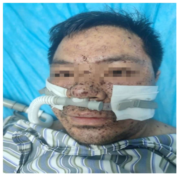
Figure 2.
Mucocutaneous involvement. .
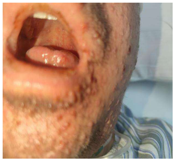
Figure 3.
Chest CT on admission.
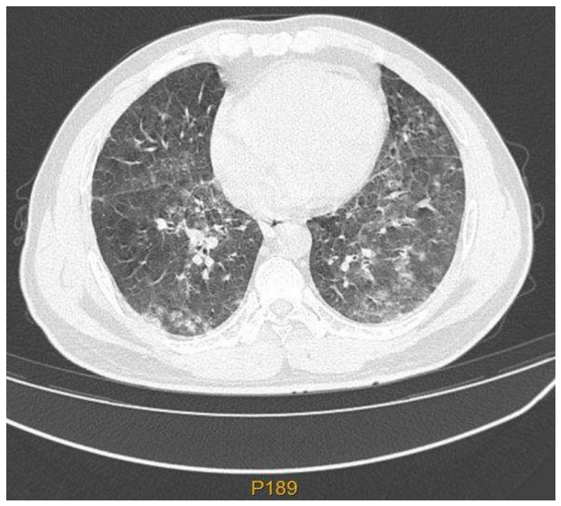
Figure 4.
Chest CT on admission.
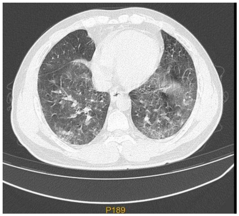

Table 1.
Laboratory findings on admission.
| Parameter | Result | Reference range |
| Platelet count | 36×10⁹/L | 125–350×10⁹/L |
| C-reactive protein | 98.64 mg/L | <10 mg/L |
| Alanine aminotransferase (ALT) | 617.9 U/L | 7–40 U/L |
| Aspartate aminotransferase (AST) | 1055.2 U/L | 8–40 U/L |
| Total bilirubin | 52.8 μmol/L | 0–23 μmol/L |
| Creatinine | 121.4 μmol/L | 57–97 μmol/L |
| Lactate dehydrogenase (LDH) | 2993.6 U/L | 120–250 U/L |
| Troponin T | 17.91 pg/mL | <14 pg/mL |
| Creatine kinase-MB (CK-MB) | 38.7 U/L | 0–24 U/L |
| D-dimer | 161.28 mg/L | 0–0.5 mg/L |
| Prothrombin time (PT) | 13.8 s | 9.4–12.5 s |
| Procalcitonin | 1.28 ng/mL | 0–0.5 ng/mL |
| CD4⁺ T cells | 373.6/μL | 518.8–963.9/μL |
| CD8⁺ T cells | 1450.3/μL | 326.7–640.2/μL |
Disclaimer/Publisher’s Note: The statements, opinions and data contained in all publications are solely those of the individual author(s) and contributor(s) and not of MDPI and/or the editor(s). MDPI and/or the editor(s) disclaim responsibility for any injury to people or property resulting from any ideas, methods, instructions or products referred to in the content. |
© 2026 by the authors. Licensee MDPI, Basel, Switzerland. This article is an open access article distributed under the terms and conditions of the Creative Commons Attribution (CC BY) license (http://creativecommons.org/licenses/by/4.0/).
Copyright: This open access article is published under a Creative Commons CC BY 4.0 license, which permit the free download, distribution, and reuse, provided that the author and preprint are cited in any reuse.