

Submitted:
08 September 2026
Posted:
09 September 2026
You are already at the latest version
Preprints on COVID-19 and SARS-CoV-2
Abstract
The global emergence of coronavirus disease 2019 (COVID-19) was caused by severe acute respiratory syndrome coronavirus 2 (SARS-CoV-2). The clinical manifestations of COVID-19 in patients range from asymptomatic disease to severe multiorgan dysfunction and even death. Moreover, the growing population of clinically immunodeficient patients and the use of single clinical antifungal agents have gradually increased the incidence of Aspergillus fumigatus infections, the complex clinical manifestations of which are yet to be fully clarified. The mortality rate among COVID-19 patients coinfected with A. fumigatus has risen significantly. Therefore, a comprehensive understanding of the diagnosis and treatment of A. fumigatus infections, SARS-CoV-2 infections, and their coinfections is crucial. In this review, we first provide a comprehensive description of the life cycle of A. fumigatus, together with an in-depth analysis of its characteristics, diagnosis, and treatment strategies for associated diseases. Then the basic structure of SARS-CoV-2, the evolution of variant strains, and key aspects of its prevention and control was discussed. Finally, the symptoms, diagnosis, and treatment of COVID-19 coinfection with A. fumigatus are summarized. In this paper, we provide methodological references and data to support the clinical prevention and treatment of A. fumigatus and SARS-CoV-2 infections.
Keywords:
COVID-19
; Aspergillus fumigatus
; coinfection
; diagnosis
; prevention
; treatment
1. Introduction
Severe acute respiratory syndrome coronavirus 2 (SARS-CoV-2) is an enveloped RNA-β coronavirus, characterized by round or oval particles. It shares genetic similarities with SARS-CoV and primarily causes lung infections. The symptoms of SARS-CoV-2 infections range from mild influenza-like symptoms to acute respiratory distress syndrome [1,2]. Since the emergence of SARS-CoV-2 in 2019, there have been instances of coinfection with the novel coronavirus and Aspergillus in the intensive/critical care units of hospitals, with a strikingly high incidence rate of 33.3% for COVID-19 combined with invasive aspergillosis [3]. However, in comparison to the coinfection of SARS-CoV or Middle East respiratory syndrome coronavirus (MERS-CoV) with fungal, there have been relatively few reports of secondary fungal infections associated with COVID-19 [4,5].
According to the World Health Organization (WHO), respiratory infections caused by fungal, bacterial, and viral pathogens ranked eighth among the top 10 causes of mortality in the United States between 2011 and 2018. Fungal infections alone accounted for approximately 1.6 million deaths worldwide, exceeding the number of deaths from malaria and approaching that from tuberculosis [6]. The number of fungal species capable of causing human infections exceeds 600, but the Aspergillus accounts for approximately 70% of these infections [7]. Over 350 species of Aspergillus have been identified so far. However, the primary causative agents of human invasive aspergillosis are A. fumigatus, A. flavus, A. niger, A. nidulans, and A. terreus. Among these species, infections caused by A. fumigatus rank highest in terms of life-threatening cases [8], and affect over 300,000 patients worldwide annually [9].
A. fumigatus is a saprophytic pathogenic fungus that can survive at a wide range of temperatures and pHs [10,11]. Its hydrophobic conidia are small and can penetrate the respiratory tract to reach the lungs. In people with normal immune function, these conidia generally do not cause disease. However, in patients with compromised immunity, such as those with neutropenia, malignant tumors, HIV, organ transplantation, or novel coronavirus infection, it often causes allergic bronchopulmonary aspergillosis (ABPA), chronic pulmonary aspergillosis (CPA), or invasive aspergillosis (IA) [12]. These patients diagnosed with ABPA, CPA or IA have a higher incidences of morbidity and mortality [13]. Since the emergence of COVID-19, pulmonary aspergillosis caused by coinfections of A. fumigatus and SARS-CoV-2 has emerged as a significant threat to public health [14,15]. Therefore, to prevent and treat both SARS-CoV-2 and A. fumigatus infections, a robust and comprehensive understanding of SARS-CoV-2, A. fumigatus, and their coinfection must be established.
2. Results
2.1. Occurrence, Diagnosis, and Treatment of Aspergillus Infections
In the life cycle of A. fumigatus, spores (ascospores and conidia) are the main form of transmission, followed by the mycelia. However, both can lead to disease in immunodeficient patients. The ascospore is the sexual reproductive body of A. fumigatus. It is yellowish white or greenish white, and pod-like, with a diameter of 4–5 μm. At present, the biological function of the ascospore is not completely clear. It is speculated that it is very important for the survival of A. fumigatus in harsh environments (for example, in compost at 75 °C). The formation of sexual propagules of A. fumigatus usually due to ascospore formation during the hybridization of MAT1-1 and MAT1-2 strain. However, the conditions required for formation of sexual propagules in vitro are complex, including induction on oatmeal agar in the dark, with low ventilation, at a temperature of 30 °C for 6–12 months [16]. Therefore, sexually propagating strains of A. fumigatus are rarely isolated. Consequently, the pathogenesis of ascospores in the host organism is not discussed in this paper. Conidia, also known as asexual propagules, have a strong reproductive capacity. Each conidial head of A. fumigatus can produce thousands of conidia, with a diameter of about 2–3 μm, producing concentrations of 1–100 conidia /m3 in indoor and outdoor air, which can even reach 108 cells/m3 [17]. Compared with the spores of other Aspergillus species, the A. fumigatus conidium is more hydrophobic and can spread continuously in the air with the airflow. When A. fumigatus conidia encounter water containing nutrient substances, they begin to germinate and eventually form mycelia. Under static culture conditions, the germinated mycelia become embedded in the extracellular matrix (ECM) and grow into agglutinative and hydrophobic mycelia, which are similar to the biofilms of bacteria or yeast [18], or grow into individual mycelia under shock and in liquid culture. The typical structure of A. fumigatus biofilm with hyphae covered by an ECM composed of polysaccharides, galactomannan, and galactosaminogalactan [19]. The formation of this biofilm greatly affects the resistance of the fungal strain itself and its pathogenicity in the host [19]. A previous study demonstrated that the resistance of A. fumigatus biofilms to voriconazole and amphotericin B differs in its various developmental stages [20]. It has also been shown that under static culture conditions, biofilms formed in vitro were similar to those formed in patients infected with Aspergillus, and that their mycelia were more robust than those formed in shaken liquid culture [21]. The reason for the stronger invasion ability of A. fumigatus strains incubated after biofilm formation may be closely related to the components of the ECM, such as galactomannan, which should be paid particular attention to. Serum galactomannan content not only serves as the most useful diagnostic marker for patients with IA [22] , but also can bind to C-type lectin receptors on host macrophages and dendritic cells, thereby inhibiting T cell activation, promotes the initial response of A. fumigatus in the host lung [23].
2.2. Pathogenic Mechanism of A. fumigatus
The lung is the primary target organ of A. fumigatus. Most conidia are first cleared by the ciliated mucous respiratory system in immunocompetent hosts, so only a few conidia reach the lungs through the upper respiratory tract, where they adhere to the pulmonary epithelial cells [24]. However, these conidia stimulate the C-type lectin receptor dectin-1 in the pulmonary epithelial cells, activating the initial immune response of the host [25]. In the lung, resident phagocytes recognize the conidia and release tumor necrosis factor α (TNF-α), interleukin-1β (IL-1β), MCP-1, and other inflammatory factors through the toll like receptor 2 (TLR2)/TLR4, mitogen-activated protein kinase (MAPK), nuclear factor κB (NF-κB), and other signaling pathways. This further activates the host immune function and recruits other immune cells to eliminate the conidia [26,27]. However, if the conidia entering the lung are not effectively removed, they swell and germinate into mycelia, which destroy the tissues and organs of the host, resulting in ABPA, CPA, or IA. The mycelia invading these tissues can then spread in the blood to infect other tissues and organs, such as the central nervous system (about 9% of all cases) [28], heart (endocarditic aspergillosis, with mortality up to 100%) [29,30], kidneys, thyroid, sinuses (2.7%), and trachea–bronchus (2.3%), causing multiorgan infections and death [30].
2.3. Allergic Bronchopulmonary Aspergillosis (ABPA)
The pathogenesis of ABPA, a complex immune-related lung disease, remains poorly understood, and it is most commonly observed in patients with cystic fibrosis or bronchial asthma [31]. The prevalence of ABPA in patients with bronchial asthma or who are sensitized to Aspergillus is 12.9% and 40%, respectively [32]. In these susceptible hosts, the repeated inhalation of Aspergillus spores can induce a hypersensitivity reaction. In response to the excessive mucus in the patient and the limited clearance of the mucociliary system, the inhaled A. fumigatus conidia can survive and germinate, release foreign proteases, and cause a cascade of inflammatory reactions, culminating in ABPA [33]. The first step in the formation of ABPA is Aspergillus sensitization, which can be as high as 6.4% in the general population and up to 28% in chest hospitals [34]. In India, up to 38.5% of asthma is characterized by Aspergillus sensitization, the highest incidence worldwide [35,36]. Moreover, because of its symptomatic resemblance to asthma and other respiratory conditions, such as tuberculosis, ABPA is frequently misdiagnosed. A staggering 70% of ABPA patients in China are misdiagnosed, and approximately 21% are erroneously diagnosed as having tuberculosis [37]. Consequently, misdiagnosis seriously hinders the timely initiation of treatment and adversely affects the overall quality of life of these patients.
The clinical manifestations of ABPA include refractory asthma, hemoptysis, pyrexia, fatigue, unintended weight loss, and brown mucus obstruction. Radiographic findings include central bronchiectasis and recurrent mucous impaction [38]. Accurate, timely, and effective diagnosis is a fundamental prerequisite for the treatment of ABPA. However, at present, no single detection method exists to ensure the diagnosis of ABPA by clinicians. The minimum criteria for a clinical diagnosis of ABPA include: (i) a history or symptoms of asthma; (ii) a positive immediate skin response to A. fumigatus antigen; (iii) levels of serum immunoglobulin E (IgE) antibodies and total serum IgE ≥ 1000 U/mL; (iv) increased or positive levels of serum IgG antibodies directed against A. fumigatus; (v) abnormal findings on chest imaging; (vi) other criteria, such as a total eosinophil count ≥ 50 cells/µL in patients not receiving steroids [39,40,41]. At present, the measurement of A. fumigatus antigen-specific IgE levels is the most sensitive method for detecting ABPA. It is recommended that all asthma patients be screened for A. fumigatus-specific IgE levels to allow the early diagnosis of ABPA [42]. The antigen activity of A. fumigatus also plays a crucial role in the diagnosis of ABPA. IgE against recombinant A. fumigatus antigens (f1 and f2) potentially detect true Aspergillus sensitisation [43]. Importantly, even if certain patients do not fully meet the diagnostic criteria for ABPA and cannot be officially diagnosed as suffering ABPA, they should still be classified as high-risk individuals requiring follow-up for ABPA.
The therapeutic objective for patients with ABPA is to effectively manage inflammation, mitigate disease progression, and limit the advance of pulmonary injury [44]. Glucocorticoids suppress an excessive immune response, and antifungal medications reduce the fungal burden within the respiratory tract [45]. Consequently, the treatment for ABPA primarily entails a combination therapy involving glucocorticoids and antifungal drugs. After 6 weeks of glucocorticoid treatment, the efficacy of antifungal drugs is enhanced [39]. At present, the conventional clinical treatment for ABPA is oral prednisolone 0.5 mg/kg daily for 2 weeks, gradually reduced to alternate doses for 8 weeks, and then reduced by 5 mg every 2 weeks for 3–5 months [45]. Triazole antifungals, especially itraconazole, may also be effective in patients who do not respond to glucocorticoid therapy, and in those who cannot gradually reduce their glucocorticoids [46]. The administration of itraconazole at a dose of 200 mg twice daily for a period of 16 weeks has shown significant efficacy [47]. For patients with ABPA have severe atelectasis or may require bronchoscopy, it is critical that interventions, such as mucus removal techniques, aerosolized hypertonic saline, and chest physical therapy, could be used. However, the side effects of glucocorticoids and itraconazole make the treatment of ABPA difficult, and it is unclear whether prolonged treatment with glucocorticoids or itraconazole improves the long-term outcome, especially in terms of preventing or reversing bronchiectasis.
2.4. Chronic Pulmonary Aspergillosis (CPA)
CPA, caused by A. fumigatus infection, involves the progressive destruction of the lung parenchyma, and usually occurs in patients with normal or mild immune impairment of the lung, such as chronic obstructive pulmonary disease, sarcoidosis, previous or concurrent tuberculosis, or nontuberculous mycobacillosis [48]. The most prevalent form of CPA is chronic cavitary pulmonary aspergillosis, which if left untreated, can progress to chronic fibrotic pulmonary aspergillosis [49]. A recent investigation of the global prevalence of CPA found that it exceeds 3 million cases, with rates of 0.6 per 100,000 in Western Europe and the United States, and 42.9 per 100,000 in the Democratic Republic of the Congo and Nigeria [50]. Tuberculosis is the most common predisposing factor for CPA [51]. According to statistical data, the prevalence of tuberculosis in CPA patients ranges from 15.3% in England to as high as 93% in South Korea [48,52]. Moreover, the combination incidence of CPA and tuberculosis is associated with a significantly elevated mortality rate of 50%–85% [53]. Therefore, the high mortality rate of CPA has led to the global recognition of its importance [54].
According to guidelines developed by the European Society for Clinical Microbiology and Infectious Diseases/European Respiratory Society and the Infectious Diseases Society of America, the diagnosis of CPA is primarily based on clinical manifestations, radiographic findings, fungal morphology, and a molecular biological examination [55]. Its clinical course lasts at least 3 months, and is characterized by weight loss, fatigue, perspiration, anorexia, chronic expectoration, dyspnea, chest discomfort, intermittent hemoptysis accompanied by cough or hemoptysis associated with weight loss. The persistent or progressive findings on lung imaging include cavitation with or without associated fungal nodules [56], and varying degrees of pulmonary or pleural fibrosis. Microbiological or histological evidence of Aspergillus infection, which include positive fungal cultures of patient samples, the direct microscopic detection of A. fumigatus, or the detection of high concentrations of anti-A. fumigatus IgG antibodies [49,51,57].
An emerging body of research indicates that prolonged antifungal treatment can improve symptoms such as cough, weight loss, and fatigue. Although there is no standardized treatment protocol for CPA at present, azole antifungal medications, including itraconazole, voriconazole, and posaconazole, are considered the primary therapeutic option for CPA patients. Typically, long-term antifungal treatment (> 6 months) is required to effectively ameliorate the symptoms of CPA patients [58], including weight gain, reductions in Aspergillus IgG titers, and the prevention of the progressive destruction of lung tissue and the development of pulmonary fibrosis [59]. A comparison of CPA patients treated with itraconazole for 6 months and CPA patients treated without antifungal therapy found a 64% deterioration rate and 36% stabilization rate in the no-treatment group, but 24% deterioration and 35% improvement in the itraconazole group. However, 30% of the patients relapsed 6 months after the discontinuation of itraconazole [60]. When voriconazole was administered to 41 patients with CPA, there was a notable improvement in 32% of patients [61]. Surgical intervention can be considered for the treatment of various forms of CPA if the physical condition of the patient is suitable. For simple aspergilloma, thoracoscopic surgical resection entails a favorable prognosis. Simultaneous perioperative antifungal treatment should also be considered. This therapeutic approach is also applicable to patients with chronic cavitary pulmonary aspergillosis. However, those who are ineligible for surgical resection should receive long-term antifungal medication to improve their overall health status and prevent worsening of the disease [49,62]. If a solitary Aspergillus nodule is not surgically excised, antifungal therapy is indicated, whereas patients with multiple Aspergillus nodules in the lungs require both antifungal therapy and regular follow-up [63].
2.5. Invasive Aspergillosis (IA)
IA, the most serious form of aspergillosis, is an opportunistic infection in immunodeficient patients [64]. At present, despite continuous improvements in medical standards, the incidence of IA is still increasing as the number of patients with immune deficiencies increases [65,66]. The lung is the organ primarily affected in IA, but mycelial fragments infiltrating the lung tissue can disseminate to distant organs in the blood through capillaries. Patients with diffuse Aspergillus, particularly that involving the central nervous system, have the highest mortality rate [67,68]. The fatality rate of IA patients exceeds 50%, and is as high as 95% in some regions [6].
The timely diagnosis of IA improves the prognosis of high-risk patients. Compared with ABPA and CPA patients, the clinical characteristics of patients with IA are unique. Their various pathological characteristics are mainly determined by the immune status of the patient. For example, in non-neutropenic patients, the computed tomography (CT) halo sign is a highly effective diagnostic indicator of IA, which is characterized by an extensive suppurative granulomatous inflammatory response, inflammatory necrosis, and extensive cavitation [69,70]. Necrotic infection of the bronchial anastomosis may be observed in lung transplant recipients [71]. However, the common clinical features of patients with IA include low fever, dry cough, sputum production, shortness of breath, tissue necrosis at the site of Aspergillus invasion, and cavitation of the lung tissue. With depending on the effectiveness of treatment, “halo syndrome” is also observed in some IA patients, which persists for a short time and disappears within a week. The “air crescent” sign is also observed in IA patients in the later stage of the disease [69,72]. The gold standard for the diagnosis of IA is a histopathological examination and microbial culture positive. However, because these interventions are traumatic, they are rarely used clinically. Therefore, the use of noninvasive biochemical markers for the auxiliary diagnosis of IA, such as fungal cell-wall antigens (galactomannan and β-d-glucan) in the serum or bronchoalveolar lavage fluid (BALF), PCR, and high-resolution CT scans [73], have become the focus of research in recent years.
The rapid progression of IA and its high mortality rate require that its treatment is timely and accurate. At present, antifungal drugs are mainly used for the clinical treatment of IA patients, of which azole drugs are the most widely used [74]. For a long time, amphotericin B deoxycholic acid (oral use, 3–10 mg/kg/d) was considered the only treatment for IA. However, given its severe toxic side effects on patients and the widespread emergence of resistant strains, amphotericin B deoxycholic acid is used with caution [75]. In the past 20 years, several new drugs have been developed with good efficacy in the treatment of IA, such as itraconazole (oral, 400 mg/day) [76], isavuconazole, voriconazole (oral use, 6 mg/kg/12 h for adults and 7 mg/kg/12 h for children), and carpofungin [72,77,78,79]. Voriconazole is considered the gold standard treatment for IA [80]. Itraconazole is used as an antifungal treatment in refractory or intolerant IA patients [81]. Some patients with IA also choose surgical treatment. However, surgical treatment should be used cautiously only in patients with massive hemoptysis or local disease, which are difficult to treat with drug therapy [82,83].
3. SARS-CoV-2 Infection
Coronaviruses are a broad subfamily of RNA viruses. In some coronaviruses, the viral envelope consists of a lipid bilayer, membrane, envelope, and spike, and also includes the structural protein. There are currently seven subtypes of coronaviruses that can infect humans, of which the β-coronaviruses cause severe illness and even death. SARS-CoV-2, a β-coronavirus and novel zoonotic coronavirus, began circulating at the end of 2019. It is closely related to SARS-CoV [84], and their genomic sequences share up to 79.6% homology [85]. The molecular structure of SARS-CoV-2 consists of membrane proteins, envelope proteins, and the structural spike protein, and it is mainly transmitted in aerosols, droplets, and pollutants. Two regional receptor-binding domains and fusion peptides of the SARS-CoV-2 spike protein are critical for viral infection. As a receptor attached to human cells, the membrane angiotensin-converting enzyme 2 (ACE2) binding domain binds to the virus and is cut and exposed to fusion peptides. Membrane fusion occurs and the viral RNA is released into the host cell, triggering the viral replicative cycle [86,87]. The spike protein is not only an important site of SARS-CoV-2 mutation, but also an important research target for the prevention and treatment of SARS-CoV-2 infection.
In the clinical context, COVID-19, caused by SARS-CoV-2 infection, is a multiorgan disease with a clinical incubation period of 6.3 days [88]. The clinical manifestations of COVID-19 are varied and depend upon the age and immune status of the patient. They include fever (>80%), dry cough (> 60%), shortness of breath (> 15%), and headache and fatigue (35%). However, in addition to other complications, such as loss of smell and smell disorders, many patients will also develop severe pneumonia, respiratory failure, hypercoagulability, organ failure, and even death [89,90,91]. People of all ages are susceptible to SARS-CoV-2. The signs and symptoms of COVID-19 in children are similar to those in adults, but milder [92,93]. COVID-19 is more likely to cause severe illness and death in older people. In the United States, people over the age of 65 years account for about 79.5% of all COVID-19-related deaths [94]. The WHO reported 761,071,826 confirmed COVID-19 cases globally and 6879,677 deaths up until March 21, 2023. There were fewer confirmed COVID-19 cases than infections due to the limitations of testing techniques. Europe topped the list with 274,391,717 confirmed cases of COVID-19 (https://www.who.int/emergencies/diseases/novel-coronavirus-2019). The catastrophic impact of COVID-19 on many countries and regions is closely linked to the emergence of variant strains of SARS-CoV-2 [95].
3.1. Mutation of SARS-CoV-2
Most mutations caused by the changes of smallest nucleotide, and do not significantly alter the clinical effects of SARS-CoV-2, including its infectivity, postinfection pathophysiological effects, or patient prognosis. However, many mutant strains show significant variations in their infectivity, patient reinfection rate, and vaccine resistance. During the COVID-19 pandemic, five highly concerning mutants with widely different virological specificities emerged: B.1.1.7 (Alpha), B.1.351 (Beta), P.1 (Gamma), B.1.617.2 (Delta), and B.1.1.529 (Omicron) [96]. The B.1.1.7 strain was first detected in southeast England in September 2020 and spread rapidly around the world. The change in the transmission and infection ability of this strain were closely related to the deletion of amino acids 69–70 and 144, and amino acid mutations N501Y, A570D, P681H, T716I, S982A, and D1118H in the spike protein [97]. Strain B.1.351 was first detected in South Africa in late 2020 and quickly became the dominant strain due to its strong transmissibility [98]. In addition to the D614G mutation, it contained another nine mutation sites in the spike protein: L18F, D80A, D215G, R246I, K417N, E484K, N501Y, D614G, and A701V [99,100]. The P.1 strain was first detected in Brazil in January 2021. Compared with the original strain, 10 significant mutations were detected in the spike protein: L18F, T20N, P26S, D138Y, R190S, H655Y, T1027I, V1176, K417T, E484K, and N501Y [101]. The immune escape and infectivity of strain P.1 were significantly increased and its infectivity was 1.4–2.2 times that of the original strain [101,102]. The fourth mutant of concern was B.1.617.2, first detected in India in December 2020 [98]. This strain caused the second wave of deadly infections in India from the end of March 2021 to the beginning in the following year. The more than 400,000 COVID-19 infections in India and more than 4,000 deaths were mainly caused by B.1.617.2 infections [95]. Similar to strain P.1, B.1.617.2 also had 10 mutations in the spike protein: T19R, G142D*, 156del, 157del, R158G, L452R, T478K, D614G, P681R, and D950N. Another mutant of great concern was strain B.1.1.529, which was first identified by WHO in South Africa in November 2021 [103]. Compare to the previous strains involved in the pandemic, the B.1.1.529 strain had 30 mutation sites in the viral envelope, nucleocapsid protein, spike protein receptor-binding region, spike egg N-binding domain, spike binding domain, and other nonstructural proteins [104]. The strain also has multiple subtypes, including BA.1, BA.2, BA.3, BA.4, and BA.5. Strain B.1.1.529 is 13 times more infectious than the original strain and even 2.8 times more infectious than strain B.1.617.2 [105]. Although most of the mutant strains are significantly more contagious than the original strain, their impacts on human morbidity and mortality are still under investigation.
3.2. Clinical Diagnosis of SARS-CoV-2 Infection
In the face of the rapid and diverse evolution of mutant SARS-CoV-2 strains, the timely and accurate diagnosis of infection is essential. At present, there are four main types of clinical diagnostic tools for COVID-19: 1) viral genetic testing; 2) human antibody detection; 3) viral antigen detection; and 4) chest CT examination. Among these, reverse transcription (RT)–real-time quantitative PCR (qPCR) used to detect viral genes is the most accurate and reliable method, and is the gold standard for the diagnosis of COVID-19 [106,107]. The major sources of samples for analysis are the nasopharynx, oropharynx, nasal swabs, upper and lower respiratory tract aspirates, BALF, and sputum. Chest CT is also used for the routine examination of patients suspected of COVID-19 infection. It is a routine scanning technology for the diagnosis of pneumonia, which is used not only for the diagnosis of COVID-19, but also during follow-up to monitor the extent of disease lesions [108]. The typical imaging feature of the lungs of COVID-19 patients are multiple ground-glass lesions in the lungs [109,110], with consolidation and interlobular thickening [111]. If the infection is not controlled in a timely way, severe cases develop “white lung” (diffuse lesions in the lung, with significantly increased tissue density), which seriously affects the lung function of patients [112]. Therefore, the combined use of RT–qPCR and chest CT to both diagnose and treat COVID-19 patients is particularly important. Moreover, more attention must be given to the occurrence and development of the disease.
3.3. Prevention and Treatment of SARS-CoV-2 Infection
The current response to COVID-19 has three main foci: personal protection, preinfection vaccination, and postinfection drug treatment. Personal protection includes increased physical exercise, wearing a mask, reducing close contact with others, and optimizing personal hygiene. Vaccination is currently the most effective and cost-effective intervention for controlling the transmission of SARS-CoV-2 and therefore maintaining public health [113]. Research and the development of COVID-19 vaccines has been intensified since the COVID-19 pandemic. As of November 17, 2022, there are 92 vaccines in phase 3 clinical trials; more than 50 vaccines have been approved for use in at least one country; and 11 vaccines are currently listed for emergency use by WHO (COVID19 Vaccine Tracker, trackvaccines.org). As of November 17, 2023, two RNA vaccines, four viral vector vaccines, three inactivated viral vaccines, and two protein subunit vaccines are available (Table 1). The RNA vaccine is one of the first vaccines to be widely used to prevent COVID-19, given its simple design, rapid production, and ready induction of both humoral and cellular immunity in the body [114]. Although the research into and development of vaccines for the prevention of COVID-19 has never stopped because the virus mutates rapidly and the persistence of the body’s antibodies after vaccination is limited, there is still a steady stream of COVID-19 patients. Clinical drug treatments for COVID-19 patients are mainly divided into western medicines and traditional Chinese medicines. Western medicines include remdesivir, favipiravir, and molnupiravir [115], nucleotide analogues that selectively inhibit the viral RNA-dependent RNA polymerase or bind to viral RNA, causing lethal mutations [116,117]. Traditional Chinese medicines mainly include compounds or injections, such as lung detoxification decoctions, Maxingshigan decoction, Shenfu injection, and Shengmai injection. However, in the treatment of COVID-19 patients, traditional Chinese medicines mainly act as antiviral drugs or repair lung damage by regulating the inflammatory process. Most Chinese medicines also function in alleviating the “cytokine storm” [118,119]. Although a variety of drugs have been developed to treat COVID-19, many patients still have a poor prognosis after infection, often in terms of cognitive, mental, or organ dysfunction [120]. These adverse outcomes may be caused by: 1) the invasive damage SARS-CoV-2 causes the host [121]; or 2) the toxic effects of the drugs given during treatment. Although most patients recover from SARS-CoV-2 infection, many suffer irreversible physical damage, such as the loss of taste or vision, a decline in lung function, anxiety, or other psychological problems [122,123].
4. Co-Infection of SARS-CoV-2 and A. fumigatus
Compared with coinfections of bacteria and viruses, relatively little is known about coinfections of SARS-CoV-2 and fungi. Among these coinfections, A. fumigatus is the most common fungus [124]. The incidence of A. fumigatus infection in severely critically ill COVID-19 patients is reported to be as high as 33.3% [3]. And related co-infection cases have been reported in the world, such as the Netherlands, Belgium, China and so on [125]. Acute respiratory distress syndrome is the most common complication, requiring intensive care unit and mechanical ventilation support. The overall fatality rate can reach 64.7%, of which liver injury and acute kidney injury were the most common. Therefore, it is necessary to fully understand the pathogenesis and disease characteristics of COVID-19-associated pulmonary aspergillosis.
In general, after infection with SARS-COV-2, the body produces a severe inflammatory response and releases a large number of inflammatory factors, such as IL-1β, IL-6, IL-10, IL-17A, and TNF-α. IL-1β, IL-6, and IL-10 are the cytokines that occur at the highest levels in severely affected COVID-19 patients, and the extreme elevation of these cytokines can lead to a cytokine storm [126]. A retrospective study showed that one of the predictors of death from COVID-19 includes a large increase in IL-6 [127]. Although an appropriate inflammatory response underpins the body’s killing effect on pathogenic bacteria, an excessive inflammatory response destroys the bronchial mucosa, causes alveolar damage, and creates an environment conducive to the growth of Aspergillus [128]. The excessive release of inflammatory factors also induces lymphocytopenia, lymphocyte dysfunction, and granulocyte and monocyte abnormalities, which disturb immune homeostasis and are other important factors in Aspergillus infection. Moreover, the treatment of patients infected with SARS-COV-2 also affects the outcome of Aspergillus infection to a certain extent. Treatment with tolumab (a monoclonal antibody directed against the IL-6 receptor) in COVID-19 patients inhibits the development of protective T cells, resulting in a deficient immune response to A. fumigatus infection. It has been demonstrated that corticosteroids inhibit the PI3 Kinase Akt signaling pathway and increase the germination of extracellular A. fumigatus conidia, which attach to the surfaces of bronchial epithelial cells. Therefore, the widespread use of corticosteroids in COVID-19 patients is one of the factors that increase their susceptibility to A. fumigatus [129,130].
More attention must also be paid to the careful selection of therapeutic drugs for COVID-19 patients coinfected with A. fumigatus because most of these patients have usually resistant A. fumigatus conidia [131,132]. When amphotericin B is used to treat severe fungal infections, it may allow SARS-CoV2 to evade the surveillance of interferon induced transmembrane protein 3 (IFITM3), which will enhance the viral infection [133]. Interestingly, itraconazole and posaconazole are believed not only to have antifungal effects, but also to inhibit influenza virus infection through an interferon (IFN)-mediated antiviral response and an induced imbalance in cellular cholesterol [134]. Although, voriconazole, as a clinical first-line treatment drug against A. fumigatus infection, may cause cardiac toxicity and drug-induced arrhythmia in the treatment of COVID-19 patients with associated Aspergillus [135], isavuconazole—a new anti-mold azole, which does not have the side effect of cardiac toxicity, may deserve further investigation regarding its potential role in the treatment of COVID-19 complicated IPA [3]. Therefore, in the treatment of patients with COVID-19-related A. fumigatus infection, it is necessary to carefully select therapeutic drugs. Although they are also potential antiviral drugs for COVID-19 infection, although their effectiveness requires verification.
The early diagnosis and treatment of COVID-19 patients coinfected with a fungus will improve the prognosis of the patients [136]. Therefore, timely and effective detection is very important for the rehabilitation of such patients. At present, the most commonly used detection methods for COVID-19-associated pulmonary aspergillosis are imaging and clinical laboratory tests. The imaging features of COVID-19-associated pulmonary aspergillosis are primarily nodules and dendritic signs with cavities. Other features include peripheral nodules, air crescent, nodular firmness, ground glass shadow, crazy pavement, pleural effusion, and lung cyst [137,138]. The clinical laboratory examinations undertaken predominantly include fungal culture, PCR, galactomannan test, and β-D-glucan test. However, in the galactomannan test, the type of sample tested affects accuracy of result, and the result of the galactomannan test is always higher for BALF than for serum [132]. It is noteworthy that although most case reports describe a predisposition to IPA within 2 weeks of the commencement of mechanical ventilation, a severely infected COVID-19 patient was diagnosed with IPA 8 months after recovery [139]. This suggests that the prevention of fungal infection or repeated infection in COVID-19 patients is not only important in the period of infection, but also during recovery.
5. Discussion
In this paper, we have reviewed the life cycle, pathogenic mechanisms, and diagnosis and treatment of common pulmonary A. fumigatus infection. Although the healthy body can clear A. fumigatus infection through a series of innate and adaptive immune responses (Figure 1), the prevention of A. fumigatus infection in immunodeficient patients is very difficult. We then described the basic characteristics, epidemiological features, mutation process, and preventive measures for SARS-CoV-2 circulating globally in recent years. The damage pathway of SARS-CoV-2 after it enters into the body is already understood (Figure 2). Finally, we have summarized the serious consequences of COVID-19 combined with A. fumigatus infection, the selection of appropriate therapeutic drugs, and the necessary detection methods. Data show that SARS-CoV-2 infection increases the occurrence of A. fumigatus infection in immunodeficient patients, and is also common in immunocompetent individuals. Moreover, data also suggest that fungal infections should be taken seriously regardless of whether COVID-19 patients are infected or convalescent. More attention must be paid to prospective studies of the prevention and treatment of COVID-19 combined with pulmonary aspergillosis in the clinical context.
Compared with other viruses, SARS-CoV-2 is relatively new and has the characteristics of ready transmissibility, pathogenicity, and rapid mutation. Aspergillus fumigatus is widely present in the air, and its pathogenicity should not be underestimated. Few investigations have reported the interaction between coronaviruses and microorganisms (except for Prevotella and Chlamydia trachomatis) [140,141], and the interaction between COVID-19 and pulmonary aspergillosis has not yet been clarified. Therefore, it is important to construct a research model that can effectively investigate the interaction between SARS-CoV-2 and A. fumigatus in vitro and in vivo [142].
Author Contributions
MZ had the idea for the article. JYR, ZYS and XZ performed the literature search and data analysis. XMG, JMW and WQL drafted and/or critically revised the work. All authors read and approved the manuscript.
Funding
This work was supported by the [Sichuan Province Science and Technology Support Program] under Grant [number 2023NSFSC0529]; [Sichuan Province Science and Technology Support Program] under Grant [number 2022YFS0629]; and [Technology Strategic Cooperation Project of Xuyong county People’s Hospital–Southwest Medical University] under Grant [number 2023XYXNYD08], and Chongqing public health key specialty (discipline) project.
Informed Consent Statement
Not applicable.
Conflicts of Interest
The authors declare that they have no competing interests.
References
- Guan, W.J.; Ni, Z.Y.; et al. Clinical Characteristics of Coronavirus Disease 2019 in China. N Engl. J. Med. 2020, 382((18)), 1708–1720. [Google Scholar] [CrossRef] [PubMed]
- Wang, D.; Hu, B.; Hu, C.; et al. Clinical Characteristics of 138 Hospitalized Patients With 2019 Novel Coronavirus-Infected Pneumonia in Wuhan, China. Jama 2020, 323((11)), 1061–1069. [Google Scholar] [CrossRef] [PubMed]
- Lai, C.C.; Yu, W.L. COVID-19 Associated with Pulmonary Aspergillosis: A Literature Review. J. Microbiol. Immunol. Infect. 2021, 54((1)), 46–53. [Google Scholar] [CrossRef] [PubMed]
- Lee, N.; Hui, D.; Wu, A.; et al. A Major Outbreak of Severe Acute Respiratory Syndrome in Hong Kong. N Engl. J. Med. 2003, 348((20)), 1986–94. [Google Scholar] [CrossRef] [PubMed]
- Assiri, A.; Al-Tawfiq, J.A.; et al. Epidemiological, Demographic, and Clinical Characteristics of 47 Cases Of Middle East Respiratory Syndrome Coronavirus Disease from Saudi Arabia: a descriptive study. Lancet Infect. Dis. 2013, 13((9)), 752–61. [Google Scholar] [CrossRef] [PubMed]
- Bongomin, F.; Gago, S.; Oladele, R.O.; et al. Global and Multi-National Prevalence of Fungal Diseases-Estimate Precision. J. Fungi 2017, 3((4)), 57. [Google Scholar] [CrossRef] [PubMed]
- Earle, K.; Valero, C.; Conn, D.P.; et al. Pathogenicity and Virulence of Aspergillus fumigatus. Virulence 2023, 14((1)), 2172264. [Google Scholar] [CrossRef] [PubMed]
- Thompson, G.R., 3rd; Young, J.H. Aspergillus Infections. N Engl. J. Med. 2021, 385((16)), 1496–1509. [Google Scholar] [CrossRef] [PubMed]
- Earl Kang, S.; Celia, B.N.; Bensasson, D. Sporulation Environment Drives Phenotypic Variation in the Pathogen Aspergillus fumigatus. G3 2021, 7(11(8)), jkab208. [Google Scholar]
- Wassano, N.S.; Goldman, G.H.; Damasio, A. Aspergillus fumigatus. Trends Microbiol. 2020, 28(7), 594–595. [Google Scholar] [CrossRef] [PubMed]
- Latgé, J.P. Aspergillus fumigatus and Aspergillosis. Clin. Microbiol. Rev. 1999, 12((2)), 310–50. [Google Scholar] [CrossRef] [PubMed]
- Zeng, M.; Zhou, X.; Yang, C.; et al. Comparative Analysis of the Biological Characteristics and Mechanisms of Azole Resistance of Clinical Aspergillus fumigatus Strains. Front Microbiol. 2023, 14, 1253197. [Google Scholar] [CrossRef] [PubMed]
- Zhang, Q.; Liu, F.; Zeng, M.; et al. Antifungal Activity of Sodium New Houttuyfonate Against Aspergillus fumigatus in Vitro and in Vivo. Front Microbiol. 2022, 13, 856272. [Google Scholar] [CrossRef] [PubMed]
- Giacobbe, D.R.; Prattes, J.; Wauters, J.; et al. Prognostic Impact of Bronchoalveolar Lavage Fluid Galactomannan and Aspergillus Culture Results on Survival in COVID-19 Intensive Care Unit Patients: a Post Hoc Analysis from the European Confederation of Medical Mycology (ECMM) COVID-19-Associated Pulmonary Aspergillosis Study. J. Clin. Microbiol. 2022, 60((4)), e0229821. [Google Scholar] [CrossRef] [PubMed]
- Hoenigl, M. Invasive Fungal Disease Complicating Coronavirus Disease 2019: When It Rains, It Spores. Clin. Infect. Dis. 2021, 73((7)), e1645–e1648. [Google Scholar] [CrossRef] [PubMed]
- O’Gorman, C.M.; Fuller, H.; Dyer, P.S. Discovery of a Sexual Cycle in the Opportunistic Fungal Pathogen Aspergillus fumigatus. Nature 2009, 457((7228)), 471–4. [Google Scholar] [CrossRef] [PubMed]
- Wéry, N. Bioaerosols from Composting Facilities--A Review. Front Cell Infect. Microbiol. 2014, 4, 42. [Google Scholar] [CrossRef] [PubMed]
- Beauvais, A.; Fontaine, T.; Aimanianda, V.; et al. Aspergillus Cell Wall and Biofilm. Mycopathologia 2014, 178((5-6)), 371–7. [Google Scholar] [CrossRef] [PubMed]
- Beauvais, A.; Latgé, J.P. Aspergillus Biofilm in Vitro and in Vivo. Microbiol. Spectr. 2015, 3((4)). [Google Scholar] [CrossRef]
- Kirchhoff, L.; Dittmer, S.; Furnica, D.T.; et al. Inhibition of Azole-resistant Aspergillus fumigatus Biofilm at Various Formation Stages by Antifungal Drugs, Including Olorofim. J. Antimicrob. Chemother. 2022, 77((6)), 1645–1654. [Google Scholar] [CrossRef] [PubMed]
- Beauvais, A.; Schmidt, C.; Guadagnini, S.; et al. An Extracellular Matrix Glues Together the Aerial-Grown Hyphae of Aspergillus fumigatus. Cell Microbiol. 2007, 9((6)), 1588–600. [Google Scholar] [CrossRef] [PubMed]
- Leeflang, M.M.; Debets-Ossenkopp, Y.J.; Visser, C.E.; et al. Galactomannan Detection for Invasive Aspergillosis in Immunocompromized Patients. Cochrane Database Syst. Rev. 2008, (4), Cd007394. [Google Scholar] [CrossRef] [PubMed]
- Fontaine, T.; Delangle, A.; Simenel, C.; et al. Galactosaminogalactan, A New Immunosuppressive Polysaccharide of Aspergillus fumigatus. PLoS Pathog. 2011, 7((11)), e1002372. [Google Scholar] [CrossRef] [PubMed]
- Sheppard, D.C. Molecular Mechanism of Aspergillus fumigatus Adherence to Host Constituents. Curr. Opin. Microbiol. 2011, 14((4)), 375–9. [Google Scholar] [CrossRef] [PubMed]
- Heyl, K.A.; Klassert, T.E.; Heinrich, A.; et al. Dectin-1 is Expressed in Human Lung and Mediates the Proinflammatory Immune Response to Nontypeable Haemophilus influenzae. mBio 2014, 5((5)), e01492-14. [Google Scholar] [CrossRef] [PubMed]
- Dubourdeau, M.; Athman, R.; Balloy, V.; et al. Interaction of Aspergillus fumigatus with the Alveolar Macrophage. Med. Mycol. 2006, 44 (Supplement_1), S213–s217. [Google Scholar] [CrossRef] [PubMed]
- Dubourdeau, M.; Athman, R.; Balloy, V.; et al. Aspergillus fumigatus Induces Innate Immune Responses in Alveolar Macrophages Through the MAPK Pathway Independently of TLR2 and TLR4. J. Immunol. 2006, 177((6)), 3994–4001. [Google Scholar] [CrossRef] [PubMed]
- Lin, S.J.; Schranz, J.; Teutsch, S.M. Aspergillosis Case-Fatality Rate: Systematic Review of the Literature. Clin. Infect. Dis. 2001, 32((3)), 358–66. [Google Scholar] [CrossRef] [PubMed]
- Biso, S.; Lekkham, R.; Climaco, A. Aspergillus Pericarditis with Tamponade in a Renal Transplant Patient. Case Rep. Cardiol. 2017, 2017, 7134586. [Google Scholar] [CrossRef] [PubMed]
- Kalokhe, A.S.; Rouphael, N.; El Chami, M.F.; et al. Aspergillus Endocarditis: A Review of the Literature. Int. J. Infect. Dis. 2010, 14((12)), e1040-7. [Google Scholar] [CrossRef] [PubMed]
- Zeng, Y.; Xue, X.; Cai, H.; et al. Clinical Characteristics and Prognosis of Allergic Bronchopulmonary Aspergillosis: A Retrospective Cohort Study. J. Asthma Allergy 2022, 15, 53–62. [Google Scholar] [CrossRef] [PubMed]
- Agarwal, R.; Aggarwal, A.N.; Gupta, D.; et al. Aspergillus Hypersensitivity and Allergic Bronchopulmonary Aspergillosis in Patients with Bronchial Asthma: Systematic Review and Meta-analysis. Int. J. Tuberc. Lung Dis. 2009, 13((8)), 936–44. [Google Scholar] [PubMed]
- Greenberger, P.A. When to Suspect and Work up Allergic Bronchopulmonary Aspergillosis. Ann. Allergy Asthma Immunol. 2013, 111((1)), 1–4. [Google Scholar] [CrossRef] [PubMed]
- Salo, P.M.; Arbes, S.J., Jr.; Jaramillo, R.; et al. Prevalence of Allergic Sensitization in the United States: Results from the National Health and Nutrition Examination Survey (NHANES) 2005-2006. J. Allergy Clin. Immunol. 2014, 134((2)), 350–9. [Google Scholar] [CrossRef] [PubMed]
- Agarwal, R.; Gupta, D.; Aggarwal, A.N.; et al. Clinical Significance of Hyperattenuating Mucoid Impaction in Allergic Bronchopulmonary Aspergillosis: an Analysis of 155 Patients. Chest 2007, 132((4)), 1183–90. [Google Scholar] [PubMed]
- Agarwal, R.; Denning, D.W.; Chakrabarti, A. Estimation of the Burden of Chronic and Allergic Pulmonary Aspergillosis in India. PLoS ONE 2014, 9((12)), e114745. [Google Scholar] [CrossRef] [PubMed]
- Chen, H.; Zhang, X.; Zhu, L.; et al. Clinical and Immunological Characteristics of Aspergillus fumigatus-Sensitized Asthma and Allergic Bronchopulmonary Aspergillosis. Front Immunol. 2022, 13, 939127. [Google Scholar] [CrossRef] [PubMed]
- Hochhegger, B.; Patel, P.; Marchiori, E. Allergic Bronchopulmonary Aspergillosis Presenting as High-Attenuation Mucous Impaction. Rev. Soc. Bras. Med. Trop. 2021, 54, e0435. [Google Scholar] [CrossRef] [PubMed]
- Agarwal, R.; Chakrabarti, A.; Shah, A.; et al. Allergic Bronchopulmonary Aspergillosis: Review of Literature and Proposal of New Diagnostic and Classification Criteria. Clin. Exp. Allergy 2013, 43((8)), 850–73. [Google Scholar] [CrossRef] [PubMed]
- Agarwal, R.; Sehgal, I.S.; Dhooria, S.; et al. Developments in the Diagnosis and Treatment of Allergic Bronchopulmonary Aspergillosis. Expert Rev. Respir. Med. 2016, 10((12)), 1317–1334. [Google Scholar] [CrossRef] [PubMed]
- Asano, K.; Hebisawa, A.; Ishiguro, T.; et al. New Clinical Diagnostic Criteria for Allergic Bronchopulmonary Aspergillosis/Mycosis and its Validation. J. Allergy Clin. Immunol. 2021, 147((4)), 1261–1268.e5. [Google Scholar] [CrossRef] [PubMed]
- Agarwal, R.; Chakrabarti, A. Allergic Bronchopulmonary Aspergillosis in Asthma: Epidemiological, Clinical and Therapeutic Issues. Future Microbiol. 2013, 8((11)), 1463–74. [Google Scholar] [CrossRef] [PubMed]
- Muthu, V.; Singh, P.; Choudhary, H.; et al. Role of Recombinant Aspergillus fumigatus Antigens in Diagnosing Aspergillus Sensitisation Among Asthmatics. Mycoses 2020, 63((9)), 928–936. [Google Scholar] [CrossRef] [PubMed]
- Eraso, I.C.; Sangiovanni, S.; Morales, E.I.; et al. Use of Monoclonal Antibodies for Allergic Bronchopulmonary Aspergillosis in Patients with Asthma and Cystic Fibrosis: Literature Review. Ther. Adv. Respir. Dis. 2020, 14, 1753466620961648. [Google Scholar] [CrossRef] [PubMed]
- Agarwal, R.; Sehgal, I.S.; Dhooria, S.; et al. Allergic Bronchopulmonary Aspergillosis. Indian J. Med. Res. 2020, 151((6)), 529–549. [Google Scholar] [CrossRef] [PubMed]
- Agarwal, R.; Dhooria, S.; Singh Sehgal, I.; et al. A Randomized Trial of Itraconazole vs Prednisolone in Acute-Stage Allergic Bronchopulmonary Aspergillosis Complicating Asthma. Chest 2018, 153((3)), 656–664. [Google Scholar] [CrossRef] [PubMed]
- Stevens, D.A.; Schwartz, H.J.; Lee, J.Y.; et al. A randomized Trial of Itraconazole in Allergic Bronchopulmonary Aspergillosis. N Engl. J. Med. 2000, 342((11)), 756–62. [Google Scholar] [CrossRef] [PubMed]
- Smith, N.L.; Denning, D.W. Underlying Conditions in Chronic Pulmonary Aspergillosis Including Simple Aspergilloma. Eur. Respir. J. 2011, 37((4)), 865–72. [Google Scholar] [CrossRef] [PubMed]
- Denning, D.W.; Cadranel, J.; Beigelman-Aubry, C.; et al. Chronic Pulmonary Aspergillosis: Rationale and Clinical Guidelines for Diagnosis and Management. Eur. Respir. J. 2016, 47((1)), 45–68. [Google Scholar] [CrossRef] [PubMed]
- Zarif, A.; Thomas, A.; Vayro, A. Chronic Pulmonary Aspergillosis: A Brief Review. Yale J. Biol. Med. 2021, 94((4)), 673–679. [Google Scholar] [PubMed]
- Kosmidis, C.; Denning, D.W. The Clinical Spectrum of Pulmonary Aspergillosis. Thorax 2015, 70((3)), 270–7. [Google Scholar] [CrossRef] [PubMed]
- Nam, H.S.; Jeon, K.; Um, S.W.; et al. Clinical Characteristics and Treatment Outcomes of Chronic Necrotizing Pulmonary Aspergillosis: A Review of 43 Cases. Int. J. Infect. Dis. 2010, 14((6)), e479-82. [Google Scholar] [CrossRef] [PubMed]
- Shinfuku, K.; Suzuki, J.; Takeda, K.; et al. Validity of Platelia Aspergillus IgG and Aspergillus Precipitin Test To Distinguish Pulmonary Aspergillosis from Colonization. Microbiol. Spectr. 2023, 11((1)), e0343522. [Google Scholar] [CrossRef] [PubMed]
- Denning, D.W.; Pleuvry, A.; Cole, D.C. Global Burden of Chronic Pulmonary Aspergillosis as A Sequel to Pulmonary Tuberculosis. Bull. World Health Organ 2011, 89((12)), 864–72. [Google Scholar] [CrossRef] [PubMed]
- Denning, D.W.; Riniotis, K.; Dobrashian, R.; et al. Chronic Cavitary and Fibrosing Pulmonary and Pleural Aspergillosis: Case Series, Proposed Nomenclature Change, and Review. Clin. Infect. Dis. 2003, 37 Suppl 3, S265–80. [Google Scholar] [CrossRef] [PubMed]
- Takazono, T.; Izumikawa, K. Recent Advances in Diagnosing Chronic Pulmonary Aspergillosis. Front Microbiol. 2018, 9, 1810. [Google Scholar] [CrossRef] [PubMed]
- Patterson, T.F.; Thompson, G.R., 3rd; Denning, D.W.; et al. Executive Summary: Practice Guidelines for the Diagnosis and Management of Aspergillosis: 2016 Update by the Infectious Diseases Society of America. Clin. Infect. Dis. 2016, 63((4)), 433–42. [Google Scholar] [CrossRef] [PubMed]
- Sales Mda, P. Chapter 5--Aspergillosis: from Diagnosis to Treatment. J. Bras. Pneumol. 2009, 35((12)), 1238–44. [Google Scholar] [PubMed]
- Jain, L.R.; Denning, D.W. The Efficacy and Tolerability of Voriconazole in the Treatment of Chronic Cavitary Pulmonary Aspergillosis. J. Infect. 2006, 52((5)), e133-7. [Google Scholar] [CrossRef] [PubMed]
- Agarwal, R.; Vishwanath, G.; Aggarwal, A.N.; et al. Itraconazole in Chronic Cavitary Pulmonary Aspergillosis: A Randomised Controlled Trial and Systematic Review of Literature. Mycoses 2013, 56((5)), 559–70. [Google Scholar] [CrossRef] [PubMed]
- Cadranel, J.; Philippe, B.; Hennequin, C.; et al. Voriconazole for Chronic Pulmonary Aspergillosis: A Prospective Multicenter Trial. Eur. J. Clin. Microbiol. Infect. Dis. 2012, 31((11)), 3231–9. [Google Scholar] [CrossRef] [PubMed]
- Sakuraba, M.; Yamasaki, H.; Kusudo, S.; et al. [Assessment of Surgical Treatment for Chronic Pulmonary Aspergillosis]. Kyobu Geka 2018, 71((5)), 323–328. [Google Scholar] [PubMed]
- Godet, C.; Philippe, B.; Laurent, F.; et al. Chronic Pulmonary Aspergillosis: an Update on Diagnosis and Treatment. Respiration 2014, 88((2)), 162–74. [Google Scholar] [CrossRef] [PubMed]
- Marr, K.A.; Patterson, T.; Denning, D. Aspergillosis. Pathogenesis, Clinical Manifestations, and Therapy. Infect. Dis. Clin. North Am. 2002, 16((4)), 875–94, vi. [Google Scholar] [PubMed]
- El-Mahallawy, H.A.; Attia, I.; Ali-El-Din, N.H.; et al. A prospective Study on Fungal Infection in Children with Cancer. J. Med. Microbiol. 2002, 51((7)), 601–673. [Google Scholar] [CrossRef] [PubMed]
- Enoch, D.A.; Ludlam, H.A.; Brown, N.M. Invasive Fungal Infections: A Review of Epidemiology and Management Options. J. Med. Microbiol. 2006, 55 (Pt 7), 809–818. [Google Scholar] [CrossRef] [PubMed]
- Abad, A.; Fernández-Molina, J.V.; Bikandi, J.; et al. What Makes Aspergillus fumigatus A Successful Pathogen? Genes And Molecules Involved in Invasive Aspergillosis. Rev. Iberoam. Micol. 2010, 27((4)), 155–82. [Google Scholar] [CrossRef] [PubMed]
- Heinz, W.J.; Einsele, H. Caspofungin for Treatment of Invasive Aspergillus Infections. Mycoses 2008, 51 Suppl 1, 47–57. [Google Scholar] [CrossRef] [PubMed]
- Caillot, D.; Couaillier, J.F.; Bernard, A.; et al. Increasing Volume and Changing Characteristics of Invasive Pulmonary Aspergillosis on Sequential Thoracic Computed Tomography Scans in Patients with Neutropenia. J. Clin. Oncol. 2001, 19((1)), 253–9. [Google Scholar] [CrossRef] [PubMed]
- Cornillet, A.; Camus, C.; Nimubona, S.; et al. Comparison of Epidemiological, Clinical, and Biological Features of Invasive Aspergillosis in Neutropenic and Nonneutropenic Patients: A 6-Year Survey. Clin. Infect. Dis. 2006, 43((5)), 577–84. [Google Scholar] [CrossRef] [PubMed]
- Stergiopoulou, T.; Meletiadis, J.; Roilides, E.; et al. Host-Dependent Patterns of Tissue Injury in Invasive Pulmonary Aspergillosis. Am. J. Clin. Pathol. 2007, 127((3)), 349–55. [Google Scholar] [CrossRef] [PubMed]
- Ullmann, A.J.; Aguado, J.M.; Arikan-Akdagli, S.; et al. Diagnosis and Management of Aspergillus Diseases: Executive Summary of the 2017 ESCMID-ECMM-ERS Guideline. Clin. Microbiol. Infect. 2018, 24 Suppl 1, e1–e38. [Google Scholar] [CrossRef] [PubMed]
- El-Baba, F.; Gao, Y.; Soubani, A.O. Pulmonary Aspergillosis: What the Generalist Needs to Know. Am. J. Med. 2020, 133((6)), 668–674. [Google Scholar] [CrossRef] [PubMed]
- Zhang, Q.; Liu, F.; Zeng, M.; et al. Drug Repurposing Strategies in the Development of Potential Antifungal Agents. Appl. Microbiol. Biotechnol. 2021, 105((13)), 5259–5279. [Google Scholar] [CrossRef] [PubMed]
- Marr, K.A.; Schlamm, H.T.; Herbrecht, R.; et al. Combination Antifungal Therapy for Invasive Aspergillosis: A Randomized Trial. Ann. Intern Med. 2015, 162((2)), 81–9. [Google Scholar] [CrossRef] [PubMed]
- Groll, A.H.; Wood, L.; Roden, M.; et al. Safety, Pharmacokinetics, and Pharmacodynamics of Cyclodextrin Itraconazole in Pediatric Patients with Oropharyngeal Candidiasis. Antimicrob. Agents Chemother. 2002, 46((8)), 2554–63. [Google Scholar] [CrossRef] [PubMed]
- Walsh, T.J.; Karlsson, M.O.; Driscoll, T.; et al. Pharmacokinetics and Safety of Intravenous Voriconazole in Children After Single- or Multiple-Dose Administration. Antimicrob. Agents Chemother. 2004, 48((6)), 2166–72. [Google Scholar] [CrossRef] [PubMed]
- Herbrecht, R.; Denning, D.W.; Patterson, T.F.; et al. Voriconazole Versus Amphotericin B for Primary Therapy of Invasive Aspergillosis. N Engl. J. Med. 2002, 347((6)), 408–15. [Google Scholar] [CrossRef] [PubMed]
- Viscoli, C.; Herbrecht, R.; Akan, H.; Baila, L.; et al. An EORTC Phase II Study of Caspofungin as First-line Therapy of Invasive Aspergillosis in Haematological Patients. J. Antimicrob. Chemother. 2009, 64((6)), 1274–81. [Google Scholar] [CrossRef] [PubMed]
- Ledoux, M.P.; Guffroy, B.; Nivoix, Y.; et al. Invasive Pulmonary Aspergillosis. Semin Respir. Crit. Care Med. 2020, 41((1)), 80–98. [Google Scholar] [CrossRef] [PubMed]
- Walsh, T.J.; Anaissie, E.J.; Denning, D.W.; et al. Treatment of Aspergillosis: Clinical Practice Guidelines of the Infectious Diseases Society of America. Clin. Infect. Dis. 2008, 46((3)), 327–60. [Google Scholar] [CrossRef] [PubMed]
- Patterson, T.F.; Thompson, G.R., 3rd; Denning, D.W.; et al. Practice Guidelines for the Diagnosis and Management of Aspergillosis: 2016 Update by the Infectious Diseases Society of America. Clin. Infect. Dis. An. Off. Publ. Infect. Dis. Soc. Am. 2016, 63((4)), e1–e60. [Google Scholar] [CrossRef] [PubMed]
- Sehulster, L.; Chinn, R.Y. Guidelines for environmental infection control in health-care facilities. Recommendations of CDC and the Healthcare Infection Control Practices Advisory Committee (HICPAC). MMWR Recomm. Rep. 2003, 52((Rr-10)), 1–42. [Google Scholar] [PubMed]
- Cevik, M.; Bamford, C.G.G.; Ho, A. COVID-19 Pandemic-a Focused Review for Clinicians. Clin. Microbiol. Infect. 2020, 26((7)), 842–847. [Google Scholar] [CrossRef] [PubMed]
- Zhou, P.; Yang, X.L.; Wang, X.G.; et al. A Pneumonia Outbreak Associated with A New Coronavirus of Probable Bat Origin. Nature 2020, 579((7798)), 270–273. [Google Scholar] [CrossRef] [PubMed]
- Hoffmann, M.; Kleine-Weber, H.; Schroeder, S.; et al. SARS-CoV-2 Cell Entry Depends on ACE2 and TMPRSS2 and Is Blocked by a Clinically Proven Protease Inhibitor. Cell 2020, 181((2)), 271–280.e8. [Google Scholar] [CrossRef] [PubMed]
- Shang, J.; Wan, Y.; Luo, C.; et al. Cell entry mechanisms of SARS-CoV-2. Proc. Natl. Acad. Sci. U S A 2020, 117((21)), 11727–11734. [Google Scholar] [CrossRef] [PubMed]
- Xin, H.; Wong, J.Y.; Murphy, C.; et al. The Incubation Period Distribution of Coronavirus Disease 2019: A Systematic Review and Meta-analysis. Clin. Infect. Dis. An. Off. Publ. Infect. Dis. Soc. Am. 2021, 73((12)), 2344–2352. [Google Scholar] [CrossRef] [PubMed]
- Heymann, D.L.; Shindo, N. COVID-19: What is Next for Public Health? Lancet 2020, 395((10224)), 542–545. [Google Scholar] [CrossRef] [PubMed]
- Zhang, B.; Zhou, X.; Qiu, Y.; et al. Clinical Characteristics of 82 Cases of Death from COVID-19. PLoS ONE 2020, 15((7)), e0235458. [Google Scholar] [CrossRef] [PubMed]
- Umakanthan, S.; Sahu, P.; Ranade, A.V.; et al. Origin, Transmission, Diagnosis and Management of Coronavirus Disease 2019 (COVID-19). Postgrad. Med. J. 2020, 96((1142)), 753–758. [Google Scholar] [PubMed]
- Liu, W.; Zhang, Q.; Chen, J.; et al. Detection of COVID-19 in Children in Early January 2020 in Wuhan, China. N Engl. J. Med. 2020, 382((14)), 1370–1371. [Google Scholar] [CrossRef] [PubMed]
- Zhu, F.; Ang, J.Y. COVID-19 Infection in Children: Diagnosis and Management. Curr. Infect. Dis. Rep. 2022, 24((4)), 51–62. [Google Scholar] [CrossRef] [PubMed]
- Yamamoto, V.; Bolanos, J.F.; Fiallos, J.; et al. COVID-19: Review of a 21st Century Pandemic from Etiology to Neuro-psychiatric Implications. J. Alzheimers Dis. 2020, 77((2)), 459–504. [Google Scholar] [CrossRef] [PubMed]
- Cascella, M.; Rajnik, M.; Aleem, A.; et al. Features, Evaluation, and Treatment of Coronavirus (COVID-19). In StatPearls, StatPearls Publishing Copyright © 2023, StatPearls Publishing LLC.: Treasure Island (FL) ineligible companies. Disclosure: Michael Rajnik Declares No Relevant Financial Relationships with Ineligible Companies. In Disclosure: Abdul Aleem Declares No Relevant Financial Relationships with Ineligible Companies. Disclosure: Scott Dulebohn Declares No Relevant Financial Relationships with Ineligible Companies. Disclosure: Raffaela Di Napoli Declares No Relevant Financial Relationships With Ineligible Companies.; 2023. [Google Scholar]
- Huang, F.; Chen, L.; Guo, W.; et al. Identifying COVID-19 Severity-Related SARS-CoV-2 Mutation Using a Machine Learning Method. Life 2022, 12((6)), 806. [Google Scholar] [CrossRef] [PubMed]
- Leung, K.; Shum, M.H.; Leung, G.M.; et al. Early Transmissibility Assessment of the N501Y Mutant Strains of SARS-CoV-2 in the United Kingdom, October to November 2020. Euro Surveill. 2021, 26((1)), 2002106. [Google Scholar] [CrossRef] [PubMed]
- Zhan, Y.; Yin, H.; Yin, J.Y. 1.617.2 (Delta) Variant of SARS-CoV-2: features, Transmission and Potential Strategies. Int. J. Biol. Sci. 2022, 18((5)), 1844–1851. [Google Scholar] [CrossRef] [PubMed]
- Wibmer, C.K.; Ayres, F.; Hermanus, T.; et al. SARS-CoV-2 501Y.V2 escapes neutralization by South African COVID-19 donor plasma. bioRxiv 2021. [Google Scholar] [CrossRef] [PubMed]
- McCallum, M.; Marco, A.; Lempp, F.; et al. N-Terminal Domain Antigenic Mapping Reveals A Site of Vulnerability for SARS-CoV-2. bioRxiv 2021. [Google Scholar] [CrossRef] [PubMed]
- Faria, N.R.; Mellan, T.A.; Whittaker, C.; et al. Genomics and Epidemiology of a Novel SARS-CoV-2 Lineage in Manaus, Brazil. In medRxiv; 2021. [Google Scholar]
- Wang, P.; Casner, R.G.; Nair, M.S.; et al. Increased Resistance of SARS-CoV-2 Variant P.1 to Antibody Neutralization. bioRxiv 2021. [Google Scholar] [CrossRef] [PubMed]
- Vaughan, A. Omicron Emerges. New Sci. 2021, 252((3363)), 7. [Google Scholar] [CrossRef] [PubMed]
- Gu, H.; Krishnan, P.; Ng, D.Y.M.; et al. Probable Transmission of SARS-CoV-2 Omicron Variant in Quarantine Hotel, Hong Kong, China, November 2021. Emerg. Infect. Dis. 2022, 28((2)), 460–462. [Google Scholar] [CrossRef] [PubMed]
- Chen, J.; Wang, R.; Gilby, N.B.; et al. Omicron (B.1.1.529): Infectivity, vaccine breakthrough, and antibody resistance. In ArXiv; 2021. [Google Scholar]
- Yang, W.; Cao, Q.; Qin, L.; et al. Clinical Characteristics and Imaging Manifestations of the 2019 Novel Coronavirus Disease (COVID-19):A Multi-Center Study in Wenzhou City, Zhejiang, China. J. Infect. 2020, 80((4)), 388–393. [Google Scholar] [CrossRef] [PubMed]
- Yang, W.; Yan, F. Patients with RT-PCR-confirmed COVID-19 and Normal Chest CT. Radiology 2020, 295((2)), E3. [Google Scholar] [CrossRef] [PubMed]
- Dai, W.C.; Zhang, H.W.; Yu, J.; et al. CT Imaging and Differential Diagnosis of COVID-19. Can. Assoc. Radiol. J. J. l’Association Can. Des. Radiol. 2020, 71((2)), 195–200. [Google Scholar] [CrossRef] [PubMed]
- Huang, P.; Liu, T.; Huang, L.; et al. Use of Chest CT in Combination with Negative RT-PCR Assay for the 2019 Novel Coronavirus but High Clinical Suspicion. Radiology 2020, 295((1)), 22–23. [Google Scholar] [CrossRef] [PubMed]
- Chung, M.; Bernheim, A.; Mei, X.; et al. CT Imaging Features of 2019 Novel Coronavirus (2019-nCoV). Radiology 2020, 295((1)), 202–207. [Google Scholar] [CrossRef] [PubMed]
- Wu, J.; Wu, X.; Zeng, W.; et al. Chest CT Findings in Patients With Coronavirus Disease 2019 and Its Relationship With Clinical Features. Investig. Radiol. 2020, 55((5)), 257–261. [Google Scholar] [CrossRef] [PubMed]
- Pan, Y.; Guan, H.; Zhou, S.; et al. Initial CT Findings and Temporal Changes in Patients with the Novel Coronavirus Pneumonia (2019-nCoV): A Study of 63 Patients in Wuhan, China. Eur. Radiol. 2020, 30((6)), 3306–3309. [Google Scholar] [CrossRef] [PubMed]
- Fang, E.; Liu, X.; Li, M.; et al. Advances in COVID-19 mRNA Vaccine Development. Signal Transduct. Target. Ther. 2022, 7((1)), 94. [Google Scholar] [CrossRef] [PubMed]
- Sahin, U.; Karikó, K.; Türeci, Ö. mRNA-based Therapeutics--Developing A New Class of Drugs. Nat. Rev. Drug Discov. 2014, 13((10)), 759–80. [Google Scholar] [CrossRef] [PubMed]
- García-Lledó, A.; Gómez-Pavón, J.; González Del Castillo, J.; et al. Pharmacological Treatment of COVID-19: An Opinion Paper. Rev. Esp. Quimioter. 2022, 35((2)), 115–130. [Google Scholar] [CrossRef] [PubMed]
- Ghasemnejad-Berenji, M.; Pashapour, S. Favipiravir and COVID-19: A Simplified Summary. Drug Res. (Stuttg) 2021, 71((3)), 166–170. [Google Scholar] [CrossRef] [PubMed]
- Tian, L.; Pang, Z.; Li, M.; et al. Molnupiravir and Its Antiviral Activity Against COVID-19. Front Immunol. 2022, 13, 855496. [Google Scholar] [CrossRef] [PubMed]
- Wang, Y.X.; Yang, Z.; Wang, W.X.; et al. Methodology of Network Pharmacology for Research on Chinese herbal Medicine Against COVID-19: A review. J. Integr. Med. 2022, 20((6)), 477–487. [Google Scholar] [CrossRef] [PubMed]
- Li, Y.; Xu, H.; Lang, H.; et al. The Efficacy and Safety of Chinese traditional Medicine Injections on Patients with Coronavirus Disease 2019: A Protocol for Systematic Review And Meta Analysis. Medicine 2020, 99((31)), e21024. [Google Scholar] [PubMed]
- Carod-Artal, F.J. Post-COVID-19 Syndrome: Epidemiology, Diagnostic Criteria and Pathogenic Mechanisms Involved. Rev. Neurol. 2021, 72((11)), 384–396. [Google Scholar] [PubMed]
- Kirtipal, N.; Kumar, S.; Dubey, S.K.; et al. Understanding on the Possible Routes for SARS CoV-2 Invasion via ACE2 in the Host Linked with Multiple Organs Damage. Infection, genetics and evolution: journal of molecular epidemiology and evolutionary genetics in infectious diseases 2022, 99, 105254. [Google Scholar] [CrossRef] [PubMed]
- Chirakkal, P.; Al Hail, A.N.; Zada, N.; et al. COVID-19 and Tinnitus. Ear Nose Throat J. 2021, 100((2_) suppl, 160s–162s. [Google Scholar] [CrossRef] [PubMed]
- Cortés-Telles, A.; López-Romero, S.; Figueroa-Hurtado, E.; et al. Pulmonary Function and Functional Capacity in COVID-19 Survivors with Persistent Dyspnoea. Respir. Physiol. Neurobiol. 2021, 288, 103644. [Google Scholar] [CrossRef] [PubMed]
- Chong, W.H.; Saha, B.K.; Ananthakrishnan, R.; et al. State-of-the-art Review of Secondary Pulmonary Infections in Patients with COVID-19 Pneumonia. Infection 2021, 49((4)), 591–605. [Google Scholar] [CrossRef] [PubMed]
- Liu, W.L.; Yu, W.L.; Chan, K.S.; et al. Aspergillosis Related to Severe Influenza: A Worldwide Phenomenon? Clin. Respir. J. 2019, 13((8)), 540–542. [Google Scholar] [CrossRef] [PubMed]
- Hernández, P.P.; Mahlakoiv, T.; Yang, I.; et al. Interferon-λ and Interleukin 22 Act Synergistically for the Induction of Interferon-Stimulated Genes and Control of Rotavirus Infection. Nat. Immunol. 2015, 16((7)), 698–707. [Google Scholar] [CrossRef] [PubMed]
- Mehta, P.; McAuley, D.F.; Brown, M.; et al. COVID-19: Consider Cytokine Storm Syndromes and Immunosuppression. Lancet 2020, 395((10229)), 1033–1034. [Google Scholar] [CrossRef] [PubMed]
- Arastehfar, A.; Carvalho, A.; van de Veerdonk, F.L.; et al. COVID-19 Associated Pulmonary Aspergillosis (CAPA)-From Immunology to Treatment. J. Fungi 2020, 6((2)), 91. [Google Scholar] [CrossRef] [PubMed]
- Wang, X.; Hu, J.; Price, S.R. Inhibition of PI3-kinase Signaling by Glucocorticoids Results in Increased Branched-Chain Amino Acid Degradation in Renal Epithelial Cells. Am. J. Physiol. Cell Physiol. 2007, 292((5)), C1874–9. [Google Scholar] [CrossRef] [PubMed]
- Honda, H.; Kida, H.; Yoshida, M.; et al. Recurrent Allergic Bronchopulmonary Aspergillosis in A Patient with Rheumatoid Arthritis Treated with Etanercept and Tocilizumab. Mod. Rheumatol. 2011, 21((6)), 660–4. [Google Scholar] [CrossRef]
- Mohamed, A.; Hassan, T.; Trzos-Grzybowska, M.; et al. Multi-triazole-resistant Aspergillus fumigatus and SARS-CoV-2 Co-Infection: A Lethal Combination. Med. Mycol. Case Rep. 2021, 31, 11–14. [Google Scholar] [CrossRef] [PubMed]
- Meijer, E.F.J.; Dofferhoff, A.S.M.; Hoiting, O.; et al. Azole-Resistant COVID-19-Associated Pulmonary Aspergillosis in an Immunocompetent Host: A Case Report. J. Fungi 2020, 6((2)), 79. [Google Scholar] [CrossRef] [PubMed]
- Zheng, M.; Zhao, X.; Zheng, S.; et al. Bat SARS-Like WIV1 Coronavirus Uses the ACE2 of Multiple Animal Species As Receptor and Evades IFITM3 Restriction via TMPRSS2 Activation of Membrane Fusion. Emerg. Microbes Infect. 2020, 9((1)), 1567–1579. [Google Scholar] [CrossRef] [PubMed]
- Schloer, S.; Goretzko, J.; Kühnl, A.; et al. The clinically Licensed Antifungal Drug Itraconazole Inhibits Influenza Virus in Vitro and in Vivo. Emerg. Microbes Infect. 2019, 8((1)), 80–93. [Google Scholar] [CrossRef] [PubMed]
- Gueta, I.; Loebstein, R.; Markovits, N.; et al. Voriconazole-induced QT Prolongation Among Hemato-Oncologic Patients: Clinical Characteristics and Risk Factors. Eur. J. Clin. Pharmacol. 2017, 73((9)), 1181–1185. [Google Scholar] [CrossRef] [PubMed]
- Ezeokoli, O.T.; Gcilitshana, O.; Pohl, C.H. Risk Factors for Fungal Co-Infections in Critically Ill COVID-19 Patients, with a Focus on Immunosuppressants. J. Fungi 2021, 7((7)), 545. [Google Scholar] [CrossRef] [PubMed]
- Wang, J.; Yang, Q.; Zhang, P.; et al. Clinical Characteristics of Invasive Pulmonary Aspergillosis in Patients with COVID-19 in Zhejiang, China: A Retrospective Case Series. Crit. Care 2020, 24((1)), 299. [Google Scholar] [CrossRef] [PubMed]
- Koehler, P.; Cornely, O.A.; Böttiger, B.W.; et al. COVID-19 Associated Pulmonary Aspergillosis. Mycoses 2020, 63((6)), 528–534. [Google Scholar] [CrossRef] [PubMed]
- Bhopalwala, H.; Mishra, V.; Do, T.V.; et al. COVID-19 Infection and Late Manifestation of Pulmonary Aspergillosis. J. Investig. Med. High Impact Case Rep. 2022, 10, 23247096211063332. [Google Scholar] [CrossRef] [PubMed]
- Khan, A.A.; Khan, Z. COVID-2019-associated Overexpressed Prevotella Proteins Mediated Host-pathogen Interactions and Their Role in Coronavirus Outbreak. Bioinformatics 2020, 36((13)), 4065–4069. [Google Scholar] [CrossRef] [PubMed]
- Khan, A.; Ashraf, M.T.; Khan, Z. Protein-protein Interactions of HPV-Chlamydia Trachomatis-human and Their Potential in Cervical Cancer. Future Microbiol. 2020, 15, 509–520. [Google Scholar] [CrossRef] [PubMed]
- Khan, A.; Farooq, F.; Jain, S.K.; et al. Comparative Host-Pathogen Interaction Analyses of SARS-CoV2 and Aspergillus fumigatus, and Pathogenesis of COVID-19-Associated Aspergillosis. Microb. Ecol. 2022, 84((4)), 1236–1244. [Google Scholar] [CrossRef] [PubMed]
Figure 1.
Outcome of A. fumigatus invasion of hosts with various immune functions. A. fumigatus conidia reach the lungs through the respiratory tract. In a normal host, the conidia are recognized by the epithelial and innate immune cells, increasing the release of inflammatory chemokines. These cells then recruit more adaptive immune cells eliminate A. fumigatus. However, in immunodeficient patients, the host immune cells are dysfunctional, their inflammatory response and cytokine release are impaired, and they cannot clear A. fumigatus conidia. The conidia in the body swell and germinate, causing tissue damage or even invasive damage to other tissues and organs via the blood flow. IL: interleukin; ROS: reactive oxygen species; TNF: tumor necrosis factor.
Figure 1.
Outcome of A. fumigatus invasion of hosts with various immune functions. A. fumigatus conidia reach the lungs through the respiratory tract. In a normal host, the conidia are recognized by the epithelial and innate immune cells, increasing the release of inflammatory chemokines. These cells then recruit more adaptive immune cells eliminate A. fumigatus. However, in immunodeficient patients, the host immune cells are dysfunctional, their inflammatory response and cytokine release are impaired, and they cannot clear A. fumigatus conidia. The conidia in the body swell and germinate, causing tissue damage or even invasive damage to other tissues and organs via the blood flow. IL: interleukin; ROS: reactive oxygen species; TNF: tumor necrosis factor.
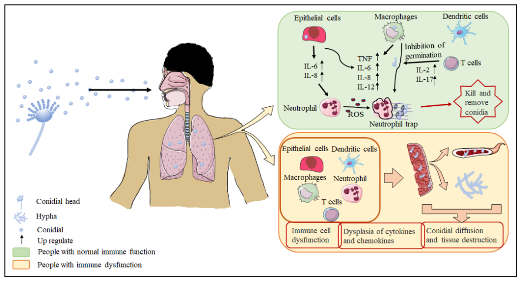
Figure 2.
A. Transmission and replication of SARS-CoV-2. SARS-CoV-2 is transmitted by direct contact, droplets, and aerosols. It enters the body through the respiratory tract and multiplies in the host to produce new virus. B. Inflammatory response caused by the invasion of SARS-CoV-2. After the virus enters the body, it is recognized by epithelial cells, which recruit T cells and CD4+/CD8+ monocytes through type III IFN. These then activate macrophages to increase the production of inflammatory factors and chemokines; recruit downstream NK cells, dendritic cells, and megakaryocytes to secrete inflammatory factors; and trigger neutrophil traps to eliminate the virus. However, the release of excessive inflammatory factors causes a cytokine storm, entailing acute inflammatory damage to tissues. AP-1: activator protein 1; CD: cluster of differentiation; CCL: C-C motif ligand; CXCL: C-X-C ligand; GM-CSF: granulocyte-macrophage colony stimulating factor; IFN: interferon; IL: interleukin; IRF: interferon regulatory factor; MIP: macrophage inflammatory protein; MIG: membrane-bound immunoglobulin; NF-κB: nuclear factor kappa-B; PRR: pattern recognition receptor; ROS: reactive oxygen species; TLR: toll-like receptor; TNF: tumor necrosis factor.
Figure 2.
A. Transmission and replication of SARS-CoV-2. SARS-CoV-2 is transmitted by direct contact, droplets, and aerosols. It enters the body through the respiratory tract and multiplies in the host to produce new virus. B. Inflammatory response caused by the invasion of SARS-CoV-2. After the virus enters the body, it is recognized by epithelial cells, which recruit T cells and CD4+/CD8+ monocytes through type III IFN. These then activate macrophages to increase the production of inflammatory factors and chemokines; recruit downstream NK cells, dendritic cells, and megakaryocytes to secrete inflammatory factors; and trigger neutrophil traps to eliminate the virus. However, the release of excessive inflammatory factors causes a cytokine storm, entailing acute inflammatory damage to tissues. AP-1: activator protein 1; CD: cluster of differentiation; CCL: C-C motif ligand; CXCL: C-X-C ligand; GM-CSF: granulocyte-macrophage colony stimulating factor; IFN: interferon; IL: interleukin; IRF: interferon regulatory factor; MIP: macrophage inflammatory protein; MIG: membrane-bound immunoglobulin; NF-κB: nuclear factor kappa-B; PRR: pattern recognition receptor; ROS: reactive oxygen species; TLR: toll-like receptor; TNF: tumor necrosis factor.
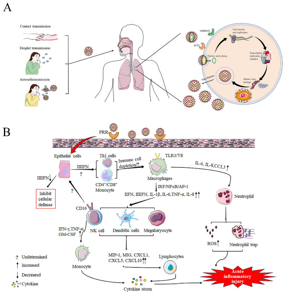

Table 1.
Vaccines listed for emergency use by the World Health Organization.
| Classification | Trademark registration | Approve countries number | Statistical cut-off time | Administration method |
|---|---|---|---|---|
| RNA vaccine | Spikevax | 88 | Apr-30, 2021 | injection |
| Comirnaty | 149 | Dec-31, 2020 | injection | |
| Viral vector vaccine | Convidecia | 10 | May-19, 2022 | injection |
| Jcovden | 113 | Mar-12, 2021 | injection | |
| Vaxzevria | 149 | Feb-15, 2021 | injection | |
| Covishield | 49 | Feb-15, 2021 | injection | |
| Inactivated virus vaccine | Covaxin | 14 | Nov-3, 2021 | injection |
| Covilo | 93 | May-7, 2021 | injection | |
| CoronaVac | 56 | Jun-1, 2021 | injection | |
| Protein subunit vaccine | Nuvaxovid | 40 | Dec-20, 2021 | injection |
Disclaimer/Publisher’s Note: The statements, opinions and data contained in all publications are solely those of the individual author(s) and contributor(s) and not of MDPI and/or the editor(s). MDPI and/or the editor(s) disclaim responsibility for any injury to people or property resulting from any ideas, methods, instructions or products referred to in the content. |
© 2026 by the authors. Licensee MDPI, Basel, Switzerland. This article is an open access article distributed under the terms and conditions of the Creative Commons Attribution (CC BY) license.
Copyright: This open access article is published under a Creative Commons CC BY 4.0 license, which permit the free download, distribution, and reuse, provided that the author and preprint are cited in any reuse.