

Submitted:
07 September 2026
Posted:
08 September 2026
You are already at the latest version
Abstract
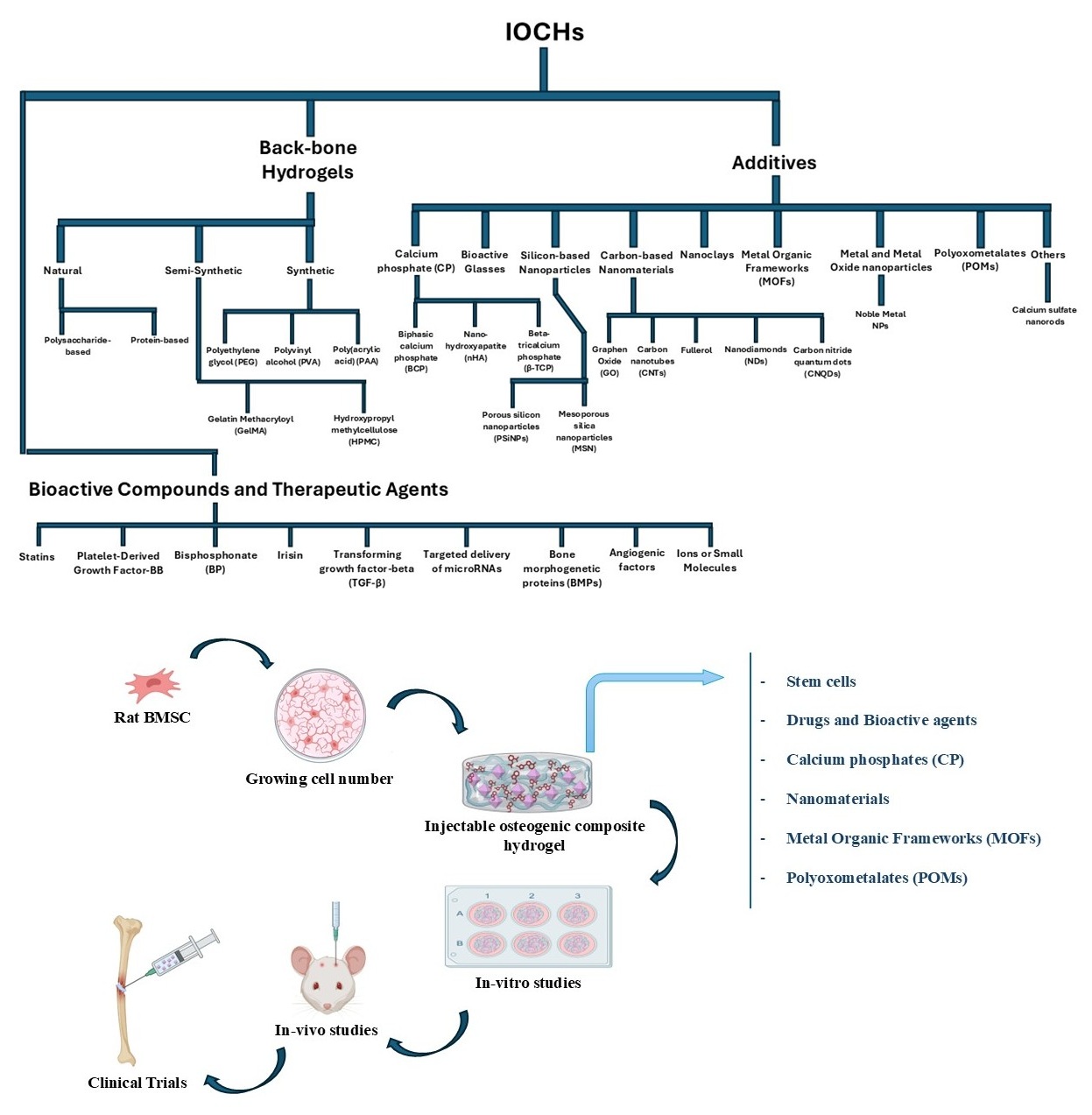
In search for possible strategies to regenerate bony tissue of the non-load bearing regions, injectable hydrogels have received substantial attention considering their tunable mechanical properties, high biocompatibility, and customizable bioactivity along with the possibility for minimally invasive delivery. Currently, many injectable hydrogels (e.g., collagen, hyaluronic acid, chitosan, polyethylene glycol, alginate, fibrin, etc.) have found their utility in bone tissue repair and augmentation. However, they can be further tailored by incorporating various bioactive additives to achieve desirable functionality, known as composite hydrogel. Depending on the nature of bioactive additives, such injectable osteogenic composite hydro-gels (IOCHs) can serve as a reservoir for sustained release therapeutic drugs, antimicrobials, growth factors, and other biomolecules and act as temporary matrices to recruit and ac-commodate the cells of interest, thereby affording a favorable stimulatory microenvironment for bone regeneration. As noted above, the combination of osteogenic materials (such as bioglass, clays, calcium phosphate (CaP), metal-organic frameworks (MOFs), and carbon-based nanoparticles) with hydrogels can endow them with the capacity to promote osteogenic differentiation of those recruited cells in addition to improved mechanical strength and struc-tural stability of the hydrogel. This review summarizes a significant number of injectable osteogenic composite hydrogels and compares their bioactivity with particular emphasis on bone regeneration. In addition, this review also discusses the advantages and drawbacks as-sociated with the inclusion of various additives to formulate bioactive hydrogels. The review concludes with highlights of the foreseeable challenges using IOCHs as well as the prospects for both research and clinical applications in BTE.
Keywords:
bone regeneration
; injectable composite hydrogel
; osteogenesis
; non-load bearing
1. Introduction
Our skeletal system, a vital organ system in the human body, plays crucial roles in movement, protection, and supporting other organs [1]. Despite its self-healing capacity, large bone defects, especially those of critical sizes (i.e., 5 cm for femur and tibia) caused by various pathophysiological conditions such as trauma, tumors, osteoporosis, arthritis, and infections, often require external interventions for timely repair. Although autologous bone grafts are considered the preferred option for functional recovery, their use is limited by donor availability and concerns about morbidity at donor sites. Thus, alternatives such as decellularized allografts or xenografts are also adopted regardless of the noted immunorejection. On the other hand, growing interest has been seen with those engineered grafts, which can be tailored to facilitate optimal bone tissue regeneration (BTR). Among them, injectable hydrogels offer noticeable advantages such as non-invasiveness, adaptability to irregular geometries, and compatibility with different forms of bone defects. Interestingly, the water-rich nature of hydrogels, similar to living tissue, can define a biocompatible environment for cell survival and growth. Moreover, their physical, chemical, and mechanical properties can be easily adjusted to suit specific requirements, thereby enhancing their utility in BTR. In recognition, injectable hydrogels have been extensively explored in recent years to mimic the native ECM with the intention to formulate a conducive environment for guided cell growth and differentiation. To harness the osteogenic capacity of the hydrogel, osteogenic factors such as BMPs are oftentimes added into the hydrogel. Furthermore, recognition of the low mechanical strength of hydrogels, limiting their wide adoption for bone regeneration, motivates the development of composite hydrogels that combine the flexibility of hydrogel networks with other additives to enhance the mechanical properties. As such, composite hydrogels incorporating various inorganic/organic additives such as bioceramics, bioglass particles, carbon nanotubes, and the like have been investigated for BTR. This review focuses on IOCHs with detailed discussion on the composition and fabrication method along with their advantages and drawbacks. Furthermore, this review also provides perspectives on the future endeavors in further development of IOCHs followed by concluding remarks.
1.1. Repair of Bone Defects and the Need for Exogenous Interventions
The longitudinal threshold for spontaneous bone healing depends on anatomical location and bone type. Commonly described as critical-size bone defects, these include a maximum defect size of 3 cm for the radius and ulna, 5 cm for the femur and tibia, and 6 cm for the humerus [2]. This means once the threshold is reached, bone cannot heal naturally anymore [3]. Failure to heal the critical size (or larger) defects leads to the non-union fractures [4].
Bone typically exhibits anisotropic mechanical properties, i.e., strong against compression but weak against tensile, bending, or torsional stress due to its unique hierarchical organization, e.g., the packed array of parallel osteons (i.e., the repeating building blocks of cortical bone). For example, when bone is subjected to 3-point bending stress, tensile stress at the midpoint can initiate surface-to-core fractures. Similarly, when bone experiences a twist, a fracture can occur to the region with the smallest diameter, and the crack oftentimes initiates perpendicular to the diagonal line of the twist plane [5].
Small fractures typically heal through four overlapping stages: hematoma formation, cartilaginous callus formation, bony callus formation, and remodeling. Hematoma (mainly composed of fibrin) immediately forms within the fracture due to disrupted vessels, which supports the infiltration of BMSCs and subsequent differentiation into chondroblasts for cartilaginous callus formation. Hypoxia, nutrient limitation, and local inflammation encourage the infiltration of vascular networks, enabling nutrition/waste exchange to promote mineralization. Calcification of cartilaginous callus leads to the transition to woven bone. Osteoblasts and osteoclasts then remodel the hard callus into mature lamellar bone with stabilized vasculature [6].
However, as a key step of bone regeneration, hematoma formation to bridge a large defect becomes impossible without exogenous intervention, and such a limited hematoma mass cannot accommodate suffi-cient BMSCs to form a large cartilaginous callus. On the other hand, exogenous assistance like autografts, allografts, and xenografts can help to reduce the gap size and support the hematoma formation.
Despite the advantages of autografts (gold standard) [6,7,8,9], autografts often experience limited availabil-ity, donor site morbidity, and implant bone resorption [10,11], preventing wide adoption. Decellularized allografts and xenografts could address some of the autograft-associated challenges [12]; however, the lack of cellular components in these grafts does not favor timely regeneration. Recognition of these limitations has motivated the search for more effective alternatives suitable for bone tissue regeneration (BTR).
2. IOCHs Are Required for BTR
IOCHs emulate the early regenerative microenvironment of hematomas to support bone healing. To be effective, they must be biocompatible, injectable, and have tunable mechanical properties, including os-teoinduction and antimicrobial activity. IOCHs should enable controlled degradation to facilitate new bony tissue formation while providing temporary support for cell adhesion, proliferation, and guided differentia-tion. Including bioactive molecules, such as bone morphogenic proteins, can enhance these properties with sustained release, ensuring optimal conditions for BTR in clinical settings [13,14,15].
2.1. Physiochemical and Mechanical Properties of IOCHs
IOCHs are designed to provide mechanical support, promote cellular activity, and facilitate BTR [16]. However, their limited mechanical strength, especially in load-bearing applications, remains a challenge. Nanostructure and porosity affect mechanical performance—higher porosity enhances cell infiltration and nutrient diffusion but can weaken structural integrity. To enhance mechanical strength, researchers incor-porate conjugated polymers and reinforcing materials, forming composites with improved durability, flexi-bility, and tailored properties compared to polymers alone [17,18,19]. Yu et al. devised an injectable organic-inorganic nanocomposite GelMA hydrogel system enriched with strontium (Sr)-substituted xonotlite (Sr-CSH) nanofibers, significantly enhancing mechanical strength [20]. Another promising approach is double-network hydrogels, where a rigid and brittle polyelectrolyte forms the first network, and a soft and ductile neutral polymer forms the second network, creating strong interpenetrating entanglement and efficient energy dissipation from the two networks. Li et al. used GG (a negatively charged, hydrophilic polysaccharide) as the first network, a crosslinking agent as the second network, and 2-methacrylamidoethyl dihydrogen phosphate (MDP) to facilitate in situ mineralization, improving hydrogel stiffness and elasticity [21,22,23]. By optimizing reinforcing agents, IOCHs can be fine-tuned to match native tissue mechanics, ensuring resilience under physiological forces during tissue integration. The ability to replicate the ECM environment is key for progenitor cell differentiation and functional bone tissue regeneration [16,17,18].
3. Backbone Hydrogels (Based on Hydrogel Type: Natural, Semi-Synthetic, and Synthetic)
Effective cell adhesion to the ECM is essential for cell growth, osteogenic differentiation, and mainte-nance of cellular functions such as survival, migration, proliferation, and polarity. Consequently, hydrogel matrices used in stem cell delivery and tissue engineering must mimic the properties of bone ECM to support these processes [24,25]. The precursor polymers used in hydrogel synthesis dictate key properties such as hy-drophilicity, mechanical strength, elasticity, biocompatibility, and degradation rate. Hydrogel polymers are classified into natural, semi-synthetic, and synthetic categories (Table 3). Natural polymers, such as collagen, gelatin, and alginate, provide biocompatibility, biodegradability, and inherent cell adhesion properties but may exhibit batch variability, limited modifiability, and risk of disease transmission. Semi-synthetic polymers, including GelMA and hydroxypropyl methylcellulose (HPMC), undergo chemical modifications to blend the advantages of biopolymers with the controlled properties of synthetic materials. Synthetic polymers, such as polyethylene glycol and polyacrylic acid, provide enhanced mechanical strength, uniformity, and longevity but often require biocompatibility modifications and may produce acidic degradation byproducts [14,26]. IOCHs integrate synthetic and natural polymers, incorporating inorganic additives such as nHAp, nano-bioactive glasses (nBGs), carbon-based nanomaterials (e.g., carbon nanotubes, graphene oxide nanosheets, nanodiamonds), metallic nanomaterials (e.g., iron oxides, gold), and MOFs or polymeric nanoparticles (e.g., PLGA, dendrimers, micelles, hyperbranched polyesters). These additives enhance the biological activity, structural integrity, and customizable properties of IOCHs [27,28,29,30,31,32,33,34,35,36]. Among these additives, those utilized in IOCHs are listed in Table 4. While some fillers discussed in this review exist on the micro-scale rather than the nanoscale, their integration remains relevant for hydrogel optimization. This review examines key design factors of nanocomposite hydrogels, including the reasoning behind nanofiller selection, their impact on hydrogel matrices, the processes driving gelation, and the physicochemical, mechanical, and biological characteristics of the resultant composites.
3.1. Natural Polymers
3.a.1. Polysaccharide-Based Backbone Hydrogels
3.a.i.1. Chitosan
Chitosan(CS)-based hydrogels exhibit pH-responsive behaviors, driven by protonation/ deprotonation of amino groups, allowing CS to form non-Newtonian, shear-thinning fluids in acidic solutions (pH < 6.5, pKa∼6.3). This enables complexation with metal ions, polymers, lipids, proteins, and DNA [37]. Combining
CS with natural or synthetic polymers such as alginate, hyaluronic acid, and polyethylene glycol enhances antibacterial activity, solubility, and adhesion [38,39,40]. To enhance physicochemical properties, CS undergoes chemical modification, with hydroxybutyl chitosan (HBCS) being a significant derivative. HBCS increases water solubility and introduces reversible temperature-responsive behavior while retaining biocompatibility, making it ideal for biomedical applications [41,42,43].
Wan et al. developed a thermo-responsive hydroxybutyl chitosan hydrogel incorporating polydopamine-coated magnesium-calcium carbonate microspheres, which respond to near-infrared (NIR) light. This com-posite enables the sequential release of therapeutic agents: first, aspirin is released to reduce early inflamma-tion, aiding tissue regeneration. Then, under NIR light, BMP-2 is released, stimulating bone formation, as demonstrated by in vitro osteogenesis studies (ALP staining, ARS staining, and absorbance analysis). Using an SD rat calvaria-defect model, in vivo studies confirmed that controlled BMP-2 release under NIR light significantly enhances bone regeneration. Notably, hydrogel composites irradiated with NIR light exhibit superior osteogenic capability, underscoring the potential of sequential drug delivery systems in BTR [44].
Zhou et al. developed a composite hydrogel dressing using CMCS structured through Fe3+-induced self-assembly and combined with ciprofloxacin (CIP) for antibacterial efficacy. This injectable, moldable hydrogel, CMCS/Fe3+/CIP, promotes osteogenesis in infected microenvironments without additional additives [45].
Karimi et al. introduced a rapid in situ crosslinking method to formulate an injectable oxidized car-boxymethyl chitosan/pullulan (CMCS/OPL) hydrogel. To augment osteogenesis, parathyroid hormone (PTH) was incorporated, resulting in a (CMCS/OPL/PTH) hydrogel with high porosity (> 90%), suitable degradation over 28 days, and continuous peptide release. The IOCH exhibited favorable cell morphology, increased calcium deposition, and effective bone regeneration when tested in mandibular defects in Sprague-Dawley rats [46].
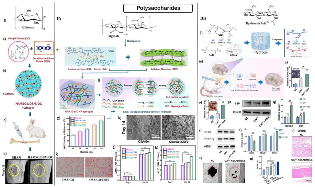
Datta et al. developed a DBM/OC IOCH using decellularized bone extracellular matrix (DBM) and oleoyl chitosan (OC) (Fig. 1-I), designed as a carrier for human amnion-derived stem cells (HAMSCs). The hydrogel mimicked natural 3D tissue environments, exhibiting enhanced mechanical strength, antimicro-bial properties, and superior cell proliferation and differentiation. The formation of the DBM/OC hydrogel
(Fig. 1-a) and subsequent encapsulation of HAMSCs (Fig. 1-b) enabled the creation of an IOCH with potent osteoinductive properties. Including stem cells can continuously provide both cells and growth fac-tors/cytokines at the defect site, allowing the opportunity for enhanced bone repair. After implantation into a rabbit tibial defect model (Fig. 1-c), the HAMSCs/DBM/OC hydrogel achieved ∼83% defect closure, significantly outperforming control groups (∼35%). Micro-CT imaging after 8 weeks (Fig. 1-d) confirmed greater bone regeneration in the HAMSC/DBM/OC hydrogel compared to SHAM (control), highlighting the therapeutic potential of this composite hydrogel system for clinical bone repair [47].
Figure 1.
Impact of polysaccharide-based IOCHs on osteogenesis. (I) Chitosan-based IOCH: (a) Fabrication of DBM/OC biohybrid hydrogel, (b) Encapsulation of HAMSCs to form HAMSCs/DBM/OC hydrogel, (c) IOCH implantation in rabbit tibial defect for bone regeneration assessment, (d) Micro-CT top view of regenerated bone after 8 weeks. (II) Alginate-based IOCH: (e) Schematic of OSA/Gel/CNF hydrogel synthesis, (f) Synergistic self-healing mechanism, (g) Self-healing performance over time, (h) SEM images of biomineralization after 14 days, (i-j) ARS staining and statistical analysis of mineralization, (k) ALP activity assessment. (III) Hyaluronic acid-based IOCH: (l) Fabrication of OADC (dynamic/covalent dual-crosslinked HA) hydrogel, (m) Regenerative outcomes induced by OADC hydrogels, (n) ALP staining and quantification showing early osteogenic activity, (o) ARS staining and analysis of mineral deposition, (p-q) Western blotting and semiquantitative analysis of BSP and RUNX2 in BMSCs on different hydrogels, (r-s) Western blotting and analysis of iNOS, PPAR-γ, and ARG-1 in THP-1 cells, (t) 3D reconstructions of bone regeneration after 8 weeks with OADC-ASA+BMSCs hydrogel, (u) BV/TV ratio quantifying bone volume, (v) H&E staining showing tissue regeneration in OADC-ASA+BMSCs group [47,48,49].
Figure 1.
Impact of polysaccharide-based IOCHs on osteogenesis. (I) Chitosan-based IOCH: (a) Fabrication of DBM/OC biohybrid hydrogel, (b) Encapsulation of HAMSCs to form HAMSCs/DBM/OC hydrogel, (c) IOCH implantation in rabbit tibial defect for bone regeneration assessment, (d) Micro-CT top view of regenerated bone after 8 weeks. (II) Alginate-based IOCH: (e) Schematic of OSA/Gel/CNF hydrogel synthesis, (f) Synergistic self-healing mechanism, (g) Self-healing performance over time, (h) SEM images of biomineralization after 14 days, (i-j) ARS staining and statistical analysis of mineralization, (k) ALP activity assessment. (III) Hyaluronic acid-based IOCH: (l) Fabrication of OADC (dynamic/covalent dual-crosslinked HA) hydrogel, (m) Regenerative outcomes induced by OADC hydrogels, (n) ALP staining and quantification showing early osteogenic activity, (o) ARS staining and analysis of mineral deposition, (p-q) Western blotting and semiquantitative analysis of BSP and RUNX2 in BMSCs on different hydrogels, (r-s) Western blotting and analysis of iNOS, PPAR-γ, and ARG-1 in THP-1 cells, (t) 3D reconstructions of bone regeneration after 8 weeks with OADC-ASA+BMSCs hydrogel, (u) BV/TV ratio quantifying bone volume, (v) H&E staining showing tissue regeneration in OADC-ASA+BMSCs group [47,48,49].
Wasupalli et al. developed thermosensitive injectable hydrogels by combining chitosan, polygalacturonic acid (PgA), βGP and hydroxyapatite using hydrothermal-assisted hydrolysis. These hydrogels exhibited high compressive stiffness, uniform PEC fiber distribution, enhanced osteoblast activity (as confirmed by MTT assays), ALP activity, and collagen production. After a 14-day culture, superior cellular responses were observed in gelatin-containing hydrogels and hydrothermally treated PEC fibers, demonstrating improved bioactivity and mechanical properties [50].
Patel et al. developed bioinspired hydrogels mimicking native Col fibers, forming mineral-hydrogel nanocomposites. In a mouse calvarial defect model, CS-kappa carrageenan nanocomposites demonstrated superior bone regeneration without added growth factors, confirming their osteoconductive properties [51].
3.a.i.2. Alginate
Alginate exhibits hydrophilic and water-soluble properties, increasing viscosity in neutral conditions and forming hydrogels in the presence of polyvalent cations [16,52,53,54]. On the other hand, alginate-based IOCHs often exhibit weak mechanical properties, limiting their use in load-bearing applications. Additionally, con-trolling the release kinetics of encapsulated agents remains challenging, requiring careful optimization to prevent burst or incomplete therapeutic release. Crosslinking methods further influence hydrogel character-istics, with some crosslinkers introducing cytotoxicity risks or other undesired effects [55].
To address these limitations, Wang et al. developed a dual-network IOCH, abbreviated CPSC, incor-porating CMCS, PEG, and calcium chloride (CaCl2). By adjusting the secondary network structure, the hydrogel exhibited optimal compressive strengths ranging from 19 to 252 kPa, mimicking bone tissue-like structure. In vitro experiments demonstrated that CPSC hydrogels supported rBMSC differentiation into osteoblasts, with CPSC-5 (cranial defect filled only with the composite hydrogel) showing superior cell adhe-sion, proliferation, and osteogenesis. Micro-CT reconstruction and histological staining of rBMSCs@CPSC-5 rat cranial defect models confirmed enhanced mineralization, accelerated bone regeneration, and increased bone volume and density [56].
In another approach, Cui et al. designed a cellulose nanofiber (CNF)-reinforced oxidized alginate (OSA)/gelatin (Gel) hydrogel (Fig. 1-e) using a one-step, crosslinker-free method, leveraging dynamic imine and hydrogen bonds. The resulting OSA/Gel/CNF hydrogel achieved a high compressive modulus of 361.3 kPa, rapid gelation (∼150 seconds), and excellent injectability, along with self-healing capabilities of up to 92%, enabling minimally invasive, personalized applications (Fig. 1-f–g). The synergistic bonding interactions facilitated efficient network reconstruction upon damage, as depicted in the self-healing schematic (Fig. 1-f). Notably, increasing CNF content reduced both swelling and degradation rates, making CNFs a potential tool for modulating alginate/gelatin hydrogel degradation. In addition, biomineralization was confirmed after 14 days of incubation (Ca/P ratio ≈ 1.69), with SEM images revealing spherical hydroxyapatite-like particles on the hydrogel surface (Fig. 1-h). Osteogenic differentiation of MC3T3-E1 cells was significantly enhanced in the presence of CNFs, as evidenced by increased calcium deposition via ARS staining (Fig. 1-i–j) and elevated ALP activity (Fig. 1-k), compared to the control (OSA/Gel). These findings reinforce the osteogenic capabilities of the OSA/Gel/CNF hydrogel and its potential as a powerful platform for bone tissue repair [48].
3.a.i.3. Hyaluronic Acid
Hyaluronic acid (HA)-based IOCHs face challenges such as limited mechanical strength, rapid degrada-tion, potential immunogenicity, and high production costs [57,58,59,60]. To address these limitations, Wan et al. developed an injectable HA-ALG hydrogel without chemical cross-linkers, optimizing porosity, rheology, and exosome release. The hydrogel demonstrated high biocompatibility and superior effects on cell migration and vessel formation in vitro. Animal studies confirmed its promising potential for bone formation and angiogenesis, though issues with swelling behavior remain [61].
Guo et al. developed a dynamic/covalent dual-crosslinked HA hydrogel (OADC) incorporating acetyl-salicylic acid (ASA) and BMSCs to enhance osteogenesis and immune modulation in BTE. The hydrogel was synthesized via hydrazone bonding between oxidized HA (OHA) and aldehyde-modified HA methacrylate (ADH-HAMA), followed by photopolymerization of HAMA to reinforce mechanical stability and support cell proliferation (Fig. 1-l). In vivo, the OADC hydrogel facilitated bone regeneration in rat calvarial defects, with the ASA+BMSCs group showing the most extensive and organized new bone formation (Fig. 1-m). Early osteogenic differentiation was confirmed by ALP staining and quantification, which showed increased ALP-positive areas in the OADC and OADC-ASA groups (Fig. 1-n), while ARS staining revealed enhanced mineral deposition, particularly in the OADC-ASA group (Fig. 1-o). Western blotting demonstrated significantly upregulated expression of osteogenic markers BSP and RUNX2 in BMSCs cultured on the OADC-ASA hy-drogel, supporting enhanced osteogenic differentiation (Fig. 1-p–q). Additionally, immunomodulatory effects were evidenced by modulating protein levels within THP-1 cells. These include decreased iNOS and elevated peroxisome proliferator-activated receptor γ (PPAR-γ) and ARG-1 expression, indicating M2 macrophage polarization (Fig. 1-r-s). After 8 weeks of implantation, 3D micro-CT reconstructions showed substantial bone regeneration in the OADC-ASA+BMSCs group (Fig. 1-t), with BV/TV analysis confirming the highest bone volume among all groups (Fig. 1-u), and H&E staining revealing mature bone tissue formation at the defect site (Fig. 1-v) [49].
3.a.i.4. Heparin (Hep)
Heparin, renowned for its potent negative charge, has been widely used in the fabrication of heparin-based hydrogels [62,63,64,65,66,67,68,69]. Kocak et al. developed injectable composite hydrogels incorporating chitosan, hydroxyapatite, and heparin (Hep) for bone regeneration, evaluating different Hep concentrations. These hydrogels form at 37°C within 5-10 minutes, demonstrating convenient rheology and injectability. Freeze-dried samples revealed porous structures that supported cell activity and angiogenesis. Interestingly, higher Hep concentrations increased elastic modulus but lowered initial gelation temperature, potentially impact-ing injectability. Meanwhile, lower Hep concentrations promoted angiogenesis, highlighting the need for optimization in bone regeneration applications [70].
3.a.i.5. Chondroitin Sulfate
Natural chondroitin sulfate (CSe) has structural complexity, inconsistent composition, and high water solubility, which limit its biomedical applications [71,72]. Additionally, unpredictable sulfonation levels further constrain its physiological functionality, requiring modification to optimize its performance. To ad-dress these limitations, CSe is often combined with other polymers to enhance mechanical strength and stability. Cheng et al. fabricated ECM-mimicking hydrogels using photopolymerized CSe analogues, where saccharide and sulfonate unit compositions were systematically varied. This approach allowed precise con-trol over mechanical properties, swelling behavior, degradation rates, and cell compatibility. Cell culture experiments demonstrated that specific saccharide and sulfonate unit ratios significantly enhanced adhesion, dispersion, proliferation, and differentiation of BMSCs. Moreover, BMSCs exposed to these hydrogels under different culture conditions exhibited selective osteogenic differentiation, emphasizing their potential for bone regenerative applications [73].
3.a.2. Protein-Based Backbone Hydrogels
3.a.i.1. Collagen
Collagen presents challenges such as limited mechanical strength, and functional constraints in pure form. To overcome these limitations, researchers have combined collagen with other polymers such as chitosan, alginate, PLGA, HA, and PEG, to improve its structural integrity and functional performance [29,74,75]. Mineralized collagen fibrils, composed of intertwined collagen (Col) and HAp, can also serve as scaffolds for bone mineralization by facilitating calcium and phosphate deposition, enhancing structural integrity, and supporting bone metabolism [76,77,78].
Huang et al. incorporated Col into a CS/β-GP/nHAp system, creating a biocompatible substrate for rBMSCs. In vivo studies involving Wistar rats confirmed cell survival within the gel for 28 days and reduced inflammatory responses compared to non-rBMSC-loaded gels, highlighting its regenerative potential [79].
3.a.i.2. Gelatin
Gelatin-based hydrogels tend to have weaker mechanical properties than synthetic polymers and limited water resistance, necessitating modifications to improve their functionality [26,41,42,43,44,45]. Researchers have reinforced gelatin-based hydrogels with eggshell-derived nanoparticles which improves their biological and mechanical tunability for BTE. To further overcome the shortcomings of these hydrogels, various cross-linking strategies were adopted to enhance their stability [80,81].
Yu et al. developed a nanocomposite GelMA hydrogel system infused with Sr-substituted xonotlite (Sr-CSH) nanofibers, significantly enhancing mechanical strength. The resulting composite hydrogel exhibited mechanical strength over three times that of pure gelatin hydrogels [20].
Yuan et al. introduced an injectable hydrogel for treating femoral head necrosis, composed of Gel cross-linked with hydroxypropyl-β-cyclodextrin (HPβCD) and infused with BMSCs. This hydrogel gelled within 6 minutes, exhibited high water absorption, and accelerated biodegradation and swelling, contributing to enhanced mechanical strength and surface properties. With BMSC viability exceeding 90%, the hydrogel facilitated efficient differentiation into osteocytes within 14 days—a rate superior to conventional osteogenic medium. In in vivo studies, HPβCD-Gel/BMSC hydrogels increased vessel density and diameter between weeks 2 and 8, promoting new bone formation in femoral head defects [82].
3.2. Semi-Synthetic Polymers
3.a.1. Gelatin Methacryloyl (GelMA)
GelMA is a semi-synthetic hydrogel that supports osteogenic differentiation. Mokhtarzade et al. devel-oped a novel gradient GelMA/agarose scaffold to mimic the native osteochondral (OC) environment, aiming for OC repair. They designed an injectable four-layered scaffold by varying hydrogel concentrations (10% and 15% w/v) and adjusting the GelMA:agarose ratio (95:5, 90:10, 85:15). The scaffold featured lower and upper layers mimicking the osseous and chondral ECM, respectively. These layers exhibited porosity between 76% and 96%, with an average pore size of 115 µm. The scaffold demonstrated uniform swelling behavior without layer disintegration along with a consistent weight loss process during degradation over four weeks. Its compressive moduli ranged from 12 to 76 kPa, with structural preservation of 40% and 70% for 10% w/v and 15% w/v compositions, respectively. Additionally, injectability tests showed viscosity values between 13.05 and 68.19 Pa.s at 50% torque. Cytotoxicity assays revealed high cell viability, exceeding 91% after one day and 86% after seven days, confirming the biocompatibility and effectiveness of GelMA-based scaffolds for osteochondral tissue engineering [83]. Cryogelation is an optimal methodology for delivery of cells. To address poor cell viability in current delivery methods, Yuan et al. developed porous shape-memory cryogel microspheres (CMS) from GelMA using this technology. By applying a gradient-cooling cryogelation process, they optimized pore size to 15.5 ± 6.0 µm in a 30-minute gradient-cooled group (CMS-30). Compared to hydrogel microspheres (HMS), CMS promoted adhesion and proliferation of human BMSCs (hBMSCs) and HUVECs over seven days, while preserving high levels of stemness. Additionally, CMS improved cell pro-tection during injection, with CMS-30 specifically enhancing osteogenic differentiation of hBMSCs in bone differentiation media. CMS demonstrated versatility as building blocks for delivering multiple cell types. in vivo experiments, subcutaneous co-injection of hBMSC-loaded and HUVEC-loaded CMS-30 (1:1 ratio) into nude mice for two months resulted in the development of vascularized bone-like tissue, characterized by elevated levels of OCN and platelet endothelial cell adhesion molecule-1 (PECAM-1) CD31 [84].
3.a.2. Hydroxypropyl Methylcellulose (HPMC)
HPMC is a semi-synthetic, non-ionic cellulose ether polymer approved by the FDA. Valued for its hydrophilicity and biodegradability, HPMC serves as an ideal scaffold material, providing mechanical support for BTE. It has been shown to promote osteoblastic survival, proliferation, and differentiation, making it an optimal candidate for regenerative applications [85,86]. To improve HPMC’s properties, silanization is employed, which facilitates three-dimensional network formation, transitioning the fluid gel into a gelatinous state. This silanized HPMC (Si-HPMC) hydrogel boasts injectability and tunable properties, functioning as a scaffold that mimics the natural extracellular matrix. Si-HPMC supports cell adhesion, proliferation, and differentiation while enabling controlled release of bioactive molecules, improving therapeutic outcomes. Despite its advantages, challenges remain, including gelation kinetics, mechanical properties, and regulatory concerns. However, ongoing research efforts are dedicated to overcoming these limitations [86,87,88].
3.3. Synthetic Polymers
3.a.1. Polyethylene Glycol (PEG)
PEG-based IOCHs are biomaterials that utilize PEG, a synthetic hydrophilic polymer, to create a three-dimensional network capable of retaining large amounts of water [89,90]. The hydrophilicity of PEG is adjustable, increasing with higher molecular weight and decreasing with rising temperature, allowing for controlled swelling behavior. By modifying molecular weight and relative density, PEG-based IOCHs can be tailored to meet specific biomechanical requirements [91]. Additionally, some PEG derivatives exhibit pH sensitivity, enabling the formation of pH-responsive hydrogels in acidic microenvironments. This feature facilitates targeted drug release and tissue regeneration. PEG-based hydrogels can also incorporate bioactive molecules, nanoparticles, or other polymers to enhance controlled drug delivery or mechanical reinforcement [92]. While PEG hydrogels have not yet seen widespread clinical use for treating bone defects, their unique properties provide structural support, allowing bone repair through internal healing mechanisms. Research into PEG-based hydrogels for bone regeneration is an emerging field with significant potential for improving bone defect treatments effectively.
3.a.2. Polyvinyl Alcohol (PVA)
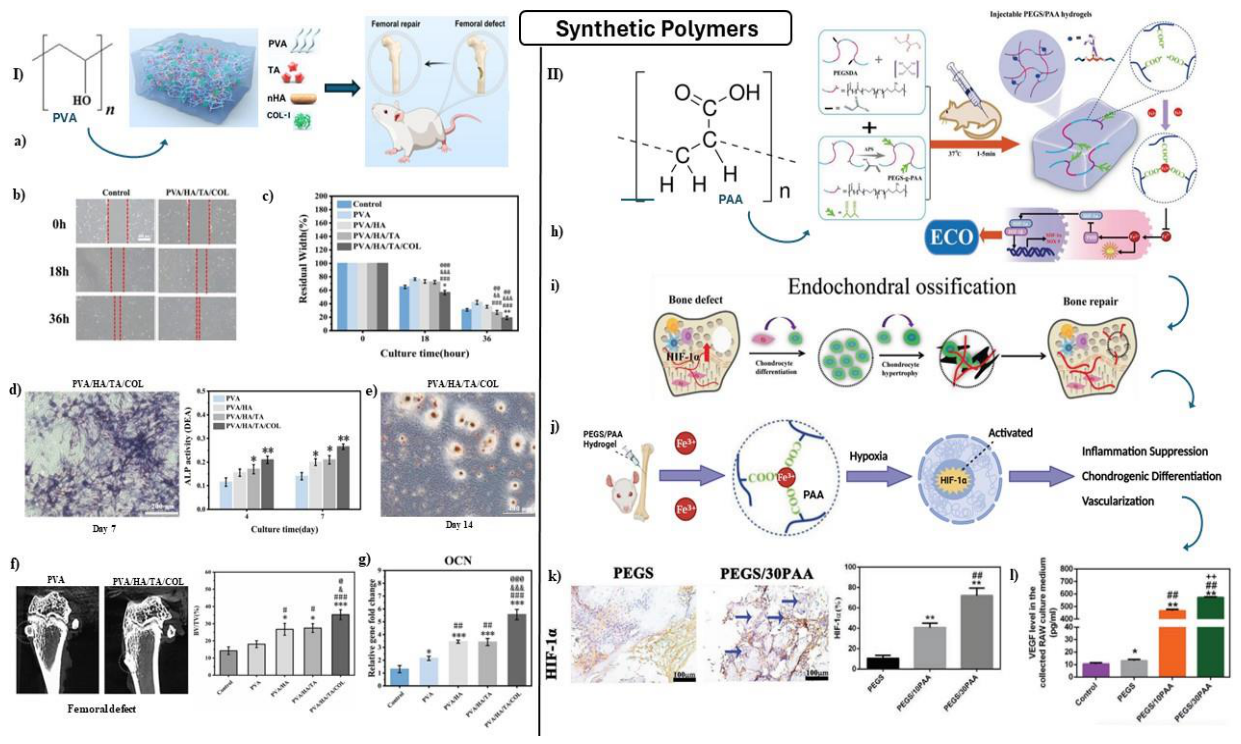
To enhance tissue integration, researchers have functionalized PVA hydrogels with nHAp, chitosan, and other molecules. However, challenges such as limited long-term stability, significant swelling potential, and controlled degradation kinetics require careful optimization. Additionally, while PVA is generally biocom-patible, certain formulations may induce immune responses [93,94]. Xiang et al. engineered a durable IOCH by physically crosslinking PVA with tannic acid (TA) and HAp. The resulting hydrogel exhibited a porous microstructure and significantly improved mechanical strength. To enhance biological interaction, researchers incorporated collagen type I (Col-I) into the hydrogel matrix (Fig. 2-a). In vitro studies demon-strated that PVA/HAp/TA/Col-I hydrogels supported superior cell migration, as shown by the scratch assay and quantification of residual scratch width after 18 hours (Fig. 2-b–c). Enhanced osteogenic differentiation was confirmed by superior ALP activity (Fig. 2-d) and increased calcium deposition via ARS staining after 14 days (Fig. 2-e). In vivo studies further confirmed bone regeneration potential in a rat femoral defect model, where PVA/HAp/TA/Col-I-treated defects exhibited significantly higher BV/TV values compared to other groups (Fig. 2-f). Additionally, RT-qPCR analysis revealed upregulated OCN gene expression in the defect regions, further supporting the hydrogel’s osteoinductive potential (Fig. 2-g). While all hydrogel groups contributed to bone regeneration, PVA/HAp/TA/Col-I demonstrated the highest capacity, addressing key limitations of traditional PVA hydrogels such as inadequate cell adhesion and low compressive strength [95].
Figure 2.
Impact of synthetic polymer-based IOCHs on osteogenesis. (I) Polyvinyl alcohol (PVA)-based IOCH: (a) Fabrication and implantation of PVA/HA/TA/COL hydrogels for rat femoral defect treatment, (b) Cell scratch assay of MC3T3-E1 cells with control and PVA/HA/TA/COL hydrogels, (c) Quantification of residual scratch width, (d) ALP staining and activity after 7 days, (e) ARS staining after 14-day culture with PVA/HA/TA/COL IOCH, (f) Micro-CT images and BV/TV quantification, (g) RT-qPCR analysis of OCN gene expression in femoral defects at 8 weeks. (II) Poly(acrylic acid) (PAA)-based IOCH: (h) Preparation of PEGS/PAA injectable hydrogels, (i) Schematic of HIF-1α-induced ECO pathway, (j) PEGS/PAA hydrogel performance post-injection, (k) Immunohistochemical analysis of HIF-1α in vivo (blue arrows: HIF-1α-positive cells), (l) ELISA detection of VEGF secreted by RAW264.7 cells [95,96].
Figure 2.
Impact of synthetic polymer-based IOCHs on osteogenesis. (I) Polyvinyl alcohol (PVA)-based IOCH: (a) Fabrication and implantation of PVA/HA/TA/COL hydrogels for rat femoral defect treatment, (b) Cell scratch assay of MC3T3-E1 cells with control and PVA/HA/TA/COL hydrogels, (c) Quantification of residual scratch width, (d) ALP staining and activity after 7 days, (e) ARS staining after 14-day culture with PVA/HA/TA/COL IOCH, (f) Micro-CT images and BV/TV quantification, (g) RT-qPCR analysis of OCN gene expression in femoral defects at 8 weeks. (II) Poly(acrylic acid) (PAA)-based IOCH: (h) Preparation of PEGS/PAA injectable hydrogels, (i) Schematic of HIF-1α-induced ECO pathway, (j) PEGS/PAA hydrogel performance post-injection, (k) Immunohistochemical analysis of HIF-1α in vivo (blue arrows: HIF-1α-positive cells), (l) ELISA detection of VEGF secreted by RAW264.7 cells [95,96].
3.a.3. Poly(Acrylic Acid) (PAA)
PAA’s low antigenic response in biological environments makes it a popular material in tissue engineer-ing. However, research has primarily focused on improving the mechanical properties of bone cement, as PAA’s high hydrophilicity leads to mechanical weakening and softening in vivo. Sun et al. introduced in-jectable poly(glycerol sebacate)-co-poly(ethylene glycol)/polyacrylic acid (PEGS/PAA) hydrogels to address the limitations of traditional intramembranous ossification (IMO) in long bones and craniofacial bones, in-stead promoting bone regeneration through endochondral ossification (ECO). These PEGS/PAA hydrogels were synthesized via a thiol-Michael addition click reaction and designed with a semi-interpenetrating net-work structure that rapidly solidifies upon injection (Fig. 2-h). The incorporation of PAA enabled iron ion chelation at the defect site, mimicking a hypoxic microenvironment that activated the HIF-1α signaling path-way (Fig. 2-i). This response suppressed inflammation, encouraged early chondrogenic differentiation, and promoted vascularization in later stages, facilitating typical ECO progression (Fig. 2-j). Immunohistochemical analysis confirmed elevated HIF-1α expression in cells surrounding the PEGS/PAA hydrogels in vivo (Fig. 2-k), while ELISA assays showed significantly increased VEGF secretion by RAW264.7 cells, indicating enhanced angiogenic potential (Fig. 2-l). These findings demonstrate the capacity of the PEGS/PAA hydro-gel to sustain HIF-1α expression, thereby maintaining a stable hypoxia-mimicking microenvironment that facilitates a balanced chondrogenic-to-osteogenic transition, accelerating bone regeneration more effectively than PEGS alone [96].
4. Bioactive Compounds and Therapeutic Agents
IOCHs can incorporate various drugs and bioactive agents to enhance bone regeneration and therapeutic effectiveness. These formulations typically contain growth factors such as BMPs, TGF-β, and insulin-like growth factor (IGF), which stimulate osteogenesis and bone formation. Additionally, small molecules like dexamethasone (Dex), ascorbic acid, and beta-glycerophosphate support the osteogenic differentiation of stem cells [97].
To address challenges such as insufficient vascularization, Chu et al. developed an injectable thermosensi-tive liposome-hydrogel composite scaffold as a sustained-release carrier for bFGF and dexamethasone (Dex). In vitro studies demonstrated that the bFGF/Dex lipo-gel composite scaffold significantly enhanced cell adhesion, proliferation, and osteogenic differentiation of HUVECs and hBMSCs through sustained release of bFGF and Dex. The scaffold exhibited excellent biocompatibility, thermosensitivity, injectability, and sus-tained release behavior. In vivo studies using a rabbit cranial defect model confirmed the scaffold’s ability to promote new bone formation, vascularization, and neurogenesis, outperforming autologous bone grafts. These findings suggest that the bFGF/Dex lipo-gel composite scaffold has great clinical potential for bone regeneration by addressing insufficient vascularization and innervation, thereby improving the efficiency of new bone regeneration [98].
Beyond these osteogenic components, IOCHs frequently incorporate antibiotics to mitigate infection risks and anti-inflammatory agents to reduce inflammation and pain at implantation sites. Angiogenic factors such as VEGF and FGF are also included to promote new blood vessel formation, facilitating nutrient and oxygen delivery to developing bone tissue. The integration of these drugs and bioactive agents within the composite hydrogel matrix establishes a multifunctional therapeutic platform capable of simultaneously promoting osteogenesis, angiogenesis, and antimicrobial activity.
Gao et al. developed a new drug delivery system (DDS) utilizing CS and nanoparticles (NPs) for the con-trolled release of VD3 (1,25-dihydroxyvitamin D3) while maintaining favorable biological properties. Their investigation confirmed the hydrogel’s robust mechanical strength, controlled degradation rate, and efficient drug release profile. In vitro studies using MC3T3-E1 and RAW264.7 cells showcased promising biological activity, highlighting its potential for clinical applications. Macrophage polarization was assessed through two key markers: ARG-1, which is associated with M2 macrophages and supports tissue repair and regen-eration, and iNOS, which is characteristic of M1 macrophages, known for their role in pathogen defense and inflammation modulation. The study observed a transition from M1 (pro-inflammatory) to M2 (anti-inflammatory) macrophages upon exposure to the VD3-NPs/CS-GP hydrogel, suggesting an immunomod-ulatory effect beneficial for osteogenic differentiation. ALP activity and ARS staining further validated the hydrogel’s capability to enhance osteogenesis, especially under inflammatory conditions, demonstrating its effectiveness in fostering bone formation [99].
To combat the significant clinical challenges faced when treating bone defects, plant-derived hydrogels have recently demonstrated a potential to provide intervention in such defects. Xie et al. developed an injectable Sr-LBP/Alg hydrogel composed of sodium alginate, strontium chloride, and Lycium barbarum polysaccharide (LBP), a bioactive compound known for its osteogenic and anti-inflammatory properties. The hydrogel rapidly crosslinks in situ, forming a porous, biocompatible scaffold capable of sustained release of LBP and Sr2+ ions. In vitro assays demonstrated enhanced osteoblast proliferation, mineralization, and expression of osteogenic markers (ALP, COL-1, OPN, RUNX-2), while angiogenesis was confirmed via tube formation assays with HUVECs. In vivo, the hydrogel significantly promoted bone regeneration and neovascularization in a rat femoral defect model, as evidenced by micro-CT, histological staining, and immunofluorescence analysis. These findings demonstrate the hydrogel’s potential as a minimally invasive, bioactive scaffold for BTE and regenerative medicine [100].
Repairing critical-sized calvarial defects remains a major clinical challenge due to the limitations of cur-rent graft materials, including poor biodegradability, low osteoinductive capacity, and mechanical fragility. Chen et al. developed a biofunctional injectable hydrogel (CPMg-NBF) composed of phosphate-functionalized methacryloyl chitosan coordinated with magnesium oxide nanoparticles and loaded with neobavaisoflavone (NBF), a bioactive compound derived from traditional Chinese medicine. This supramolecular hydrogel ex-hibits spongy-like architecture, anti-fracture properties, and sustained release of Mg2+ and NBF, promoting osteogenesis through activation of the p38 MAPK and NOTCH signaling pathways. In vitro assays con-firmed enhanced osteoblast viability, ALP activity, mineralization, and calcium phosphate deposition, while in vivo implantation in a rat calvarial defect model demonstrated superior bone regeneration, collagen matrix development, and neovascularization compared to control hydrogels. These findings position CPMg-NBF as a promising candidate for minimally invasive, mechanically stable, and biologically active bone grafting applications [101].
4.1. Biphosphonate (BP)
BPs are widely used drugs for osteoporosis, preventing osteoclast activity and reducing fracture risk [102]. Barpour et al. developed an amidated-pectin/chitosan/nano-crystalline cellulose scaffold incorporat-ing nHAp and the FDA-approved form of BP, alendronate (ALN). This injectable chitosan-based composite hydrogel improved ADSC proliferation and viability while also increasing anti-inflammatory protein lev-els in ALN-containing groups [103]. Yu et al. introduced a bisphosphonate-containing IOCH utilizing an ALN-Ca2+/Mg2+-doped supramolecular structure based on modified gelatin supramolecular hydrogel. This hydrogel exhibited high durability against compressive and tensile strain and quick self-healing after mechan-ical disruption. Critically, it was injected in a liquid state and transitioned to a gel post-injection, allowing precise molding to the target geometry while preserving encapsulated cells. Compared with conventional host-guest macromer (HGM) hydrogels, the Ca2+/Mg2+-doped system exhibited superior mechanical prop-erties due to the additional physical crosslinking. The addition of acrylate ALN and Ca2+/Mg2+ enhanced in vitro BMSC proliferation, migration, and osteogenic differentiation. In vivo analysis of rat cranial defects confirmed greater bone regeneration compared to hydrogels lacking bisphosphonate additives [104]. Klara et al. introduced a multifunctional IOCH by modifying mesoporous silica particles (MSPs) with NH2, enabling crosslinking with HAp and alendronate (ALN). This is denoted as MSP-NH2-HAp-ALN. These hybrids were immobilized within a collagen/chitosan/hyaluronic acid hydrogel, demonstrating high biocompatibility without hemolytic effects, ensuring blood and liver cell safety. The hydrogel supports hBMSC differentiation into osteoblasts while reducing osteoclast precursor (RAW 264.7 cell) viability. Notably, even the lowest ALN concentration exhibited substantial cytotoxicity, decreasing osteoclast precursor viability to 20% by day 3 and 10% by day 7, suggesting a strong osteoclast-suppressive effect. ALN release was prolonged for up to 20 days, minimizing burst release, with mechanical and degradation properties varying based on MSP-NH2-HAp-ALN content [105].
4.2. Irisin
Irisin is a hormone-like molecule released during exercise, known to induce osteoblast differentiation and bone regeneration, making it a promising osteoporosis drug [106]. Chen et al. developed an injectable natural polymer-based IOCH encapsulating irisin to prevent aseptic loosening in prosthetic joints caused by wear particles, primarily titanium debris. Their hydrogel, composed of gelatin cross-linked with oxidized starch, effectively entrapped and delivered irisin. The hydrogel remains in a liquid state during injection and transitions into a gel at human body temperature, ensuring precise delivery and structural stability. This hierarchical cross-linked hydrogel system, formed by gelatin molecular chains and dynamic imine bonds, exhibits favorable injectability and self-healing properties. In in vivo and in vitro studies, the hydrogel was shown to modulate the Wnt/β-catenin and NF-κB signaling pathways via integrin αV activation. The Wnt/β-catenin pathway regulates cell fate and tissue homeostasis, while the NF-κB pathway orchestrates immune and inflammatory responses. By activating integrin αV, the hydrogel promotes osteogenic differ-entiation of BMSCs, impedes osteoclastic activity, reduces inflammation, and ultimately mitigates aseptic loosening induced by titanium wear particles [107].
4.3. Platelet-Derived Growth Factor-BB (PDGF-BB)
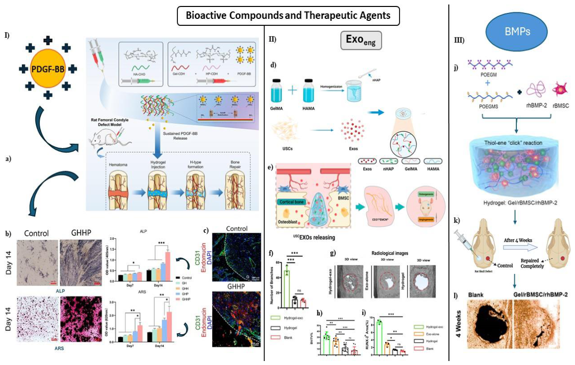
PDGF-BB is a dimer isoform of the PDGF family, known for its ability to attract and stimulate cell divi-sion. It binds to PDGF receptor β, encouraging mesenchymal stem cells and bone marrow-derived endothelial progenitor cells to migrate and differentiate, supporting bone formation and H-type blood vessel develop-ment. Wei et al. developed an injectable heparin-conjugated hydrogel (GHH) to enable controlled delivery and binding of PDGF-BB, facilitating rapid vascularized bone regeneration. The PDGF-BB-loaded GHH hydrogel (GHHP) was formed by in situ mixing of ECM components—Gel-CDH (amino-modified gelatin), HA-CHO (aldehyde-modified hyaluronic acid), and HP-CDH (amino-modified heparin)—with PDGF-BB, creating a stable injectable matrix (Fig. 3-a). In vitro, GHHP promoted osteogenic differentiation of BMSCs, as evidenced by increased ALP activity and calcium deposition at day 14 (Fig. 3-b). In vivo, GHHP facili-tated the formation of H-type blood vessels—characterized by co-expression of CD31 and endomucin—near the bone growth plate, indicating enhanced angiogenesis and osteogenesis (Fig. 3-c). These findings high-light GHHP’s ability to mimic the natural coupling of vascular and bone regeneration, offering a promising strategy for treating bone defects through sustained PDGF-BB delivery [67].
Figure 3.
Impact of bioactive compounds and therapeutic agents on osteogenesis. (I) Platelet-derived growth factor BB (PDGF-BB)-based IOCH: (a) Schematic of GHHP hydrogel preparation (PDGF-BB-loaded GHH hydrogel) and role in bone regeneration, (b) In vitro osteogenic differentiation of human BMSCs assessed by ALP and ARS staining at day 14 with quantification, (c) Immunofluorescence imaging of type-H vessels (endomucin and CD31). (II) Engineered exosome (Exoeng)-based IOCH: (d) Preparation of composite hydro-gels, (e) Photocrosslinked hydrogels with in situ adhesion supporting exosome function for osteogenesis and angiogenesis in rat cranial bone regeneration, (f) Semi-quantitative tube formation assay, (g-h) Micro-CT 3D view of cranial bone regeneration at week 4 and BV/TV analysis, (i) Semi-quantitative results of RUNX-2 immunohistochemical staining. (III) Bone morphogenetic protein (BMP)-based IOCH: (j) Illustration of in-jectable Gel/rBMSC/rhBMP-2 hydrogel containing rBMSCs and rhBMP-2 synthesized via “click” chemistry, (k) Injection of Gel/rBMSC/rhBMP-2 into rat cranial bone defect promoting complete regeneration after 4 weeks, (l) 3D micro-CT reconstruction of defect regions in Blank and Gel/rBMSC/rhBMP-2 groups at 4 weeks post-implantation [67,108,109].
Figure 3.
Impact of bioactive compounds and therapeutic agents on osteogenesis. (I) Platelet-derived growth factor BB (PDGF-BB)-based IOCH: (a) Schematic of GHHP hydrogel preparation (PDGF-BB-loaded GHH hydrogel) and role in bone regeneration, (b) In vitro osteogenic differentiation of human BMSCs assessed by ALP and ARS staining at day 14 with quantification, (c) Immunofluorescence imaging of type-H vessels (endomucin and CD31). (II) Engineered exosome (Exoeng)-based IOCH: (d) Preparation of composite hydro-gels, (e) Photocrosslinked hydrogels with in situ adhesion supporting exosome function for osteogenesis and angiogenesis in rat cranial bone regeneration, (f) Semi-quantitative tube formation assay, (g-h) Micro-CT 3D view of cranial bone regeneration at week 4 and BV/TV analysis, (i) Semi-quantitative results of RUNX-2 immunohistochemical staining. (III) Bone morphogenetic protein (BMP)-based IOCH: (j) Illustration of in-jectable Gel/rBMSC/rhBMP-2 hydrogel containing rBMSCs and rhBMP-2 synthesized via “click” chemistry, (k) Injection of Gel/rBMSC/rhBMP-2 into rat cranial bone defect promoting complete regeneration after 4 weeks, (l) 3D micro-CT reconstruction of defect regions in Blank and Gel/rBMSC/rhBMP-2 groups at 4 weeks post-implantation [67,108,109].
4.4. Transforming Growth Factor-Beta (TGF-β)
TGF-β is a multifunctional cytokine that regulates cell growth, differentiation, migration, and apopto-sis. In bone tissue engineering and regenerative medicine, TGF-β plays a critical role in osteogenesis by stimulating mesenchymal stem cell (MSC) proliferation and differentiation into osteoblasts. It also enhances the production of extracellular matrix proteins, such as collagen and osteocalcin, which are essential for bone formation and mineralization. Additionally, TGF-β regulates osteoclast activity, promoting bone re-sorption and homeostasis. For example, Li et al. developed an advanced biomaterial platform incorporating gelatin-heparin microspheres (MS) loaded with TGF-β1 (MS/TGF-β1) into an injectable lithium-heparin hydrogel (Li-gel). The Li-gel serves as both a carrier for MS/TGF-β1 and a source of lithium ions, which further promotes osteogenesis. Additionally, the release of TGF-β1 guides macrophage polarization toward the M2 phenotype, which is crucial for tissue repair and regeneration by suppressing inflammation, releasing growth factors, and facilitating vascular formation. Both in vitro and in vivo experiments demonstrated that Li-gel@MS/TGF-β1 enhances osteogenesis and angiogenesis through the TGF-β/Snail pathway, reinforcing its potential for bone regeneration applications [110].
4.5. Targeted Delivery of microRNAs
MicroRNA-29c (miR29c) plays a key role in bone regeneration and metabolism, targeting genes involved in bone formation and mineralization, including collagens and extracellular matrix proteins. Tetrahedral framework nucleic acids (tFNAs) serve as delivery systems for miRNA molecules, enhancing the osteogenic potential of surrounding cells such as MSCs by promoting differentiation into osteoblasts and stimulating bone matrix protein production. Additionally, tFNAs provide stability, controlled release, and protection against miRNA degradation, making them ideal carriers in injectable osteogenic hydrogels. Sun et al. investigated GelMA-stFNAs-miR29c, a novel DNA nanomaterial, for its osteogenic effects in vitro. The GelMA-stFNAs-miR29c group exhibited significantly higher expression of osteogenesis-related proteins and mRNA levels compared to control groups, while also activating the Wnt signaling pathway. Notably, the GelMA-stFNAs-miR29c hydrogel promoted mineralized nodule formation and exhibited superior bone re-generation and repair in vivo, reinforcing its potential for bone tissue engineering [111]. Similarly, miR-26a, a small non-coding RNA molecule, regulates cell proliferation, differentiation, apoptosis, and development. Gan et al. incorporated cholesterol-modified miR-26a (Chol-miR-26a) into an injectable PEG hydrogel, forming a cleavable ester bond through ultraviolet (UV) exposure. Upon UV activation, the Gel-c-miR-26a (MLCaged) hydrogel selectively released Chol-c-miR-26a, significantly enhancing bone regeneration compared to non-UV-activated MLCaged. The MLCaged hydrogel demonstrated increased alkaline phosphatase activity, calcium deposition in vitro, and successful repair of critical-sized skull defects in a rat model, confirming its osteogenic efficacy [90].
Huang et al. addressed the challenges of treating bone and joint tuberculosis (BJTB) by developing a multifunctional bioimplantable scaffold with both osteoinductive and antituberculosis properties. The scaffold, composed of oxidized hyaluronic acid (OHA), CMCS, and nHAP, formed an injectable hydrogel via Schiff base chemistry. To enhance therapeutic efficacy, engineered exosomes (Exoeng) generated by stimulating BMSCs with Fe3O4 nanoparticles and an alternating magnetic field (AMF) were incorporated alongside drug-loaded liposomes containing rifampicin (RFP), isoniazid (INH), and pyrazinamide (PZA). This composite system demonstrated potent anti-tuberculosis activity and promoted bone repair in vitro. ARS staining on day 14 revealed significantly greater calcium deposition in the Fe3O4-AMF-Exo Gel group compared to other treatments, indicating enhanced mineralization. Additionally, RT-qPCR analysis showed upregulated RUNX2 expression, confirming superior osteogenic differentiation in this group. These results demonstrate the scaffold’s dual functionality in promoting bone regeneration while delivering sustained anti-tuberculosis therapy [112].
Lu et al. designed a novel composite hydrogel integrating homogenously mixed GelMA-HAMA and nHAP as a drug delivery system for slow-release human urine-derived stem cell exosomes (human USCEXOs) to enhance osteogenesis. In the hydrogel, HAMA provides a structural framework that enables sustained release of USCEXOs over time, supporting prolonged osteogenic signaling (Fig. 3-d). Each component syn-ergizes to create an efficient matrix for bone regeneration, with photocrosslinked hydrogels exhibiting in situ adhesion and retention of exosome bioactivity, thereby promoting both osteogenesis and angiogenesis in rat cranial bone regeneration (Fig. 3-e). These angiogenic abilities were promoted from endothelial pro-genitor cells (EPCs), confirmed by semi-quantitative tube formation assays (Fig. 3-f). In vitro experiments effectively demonstrated the strong osteogenic abilities of the USCEXOs/GelMA-HAMA/nHAP composite hydrogel, as evidenced by 3D reconstructions of micro-CT imaging and BV/TV of regenerated bone tissue.
The BV/TV of the Hydrogel-exo group was 32.5 ± 6.4%, compared to the Exo-alone group (23.3 ± 5.9%), Hydrogel group (12 ± 6%), and Blank group (7.6 ± 6%), clearly showing the impact of USCEXOs and the hydrogel combined (Fig. 3-g–h). RUNX-2 immunohistochemistry results further validated its ability to repair cranial bone defects, with significantly higher positive regions obtained in the Hydrogel-exo group compared to the rest of the groups (Fig. 3-i) [108].
4.6. Bone Morphogenetic Proteins (BMPs)
BMPs hold significant promise for use in injectable osteogenic hydrogels for bone tissue engineering. Despite FDA approval for biomedical applications, BMPs face limitations such as high cost and instability under physiological conditions. To address these limitations, researchers have developed BMP mimetic peptides, such as BMP-2 mimetic peptide BP, which replicates BMP activity while offering cost-effectiveness and storage stability. Several biomaterial carriers have been explored to extend BP’s in vivo lifespan, ensuring sustained therapeutic effects. Additionally, extracellular signaling pathways, including Wnt/β-catenin and BMP pathways, regulate myeloma cell survival and bone formation, highlighting BMPs’ role in bone-related disorders.
Park et al. investigated an injectable HA hydrogel scaffold incorporating BMP-2 mimetic peptide (BP) for bone tissue engineering. This scaffold, formed through click-cross linking (Cx) of HA, demonstrated prolonged stability in vitro and in vivo, confirmed via real-time fluorescence imaging. BP catalyzed osteogenic differentiation in hDPSCs, while the Cx-HA scaffold provided an optimal microenvironment for hDPSCs with excellent biocompatibility. In vitro, BP exhibited osteogenic properties comparable to traditional BMP-2, and when loaded into the Cx-HA hydrogel, it maintained injectability and transitioned into a solid scaffold post-injection, ensuring sustained osteogenic induction [113].
Grab et al. developed an HA-based hydrogel incorporating BMP-6, designed to mimic bone marrow stiffness and target myeloma cell survival. By integrating heparin, they prolonged BMP-6 activity, enabling gradual release and enhanced bone formation. Additionally, fibronectin coating aided in mesenchymal stem cell attachment, ultimately inducing myeloma cell death and promoting stromal cell differentiation into bone-forming cells [114].
Lao et al. introduced a fully biodegradable PEG-based injectable hydrogel, synthesized via thiol-ene “click” chemistry under physiological conditions. The hydrogel demonstrated excellent biocompatibility, mechanical strength, low swelling, and a controlled degradation profile. When loaded with rhBMP-2, the hydrogel supported cell survival and induced rBMSC proliferation and osteogenic differentiation (Fig. 3-j). When injected into critical-sized cranial bone defects in rats, the Gel/rBMSC/rhBMP-2 formulation acceler-ated near-complete bone regeneration within four weeks (Fig. 3-k). Micro-CT imaging confirmed substantial new bone formation in the treated group compared to controls (Fig. 3-l), highlighting the hydrogel’s potential as a clinically relevant bone substitute for craniomaxillofacial repair [109].
Sun et al. developed BMP-2@PNH-TA, a novel bioactive hydrogel combining poly(N-isopropylacrylamide) (PNIPAM), Fe-Tannin (Fe-TA), and O-hydroxypropyltrimethyl ammonium chloride chitosan (O-HACC). This hydrogel integrates osteogenic and antibacterial effects, utilizing near-infrared (NIR)-induced photother-mal therapy (PTT). Through a sponge-like mechanism, the hydrogel releases BMP-2 and tannin at low NIR power to promote bone formation, while high-power NIR stimulation releases O-HACC to eliminate bacteria. This tailored PTT approach significantly accelerates bone regeneration, reducing healing time by nearly 50% in vivo [115].
4.7. Statins
Statins, mold fungi-derived drugs originally developed for cholesterol-lowering, have gained attention in bone tissue engineering due to their ability to reduce osteoblast apoptosis, enhance vascularization, and promote bone mineralization. These drugs upregulate VEGF and FGF-2, facilitating coordinated vascu-larization, which is essential for bone reconstruction. Additionally, statins increase BMP gene expression, leading to osteoblast proliferation and differentiation, while inhibiting osteoclastogenesis by modulating cell signaling pathways [116].
Petit et al. developed a thermosensitive chitosan hydrogel loaded with statins to achieve spatially controlled drug release at the lesion site. Due to the water-insolubility of atorvastatin and lovastatin, they created an emulsion with chitosan, incorporating glycerophosphate salt to ensure irreversible gelation at 37◦C, stabilizing the hydrogel at body temperature. When applied to Porphyromonas gingivalis-infected oral epithelial cells and gingival fibroblasts, the hydrogel significantly reduced pro-inflammatory markers and pro-osteoclastic RANKL expression while inducing bone sialoprotein 2 (BSP2) expression in osteoblasts, underscoring its pro-healing properties. An in vitro release study demonstrated that 70% of the statins were released within 24 hours, following a linear release profile. Researchers suggested that this early release phase aids the transition from inflammation to healing. In an experiment infecting Human Oral Epithelial Cells (ECs) with P. gingivalis, treatment with atorvastatin and lovastatin TPGS (D-α-Tocopherol polyethylene glycol succinate) nanoemulsions resulted in significantly decreased expression of TNF-α, IL-1β, and RANKL, confirming anti-inflammatory and anti-resorptive effects. In an in vivo calvarial bone defect model, the statin-chitosan composite hydrogel significantly increased new bone formation, outperforming systemic statin administration [117].
4.8. Angiogenic Factors
Among the plethora of growth factors used in IOCHs, FGF and VEGF play distinct roles in bone regeneration and angiogenesis. FGF enhances osteogenesis and tissue repair, while VEGF primarily promotes angiogenesis, ensuring nutrient and oxygen delivery to regenerating bone tissue.
Divband et al. developed injectable hydrogels using chitosan biguanidine and carboxymethylcellulose, incorporating VEGF and recombinant BMP-2 through in situ formation. The sequential release of these growth factors mimicked human bone growth patterns, facilitating vascularization and bone regeneration. The hydrogels significantly enhanced the proliferation and differentiation of DPSCs in the presence of VEGF and BMP-2, as confirmed by Western blot and qRT-PCR analyses, which showed increased expression of ALP, collagen 1, and osteocalcin genes [118].
Sivashanmugam et al. created an IOCH using chitin and PLGA, integrating calcium sulfate (CaSO4) and FGF-18 to increase hydrogel stiffness and mechanical strength. The incorporation of CaSO4 facilitated sustained FGF-18 release, crucial for osteogenesis. In vitro experiments showcased heightened osteogenic differentiation, with elevated ALP expression and upregulation of key osteogenic genes (RUNX2, ALP, BMP-2, osteocalcin, and osteopontin). Immunofluorescence staining and ARS staining confirmed its osteogenic potential, while FGF-18 also promoted endothelial cell migration, indicating angiogenic properties. In vivo evaluation demonstrated superior bone healing with the FGF-18-loaded hydrogel, corroborated by histolog-ical staining and longitudinal live animal micro-computed tomography (µ-CT) scans [119].
Guo et al. utilized bFGF despite its instability under physiological conditions. To address this, they em-ployed recombinant human collagen (rhCol) cross-linked by transglutaminase (TG) and loaded with bFGF, forming rhCol/bFGF hydrogels. These hydrogels exhibited a porous structure and robust mechanical properties, inducing cell proliferation, migration, and adhesion. Controlled degradation facilitated sustained bFGF release, maximizing its effectiveness in promoting bone regeneration. Molecular analyses confirmed upregulated bone-related protein expression, while in vivo experiments in rat cranial defects demonstrated the accelerated bone repair facilitated by rhCol/bFGF hydrogels [120].
4.9. Natural Peptide Therapeutics
Recently, Yu et al. sought to enhance bone defect treatment and peptide bioavailability by incorpo-rating antioxidant-active peptides (DBPs) from deer antler blood into an oxidized sodium alginate/amino gelatine injectable hydrogel (OSA/N-Gel). The resulting bioscaffold, OSA/N-gel/DGP, features a loose, porous structure that facilitates nutrient flow and gradual DBP release for sustained bone repair. In vitro studies demonstrated the scaffold’s pro-proliferative and pro-mineralizing effects on osteoblasts, promoting the expression of osteogenesis-related genes and activating the Wnt/β-catenin signaling pathway. OSA/N-gel/DBP significantly outperformed OSA/N-gel alone, indicating DBP’s strong osteogenic properties. This innovative approach successfully repaired bone defects and expanded the application of antler bone in bone regeneration [121].
Despite advancements in polymer scaffolds for bone defect repair, effective regeneration in osteoporotic bones remains difficult, especially when combined with osteoporosis medications. Yu et al. developed a drug delivery system using mesoporous bioactive glass (MBG) and photo-crosslinked hyaluronic acid methacrylate (HAMA), loaded with the osteogenesis-promoting peptide DWIVA (D5) and osteoclastogenesis-inhibiting drug alendronate (ALN). The MBG@D5-Gel complex enables controlled release of these agents, enhancing bone regeneration in osteoporotic conditions by inhibiting osteoclastogenesis and promoting osteogenic dif-ferentiation. In vitro testing, a Live/Dead Cell Viability Assay Kit, Micro-CT analysis, and more were done to successfully ensure the safety and efficacy of this IOCH. The dual-action system optimizes the pathologi-cal microenvironment of osteoporosis, facilitating the repair of osteoporotic bone defects and showing great potential as a biomimetic implant material [122].
4.10. Ions or Small Molecules
Ions such as magnesium (Mg2+) and strontium (Sr2+) play a crucial role in bone regeneration, supporting osteogenic differentiation, biomineralization, and scaffold stability. Incorporating these ions into injectable osteogenic hydrogels (IOCHs) enhances their biocompatibility and regenerative potential. Zhou et al. syn-thesized injectable MgO/MgCO3@PLGA (PMM) hydrogels, namely, injectable biomimetic porous hydrogels (IBPHs), to accelerate bone regeneration. These hydrogels exhibited excellent injectability, undergoing a liquid-to-solid phase transition in situ, enabling them to fully conform to irregular bone defects. Moreover, the controlled release of Mg2+, regulated by the weight ratio of MgO and MgCO3 particles, significantly pro-moted proliferation, osteogenic differentiation, migration, and biomineral deposition in immortalized mouse embryonic fibroblasts. Micro-CT imaging and histological analysis confirmed that PMM hydrogels stimu-lated bone regeneration in rat calvarial defects, doubling bone volume fraction compared to the control group [123].
Chen et al. developed CSMP-MgO injectable hydrogels, integrating magnesium oxide nanoparticles (NPs) into a water-soluble phosphocreatine-functionalized CS (CSMP) solution. The hydrogel formed when MgO NP concentration exceeded 2.5 mg/mL, with gelation time decreasing as NP concentration increased. The resulting porous structure (50–100 µm) facilitated controlled Mg2+ release, promoting calcium phosphate deposition, osteogenic differentiation, and endothelial tube formation in HUVECs. CSMP-MgO (5) hydrogels facilitated new bone formation in critical-sized calvarial defects in rats, confirming their osteogenic potential [32].
Zhang et al. investigated the cytotoxicity of Alg-DA (dopamine grafted to alginate (Alg) via an amidation reaction, followed by mixing with strontium ions (Sr) in the Alg-DA aqueous solution). The Alg-DA/Sr2+ DC hydrogel demonstrated superior biocompatibility and osteogenic potential, confirmed by MC3T3-E1 cell viability assays. ALP staining and activity assays showed the highest ALP expression on days 3 and 7, indicating enhanced osteogenic differentiation. Micro-CT analysis at 4 weeks post-implantation revealed significant new bone formation, with the highest bone formation rate among tested groups. Histological staining, including hematoxylin & eosin (H&E) and Masson’s trichrome (MT) confirmed collagen fiber formation and maturation, reinforcing the hydrogel’s regenerative potential [124].
5. Additives
5.1. Calcium Phosphate (CP)
CP is widely used in injectable hydrogels for biomedical applications, particularly in BTE and regen-erative medicine. As a bioactive component, it enhances both the biological and mechanical properties of hydrogels. When integrated into the hydrogel matrix, calcium phosphate supports bone regeneration by serving as a scaffold for new bone formation and promoting osteogenic differentiation of stem cells [125,126]. A commonly used form of calcium phosphate in injectable hydrogels is HAp, a mineral naturally found in bone tissue. HAp is bioactive and closely mimics the composition and structure of natural bone. Dispersing HAp within a hydrogel improves its mechanical strength and stability while also promoting cell adhesion, proliferation, and differentiation [127,128]. Other calcium phosphate variants, such as TCP and BCP, may also be employed depending on specific application requirements. These materials offer distinct degradation rates and bioactivity profiles, enabling tailored properties for optimized BTR outcomes [32,129].
Cheng et al. developed an innovative injectable composite hydrogel combining 4-arm-PEG-thiol (4-arm-PEG-SH) with liposome-calcium phosphate nanoparticles (Lip#CaP). Their goal was to create a multifunc-tional “three-in-one” hydrogel platform integrating osteogenesis, angiogenesis, and antibacterial properties. By carefully optimizing cargo types and concentrations, the platform maintained precise control over key parameters such as drug release kinetics, swelling behavior, degradation rate, injectability, and self-healing capacity. Notably, the hydrogel exhibited potent antibacterial activity against Escherichia coli, Staphylo-coccus aureus, and Staphylococcus epidermidis, with over 50% inhibition observed. In vitro experiments demonstrated significant enhancements in angiogenesis, evidenced by increased vessel length and junction formation when culturing HUVECs. Furthermore, osteogenesis was substantially improved, as indicated by increased alkaline phosphatase activity and extracellular matrix mineralization of MC3T3-E1 cells. In a rat calvarial critical-size defect model, the hydrogel facilitated robust osteogenesis and angiogenesis over eight weeks before fully degrading [130].
Wang et al. designed a composite hydrogel system incorporating calcium phosphate cement (CPC) with GelMA and poly(N-hydroxyethyl acrylamide) (PHEAA), achieving rapid polymerization and crosslinking through UV photoactivation. Osteogenic gene expression analysis in MC3T3 cells via qRT-PCR revealed significantly higher levels in cells cultured on the GelMA-PHEAA/CPC hydrogel compared to other formu-lations. The consistent upregulation of osteogenic markers—including Runx2, OPN, OCN, ALP, COL I, and OSX—was attributed to the formation of a structurally robust tissue, reduced CPC rupture risk (as ceramic materials can be prone to rupturing when implanted), and improved CPC storage conditions facilitated by HEAA. Immunofluorescence staining further confirmed that the GelMA-PHEAA/CPC hydrogel notably enhanced COL I and OCN expression compared to alternative hydrogels, reinforcing its bioactivity [126].
5.a.1. Nano-Hydroxyapatite (nHAp)
nHAp consists of hydroxyapatite particles reduced to the nanoscale range, offering superior biocompati-bility and bioactivity due to its resemblance to natural bone. Its high surface area enhances integration into biomaterials, particularly hydrogels, where it strengthens mechanical properties and promotes osteogenic differentiation [29,131]. Cao et al. devised an injectable, bilayer hydrogel scaffold incorporating carbonyl hydrazide grafted collagen (COL-CDH), oxidized chondroitin sulfate (OCS) derivatives, and PEG diacry-late (PEGDA) for cartilage repair, with a zinc-doped hydroxyapatite layer supporting subchondral bone regeneration. This structure facilitated stem cell differentiation and calcium deposition, accelerating bone formation. This was further supported via micro-CT and tissue staining analysis [71].
Shi et al. introduced FHCS, a hydrogel composed of pluronic F-127, carboxymethyl chitosan/sodium alginate nanoparticles (nCS), and nHAp, improving osteogenic potential through BMP/Smad pathway acti-vation. In vivo, FHCS-5 (5 mg/ml of nCS included) significantly promoted bone regeneration in rat calvarial defects as observed through CT scanning [40].
Pan et al. developed an IOCH containing N-carboxyethyl chitosan (CEC), hyaluronic acid-aldehyde (HA-ALD), adipic acid dihydrazide (ADH), and 25% w/v nHAp. It promoted osteogenic differentiation and enhanced alveolar ridge preservation in a rat mandibular defect model, demonstrating both osteoconductive and osteoinductive properties [132].
Ma et al. designed an injectable hydrogel incorporating HAp-containing nanoparticles and CMCS via Schiff base reaction, resulting in a porous, self-healing structure with uniform HAp nanoparticle distribution. Cytocompatibility assays confirmed favorable interaction with L929 cells [133]. They also integrated nHAp into a Gel-DA hydrogel using polydopamine-functionalized nHAp (PHAp), significantly bolstering compres-sive strength and cell adhesion and proliferation. Gel-PHAp hydrogels accelerated bone repair efficiency in a rat femoral defect model [134].
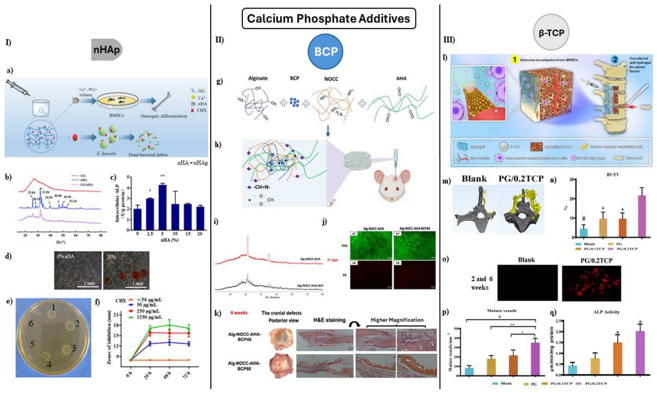
To tackle the dual challenge of infection and bone regeneration, Xu et al. developed a GG-based IOCH, incorporating chlorhexidine (CHX) and nHAp. This dual-function hydrogel demonstrated strong mechanical properties, effective biodegradability, and biocompatibility while supporting BMSC growth and osteogenic differentiation (Fig. 4-a). XRD confirmed the crystalline structure of nHAp within the hydrogel (Fig. 4-b). ALP activity peaked at 5% nHAp concentration, indicating optimal osteoinductive potential (Fig. 4-c), while ARS staining revealed enhanced mineralized nodule formation at this concentration (Fig. 4-d). Antibacterial testing showed that CHX-loaded hydrogels produced clear inhibition zones against E. faecalis in a dose-dependent manner, with 50 µg/mL CHX demonstrating effective antimicrobial activity (Fig. 4e–f). These results highlight the hydrogel’s promise for treating infectious bone defects, particularly in cases like refractory periapical periodontitis [135].
Figure 4.
Impact of calcium phosphate additives on osteogenesis. (I) Nanohydroxyapatite (nHAp)-based IOCH: (a) Schematic of hydrogel’s dual functions: osteogenic induction and antibacterial activity, (b) XRD patterns of GG with/without nHAp, (c) ALP quantification of BMSCs cultured in nHAp hydrogel extracts, (d) ARS staining for osteogenesis, (e) CHX-induced inhibition zones and E. faecalis colony formation in agar medium, (f) Time-dependent inhibition zone production of hydrogels (0–72 h). (II) Biphasic calcium phos-phate (BCP)-based IOCH: (g) Fabrication of Alg-NOCC-AHA-BCP hydrogel, (h) In vivo bone regeneration in mouse calvarial defect model, (i) XRD comparison of Alg-NOCC-AHA (red) vs. Alg-NOCC-AHA-BCP (black), (j) Live/dead staining of MC3T3 cells on hydrogels after 7 days (A7-B8), (k) Mouse cranial defect images (red dashed circles) and H&E staining (red dashed squares) after 4 weeks; higher magnification shows osteocytes (white arrows) and woven bone (white dashed circles). (III) β-TCP-based IOCH: (l) Exosome visualization, (m) 3D micro-CT reconstruction, (n) BV/TV quantification, (o) In vivo fluorochrome labeling and histomorphometry with alizarin red after 2 and 6 weeks, (p) Immunofluorescence showing mature blood vessels, (q) ALP activity analysis [17,135,136].
Figure 4.
Impact of calcium phosphate additives on osteogenesis. (I) Nanohydroxyapatite (nHAp)-based IOCH: (a) Schematic of hydrogel’s dual functions: osteogenic induction and antibacterial activity, (b) XRD patterns of GG with/without nHAp, (c) ALP quantification of BMSCs cultured in nHAp hydrogel extracts, (d) ARS staining for osteogenesis, (e) CHX-induced inhibition zones and E. faecalis colony formation in agar medium, (f) Time-dependent inhibition zone production of hydrogels (0–72 h). (II) Biphasic calcium phos-phate (BCP)-based IOCH: (g) Fabrication of Alg-NOCC-AHA-BCP hydrogel, (h) In vivo bone regeneration in mouse calvarial defect model, (i) XRD comparison of Alg-NOCC-AHA (red) vs. Alg-NOCC-AHA-BCP (black), (j) Live/dead staining of MC3T3 cells on hydrogels after 7 days (A7-B8), (k) Mouse cranial defect images (red dashed circles) and H&E staining (red dashed squares) after 4 weeks; higher magnification shows osteocytes (white arrows) and woven bone (white dashed circles). (III) β-TCP-based IOCH: (l) Exosome visualization, (m) 3D micro-CT reconstruction, (n) BV/TV quantification, (o) In vivo fluorochrome labeling and histomorphometry with alizarin red after 2 and 6 weeks, (p) Immunofluorescence showing mature blood vessels, (q) ALP activity analysis [17,135,136].
GG-based hydrogels with nHAp and magnesium sulfate (MgSO4) have shown promising results in pro-moting osteogenesis and angiogenesis. The optimized GG/5%nHAp/MgSO4 formulation supported mes-enchymal stem cell differentiation and blood vessel formation in cranial defect models, accelerating bone repair [137].
Li et al. improved nHAp stability by utilizing strongly negatively charged thiolated hyaluronic acid (HA-SH), enhancing dispersion and osteointegration. Their HA-SS-nHAp/Col hybrid hydrogel exhibited superior mechanical properties, homogeneous mineralization, and increased rBMSC osteodifferentiation in vitro and in vivo [138].
Liu et al. developed an injectable hydrogel incorporating CSN into Col-HA matrices, facilitating sus-tained Ca2+ release and in situ HAp formation. This structure supported enhanced osteogenic differentiation and bone formation while maintaining excellent injectability and compressive strength [139].
Luo et al. introduced a lithium-doped nHAp hydrogel (Li-nHAp@Gel) aimed at immune regulation in osteonecrosis treatment. Sustained, long-term lithium-ion release induced M2 macrophage polarization, promoting osteogenesis and angiogenesis. In vivo studies in a rabbit GI-ONFH model confirmed its ability to enhance vascularization and bone regeneration through upregulated BMP-2 and VEGF signaling [140].
5.a.2. Biphasic Calcium Phosphate (BCP)
BCP consists of HAp and β-TCP, combining mechanical stability and bioactivity with faster resorp-tion and ion release for bone regeneration [141]. Vu et al. developed a mineralized IOCH using alginate, N,O-carboxymethyl chitosan (NOCC), and aldehyde HA (AHA), incorporating BCP to enhance osteogenic performance. The hydrogel was formed via in situ Schiff base crosslinking, eliminating the need for chem-ical linkers or light-based curing (Fig. 4-g). BCP addition improved the scaffold’s mechanical strength, degradation control, and cytocompatibility. The porous scaffold structure supported cell proliferation and compression strength, with 40% BCP loading proving optimal ratio that avoids aggregation seen at 60% loading, while supporting bone regeneration. Live/dead staining of MC3T3 cells on Alg-NOCC-AHA (A7, A8) and Alg-NOCC-AHA-BC40 (B7, B8) showed high viability and confluency after 7 days, with minimal cell death (Fig. 4-j). In vivo, the BCP40-loaded hydrogel facilitated robust bone regeneration in a mouse calvarial defect model, with histology revealing extensive woven bone, reduced fibrous tissue, and abundant osteocytes resembling native bone architecture (Fig. 4-k). These findings highlight the hydrogel’s potential as a biomaterial for treating critical-sized bone defects [136].
5.a.3. Beta-Tricalcium Phosphate (β-TCP)
β-TCP is a highly bioactive, osteoconductive bioceramic that enhances mechanical stability, gel stiff-ness, and ALP activity, promoting osteogenic differentiation in bone tissue engineering [142,143,144]. Kang et al. developed an injectable bone graft by combining two β-TCP forms with demineralized bone matrix (DBM) particles in a thermo-sensitive hydrogel (hDBM). While micron-sized TCP powder (pTCP) in hDBM induced inflammation and fibrous dysplasia, TCP granules (gTCP) with interconnected channels supported osteogenesis and acted as a scaffold, synergistically enhancing bone formation through growth factor loading [145].
Zhang et al. developed a PEG methacrylate (PEGMC)-based hydrogel incorporating β-TCP to enhance spinal fusion outcomes. The PG/TCP hydrogel, particularly at 20% w/v TCP loading (PG/0.2TCP), signif-icantly enhanced osteogenesis and angiogenesis by altering the rBMSC microenvironments and influencing exosome secretion (Fig. 4-l). Micro-CT analysis revealed complete bony bridging and higher mineral den-sity in the PG/0.2TCP group compared to lower TCP concentration and controls (Fig. 4-m), with BV/TV quantification confirming superior bone volume (Fig. 4-n). Fluorochrome labeling and Van Gieson’s stain-ing further demonstrated enhanced mineralization and new bone formation at 2 and 6 weeks (Fig. 4-o). Immunofluorescence staining showed increased formation of mature blood vessels (Fig. 4-p), while ALP activity assays confirmed elevated osteogenic differentiation of rBMSCs (Fig. 4-q). These results highlight PG/0.2TCP as a promising biomaterial for promoting spinal fusion through its superior cell attachment, pro-liferation, and osteogenic gene expression, with exosome-mediated angiogenic effects contributing to bone formation [17].
5.2. Bioactive Glasses
Reactivity and crosslinking potential of bioglass can be enhanced by functionalizing it with thiol (-SH) groups, which changes its name to Thiolated bioglass (TBG) [146,147,148]. Yao et al. formulated a multi-network hydrogel with enhanced mechanical strength and resistance to degradation, tailored for bone repair applications. It is comprised of thiolated chitosan (TCH), SF, and TBG nanoparticles with sulfhydryl self-crosslinking, diepoxide, and enzyme-mediated bonding. Mesoporous TBG NPs served as stromal cell-derived factor-1 (SDF-1) carriers, promoting cell recruitment and bone matrix deposition while improving strength, elasticity, and degradation resistance [128].
Wu et al. engineered injectable Cu-BG NP hydrogels, integrating copper-containing bioactive glass nanoparticles into CS/SF/GP composites. This hydrogel demonstrated porosity, controlled release of Si, Ca, and Cu ions, and supported MC3T3-E1 and HUVEC proliferation. In vivo, the IOCH efficiently repaired rat calvarial defects within 8 weeks, forming vascularized bone tissue without added cells or growth factors [149].
Zhou et al. developed Gel/OCS hydrogels combined with mesoporous bioactive glass nanoparticles (MBGNs), improving mechanical strength, storage modulus, and compressive resilience while maintaining injectability. In vitro studies confirmed enhanced rat BMSC proliferation and osteogenic differentiation, while in vivo rat cranial defect models showed accelerated bone restoration [150].
5.3. Silicon-Based Nanoparticles
5.a.1. Mesoporous Silica Nanoparticles (MSN)
MSN functions as highly efficient nanocarriers due to their substantial surface area and pore volume, enabling functionalization for bone repair applications. These materials exhibit pro-osteogenic activity, pro-moting bone regeneration. Zhu et al. developed an IOCH by integrating amino-functionalized mesoporous silica nanoparticles (MSN-NH2) with GelMA and loading it with processed pyritum (PP). The resulting PP/MSNs-NH2@GelMA hydrogel demonstrated enhanced mechanical strength and biocompatibility, sup-porting BMSC proliferation and osteogenic differentiation. The incorporation of PP introduced multiple bioactive metal ions into the hydrogel matrix, which contributed to osteogenic stimulation and bone regener-ation (Fig. 5-a–c). In vitro, the hydrogel significantly increased ALP activity and mineralization (Fig. 5-d–e), while western blot analysis confirmed upregulation of key osteogenic markers, including Col1, Runx2, ALP, and OCN (Fig. 5-f). Collectively, these findings underscore the potential of PP/MSNs-NH2@GelMA as a promising osteoinductive hydrogel system for effective bone defect repair [151].
Figure 5.
Impact of additional additives on osteogenesis. (I) MSN and PP-enhanced IOCH: (a) Synthesis of MSNs-NH2 (CTAB: cetyltrimethylammonium bromide; TEOS: tetraethyl orthosilicate), (b) Fabrication of PP/MSNs-NH2@GelMA hydrogel, (c) MSN and PP-enhanced hydrogel activates BMSCs and osteogenic signaling to accelerate cranial bone defect repair, (d) ALP staining and quantification in BMSCs, (e) ARS staining and quantification, (f) Western blot of Col1, Runx2, ALP, and OCN in BMSCs. (II) Carbon nanotube (CNT)-enhanced IOCH: (g) Schematic of injectable gel with phosphate ion release and electrical responsiveness, (h) Gene expression changes under electrical stimulation, (i) ALP activity after 14 days on gel surfaces, (j) OCN content after 21 days on gel surfaces, (k) X-ray images pre- and post-injection in rabbit femur defect. (III) ZnO-MP-enhanced IOCH: (l) Hydrogel system illustration: MC with pDA and ZnO-MPs; NIR irradiation enhances Zn2+ release, (m) Animal model: hydrogel implanted in tooth socket post-extraction with NIR irradiation 4 times post-surgery, (n) Temperature profiles at 2, 4, and 6 days post-implantation, (o) Comparative ALP expression in BMSCs under osteogenic stimulation, (p) Micro-CT images of extracted tooth socket (occlusal and coronal views), (q) Graphical quantification of bone volume (BV), (r-s) Histology of tooth socket by Masson staining of control and ZnO-MPs/MC@pDA gel + laser group with corresponding collagen proportion [151,152,153].
Figure 5.
Impact of additional additives on osteogenesis. (I) MSN and PP-enhanced IOCH: (a) Synthesis of MSNs-NH2 (CTAB: cetyltrimethylammonium bromide; TEOS: tetraethyl orthosilicate), (b) Fabrication of PP/MSNs-NH2@GelMA hydrogel, (c) MSN and PP-enhanced hydrogel activates BMSCs and osteogenic signaling to accelerate cranial bone defect repair, (d) ALP staining and quantification in BMSCs, (e) ARS staining and quantification, (f) Western blot of Col1, Runx2, ALP, and OCN in BMSCs. (II) Carbon nanotube (CNT)-enhanced IOCH: (g) Schematic of injectable gel with phosphate ion release and electrical responsiveness, (h) Gene expression changes under electrical stimulation, (i) ALP activity after 14 days on gel surfaces, (j) OCN content after 21 days on gel surfaces, (k) X-ray images pre- and post-injection in rabbit femur defect. (III) ZnO-MP-enhanced IOCH: (l) Hydrogel system illustration: MC with pDA and ZnO-MPs; NIR irradiation enhances Zn2+ release, (m) Animal model: hydrogel implanted in tooth socket post-extraction with NIR irradiation 4 times post-surgery, (n) Temperature profiles at 2, 4, and 6 days post-implantation, (o) Comparative ALP expression in BMSCs under osteogenic stimulation, (p) Micro-CT images of extracted tooth socket (occlusal and coronal views), (q) Graphical quantification of bone volume (BV), (r-s) Histology of tooth socket by Masson staining of control and ZnO-MPs/MC@pDA gel + laser group with corresponding collagen proportion [151,152,153].
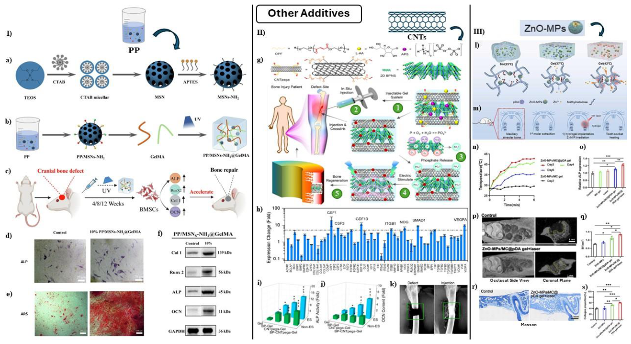
5.a.2. Porous Silicon Nanoparticles (PSiNPs)
Aghajanzadeh et al. investigated PSiNPs in Gel/aldehyde-modified xanthan (Gel-AXG) hydrogels, find-ing that higher PSiNP concentrations reduced swelling, porosity, and degradation rate, optimizing mechanical properties such as compressive strength (0.6223 MPa at 90% strain) and Young’s modulus (0.054 MPa). The Gel-AXG-8%Si formulation (containing 8% wt PSiNP) supported hydroxyapatite formation and sustained Si ion release, enhancing cell attachment, proliferation, and ALP activity in MG-63 cells [154].
Guan et al. developed a composite hydrogel by integrating nano bioactive glass (NBG) and nHA to compensate for alginate’s osteoinductive activity while mimicking natural bone composition. Within the NBG/n-HA/SA hydrogel, they could control the gelling time (4–8 minutes) by adjusting D-gluconic acid δ-lactone (GDL) dosage. This adjustment resulted in high compressive strength (170–220 kPa), and a porous structure with size of ∼40 µm. In vitro, the hydrogel facilitated bone-like apatite deposition, while cell studies confirmed biocompatibility, ALP activity enhancement, and osteogenic potential [155].
5.4. Carbon-Based Nanomaterials
Biomedical engineering researchers have extensively investigated lightweight and flexible carbon-based nanomaterials for bone tissue regeneration. These materials span zero-dimensional (fullerene, carbon dots, NDs), one-dimensional (CNTs), and two-dimensional (graphenic materials). Their unique mechanical, elec-trical, and thermal properties facilitate the differentiation of osteoblast-like human cells via interactions with proteins and nucleic acids, highlighting their potential in regenerative medicine [156,157,158]. Wei et al. addressed the challenges of carbon nanomaterial clearance by developing an injectable temperature-sensitive hydrogel composed of CS/HA/βGP incorporating large carbon particles (CPs, 491 nm diameter). This hy-drogel conformed to irregular bone defects, fostering new bone growth while exhibiting strong photothermal performance with a remarkable 98.4% tumor inhibition rate. Additionally, it facilitated bone regeneration, yielding a BV/TV ratio of 76.2%, forming a porous scaffold-like structure [159].
5.a.1. Graphen Oxide (GO)
GO has shown great potential for promoting osteogenic differentiation in both in vitro and in vivo studies. However, its delivery via injectable hydrogel systems remains limited, with thermosensitive hydro-gels being the primary approach employed [160,161]. While GO offers enhanced mechanical and bioactive properties, concerns regarding implant-associated inflammation as well as metal ion release when GO is com-posited with metals still persist [162,163]. Efforts to improve GO and CNT biocompatibility have focused on modifications in synthesis, purification techniques, and surface functionalization, aiming to enhance hy-drophilicity and minimize toxicity [164,165,166,167,168]. Encapsulation strategies have been proposed to prevent direct interaction of carbon-based hydrogels with cells and tissues, thus reducing adverse effects [161,169]. GO can be functionalized via hydroxyl and carboxyl modifications, enhancing electrostatic bonding [170,171].
Proteins are able to covalently bind to GO, improving hydrogel stability under various conditions, includ-ing heat, pH fluctuations, and while existing in an environment containing organic solvents [172]. Graphenic materials such as graphene, reduced graphene oxide (rGO), and GO possess high mechanical strength, elec-trical conductivity, and biocompatibility, making them valuable for hydrogel reinforcement [173,174]. GO’s oxygen-containing functional groups and large surface area enhance hydrogel flexibility and tensile strength, improving polymer interactions and scaffold durability [175]. Highly reduced rGO, while exhibiting lower hydrophilicity, alters protein adsorption and cellular responses, potentially increasing reactive oxygen species levels and playing role in angiogenesis [175,176,177].
Jiang et al. devised an injectable alginate-sericin-GO (Alg/Ser/GO) hydrogel using enzymatic crosslink-ing, which enabled bioimaging functionality and controlled degradation. GO contributed to mineralization, while sericin facilitated M2 macrophage polarization, further stimulating osteogenic differentiation of BM-SCs. The hydrogel attracted macrophages, suppressed inflammation, and enhanced bone repair in rats with femoral defects [171].
Lee et al. developed an injectable hydrogel system incorporating GO into glycol chitosan (gC) and HA (gC/oHA4GO36), optimizing GO concentration to maximize osteogenesis without adverse effects. In vivo implantation in rat calvarial defects demonstrated significant new bone formation in the gC/oHA4GO72 group, confirming effective clot formation, hydrogel stabilization, and enhanced osteogenic differentiation [160].
Amiryaghoubi et al. developed a thermosensitive injectable hydrogel containing a poly(N-isopropylacrylamide) (PNIPAAm)-based copolymer and GO, improving mineral deposition and ALP activity. The oxygen- and amine-containing functional groups in GO and CS facilitated osteogenic differentiation in hDPSCs cultured in both normal and osteogenic conditions. Additionally, the hydrogel enhanced the absorption of osteogenic inducers, reinforcing its bone regeneration potential [28].
Wang et al. investigated SF-based hydrogels, noting that low concentrations of GO improved scaffold porosity, mechanical properties, and cell encapsulation. BMSCs displayed enhanced growth, proliferation, and osteogenic differentiation compared to SF hydrogels without GO or those with excessive GO incorpora-tion, confirming its osteogenic benefits [178].
Wu et al. developed an antibacterial hydrogel, incorporating ASOs onto GO/alginate through nonco-valent interactions. This hydrogel efficiently delivered hUCMSCs to target sites, enhancing tissue healing. However, it exhibited rapid degradation, influencing encapsulated cell fate based on tissue microenviron-ments. The hydrogel inhibited bacterial growth by inducing oxidative stress via GO and suppressing gene transcription through ASOs [179].
Geng et al. examined graphene quantum dots (GQDs) to assess the role of surface charge in osteogenic differentiation. They observed that negatively charged GQDs significantly enhanced hMSCs osteogenic differentiation, activating specific signaling pathways, while positively charged GQDs had no observable impact. The constructed GQD/GelMA hydrogel scaffold accelerated osteogenic differentiation and bone regeneration in vivo, offering a promising approach for BTE [180].
5.a.2. Carbon Nanotubes (CNTs)
CNTs have also been in use for improving IOCHs bone regenerative capabilities. For example, Kaur et al. investigated a hydrogel composed of CS, collagen (Coll), and carboxylated single-walled CNTs (COOH-SWCNTs). These hydrogels exhibited thermoresponsive behavior, solidifying at 37◦C while demonstrating strong bioactivity through calcium phosphate deposition, forming an HAp layer within one day of incubation in simulated body fluid (SBF). The charged functional groups of COOH-SWCNTs attracted calcium and phosphate ions, facilitating early osteogenesis. Additionally, COOH-SWCNT incorporation significantly enhanced cell proliferation and osteogenic differentiation compared to CS/Coll-only hydrogels, reinforcing its bone tissue engineering potential [75].
Liu et al. introduced an IOCH composed of CNT-poly(ethylene glycol)-acrylate (CNTpega) and black phosphorus (BP), crosslinked within a biodegradable oligo(poly(ethylene glycol) fumarate) (OPF) matrix. This BP-CNTpega-gel, once injected in the defect site of the patient, exhibited enhanced mechanical strength, sustained phosphate ion release, and electrical conductivity, supporting bone tissue regeneration (Fig. 5-g). These properties in combination with electrical stimulation promoted MC3T3 preosteoblast adhesion, prolif-eration, and osteogenic differentiation, validating the hydrogel’s potential for bone repair applications. Gene expression screening revealed significant upregulation of multiple osteogenic pathway genes in response to stimulation (Fig. 5-h). After 14 days, cells cultured on BP-CNTpega-gel showed the highest ALP activity (Fig. 5-i), and by day 21, osteocalcin (OCN) levels in the culture medium were significantly elevated, indi-cating enhanced mineralization (Fig. 5-j). X-ray imaging confirmed successful in situ gelation and defect filling in rabbit femur models, demonstrating the hydrogel’s potential for clinical bone repair applications (Fig. 5-k) [152].
5.a.3. Fullerol
Fullerol, derived from fullerene C60, is a hydroxyl (-OH)-functionalized carbon structure, enhancing wa-ter solubility and biocompatibility, making it suitable for medical applications [156,181]. Fullerol presents several benefits compared to carbon nanotubes, notably lower toxicity and improved antioxidant properties.
Yang et al. developed fullerol-hydrogel microfluidic spheres (FMSs) to regulate redox homeostasis in stem cells and promote refractory bone healing in situ. Their study revealed a significant increase in bone volume (BV) fraction across experimental groups, with the GelMA/Ful + BMSCs group achieving the highest bone healing rate (38.5 ± 2.8%), compared to GelMA + BMSCs (23.7 ± 2.9%), BMSCs alone (12.6 ± 2.1%), and blank control (4.1 ± 1.5%). These findings confirmed the osteogenic efficacy of fullerol-functionalized hydrogels. Additionally, FMSs shielded stem cells from oxidative stress-induced damage, enhancing their os-teogenic differentiation by activating the forkhead box O1 (FoxO1) signaling pathway. In rat calvarial defect models, the injection of stem cell-laden FMSs resulted in notable new bone formation, further reinforcing the therapeutic value of fullerol-based hydrogels in BTE [182].
5.a.4. Nanodiamonds (NDs)
NDs are carbon-based nanomaterials recognized for their high biocompatibility, stability, and resilience, making them valuable in biomedical applications [183]. Their incorporation into hydrogels enhances mechan-ical properties, and when combined with VEGF, they enable sustained VEGF release, suggesting potential in tissue engineering [184]. NDs exhibit lower toxicity than graphene or carbon nanotubes (CNTs), making them suitable for permanent bone implants [185,186]. However, NDs are not currently employed in IOCHs, though their unique properties position them as promising candidates for future research and development in this field.
5.a.5. Carbon Nitride Quantum Dots (CNQDs)
CNQDs are 10 nm-sized nanoparticles that can adopt spherical, crystalline, or amorphous arrangements, exhibiting high biocompatibility with minimal toxicity. Their fluorescence enables precise imaging, making them valuable in biomedical applications. CNQDs serve as versatile carriers for therapeutic agents, con-tribute to antioxidant activity, and promote osteogenic differentiation, facilitating bone tissue formation. Certain CNQDs also demonstrate photothermal effects and are capable of enhancing mechanical properties of scaffolds [187,188]. Furthermore, addition of CNQDs into hydrogels resulted in smaller pore structures at higher concentrations, leading to a more uniform hydrogel system with enhanced durability, hardness, Young modulus, biostability, and cell attachment. MTT assay results revealed that CNQD-based hydrogels exhibited no cytotoxicity; instead, they fostered robust cell adhesion and proliferation (osteosarcoma cell line MG63) [189]. While these two results are promising indicators for bone regeneration, further testing is necessary to determine if CNQD containing hydrogels are capable of promoting osteogenesis.
5.5. Nanoclays
Nanoclays play a key role in hydrogel engineering, enhancing self-healing and shape-memory properties. Upon hydration, nanoclays exhibit plastic or viscoelastic behavior, forming pastes or gels that reinforce hydrogel matrices. Additionally, clay minerals facilitate osteogenic differentiation of stem cells, supporting bone tissue regeneration [190,191,192].
Zhang et al. devised a self-healing and pro-osteogenic injectable hydrogel using Lap nanosheets and guanidinylated chitosan. Laponite acted as a physical crosslinker with osteoinductive properties, forming a network structure with the cationic guanidine groups on chitosan chains while also serving as a carrier for demineralized bone matrix (DBM). This hydrogel enhanced adhesion and osteogenic differentiation of mesenchymal stem cells by activating the Wnt/β-catenin signaling pathway, promoting bone healing through self-healing and injectable properties for minimally invasive applications [190].
Andrade et al. fabricated a GelMA–Lap hydrogel composed of 1 w/v% GelMA and 6% laponite. The hydrogel showed no acute toxicity in Artemia salina lethality assays, and MTT analysis indicated cell via-bilities above 80%. In addition, osteogenic differentiation assays demonstrated that the composite hydrogel supported osteogenic activity, suggesting its suitability for further in vivo evaluation [193].
Kazemi-Aghdam et al. developed an injectable chitosan/mHNTs hydrogel, enhancing mechanical strength and osteoinductivity for bone tissue engineering. Encapsulated MSCs showed enhanced proliferation and osteogenic differentiation, while mHNT incorporation improved scaffold strength and hASCs proliferation [194].
Li et al. introduced a subperiosteal injectable hydrogel combining hydroxyapatite, laponite, and alginate, cross-linked via CaCO3 and Glucono-δ-lactone (GDL) ionic interactions. The Lap-10 (0.1 w/v%) group exhibited significantly higher ALP activity and osteogenic effects, while Lap-5 (0.05 w/v%) and Lap-20 (0.2 w/v%) had lower activity due to laponite agglomeration, limiting cell endocytosis of Li+, Mg2+, and Si(OH)4. RT-qPCR analysis confirmed osteogenesis-related gene upregulation, particularly Runx-2 expression in Lap-20 and strong ALP, Col-1, and OPN gene expression in Lap-10. In rat cranial bone defects, Lap-10 hydrogels displayed superior bone regeneration, confirmed through H&E staining, with significant new bone formation at 8 and 12 weeks [195].
Despite the fact that Laponite has been used as an osteogenic component in composite hydrogels, op-timizing its concentration to achieve the highest osteoinductive potential remains a challenge. Miao et al. investigated the ideal proportion of Laponite in gelatin-alginate hydrogel composites to enhance bone regen-eration. Their study identified 2% Laponite as the optimal concentration, balancing mechanical strength, pore architecture, and biocompatibility. In vitro, this formulation significantly promoted BMSC adhesion, ALP activity, and expression of osteogenic markers (Runx2, Col1a, ALP, and OCN). In vivo, implantation into rat femoral condylar defects resulted in superior bone regeneration, collagen deposition, and miner-alization compared to other concentrations. Transcriptomic and protein analyses revealed that Laponite significantly activates the MAPK-Erk signaling pathway in comparison with control group, meaning that osteogenic differentiation in BMSCs is induced through this pathway. These findings establish 2% Laponite hydrogel as a promising scaffold for bone defect repair, with mechanistic insights supporting its clinical translation [196].
To overcome the limitations of hypoxia and insufficient osteoinductivity in critical-sized bone defects, Xu et al. developed an injectable thermosensitive hydrogel composed of Pluronic F127 (PF-127), calcium peroxide-loaded polycaprolactone microspheres (ORMs), and osteoinductive nanoparticles—HAp and Lap (LAP). The hydrogel demonstrated sustained oxygen release for up to 21 days, effectively supporting the survival and proliferation of BMSCs and endothelial cells under anoxic conditions. In vitro studies re-vealed enhanced expression of RUNX2, ALP, OCN, and OPN, while in vivo implantation in a rat cranial defect model confirmed superior bone regeneration and vascularization in the PF-127/HAp/LAP/ORMs group compared to controls. These findings deepen the hydrogel’s potential as a minimally invasive and multifunctional platform for bone tissue regeneration, offering a biomimetic microenvironment conducive to vascularized osteogenesis [197].
5.6. Metal and Metal Oxide Nanoparticles
Metal and metal-oxide nanoparticles, including magnetic materials, have gained attention in regenerative medicine due to their unique properties and potential applications in BTE and regenerative therapies. These nanoparticles—silver (Ag), gold (Au), iron oxide (Fe2O3, Fe3O4), titania (TiO2), ZnO, and copper oxide (CuO)—offer a wide range of functionalities to enhance the performance of IOCHs [149,198,199,200,201,202,203,204,205,206].
Magnetic nanoparticles (MNPs), particularly magnetic iron oxide nanoparticles (IONPs), exhibit re-sponsiveness to magnetic fields, making them valuable for stimulating cell signaling, accelerating vascular network formation, and enhancing tissue development [207,208,209,210]. Additionally, IONPs contribute to labeling, imaging, thermal therapy, disease targeting, and improving hydrogel-tissue adhesion [211,212,213].
To address the complex and hostile microenvironment of osteomyelitis that traditionally hinders both bacterial eradication and osteogenesis, Zhou et al. combined CMCS with Fe3+ ions and an antibiotic named ciprofloxacin (CIP) to develop a CMCS/Fe3+/CIP hydrogel. This injectable and moldable hydrogel filled irregular cavities, resisted mechanical stress, and promoted osteogenesis in infected environments. From the tissue regenerations step-wise perspective, it accelerated three phases of osteomyelitis repair during tissue regeneration; anti-infection, proliferation, and remodeling [45].
Repair of large bone defects remains highly challenging, largely because current bioinks are unable to reproduce the anisotropically organized matrix of native bone, even though this architectural feature has been shown to significantly enhance stem cell osteogenic differentiation via mechanotransduction. Magnetic anisotropic hydrogels (MAHs) are highly promising for bone tissue engineering because of their mechanotrans-ductive and osteoinductive functions, yet their fabrication by bioprinting remains technically challenging. Bioprinting requires the ink to rapidly transition from liquid to solid to maintain structural stability, while anisotropic alignment requires consistent fluidity for magnetic components to freely assemble under magnetic induction. Xu et al. overcame these challenges by developing a biomimetic, anisotropic-structured MAH us-ing a novel continuous Liquid-in-Liquid bioprinting method paired with magnetic induction. This approach enabled a GelMA/HAMA base to encapsulate BMSCs while Fe3O4 microfibers formed aligned microscale geometric cues that promote BMSC elongation and osteogenic bioactivity through biomechanical signaling pathways. These anisotropic cues further strengthen mechanotransduction (e.g., MAPK, PI3K-Akt, Wnt, and calcium signaling), orchestrating BMSC elongation and osteogenic differentiation. In vitro assays con-firmed high viability and increased osteogenic markers, and In vivo, the BMSC-laden scaffold accelerated collagen matrix development, neovascularization, and complete bridging of a rabbit cranial defect by 12 weeks. This novel approach demonstrates strong potential for advanced applications in BTE and defect repair [214].
To improve guided tissue regeneration (GTR) methods for periodontitis treatment, Xu et al. introduced an injectable sodium alginate hydrogel composite doped with cubic cuprous oxide (Cu2O) and polydopamine-coated titanium dioxide (TiO2@PDA) nanoparticles named CTP-SA. This hydrogel transitioned from liquid to solid post-injection, adapting to various bone defects. By applying blue light in early healing stage, TiO2@PDA creates ROS capable of eliminating bacteria. During this procedure, Cu+ oxidized to Cu2+, through which osteogenesis is promoted. During the late stage of healing, near-infrared (NIR) is applied to the defect area, increasing the local temperature to ∼ 40–42 °C by means of TiO2@PDA, which is known to induce osteoblast differentiation and regulate osteogenic gene expression. This dual-light modulation (blue and NIR) enabled simultaneous antibacterial and osteogenic functionality, addressing patient-specific needs during healing [215].
Wang et al. developed proanthocyanidin (PC)-coordinated zinc-based injectable composite hydrogels (iPZCHs) for infected bone defect repair. These hydrogels, containing antimicrobial and antioxidant PC-coordinated ZnO-MPs and thioether-grafted sodium alginate (TSA), rapidly disintegrated in ROS environ-ments, releasing Zn2+ and Ag+ for antibacterial effects while creating areas where they will be replaced by regenerated bone. The antioxidant PC molecules scavenged excess ROS, enhancing Zn2+-driven im-munomodulation and osteogenesis through M2 macrophage polarization, demonstrating strong therapeutic potential [206].
Dhivya et al. crafted a thermosensitive injectable hydrogel (Zn-CS/nHAp/β-GP) containing zinc-doped chitosan, nHAp, and β-GP. The hydrogel underwent a sol–gel transition at 37◦C and exhibited swelling behavior, protein adsorption capability, biomineralization potential, and non-cytotoxicity for MSCs. nHAp incorporation further promoted osteoblast differentiation in vitro and accelerated bone formation in vivo, validated by increased apatite and collagen deposition, reinforcing its possible application for BTE [216].
In more recent work regarding the clinical treatment of osteosarcoma, Zhang et al. developed a mul-tifunctional injectable and in situ cross-linkable composite hydrogel formulated using magnesium-peroxide nanoparticles, horse radish peroxidase (HRP), and dopamine-conjugated gelatin (MgO2@GelDA). MgO2@GelDA demonstrated excellent gel stability, injectability, shape adaptability, tissue adhesion, and rapid hemostatic ability. MgO2@GelDA integrates the osteogenic and angiogenic capabilities of Mg2+ ions with immunomod-ulatory effects to enhance the local microenvironment. GelDA, a natural polymer, provides a biocompatible and adhesive matrix, while MgO2, a metal oxide nanoparticle, serves as both a crosslinking agent and a bioactive filler. In vitro, the group containing 10mg/mL MgO2 nanoparticles enhanced ALP activity, min-eralization, and expression of BMP2, Runx2, and OCN in hBMSCs. The hydrogel’s robust photothermal capabilities and early H2O2 release, enhance tumor suppression. In vivo, it significantly improved bone regen-eration in a rat skull defect model, confirmed by micro-CT and histological staining. These findings suggest significant potential for the clinical application of MgO2@GelDA in postsurgical osteosarcoma treatment and bone regeneration [217].
5.a.1. Noble Metal NPs
The incorporation of metallic nanoparticles such as silver and gold into hydrogels enhances electrical conductivity, magnetic properties, and osteogenic differentiation potential in both in vitro and in vivo set-tings. Their rapid response to external magnetic fields allows for precise manipulation of cell signaling. Silver nanoparticles are renowned for their antimicrobial properties and wound-healing applications, while gold nanoparticles promote MSC proliferation and osteogenic differentiation.
To reduce loss of alveolar bone, Ge et al. developed a photothermal hydrogel composed of ZnO-MPs and polydopamine embedded in a thermosensitive methylcellulose (MC) matrix named ZnO-MP/MC@pDA hydrogel. Upon near-infrared (NIR) irradiation, pDA converted light energy into heat, elevating the hydrogel temperature to approximately 42◦C and accelerating Zn2+ release (Fig. 5-l). This system retained photother-mal responsiveness over several days and was implanted into the rats’ maxillary first molar sockets, followed by four rounds of NIR stimulation (Fig. 5-m). Temperature profiling confirmed consistent heat generation post-implantation (Fig. 5-n). In vitro, the hydrogel enhanced ALP expression and calcium deposition in BMSCs (Fig. 5-o), while micro-CT imaging revealed substantial bone regeneration in the NIR-laser-treated group (Fig. 5-p), supported by increased bone volume (Fig. 5-q). Masson staining of the tooth socket re-vealed enhanced collagen deposition and bone formation in the ZnO-MP/MC@pDA gel plus NIR-laser group compared to controls, confirming the synergistic effect of sustained Zn2+ release and mild heat stimulation on alveolar bone regeneration (Fig. 5-r). Collagen analysis further indicated that laser-induced heating am-plified regenerative potential (Fig. 5-s). RNA sequencing identified activation of the Wnt signaling pathway, suggesting a molecular mechanism driving both osteogenesis and collagen synthesis [153].
De Mori et al. introduced a chitosan-hydroxyapatite-silver nanowire (CS-HACS-AgNWs) scaffold, merg-ing bioactivity with antibacterial properties. In vitro testing demonstrated strong AgNW antibacterial effects against both gram-positive and gram-negative bacteria. The scaffold enhanced calcium/phosphate deposition, improved gel strength, reduced gelation time, and maintained sustained Ag+ ion release within therapeutic ranges. Additionally, it suppressed bacterial growth and biofilm formation, including resistant strains, while supporting cell proliferation [218].
Liu et al. developed a bovine serum albumin (BSA)-based hydrogel with injectability, self-healing, and antibacterial capabilities for bone defect regeneration. The hydrogel utilized Sulfur-Silver (S-Ag) coordi-nation as the crosslinking mechanism, which facilitated osteogenic differentiation in vitro and produced accelerated bone repair in large cranial defect rabbit models compared to Bio-OSS, a commercially used spongious bovine bone substitute. The hydrogel’s gradual BSA protein and Ag+ release reinforced bone regeneration and bacterial inhibition, particularly against P. gingivalis and F. nucleatum. Moreover, the Ag ion contained in the hydrogel was gradually released as the hydrogel was degraded which offered a considerable antibacterial effect [219].
5.7. Metal Organic Frameworks (MOFs)
MOFs are crystalline materials composed of metal ions or clusters connected by organic ligands, forming porous structures with large surface areas. Their tunable structures and properties enable precise control over porosity, surface area, pore size, and morphology, thereby allowing researchers to design MOFs with tailored functionalities for specific applications [220,221,222]. Their porous nature enhances hydrogel swelling and injectability, making them valuable for incorporation into IOCH compositions. Nano-MOFs can serve multiple roles in IOCHs, including acting as cross-linkers to strengthen hydrogel networks, providing sites for drug loading and release, and modulating biological functions such as cell adhesion, proliferation, and differentiation [223,224,225]. Studies show that MOF-modified composites enhance wettability, surface en-ergy, cell attachment, growth, and osteogenic differentiation of ADSCs [224,226]. Additionally, integrating MOFs into postoperative biodegradable barrier membranes strengthens bioactivity by promoting osteogene-sis, modulating inflammation, and supporting responsive therapeutic drug/ion release; similarly, nano-MOF-functionalized bone scaffolds provide localized anticancer activity to target residual tumor cells post-resection [227,228].
Cobalt (Co2+) ions mimic hypoxia and promote angiogenesis, supporting bone repair. However, their release profile must be carefully managed to align with the bone regeneration process. ZIF-67, part of the zeolitic imidazolate framework family, is a MOF composed of cobalt ions coordinated with 2-methylimidazole (MeIm). Its high surface area and tunable pore size enable efficient therapeutic delivery, and its biocompati-bility alongside its controlled release properties further strengthens its potential for biomedical applications. Sun et al. developed an eIm/ZIF-67 nanocomposite hydrogel via in situ photo-crosslinking, integrating 2-ethylimidazole (eIm) into ZIF-67 to regulate Co2+ release. By combining 75% eIm/ZIF-67 with GelMA, they achieved sustained Co2+ release over 21 days, supporting early-stage angiogenesis, which is crucial for bone formation. In vitro experiments revealed that GelMA@eIm/ZIF-67 was less cytotoxic than GelMA@CoCl2 toward both BMSCs and HUVECs, stimulated angiogenic activity in HUVECs, and promoted osteogenic differentiation of BMSCs. In vivo rat calvaria defect models have also confirmed enhanced bone forma-tion and neovascularization. ZIF-8, another MOF subclass, is composed of zinc ions (Zn2+) coordinated with 2-methylimidazole (MeIm). It possesses low cytotoxicity, remarkable chemical stability, and pH re-sponsiveness, rendering it highly promising for bone regeneration. Zn2+, a vital trace element, contributes to bone growth and antibacterial effects as ZIF-8 degrades. Its uniform pore structure and ample surface area enable controlled drug release when it is included in IOCHs [229]. Qiao et al. developed an injectable composite hydrogel modified with nano simvastatin-laden zeolitic imidazolate framework-8 (nano SIM@ZIF-8). TEM imaging confirmed SIM presence within the ZIF-8 MOF, while mechanical testing revealed that nano SIM@ZIF-8 enhanced hydrogel strength, with PEG diacrylate (PEGDA) contributing to the improve-ment. This composite, nano SIM@ZIF-8/PEGDA/SA (nSZPS), exhibited excellent injectability within the PEGDA/sodium alginate (PEGDA/SA) system, as validated by rheological assessments. Sustained SIM and Zn2+ release demonstrated exceptional biocompatibility, boosting osteogenic differentiation while suppress-ing adipogenic differentiation of BMSCs in vitro. Gene expression analysis confirmed significantly increased levels of osteogenic genes, including ALP, OCN, and OPN, though RUNX2 showed no difference compared to control groups. In vivo experiments on hyperlipidemic rat bone defects revealed enhanced osseointegration and lipid-lowering effects, possibly linked to PPARγ and Wnt/β-CATENIN interactions, with H&E staining and µ-CT scanning reinforcing these findings [230].
Repairing bone defects in diabetic patients poses a challenge due to issues like glucose fluctuations, oxidative damage from ROS, and the inflammatory conditions within the affected area. Gong et al. tackled these by creating an injectable, UV-curable hydrogel in which GelMA and hydroxyapatite nanowires (HAP NWs) were incorporated and it expands upon gas exposure. Gallic acid and magnesium ion (Mg2+) MOFs loaded with glucose oxidase (GOx) were included, creating the GOx-MOF/HAP-GelMA composite hydrogel. Upon injection and quick solidification, controlled Gallic acid release neutralized hydrogen peroxide (H2O2) produced during glucose decomposition, reducing oxidative damage on cells [231].
Sun et al. investigated GelMA@eIm/ZIF-67 nanocomposite hydrogels for osteogenic differentiation of BMSCs, assessing ALP activity and mineralized nodule formation. ALP staining and activity assays con-ducted on day 3 showed enhanced osteogenic differentiation in the Gel@eZIF100 and Gel@eZIF200 groups, while ARS staining after 14 days of In vitro culture confirmed higher mineralization in Gel@eZIF200 com-pared to Gel@eZIF100. These results indicate that GelMA@eIM/ZIF-67 hydrogels support osteogenesis in a dose-dependent manner [229].
5.8. Polyoxometalates (POMs)
POMs are molecular clusters composed of metal oxides, primarily from early transition metals like tung-sten, molybdenum, and vanadium, surrounded by oxide ions. These compounds exhibit anti-inflammatory and osteogenic properties, making them valuable for regenerative medicine applications [232,233,234].
Integration of Molybdenum (Mo)-based POM nanoclusters into GelMA hydrogels enables a controlled, localized, and slow release of POM itself, with GelMA serving as the drug-delivery matrix and POM as the active therapeutic agent. This delivery strategy is especially relevant for diabetic bone defects, sustain-ing antioxidant activity at the defect site while minimizing systemic side effects. As a versatile platform, the hydrogel improves local bioavailability and therapeutic efficacy. Liao et al. developed a GelMA/POM hydrogel, combining GelMA’s biocompatibility with Mo-based POM’s redox activity to scavenge ROS, sup-porting osteoblast proliferation and enhancing osteogenic differentiation. Their in vitro data confirm that GelMA/POM promotes osteogenesis, and the sustained release of POM counters ROS-induced oxidative stress, consequently optimizing the healing microenvironment. Moreover, additional analyses suggest activa-tion of the PI3K/Akt signaling pathway. In vivo results in a diabetic femoral-defect model show accelerated bone repair with minimal systemic toxicity, as the hydrogel remains localized in the defective bone tissue and gradually metabolizes [235].
5.9. Others
5.a.1. Calcium Sulfate Nanorods
To address limitations of natural polymer-based hydrogels, Liu et al. devised an injectable collagen-HA (Col-HA) hydrogel incorporating CSN. The hydrogel was synthesized using a bio-orthogonal reaction between norbornene (Nb) and tetrazine (Tz), forming a honeycomb-like porous structure at 5% (w/v) Col-Nb and HA-Tz concentration. Increasing CSN concentration (0–5%) reduced pore diameter, enhancing hydrogel strength and compressive strain while preserving injectability. In vitro release experiments confirmed a sustained Ca2+ ion release for 28 days, facilitating spontaneous hydroxyapatite (HAp) formation via interactions with phosphate in the pericellular environment. The released Ca2+ promoted osteogenic differentiation, with 5%CSN@Col-HA samples demonstrating superior performance over 1%CSN@Col-HA and Col-HA samples both in vitro and in vivo. Moreover, Col-HA enhanced cell attachment, reinforcing its biocompatibility and regenerative potential [139].
6. Future Directions and Challenges
Aging, diabetes, smoking, and other pathological conditions negatively impact fracture healing, primarily due to heightened inflammatory responses. Pro-inflammatory cytokines increase with age, contributing to osteoporosis, a leading cause of bone fractures. Research suggests this inflammatory rise results from declining immune system functionality, affecting B cell and T cell proliferation and differentiation. Aging reduces hematopoietic compartments, decreasing T cell numbers and their capacity for proliferation. Xing et al. demonstrated that younger bone marrow cell transplants in older mice accelerated callus formation and remodeling, reinforcing the significance of immune health in bone healing [236].
Future research in IOCHs must prioritize nanoparticle modifications, including pre- and post-functionalization techniques, to optimize bioactivity, therapeutic delivery, and tissue targeting. One promising approach is colloidal crystal engineering, which precisely arranges nanoparticles (NPs) to tailor optical, electronic, and mechanical properties, improving material selection and composite hydrogel integration. Beyond nanopar-ticle modification, innovative strategies for incorporating diverse biomolecules and growth factors into hy-drogels will expand their utility in tissue regeneration. Controlled release mechanisms enhance bioactivity, ensuring sustained effects.
A comprehensive understanding of hydrogel-nanoparticle interactions is essential for optimizing tissue re-generation outcomes. Studies should investigate nanoparticle influence on hydrogel degradation, mechanical properties, and cellular response, ensuring clinical feasibility through rigorous preclinical studies on safety, efficacy, and long-term effects. Additionally, scalability and reproducibility improvements in manufacturing processes will be essential for real-world application. With continued research and testing, injectable com-posite hydrogel technology holds immense potential for tailored therapeutic applications across healthcare.
7. Conclusions
Composite hydrogels represent the future gold standard therapy for bone defect repair, with extensive research validating their in vitro and in vivo regenerative efficacy. Their diverse compositions present significant opportunities for further optimization. Moving forward, research must prioritize the identification of the optimal formulations and the transition to clinical trials, ensuring scalable, effective alternatives to autografts for widespread clinical application.

Table 1.
List of abbreviations.
| Abbr. | Explanation |
|---|---|
| BTE | Bone Tissue Engineering |
| ECM | Extracellular Matrix |
| BMPs | Bone Morphogenetic Proteins |
| MSCs | Mesenchymal Stem Cells |
| BMSCs | Bone Marrow Mesenchymal Stem Cells |
| rBMSCs | Rat Bone Marrow Mesenchymal Stem Cells |
| ADSCs | Adipose-derived Mesenchymal Stem Cells |
| hDPSCs | Human Dental Pulp Stem Cells |
| HUVECs | Human Umbilical Vein Endothelial Cells CD31 or PECAM-1 |
| Platelet endothelial cell adhesion molecule-1 VEGF | Vascular |
| Endothelial Growth Factor | |
| FGF | Fibroblast Growth Factor |
| bFGF | Basic Fibroblast Growth Factor |
| TGF-β | Transforming Growth Factor-Beta |
| rhBMP-2 | Recombinant Human Bone Morphogenetic Protein-2 |
| iNOS | Inducible Nitric Oxide Synthase |
| ARG-1 | Arginase-1 |
| ROS | Reactive Oxygen Species |
| ALP | Alkaline Phosphatase |
| ARS | Alizarin Red S |
| HA | Hyaluronic Acid |
| Gel | Gelatin |
| GelMA | Gelatin Methacrylate |
| GelDA | Dopamine-conjugated Gelatin |
| CS | Chitosan |
| CMCS | Carboxymethyl Chitosan |
| CSe | Chondroitin Sulfate |
| OCS | Oxidized Chondroitin Sulfate |
| PEG | Polyethylene Glycol |
| PLGA | Poly(lactic-co-glycolic acid) |
| MC | Methylcellulose |
| SF | Silk Fibroin |
| GG | Gellan Gum |
| pdA | Polydopamine |
| SS | Disulfide |
| HAp | Hydroxyapatite |
| nHAp | Nano-hydroxyapatite |
| β-TCP | β-Tricalcium Phosphate |
| BCP | Biphasic Calcium Phosphate |
| CSN | Calcium Sulfate Nanorods |
| NDs | Nanodiamonds |
| Lap | Laponite |
| ZnO-MP | Zinc Oxide Microsphere |
| GP | Glycerophosphate |
| XRD | X-Ray Diffraction |
| µ-CT | Micro-computed Tomography |
Table 2.
Definitions of key biological, cellular, and biomaterial terms used in BTE.
| Name | Definition |
|---|---|
| MC3T3-E1 | A mouse osteoblastic cell line commonly used in research related to bone biology |
| MG-63 cells | A type of human osteosarcoma cell line commonly used in biomedical research |
| Multiple myeloma cells | Cancerous plasma cells that grow uncontrollably in the bone |
| marrow | |
| Exosome | Extracellular vesicles secreted from cell to ECM, with a size range of 30–150 nm |
| HIF-1α signaling pathway | Regulates cellular responses to low oxygen levels (hypoxia); promotes bone regeneration |
| Wnt/β-catenin | Key signaling pathway regulating cell fate, tissue homeosta-sis, and embryonic growth |
| NF-κB | A signaling cascade crucial for regulating immune and in-flammatory responses |
| PI3K/Akt signaling pathway | Promotes osteoblast differentiation from MSCs |
| SIPN | Semi-interpenetrating network: a hydrogel containing a cross-linked polymer network while the other hydrogel chains are not cross-linked |
| Hydrogel secondary network structure. | Referred to as the structure in which two or more polymeric networks are entangled |
| Hydrogel self-healing | Some hydrogels have the capability to reconstruct their original structure by having no external parameter involved |
| Hydrogels dynamic adjustments | Some hydrogels can change their properties regarding their dynamic environment (e.g., pH, and temperature) |
| Supramolecular hydrogel | A hydrogel formed from non-covalent reactions |
| Cryogels | Three-dimensional porous structures with a highly intercon-nected pore network |
| CMS | Cryogel microspheres: a type of hydrogel with a three-dimensional porous structure |
| Biodegradable barrier membranes | A resorbable membrane that blocks soft-tissue invasion while preserving a regenerative space to facilitate bone regeneration |
| Hemostatic Agents | Substances used to stop bleeding by accelerating and promoting the body’s natural coagulation abilities |
| Mechanotransduction | The process by which cells detect mechanical cues in their environment and convert them into biochemical signals that regulate cellular behavior and function |
| Anisotropic | Materials that exhibit direction-dependent physical or me-chanical properties, such that their responses differ based on the orientation of applied forces or stimuli |
Supplementary Materials
The following supporting information can be downloaded at the website of this paper posted on Preprints.org.
References
- Su, N.; Yang, J.; Xie, Y.; Du, X.; Chen, H.; Zhou, H.; Chen, L. Bone function, dysfunction and its role in diseases including critical illness. Int. J. Biol. Sci. 2019, 15(4), 776. [Google Scholar] [CrossRef] [PubMed]
- S. Stewart, S. Bryant, J. Ahn, K. Hankenson, Bone regeneration. translational regenerative medicine, Translational regenerative medicine (2015).
- Schemitsch, E. H. Size matters: defining critical in bone defect size! J. Orthop. Trauma 2017, 31, S20–S22. [Google Scholar] [CrossRef] [PubMed]
- Minardi, S.; Corradetti, B.; Taraballi, F.; Sandri, M.; Van Eps, J.; Cabrera, F. J.; Weiner, B. K.; Tampieri; Tasciotti, E. Evaluation of the osteoinductive potential of a bio-inspired scaffold mimick-ing the osteogenic niche for bone augmentation. Biomaterials 2015, 62, 128–137. [Google Scholar] [CrossRef] [PubMed]
- B. Mubeen, I. Ahmed, A. Jameel, Study of mechanical properties of bones and mechanics of bone fracture, in: Proceedings of 60th Congress of ISTAM, 2015, pp. 16–19.
- Eldesoqi, K.; Henrich, D.; El-Kady, A. M.; Arbid, M. S.; Abd El-Hady, B. M.; Marzi, I.; Seebach, C. Safety evaluation of a bioglass–polylactic acid composite scaffold seeded with progenitor cells in a rat skull critical-size bone defect. PLoS ONE 2014, 9(2), e87642. [Google Scholar] [CrossRef] [PubMed]
- Zwingenberger, S.; Niederlohmann, E.; Vater, C.; Rammelt, S.; Matthys, R.; Bernhardt, R.; Val-ladares, R. D.; Goodman, S. B.; Stiehler, M. Establishment of a femoral critical-size bone defect model in immunodeficient mice. J. Surg. Res. 2013, 181(1), e7–e14. [Google Scholar] [CrossRef] [PubMed]
- Sowa, D. T.; Weiland, A. J. Clinical applications of vascularized bone autografts. Orthop. Clin. N. Am. 1987, 18(2), 257–273. [Google Scholar] [CrossRef]
- Friedlaender, G. E.; Perry, C. R.; Cole, J. D.; Cook, S. D.; Cierny, G.; Muschler, G. F.; Zych, G. A.; Calhoun, J. H.; LaForte, A. J.; Yin, S. Osteogenic protein-1 (bone morphogenetic protein-7) in the treatment of tibial nonunions: a prospective, randomized clinical trial comparing rhop-1 with fresh bone autograft. JBJS 2001, 83(1), S151–S158. [Google Scholar]
- Jimi, E.; Hirata, S.; Osawa, K.; Terashita, M.; Kitamura, C.; Fukushima, H. The current and future therapies of bone regeneration to repair bone defects. Int. J. Dent. 2012, 2012(1), 148261. [Google Scholar] [CrossRef] [PubMed]
- Chen, C. M.; Disa, J. J.; Lee, H.-Y.; Mehrara, B. J.; Hu, Q.-Y.; Nathan, S.; Boland, P.; Healey, J.; Cordeiro, P. G. Reconstruction of extremity long bone defects after sarcoma resection with vascularized fibula flaps: a 10-year review. Plast. Reconstr. Surg. 2007, 119(3), 915–924. [Google Scholar] [CrossRef] [PubMed]
- Gharedaghi, M.; Peivandi, M. T.; Mazloomi, M.; Shoorin, H. R.; Hasani, M.; Seyf, P.; Khazaee, F. Eval-uation of clinical results and complications of structural allograft reconstruction after bone tumor surgery. Arch. Bone Jt. Surg. 2016, 4(3), 236. [Google Scholar] [PubMed]
- Budi, H. S.; Al-azzawi, M. F. J.; Al-Dolaimy, F.; Alahmari, M. M.; Abullais, S. S.; Ebrahimi, S.; Khlewee, I. H.; Alawady, A. H. R.; Alsaalamy, A. H.; Shayan, F. K. Injectable and 3d-printed hydrogels: state-of-the-art platform for bone regeneration in dentistry. Inorg. Chem. Commun. 2024, 161, 112026. [Google Scholar] [CrossRef]
- Mellati; Hasanzadeh, E.; Gholipourmalekabadi, M.; Enderami, S. E. Injectable nanocomposite hydro-gels as an emerging platform for biomedical applications: A review. Mater. Sci. Eng. C 2021, 131, 112489. [Google Scholar] [CrossRef] [PubMed]
- Utech, S.; Boccaccini, A. R. A review of hydrogel-based composites for biomedical applications: en-hancement of hydrogel properties by addition of rigid inorganic fillers. J. Mater. Sci. 2016, 51(1), 271–310. [Google Scholar] [CrossRef]
- Tran, H. D.; Park, K. D.; Ching, Y. C.; Huynh, C.; Nguyen, D. H. A comprehensive review on polymeric hydrogel and its composite: Matrices of choice for bone and cartilage tissue engineering. J. Ind. Eng. Chem. 2020, 89, 58–82. [Google Scholar] [CrossRef]
- Zhang, B.; Huang, J.; Liu, J.; Lin, F.; Ding, Z.; Xu, J. Injectable composite hydrogel promotes os-teogenesis and angiogenesis in spinal fusion by optimizing the bone marrow mesenchymal stem cell microenvironment and exosomes secretion. Mater. Sci. Eng. C 2021, 123, 111782. [Google Scholar] [CrossRef] [PubMed]
- Biglari, N.; Zare, E. N. Conjugated polymer-based composite scaffolds for tissue engineering and re-generative medicine. Alex. Eng. J. 2024, 87, 277–299. [Google Scholar] [CrossRef]
- Gulfam, M.; Jo, S.-H.; Jo, S.-W.; Vu, T. T.; Park, S.-H.; Lim, K. T. Highly porous and injectable hydrogels derived from cartilage acellularized matrix exhibit reduction and nir light dual-responsive drug release properties for application in antitumor therapy. NPG Asia Mater. 2022, 14(1), 8. [Google Scholar] [CrossRef]
- Yu, X.; Wang, X.; Li, D.; Sheng, R.; Qian, Y.; Zhu, R.; Wang, X.; Lin, K. Mechanically reinforced injectable bioactive nanocomposite hydrogels for in-situ bone regeneration. Chem. Eng. J. 2022, 433, 132799. [Google Scholar] [CrossRef]
- Morales, M. E.; Ruiz, M. A. Microencapsulation of probiotic cells: applications in nutraceutic and food industry. Nutraceuticals 2016, 1, 627–668. [Google Scholar] [CrossRef]
- Chen, Q.; Chen, H.; Zhu, L.; Zheng, J. Fundamentals of double network hydrogels. J. Mater. Chem. B 2015, 3(18), 3654–3676. [Google Scholar] [CrossRef] [PubMed]
- Li; Xu, H.; Yu, P.; Xing, J.; Ding, C.; Yan, X.; Xie, J.; Li, J. Injectable hydrogels based on gellan gum promotes in situ mineralization and potential osteogenesis. Eur. Polym. J. 2020, 141, 110091. [Google Scholar] [CrossRef]
- Lin, X.; Patil, S.; Gao, Y.-G.; Qian, A. The bone extracellular matrix in bone formation and regeneration. Front. Pharmacol. 2020, 11, 757. [Google Scholar] [CrossRef] [PubMed]
- Chen, J.; Sun, T.; You, Y.; Wu, B.; Wang, X.; Wu, J. Proteoglycans and glycosaminoglycans in stem cell homeostasis and bone tissue regeneration. Front. Cell Dev. Biol. 2021, 9, 760532. [Google Scholar] [CrossRef] [PubMed]
- Lavanya, K.; Chandran, S. V.; Balagangadharan, K.; Selvamurugan, N. Temperature-and ph-responsive chitosan-based injectable hydrogels for bone tissue engineering. Mater. Sci. Eng. C 2020, 111, 110862. [Google Scholar] [CrossRef] [PubMed]
- Fardjahromi, M. A.; Nazari, H.; Tafti, S. A.; Razmjou, A.; Mukhopadhyay, S.; Warkiani, M. E. Metal-organic framework-based nanomaterials for bone tissue engineering and wound healing. Mater. Today Chem. 2022, 23, 100670. [Google Scholar] [CrossRef]
- Amiryaghoubi, N.; Pesyan, N. N.; Fathi, M.; Omidi, Y. Injectable thermosensitive hybrid hydrogel containing graphene oxide and chitosan as dental pulp stem cells scaffold for bone tissue engineering. Int. J. Biol. Macromol. 2020, 162, 1338–1357. [Google Scholar] [CrossRef] [PubMed]
- Hassanzadeh; Ashrafihelan, J.; Salehi, R.; Rahbarghazi, R.; Firouzamandi, M.; Ahmadi, M.; Khak-sar, M.; Alipour, M.; Aghazadeh, M. Development and biocompatibility of the injectable collagen/nano-hydroxyapatite scaffolds as in situ forming hydrogel for the hard tissue engineering application. Arti-Ficial Cells Nanomed. Biotechnol. 2021, 49(1), 136–146. [Google Scholar] [CrossRef] [PubMed]
- Deo, K. A.; Lokhande, G.; Gaharwar, A. K. Nanostructured hydrogels for tissue engineering and regen-erative medicine. Encyclopedia of tissue engineering and regenerative medicine 2019, 1, 3. [Google Scholar]
- Chen, M.; Li, Y.; Hou, W.-X.; Peng, D.-Y.; Li, J.-K.; Zhang, H.-X. The antibacterial effect, biocom-patibility, and osteogenesis of vancomycin-nanodiamond composite scaffold for infected bone defects. Int. J. Nanomed. 2023, 1365–1380. [Google Scholar] [CrossRef] [PubMed]
- Chen, Y.; Sheng, W.; Lin, J.; Fang, C.; Deng, J.; Zhang, P.; Zhou, M.; Liu, P.; Weng, J.; Yu, F.; et al. Magnesium oxide nanoparticle coordinated phosphate-functionalized chitosan injectable hydrogel for osteogenesis and angiogenesis in bone regeneration. ACS Appl. Mater. Interfaces 2022, 14(6), 7592–7608. [Google Scholar] [CrossRef] [PubMed]
- Wang, L.; Li, Y.; Jiang, S.; Zhang, Z.; Zhao, S.; Song, Y.; Liu, J.; Tan, F. Alginate hydrogels containing different concentrations of magnesium-containing poly (lactic-co-glycolic acid) microspheres for bone tissue engineering. Biomed. Mater. 2023, 18(5), 055022. [Google Scholar] [CrossRef] [PubMed]
- Shaikh; Kesharwani, P.; Gajbhiye, V. Dendrimer as a momentous tool in tissue engineering and regenerative medicine. J. Control. Release 2022, 346, 328–354. [Google Scholar] [CrossRef] [PubMed]
- Liu, F.; Wang, X.; Li, Y.; Ren, M.; He, P.; Wang, L.; Xu, J.; Yang, S.; Ji, P. Dendrimer-modified gelatin methacrylate hydrogels carrying adipose-derived stromal/stem cells promote cartilage regeneration. Stem Cell Res. Ther. 2022, 13(1), 26. [Google Scholar] [CrossRef] [PubMed]
- Yan, S.; Ren, J.; Jian, Y.; Wang, W.; Yun, W.; Yin, J. Injectable maltodextrin-based micelle/hydrogel composites for simvastatin-controlled release. Biomacromolecules 2018, 19(12), 4554–4564. [Google Scholar] [CrossRef] [PubMed]
- Tang, G.; Tan, Z.; Zeng, W.; Wang, X.; Shi, C.; Liu, Y.; He, H.; Chen, R.; Ye, X. Recent advances of chitosan-based injectable hydrogels for bone and dental tissue regeneration. Front. Bioeng. Biotechnol. 2020, 8, 587658. [Google Scholar] [CrossRef] [PubMed]
- Lee, E. J.; Kang, E.; Kang, S.-W.; Huh, K. M. Thermo-irreversible glycol chitosan/hyaluronic acid blend hydrogel for injectable tissue engineering. Carbohydr. Polym. 2020, 244, 116432. [Google Scholar] [CrossRef] [PubMed]
- Yang, J.; Jing, X.; Wang, Z.; Liu, X.; Zhu, X.; Lei, T.; Li, X.; Guo, W.; Rao, H.; Chen, M.; et al. In vitro and in vivo study on an injectable glycol chitosan/dibenzaldehyde-terminated polyethylene glycol hydrogel in repairing articular cartilage defects. Front. Bioeng. Biotechnol. 2021, 9, 607709. [Google Scholar] [CrossRef] [PubMed]
- Shi, Z.; Yang, F.; Pang, Q.; Hu, Y.; Wu, H.; Yu, X.; Chen, X.; Shi, L.; Wen, B.; Xu, R.; et al. The osteoge-nesis and the biologic mechanism of thermo-responsive injectable hydrogel containing carboxymethyl chitosan/sodium alginate nanoparticles towards promoting osteal wound healing. Int. J. Biol. Macromol. 2023, 224, 533–543. [Google Scholar] [CrossRef] [PubMed]
- Li, J.; Shi, Q.; Wu, X.; Li, C.; Chen, X. In vitro and in vivo evaluation of 3d biodegradable thermo/ph sensitive sol-gel reversible hydroxybutyl chitosan hydrogel. Mater. Sci. Eng. C 2020, 108, 110419. [Google Scholar] [CrossRef] [PubMed]
- Xu, P.; Chen, P.; Sun, Y.; Nuliqiman, M.; Zhou, Y.; Cao, J.; Yu, S.; Huang, J.; Ye, J. A novel injectable thermo/photo dual-crosslinking hydrogel based on modified chitosan for fast sealing open globe injury. Carbohydr. Polym. 2024, 331, 121854. [Google Scholar] [CrossRef] [PubMed]
- Vaidya, G.; Pramanik, S.; Kadi, A.; Rayshan, A. R.; Abualsoud, B. M.; Ansari, M. J.; Masood, R.; Michaelson, J. Injecting hope: chitosan hydrogels as bone regeneration innovators. J. Bioma-Terials Sci. Polym. Ed. 2024, 35(5), 756–797. [Google Scholar] [CrossRef] [PubMed]
- Wan, Z.; Dong, Q.; Guo, X.; Bai, X.; Zhang, X.; Zhang, P.; Liu, Y.; Lv, L.; Zhou, Y. A dual-responsive polydopamine-modified hydroxybutyl chitosan hydrogel for sequential regulation of bone regeneration. Carbohydr. Polym. 2022, 297, 120027. [Google Scholar] [CrossRef] [PubMed]
- Zhou, T.; Zhou, H.; Wang, F.; Zhang, P.; Shang, J.; Shi, L. An injectable carboxymethyl chitosan hydrogel scaffold formed via coordination bond for antibacterial and osteogenesis in osteomyelitis. Carbohydr. Polym. 2024, 324, 121466. [Google Scholar] [CrossRef] [PubMed]
- Karimi; Peyravian, N.; Delattre, C.; Ghasemian, M.; Michaud, P.; Amini, N.; Roudmiane, M. M. M.; Milan, P. B.; et al. Designing and synthesis of in-situ hydrogel based on pullulan/carboxymethyl chitosan containing parathyroid hormone for bone tissue engineering. Materialia 2024, 33, 102026. [Google Scholar] [CrossRef]
- Datta, S.; Rameshbabu, A. P.; Bankoti, K.; Roy, M.; Gupta, C.; Jana, S.; Das, A. K.; Sen, R.; Dhara, S. De-cellularized bone matrix/oleoyl chitosan derived supramolecular injectable hydrogel promotes efficient bone integration. Mater. Sci. Eng. C 2021, 119, 111604. [Google Scholar] [CrossRef] [PubMed]
- Cui, S.; Zhang, S.; Coseri, S. An injectable and self-healing cellulose nanofiber-reinforced alginate hy-drogel for bone repair. Carbohydr. Polym. 2023, 300, 120243. [Google Scholar] [CrossRef] [PubMed]
- Guo, K.; Li, G.; Yu, Q.; Yang, Y.; Liu, H.; Zhao, Y.; Huang, Y.; Zhang, H.; Li, W. Injectable hyaluronate-based hydrogel with a dynamic/covalent dual-crosslinked architecture for bone tissue engineering: Enhancing osteogenesis and immune regulation. Int. J. Biol. Macromol. 2024, 282, 137249. [Google Scholar] [CrossRef] [PubMed]
- Wasupalli, G. K.; Verma, D. Thermosensitive injectable hydrogel based on chitosan-polygalacturonic acid polyelectrolyte complexes for bone tissue engineering. Carbohydr. Polym. 2022, 294, 119769. [Google Scholar] [CrossRef] [PubMed]
- Patel; Zaky, S. H.; Schoedel, K.; Li, H.; Sant, V.; Beniash, E.; Sfeir, C.; Stolz, D. B.; Sant, S. De-sign and evaluation of collagen-inspired mineral-hydrogel nanocomposites for bone regeneration. Acta Biomater. 2020, 112, 262–273. [Google Scholar] [CrossRef] [PubMed]
- Zhang, Q.; Yan, Y.; Li, Z.; Du, J.; Zhang, K.; Zhang, L.; Wang, T.; Bianco, A.; Ge, S.; Ma, B. A uniform-unsaturated crosslinking strategy to construct injectable alginate hydrogel. Int. J. Biol. Macromol. 2024, 254, 127726. [Google Scholar] [CrossRef] [PubMed]
- Li, J.; Chen, G.; Xu, X.; Abdou, P.; Jiang, Q.; Shi, D.; Gu, Z. Advances of injectable hydrogel-based scaffolds for cartilage regeneration. Regen. Biomater. 2019, 6(3), 129–140. [Google Scholar] [CrossRef] [PubMed]
- Rana, K.; Gupta, V. K.; Hart, P.; Thakur, V. K. Cellulose-alginate hydrogels and their nanocompos-ites for water remediation and biomedical applications. Environ. Res. 2024, 243, 117889. [Google Scholar] [CrossRef] [PubMed]
- Lee, K. Y.; Mooney, D. J. Alginate: properties and biomedical applications. Prog. Polym. Sci. 2012, 37(1), 106–126. [Google Scholar] [CrossRef] [PubMed]
- Wang, H.; Yang, J.; Tian, W.; Peng, K.; Xue, Y.; Zhao, H.; Ma, X.; Shi, R.; Chen, Y. A sodium alginate/carboxymethyl chitosan dual-crosslinked injectable hydrogel scaffold with tunable soft-ness/hardness for bone regeneration. Int. J. Biol. Macromol. 2024, 257, 128700. [Google Scholar] [CrossRef] [PubMed]
- Xu, Q.; Torres, J. E.; Hakim, M.; Babiak, P. M.; Pal, P.; Battistoni, C. M.; Nguyen, M.; Panitch, A.; Solorio, L.; Liu, J. C. Collagen-and hyaluronic acid-based hydrogels and their biomedical applications. Mater. Sci. Eng. R Rep. 2021, 146, 100641. [Google Scholar] [CrossRef] [PubMed]
- M. N. Collins, F. Zamboni, A. Serafin, G. Ren, A. Thanusha, M. Culebras, The role of hyaluronic acid in tissue engineering, in: polysaccharides of microbial origin: biomedical applications, Springer, 2021,pp. 1–55.
- Halperin-Sternfeld, M.; Pokhojaev, A.; Ghosh, M.; Rachmiel, D.; Kannan, R.; Grinberg, I.; Asher, M.; Aviv, M.; Ma, P. X.; Binderman, I.; et al. Immunomodulatory fibrous hyaluronic acid-fmoc-diphenylalanine-based hydrogel induces bone regeneration. J. Clin. Periodontol. 2023, 50(2), 200–219. [Google Scholar] [CrossRef] [PubMed]
- Deng, Z.; Li, M.; Hu, Y.; He, Y.; Tao, B.; Yuan, Z.; Wang, R.; Chen, M.; Luo, Z.; Cai, K. Injectable biomimetic hydrogels encapsulating gold/metal–organic frameworks nanocomposites for enhanced an-tibacterial and wound healing activity under visible light actuation. Chem. Eng. J. 2021, 420, 129668. [Google Scholar] [CrossRef]
- Wan, L.; He, Y.; Wang, A.; Pan, J.; Xu, C.; Fu, D.; Ye, Q.; Wu, F. Development of an integrated device utilizing exosome-hyaluronic acid-based hydrogel and investigation of its osteogenic and angiogenic characteristics. Mater. Des. 2024, 237, 112565. [Google Scholar] [CrossRef]
- Zare, E. N.; Khorsandi, D.; Zarepour, A.; Yilmaz, H.; Agarwal, T.; Hooshmand, S.; Mohammadine-jad, R.; Ozdemir, F.; Sahin, O.; Adiguzel, S.; et al. Biomedical applications of engineered heparin-based materials. Bioact. Mater. 2024, 31, 87–118. [Google Scholar] [CrossRef] [PubMed]
- Perkins, S. J.; Fung, K. W.; Khan, S. Molecular interactions between complement factor h and its heparin and heparan sulfate ligands. Front. Immunol. 2014, 5 126. [Google Scholar] [CrossRef] [PubMed]
- Palhares, L. C.; London, J. A.; Kozlowski, A. M.; Esposito, E.; Chavante, S. F.; Ni, M.; Yates, E. A. Chemical modification of glycosaminoglycan polysaccharides. Molecules 2021, 26(17), 5211. [Google Scholar] [CrossRef] [PubMed]
- Ansari, M.; Darvishi, A.; Sabzevari, A. A review of advanced hydrogels for cartilage tissue engineering. Front. Bioeng. Biotechnol. 2024, 12, 1340893. [Google Scholar] [CrossRef] [PubMed]
- Li, X.; Xu, Q.; Johnson, M.; Wang, X.; Lyu, J.; Li, Y.; McMahon, S.; Greiser, U.; Wang, W.; et al. A chondroitin sulfate based injectable hydrogel for delivery of stem cells in cartilage regeneration. Biomater. Sci. 2021, 9(11), 4139–4148. [Google Scholar] [CrossRef] [PubMed]
- Wei, J.; Duan, D.; Jing, Y.; Huang, S.; Liu, J.; Lou, A.; Xiao, T.; Chen, Y.; Chen, X.; Zeng, Y.; et al. Heparin-conjugated injectable hydrogels with sustained releasing capability for promotion of h-type vessels formation and rat femoral bone defects repair. Mater. Des. 2023, 235, 112407. [Google Scholar] [CrossRef]
- Shamskhou, E. A.; Kratochvil, M. J.; Orcholski, M. E.; Nagy, N.; Kaber, G.; Steen, E.; Balaji, S.; Yuan, K.; Keswani, S.; Danielson, B.; et al. Hydrogel-based delivery of il-10 improves treatment of bleomycin-induced lung fibrosis in mice. Biomaterials 2019, 203, 52–62. [Google Scholar] [CrossRef] [PubMed]
- Summonte, S.; Racaniello, G. F.; Lopedota, A.; Denora, N.; Bernkop-Schnürch, A. Thiolated polymeric hydrogels for biomedical application: Cross-linking mechanisms. J. Control. Release 2021, 330, 470–482. [Google Scholar] [CrossRef] [PubMed]
- Kocak, F. Z.; Yar, M.; Rehman, I. U. Hydroxyapatite-integrated, heparin-and glycerol-functionalized chitosan-based injectable hydrogels with improved mechanical and proangiogenic performance. Inter-Natl. J. Mol. Sci. 2022, 23(10), 5370. [Google Scholar] [CrossRef] [PubMed]
- Cao, Y.; Zhang, H.; Qiu, M.; Zheng, Y.; Shi, X.; Yang, J. Biomimetic injectable and bilayered hydrogel scaffold based on collagen and chondroitin sulfate for the repair of osteochondral defects. Int. J. Biol. Macromol. 2024, 257, 128593. [Google Scholar] [CrossRef] [PubMed]
- Wang, H.; Li, Q.; Jiang, Y.; Wang, X. Functional hydrogels with chondroitin sulfate release properties regulate the angiogenesis behaviors of endothelial cells. Gels 2022, 8(5), 261. [Google Scholar] [CrossRef] [PubMed]
- Cheng, K.; Zhu, Y.; Wang, D.; Li, Y.; Xu, X.; Cai, H.; Chu, H.; Li, J.; Zhang, D. Biomimetic synthesis of chondroitin sulfate-analogue hydrogels for regulating osteogenic and chondrogenic differentiation of bone marrow mesenchymal stem cells. Mater. Sci. Eng. C 2020, 117, 111368. [Google Scholar] [CrossRef] [PubMed]
- Chun, Y. H.; Park, S.-K.; Kim, E. J.; Lee, H. J.; Kim, H.; Koh, W.-G.; Cunha, G. F.; Myung, D.; Na, K.-S. In vivo biocompatibility evaluation of in situ-forming polyethylene glycol-collagen hydrogels in corneal defects. Sci. Rep. 2021, 11(1), 23913. [Google Scholar] [CrossRef] [PubMed]
- Kaur, K.; Sa’Paiva, S.; Caffrey, D.; Cavanagh, B. L.; Murphy, C. M. Injectable chitosan/collagen hydrogels nano-engineered with functionalized single wall carbon nanotubes for minimally invasive applications in bone. Mater. Sci. Eng. C 2021, 128, 112340. [Google Scholar] [CrossRef] [PubMed]
- Li, Z.; Du, T.; Ruan, C.; Niu, X. Bioinspired mineralized collagen scaffolds for bone tissue engineering. Bioact. Mater. 2021, 6(5), 1491–1511. [Google Scholar] [CrossRef] [PubMed]
- Murshed, M. Mechanism of bone mineralization. Cold Spring Harb. Perspect. Med. 2018, 8(12), a031229. [Google Scholar] [CrossRef] [PubMed]
- Al-Qudsy, L.; Hu, Y.-W.; Xu, H.; Yang, P.-F. Mineralized collagen fibrils: an essential component in determining the mechanical behavior of cortical bone. ACS Biomater. Sci. Eng. 2023, 9(5), 2203–2219. [Google Scholar] [CrossRef] [PubMed]
- Huang, Z.; Yu, B.; Feng, Q.; Li, S.; Chen, Y.; Luo, L. In situ-forming chitosan/nano-hydroxyapatite/collagen gel for the delivery of bone marrow mesenchymal stem cells. Carbohydr. Polym. 2011, 85(1), 261–267. [Google Scholar] [CrossRef]
- Hu, Z.; Zhou, Y.; Wu, H.; Hong, G.; Chen, M.; Jin, W.; Lu, W.; Zuo, M.; Xie, Z.; Shi, J. An injectable photopolymerizable chitosan hydrogel doped anti-inflammatory peptide for long-lasting periodontal pocket delivery and periodontitis therapy. Int. J. Biol. Macromol. 2023, 252, 126060. [Google Scholar] [CrossRef] [PubMed]
- Wu, X.; Stroll, S. I.; Lantigua, D.; S. Suvarnapathaki, G. Camci-Unal, Eggshell particle-reinforced hydrogels for bone tissue engineering: an orthogonal approach. Biomater. Sci. 2019, 7(7), 2675–2685. [Google Scholar] [CrossRef] [PubMed]
- Yuan, S.; Han, Y.; Xiang, D.; Wang, B.; Chen, Y.; Hao, Y. An injectable hydroxypropyl-β-cyclodextrin cross-linked gelatin-based hydrogel loaded bone mesenchymal stem cell for osteogenic and in vivo bone regeneration of femoral head necrosis. Nanomed. Nanotechnol. Biol. Med. 2022, 41, 102521. [Google Scholar] [CrossRef] [PubMed]
- Mokhtarzade; Imani, R.; Shokrollahi, P. A gradient four-layered gelatin methacrylate/agarose con-struct as an injectable scaffold for mimicking osteochondral tissue. J. Mater. Sci. 2023, 58(13), 5735–5755. [Google Scholar] [CrossRef]
- Yuan, Z.; Yuan, X.; Zhao, Y.; Cai, Q.; Wang, Y.; Luo, R.; Yu, S.; Wang, Y.; Han, J.; Ge, L.; et al. Injectable gelma cryogel microspheres for modularized cell delivery and potential vascularized bone regeneration. Small 2021, 17(11), 2006596. [Google Scholar] [CrossRef] [PubMed]
- Feroz, S.; Dias, G. Hydroxypropylmethyl cellulose (hpmc) crosslinked keratin/hydroxyapatite (ha) scaf-fold fabrication, characterization and in vitro biocompatibility assessment as a bone graft for alveolar bone regeneration. Heliyon 2021, 7(11). [Google Scholar] [CrossRef] [PubMed]
- Iqbal, H.; Ali, M.; Zeeshan, R.; Mutahir, Z.; Iqbal, F.; Nawaz, M. A. H.; Shahzadi, L.; Chaudhry, A. A.; Yar, M.; Luan, S.; et al. Chitosan/hydroxyapatite (ha)/hydroxypropylmethyl cellulose (hpmc) spongy scaffolds-synthesis and evaluation as potential alveolar bone substitutes. Colloids Surf. B Biointerfaces 2017, 160, 553–563. [Google Scholar] [CrossRef] [PubMed]
- Chichiricco, P. M.; Matricardi, P.; Colaço, B.; Gomes, P.; Jérôme, C.; Lesoeur, J.; Veziers, J.; Réthoré, G.; Weiss, P.; Struillou, X.; et al. Injectable hydrogel membrane for guided bone regeneration. Bioengi-neering 2023, 10(1), 94. [Google Scholar] [CrossRef] [PubMed]
- Moussa, L.; Pattappa, G.; Doix, B.; Benselama, S.-L.; Demarquay, C.; Benderitter, M.; Sémont, A.; Tamarat, R.; Guicheux, J.; Weiss, P.; et al. A biomaterial-assisted mesenchymal stromal cell therapy alleviates colonic radiation-induced damage. Biomaterials 2017, 115, 40–52. [Google Scholar] [CrossRef] [PubMed]
- Hoang Thi, T. T.; Pilkington, E. H.; Nguyen, D. H.; Lee, J. S.; Park, K. D.; Truong, N. P. The impor-tance of poly (ethylene glycol) alternatives for overcoming peg immunogenicity in drug delivery and bioconjugation. Polymers 2020, 12(2), 298. [Google Scholar] [CrossRef] [PubMed]
- Gan, M.; Zhou, Q.; Ge, J.; Zhao, J.; Wang, Y.; Yan, Q.; Wu, C.; Yu, H.; Xiao, Q.; Wang, W.; et al. Precise in-situ release of microrna from an injectable hydrogel induces bone regeneration. Acta Biomater. 2021, 135, 289–303. [Google Scholar] [CrossRef] [PubMed]
- Sun, S.; Cui, Y.; Yuan, B.; Dou, M.; Wang, G.; Xu, H.; Wang, J.; Yin, W.; Wu, D.; Peng, C. Drug delivery systems based on polyethylene glycol hydrogels for enhanced bone regeneration. Front. Bioeng. Biotechnol. 2023, 11, 1117647. [Google Scholar] [CrossRef] [PubMed]
- Shi, J.; Yu, L.; Ding, J. Peg-based thermosensitive and biodegradable hydrogels. Acta Biomater. 2021, 128, 42–59. [Google Scholar] [CrossRef] [PubMed]
- Kumar; Han, S. S. Pva-based hydrogels for tissue engineering: A review. Int. J. Polym. Mater. Polym. Biomater. 2017, 66(4), 159–182. [Google Scholar] [CrossRef]
- Wang, M.; Bai, J.; Shao, K.; Tang, W.; Zhao, X.; Lin, D.; Huang, S.; Chen, C.; Ding, Z.; Ye, J. Poly (vinyl alcohol) hydrogels: the old and new functional materials. Int. J. Polym. Sci. 2021, 2021(1), 2225426. [Google Scholar] [CrossRef]
- Xiang, C.; Wang, Z.; Zhang, Q.; Guo, Z.; Li, X.; Chen, W.; Wei, X.; Li, P. Tough physically crosslinked poly (vinyl alcohol)-based hydrogels loaded with collagen type i to promote bone regeneration in vitro and in vivo. Int. J. Biol. Macromol. 2024, 261, 129847. [Google Scholar] [CrossRef] [PubMed]
- Sun, L.; Ma, Y.; Niu, H.; Liu, Y.; Yuan, Y.; Liu, C. Recapitulation of in situ endochondral ossification using an injectable hypoxia-mimetic hydrogel. Adv. Funct. Mater. 2021, 31(5), 2008515. [Google Scholar] [CrossRef]
- Langenbach, F.; Handschel, J. Effects of dexamethasone, ascorbic acid and β-glycerophosphate on the osteogenic differentiation of stem cells in vitro. Stem Cell Res. Ther. 2013, 4(5), 117. [Google Scholar] [CrossRef] [PubMed]
- Chu, C.; Qiu, J.; Zhao, Q.; Xun, X.; Wang, H.; Yuan, R.; Xu, X. Injectable dual drug-loaded thermosensi-tive liposome-hydrogel composite scaffold for vascularised and innervated bone regeneration. Colloids Surf. B Biointerfaces 2025, 245, 114203. [Google Scholar] [CrossRef] [PubMed]
- Gao, R.; Xu, S.; Chen, C.; Liu, D.; He, Y.; Zang, Y.; Dong, X.; Ma, G.; Liu, H. Impact of 1, 25-dihydroxyvitamin d3 plga-nanoparticles/chitosan hydrogel on osteoimmunomodulation. Int. J. Biol. Macromol. 2023, 247, 125624. [Google Scholar] [CrossRef] [PubMed]
- Xie, K.; Liu, Z.; Wang, Y.; Zhu, L.; Yang, J.; Wang, Y.; Zhang, M. Injectable functionalised lycium barbarum polysaccharide/alginate hydrogel particles for the treatment of bone defects. Mater. Today Bio 2025, 101919. [Google Scholar] [CrossRef] [PubMed]
- Chen, Y.; Qiu, Z.; Hu, X.; Wang, T.; Li, G.; Tang, Z.; Fang, C.; Sheng, W.; Zhao, J.; Yu, F.; et al. Bio-functional supramolecular injectable hydrogel with spongy-like metal-organic coordination for effective repair of critical-sized calvarial defects. Asian J. Pharm. Sci. 2025, 20(1), 100988. [Google Scholar] [CrossRef] [PubMed]
- McClung, M.; Harris, S. T.; Miller, P. D.; Bauer, D. C.; Davison, K. S.; Dian, L.; Hanley, D. A.; Kendler, D. L.; Yuen, C. K.; Lewiecki, E. M. Bisphosphonate therapy for osteoporosis: benefits, risks, and drug holiday. Am. J. Med. 2013, 126(1), 13–20. [Google Scholar] [CrossRef] [PubMed]
- Barpour, N.; Ghorbani, M.; Baradaran, B.; Jodari-Mohammadpour, Z.; Nejati-Koshki, K.; Abdollahpour-Alitappeh, M.; Dabbaghi, R.; Gharibi, T. Development of an injectable chitosan-based hydrogel containing nano-hydroxy-apatite and alendronate for msc-based therapy. Int. Jour-Nal. Biol. Macromol. 2024, 261, 129737. [Google Scholar] [CrossRef] [PubMed]
- Yu, T.; Hu, Y.; He, W.; Xu, Y.; Zhan, A.; Chen, K.; Liu, M.; Xiao, X.; Xu, X.; Feng, Q.; et al. An injectable and self-healing hydrogel with dual physical crosslinking for in-situ bone formation. Mater. Today Bio 2023, 19, 100558. [Google Scholar] [CrossRef] [PubMed]
- Klara, J.; Hinz, A.; Bzowska, M.; Horak, W.; Lewandowska-Łańcucka, J. In vitro/ex vivo evaluation of multifunctional collagen/chitosan/hyaluronic acid hydrogel-based alendronate delivery systems. Inter-Natl. J. Biol. Macromol. 2024, 262, 130142. [Google Scholar] [CrossRef] [PubMed]
- Chen, Z.; Zhang, Y.; Zhao, F.; Yin, C.; Yang, C.; Wang, X.; Wu, Z.; Liang, S.; Li, D.; Lin, X.; et al. Recom-binant irisin prevents the reduction of osteoblast differentiation induced by stimulated microgravity through increasing β-catenin expression. Int. J. Mol. Sci. 2020, 21(4), 1259. [Google Scholar] [CrossRef] [PubMed]
- Chen, X.; Xu, C.; Geng, T.; Geng, Y.; Li, Z.; Li, Y.; Wu, P.; Lei, N.; Zhuang, X.; Zhao, S. Injectable self-healing oxidized starch/gelatin hybrid hydrogel for preventing aseptic loosening of bone tissue engineering. ACS Appl. Mater. Interfaces 2024, 16(5), 5368–5381. [Google Scholar] [CrossRef] [PubMed]
- Lu, W.; Zeng, M.; Liu, W.; Ma, T.; Fan, X.; Li, H.; Wang, Y.; Wang, H.; Hu, Y.; Xie, J. Human urine-derived stem cell exosomes delivered via injectable gelma templated hydrogel accelerate bone regeneration. Mater. Today Bio 2023, 19, 100569. [Google Scholar] [CrossRef] [PubMed]
- Lao, W.; Fan, L.; Zhang, Q.; Lou, C.; Li, H.; Li, Y.; Wu, S.; Li, X.; Luo, Q.; Zhu, W.; et al. Click-based injectable bioactive peg-hydrogels guide rapid craniomaxillofacial bone regeneration by the spatiotem-poral delivery of rhbmp-2. J. Mater. Chem. B 2023, 11(14), 3136–3150. [Google Scholar] [CrossRef] [PubMed]
- Li, D.; Yang, Z.; Zhao, X.; Luo, Y.; Zhou, W.; Xu, J.; Hou, Z.; Kang, P.; Tian, M. Osteoimmunomodula-tory injectable lithium-heparin hydrogel with microspheres/tgf-β1 delivery promotes m2 macrophage polarization and osteogenesis for guided bone regeneration. Chem. Eng. J. 2022, 435, 134991. [Google Scholar] [CrossRef]
- Sun, J.; Gao, Y.; Yao, Y.; Li, Y.; Feng, M.; Bai, L.; Chen, X.; Ge, Y.; Lin, Y.; Cai, X. Bone tissue engineering based on sustained release of mir29c-modified framework nucleic acids from an injectable hydrogel. Chem. Eng. J. 2024, 487, 150706. [Google Scholar] [CrossRef]
- Huang, J.; Li, H.; Mei, Y.; Yi, P.; Ren, Y.; Wang, Y.; Han, L.; Tang, Q.; Liu, D.; Chen, W.; et al. An injectable hydrogel bioimplant loaded with engineered exosomes and triple anti-tuberculosis drugs with potential for treating bone and joint tuberculosis. Int. J. Nanomed. 2025, 1285–1302. [Google Scholar] [CrossRef] [PubMed]
- Park, S. H.; Park, J. Y.; Ji, Y. B.; Ju, H. J.; Min, B. H.; Kim, M. S. An injectable click-crosslinked hyaluronic acid hydrogel modified with a bmp-2 mimetic peptide as a bone tissue engineering scaffold. Acta Biomater. 2020, 117, 108–120. [Google Scholar] [CrossRef] [PubMed]
- Grab, L.; Seckinger, A.; Horn, P.; Hose, D.; Cavalcanti-Adam, E. A. Hyaluronan hydrogels delivering bmp-6 for local targeting of malignant plasma cells and osteogenic differentiation of mesenchymal stromal cells. Acta Biomater. 2019, 96, 258–270. [Google Scholar] [CrossRef] [PubMed]
- Sun, J.; Wei, H.; Lei, H.; Chen, T.; Wo, K.; Zhang, J.; Feng, G.; Wang, J.; Zhang, Y.; Wang, Y.; et al. Bmp-2@ pnh-ta hydrogel with osteogenic and antibacterial properties for integrated photothermal therapy. Mater. Des. 2023, 230, 111954. [Google Scholar] [CrossRef]
- Tozzi, G.; De Mori, A.; Oliveira, A.; Roldo, M. Composite hydrogels for bone regeneration. Materials 2016, 9(4), 267. [Google Scholar] [CrossRef] [PubMed]
- Petit, C.; Batool, F.; Stutz, C.; Anton, N.; Klymchenko, A.; Vandamme, T.; Benkirane-Jessel, N.; Huck, O. Development of a thermosensitive statin loaded chitosan-based hydrogel promoting bone healing. In-Ternational J. Pharm. 2020, 586, 119534. [Google Scholar] [CrossRef] [PubMed]
- Divband, B.; Aghazadeh, M.; Al-Qaim, Z. H.; Samiei, M.; Hussein, F. H.; Shaabani, A.; Shahi, S.; Sedghi, R. Bioactive chitosan biguanidine-based injectable hydrogels as a novel bmp-2 and vegf carrier for osteo-genesis of dental pulp stem cells. Carbohydr. Polym. 2021, 273, 118589. [Google Scholar] [CrossRef] [PubMed]
- Sivashanmugam; Charoenlarp, P.; Deepthi, S.; Rajendran, A.; Nair, S. V.; Iseki, S.; Jayakumar, R. Injectable shear-thinning caso4/fgf-18-incorporated chitin–plga hydrogel enhances bone regeneration in mice cranial bone defect model. ACS Appl. Mater. Interfaces 2017, 9(49), 42639–42652. [Google Scholar] [CrossRef] [PubMed]
- Guo, Y.; Hu, Z.; Chen, J.; Zhang, Z.; Liu, Q.; Li, J.; Yang, J.; Ma, Z.; Zhao, J.; Hu, J.; et al. Injectable tg-linked recombinant human collagen hydrogel loaded with bfgf for rat cranial defect repair. Int. J. Biol. Macromol. 2023, 236, 123864. [Google Scholar] [CrossRef] [PubMed]
- Yu, T.; Ding, Q.; Wang, N.; Zhang, S.; Cheng, Z.; Zhao, C.; Li, Q.; Ding, C.; Liu, W. Cranial repair-promoting effect of oxidised sodium alginate/amino gelatine injectable hydrogel loaded with deer antler blood peptides. Int. J. Biol. Macromol. 2025, 305, 141116. [Google Scholar] [CrossRef] [PubMed]
- Yu, L.; Wang, W.; Lv, C.; Chen, Q.; Yang, P.; Qi, Z.; Yu, H.; Cao, R.; Li, W.; Qin, Y.; et al. Dual functional hydrogel of osteoclastic-inhibition and osteogenic-stimulation for osteoporotic bone defect regeneration. Mater. Today Bio 2025, 31, 101550. [Google Scholar] [CrossRef] [PubMed]
- Zhou, H.; Yu, K.; Jiang, H.; Deng, R.; Chu, L.; Cao, Y.; Zheng, Y.; Lu, W.; Deng, Z.; Liang, B. A three-in-one strategy: injectable biomimetic porous hydrogels for accelerating bone regeneration via shape-adaptable scaffolds, controllable magnesium ion release, and enhanced osteogenic differentiation. Biomacromolecules 2021, 22(11), 4552–4568. [Google Scholar] [CrossRef] [PubMed]
- Zhang, H.; Cai, Q.; Zhu, Y.; Zhu, W. A simple hydrogel scaffold with injectability, adhesivity and osteogenic activity for bone regeneration. Biomater. Sci. 2021, 9(3), 960–972. [Google Scholar] [CrossRef] [PubMed]
- Jeong, J.; Kim, J. H.; Shim, J. H.; Hwang, N. S.; Heo, C. Y. Bioactive calcium phosphate materials and applications in bone regeneration. Biomater. Res. 2019, 23(1), 4. [Google Scholar] [CrossRef] [PubMed]
- Wang, Y.; Peng, Z.; Zhang, D.; Song, D. Tough, injectable calcium phosphate cement based composite hydrogels to promote osteogenesis. Gels 2023, 9(4), 302. [Google Scholar] [CrossRef] [PubMed]
- Ressler; Ródenas-Rochina, J.; Ivanković, M.; Ivanković, H.; Rogina, A.; Ferrer, G. G. Injectable chitosan-hydroxyapatite hydrogels promote the osteogenic differentiation of mesenchymal stem cells. Carbohydr. Polym. 2018, 197, 469–477. [Google Scholar] [CrossRef] [PubMed]
- Yao, S.; Xu, Y.; Zhou, Y.; Shao, C.; Liu, Z.; Jin, B.; Zhao, R.; Cao, H.; Pan, H.; Tang, R. Calcium phosphate nanocluster-loaded injectable hydrogel for bone regeneration. ACS Appl. Bio Mater. 2019, 2(10), 4408–4417. [Google Scholar] [CrossRef] [PubMed]
- Hou, X.; Zhang, L.; Zhou, Z.; Luo, X.; Wang, T.; Zhao, X.; Lu, B.; Chen, F.; Zheng, L. Calcium phosphate-based biomaterials for bone repair. J. Funct. Biomater. 2022, 13(4), 187. [Google Scholar] [CrossRef] [PubMed]
- Cheng, R.; Xin, T.; Liu, L.; Wang, F.; Ye, T.; Deng, L.; Cui, W. A “three-in-one” injectable hydrogel plat-form with osteogenesis, angiogenesis and antibacterial for guiding bone regeneration. Appl. Mater. Today 2020, 20, 100763. [Google Scholar] [CrossRef]
- Shi, Z.; Zhong, Q.; Chen, Y.; Gao, J.; Pan, X.; Lian, Q.; Chen, R.; Wang, P.; Wang, J.; Shi, Z.; et al. Nanohydroxyapatite, nanosilicate-reinforced injectable, and biomimetic gelatin-methacryloyl hydrogel for bone tissue engineering. Int. J. Nanomed. 2021, 5603–5619. [Google Scholar] [CrossRef] [PubMed]
- Pan, Y.; Zhao, Y.; Kuang, R.; Liu, H.; Sun, D.; Mao, T.; Jiang, K.; Yang, X.; Watanabe, N.; Mayo, K. H.; et al. Injectable hydrogel-loaded nano-hydroxyapatite that improves bone regeneration and alveolar ridge promotion. Mater. Sci. Eng. C 2020, 116, 111158. [Google Scholar] [CrossRef] [PubMed]
- Ma, L.; Su, W.; Ran, Y.; Ma, X.; Yi, Z.; Chen, G.; Chen, X.; Deng, Z.; Tong, Q.; Wang, X.; et al. Synthesis and characterization of injectable self-healing hydrogels based on oxidized alginate-hybrid-hydroxyapatite nanoparticles and carboxymethyl chitosan. Int. J. Biol. Macro-Mol. 2020, 165, 1164–1174. [Google Scholar] [CrossRef] [PubMed]
- Ma, W.; Chen, H.; Cheng, S.; Wu, C.; Wang, L.; Du, M. Gelatin hydrogel reinforced with mussel-inspired polydopamine-functionalized nanohydroxyapatite for bone regeneration. Int. J. Biol. Macromol. 2023, 240, 124287. [Google Scholar] [CrossRef] [PubMed]
- Xu, L.; Bai, X.; Yang, J.; Li, J.; Xing, J.; Yuan, H.; Xie, J.; Li, J. Preparation and characterisation of a gellan gum-based hydrogel enabling osteogenesis and inhibiting enterococcus faecalis. Int. J. Biol. Macromol. 2020, 165, 2964–2973. [Google Scholar] [CrossRef] [PubMed]
- Vu, B. T.; Le, A. N.-M.; Tang, T.-N.; Dang, N. N.-T.; Duong, T.-T.; Hua, H. T.-N.; Phan, T. B.; Ta, H. T.-K.; Pham, V. H.; Tran, Q. N.; et al. Fabrication of novel bone substitute alginate–n, o-carboxymethyl chitosan–aldehyde hyaluronic acid–biphasic calcium phosphate for bone regeneration. React. Funct. Polym. 2023, 191, 105691. [Google Scholar]
- Liu, H.; Li, K.; Guo, B.; Yuan, Y.; Ruan, Z.; Long, H.; Zhu, J.; Zhu, Y.; Chen, C. Engineering an injectable gellan gum-based hydrogel with osteogenesis and angiogenesis for bone regeneration. Tissue Cell 2024, 86, 102279. [Google Scholar] [CrossRef] [PubMed]
- Li, X.; Chen, M.; Wang, P.; Yao, Y.; Han, X.; Liang, J.; Jiang, Q.; Sun, Y.; Fan, Y.; Zhang, X. A highly in-terweaved ha-ss-nhap/collagen hybrid fibering hydrogel enhances osteoinductivity and mineralization. Nanoscale 2020, 12(24), 12869–12882. [Google Scholar] [CrossRef] [PubMed]
- Liu, X.; Zhang, Y.; Hussain, Z.; Zheng, P.; Xu, M.; Zhao, H.; Liu, Y.; Cao, Y.; Ullah, I.; Osaka, A.; et al. Self-biomineralized in situ injectable caso4 nanorods-enriched collagen-hyaluronic acid composite hydrogels for biomimetic bone reconstruction in a minimally invasive manner. Appl. Mater. Today 2023, 30, 101693. [Google Scholar] [CrossRef]
- Luo, Y.; Yang, Z.; Zhao, X.; Li, D.; Li, Q.; Wei, Y.; Wan, L.; Tian, M.; Kang, P. Immune regulation enhances osteogenesis and angiogenesis using an injectable thiolated hyaluronic acid hydrogel with lithium-doped nano-hydroxyapatite (li-nha) delivery for osteonecrosis. Mater. Today Bio 2024, 25, 100976. [Google Scholar] [CrossRef] [PubMed]
- Duta, L.; Dorcioman, G.; Grumezescu, V. A review on biphasic calcium phosphate materials derived from fish discards. Nanomaterials 2021, 11(11), 2856. [Google Scholar] [CrossRef] [PubMed]
- Ritschl, L.; Schilling, P.; Wittmer, A.; Bohner, M.; Bernstein, A.; Schmal, H.; Seidenstuecker, M. Compos-ite material consisting of microporous beta-tcp ceramic and alginate-dialdehyde-gelatin for controlled dual release of clindamycin and bone morphogenetic protein 2. J. Mater. Sci. Mater. Med. 2023, 34(8), 39. [Google Scholar] [CrossRef] [PubMed]
- Luginbuehl, V.; Wenk, E.; Koch, A.; Gander, B.; Merkle, H. P.; Meinel, L. Insulin-like growth factor releasing alginate-tricalciumphosphate composites for bone regeneration. Pharm. Res. 2005, 22(6), 940–950. [Google Scholar] [CrossRef] [PubMed]
- Tan, N.; Sabalic-Schoener, M.; Nguyen, L.; D’Aiuto, F. β-tricalcium phosphate-loaded chitosan-based thermosensitive hydrogel for periodontal regeneration. Polymers 2023, 15(20), 4146. [Google Scholar] [CrossRef] [PubMed]
- Kang, H.-J.; Park, S.-S.; Tripathi, G.; Lee, B.-T. Injectable demineralized bone matrix particles and their hydrogel bone grafts loaded with β-tricalcium phosphate powder and granules: A comparative study. Mater. Today Bio 2022, 16, 100422. [Google Scholar] [CrossRef] [PubMed]
- Kargozar, S.; Kermani, F.; Mollazadeh Beidokhti, S.; Hamzehlou, S.; Verné, E.; Ferraris, S.; Baino, F. Functionalization and surface modifications of bioactive glasses (bgs): tailoring of the biological re-sponse working on the outermost surface layer. Materials 2019, 12(22), 3696. [Google Scholar] [CrossRef] [PubMed]
- Min, Q.; Tan, R.; Zhang, Y.; Wang, C.; Wan, Y.; Li, J. Multi-crosslinked strong and elastic bioglass/chitosan-cysteine hydrogels with controlled quercetin delivery for bone tissue engineering. Pharmaceutics 2022, 14(10), 2048. [Google Scholar] [CrossRef] [PubMed]
- Szwed-Georgiou; Płocinski, P.; Kupikowska-Stobba, B.; Urbaniak, M. M.; Rusek-Wala, P.; Szus-takiewicz, K.; Piszko, P.; Krupa, A.; Biernat, M.; Gazinska, M.; et al. Bioactive materials for bone re-generation: biomolecules and delivery systems. ACS Biomater. Sci. Eng. 2023, 9(9), 5222–5254. [Google Scholar] [CrossRef] [PubMed]
- Wu, J.; Zheng, K.; Huang, X.; Liu, J.; Liu, H.; Boccaccini, A. R.; Wan, Y.; Guo, X.; Shao, Z. Thermally trig-gered injectable chitosan/silk fibroin/bioactive glass nanoparticle hydrogels for in-situ bone formation in rat calvarial bone defects. Acta Biomater. 2019, 91, 60–71. [Google Scholar] [CrossRef] [PubMed]
- Zhou, L.; Fan, L.; Zhang, F.-M.; Jiang, Y.; Cai, M.; Dai, C.; Luo, Y.-A.; Tu, L.-J.; Zhou, Z.-N.; Li, X.-J.; et al. Hybrid gelatin/oxidized chondroitin sulfate hydrogels incorporating bioactive glass nanoparticles with enhanced mechanical properties, mineralization, and osteogenic differentiation. Bioact. Mater. 2021, 6(3), 890–904. [Google Scholar] [CrossRef] [PubMed]
- Zhu, X.; Liu, H.; Mei, C.; Chen, F.; Guo, M.; Wei, C.; Wang, D.; Luo, M.; Hu, X.; Zhao, Y.; et al. A composite hydrogel loaded with the processed pyritum promotes bone repair via stimulate the osteogenic differentiation of bmscs. Biomater. Adv. 2024, 160, 213848. [Google Scholar] [CrossRef] [PubMed]
- Liu, X.; George, M. N.; Li, L.; Gamble, D.; Miller, A. L., II; Gaihre, B.; Waletzki, B. E.; Lu, L. Injectable electrical conductive and phosphate releasing gel with two-dimensional black phosphorus and carbon nanotubes for bone tissue engineering. ACS Biomater. Sci. Eng. 2020, 6(8), 4653–4665. [Google Scholar] [CrossRef] [PubMed]
- Ge, Y.; Yu, Y.; Feng, C.; Ran, Q.; Fu, Y.; Shi, Y.; Xie, Y.; Cheng, Z.; Fan, X. Enhanced bone healing of photothermal hydrogel via mild heat stimulation and zn2+ release. Mater. Today Commun. 2023, 37, 107392. [Google Scholar] [CrossRef]
- Aghajanzadeh, M. S.; Imani, R.; Nazarpak, M. H.; McInnes, S. J. Augmented physical, mechanical, and cellular responsiveness of gelatin-aldehyde modified xanthan hydrogel through incorporation of silicon nanoparticles for bone tissue engineering. Int. J. Biol. Macromol. 2024, 259, 129231. [Google Scholar] [CrossRef] [PubMed]
- Guan, Y.; Mao, Y.; Liao, J. Preparation and properties of nano silica-based bioactive glass/apatite/sodium alginate composite hydrogel. J. Mech. Behav. Biomedi-Cal. Mater. 2022, 136, 105515. [Google Scholar] [CrossRef] [PubMed]
- Chen, Y.; Li, X. The utilization of carbon-based nanomaterials in bone tissue regeneration and en-gineering: respective featured applications and future prospects. Med. Nov. Technol. Devices 2022, 16, 100168. [Google Scholar] [CrossRef]
- Adorinni, S.; Rozhin, P.; Marchesan, S. Smart hydrogels meet carbon nanomaterials for new frontiers in medicine. Biomedicines 2021, 9(5), 570. [Google Scholar] [CrossRef] [PubMed]
- Govindarajan, D.; Saravanan, S.; Sudhakar, S.; Vimalraj, S. Graphene: a multifaceted carbon-based material for bone tissue engineering applications. 2023, ACS omega 9(1), 67–80. [Google Scholar] [CrossRef] [PubMed]
- Wei, C.; Jin, X.; Wu, C.; Zhang, W. Injectable composite hydrogel based on carbon particles for pho-tothermal therapy of bone tumor and bone regeneration. J. Mater. Sci. Technol. 2022, 118, 64–72. [Google Scholar] [CrossRef]
- Lee, S. J.; Nah, H.; Heo, D. N.; Kim, K.-H.; Seok, J. M.; Heo, M.; Moon, H.-J.; Lee, D.; Lee, J. S.; An, S. Y.; et al. Induction of osteogenic differentiation in a rat calvarial bone defect model using an in situ forming graphene oxide incorporated glycol chitosan/oxidized hyaluronic acid injectable hydrogel. Carbon 2020, 168, 264–277. [Google Scholar] [CrossRef]
- Saravanan, S.; Vimalraj, S.; Anuradha, D. Chitosan based thermoresponsive hydrogel containing graphene oxide for bone tissue repair. Biomed. Pharmacother. 2018, 107, 908–917. [Google Scholar] [CrossRef] [PubMed]
- Zhou, Z.; Zhou, A.; Jalil, A. T.; Saleh, M. M.; Huang, C. Carbon nanoparticles-based hydrogel nanocom-posite induces bone repair in vivo. Bioprocess Biosyst. Eng. 2023, 46(4), 577–588. [Google Scholar] [CrossRef] [PubMed]
- Liu, H.; Chen, J.; Qiao, S.; Zhang, W. Carbon-based nanomaterials for bone and cartilage regeneration: a review. ACS Biomater. Sci. Eng. 2021, 7(10), 4718–4735. [Google Scholar] [CrossRef] [PubMed]
- Lu, Y.-J.; Lan, Y.-H.; Chuang, C.-C.; Lu, W.-T.; Chan, L.-Y.; Hsu, P.-W.; Chen, J.-P. Injectable thermo-sensitive chitosan hydrogel containing cpt-11-loaded egfr-targeted graphene oxide and slp2 shrna for localized drug/gene delivery in glioblastoma therapy. Int. J. Mol. Sci. 2020, 21(19), 7111. [Google Scholar] [CrossRef] [PubMed]
- Mahor; Singh, P. P.; Bharadwaj, P.; Sharma, N.; Yadav, S.; Rosenholm, J. M.; Bansal, K. K. Carbon-based nanomaterials for delivery of biologicals and therapeutics: a cutting-edge technology. 2021, C 7(1), 19. [Google Scholar] [CrossRef]
- Toh, R. J.; Ambrosi, A.; Pumera, M. Bioavailability of metallic impurities in carbon nanotubes is greatly enhanced by ultrasonication. Chemistry–A Eur. J. 2012, 18(37), 11593–11596. [Google Scholar] [CrossRef] [PubMed]
- Xuan, L.; Ju, Z.; Skonieczna, M.; Zhou, P.-K.; Huang, R. Nanoparticles-induced potential toxicity on human health: applications, toxicity mechanisms, and evaluation models. MedComm 2023, 4(4), e327. [Google Scholar] [CrossRef] [PubMed]
- Liu, Y.; Zhao, Y.; Sun, B.; Chen, C. Understanding the toxicity of carbon nanotubes. Acc. Chem. Res. 2013, 46(3), 702–713. [Google Scholar] [CrossRef] [PubMed]
- Geng, B.; Li, P.; Fang, F.; Shi, W.; Glowacki, J.; Pan, D.; Shen, L. Antibacterial and osteogenic carbon quantum dots for regeneration of bone defects infected with multidrug-resistant bacteria. Carbon 2021, 184, 375–385. [Google Scholar] [CrossRef]
- Li, Z.; Xiang, S.; Lin, Z.; Li, E. N.; Yagi, H.; Cao, G.; Yocum, L.; Li, L.; Hao, T.; Bruce, K. K.; et al. Graphene oxide-functionalized nanocomposites promote osteogenesis of human mesenchymal stem cells via enhancement of bmp-smad1/5 signaling pathway. Biomaterials 2021, 277, 121082. [Google Scholar] [CrossRef] [PubMed]
- Jiang, L.-B.; Ding, S.-L.; Ding, W.; Su, D.-H.; Zhang, F.-X.; Zhang, T.-W.; Yin, X.-F.; Xiao, L.; Li, Y.-L.; Yuan, F.-L.; et al. Injectable sericin based nanocomposite hydrogel for multi-modal imaging-guided immunomodulatory bone regeneration. Chem. Eng. J. 2021, 418, 129323. [Google Scholar] [CrossRef]
- Chaudhary, K.; Kumar, K.; Venkatesu, P.; Masram, D. T. Protein immobilization on graphene oxide or reduced graphene oxide surface and their applications: Influence over activity, structural and thermal stability of protein. Adv. Colloid Interface Sci. 2021, 289, 102367. [Google Scholar] [CrossRef] [PubMed]
- Smith, T.; LaChance, A. M.; Zeng, S.; Liu, B.; Sun, L. Synthesis, properties, and applications of graphene oxide/reduced graphene oxide and their nanocomposites. Nano Mater. Sci. 2019, 1(1), 31–47. [Google Scholar] [CrossRef]
- Plachá, D.; Jampilek, J. Graphenic materials for biomedical applications. Nanomaterials 2019, 9(12), 1758. [Google Scholar] [CrossRef] [PubMed]
- Zare; Mirshafiei, M.; Kheilnezhad, B.; Far, B. F.; Hassanpour, M.; Pishbin, E.; Vaghefi, S. S. E.; Yazdian, F.; Rashedi, H.; Hasan, A.; et al. Hydrogel-integrated graphene superstructures for tissue engineering: from periodontal to neural regeneration. Carbon 2024, 223, 118970. [Google Scholar] [CrossRef]
- Hussain, S.; Maktedar, S. S. Structural, functional and mechanical performance of advanced graphene-based composite hydrogels. Results Chem. 2023, 6, 101029. [Google Scholar] [CrossRef]
- Trembecka-Wójciga; Sobczak, J. J.; Sobczak, N. A comprehensive review of graphene-based aerogels for biomedical applications. the impact of synthesis parameters onto material microstructure and porosity. Arch. Civ. Mech. Eng. 2023, 23(2), 133. [Google Scholar] [CrossRef]
- Wang; Lu, R.; Hou, J.; Nan, X.; Xia, Y.; Guo, Y.; Meng, K.; Xu, C.; Wang, X.; Zhao, B. Application of injectable silk fibroin/graphene oxide hydrogel combined with bone marrow mesenchymal stem cells in bone tissue engineering. Colloids Surf. A Physicochem. Eng. Asp. 2020, 604, 125318. [Google Scholar] [CrossRef]
- Wu, S.; Gan, T.; Xie, L.; Deng, S.; Liu, Y.; Zhang, H.; Hu, X.; Lei, L. Antibacterial performance of graphene oxide/alginate-based antisense hydrogel for potential therapeutic application in staphylococcus aureus infection. Biomater. Adv. 2022, 141, 213121. [Google Scholar] [CrossRef] [PubMed]
- Geng, B.; Fang, F.; Li, P.; Xu, S.; Pan, D.; Zhang, Y.; Shen, L. Surface charge-dependent osteogenic behaviors of edge-functionalized graphene quantum dots. Chem. Eng. J. 2021, 417, 128125. [Google Scholar] [CrossRef]
- Rahman, A.; Abul Barkat, H.; Harwansh, R. K.; Deshmukh, R. Carbon-based nanomaterials: carbon nanotubes, graphene, and fullerenes for the control of burn infections and wound healing. Curr. Pharm. Biotechnol. 2022, 23(12), 1483–1496. [Google Scholar] [CrossRef] [PubMed]
- Yang, J.; Liang, J.; Zhu, Y.; Hu, M.; Deng, L.; Cui, W.; Xu, X. Fullerol-hydrogel microfluidic spheres for in situ redox regulation of stem cell fate and refractory bone healing. Bioact. Mater. 2021, 6(12), 4801–4815. [Google Scholar] [CrossRef] [PubMed]
- Adel; Keyhanvar, P.; Zare, I.; Tavangari, Z.; Akbarzadeh, A.; Zahmatkeshan, M. Nanodiamonds for tissue engineering and regeneration. J. Drug Deliv. Sci. Technol. 2023, 90, 105130. [Google Scholar] [CrossRef]
- Pan, F.; Khan, M.; Ragab, A. H.; Javed, E.; Alsalmah, H. A.; Khan, I.; Lei, T.; Hussain, A.; Mohamed, A.; Zada; et al. Recent advances in the structure and biomedical applications of nanodiamonds and their future perspectives. Mater. Des. 2023, 233, 112179. [Google Scholar] [CrossRef]
- Grausova, L.; Bacakova, L.; Kromka, A.; Potocky, S.; Vanecek, M.; Nesladek, M.; Lisa, V. Nanodiamond as promising material for bone tissue engineering. J. Nanosci. Nanotechnol. 2009, 9(6), 3524–3534. [Google Scholar] [CrossRef] [PubMed]
- Babuska, V.; Kasi, P. B.; Chocholata, P.; Wiesnerova, L.; Dvorakova, J.; Vrzakova, R.; Nekleionova, A.; Landsmann, L.; Kulda, V. Nanomaterials in bone regeneration. Appl. Sci. 2022, 12(13), 6793. [Google Scholar] [CrossRef]
- Lu, Y.; Li, L.; Li, M.; Lin, Z.; Wang, L.; Zhang, Y.; Yin, Q.; Xia, H.; Han, G. Zero-dimensional carbon dots enhance bone regeneration, osteosarcoma ablation, and clinical bacterial eradication. Bioconjugate Chem. 2018, 29(9), 2982–2993. [Google Scholar] [CrossRef] [PubMed]
- Zhu, L.; Kong, W.; Ma, J.; Zhang, R.; Qin, C.; Liu, H.; Pan, S. Applications of carbon dots and its modified carbon dots in bone defect repair. J. Biol. Eng. 2022, 16(1), 32. [Google Scholar] [CrossRef] [PubMed]
- Ghanbari; Salavati-Niasari, M.; Mohandes, F. Injectable hydrogels based on oxidized alginate-gelatin reinforced by carbon nitride quantum dots for tissue engineering. Int. J. Pharm. 2021, 602, 120660. [Google Scholar] [CrossRef] [PubMed]
- Zhang, X.; Fan, J.; Lee, C.-S.; Kim, S.; Chen, C.; Lee, M. Supramolecular hydrogels based on nanoclay and guanidine-rich chitosan: injectable and moldable osteoinductive carriers. ACS Appl. Mater. Interfaces 2020, 12(14), 16088–16096. [Google Scholar] [CrossRef] [PubMed]
- Kim, Y.-H.; Yang, X.; Shi, L.; Lanham, S. A.; Hilborn, J.; Oreffo, R. O.; Ossipov, D.; Dawson, J. I. Bisphosphonate nanoclay edge-site interactions facilitate hydrogel self-assembly and sustained growth factor localization. Nat. Commun. 2020, 11(1), 1365. [Google Scholar] [CrossRef] [PubMed]
- Lee, C.-S.; Hwang, H. S.; Kim, S.; Fan, J.; Aghaloo, T.; Lee, M. Inspired by nature: Facile design of nanoclay–organic hydrogel bone sealant with multifunctional properties for robust bone regeneration. Adv. Funct. Mater. 2020, 30(43), 2003717. [Google Scholar] [CrossRef] [PubMed]
- Andrade, D. B.; Soares, L. L.; Cardoso, F. L.; Lima, I. S.; Silva, J. G.; Carvalho, M. A.; Fonseca, M. G.; Brito, G. d. C.; Santos, F. E. P.; Osajima, J. A.; et al. Hydrogel based on nanoclay and gelatin methacry-late polymeric matrix as a potential osteogenic application. J. Funct. Biomater. 2023, 14(2), 74. [Google Scholar] [CrossRef] [PubMed]
- Kazemi-Aghdam, F.; Jahed, V.; Dehghan-Niri, M.; Ganji, F.; Vasheghani-Farahani, E. Injectable chitosan hydrogel embedding modified halloysite nanotubes for bone tissue engineering. Carbohydr. Polym. 2021, 269, 118311. [Google Scholar] [CrossRef] [PubMed]
- Li, Y.; Zhao, D.; Wang, Z.; Meng, Y.; Liu, B.; Li, L.; Liu, R.; Dong, S.; Wei, F. Minimally invasive bone augmentation through subperiosteal injectable hydroxylapatite/laponite/alginate nanocomposite hydrogels. Int. J. Biol. Macromol. 2023, 231, 123232. [Google Scholar] [CrossRef] [PubMed]
- Miao, S.; Zhou, J.; Lei, X.; Wang, T.; Cheng, P.; Pei, G.; Bi, L. Optimal proportion of laponite in hydrogel for promoting bone regeneration via mapk-erk pathway. Mater. Des. 2025, 114446. [Google Scholar] [CrossRef]
- Xu, Y.; Zheng, S.; Tang, Z.; Zhong, Q.; Chen, R.; Wang, P.; Fu, J.; Xie, J.; Ning, Y.; Lei, M.; et al. Injectable, oxygen-releasing, thermosensitive hydrogel promotes vascularized bone formation with prolonged oxy-gen delivery and improved osteoinductivity. Mater. Today Bio 2024, 29, 101267. [Google Scholar] [CrossRef] [PubMed]
- Lee, D.; Heo, D. N.; Nah, H. R.; Lee, S. J.; Ko, W.-K.; Lee, J. S.; Moon, H.-J.; Bang, J. B.; Hwang, Y.-S.; Reis, R. L.; et al. Injectable hydrogel composite containing modified gold nanoparticles: implication in bone tissue regeneration. Int. J. Nanomed. 2018, 7019–7031. [Google Scholar] [CrossRef] [PubMed]
- Van der Ven, C. F.; Tibbitt, M. W.; Conde, J.; van Mil, A.; Hjortnaes, J.; Doevendans, P. A.; Sluijter, J. P.; Aikawa, E.; Langer, R. S. Controlled delivery of gold nanoparticle-coupled mirna therapeutics via an injectable self-healing hydrogel. Nanoscale 2021, 13(48), 20451–20461. [Google Scholar] [CrossRef] [PubMed]
- Wei; Yuan, Z.; Cai, Q.; Mao, J.; Yang, X. Bioresorbable microspheres with surface-loaded nanosilver and apatite as dual-functional injectable cell carriers for bone regeneration. Macromol. Rapid Commun. 2018, 39(20), 1800062. [Google Scholar] [CrossRef] [PubMed]
- Wang, Y.; Yuan, Z.; Pang, Y.; Zhang, D.; Li, G.; Zhang, X.; Yu, Y.; Yang, X.; Cai, Q. Injectable, high specific surface area cryogel microscaffolds integrated with osteoinductive bioceramic fibers for enhanced bone regeneration. ACS Appl. Mater. Interfaces 2023, 15(17), 20661–20676. [Google Scholar] [CrossRef] [PubMed]
- Xia, Y.; Zhao, Y.; Zhang, F.; Chen, B.; Hu, X.; Weir, M. D.; Schneider, A.; Jia, L.; Gu, N.; Xu, H. H. Iron oxide nanoparticles in liquid or powder form enhanced osteogenesis via stem cells on injectable calcium phosphate scaffold. Nanomed. Nanotechnol. Biol. Med. 2019, 21, 102069. [Google Scholar] [CrossRef] [PubMed]
- Liao, W.; Lu, J.; Wang, Q.; Yan, S.; Li, Y.; Zhang, Y.; Wang, P.; Jiang, Q.; Gu, N. Osteogenesis of iron oxide nanoparticles-labeled human precartilaginous stem cells in interpenetrating network printable hydrogel. Front. Bioeng. Biotechnol. 2022, 10, 872149. [Google Scholar] [CrossRef] [PubMed]
- Li, B.; Zhang, L.; Wang, D.; Peng, F.; Zhao, X.; Liang, C.; Li, H.; Wang, H. Thermosensitive-hydrogel-coated titania nanotubes with controlled drug release and immunoregulatory characteristics for ortho-pedic applications. Mater. Sci. Eng. C 2021, 122, 111878. [Google Scholar] [CrossRef] [PubMed]
- Chen, H.; Feng, R.; Xia, T.; Wen, Z.; Li, Q.; Qiu, X.; Huang, B.; Li, Y. Progress in surface modification of titanium implants by hydrogel coatings. Gels 2023, 9(5), 423. [Google Scholar] [CrossRef] [PubMed]
- Wang, Y.; Zhao, Y.; Ma, S.; Fu, M.; Wu, M.; Li, J.; Wu, K.; Zhuang, X.; Lu, Z.; Guo, J. Injective pro-grammable proanthocyanidin-coordinated zinc-based composite hydrogel for infected bone repair. Ad.-Vanced Healthc. Mater. 2024, 13(6), 2302690. [Google Scholar] [CrossRef] [PubMed]
- Nikitin, A.; Ivanova, A. V.; Semkina, A. S.; Lazareva, P. A.; Abakumov, M. A. Magneto-mechanical approach in biomedicine: Benefits, challenges, and future perspectives. Int. J. Molec-Ular Sci. 2022, 23(19), 11134. [Google Scholar] [CrossRef] [PubMed]
- Ko, M. J.; Hong, H.; Choi, H.; Kang, H.; Kim, D.-H. Multifunctional magnetic nanoparticles for dynamic imaging and therapy. Adv. NanoBiomed Res. 2022, 2(11), 2200053. [Google Scholar] [CrossRef]
- Liu, Z.; Liu, J.; Cui, X.; Wang, X.; Zhang, L.; Tang, P. Recent advances on magnetic sensitive hydrogels in tissue engineering. Front. Chem. 2020, 8, 124. [Google Scholar] [CrossRef] [PubMed]
- Tang, S.; Yan, Y.; Lu, X.; Wang, P.; Xu, X.; Hu, K.; Yan, S.; Guo, Z.; Han, X.; Zhang, F.; et al. Nanocom-posite magnetic hydrogel with dual anisotropic properties induces osteogenesis through the notch-dependent pathways. NPG Asia Mater. 2024, 16(1), 16. [Google Scholar] [CrossRef]
- Farzaneh, S.; Hosseinzadeh, S.; Samanipour, R.; Hatamie, S.; Ranjbari, J.; Khojasteh, A. Fabrication and characterization of cobalt ferrite magnetic hydrogel combined with static magnetic field as a potential bio-composite for bone tissue engineering. J. Drug Deliv. Sci. Technol. 2021, 64, 102525. [Google Scholar] [CrossRef]
- Tran, H.-V.; Ngo, N. M.; Medhi, R.; Srinoi, P.; Liu, T.; Rittikulsittichai, S.; Lee, T. R. Multifunctional iron oxide magnetic nanoparticles for biomedical applications: a review. Materials 2022, 15(2), 503. [Google Scholar] [CrossRef] [PubMed]
- Wang, N.; Xie, Y.; Xi, Z.; Mi, Z.; Deng, R.; Liu, X.; Kang, R.; Liu, X. Hope for bone regeneration: The versatility of iron oxide nanoparticles. Front. Bioeng. Biotechnol. 2022, 10, 937803. [Google Scholar] [CrossRef] [PubMed]
- Xu, R.; Zhang, H.; Luo, Y.; Pan, S.; Zhang, C.; Wu, X.; Zhang, G.; Su, C.; Xia, D. Magnetically bioprinted anisotropic hydrogels promote bmsc osteogenic differentiation for bone defect repair. Mater. Today Bio 2025, 101885. [Google Scholar] [CrossRef] [PubMed]
- Xu, Y.; Zhao, S.; Weng, Z.; Zhang, W.; Wan, X.; Cui, T.; Ye, J.; Liao, L.; Wang, X. Jelly-inspired in-jectable guided tissue regeneration strategy with shape auto-matched and dual-light-defined antibac-terial/osteogenic pattern switch properties. ACS Appl. Mater. Interfaces 2020, 12(49), 54497–54506. [Google Scholar] [CrossRef] [PubMed]
- Dhivya, S.; Saravanan, S.; Sastry, T.; Selvamurugan, N. Nanohydroxyapatite-reinforced chitosan com-posite hydrogel for bone tissue repair in vitro and in vivo. J. Nanobiotechnology 2015, 13(1), 40. [Google Scholar] [CrossRef] [PubMed]
- Zhang, W.; Li, L.; Wang, Z.; Nie, Y.; Yang, Y.; Li, C.; Zhang, Y.; Jiang, Y.; Kou, Y.; Zhang, W.; et al. Injectable and adhesive mgo2-potentiated hydrogel with sequential tumor synergistic therapy and osteogenesis for challenging postsurgical osteosarcoma treatment. Biomaterials 2025, 315, 122959. [Google Scholar] [CrossRef] [PubMed]
- De Mori; Hafidh, M.; Mele, N.; Yusuf, R.; Cerri, G.; Gavini, E.; Tozzi, G.; Barbu, E.; Conconi, M.; Draheim, R. R.; et al. Sustained release from injectable composite gels loaded with silver nanowires designed to combat bacterial resistance in bone regeneration applications. Pharmaceutics 2019, 11(3), 116. [Google Scholar] [CrossRef] [PubMed]
- Liu, W.; Sun, J.; Sun, Y.; Xiang, Y.; Yan, Y.; Han, Z.; Bi, W.; Yang, F.; Zhou, Q.; Wang, L.; et al. Multifunctional injectable protein-based hydrogel for bone regeneration. Chem. Eng. J. 2020, 394, 124875. [Google Scholar] [CrossRef]
- Wong, H.-L.; Tsang, C.-Y.; Beyer, S. Metal–organic frameworks (mofs) and their composites as emerging biomaterials for osteoarthritis treatment. Biomimetics 2023, 8(1), 97. [Google Scholar] [CrossRef] [PubMed]
- Kim, H.-S.; Jang, J.; Oh, J.-S.; Lee, E.-J.; Han, C.-M.; Shin, U. S. Injectable remodeling hydrogels derived from alendronate-tethered alginate calcium complex for enhanced osteogenesis. Carbohydr. Polym. 2023, 303, 120473. [Google Scholar] [CrossRef] [PubMed]
- Luo; Yang, Y.; Ho, C.; Li, Z.; Chiu, W.; Li, A.; Dai, Y.; Li, W.; Zhang, X. Mof-integrated responsive injectable self-healing hydrogel system for bone regeneration in periodontitis. 2024. [CrossRef] [PubMed]
- Bigham; Islami, N.; Khosravi, A.; Zarepour, A.; Iravani, S.; Zarrabi, A. Mofs and mof-based composites as next-generation materials for wound healing and dressings. Small 2024, 20(30), 2311903. [Google Scholar] [CrossRef] [PubMed]
- Lim, J. Y.; Goh, L.; Otake, K.-i.; Goh, S. S.; Loh, X. J.; Kitagawa, S. Biomedically-relevant metal organic framework-hydrogel composites. Biomater. Sci. 2023, 11(8), 2661–2677. [Google Scholar] [CrossRef] [PubMed]
- Li, N.; Xie, L.; Wu, Y.; Wu, Y.; Liu, Y.; Gao, Y.; Yang, J.; Zhang, X.; Jiang, L. Dexamethasone-loaded ze-olitic imidazolate frameworks nanocomposite hydrogel with antibacterial and anti-inflammatory effects for periodontitis treatment. Mater. Today Bio 2022, 16, 100360. [Google Scholar] [CrossRef] [PubMed]
- Chen, J.; Zhang, X.; Huang, C.; Cai, H.; Hu, S.; Wan, Q.; Pei, X.; Wang, J. Osteogenic activity and antibac-terial effect of porous titanium modified with metal-organic framework films. J. Biomed. Mater. Res. Part A 2017, 105(3), 834–846. [Google Scholar] [CrossRef] [PubMed]
- Liu, Y.; Xu, Z.; Qiao, M.; Cai, H.; Zhu, Z. Metal-based nano-delivery platform for treating bone disease and regeneration. Front. Chem. 2022, 10, 955993. [Google Scholar] [CrossRef] [PubMed]
- Zhao, C.; Shu, C.; Yu, J.; Zhu, Y. Metal-organic frameworks functionalized biomaterials for promoting bone repair. Mater. Today Bio 2023, 21, 100717. [Google Scholar] [CrossRef] [PubMed]
- Sun, Y.; Liu, X.; Zhu, Y.; Han, Y.; Shen, J.; Bao, B.; Gao, T.; Lin, J.; Huang, T.; Xu, J.; et al. Tunable and controlled release of cobalt ions from metal–organic framework hydrogel nanocomposites enhances bone regeneration. ACS Appl. Mater. Interfaces 2021, 13(49), 59051–59066. [Google Scholar] [CrossRef] [PubMed]
- Qiao, M.; Xu, Z.; Pei, X.; Liu, Y.; Wang, J.; Chen, J.; Zhu, Z.; Wan, Q. Nano sim@ zif-8 modified injectable high-intensity biohydrogel with bidirectional regulation of osteogenesis and anti-adipogenesis for bone repair. Chem. Eng. J. 2022, 434, 134583. [Google Scholar] [CrossRef]
- Gong, Y.; Gan, Y.; Wang, P.; Gong, C.; Han, B.; Li, P.; Liu, E.; Yu, Z.; Sheng, L.; Wang, X. Injectable foam-like scaffolds release glucose oxidase-integrated metal–organic framework hybrids for diabetic bone defects. Appl. Mater. Today 2024, 38, 102190. [Google Scholar] [CrossRef]
- Taghiyar, H.; Yadollahi, B.; Kajani, A. A. Controlled drug delivery and cell adhesion for bone tissue regeneration by keplerate polyoxometalate (mo132)/metronidazole/pmma scaffolds. Sci. Rep. 2022, 12(1), 14443. [Google Scholar] [CrossRef] [PubMed]
- Sakthiabirami, K.; Soundharrajan, V.; Kang, J.-H.; Yang, Y. P.; Park, S.-W. Three-dimensional zirconia-based scaffolds for load-bearing bone-regeneration applications: Prospects and challenges. Materials 2021, 14(12), 3207. [Google Scholar] [CrossRef] [PubMed]
- Fang, Y.; Yu, Y.; Jiang, X.; Liu, P.; Chen, Y.; Feng, W. Self-adaptive moo3- x subnanometric wires incorporated scaffolds for osteosarcoma therapy and bone regeneration. Adv. Funct. Mater. 2023, 33(49), 2304163. [Google Scholar] [CrossRef]
- Liao, X.; Shen, M.; Li, T.; Feng, L.; Lin, Z.; Shi, G.; Pei, G.; Cai, X. Combined molybdenum gela-tine methacrylate injectable nano-hydrogel effective against diabetic bone regeneration. Int. J. Nanomed. 2023, 5925–5942. [Google Scholar] [CrossRef] [PubMed]
- Clark, D.; Nakamura, M.; Miclau, T.; Marcucio, R. Effects of aging on fracture healing. Curr. Osteo-Porosis Rep. 2017, 15(6), 601–608. [Google Scholar] [CrossRef] [PubMed]
Disclaimer/Publisher’s Note: The statements, opinions and data contained in all publications are solely those of the individual author(s) and contributor(s) and not of MDPI and/or the editor(s). MDPI and/or the editor(s) disclaim responsibility for any injury to people or property resulting from any ideas, methods, instructions or products referred to in the content. |
© 2026 by the authors. Licensee MDPI, Basel, Switzerland. This article is an open access article distributed under the terms and conditions of the Creative Commons Attribution (CC BY) license (http://creativecommons.org/licenses/by/4.0/).
Copyright: This open access article is published under a Creative Commons CC BY 4.0 license, which permit the free download, distribution, and reuse, provided that the author and preprint are cited in any reuse.