

Submitted:
07 September 2026
Posted:
08 September 2026
You are already at the latest version
Abstract
Microscopic polyangiitis (MPA) is an autoimmune condition belonging to a group of vasculitides driven by interactions between anti-neutrophil cytoplasmic antibodies (ANCAs) and proteins within neutrophilic granules.1 Hydralazine, a direct-acting vasodilator, is a drug commonly used to treat hypertension, but is also known to induce a lupus like reaction, as well as AAV. Although life-threatening, there remains mysteries regarding hydralazine-induced microscopic polyangiitis, and investigation into these mysteries could prove lifesaving for patients. A total of 62 articles and books published from 1983 to 2026 were chosen through literature searches in PubMed, Science Direct, Embase, Elsevier, and Google scholar. Search terms were “hydralazine-induced microscopic polyangiitis” or “microscopic polyangiitis” as Mesh terms, and subsequent terms included “drug-induced vasculitides,” “glomerulonephritis,” and “diffuse alveolar hemorrhage.” Inclusion criteria included global studies focused on microscopic polyangiitis or drug induced vasculitides within the last fifty years, exclusion criteria studies older than fifty years. The most reliable diagnostic methods were proven to be a combination of ELISA and IIF for antibodies and histopathological evaluation after a detailed history from the patient. A consensus among the literature points to a combination therapy including corticosteroids and other immunosuppressants or biologics. However, detriments in the literature include demographic studies, mortality rates, and pathophysiology of the condition. Further research is needed to investigate epidemiology, exact pathophysiology, and mortality rates to develop treatment guidelines or prevent the disease altogether.
Keywords:
hydralazine-induced microsopic polyangiits
; microsopic polyangiitis
; drug-induced anti-neutrophil cystoplasmic antibodyassociated vasculitides
; anti-neutrophil cystoplasmic antibody associated vasculitides
Introduction
Microscopic polyangiitis (MPA) is a type of anti-neutrophil cytoplasmic antibody (ANCA)-associated vasculitis characterized by a pattern of necrotizing vasculitis without immune complex deposition [1]. ANCA-associated vasculitides (AAV) are a group of rare autoimmune conditions that cause inflammation of the small vessels, such as capillaries, venules, and arterioles, of the human body, but most commonly the renal and pulmonary systems [2]. As with most autoimmune conditions, the manifestations can vary but are almost always severe and life-threatening.
Commonly, AAVs are precipitated by autoimmunity induced by complex interactions between environmental insults, such as lifestyle habits or infectious disease, genetics, or medications [3]. While there are many different precipitants for MPA, this literature review will focus on hydralazine-induced MPA.
In most cases, AAVs are associated with either proteinase 3 (PR3) or myeloperoxidase (MPO), which are proteins that are held within the granules of neutrophils [4]. When these granules are attacked by autoantibodies, namely ANCAs, they release these proteins, which in turn cause the clinical manifestations and complications that are seen in AAVs. The ANCA associated with MPA is directed against MPO and is therefore dubbed anti-MPO antibody. However, while hydralazine-induced MPA is known to be dose-dependent, the exact mechanism of action is unknown, although there are currently propitious theories floating among the medical sciences community.
Complications of AAV may vary from case to case, but the primary complication is glomerulonephritis. In hydralazine-induced MPA specifically, the pattern of glomerulonephritis is usually rapidly progressive glomerulonephritis (RPGN) [1]. It is important to note that renal involvement is almost always involved in MPA. The second most common system involved is the pulmonary system, with 80% of patients with confirmed hydralazine-induced MPA experiencing pulmonary symptoms. The most common pulmonary manifestation of hydralazine-induced MPA is diffuse alveolar hemorrhage (DAH). Other complications include the development of gastrointestinal (GI) bleeding, dermatological manifestations such as necrotic skin ulcers or livedo reticularis, and lastly neurological manifestations such as peripheral neuropathy, cerebral hemorrhage, or very rarely, posterior reversible encephalopathy syndrome (PRES) [1,2]. While these are complications, they are often taken as clinical signs and symptoms of MPA, due to the rapidly progressive nature of the disease, and diagnostic investigation is often initiated based off these signs and symptoms.
Graph 1.
Bar graph revealing estimated percentages of patients affected by each known complication of hydralazine-induced MPA.
Graph 1.
Bar graph revealing estimated percentages of patients affected by each known complication of hydralazine-induced MPA.
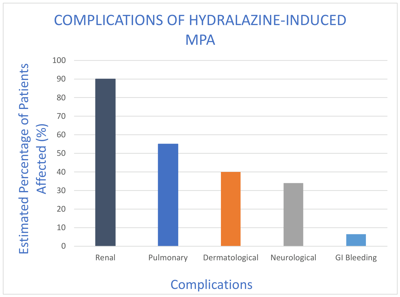
Because of the recent differentiation of MPA from the other AAVs, comprehensive demographic data for its incidence and prevalence in the US is not yet available [1]. Furthermore, there is a gap in the literature regarding incidence and prevalence of hydralazine-induced MPA alone, with most studies reviewing the incidence of hydralazine and other medications such as propylthiouracil-induced MPA [5]. However, this is a condition with the potential for relapse as well as a high potential for irreversible renal failure, and morbidity and mortality should be studied at further lengths to understand how to improve patient survival.
Diagnostic methods and management options will be discussed later in this review, but it would be remiss not to mention that management options are limited, and they tend to come with unfavorable side effects. While there are available treatment options and promising research on induction and maintenance of remission in hydralazine-induced MPA, preventative measures or methods directed toward a cure are unavailable now. This is due to the unknown mechanism of how hydralazine precipitates MPA. The potential for a full recovery from hydralazine-induced MPA is hinged on the induction of therapies for treatment as early as possible, and this is not always possible due to the variety of diagnostic methods that may take days, even weeks, to result. Further studies should be focused on extrapolating the exact incidence and prevalence, and other epidemiological factors of hydralazine-induced MPA, as well as deciphering the exact pathway in which hydralazine causes MPA, to enhance available diagnostic methods and treatment options to make progress toward prevention and cure.
Materials and Methods
Articles and books published from 1983 to 2026 were chosen through literature searches in PubMed, Science Direct, Embase, Elsevier and Google Scholar databases. Search terms were “hydralazine-induced vasculitis” or “drug-induced microscopic polyangiitis” as MESH terms and subsequent terms included “ANCA vasculitis” and “hydralazine side effects” to name a few. This investigation took place during March 2026. AI was not used for data collection, analysis, or any other purposes in this review.
Literature Review
Etiology and Pathophysiology of Hydralazine-Induced Microscopic Polyangiitis
Microscopic polyangiitis (MPA) is an autoimmune condition belonging to a group of vasculitides driven by interactions between anti-neutrophil cytoplasmic antibodies (ANCAs) and proteins within neutrophilic granules [1]. ANCA-associated vasculitides (AAVs) are characterized by inflammation of the body’s small vessels (i.e., vasculitis) that can be associated with necrosis, granuloma formation, and cellular infiltrates [3]. There are three known types of AAVs, and it is important to be able to differentiate the characteristics and clinical presentation of MPA from the other two types. It is important to note that, because none of these AAVs are known to have immune complex or complement deposition on histopathology, they are known as Pauci-Immune vasculitides [3].
MPA is characterized by a pattern of necrotizing vasculitis, mainly of the capillaries, without any immune complex deposition [1,3]. The primary manifestation of MPA is glomerulonephritis, specifically rapidly progressive glomerulonephritis (RPGN) [8]. From an immunohistology perspective, in a patient with MPA, biopsy of the capillaries shows inflammation without granuloma formation while biopsy of the kidneys revels crescentic glomerulonephritis [3]. What typically differentiates MPA from the other two types of AAVs, is lack of involvement of the upper respiratory tract. The other two types of AAVs, granulomatosis with polyangiitis (GPA) and eosinophilic granulomatosis with polyangiitis (EGPA) more commonly cause upper respiratory symptoms such as epistaxis, sinusitis, and nasal polyps [3]. Furthermore, GPA and EGPA are distinguished from MPA by the presence of granulomatous inflammation outside of the vasculature, and EGPA specifically is the only AAV characterized by eosinophilia, hence its name [4].

Table 1.
Distinguishing facts of each AAV type.
| Type of AAV | Presentation | Diagnostic Features |
| Granulomatosis with polyangiitis |
|
Anti-proteinase (c-ANCA) |
| Microscopic polyangiitis |
|
Anti-myeloperoxidase (p-ANCA) |
| Eosinophilic granulomatosis with polyangiitis |
|
Anti-proteinase (c-ANCA) |
The pathophysiology of AAVs results from a set of complex interactions between the immune system and genetics or environmental insults, such as infectious diseases, lifestyle habits, or certain medications. Hydralazine, a direct-acting vasodilator, is a drug commonly used to treat hypertension, but is also known to induce a lupus like reaction, as well as AAV. This literature review will focus on hydralazine-induced MPA.
While the exact pathophysiology of hydralazine-induced MPA is unknown, the pathway for AAV in general has been studied at length. Within the granules of neutrophils, a type of white blood cell in the immune system in charge of attacking harmful pathogens, there are proteins that can be released as a result of immune system activation. The proteins associated with AAVs are known to be either proteinase 3 (PR3) or myeloperoxidase (MPO) [4]. MPO is the major protein of peroxidase-positive granules, which serves to form reactive oxygen species that are harmful to both the body and microbial insults, while PR3 is an autoantigen that exhibits apoptosis-inducing capabilities of both healthy cells and harmful cells [6]. When the immune system is activated, neutrophils receive a signal to degranulate, and thus these proteins are released from their granules within the neutrophils. In AAV, the recognition of autoantibodies, namely ANCAs, is the inciting event causing neutrophil degranulation and the release of PR3 and MPO [4].
It is though that approximately 90% of patients with either MPA or GPA will test positive for ANCAs on serology [1]. Anti-MPO, or perinuclear ANCA (p-ANCA), is the autoantibody associated with MPA, as this is the autoantibody most found on serology testing in patients with biopsy confirmed MPA [4]. The ANCA associated with GPA is typically anti-PR3, or cytoplasmic ANCA (c-ANCA). EGPA is different from the other two types of AAVs in which most patients with this disease may not even test positive for ANCAs at all, around 60% test negatively, but if they do test positively, it is most likely to be anti-MPO rather than anti-PR3 [3]. This distinction between c-ANCA and p-ANCA in the serology is important for diagnosis of the type of AAV the patient may have, and will be explained more in depth at a later point in this review. Unfortunately, the underlying mechanisms surrounding the production of the ANCAs targeting MPO or PR3 are unknown currently, however, the bioscience community is currently proposing links between certain genetic predispositions, such as human leukocyte antigens (HLA) types and the association of MPO or PR3 ANCA vasculitides [7].
In hydralazine-induced MPA, the general pathway of AAV was used to extrapolate some promising theories on the mechanism by which the drug induces vasculitis. One theory noted in a case report in the Modern Rheumatology journal focuses on the binding interactions between hydralazine and MPO causing neutrophil apoptosis and thus, degranulation [8]. However, it is unknown the exact mechanism by which hydralazine binding to MPO induces autoimmunity. Another review by Pendergraft et al. proposed that hydralazine may indeed reverse the silencing of MPO and PR3 within neutrophil granules by way of inhibiting deoxyribonucleic acid (DNA) methylation, causing increased expression of neutrophil autoantigens and therefore increasing opportunities for autoimmunity [9]. However, there has not been any concrete evidence to support this either. Finally, a theory focusing on pharmacokinetics of how patients may acetylate hydralazine and possibly convert its components into an autoantibody [10]. Unfortunately, more research needs to be done to validate and eventually confirm this theory.
Another lacking territory of hydralazine-induced MPA is its epidemiology. Most epidemiological reviews focused on drug-induced AAV and studied the incidence and prevalence of other drugs commonly associated with AAV.
One study lead by Choi et al. in the late 90s attempted to determine the prevalence of drug-induced AAV [11]. In this study, they investigated the exposure of 30 patients with known vasculitis and high titers of anti-MPO antibodies to drugs associated with AAV, as well as the other clinical and histologic features of these patients. Choi et al. found that, of these 30 patients, ten had been exposed to hydralazine and had findings typical of AAV. Eight other patients had been exposed to propylthiouracil, allopurinol, penicillamine, and sulfasalazine, all drugs associated with drug-induced AAV. A more recent review by Pendergraft et al. in 2014 worked to extrapolate epidemiological data for different drug-induced AAVs. It was proposed that the incidence of hydralazine-induced vasculitis, not MPA, has an incidence of 5.4%-10.4% dependent on the dosage of hydralazine. While these studies are not current and were relatively small, they did confirm a link that had already been proven between hydralazine dosing and AAV. This calls for more research into the epidemiology of hydralazine-induced MPA.
In 2018, Kumar et al. performed a retrospective study to also determine the incidence of hydralazine-induced MPA [12]. They identified 323 cases of AAV and isolated the 12 cases that were positive for exposure to hydralazine. Of these 12 cases, all patients were ANCA positive, and 11 were positive for anti-MPO, pointing to a diagnosis of hydralazine-induced MPA. Of the 6 of these 12 patients who underwent kidney biopsy, all were found to have pauci-immune crescentic glomerulonephritis, confirming a true diagnosis of AAV. They also found that these 12 patients had all undergone an average duration of 22 months of hydralazine therapy with an average dose of 146 grams. While, once again, not exactly providing a consensus on incidence and prevalence of hydralazine-induced MPA, these studies provided valuable information on the dosing in which hydralazine was most likely to induce AAV.
Because hydralazine-induced MPA was confirmed to be dose-dependent, there have been many studies focusing on exactly what dosage is associated with the precipitation of hydralazine-induced vasculitis. One study does a comprehensive review of hydralazine dosing, as well as breaking down each vasculitis into its subtypes. From 2006-2019, Santoriello et al. identified 80 cases of AAV glomerulonephritis associated with hydralazine out of 1,858 cases of AAV glomerulonephritis, finding that hydralazine-induced vasculitis accounted for 4.3% of AAV in this study [13]. Over 75% of these patients had been on hydralazine for at least one year, with a mean daily dose of 250 mg/day [13]. Furthermore, Santoriello et al. differentiated each AAV into their subtypes by auto-antibody, finding that 98% of these 80 patients had anti-MPO, likely pointing to a diagnosis of hydralazine-induced MPA. While, once again, not exactly providing a consensus on incidence and prevalence of hydralazine-induced MPA, these studies provided valuable information on the dosing in which hydralazine was most likely to induce AAV. More studies like this are necessary to confirm the incidence and prevalence of hydralazine-induced MPA, as well as the dosing danger zone with which it is associated.
Graph 2.
Pie chart revealing 250 patients studied in Choi et al. review, in which 220 were found to be vasculitis negative, 30 were found to be vasculitis positive, 10 of those vasculitis positive patients had hydralazine exposure, 8 had exposure to other associated drugs, and 18 were not found to have drug exposure.
Graph 2.
Pie chart revealing 250 patients studied in Choi et al. review, in which 220 were found to be vasculitis negative, 30 were found to be vasculitis positive, 10 of those vasculitis positive patients had hydralazine exposure, 8 had exposure to other associated drugs, and 18 were not found to have drug exposure.
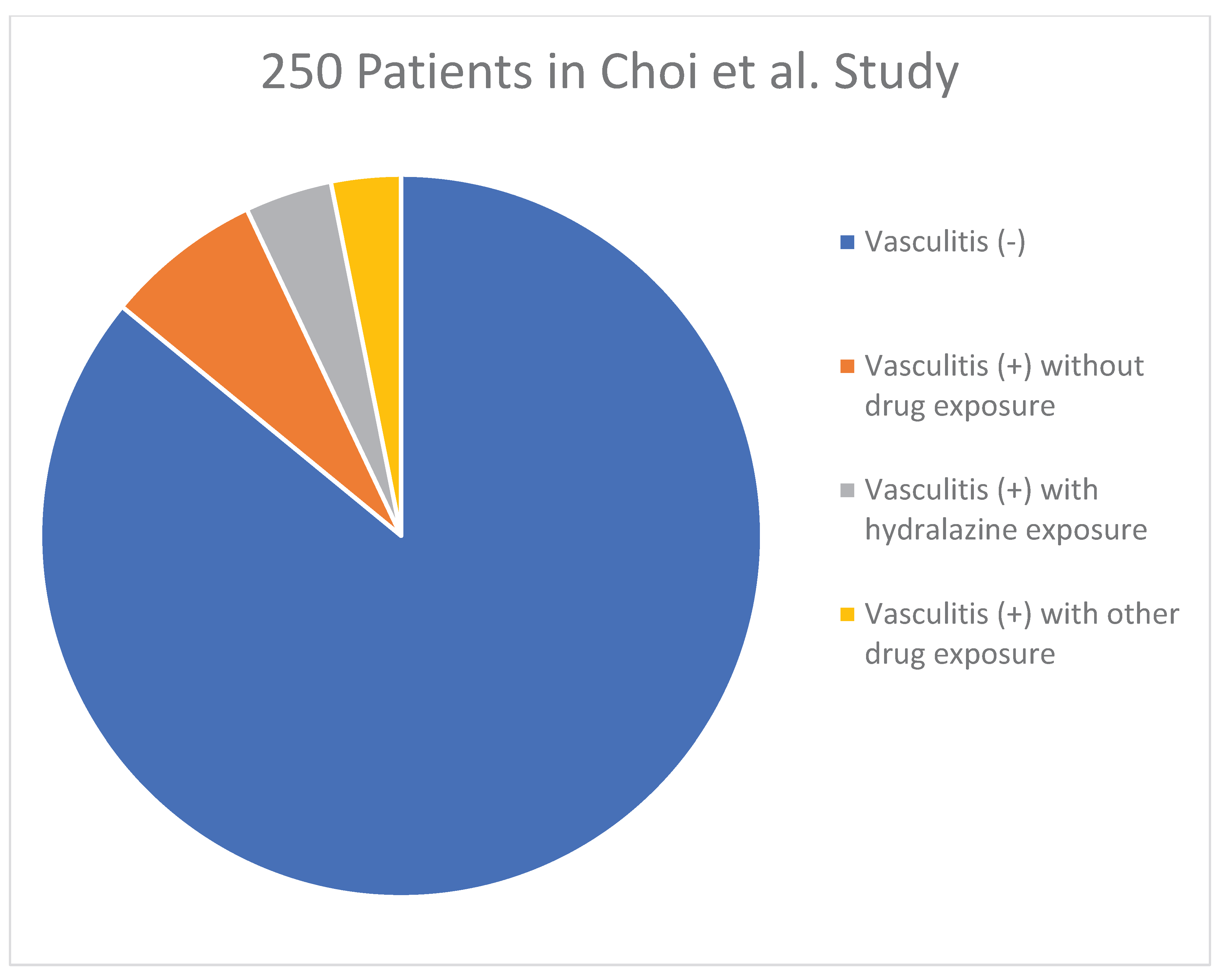
Fortunately, however, there are some studies in the literature that can provide some insight into the epidemiology of general MPA. One notable prospective study was able to estimate the global incidence and prevalence of MPA by measuring cases in a specific county of Minnesota, as well as recording the demographic characteristics of each patient with confirmed MPA [14]. From January 1996 to December 2015, 58 cases of AAV were identified, and 48% of these cases were confirmed to be MPA. They found that these 28 cases of MPA had a slight male predominance with an age range of 45 to approximately 77 years old. With this data, Berti et al. were able to calculate the annual incidence of MPA in their region to be 3.3 per 100,000 with a prevalence of 42.1 per 100,000. A recent study in 2022 by Redondo-Rodriguez et al. used systematic search in medical research databases for studies analyzing confirmed cases of AAV aged in patients over 16 years old [15]. With the data they collected, they were able to extrapolate that the global pooled incidence of MPA was 5.9 per million person-years and the individual pooled prevalence per million person was 39.2. This study also found that MPA was the second most prevalent type of AAV, with GPA being the first at a prevalence of 96.8 per million persons [15].
Unfortunately, there is a deficit in literature focusing on the incidence and prevalence of hydralazine-induced MPA, with most studies choosing to study drug-induced AAV as a broader subject.
Manifestations and Complications of Hydralazine-Induced Microscopic Polyangiitis
Hydralazine-induced MPA manifests in a comparable way that a general MPA does, primarily with glomerulonephritis. The manifestations of hydralazine-induced MPA and general MPA overlap, since they are indeed the same condition, although precipitated by differing offenses. In a study by Yokogawa et al., 68 cases of confirmed hydralazine-induced vasculitis were reviewed and the clinical signs and symptoms that these patients experienced were recorded [7]. Of all 68 patients, 81% of these patients had kidney involvement, while 19% had lung involvement, and, interestingly, 25% had dermatological manifestations.
It is important to note that MPA is considered to be a pulmonary renal syndrome (PRS), which is a term used to describe the combination of glomerulonephritis and pulmonary hemorrhage as part of a larger, multisystem autoimmune disease [16]. Other diseases considered to be a part of the PRS umbrella include other AAVs, and anti-glomerular basement membrane (anti-GBM) disease. In the literature, it has been shown that approximately 70% of PRS cases are associated with an AAV, and that a portion of these AAVs were precipitated by hydralazine [16]. As aforementioned, PRS presents with renal impairment and pulmonary distress that manifests in the form of RPGN with associated DAH. However, not every patient with hydralazine-induced MPA will develop pulmonary symptoms, and therefore, does not meet the criteria of having a PRS. Therefore, it would be remiss not to touch on each of these manifestations as separate manifestations, although they are seen together in some cases.
The type of glomerulonephritis most frequently noted in hydralazine-induced MPA is RPGN, also known as crescentic glomerulonephritis. RPGN is characterized by rapid loss of renal function over days to weeks, protein and blood in the urine as seen on urine analysis, and histopathological findings on renal biopsy [17]. There are many causes of RPGN, but the most common is ANCA-associated vasculitis, also known as pauci-immune glomerulonephritis, comprising approximately 40-50% of RPGN cases, which is found on renal biopsy [17]. There are different classifications based on the histopathology and the presence of immune complex deposition, and if RPGN is caused by an AAV and, therefore has no immune complex depositions in the glomerular basement membrane, it is known as a type III RPGN. RPGN is precipitated by the release of histones, cathepsins, cytokines, and plasminogen activator autoantibodies which all lead to inflammation and destruction of renal microtubules, as well as the formation of fibrinoid necrosis within the glomerulus and crescents in the Bowman space [17]. These are the hallmark histologic lesions of pauci-immune glomerulonephritis.
The pro-inflammatory state caused by the disease process creates an ischemic environment for the kidney, leading to loss of renal function and sequela that can accompany this. The inflammation causes destruction of the glomeruli and small vessels within the kidney, leading to loss of vascular permeability, and therefore allowing inflammatory and immune cells to infiltrate and accumulate to form fibrin deposits [18]. These deposits block the flow of blood within these structures by way of micro-thrombi, depriving the kidney of blood flow, and fibrinoid necrosis ensues.
Figure 1.
Histology slide showing Pauci-immune necrotizing crescentic glomerulonephritis with fibrinoid necrosis on a Jones silver stain [19].
Figure 1.
Histology slide showing Pauci-immune necrotizing crescentic glomerulonephritis with fibrinoid necrosis on a Jones silver stain [19].
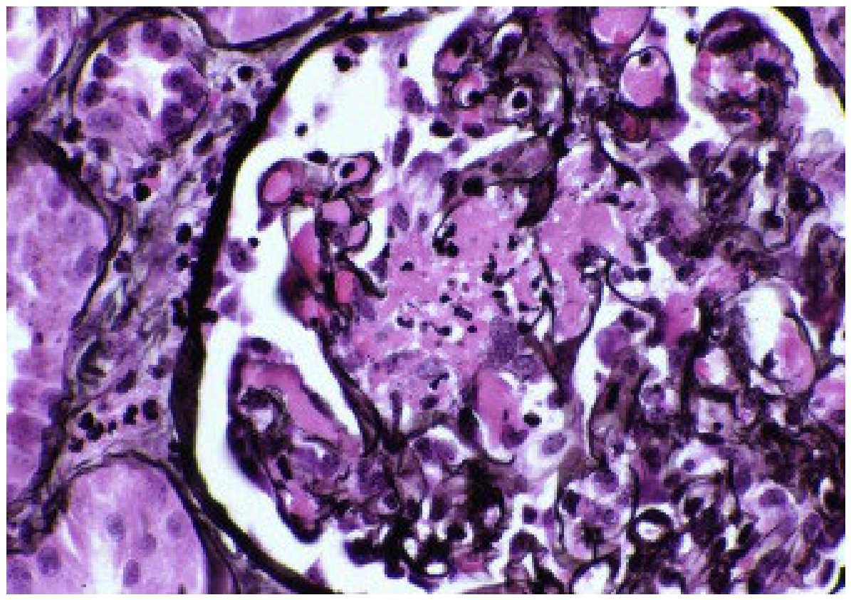
A crescent is a hyperplastic lesion, usually found adjacent to an area of fibrinoid necrosis, that affects more than 10% of the glomerulus; it is formed from the accumulation of parietal and visceral epithelial cells in the Bowman space as a result of the destruction of glomerular capillary walls due to the disease process [20]. Crescents contribute to loss of renal function by acting as another blockage, compressing the glomerulus and disrupting filtration, but also replacing functional renal tissue. Crescentic formation is classified in histopathological terms based on the composition of the crescent. If a crescent is composed of more than 75% of cells and fibrin, then it is dubbed as a cellular crescent, and a crescent with more than 75% of fibrous matrix is labeled as a fibrous crescent. If the crescent is composed of an equivocal mixture of cells, fibrin, and fibrous matrix, then it is a fibrocellular crescent [17]. If a crescent is fibrocellular or fibrous, there is a very low chance of recovery of the renal tissue, and it is considered irreversible, leading to nephron atrophy and further fibrosis of the surrounding interstitium [20].
Figure 2.
Histology slide showing Pauci-immune necrotizing crescentic glomerulonephritis with cellular crescent on a Jones silver stain [19].
Figure 2.
Histology slide showing Pauci-immune necrotizing crescentic glomerulonephritis with cellular crescent on a Jones silver stain [19].
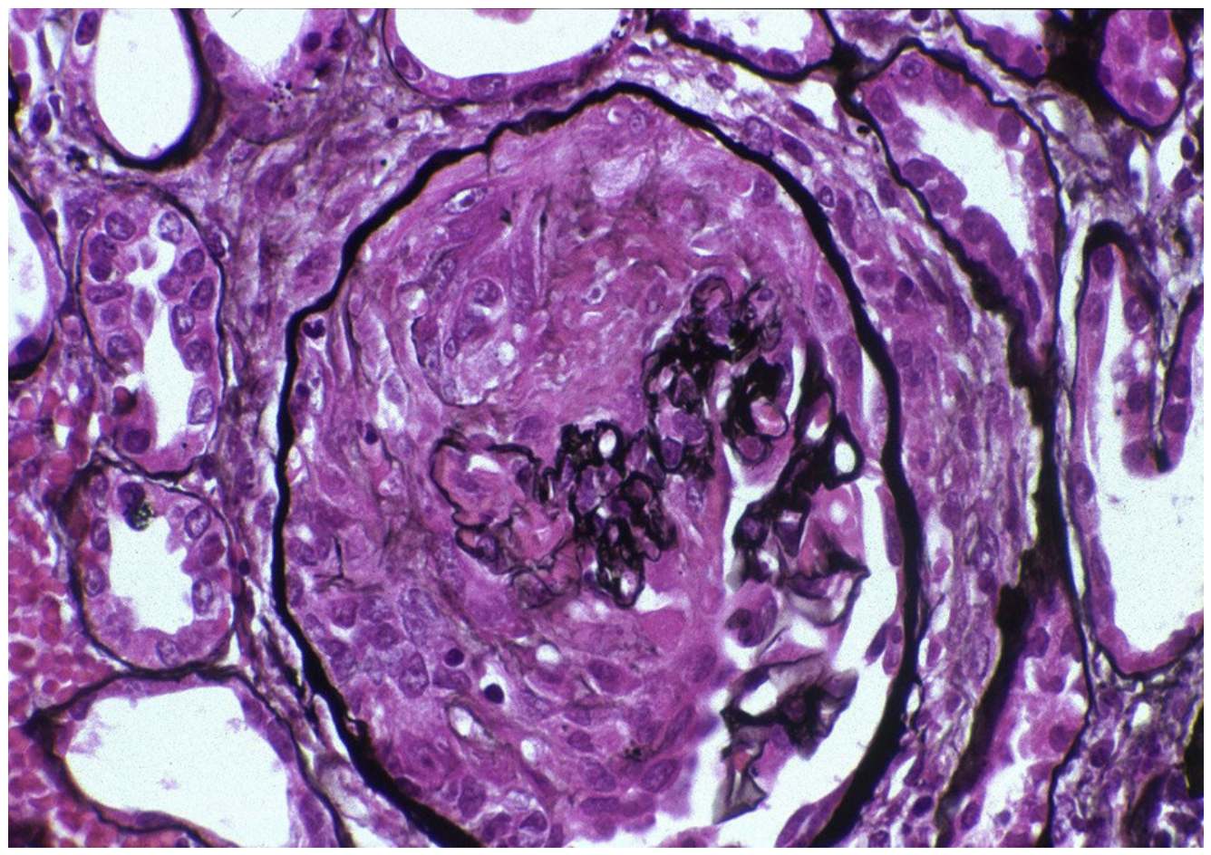
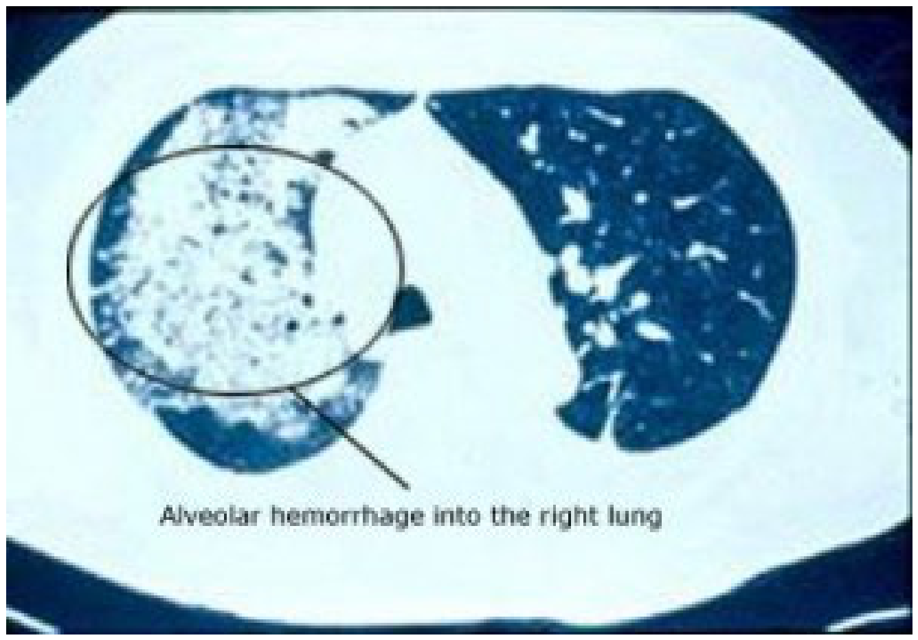
Pulmonary involvement is observed in 25-55% of patients with confirmed MPA, one study by Lane et al. noting that 29% of MPA cases involved pulmonary symptoms [21]. However, recent studies suggest that pulmonary involvement may be more common than originally thought [1]. A large single-center study by Wilke et al. in 2014 found that 80% of patients with confirmed MPA presented with pulmonary complaints, such as cough or hemoptysis, and 92% were found to have pulmonary infiltrates on chest radiographic imaging [22]. The most common manifestation of MPA is known to be DAH. Although it is unknown in the current literature exactly how many patients with confirmed hydralazine-induced MPA have been found to develop DAH, one small case review in 2009 proposed the most frequent cause of DAH to be AAV, and following literature agrees with this notion [23].
DAH is defined as diffuse bleeding from pulmonary microcirculation, which can be due to abnormalities in the lung itself or in the bronchial circulation, and other autoimmune conditions [24]. It is confirmed by bronchoscopy with biopsy. However, when DAH is associated with hydralazine-induced MPA, the hemorrhage is due to hemorrhagic capillaritis of the alveoli, which can be seen as the hallmark lesion on histology [25]. DAH is a particularly life-threatening manifestation of hydralazine-induced MPA, because not only can the hemorrhage lead to a severe anemia requiring blood transfusion, but it can also cause acute respiratory failure due to compromise of the patient’s airway filling with blood. The presence of DAH in a case of MPA can move the severity of the disease from generalized to severe, according to the European Vasculitis Study Group (EUVAS) grading system, which is a clinically useful categorization tool for vasculitis created by EUVAS that will be detailed later [24].
Figure 3.
Computed Tomography (CT) scan of the chest showing DAH in a patient with MPA [26].
Figure 3.
Computed Tomography (CT) scan of the chest showing DAH in a patient with MPA [26].
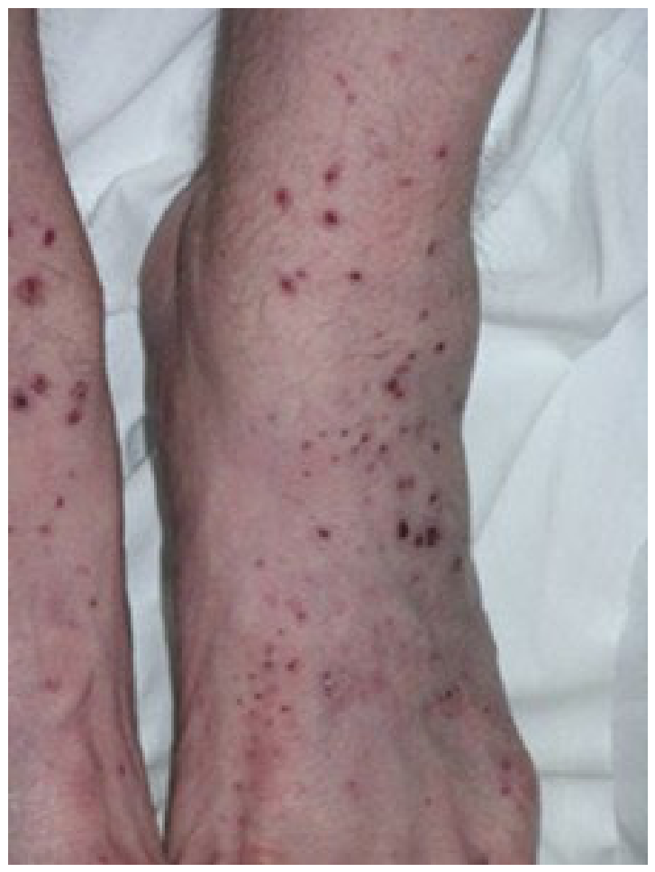
Dermatological manifestations of hydralazine-induced MPA, such as palpable purpura, skin ulcerations, necrotic plaques, and skin nodules, are found in 30-60% of patients with confirmed MPA [1]. The literature struggles to find a consensus as to what the most common dermatological manifestation may be. One study by Gibson found that the most common skin manifestation associated with MPA tends to be livedo reticularis, as it suggests as deeper vascular inflammation that is most closely associated with MPA rather than the other two subtypes of AAV [27]. Another study by Kluger et al. suggested that palpable purpura are the most common, as recorded in 26% of 162 patients with confirmed MPA [28]. However, it can be inferred that dermatological signs can take on a wide array of manifestations, and any sort should be considered for a diagnosis of AAV, especially if the patient has been treated with hydralazine.
Figure 4.
Palpable purpura over the left foot of a patient with MPA [26].
Figure 4.
Palpable purpura over the left foot of a patient with MPA [26].
Diagnostic Approaches to Hydralazine-Induced Microscopic Polyangiitis
As aforementioned, since hydralazine-induced MPA is a subset of MPA, the diagnostic methods for MPA will be discussed at length. As of right now, there are no diagnostic methods used only to diagnose hydralazine-induced MPA, as it can be inferred from the patient’s medication history combined with a diagnosis of MPA.
Diagnostic evaluation of a patient with suspected MPA requires comprehensive clinical, radiological, histopathological, and laboratory assessment, and as with most cases of every diagnosis, a detailed clinical history is the initial step. If a patient is found to have sino-nasal symptoms or signs of eosinophilia, such as allergic type symptoms, the suspicion should shift toward GPA or EGPA as those types of clinical signs are much more likely to be associated with them rather than MPA [1].
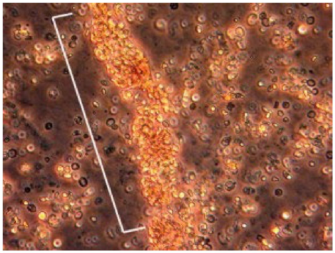
Laboratory studies that can be done to diagnose a patient with suspected MPA include the comprehensive, complete blood cell count (CBC) with manual differential, erythrocyte sedimentation rate (ESR), kidney function testing, urine analysis with urine microscopy, and complement levels (C3 and C4). In MPA, a CBC may show leukocytosis and/or anemia, but is less likely to show eosinophilia, as this would be more likely associated with EGPA. An ESR would typically be elevated in MPA, as would kidney function testing, but C3 and C4 levels are typically normal. A urine analysis with microscopy may show abnormal urine sediment, proteinuria, hematuria, and red blood cell (RBC) casts [26].
Figure 5.
RBC casts on urine microscopy [26].
Figure 5.
RBC casts on urine microscopy [26].
Serum studies, such as evaluation of p-ANCA or anti-MPO antibody positivity, have been studied at length to determine if they are truly effective at ruling in MPA. In 1998, Hagen et al. studied the diagnostic value of p-ANCA or c-ANCA measurement by indirect immunofluorescence (IIF) and by anti-PR3 antibody and anti-MPO antibody enzyme linked immunosorbent assay (ELISA) [29]. The study spanned fourteen different centers and involved 358 total patients diagnosed with idiopathic small vessel vasculitis against 740 health controls. It was found that the sensitivity of p-ANCA was 58% and the sensitivity of anti-MPO antibody was 58% in a patient with confirmed MPA [29]. The specificity was found to be 81% for p-ANCA and 91% for anti-MPO antibody. Interestingly though, if IIF and ELISA were combined, then the diagnostic specificity of p-ANCA + anti-MPO antibody increased to 99%, and the sensitivity of p-ANCA + anti-MPO antibody increased to 67% for patients with MPA [29]. Although the increase in sensitivity is almost equivocal, the increase in diagnostic specificity proved to Hagen et al.l that IIF and ELISA should be used in combination to effectively rule in MPA. Furthermore, a meta-analysis by Choi et al. in 2001 found that the combination of IIF for p-ANCA and anti-MPO antibody ELISA yielded a sensitivity of 85.5% and specificity of 98.6% in patients with MPA, yielding a recommendation that both tests should be used in combination to optimize diagnostic performance [30].
More recent studies continue to debate whether IIF and the newer ELISAs should be used separately or in combination. In 2020, Menezes et al. embarked on a study with the goal to evaluate performance of ELISA and IIF methods for ANCA detection [31]. They assessed a total of 227 samples by IIF and by ELISA and found that IIF should be considered the gold standard. Alone, ELISA was found to have a sensitivity of 62% and specificity of 99%, but in combination with IIF, the diagnostic sensitivity was increased [31]. Again, in 2021, Deka et al. aimed to evaluate the diagnostic performance of newer ELISAs compared to IIF when detecting ANCA [32]. They found that IIF detected ANCA byway of p-ANCA and c-ANCA in 17.5% of cases while ELISA detected ANCA byway of anti-PR3 antibodies and anti-MPO antibodies in 11.6% of cases out of a total of 189 serum samples. IIF is still superior to even the newer ELISAs when it comes to detecting ANCA, as the newer ELISAs have high specificity, but a persistently low sensitivity [32]. In conclusion, serologic studies for MPA should include both IIF for p-ANCA and ELISA for anti-MPO antibodies.
Imaging of the chest can be useful for the assessment of pulmonary lesions in patients who present with pulmonary complaints, such as hemoptysis or shortness of breath. A chest radiograph (CXR) or CT scan of the chest can also be used to differentiate between GPA and MPA, as GPA presents with cavitary and nodular lesions while MPA is more likely to present with DAH on imaging. Imaging is not confirmatory, and should never confirm a diagnosis of MPA, but is useful in ruling in other disease and differentiating from comparators [1].
Lastly, histopathological evaluation can be done by way of kidney biopsy. Histopathological features of MPA include crescentic glomerulonephritis, as discussed above, with fibrinoid necrosis of the glomerulus and crescent formation within the Bowman space. The presence of these, again, does not automatically confirm a diagnosis of MPA, but rather increases the likelihood that MPA is the diagnosis.
The American College of Rheumatology (ACR) and the European Alliance of Associations for Rheumatology (EULAR) are currently working to develop criteria for the diagnosis and classification of AAVs byway of cohort study [33]. Their most recent study in 2022 used 129 cases of MPA compared and 408 comparator disease cases and candidate items were then used to identify where the patient would fall into each category of MPA. The five phases of the study included identification of candidate items using consensus methodology, prospective collection of candidate items present at time of MPA diagnosis, data-driven reduction of the number of candidate items, expert panel review of cases to define MPA diagnosis, and lastly, derivation of a points-based risk score for disease classification [33]. The aim of the study was to develop and validate classification criteria for MPA, and the final classification criteria was found to be 91% sensitive (95% CI, 85%-95%) and 94% specific (95% CI, 92%-96%); with both high sensitivity and specificity, this classification criteria can both accurately rule in MPA and rule out those who are more likely to have a comparator disease [33]. A score of at least five points will classify a patient with small or medium vessel vasculitis with MPA Of note, the candidate item with the most weight for classification was found to be p-ANCA or anti-MPO positive, with six points toward classification, automatically classifying the patient as having MPA. Other candidate items and scores can be seen in the table below.
Table 2.
Displaying scoring system created during ACR/EULAR study [33].
Table 2.
Displaying scoring system created during ACR/EULAR study [33].
| Candidate Item | Score (in points) |
| p-ANCA or anti-MPO positivity | +6 |
| Pauci-immune glomerulonephritis | +3 |
| Lung fibrosis or interstitial lung disease | +3 |
| Sino-nasal signs or symptoms | -3 |
| c-ANCA or anti-PR3 positivity | -1 |
| Eosinophil count ≥1×10⁹/L | -4 |
While this criterion is not specifically used to classify patients with hydralazine-induced MPA, it proves quite useful in clinical reasoning for diagnosis. If a patient who has been on hydralazine and subsequently scores five or more points on the ACR/EULAR classification tool, it would be safe to hypothesize a diagnosis of hydralazine-induced MPA until proven otherwise.
Management Options for Patients with Hydralazine-Induced Microscopic Polyangiitis
The mainstay of management of any sort of vasculitis involves induction of a remission phase to control active disease, followed by a maintenance phase to maintain this remission and lower the risk of recurrence or complications of the vasculitis. As vasculitides are autoimmune disorders, the key component of management of treatment is immunosuppression. However, to initiate any sort of treatment, early identification is key.
Treatment recommendations generally depend on an accurate determination of the disease severity, meaning, number of organ systems involved, presence of organ damage, degree of renal disease, and presence of DAH [24]. The European Vasculitis Study Group (EUVAS) developed a grading system, seen in Table x below, to categorize a patient’s disease as limited, early and generalized, active and generalized, severe, or refractory. The management options discussed according to the EVUAS grading system are for induction of remission in a patient with vasculitis.
Table 3.
The severity of vasculitis and treatment options according to EUVAS grading [24].
Table 3.
The severity of vasculitis and treatment options according to EUVAS grading [24].
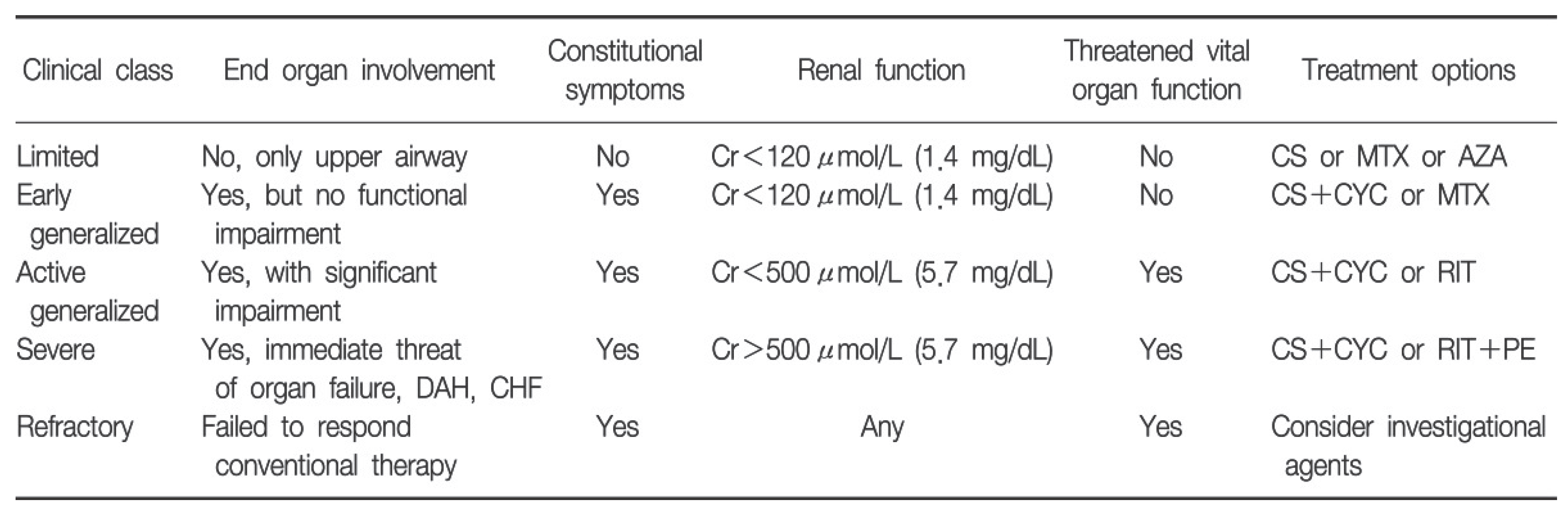
Cr: creatinine; CS: corticosteroid; MTX: methotrexate; AZA: azathioprine; CYC: cyclophosphamide; RIT: rituximab; CHF: congestive heart failure; PE: plasma exchange.
Limited disease tends to have only upper airway involvement, and renal function is preserved. In this setting, single agents, such as corticosteroids (CS), azathioprine (AZA), or methotrexate (MTX) can be used, but usually CS are the first line of therapy. The mechanism of actions of all immunosuppressive agents used for vasculitides will be discussed below.
Generalized disease is divided into two subcategories, active and early. For early generalized disease, there is no functional impairment of organs involved; treatment for this consists of corticosteroids and an additional agent such as cyclophosphamide (CYC) or MTX. Because of the more favorable side effect profile of MTX, which will be discussed in more detail later, the combination of CS and MTX is being utilized more often than CS and CYC [24].
Active generalized vasculitis is diagnosed if early generalized to include significant impairment with end organ damage. Immunosuppression for this category of disease is CS and CYC or rituximab (RIT), with newer evidence suggesting that pulsed intravenous (IV) CYC being preferrable to oral CYC due to fewer side effects and equivocal efficacy changes according to study for treatment options in GPA [34].
Severe disease is characterized by immediate threat of organ failure and functional impairment of critical organs, such as the presence of DAH or a new diagnosis of congestive heart failure (CHF) [24]. Studies suggest that patients with severe vasculitis should receive a combination of CS, CYC or RIT, and plasma exchange therapy as a last line therapy option. In a study by Klemmer et al. involving 20 patients with ANCA associated DAH, 100% of those patients had resolution of DAH with plasma exchange and 50% of these patients were discharged with improved and stable renal function [35]. It can be inferred that plasma exchange may be superior in restoring renal function and treating DAH compared to even high dose CS, but larger scale studies should be done to confirm this treatment.
And lastly, disease that does not improve and continues to progress despite all other conventional therapies available is dubbed refractory disease. The recommendation for refractory vasculitides is to seek out investigational agents. Drugs currently being studied for refractory vasculitis will be discussed toward the end of this section.
In the United Status, treatment is also based on disease severity; the mainstay of AAV tends to be CS and immunosuppressive agents, not unlike the EUVAS guidelines. For active non severe MPA, the combination of 50-75 mg prednisone per day with RIT is recommended, while mycophenolate mofetil (MMF) and MTX serve as alternatives, for the induction of remission [36]. Once remission is induced, maintenance therapy should consist of RIT, AZA, or MTX for 24-48 months with continued CS steroid therapy that eventually tapers down to 5 mg daily. Once the patient ends this trial of medications, their risk factors, kidney function, and relapse risk are reassessed and shared decision-making between the physician and patient is made to either continue maintenance treatment or discontinue it [36].
The American College of Rheumatology created guidelines for treatment of both GPA and MPA, however only GPA is included for both severe and non-severe disease [37]. The algorithm in Figure X delineates treatment recommendations for active severe MPA.
Figure 6.
Key recommendations for the treatment of GPA and MPA [37].
Figure 6.
Key recommendations for the treatment of GPA and MPA [37].
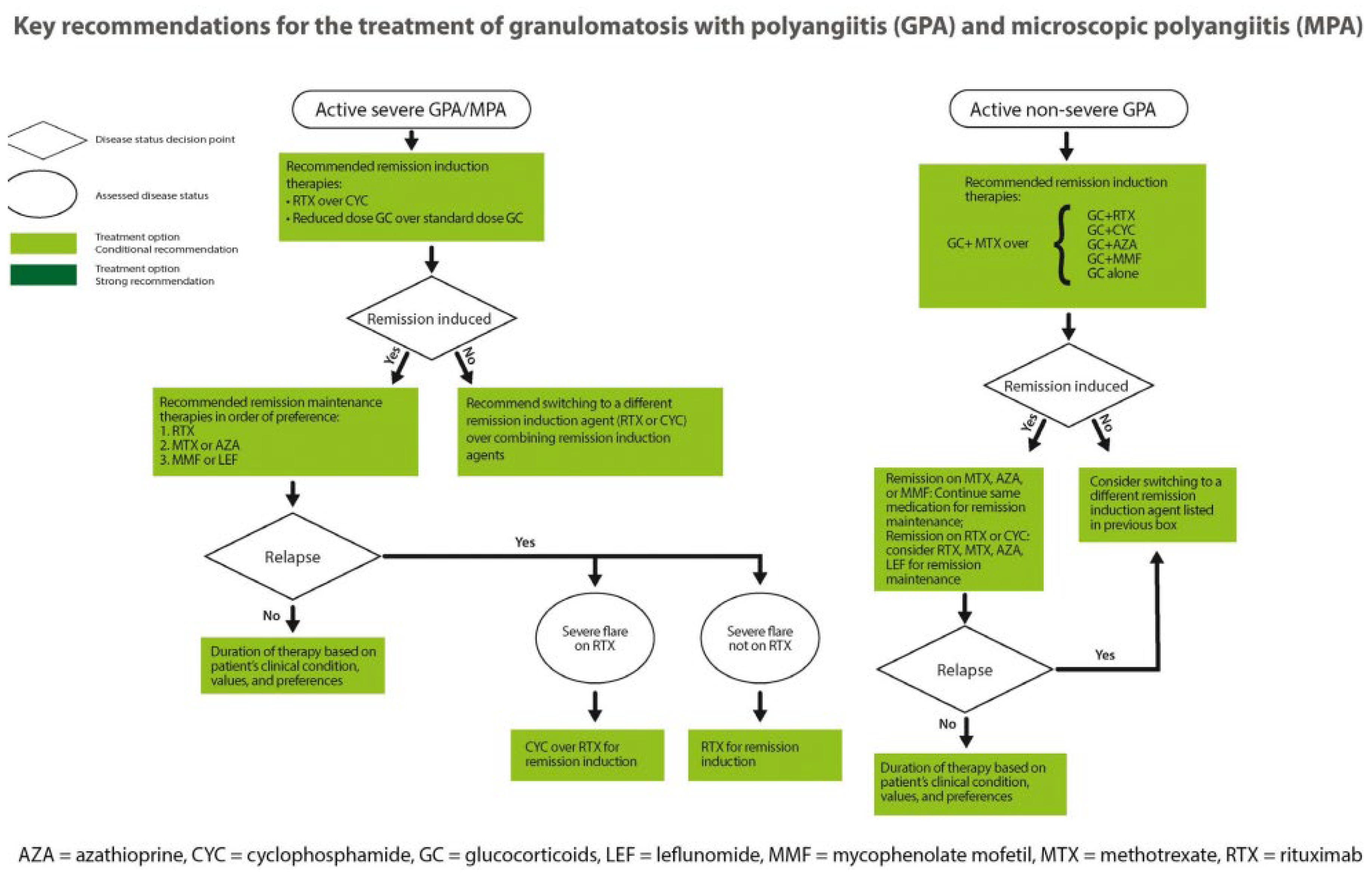
As noted in the figure above, the first course of action for active severe MPA is induction of remission with RIT or a reduced dose of CS, and if persistent, can switch to CYC. A study done by Walsh et al. in 2020 compared reduced-dose CS to high-dose CS and found a similar outcome of end stage renal disease (ESRD) or death but was associated with a decreased risk of infection [38]. Walsh et al. also found that plasma exchange therapy was not found to reduce the incidence of death or ESRD and another study done by Jayne et al. in 2007 found that there was no mortality benefit for plasma exchange versus high-dose methyl prednisone, and therefore ACR recommends against the use of plasma exchange therapies in patients with active severe MPA [38,39]. This differs from EUVAS in that plasma exchange therapy is recommended for patients with active severe disease. However, the benefits of plasma exchange in maintenance therapy will be discussed shortly.
Once remission of active severe MPA is induced, maintenance therapy is indicated with RIT as a first line therapy; alternatives for maintenance therapy of severe active MPA include MTX or AZA, as well as mycophenolate mofetil (MMF) or leflunomide (LEF).
As stated above, there are multiple different alternatives used in both the induction of remission and maintenance of MPA. The mechanism of action of each of the drugs will now be discussed as the understanding of each drug will give a deeper understanding of the disease process. The table below provides a brief summary for each of the medications that will be discussed.
| Drug | Drug Class | Treatment Phase | Adverse Effects |
| Corticosteroids | Anti-inflammatory and immunosuppressant | Induction of remission and maintenance |
|
| Azathioprine | Immunosuppressant | Maintenance |
|
| Methotrexate | DNA synthesis inhibition | Induction of remission and maintenance |
|
| Cyclophosphamide | DNA synthesis inhibition | Induction of remission and maintenance |
|
| Rituximab | Immunosuppressant | Induction of remission and maintenance |
|
| Mycophenolate | Immunosuppressant | Induction of remission and maintenance |
|
| Leflunomide | Immunosuppressant | Maintenance |
|
| Plasmapheresis | Immunomodulator | Maintenance |
|
Corticosteroids
Corticosteroids are the backbone of microscopic polyangiitis management, as they are used in both the induction of remission and disease maintenance therapy. Corticosteroids are immunosuppressive agents that are indicated in pro-inflammatory disease, autoimmune disease, and adrenal insufficiency. Corticosteroids work on the glucocorticoid receptor, which once activated, will cause inhibition of gene expression and translation of pro-inflammatory cytokines, chemokines, cell adhesion molecules, and other pro-inflammatory enzymes [40]. In MPA, this works to induce remission by suppressing the immune system and blunting the effects of the anti-MPO.
Azathioprine
Azathioprine is a purine analog that converts mercaptopurine and thioguanine via hypoxanthine-guanine phosphoribosyl transferase (HPRT) and thiopurine methyltransferase (TPMT) enzymes. These enzymes will then go on to inhibit purine synthesis, leading to inhibition of DNA synthesis of immune cells [41]. Azathioprine works to inhibit immune cell synthesis, therefore blunting the response that contributes autoimmunity in these patients. It serves useful in the maintenance phase of treatment.
Methotrexate
Methotrexate is a folate analogue that will inhibit dihydrofolate reductase, an enzyme that contributes to the synthesis of purines and pyrimidines. By blocking this enzyme, it leads to reduction in synthesis of purines and pyrimidines, DNA and RNA [42]. It can serve both as an alternative agent for remission induction in non-severe MPA, or in maintenance therapy.
Cyclophosphamide
Cyclophosphamide is often used interchangeably with MTX due to their similar mechanisms of action. Cyclophosphamide is a drug derived from mustard seeds and acts as an antineoplastic but also has immunosuppressive effects. It will metabolize into aldophosphamide via liver enzymes which is then cleaved into the active DNA alkylating agent phosphoramide. DNA alkylation has a silencing effect, thus causing inhibition of protein synthesis through DNA and RNA crosslinking [43]. While rituximab is preferred over cyclophosphamide, it can be used as an agent for both remission induction and maintenance therapy.
Rituximab
Rituximab, the other drug that serves as the backbone of MPA treatment, is an anti-CD20 monoclonal antibody that is a powerful immunosuppressant used in many different cancers, autoimmune diseases, and inflammatory diseases. Since it is a monoclonal antibody, it will target CD20 expressed by B-cells, an immune system component, and attach itself. Once attached, rituximab tags the B cells for destruction and natural killer (NK) cells will recognize the attached rituximab and work to destroy the cells that rituximab is attached to [44]. In MPA, rituximab works to dampen the immune system by destroying the B cell, a key component in the autoimmune response, making it a powerful agent for both remission induction and maintenance therapy.
The RAVE trial was a randomized, multicenter non-inferiority study assessing the efficacy of rituximab in patients with AAV. It was completed in 2010 by Stone et al. to compare rituximab to cyclophosphamide in the rates of remission induction in patients with either GPA or MPA. It found 67% of patients treated with rituximab compared to 42% of patients treated with cyclophosphamide had successful remission induction out of 197 total patients without significant differences in rates of adverse events [45]. Because of similar findings in other studies, rituximab is a powerful mainstay of both remission induction and maintenance therapy.
Mycophenolate
Mycophenolate mofetil is a powerful immunosuppressant often used in transplant patients to prevent rejection. It has three useful mechanisms of action once it is converted into its active form: mycophenolic acid [46]. First, it depletes protein found in B and T cells and thus inhibits their proliferation. Next, it inhibits the glycosylation and expression of adhesion molecules, therefore inhibiting the recruitment of immune system cells to sites of inflammation. Lastly, it depletes tetrahydrobiopterin, decreasing the production of nitric oxide and tissue damaging oxidants. In MPA, it is useful for dampening the immune system, working against inflammation, and preventing tissue damage by decreasing production of oxidants. Mycophenolate serves as an alternative agent in non-severe MPA, but also as an alternative for maintenance therapy.
Leflunomide
Leflunomide is an immunomodulatory drug that serves to inhibit a key enzyme in the de novo synthesis of proteins used in DNA production. [47] Leflunomide is solely used as an alternative in maintenance therapy.
Plasmapheresis
Lastly, although not pharmacological, plasma exchange, or plasmapheresis, has been used to treat serious complications of autoimmune diseases such as systemic lupus erythematous (SLE), rheumatoid arthritis (RA), and many others. The process involves removing a patient’s blood and filtering it through a machine where it is separated into red blood cells, white blood cells, platelets, and plasma [48]. The plasma is then discarded, as this is the component of blood that contains harmful antibodies that are targeting the patient’s own immune cells. Once discarded, the plasma is then replaced with a substitute fluid from donors containing healthy immune cells and is then returned to the patient with the other components that were filtered.
The efficacy of plasmapheresis in MPA has been recently studied, and although it proves to be quite useful in patients with MPA complicated by DAH, it is not recommended as a therapy unless the patient has a higher risk for progression to ESRD. This recommendation is supported by data from two of the largest trials that studied the efficacy of plasmapheresis in the treatment of AAV glomerulonephritis, which were discussed above [37].
Although treatment guidelines were recently published, novel studies are currently underway to establish new drugs that could show promise in both the remission of induction and maintenance of MPA. Emerging therapies include anti-CD20 monoclonal antibodies, CAR-T cell therapies, and other precision management strategies that target specific components of the immune response [49].
Avacopan
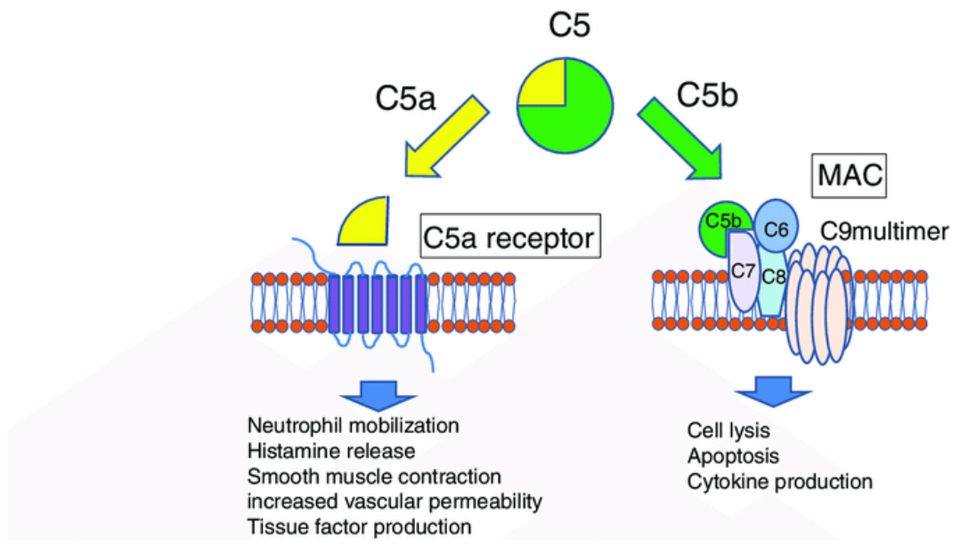
Avacopan, a drug approved for the treatment of AAVs alongside mainstay therapies, was approved in October 2021. Avacopan acts as a C5a receptor antagonist, which is a part of the alternative complement pathway that is thought to be a key component in the autoimmune dysregulation cascade associated with AAV [50]. C5a is a powerful anaphylatoxin, meaning that it contributes to inflammation and causes overactivation of the immune system. By preventing C5a binding to its receptor by way of receptor antagonism, autoimmunity and inflammation is reduced [51].
Figure 7.
The pathway of C5 in the alternative complement system[52].
Figure 7.
The pathway of C5 in the alternative complement system[52].
Although its use does not eliminate the need for corticosteroids, avacopan was found that it was successful in remission induction compared to therapy with only prednisone. These trials include the ADVOCATE, CLEAR, AND CLASSIC trials; these trials found that avacopan improved remission rates, sustained remission over time, and improved kidney function in patients with MPA or GPA compared to treatment with RIT or CYC and corticosteroids [50]. In the ADVOCATE trial, Jayne et al. 331 patients with AAV were treated with avacopan or prednisone, and remission was observed in 72.3% of patients treated with avacopan versus 70.1% of patients treated with prednisone [53]. While not a large difference, it did show that avacopan was noninferior to prednisone therapy in achieving remission. A similar study done by Jayne once again, the CLEAR trial, found similar results; they studied the efficacy of avacopan in patients with AAV and whether it could replace glucocorticoids and found that they had similar rates of clinical response and safety profiles [54]. The CLASSIC trial, by Merkel et al., aimed to determine the safety profile of avacopan; they found that avacopan was well tolerated and achieve higher remission rates when compared to standard of care corticosteroids and CYC or RIT. A difference of 15% patients receiving standard therapies experienced adverse effects and 17% of patients receiving avacopan experienced adverse effects [55].
Unfortunately, there is a current proposal for withdrawal of the drug’s approval that was filed due to controversial study methods and a worrisome drug safety profile. Because of this, Jayne et al. retracted their publication on the ADVOCATE trial. While the approval of avacopan may be undergoing dispute, it continues to encourage further research into development of therapies for MPA.
Although there are emerging therapies and deliberations on treatment guidelines, there remains many gaps in the literature regarding MPA. Further studies should be focused on long-term follow up beyond 12-16 months and further recommendations for maintenance therapy duration. Furthermore, there is residual uncertainty in regimens, not including CS, as seen in the avacopan trials.
Nonetheless, hydralazine-induced MPA treatment is clear: remove the offending agent, hydralazine, and treat for MPA. However, there is still a question of whether there are better therapies to target or even prevent hydralazine-induced MPA. While MPA is a rare adverse effect of hydralazine use, it is life-threatening and efforts should be made to study prevention of this disease. And this can only be done once we understand the true pathophysiology of the disease.
Prognosis and Mortality of Hydralazine-Induced Microscopic Polyangiitis
With the severe manifestations of hydralazine-induced MPA, this disease is certainly dangerous. Prior to current standard therapy, mortality for AAVs sat at around 80%, and although they’ve now improved, patients with AAV continue to experience high risks of chronic morbidity and early mortality [56]. Compared to the other AAV, MPA holds the worst 5-year survival estimate at 45-76%, leaving room for a 24-55% 5-year mortality rate [1,6].
As for the prognosis of disease based on renal involvement, the extent of crescentic involvement on histopathology assists in the determination of prognosis of renal recovery [17]. In the type of RPGN associated with MPA, pauci-immune glomerulonephritis, a disease pattern of crescentic lesions with more than 50% normal glomeruli holds a more favorable prognosis of over 90% of renal recovery at a 5-year follow up; however, if more than 50% of glomeruli are involved, renal recovery is less than 25% at a 5-year follow up [17]. The mortality rate of pauci-immune glomerulonephritis is quite high, even with standard therapy, at 80% in a 5-year period, or at 75% in a 5-year period with aggressive immunosuppression [17].
Unfortunately, 25% of patients with pauci-immune glomerulonephritis develop ESRD and require hemodialysis (HD) [17]. HD is used to filter waste, toxins, and extra fluids out of a patient’s blood, taking over the job of the kidneys in the setting of renal dysfunction. In a study by Lee et al. in Korea, it was found that 13 out of 34 patients with AAV were able to discontinue dialysis after approximately 2-3 months of HD [57]. The characteristics of these patients who achieved renal recovery without requirement of long-term HD were the presence of sinopulmonary manifestations, a higher GFR at diagnosis, and a higher proportion of normal glomeruli on histopathology [57]. Another study in Japan by Owaki et al. also found that kidney function recovery is a reliable predictor of prognosis [58]. Sadly, there are not many studies such as this to compare to in the United States, especially targeting patients who have hydralazine-induced MPA.
Despite advances in therapy aimed at reducing morbidities of MPA, complications can still cause significant mortality. DAH, the most common and most deadly complication of MPA, holds a mortality rate of 50-65% when combined with AAV [59,60]. In a retrospective study of 92 patients diagnosed with MPA and DAH at a medical center in China, the cumulative survival rates were found and this data was used to determine prognostic factors associated with poorer prognosis [61]. Tang et al. found that, out of 92 patients with MPA associated DAH, the 1, 3, and 5-year survival rates were 63.7%, 51.2%, and 47.3% respectively [61]. The higher rates were found in patients older than 65 years old, a Horowitz index of less than 300 millimeters of mercury (mmHg), lung involvement of at least 50%, and serum creatinine of at least 5.66 milligrams per deciliter (mg/dL) [61].
Risk factors for a poor prognosis in MPA include severity of disease using EUVAS criteria, older age, hypocomplementemia, duration of CS treatment, number of relapses into active disease, presence of DAH, and dialysis requirement [1,17,62]. As seen in the above studies, HD requirement, severity of disease, and presence of DAH were main factors in assessing the mortality rate in patients with MPA. And as aforementioned, there is little data to extrapolate mortality for hydralazine-induced MPA, and more studies are necessary to determine mortality rates and prognosis for these patients.
Conclusions
Microscopic polyangiitis (MPA) is an autoimmune condition belonging to a group of vasculitides driven by interactions between anti-neutrophil cytoplasmic antibodies (ANCAs) and proteins within neutrophilic granules [1]. Hydralazine, a direct-acting vasodilator, is a drug commonly used to treat hypertension, but is also known to induce a lupus like reaction, as well as AAV. MPA is characterized by a pattern of necrotizing vasculitis, of the capillaries, without any immune complex deposition, manifesting with pauci-immune glomerulonephritis, diffuse alveolar hemorrhage, palpable purpura, skin nodules [1,3].
The exact pathophysiology of hydralazine-induced MPA is unknown, although there are thought to be two major pathways: apoptosis driven or DNA methylation mediated [8,9]. However, more research needs to be done to validate and eventually confirm these theories. Additionally, epidemiology for this condition is unknown, as most studies aimed at determining incidence and prevalence of hydralazine-induced MPA group it with a broader subject such as drug-induced AAV.
At present, there are no methods for the diagnosis of hydralazine-induced MPA specifically, however, it can be inferred from a patient’s medication history and a confirmed diagnosis of MPA. MPA diagnostic methods include detailed history taking, serum studies, and imaging. A diagnosis of MPA beyond basic laboratory studies comes in the form of a positive p-ANCA or anti-MPO antibody assay byway of ELISA and IIF [30,31,32]. Imaging of the chest is also useful in determining if there are lower airway manifestations, such as DAH, which changes management and worsens prognosis. Another powerful diagnostic study is histopathological evaluation of a glomerular specimen from kidney biopsy; microscopically, MPA presents with fibrinoid necrosis of the glomerulus and crescent formation within the Bowman space, in others: crescentic glomerulonephritis or pauci-immune glomerulonephritis [19,20]. Lastly, the ACR/EULAR is a useful tool to classify a patient with vasculitis into a diagnosis of MPA. If a patient who has been on hydralazine and subsequently scores five or more points on the ACR/EULAR classification tool, it would be safe to hypothesize a diagnosis of hydralazine-induced MPA until proven otherwise [33].
The mainstay of management of any sort of vasculitis, including hydralazine-induced MPA, involves induction of remission phase followed by the maintenance phase to assist in prevention of relapse or co-morbidities of vasculitides. The first step in management of hydralazine-induced MPA is the removal of hydralazine from the patient’s intake, and it is recommended to add hydralazine as an allergy to avoid future use. Next, induction of remission is initiated with corticosteroids, the backbone of all autoimmune condition management. From there, the addition of other immunosuppressants or biologics depends on the severity of the disease [24]. Methotrexate, cyclophosphamide, rituximab, and mycophenolate can all be used in both the induction of remission and maintenance phases. Azathioprine, leflunomide, and plasmapheresis are used only in the maintenance phase of treatment [24]. Hemodialysis is also used as renal replacement while the patient is recovering and helps to bolster chances for renal recovery from this difficult condition.
Hydralazine-induced MPA has proven to be a life-threatening autoimmune condition, however, there are not many studies focused on this disease specifically. Most studies that mention hydralazine-induced MPA are centered on MPA or drug-induced AAV, which are much broader subjects that dilute the research on hydralazine-induced MPA. Gaps in the literature are many, and a few of the most important gaps include incidence, prevalence, mortality rates, and targeted therapies. The early identification of hydralazine-induced MPA could save a patient’s life, and while there are reliable diagnostic methods to diagnose this condition, more literature should focus on epidemiological and demographical data specifically for patients with hydralazine-induced MPA. With this data, co-morbidities and mortality rates can be studied and the academic medicine community could be urged to investigate the exact pathophysiology of the disease and create medications to either prevent or provide faster and longer lasting remission for these patients. Perhaps, the disease could be avoided altogether if more studies focused on the dosage of hydralazine that would precipitate MPA, but unfortunately a knowledge of pathophysiology would be needed for this. Hydralazine-induced MPA is a life-threatening illness that is regrettably somewhat of a mystery to the academic medicine community, and future studies should investigate epidemiology, exact pathophysiology, and mortality rates to hopefully develop medication to take alongside hydralazine, guidelines for dosages on hydralazine, or therapies for a quicker and longer lasting remission.
References
- Hashmi, M.F.; Rout, P. Microscopic polyangiitis. In StatPearls; StatPearls Publishing, 2026; Available online: http://www.ncbi.nlm.nih.gov/books/NBK531484/ (accessed on 19 March 2026).
- Eriksson, P.; Segelmark, M.; Hallböök, O. Frequency, diagnosis, treatment, and outcome of gastrointestinal disease in granulomatosis with polyangiitis and microscopic polyangiitis. J. Rheumatol. 2018, 45(4), 529–537. [Google Scholar] [CrossRef]
- Qasim, A.; Patel, J.B. Anca-associated vasculitis. In StatPearls; StatPearls Publishing, 2026; Available online: http://www.ncbi.nlm.nih.gov/books/NBK554372/ (accessed on 19 March 2026).
- Cortazar, F. ANCA-Associated Kidney Disease and Vasculitis. In National Kidney Foundation Primer on Kidney Disease, 8th ed.; Elsevier, 2023; Vol 17, pp. 176–186. Available online: https://www-clinicalkey-com.proxy.rvu.edu/#!/content/book/3-s2.0-B9780323791229000170.
- Wakamatsu, K.; Mitsuhashi, Y.; Yamamoto, T.; Tsuboi, R. Propylthiouracil-induced antineutrophil cytoplasmic antibody positive vasculitis clinically mimicking pyoderma gangrenosum. J. Dermatol. 2012, 39(8), 736–738. [Google Scholar] [CrossRef]
- Walulik, A.; Łysak, K.; Błaszkiewicz, M.; Górecki, I.; Gomułka, K. The role of neutrophils in anca-associated vasculitis: the pathogenic role and diagnostic utility of autoantibodies. IJMS 2023, 24(24), 17217. [Google Scholar] [CrossRef]
- Lyons, P.A.; Rayner, T.F.; Trivedi, S.; et al. Genetically distinct subsets within anca-associated vasculitis. N Engl. J. Med. 2012, 367(3), 214–223. [Google Scholar] [CrossRef]
- Yokogawa, N.; Vivino, F.B. Hydralazine-induced autoimmune disease: comparison to idiopathic lupus and ANCA-positive vasculitis. Mod. Rheumatol. 2009, 19(3), 338–347. [Google Scholar] [CrossRef]
- Pendergraft, W.F.; Niles, J.L. Trojan horses: drug culprits associated with antineutrophil cytoplasmic autoantibody (Anca) vasculitis. Curr. Opin. Rheumatol. 2014, 26(1), 42–49. [Google Scholar] [CrossRef]
- Hogan, J.J.; Markowitz, G.S.; Radhakrishnan, J. Drug-induced glomerular disease: immune-mediated injury. Clin. J. Am. Soc. Nephrol. 2015, 10(7), 1300–1310. [Google Scholar] [CrossRef]
- Choi, H.K.; Merkel, P.A.; Walker, A.M.; Niles, J.L. Drug-associated antineutrophil cytoplasmic antibody–positive vasculitis: Prevalence among patients with high titers of antimyeloperoxidase antibodies. Arthritis Rheum. 2000, 43(2), 405. [Google Scholar] [CrossRef]
- Kumar, B.; Strouse, J.; Swee, M.; Lenert, P.; Suneja, M. Hydralazine-associated vasculitis: Overlapping features of drug-induced lupus and vasculitis. Semin. Arthritis Rheum. 2018, 48(2), 283–287. [Google Scholar] [CrossRef]
- Santoriello, D.; Bomback, A.S.; Kudose, S.; et al. Anti-neutrophil cytoplasmic antibody associated glomerulonephritis complicating treatment with hydralazine. Kidney Int. 2021, 100(2), 440–446. [Google Scholar] [CrossRef]
- Berti, A.; Cornec, D.; Crowson, C.S.; Specks, U.; Matteson, E.L. The epidemiology of antineutrophil cytoplasmic autoantibody-associated vasculitis in olmsted county, minnesota: a twenty-year us population-based study. Arthritis Rheumatol. 2017, 69(12), 2338–2350. [Google Scholar] [CrossRef]
- Redondo-Rodriguez, R.; Mena-Vázquez, N.; Cabezas-Lucena, A.M.; Manrique-Arija, S.; Mucientes, A.; Fernández-Nebro, A. Systematic review and metaanalysis of worldwide incidence and prevalence of antineutrophil cytoplasmic antibody (Anca) associated vasculitis. J. Clin. Med. 2022, 11(9), 2573. [Google Scholar] [CrossRef]
- Saladi, L.; Shaikh, D.; Saad, M.; et al. Pulmonary renal syndrome. Medicine 2018, 97(23), e10954. [Google Scholar] [CrossRef]
- Rout, P.; Leslie, S.W. Rapidly progressive glomerulonephritis. In StatPearls; StatPearls Publishing, 2026; Available online: http://www.ncbi.nlm.nih.gov/books/NBK557430/ (accessed on 19 March 2026).
- Jog, N.R.; Caricchio, R. The Role of Necrotic cell death in the pathogenesis of immune mediated nephropathies. Clin. Immunol. 2014, 153(2), 243–253. [Google Scholar] [CrossRef]
- Fogo, A.; Lusco, M.; Najafian, B. Pauci-immune Necrotizing Crescentic Glomerulonephritis. Am. J. Kidney Dis. Atlas Ren. Pathol. Vol 68. 2026, E31-32. Available online: https://www.ajkd.org/article/s0272-6386(16)30434-6/fulltext.
- Anguiano, L.; Kain, R.; Anders, H.J. The glomerular crescent: triggers, evolution, resolution, and implications for therapy. Curr. Opin. Nephrol. Hypertens. 2020, 29(3), 302–309. [Google Scholar] [CrossRef]
- Lane, S.E.; Watts, R.A.; Shepstone, L.; Scott, D.G.I. Primary systemic vasculitis: clinical features and mortality. QJM 2005, 98(2), 97–111. [Google Scholar] [CrossRef]
- Wilke, L.; Prince-Fiocco, M.; Fiocco, G.P. Microscopic polyangiitis: a large single-center series. J. Clin. Rheumatol. 2014, 20(4), 179–182. [Google Scholar] [CrossRef]
- Buendía-Roldán, I.; Navarro, C.; Rojas-Serrano, J. Hemorragia alveolar difusa: causas y desenlaces en un instituto de tercer nivel. Reumatol. Clínica 2010, 6(4), 196–198. [Google Scholar] [CrossRef]
- Park, M.S. Diffuse alveolar hemorrhage. Tuberc. Respir. Dis. 2013, 74(4), 151. [Google Scholar] [CrossRef]
- Jennette, J.C.; Falk, R.J.; Hu, P.; Xiao, H. Pathogenesis of antineutrophil cytoplasmic autoantibody–associated small-vessel vasculitis. Annu Rev. Pathol. 2013, 8, 139–160. [Google Scholar] [CrossRef]
- Microscopic polyangiitis. Johns Hopkins Vasculitis Center. Available online: https://www.hopkinsvasculitis.org/types-vasculitis/microscopic-polyangiitis/ (accessed on 10 August 2026).
- Gibson, L.E. Cutaneous manifestations of antineutrophil cytoplasmic antibody-associated vasculitis (aav): a concise review with emphasis on clinical and histopathologic correlation. Int. J. Dermatol. 2022, 61(12), 1442–1451. [Google Scholar] [CrossRef]
- Kluger, N.; Pagnoux, C.; Guillevin, L.; Francès, C. the French Vasculitis Study Group. Comparison of cutaneous manifestations in systemic polyarteritis nodosa and microscopic polyangiitis. Br. J. Dermatol. 2008, 159(3), 615–620. [Google Scholar] [CrossRef]
- Hagen, E.C.; Daha, M.R.; Hermans, J.; et al. Diagnostic value of standardized assays for anti-neutrophil cytoplasmic antibodies in idiopathic systemic vasculitis. EC/BCR Project for ANCA Assay Standardization. Kidney Int. 1998, 53(3), 743–753. [Google Scholar] [CrossRef]
- Choi, H.K.; Liu, S.; Merkel, P.A.; Colditz, G.A.; Niles, J.L. Diagnostic performance of antineutrophil cytoplasmic antibody tests for idiopathic vasculitides: metaanalysis with a focus on antimyeloperoxidase antibodies. J. Rheumatol. 2001, 28(7), 1584–1590. [Google Scholar]
- Menezes, J.M.; Rossener, R.; da Silva, A.P.M.A.; Rodrigues, S.S.; Mangueira, C.L.P. Comparison between enzyme-linked immunosorbent assay and indirect immunofluorescence for detection of antineutrophil cytoplasmic antibodies. Einstein (Sao Paulo) 2020, 18, eAO5132. [Google Scholar] [CrossRef]
- Deka, S.; Kalita, D.; Shankar, R.; Rekha, U.S.; Sindhwani, G. A comparative evaluation of antigen-specific sandwich immunoassay and indirect immunofluorescence assay (Iif) in detecting antineutrophil cytoplasmic antibodies: are we ready to replace iif with elisa as the primary screening method? J. Lab Physicians 2021, 13(03), 286–290. [Google Scholar] [CrossRef]
- Suppiah, R.; Robson, J.C.; Grayson, P.C.; et al. 2022 american college of rheumatology/european alliance of associations for rheumatology classification criteria for microscopic polyangiitis. Arthritis Rheumatol. 2022, 74(3), 400–406. [Google Scholar] [CrossRef]
- Fauci, A.S.; Haynes, B.F.; Katz, P.; Wolff, S.M. Wegener’s granulomatosis: prospective clinical and therapeutic experience with 85 patients for 21 years. Ann. Intern Med. 1983, 98(1), 76–85. [Google Scholar] [CrossRef]
- Klemmer, P.J.; Chalermskulrat, W.; Reif, M.S.; Hogan, S.L.; Henke, D.C.; Falk, R.J. Plasmapheresis therapy for diffuse alveolar hemorrhage in patients with small-vessel vasculitis. Am. J. Kidney Dis. 2003, 42(6), 1149–1153. [Google Scholar] [CrossRef]
- Hodgens, A.; Sharman, T. Corticosteroids. In StatPearls; StatPearls Publishing, 2026; Available online: http://www.ncbi.nlm.nih.gov/books/NBK554612/ (accessed on 24 August 2026).
- Chung, S.A.; Langford, C.A.; Maz, M.; et al. 2021 american college of rheumatology/vasculitis foundation guideline for the management of antineutrophil cytoplasmic antibody–associated vasculitis. Arthritis Care Res. 2021, 73(8), 1088–1105. [Google Scholar] [CrossRef]
- Walsh, M.; Merkel, P.A.; Peh, C.A.; et al. Plasma exchange and glucocorticoids in severe anca-associated vasculitis. N Engl. J. Med. 2020, 382(7), 622–631. [Google Scholar] [CrossRef]
- Randomized trial of plasma exchange or. In journal of the american society of nephrology; Ovid. [CrossRef]
- Hellmich, B.; Sanchez-Alamo, B.; Schirmer, J.H.; et al. EULAR recommendations for the management of ANCA-associated vasculitis: 2022 update. Ann. Rheum. Dis. 2024, 83(1), 30–47. [Google Scholar] [CrossRef]
- Mohammadi, O.; Kassim, T.A. Azathioprine. In StatPearls; StatPearls Publishing, 2026; Available online: http://www.ncbi.nlm.nih.gov/books/NBK542190/ (accessed on 24 August 2026).
- Cronstein, B.N. The mechanism of action of methotrexate. Rheum. Dis. Clin. N. Am. 1997, 23(4), 739–755. [Google Scholar] [CrossRef]
- Ogino, M.H.; Tadi, P. Cyclophosphamide. In StatPearls; StatPearls Publishing, 2026; Available online: http://www.ncbi.nlm.nih.gov/books/NBK553087/ (accessed on 25 August 2026).
- Rituximab. Available online: https://www.cancerresearchuk.org/about-cancer/treatment/drugs/rituximab (accessed on 25 August 2026).
- Stone, J.H.; Merkel, P.A.; Spiera, R.; et al. Rituximab versus cyclophosphamide for anca-associated vasculitis. N Engl. J. Med. 2010, 363(3), 221–232. [Google Scholar] [CrossRef]
- Allison, A. Mechanisms of action of mycophenolate mofetil. Lupus 2005, 14((3_) suppl, 2–8. [Google Scholar] [CrossRef]
- Fox, R.I.; Herrmann, M.L.; Frangou, C.G.; et al. Mechanism of action for leflunomide in rheumatoid arthritis. Clin. Immunol. 1999, 93(3), 198–208. [Google Scholar] [CrossRef]
- Plasma exchange (Plasmapheresis) | american college of rheumatology. Available online: https://rheumatology.org/patients/plasma-exchange-plasmapheresis (accessed on 25 August 2026).
- Koirala, A.; Singh, A.; Geetha, D. ANCA vasculitis: what is in the pipeline. Curr. Opin. Nephrol. Hypertens. 2026, 35(5), 504–514. [Google Scholar] [CrossRef]
- Merkel, P.A.; George, M.D.; Yue, H.; et al. Safety of avacopan for the treatment of antineutrophil cytoplasmic antibody–associated vasculitis: combined data from three clinical trials. ACR Open Rheumatol. 2025, 7(4), e70001. [Google Scholar] [CrossRef]
- Schanzenbacher, J.; Hendrika Kähler, K.; Mesler, E.; Kleingarn, M.; Marcel Karsten, C.; Leonard Seiler, D. The role of C5a receptors in autoimmunity. Immunobiology 2023, 228(5), 152413. [Google Scholar] [CrossRef]
- Horiuchi, T.; Tsukamoto, H. Complement-targeted therapy: development of C5- and C5a-targeted inhibition. Inflamm. Regen. 2016, 36(1), 11. [Google Scholar] [CrossRef]
- Jayne, D.R.W.; Merkel, P.A.; Schall, T.J.; Bekker, P. Retracted: avacopan for the treatment of anca-associated vasculitis. N Engl. J. Med. 2021, 384(7), 599–609. [Google Scholar] [CrossRef]
- Jayne, D.R.W.; Bruchfeld, A.N.; Harper, L.; et al. Randomized trial of c5a receptor inhibitor avacopan in anca-associated vasculitis. JASN 2017, 28(9), 2756–2767. [Google Scholar] [CrossRef]
- Merkel, P.A.; Niles, J.; Jimenez, R.; et al. Adjunctive treatment with avacopan, an oral c5a receptor inhibitor, in patients with antineutrophil cytoplasmic antibody–associated vasculitis. ACR Open Rheumatol. 2020, 2(11), 662–671. [Google Scholar] [CrossRef]
- Casal Moura, M.; Merkel, P.A.; Jayne, D.; et al. Challenges in the diagnosis, classification and prognosis of ANCA-associated vasculitis. Nat. Rev. Rheumatol. 2025, 21(12), 719–736. [Google Scholar] [CrossRef]
- Lee, Y.J.; Ahn, S.M.; Oh, J.S.; et al. Recovery and long-term renal outcome of patients with anti-neutrophil cytoplasmic antibody-associated vasculitis who are on dialysis at presentation. J. Rheum. Dis. 2023, 30(4), 251–259. [Google Scholar] [CrossRef]
- Owaki, A.; Tanaka, A.; Furuhashi, K.; et al. Prognosis of microscopic polyangiitis is well predictable in the first 2 weeks of treatment. Clin. Exp. Nephrol. 2024, 28(7), 701–706. [Google Scholar] [CrossRef]
- Loftis, C.E.; Dulgheru, E.C.; White, R. Disease severity and response to induction therapy in hispanic patients with antineutrophilic cytoplasmic autoantibody-associated vasculitis-related diffuse alveolar hemorrhage. Cureus. Published online. 25 April 2022. [CrossRef]
- Fatima, A.; Shastri, P.; Ali, R.; et al. Diffuse alveolar hemorrhage: a rare case of pulmonary renal syndrome in the background of seropositive rheumatoid arthritis. Chest 2019, 155(4), 278A. [Google Scholar] [CrossRef]
- Tang, S.; Li, X.; Zhao, K.Y.; Zhou, Q.; Tang, X.K. Clinical characteristics and prognostic analysis of microscopic polyangiitis with diffuse alveolar hemorrhage. J. Rheumatol. 2021, 48(3), 410–416. [Google Scholar] [CrossRef]
- Chalkia, A.; Thomas, K.; Giannou, P.; et al. Hypocomplementemia is associated with more severe renal disease and worse renal outcomes in patients with ANCA-associated vasculitis: a retrospective cohort study. Ren. Fail. 2020, 42(1), 845–852. [Google Scholar] [CrossRef]
Disclaimer/Publisher’s Note: The statements, opinions and data contained in all publications are solely those of the individual author(s) and contributor(s) and not of MDPI and/or the editor(s). MDPI and/or the editor(s) disclaim responsibility for any injury to people or property resulting from any ideas, methods, instructions or products referred to in the content. |
© 2026 by the authors. Licensee MDPI, Basel, Switzerland. This article is an open access article distributed under the terms and conditions of the Creative Commons Attribution (CC BY) license.
Copyright: This open access article is published under a Creative Commons CC BY 4.0 license, which permit the free download, distribution, and reuse, provided that the author and preprint are cited in any reuse.