

Submitted:
06 September 2026
Posted:
08 September 2026
You are already at the latest version
Abstract
Inherited mitochondrial cardiomyopathies arise from pathogenic variants that disrupt oxidative phosphorylation, mitochondrial DNA maintenance, cardiolipin biogenesis, or mitochondrial dynamics and quality control. Although the causative genes are diverse—mtDNA mutations, nuclear respiratory-chain and assembly factors, TAZ in Barth syndrome, FXN in Friedreich ataxia, AGK in Sengers syndrome—the resulting cardiac disease converges on a shared endpoint: bioenergetic insufficiency with cristae destabilization, calcium mishandling, redox imbalance, and failure of mitophagy-dependent quality control. We synthesize how these distinct lesions funnel into common pathways of hypertrophy, fibrosis, and arrhythmogenesis, and argue that this convergence, rather than any single gene, defines the actionable therapeutic space. Mapping treatment onto these nodes, we discuss cardiolipin-directed stabilization with elamipretide—approved for Barth syndrome in 2025—gene-replacement and editing strategies (AAV-delivered TAZ and FXN, mitochondrial base editing), and cofactor, substrate-bypass, and mitophagy-modulating approaches. We propose a falsifiable central hypothesis with testable predictions, and confront the field’s principal obstacles: genetic heterogeneity requiring patient stratification, an apparent fibrotic ceiling on reversibility, and mitochondrial delivery barriers. Positioning mitochondrial quality control as the mechanistic hinge, we outline a stratified, mechanism-guided route toward cardiac-specific therapy.
Keywords:
mitochondrial cardiomyopathy
; oxidative phosphorylation
; cardiolipin
; mitophagy
; Barth syndrome
; Friedreich ataxia
; elamipretide
; gene therapy
; targeted therapy
1. Introduction
No organ operates on a narrower energetic margin than the heart. The myocardium turns over an amount of ATP far exceeding its own mass every day, and it does so almost entirely through oxidative phosphorylation, drawing flexibly on fatty acids, glucose, lactate and ketone bodies as availability and workload dictate [1]. That flexibility is a strength under physiological stress, but it also means the cardiomyocyte holds almost no bioenergetic reserve. Where ATP-generating capacity is constitutively reduced rather than transiently challenged, the shortfall cannot be concealed indefinitely, and the heart is among the first organs to declare it.
Mitochondrial cardiomyopathy is the clinical expression of that constitutive shortfall. It is defined by myocardial structural or functional abnormality attributable to impaired oxidative phosphorylation, arising from pathogenic variants in either the mitochondrial or the nuclear genome [2,3]. What distinguishes it from the sarcomeric cardiomyopathies is not severity but genetic architecture. Sarcomeric disease generally follows predictable Mendelian rules; mitochondrial disease does not. Maternal transmission, heteroplasmy, and tissue-specific variant load together decouple genotype from phenotype to a degree rarely encountered elsewhere in cardiac genetics, and the same variant may produce lethal infantile disease in one carrier and little more than exercise intolerance in another [2]. The causative gene list is correspondingly long and heterogeneous, spanning mitochondrial tRNA and protein-coding genes, large-scale deletions, nuclear-encoded respiratory-chain subunits and assembly factors, and genes governing membrane lipid biogenesis, iron-sulfur cluster assembly and organellar dynamics [3].
The clinical stakes are not incidental. In a consecutive series of 223 genetically diagnosed paediatric patients with mitochondrial disease, cardiomyopathy was present in 21% at baseline and carried a markedly lower overall survival; on multivariable analysis, left ventricular hypertrophy was the strongest independent predictor of all-cause mortality (hazard ratio 4.6, 95% CI 2.8–7.3), followed by neonatal onset and chromosomal aberration (both hazard ratio 2.9). Patients combining left ventricular hypertrophy with neonatal onset or chromosomal aberration died uniformly [4]. An earlier cohort from the same programme reported the same 21% prevalence and ten-year survival estimates of 18% with cardiomyopathy versus 67% without [5]. Among 281 patients with neonatal-onset disease, cardiomyopathy was the presenting phenotype in 38 [6]. Cardiac involvement, in other words, is neither rare nor prognostically neutral; it is the single feature that most reliably converts a metabolic diagnosis into a fatal one.
Recognition remains the bottleneck. Isolated mitochondrial cardiomyopathy can present as an apparently conventional hypertrophic phenotype, and respiratory chain enzyme analysis may be the only route to the correct diagnosis when genetic testing of sarcomeric genes returns negative [7]. Multisystem cases are diagnosed late for the opposite reason, being attributed to myositis or an inflammatory process until family history prompts genetic testing [8]. Cardiac magnetic resonance spectroscopy can demonstrate impaired myocardial energetics directly, but is not widely deployed [9]. At the extreme end, complex I deficiency has been implicated in conduction system hamartoma presenting as sudden unexplained infant death, a diagnosis obtainable only at autopsy with immunohistochemical interrogation of the respiratory chain [10]. The diagnostic delay this produces is consequential, because it is precisely the early, compensated phase in which mechanism-directed intervention is most plausible.
That prospect is no longer hypothetical. In September 2025 the United States Food and Drug Administration granted accelerated approval to elamipretide for the improvement of muscle strength in patients with Barth syndrome weighing at least 30 kg, making it the first therapy directed at the mitochondrial cause of a cardiomyopathy rather than at its haemodynamic consequences [11,12]. The evidence supporting it is instructive precisely because it is imperfect. The randomised, placebo-controlled phase of TAZPOWER did not meet its primary endpoints; sustained functional and echocardiographic gains emerged only over a 168-week open-label extension, accompanied by improvement in the monolysocardiolipin to cardiolipin ratio, and a confirmatory trial is a condition of approval [12,13]. Whatever its eventual regulatory fate, the decision establishes a principle with wide application: stabilising the lipid environment of the inner membrane can yield measurable clinical benefit while the causal mutation remains entirely uncorrected.
This observation is difficult to reconcile with a strictly gene-centric account of disease. If benefit follows from repairing a downstream structural property shared by many genotypes, then the therapeutically relevant unit is not the gene but the mechanism into which the gene feeds. We take that inference as our organising premise. The lesions catalogued above are genetically unrelated, yet the cardiac disease they produce converges on a common set of nodes: bioenergetic insufficiency compounded by cristae destabilisation, calcium mishandling, redox imbalance, and, we will argue, a failure of mitophagy-dependent quality control that determines how long damaged organelles are permitted to persist. Among these, mitochondrial quality control occupies a distinctive position. It is not simply one more consequence of energetic failure but the hinge on which the transition from compensated to decompensated disease appears to turn, and it is the node at which several otherwise unrelated therapeutic strategies converge.
Two clarifications define our scope. First, the molecular and clinical landscape of mitochondrial cardiomyopathy has recently been surveyed comprehensively [2], and we do not attempt to duplicate that account. Our aim is narrower and deliberately argumentative: to test whether mechanistic convergence, and specifically the quality-control node, is sufficient to organise both the pathobiology and the emerging therapeutic landscape, and to state that claim in a form that can be falsified. Section 7 sets out the central hypothesis together with the observations that would refute it. Second, this review is conceived as a mechanistic companion to our recent analysis of autophagy-lysosomal dysfunction in lysosomal storage disorder cardiomyopathies [15]. The two reviews share a common logic: genetically heterogeneous lesions converge on failure of intracellular quality control, allowing damaged proteins, lipids and organelles to accumulate faster than they can be cleared. Mitophagy represents the mechanistic interface between these two disease frameworks. Figure 1 summarises the argument.
2. The Mitochondrion in the Cardiomyocyte
Before tracing how distinct genetic lesions converge, it is worth setting out what the cardiomyocyte mitochondrion actually does, because the vulnerability of the heart follows from the specific demands it places on the organelle. Four functional domains are relevant: bioenergetic supply, inner-membrane architecture, calcium and redox handling, and the dynamics and quality-control machinery that determines organellar turnover. They are not independent. A defect confined to one propagates to the others, which is the structural reason convergence occurs at all.
2.1. Bioenergetic Demand and Substrate Flexibility
Mitochondria occupy roughly a third of cardiomyocyte volume, an allocation that reflects the continuous and non-negotiable nature of cardiac work. Fatty acid beta-oxidation supplies the majority of acetyl-CoA under normal conditions, with glucose, lactate and ketone bodies contributing variably; the relative shares shift with workload, substrate availability and disease state [1]. This flexibility is itself a regulated property and its loss is an early feature of myocardial disease. In heart failure with preserved ejection fraction, insulin-stimulated glucose oxidation is suppressed and fatty acid oxidation becomes the dominant source of ATP, an adaptation that sustains output but narrows the range of usable fuels [16]. In hypertrophic cardiomyopathy, the energetic problem is one of efficiency rather than supply: glycolysis becomes uncoupled from glucose oxidation, the energetic cost of contraction rises, and restoring coupling with sodium-glucose cotransporter 2 inhibition improves hypertrophy and diastolic function in a murine R403Q model [17]. The principle generalises. A heart whose maximal oxidative capacity is genetically constrained has no residual flexibility to deploy when demand rises, and the phenotype declares itself at the point where reserve is exhausted rather than at the moment the variant becomes expressed.
2.2. The Inner Membrane: Cardiolipin, Cristae and Supercomplex Assembly
Oxidative phosphorylation is not merely a set of enzymes but a spatial arrangement of them. The respiratory chain complexes reside within cristae, whose curvature concentrates the proton-motive force and permits the assembly of respiratory supercomplexes that improve electron transfer efficiency. This geometry depends on cardiolipin, the signature dimeric phospholipid of the inner membrane, which stabilises cristae architecture and holds respiratory complexes in productive association; its loss increases reactive oxygen species production and reduces ATP output [18]. Cardiolipin must be not only synthesised but remodelled to its mature acyl composition, a step catalysed by tafazzin. In human induced pluripotent stem cell-derived cardiomyocytes, cardiomyocyte maturation normally involves progressive cristae remodelling accompanied by coordinated protein and lipid changes in the inner membrane; TAFAZZIN-deficient cardiomyocytes fail to make this adaptation, resulting in damaged cristae, compromised respiration and contractile dysfunction [19]. This is a developmental as well as a bioenergetic failure, and it offers a mechanistic explanation for why Barth syndrome cardiomyopathy manifests in childhood rather than at birth. The dependence is not confined to tafazzin: loss of mitoregulin, a conserved inner-membrane microprotein that binds cardiolipin, destabilises mitochondrial membranes, promotes cardiolipin remodelling and damage, and worsens ischaemia-reperfusion injury [20]. Membrane lipid integrity is therefore a load-bearing element of cardiac bioenergetics rather than a passive scaffold, which is precisely why it is druggable.
2.3. Calcium Handling, Redox Balance and the Permeability Transition
Mitochondrial matrix calcium tunes the activity of dehydrogenases that supply reducing equivalents, coupling beat-to-beat contractile demand to substrate oxidation. The same coupling creates hazard. Calcium and reactive oxygen species jointly govern opening of the mitochondrial permeability transition pore, and their relationship is not additive but synergistic: in isolated cardiac and hepatic mitochondria, reactive oxygen species alone fail to open the pore in the absence of calcium and instead cause rupture through lipid peroxidation, whereas subtoxic concentrations of both together open it, and this synergy persists when cyclophilin D is absent [21]. Pore sensitivity is also a programmable property rather than a fixed one. In a rat model, fetal hypoxia increased cyclophilin D abundance and lowered calcium retention capacity in adult offspring hearts, rendering them more vulnerable to ischaemia-reperfusion injury decades of developmental time later [22]. For inherited disease this matters in two ways. Chronically elevated reactive oxygen species production shifts the pore towards its open configuration at calcium loads that would otherwise be tolerated, and the resulting threshold is set by history as well as genotype.
2.4. Mitochondrial Dynamics
Mitochondria are continuously remodelled by fusion and fission. Fusion, mediated by mitofusins 1 and 2 at the outer membrane and by OPA1 at the inner membrane, permits content mixing and functional complementation between genomes, buffering the effect of heteroplasmic mtDNA variants. Fission, executed by dynamin-related protein 1 with FIS1 and MFF, segregates damaged subdomains so they can be removed. The balance is tightly regulated, and its disruption impairs energy production, calcium buffering, reactive oxygen species handling and mitophagy, contributing to ischaemic, septic and diabetic cardiac disease [23]. Dynamics also determine how much stress a genetically compromised heart can absorb before decompensating. Mice heterozygous for the pathogenic p.S55L variant in CHCHD10 develop acute heart failure in the immediate postpartum period, with metabolic rewiring, depletion of NAD(H) and ADP, and failure to meet the energetic demands of labour; dietary nicotinamide riboside with pterostilbene improved postpartum survival [24]. The lesson is that the genotype-phenotype relationship is conditional on physiological load, a point with direct implications for counselling and for trial design.
2.5. Mitophagy and the Disposal of Damaged Organelles
Fission segregates damaged material, but something must remove it. Selective autophagic clearance of mitochondria, initiated canonically by PINK1 accumulation on depolarised organelles and Parkin-mediated ubiquitination and reinforced by receptor-mediated routes including FUNDC1, is the mechanism by which the cardiomyocyte limits the persistence of dysfunctional organelles [27]. Two recent findings indicate that this pathway does not respond reliably to metabolic stress and may fail exactly when it is most needed. In a murine model of heart failure with preserved ejection fraction, mitophagy was not activated despite mitochondrial dysfunction and a reduced phosphocreatine to ATP ratio, whereas high-fat feeding alone, producing comparable obesity, did activate it; enhancing fatty acid oxidation by deleting acetyl-CoA carboxylase 2 restored mitophagy and improved the phenotype [25]. More directly relevant to inherited disease, cardiomyocyte-specific deletion of carnitine palmitoyltransferase 2 produced pronounced mitochondrial stress yet suppressed rather than activated mitophagy, through impaired PINK1/Parkin signalling and dysregulation of the PINK1-processing protease PARL; deleting the deubiquitinase USP30, which antagonises this pathway, restored mitophagy, improved cardiac function and substantially extended survival [26]. A primary metabolic defect can therefore disable the very quality-control system that would otherwise contain its consequences, and that secondary failure is pharmacologically reversible. Table 1 summarises the machinery and the cardiac consequences of its failure.
Taken together, these four domains describe a system in which no component fails alone. Cardiolipin loss destabilises cristae and disassembles supercomplexes, raising reactive oxygen species output; oxidative stress lowers the calcium threshold for permeability transition; permeability transition depolarises the organelle and should recruit PINK1; and if that recruitment is blunted, the damaged organelle is retained and continues to generate reactive oxygen species. The circuit closes on itself. Figure 2 places the four domains in relation to one another and identifies the quality-control node as the point of contact with the autophagy-lysosomal axis we have described previously in lysosomal storage cardiomyopathy [15]. Section 3 turns to the genetic architecture that enters this system at different points.
3. Genetic Architecture of Inherited Mitochondrial Cardiomyopathy
The genetic architecture of mitochondrial cardiomyopathy is best understood as a two-genome problem superimposed on a threshold phenotype. Variants in mitochondrial DNA (mtDNA) are transmitted maternally, but their clinical expression is shaped by heteroplasmy, replicative segregation and tissue-specific selection. Nuclear variants follow Mendelian inheritance and affect a broader range of processes, including respiratory-chain structure and assembly, mtDNA maintenance, protein import, cardiolipin remodelling, iron-sulfur cluster biogenesis and mitochondrial dynamics [2,3]. These categories are useful diagnostically, but they should not be mistaken for discrete biological compartments. A nuclear defect in mtDNA maintenance can produce secondary mtDNA depletion or multiple deletions, while an mtDNA translation defect can destabilise nuclear-encoded respiratory complexes. The phenotype therefore reflects the final biochemical state of the myocardial mitochondrial network rather than the chromosomal location of the initiating variant.
3.1. mtDNA Point Variants and Large-Scale Rearrangements
Pathogenic mtDNA point variants associated with cardiomyopathy occur in protein-coding genes and, particularly often, in mitochondrial tRNA genes. Variants such as m.3243A>G in MT-TL1 may produce hypertrophic or dilated cardiomyopathy within multisystem disease, whereas m.4300A>G in MT-TI has been repeatedly associated with a more cardiac-predominant phenotype [2,8,9]. The same variant can yield markedly different cardiac expression because blood heteroplasmy is an imperfect surrogate for myocardial mutant load, and because the biochemical threshold differs among tissues. A low or declining blood heteroplasmy level therefore does not exclude clinically important cardiac involvement. When suspicion remains high, urine epithelial cells, skeletal muscle or, rarely, myocardial tissue may provide a more informative estimate.
Single large-scale mtDNA deletions usually present within the Pearson-Kearns-Sayre spectrum. Their cardiac risk is often electrical rather than purely contractile: progressive atrioventricular block and His-Purkinje disease can precede overt ventricular dysfunction and may cause sudden death [28]. This distinction matters because a normal ejection fraction does not establish cardiac safety. Serial electrocardiography, ambulatory rhythm surveillance and a low threshold for electrophysiological assessment should accompany structural imaging in deletion syndromes.
3.2. Nuclear OXPHOS, Assembly and mtDNA-Maintenance Genes
Nuclear-encoded disease expands the phenotype beyond classical maternal inheritance. Biallelic variants affecting structural respiratory-chain subunits or assembly factors—including NDUFS2, NDUFV2, NDUFS4, ACAD9, SCO2, COX10, COX15, BCS1L and TMEM70—may present with neonatal or infantile hypertrophic cardiomyopathy, dilated cardiomyopathy, non-compaction, lactic acidosis or rapidly progressive multisystem failure [2,3]. The clinical course is determined not simply by which complex is affected, but by residual activity, tissue distribution and whether the defect is amenable to cofactor rescue. ACAD9 deficiency is the clearest example: riboflavin improves complex I activity or clinical status in a substantial, but not universal, subset, making molecular diagnosis directly actionable [29].
Genes responsible for mtDNA replication and maintenance, including POLG, TWNK, TK2 and MPV17, can produce secondary depletion or multiple deletions. Cardiac involvement is variable and may be overshadowed by hepatic, neurological or skeletal-muscle disease. This variability reinforces a practical point: the absence of cardiomyopathy at diagnosis is not evidence against subsequent cardiac disease. Surveillance should follow the gene-disease relationship and natural history rather than the initial phenotype alone.
3.3. Cardiolipin, Cofactor and Protein-Import Disorders
Three disorders illustrate how apparently unrelated lesions converge on inner-membrane failure. Barth syndrome is caused by X-linked loss-of-function variants in TAZ, which impair tafazzin-dependent cardiolipin remodelling. The resulting increase in monolysocardiolipin relative to mature cardiolipin destabilises cristae, respiratory supercomplexes and mitochondrial maturation; clinically, affected males may cycle between dilated and hypertrophic phenotypes and remain vulnerable to ventricular arrhythmia even when systolic function improves [18,19,30]. Friedreich ataxia is usually caused by biallelic GAA-repeat expansion in FXN. Frataxin deficiency impairs iron-sulfur cluster biogenesis, disrupts respiratory enzymes and promotes iron-dependent oxidative injury, producing a characteristic hypertrophic-remodelling phase that may later evolve toward fibrosis, systolic failure and arrhythmia [31]. Sengers syndrome results from biallelic AGK variants. AGK is not only a lipid kinase but also a component of the TIM22 carrier-import complex; its loss therefore couples defective phospholipid metabolism to impaired import of metabolite carriers, explaining the combination of congenital cataracts, lactic acidosis and severe cardiomyopathy [32,33].
3.4. Dynamics Genes, Inheritance and Sex
Primary variants in OPA1, MFN2, DNM1L and related genes more commonly produce neurological disease than isolated cardiomyopathy, yet they expose a central modifier of cardiac risk: the capacity of the mitochondrial network to complement, segregate and clear damage. A myocardium with impaired fusion cannot efficiently dilute locally defective genomes; one with impaired fission cannot isolate damaged subdomains for mitophagy [23]. These variants may therefore act as direct causes in rare families and as pathway-level modifiers across many genotypes.
Inheritance also changes ascertainment. X-linked TAZ disease predominantly affects males, whereas heteroplasmic mtDNA disease can show sex-dependent penetrance despite maternal transmission. Experimental work has begun to identify biological rather than purely ascertainment-based explanations. In a mouse model of mitochondrial cardiomyopathy, spatial and single-cell transcriptomics identified an ATF3-regulated transition with sex-specific features, suggesting that stress-response state, hormonal context and cellular composition modify progression after the initiating energetic lesion [14]. These observations are not yet a basis for sex-specific treatment, but they argue that sex should be prespecified in mechanistic studies and stratified in natural-history cohorts.
4. Converging Molecular Mechanisms of Cardiac Injury
The diversity of the genes above contrasts with the limited repertoire of myocardial responses they evoke. Five processes recur: loss of energetic reserve, redox imbalance, calcium-dependent permeability transition, cristae and supercomplex disruption, and inadequate clearance of dysfunctional mitochondria. The central claim of this review is not that every genotype perturbs each process equally. It is that progression occurs when these processes become mutually reinforcing and quality control no longer prevents local mitochondrial injury from becoming a stable cellular state.
4.1. Bioenergetic Failure and Maladaptive Remodelling
Reduced ATP synthesis is initially buffered by increased substrate delivery, mitochondrial biogenesis and altered fuel selection. These compensations preserve resting output but carry a cost. Mitochondrial proliferation enlarges cardiomyocytes without restoring proportional oxidative capacity, while reliance on glycolysis or a restricted substrate range lowers efficiency and metabolic flexibility [1,16,17]. AMP-activated protein kinase, mTOR and the integrated stress response translate this energetic deficit into altered growth and protein synthesis. Hypertrophy is therefore not merely a mechanical response to reduced contractility; it is an attempted metabolic compensation. Once capillary supply, substrate oxidation and ATP transfer can no longer match cell size, the same programme becomes maladaptive.
4.2. ROS, Calcium and mPTP Opening
Electron leakage from an inefficient respiratory chain increases superoxide generation, but reactive oxygen species (ROS) are pathogenic mainly through their interactions. ROS oxidise respiratory proteins and membrane lipids, further reducing electron-transfer efficiency, and sensitise the mitochondrial permeability transition pore (mPTP) to calcium. Conversely, impaired ATP supply compromises sarcoplasmic-reticulum calcium reuptake and ionic homeostasis, increasing the calcium burden delivered to mitochondria. Experimental data show that subtoxic ROS and calcium together can open the mPTP when either alone is insufficient [21]. Recurrent transient opening wastes membrane potential; sustained opening causes swelling, outer-membrane rupture and necrotic cell loss. This provides a direct route from an inherited OXPHOS lesion to replacement fibrosis and an arrhythmogenic substrate.
4.3. Cristae Collapse and Respiratory Disorganisation
Cristae determine the local geometry in which proton pumping and ATP synthesis occur. Cardiolipin remodelling defects, OPA1 dysfunction and oxidative lipid damage all flatten or disorganise cristae, reduce respiratory supercomplex stability and increase cytochrome-c mobility [18,19,20]. The consequence is nonlinear: modest structural disruption can reduce respiratory efficiency, increase ROS and lower the threshold for apoptotic or necrotic signalling at the same time. The inner membrane is thus both the site of energy conversion and an amplifier of injury. This explains why cardiolipin-directed treatment can be biologically rational across disorders while still being clinically validated only in a narrow genotype-defined population.
4.4. Mitophagy Failure as the Mechanistic Hinge
Depolarisation should stabilise PINK1 on the outer mitochondrial membrane, recruit Parkin and mark the organelle for autophagic removal. In practice, chronic metabolic disease may suppress rather than activate this response. In fatty-acid oxidation-deficient mouse hearts, impaired PINK1/Parkin signalling allowed damaged mitochondria to persist, whereas removal of the opposing deubiquitinase USP30 restored mitophagy, cardiac function and survival [26]. This finding gives the hinge model its strongest causal support: the primary lesion was not corrected, but improving disposal changed the course of disease.
The model also has limits. Excessive mitophagy can deplete the mitochondrial pool, particularly when biogenesis cannot keep pace, and PINK1/Parkin-independent routes may dominate in some cardiac contexts [27]. Mitophagy should therefore be measured as flux, not inferred from static abundance of LC3, PINK1 or Parkin. The therapeutically desirable state is not maximal clearance but matched turnover: damaged organelles removed at a rate that preserves sufficient respiratory mass.
4.5. From Mitochondrial Injury to Hypertrophy, Fibrosis and Arrhythmia
The terminal cardiac phenotype emerges from interactions among cardiomyocytes, fibroblasts, endothelial cells and the conduction system. Energetic stress and ROS activate hypertrophic signalling and release damage-associated molecules; mPTP opening and membrane rupture cause cardiomyocyte loss; fibroblasts respond to transforming growth factor-beta and inflammatory cues by depositing extracellular matrix. Fibrosis then increases wall stiffness, impairs oxygen diffusion and separates electrically coupled myocytes. Meanwhile, ATP-dependent ion pumps, calcium cycling and membrane potential remain directly sensitive to bioenergetic failure. Arrhythmia is therefore both a primary electrophysiological consequence of mitochondrial dysfunction and a secondary consequence of scar. The relative contribution varies by genotype: large-scale mtDNA deletions may be conduction-predominant, Barth syndrome combines myocardial disease with ventricular arrhythmic risk, and Friedreich ataxia often accumulates fibrosis before terminal systolic decline [28,30,31].
5. Biomarkers: From Detection to Mechanistic Stratification
No circulating marker currently identifies mitochondrial cardiomyopathy with adequate cardiac specificity. Biomarkers are nevertheless useful when assigned a precise job: screening for systemic mitochondrial stress, quantifying myocardial phenotype, selecting a mechanistic subgroup or measuring pharmacodynamic response. Problems arise when one marker is expected to perform all four tasks.
Lactate remains clinically accessible but is sensitive to collection technique, exertion, perfusion, hepatic clearance and acute illness. A normal value does not exclude mitochondrial disease, and an elevated value does not localise the lesion to the heart. GDF-15 and FGF-21 more directly report the mitochondrial integrated stress response. In a meta-analysis, GDF-15 showed higher pooled diagnostic sensitivity and specificity than FGF-21, but both estimates varied across cohorts and reference groups [34]. FGF-21 is particularly informative in mitochondrial translation and mtDNA-maintenance disorders with skeletal-muscle involvement, and may remain normal in structural respiratory-chain or assembly defects [35]. GDF-15 is sensitive but rises with age, inflammation, malignancy, renal dysfunction and conventional heart failure. Neither should be interpreted without genotype, organ phenotype and conventional cardiac biomarkers.
Circulating cell-free mtDNA is mechanistically attractive because mitochondrial membrane injury can release mtDNA into the cytosol and circulation, where it may engage cGAS-STING and other innate immune pathways. Early studies in mitochondrial and related metabolic cardiomyopathies support an association with tissue injury and inflammatory activation, but pre-analytical variation, platelet contamination, uncertain tissue origin and inconsistent normalisation currently prevent routine use [36]. It should be treated as an exploratory pharmacodynamic marker rather than a diagnostic test.
Cardiac imaging provides the necessary organ specificity. Echocardiography should extend beyond ejection fraction to wall thickness, chamber size, diastolic indices and global longitudinal strain. Cardiac magnetic resonance (CMR) adds ventricular volumes, mass, tissue characterisation, late gadolinium enhancement and T1/extracellular-volume mapping. Phosphorus-31 magnetic resonance spectroscopy can measure the phosphocreatine-to-ATP ratio and thus interrogate energetics directly, although availability and reproducibility remain limiting [9]. Serial imaging is most informative when paired with rhythm surveillance and a circulating stress marker, because structure, electrical instability and systemic mitochondrial stress need not evolve in parallel.

Table 2.
Candidate biomarkers for inherited mitochondrial cardiomyopathy: intended use and principal limitations.
Table 2.
Candidate biomarkers for inherited mitochondrial cardiomyopathy: intended use and principal limitations.
| Biomarker | Biological Signal | Most Defensible Use | Principal Limitation | Ref. |
|---|---|---|---|---|
| Lactate/lactate-to-pyruvate ratio | Redox imbalance and impaired oxidative metabolism | Acute assessment and supportive biochemical evidence | Collection-sensitive; low cardiac and disease specificity; normal values do not exclude disease | [2,3] |
| GDF-15 | Mitochondrial integrated stress response and systemic stress | Screening and longitudinal response within a defined genotype | Elevated in ageing, inflammation, cancer, renal disease and ordinary heart failure | [34,37] |
| FGF-21 | Muscle-derived mitochondrial stress, especially translation/maintenance defects | Enrichment for mitochondrial myopathy; severity follow-up | Limited sensitivity for structural OXPHOS/assembly defects; influenced by metabolic disease | [35,37] |
| Cell-free mtDNA | Mitochondrial membrane injury and inflammatory signalling | Exploratory pharmacodynamic and injury marker | Pre-analytical instability, platelet contamination and uncertain tissue source | [36] |
| hs-troponin/NT-proBNP | Myocyte injury/wall stress | Conventional cardiac risk and decompensation monitoring | Not specific to mitochondrial mechanism | [2] |
| Echocardiographic strain | Subclinical contractile dysfunction | Serial surveillance before ejection fraction falls | Load- and vendor-dependent | [2,4] |
| CMR LGE and T1/ECV | Replacement and diffuse fibrosis | Risk stratification and estimation of reversibility | Sedation, device and access constraints; fibrosis is a late marker | [2,9] |
| 31P-MRS PCr/ATP | Myocardial energetic reserve | Mechanistic trials and target engagement | Limited availability and inter-site standardisation | [9] |
The practical implication is a layered panel rather than a single threshold. A trial could use genotype and baseline CMR fibrosis for biological stratification, strain and rhythm burden for cardiac phenotype, and GDF-15 or FGF-21 for systemic response. The panel should be chosen before enrolment and tied to the proposed mechanism; otherwise, an apparent biomarker response may be statistically real but biologically uninformative.
6. Targeted Therapy
Supportive heart-failure and arrhythmia management remains essential, but it does not resolve the initiating mitochondrial lesion. Targeted strategies can be grouped by the level at which they intervene: membrane architecture, gene dosage or sequence, electron and substrate flow, and organelle turnover. Each level carries a different expectation of reversibility.
6.1. Cardiolipin-Directed Therapy: Elamipretide/Forzinity
Elamipretide is a mitochondria-targeting tetrapeptide that associates with cardiolipin and is proposed to stabilise inner-membrane architecture, improve electron transfer and reduce ROS generation [18]. Its attraction lies in acting downstream of TAZ: it does not restore tafazzin or normalise every cardiolipin species, but it may improve the physical environment in which respiratory complexes operate. In TAZPOWER, the initial randomised crossover phase in 12 participants did not meet either primary endpoint. Improvements in six-minute walk distance, symptoms, strength and selected cardiac measures emerged during open-label extension, with the obvious limitations of small sample size, survivor selection and absence of a concurrent long-term control [13,38].
The FDA granted accelerated approval to Forzinity in September 2025 to improve muscle strength in adults and children with Barth syndrome weighing at least 30 kg; continued approval is contingent on confirmatory evidence [11,12]. This is a consequential precedent, but its indication and evidentiary basis should be stated narrowly. It is not an approval for mitochondrial cardiomyopathy as a class, and the pivotal surrogate was muscle strength rather than reduction in heart-failure events. Cardiac benefit remains plausible and supported by longitudinal imaging, but not definitively established. Extension to other cardiolipin-disrupting genotypes should therefore require evidence of target engagement rather than mechanistic analogy alone.
6.2. Gene Replacement and Editing
Nuclear-encoded defects are conceptually suited to gene replacement because a conventional transgene can be expressed in the nucleus and its product directed to mitochondria. AAV9-mediated TAZ replacement improved cardiolipin composition, mitochondrial respiration and cardioskeletal function in mouse models, and separate work showed prevention and reversal of heart failure after systemic AAV delivery [39,40]. In a cardiac Friedreich ataxia model, AAV-mediated FXN expression prevented and even reversed severe cardiomyopathy after functional decline had begun [41]. These experiments establish biological reversibility, but they also expose dose as a central risk: frataxin overexpression can be toxic, and cardiac rescue must not be purchased at the cost of hepatic or dorsal-root-ganglion injury. Pre-existing anti-AAV immunity, manufacturing scale, durability in paediatric hearts and inability to redose remain practical constraints. Early-phase clinical programmes for FXN cardiomyopathy are testing safety and preliminary efficacy, but clinical benefit cannot yet be assumed [42].
mtDNA editing solves a different problem. Mitochondria do not efficiently import guide RNAs, limiting conventional CRISPR-Cas systems. Protein-only DddA-derived cytosine base editors (DdCBEs) and TALE-linked adenine editors circumvent that barrier and have installed targeted mtDNA changes in cells and, through split AAV delivery, in post-mitotic mouse tissues including heart [43,44]. The advance is substantial, yet the therapeutic gap remains wide. Editing windows cover only a subset of nucleotide changes; each editor requires custom DNA-binding arrays; bystander and low-frequency off-target edits are difficult to exclude across high-copy genomes; and two AAV components must reach the same cell and mitochondrion. Most importantly, a clinically useful intervention must shift heteroplasmy beyond a biochemical threshold across enough cardiomyocytes without provoking immune or genotoxic injury. At present, mtDNA base editing is a platform for proof of principle and disease modelling, not a cardiac therapy ready for clinical deployment.
6.3. Cofactor, Substrate-Bypass and Mitophagy Modulation
Cofactor therapy is most persuasive when a genotype identifies the missing chemical function. Riboflavin supplies flavin adenine dinucleotide and can improve complex I assembly and clinical status in many patients with ACAD9 deficiency, although non-response remains common [29]. Coenzyme Q10 or related quinones may support electron transfer in primary CoQ deficiency, but evidence from heterogeneous mitochondrial disease should not be extrapolated to every OXPHOS defect. Idebenone illustrates the problem: early small studies in Friedreich ataxia suggested regression of cardiac hypertrophy, whereas a larger randomised trial did not demonstrate reduced left ventricular mass or improved function [45,46]. A biochemical rationale is necessary, but it is not a substitute for adequately controlled cardiac outcomes.
Substrate-bypass strategies attempt to feed electrons downstream of a blocked complex, replenish NAD+, or restore fuel flexibility. Their effects are likely to be context-dependent: increasing fatty-acid oxidation may help when flux is suppressed but worsen oxygen cost or toxic-intermediate accumulation in other defects. The same caution applies to NAD+ precursors, ketone strategies and anaplerotic substrates. Target engagement should be demonstrated by respirometry, isotope tracing or myocardial energetic measurements before changes in exercise capacity are attributed to cardiac rescue.
Mitophagy modulation is attractive because it could act across genotypes. USP30 inhibition has causal support in fatty-acid oxidation-deficient cardiomyopathy, where genetic removal of this brake restored mitophagy and prolonged survival [26]. Pharmacological translation must nevertheless avoid indiscriminate organelle depletion. Candidate agents should be advanced only when they increase mitophagic flux, preserve or improve respiratory capacity, and do not trigger compensatory fibrosis or loss of mitochondrial mass. The therapeutic objective is restoration of turnover, not activation of autophagy as an abstract endpoint.
Table 3.
Mechanism-directed therapeutic strategies and the evidence needed for translation.
| Strategy | Mechanistic Rationale | Current Evidence | Critical Limitation/Decision Criterion |
|---|---|---|---|
| Elamipretide (Forzinity) | Stabilises cardiolipin-associated inner-membrane function | Accelerated approval for muscle strength in Barth syndrome; small randomised and open-label data | Cardiac outcome not yet confirmed; indication cannot be generalised without target-engagement data [11,12,13,38] |
| AAV-TAZ | Restores tafazzin and cardiolipin remodelling | Prevention/reversal in murine Barth cardiomyopathy | Dose, immunity, durability and paediatric redosing [39,40] |
| AAV-FXN | Restores iron-sulfur cluster biogenesis | Reversal in a murine cardiac model; early clinical testing | Narrow expression window and systemic AAV toxicity [41,42] |
| mtDNA base editing | Corrects or shifts a pathogenic mtDNA allele without guide RNA | Targeted editing in cells and mouse post-mitotic tissues | Editing scope, bystanders, dual-AAV delivery, heteroplasmy threshold and off-target surveillance [43,44] |
| Riboflavin/genotype-matched cofactors | Restores a limiting cofactor or residual enzyme function | Clinically responsive subset in ACAD9 deficiency | Response is genotype- and variant-dependent [29] |
| Quinones/electron bypass | Supports electron transfer and limits oxidative stress | Mixed clinical evidence; conflicting cardiac results in Friedreich ataxia | Weak disease specificity and uncertain myocardial target engagement [45,46] |
| Mitophagy enhancement/USP30 inhibition | Removes persistently depolarised ROS-producing mitochondria | Genetic proof of concept in murine metabolic cardiomyopathy | Risk of mitochondrial depletion; human cardiac pharmacodynamics undefined [26] |
7. A Falsifiable Framework
Our central hypothesis is that progression of inherited mitochondrial cardiomyopathy is determined less by the absolute magnitude of the initiating respiratory defect than by failure to match mitochondrial damage with effective turnover. In this formulation, bioenergetic insufficiency creates damaged organelles, but persistent cardiac remodelling requires inadequate mitophagic flux relative to that damage. The hypothesis predicts a measurable transition from compensated dysfunction to self-sustaining injury and, importantly, states what would disprove it.
Three predictions follow. First, across genotypes with comparable impairment of maximal respiration, models with lower mitophagic flux should show greater ROS persistence, calcium-triggered mPTP opening, fibrosis and arrhythmic instability. Second, restoring flux without correcting the causal variant should improve cardiac function and reduce downstream injury, provided intervention occurs before extensive replacement fibrosis. Third, once fibrosis exceeds a definable threshold, normalising mitochondrial flux should improve cell-level energetics without proportionate recovery of organ-level function. The model would be weakened if mitophagic flux did not track progression after adjustment for respiratory impairment, or if selective restoration of flux failed to alter phenotype despite adequate target engagement and preserved mitochondrial mass.
7.1. Stage 1: Isogenic iPSC-Cardiomyocyte Experiments
The first stage should compare isogenic iPSC-derived cardiomyocytes carrying representative lesions in TAZ, FXN, an OXPHOS assembly gene and an mtDNA variant. Static markers are insufficient. Mitophagy should be quantified with flux reporters under basal conditions and after controlled energetic or pacing stress, alongside Seahorse respiratory reserve, ATP/ADP ratio, membrane potential, ROS, calcium transients, mPTP sensitivity and contractile force. CRISPR correction of nuclear variants, heteroplasmy-shifted mtDNA controls and pathway-specific rescue with TAZ, FXN or USP30 suppression would separate cause from association. The key analysis is whether improvement in flux mediates functional rescue independently of the change in maximal respiration.
7.2. Stage 2: In Vivo Intervention and Timing
The second stage should test early and late intervention in genotype-specific mouse models. Animals would receive gene replacement, cardiolipin stabilisation or a validated mitophagy intervention at a prehypertrophic stage and again after CMR or histological evidence of fibrosis. Longitudinal echocardiographic strain, CMR, telemetry, exercise physiology and circulating GDF-15/FGF-21 would be paired with terminal respirometry, flux reporters and fibrosis quantification. A factorial design combining causal replacement with quality-control rescue would determine whether the interventions are additive, redundant or stage-dependent. Demonstrating benefit only before fibrosis would support a reversibility ceiling; equivalent late rescue would refute that component of the framework.
7.3. Stage 3: Human Correlative Validation
The human stage should be prospective and genotype-stratified rather than a pooled rare-disease registry. At baseline and serial follow-up, participants would undergo ECG and ambulatory rhythm monitoring, echocardiographic strain, CMR with fibrosis mapping where feasible, and measurement of GDF-15, FGF-21, lactate, conventional cardiac biomarkers and exploratory cell-free mtDNA. Patient-derived cells could provide ex vivo respiratory and mitophagy phenotypes. The primary correlative question is whether a composite quality-control signature predicts progression beyond genotype, age, ventricular mass and baseline fibrosis. Such a study would not prove causality, but failure of the signature to add predictive value would directly challenge the proposed hinge.
This framework also changes trial design. Enrolment should be stratified by mechanism and fibrotic stage; pharmacodynamic evidence should precede efficacy interpretation; and cardiac outcomes should be selected according to the expected time scale. A short trial can reasonably test target engagement, strain or energetic reserve. It cannot establish prevention of fibrosis or sudden death. Conversely, a long natural-history comparator is essential when randomisation is constrained by an ultrarare population.
8. Conclusions and Outlook
Inherited mitochondrial cardiomyopathy is genetically heterogeneous but mechanistically compressible. mtDNA variants, OXPHOS and assembly defects, cardiolipin-remodelling disorders, frataxin deficiency, AGK-related protein-import failure and dynamics defects enter the mitochondrial system at different points. They converge on a restricted network of energetic failure, cristae disruption, redox-calcium coupling and inadequate disposal of damaged organelles. This convergence explains recurrent cardiac phenotypes and creates therapeutic opportunities that a gene list alone cannot reveal.
The field should resist two opposite errors. The first is to treat all mitochondrial cardiomyopathies as one disease and generalise a positive result across genotypes. The second is to regard every variant as mechanistically unique and therefore untreatable outside a bespoke gene therapy. A stratified approach lies between them: correct the gene when feasible, stabilise a shared structural or metabolic node when target engagement is demonstrable, and restore quality-control flux when damaged mitochondria persist. Genotype, mitochondrial stress, rhythm burden and fibrotic stage should jointly determine treatment rather than serve as parallel descriptions.
Forzinity establishes that a downstream mitochondrial target can support regulatory approval in a genetically defined cardiomyopathy, but it also shows why evidentiary discipline matters. Gene replacement has reversed established cardiac disease in animals, while mtDNA editing now reaches post-mitotic tissue; neither achievement removes the barriers of dose, delivery, heteroplasmy, durability and late fibrosis. The next decisive advance will therefore be a trial architecture, not a single molecule: early identification, mechanism-matched intervention, direct evidence of target engagement and cardiac endpoints chosen for the stage of disease. Under that framework, mitochondrial quality control is not a slogan. It is a testable determinant of whether energetic injury remains reversible.
Author Contributions
Conceptualization, C.-L.L., H.-Y.L. and S.-P.L.; methodology (review design and literature search strategy), C.-L.L., C.-K.C. and Y.-R.T.; investigation (literature search and study selection), C.-L.L., Y.-T.L. and J.-Y.W.; validation (clinical interpretation and assessment of nursing-care applicability), Y.-H.C., H.-C.C. and H.-Y.H.; writing—original draft preparation, C.-L.L.; writing—review and editing, C.-K.C., Y.-H.C., H.-C.C., Y.-R.T., Y.-T.L., J.-Y.W., H.-Y.H., H.-Y.L. and S.-P.L.; visualization, C.-L.L.; supervision, H.-Y.L. and S.-P.L.; project administration, H.-Y.L. All authors have read and agreed to the published version of the manuscript.
Funding
This work was supported by MacKay Memorial Hospital (MMH-E-112-13, MMH-MM-112-14, MMH-E-113-13, MMH-MM-113-13, MMH-E-114-13, and MMH-E-115-13) and the National Science and Technology Council, Taiwan (NSTC-112-2314-B-195-003, NSTC-112-2314-B-195-014-MY3, NSTC-112-2811-B-195-001, NSTC-113-2314-B-195-003, NSTC-113-2314-B-195-004, NSTC-113-2314-B-195-021, NSTC-113-2314-B-715-002, NSTC-113-2811-B-195-001, NSTC-114-2314-B-195-001, NSTC-114-2314-B-195-002, NSTC-114-2314-B-715-001, NSTC-114-2811-B-195-002, NSTC-115-2314-B-195-012-MY3, NSTC-115-2314-B-195-013, NSTC-115-2314-B-195-001, NSTC-115-2314-B-715-001, NSTC-115-2314-B-195-002, and NSTC-115-2124-M-001-016). The funders had no role in the conception of the review; selection or interpretation of the literature; preparation of the manuscript; or the decision to publish.
Institutional Review Board Statement
Not applicable.
Informed Consent Statement
Not applicable.
Data Availability Statement
No new data were created or analyzed in this study. Data sharing is not applicable to this article.
Acknowledgments
The authors thank their colleagues in the Division of Pediatric Genetics and the Department of Nursing, MacKay Memorial Hospital, Taipei, for helpful discussions and clinical insights during the preparation of this review. During manuscript preparation, the authors used OpenAI ChatGPT to assist with language editing, structural refinement, and the preliminary organization of schematic figures. All AI-assisted material was critically reviewed, revised, and verified by the authors, who take full responsibility for the scientific accuracy, interpretation, and final content of the manuscript.
Conflicts of Interest
The authors declare no conflicts of interest.
Abbreviations
The following abbreviations are used in this manuscript:
| 31P-MRS | phosphorus-31 magnetic resonance spectroscopy |
| AAV | adeno-associated virus |
| ACAD9 | acyl-CoA dehydrogenase family member 9 |
| ADP | adenosine diphosphate |
| AGK | acylglycerol kinase |
| ATP | adenosine triphosphate |
| ATF3 | activating transcription factor 3 |
| BNIP3 | BCL2/adenovirus E1B 19-kDa interacting protein 3 |
| cGAS | cyclic GMP-AMP synthase |
| CMR | cardiac magnetic resonance |
| CoQ | coenzyme Q |
| CRISPR | clustered regularly interspaced short palindromic repeats |
| CypD | cyclophilin D |
| DdCBE | DddA-derived cytosine base editor |
| DNM1L | dynamin 1-like |
| DRP1 | dynamin-related protein 1 |
| ECG | electrocardiography |
| ECV | extracellular volume |
| FAO | fatty acid oxidation |
| FDA | United States Food and Drug Administration |
| FGF-21 | fibroblast growth factor 21 |
| FIS1 | mitochondrial fission 1 protein |
| FUNDC1 | FUN14 domain-containing protein 1 |
| FXN | frataxin |
| GDF-15 | growth differentiation factor 15 |
| IMM | inner mitochondrial membrane |
| iPSC-CM | induced pluripotent stem cell-derived cardiomyocyte |
| LC3 | microtubule-associated protein 1 light chain 3 |
| LGE | late gadolinium enhancement |
| MFF | mitochondrial fission factor |
| MFN1 | mitofusin 1 |
| MFN2 | mitofusin 2 |
| mPTP | mitochondrial permeability transition pore |
| mtDNA | mitochondrial DNA |
| NAD+ | oxidized nicotinamide adenine dinucleotide |
| NAD(H) | oxidized and reduced nicotinamide adenine dinucleotide |
| NIX | NIP3-like protein X (BNIP3L) |
| NT-proBNP | N-terminal pro-B-type natriuretic peptide |
| OMM | outer mitochondrial membrane |
| OPA1 | optic atrophy protein 1 |
| OXPHOS | oxidative phosphorylation |
| PARL | presenilin-associated rhomboid-like protein |
| PCr | phosphocreatine |
| PINK1 | PTEN-induced putative kinase 1 |
| PPIF | peptidyl-prolyl cis-trans isomerase F |
| ROS | reactive oxygen species |
| STING | stimulator of interferon genes |
| TALE | transcription activator-like effector |
| TAZ | tafazzin |
| TIM22 | translocase of inner mitochondrial membrane 22 |
| USP30 | ubiquitin-specific peptidase 30 |
References
- Mericskay, M.; Zuurbier, C.J.; Heather, L.C.; Karlstaedt, A.; Inserte, J.; Bertrand, L.; Kararigas, G.; Ruiz-Meana, M.; Maack, C.; Schiattarella, G.G. Cardiac intermediary metabolism in heart failure: Substrate use, signalling roles and therapeutic targets. Nat. Rev. Cardiol. 2025, 22, 704–727. [Google Scholar] [CrossRef] [PubMed]
- Imai-Okazaki, A.; Pei, L.; Wallace, D.C. Mitochondrial cardiomyopathy: Bridging molecular mechanisms and clinical frontiers. Nat. Rev. Cardiol. 2026. [Google Scholar] [CrossRef] [PubMed]
- Moudian, I.; Bakkach, J.; Zian, Z.; Ghailani Nourouti, N.; Barakat, A.; Bennani Mechita, M. Genetic underpinnings of mitochondrial cardiomyopathy: A scoping 2010-2024 update. DNA Cell Biol. 2025, 44, 473–485. [Google Scholar] [CrossRef] [PubMed]
- Imai-Okazaki, A.; Matsunaga, A.; Yatsuka, Y.; Nitta, K.R.; Kishita, Y.; Sugiura, A.; Sugiyama, Y.; Fushimi, T.; Shimura, M.; Ichimoto, K.; et al. Long-term prognosis and genetic background of cardiomyopathy in 223 pediatric mitochondrial disease patients. Int. J. Cardiol. 2021, 341, 48–55. [Google Scholar] [CrossRef] [PubMed]
- Imai-Okazaki, A.; Kishita, Y.; Kohda, M.; Mizuno, Y.; Fushimi, T.; Matsunaga, A.; Yatsuka, Y.; Hirata, T.; Harashima, H.; Takeda, A.; et al. Cardiomyopathy in children with mitochondrial disease: Prognosis and genetic background. Int. J. Cardiol. 2019, 279, 115–121. [Google Scholar] [CrossRef] [PubMed]
- Ebihara, T.; Nagatomo, T.; Sugiyama, Y.; Tsuruoka, T.; Osone, Y.; Shimura, M.; Tajika, M.; Matsuhashi, T.; Ichimoto, K.; Matsunaga, A.; et al. Neonatal-onset mitochondrial disease: Clinical features, molecular diagnosis and prognosis. Arch. Dis. Child. Fetal Neonatal Ed. 2022, 107, 329–334. [Google Scholar] [CrossRef] [PubMed]
- Abe, R.; Kitamura, M.; Takayama, M. Diagnostic utility of respiratory chain enzyme analysis in isolated mitochondrial cardiomyopathy mimicking hypertrophic cardiomyopathy. Eur. Heart J. 2025, 46, 1079. [Google Scholar] [CrossRef] [PubMed]
- Kadisa, A.; Vereskuns, R.; Tarasovs, M.; Micule, I.; Inashkina, I. Mitochondrial cardiomyopathy with skeletal muscle myopathy caused bym.3260A>G mutation in MT-TL1 gene: A case report. J. Med. Case Rep. 2025, 19, 573. [Google Scholar] [CrossRef] [PubMed]
- Dong, Y.; Yang, W.; Lu, M. Myocardial energy impairment in MT-TI m.4300A. Eur. Heart J. 2026, 47, 3308. [Google Scholar] [CrossRef] [PubMed]
- Kaimori, R.; Sakai, K.; Takeda, A.; Hayata, R.; Yano, S.; Nishida, H.; Daa, T.; Mori, S. Immunohistochemically-confirmed mitochondrial cardiomyopathy presenting as a conduction system hamartoma: A case report. Cardiovasc. Pathol. 2025, 79, 107764. [Google Scholar] [CrossRef] [PubMed]
- Shirley, M. Elamipretide: First approval. Drugs 2025, 86, 377–383. [Google Scholar] [CrossRef] [PubMed]
- Zhao, C.; Zhuang, X.; Gao, J. Elamipretide: The first cardiolipin-directed mitochondrial therapeutic for Barth syndrome approved under accelerated approval. Drug Discov. Ther. 2025, 19, 435–436. [Google Scholar] [CrossRef] [PubMed]
- Thompson, W.R.; Manuel, R.; Abbruscato, A.; Carr, J.; Campbell, J.; Hornby, B.; Vaz, F.M.; Vernon, H.J. Long-term efficacy and safety of elamipretide in patients with Barth syndrome: 168-week open-label extension results of TAZPOWER. Genet. Med. 2024, 26, 101138. [Google Scholar] [CrossRef] [PubMed]
- Qaqorh, T.; Takahashi, Y.; Sameshima, K.; Otani, K.; Yazawa, I.; Nishida, Y.; Tonai, K.; Fujihara, Y.; Honda, M.; Oki, S.; et al. Atf3 controls transitioning in female mitochondrial cardiomyopathy as identified by spatial and single-cell transcriptomics. Sci. Adv. 2025, 11, eadq1575. [Google Scholar] [CrossRef] [PubMed]
- Lee, C.-L.; Chuang, C.-K.; Chang, Y.-H.; Chiu, H.-C.; Tu, Y.-R.; Lo, Y.-T.; Wu, J.-Y.; Lin, H.-Y.; Lin, S.-P. Autophagy-Lysosomal Dysfunction as a Converging Mechanism of Cardiomyopathy in Lysosomal Storage Disorders: From Pathobiology to Targeted Therapy. Int. J. Mol. Sci. 2026, 27, 6418. [Google Scholar] [CrossRef] [PubMed]
- Sun, Q.; Guven, B.; Wagg, C.S.; Almeida de Oliveira, A.; Silver, H.; Zhang, L.; Chen, B.; Wei, K.; Ketema, E.B.; Karwi, Q.G.; et al. Mitochondrial fatty acid oxidation is the major source of cardiac adenosine triphosphate production in heart failure with preserved ejection fraction. Cardiovasc. Res. 2024, 120, 360–371. [Google Scholar] [CrossRef] [PubMed]
- Baka, T.; Moore, J.; Qin, F.; Yurista, S.R.; Zhang, A.; He, H.; Chambers, J.M.; Croteau, D.; Goel, R.K.; Smith, H.; et al. Empagliflozin enhances metabolic efficiency and improves left ventricular hypertrophy in a hypertrophic cardiomyopathy mouse model. Eur. Heart J. 2025, 46, 4105–4119. [Google Scholar] [CrossRef] [PubMed]
- Tung, C.; Varzideh, F.; Farroni, E.; Mone, P.; Kansakar, U.; Jankauskas, S.S.; Santulli, G. Elamipretide: A review of its structure, mechanism of action, and therapeutic potential. Int. J. Mol. Sci. 2025, 26, 944. [Google Scholar] [CrossRef] [PubMed]
- Senoo, N.; Sheridan, M.S.; Wohlfarter, Y.; Primrose, M.T.; Tampakakis, E.; Keller, M.A.; Claypool, S.M. Disturbed mitochondrial maturation in cardiolipin remodeling-deficient cardiomyocytes. iScience 2026, 29, 115111. [Google Scholar] [CrossRef] [PubMed]
- Stein, C.S.; Zhang, X.; Witmer, N.H.; Pennington, E.R.; Hahn, S.; Straub, A.C.; Shaikh, S.R.; Boudreau, R.L. Mitoregulin supports mitochondrial membrane integrity and protects against cardiac ischaemia-reperfusion injury. Cardiovasc. Res. 2026, 122, 379–396. [Google Scholar] [CrossRef] [PubMed]
- Mendoza, A.; Patel, P.; Robichaux, D.; Ramirez, D.; Karch, J. Inhibition of the mPTP and lipid peroxidation is additively protective against I/R injury. Circ. Res. 2024, 134, 1292–1305. [Google Scholar] [CrossRef] [PubMed]
- Smith, K.L.M.; Pasdois, P.; Pires, M.; Lock, M.C.; Galli, G.L.J. Fetal programming of the cardiac mitochondrial permeability transition pore in male offspring from hypoxic pregnancies. Redox Biol. 2025, 89, 103975. [Google Scholar] [CrossRef] [PubMed]
- Zhang, T.; Li, Z.; Xu, Y.; Xu, C.; Wang, H.; Rui, T. Regulation of mitochondrial dynamics in cardiomyocytes: Implications for cardiac health and disease. Front. Cell Dev. Biol. 2025, 13, 1652683. [Google Scholar] [CrossRef] [PubMed]
- Sayles, N.M.; Casalena, G.; Zhao, D.; Dellinger, R.W.; Holmes, H.E.; Manzo, O.; Galkin, A.; Di Lorenzo, A.; Manfredi, G. Pregnancy precipitates metabolic imbalance and accelerates death in an animal model of mitochondrial cardiomyopathy. Mol. Metab. 2026, 107, 102352. [Google Scholar] [CrossRef] [PubMed]
- Yoshii, A.; McMillen, T.S.; Wang, Y.; Zhou, B.; Chen, H.; Banerjee, D.; Herrero, M.; Wang, P.; Muraoka, N.; Wang, W.; et al. Blunted cardiac mitophagy in response to metabolic stress contributes to HFpEF. Circ. Res. 2024, 135, 1004–1017. [Google Scholar] [CrossRef] [PubMed]
- Sun, N.; Barta, H.; Chaudhuri, S.; Chen, K.; Jin, J.; Luo, H.; Yang, M.; Krigman, J.; Zhang, R.; Sanghvi, S.; et al. Mitophagy mitigates mitochondrial fatty acid beta-oxidation deficient cardiomyopathy. Nat. Commun. 2025, 16, 5465. [Google Scholar] [CrossRef] [PubMed]
- Javadifar, A.; Tahani, M.; Khayat, S.; Nasab, S.R.; Karav, S.; Kesharwani, P.; Sahebkar, A. Targeting mitophagy in the heart: Exploring the therapeutic potential of microRNAs. Mech. Ageing Dev. 2025, 226, 112082. [Google Scholar] [CrossRef] [PubMed]
- Goldstein, A.; Falk, M.J. Mitochondrial DNA Deletion Syndromes. In GeneReviews®; Adam, M.P., Feldman, J., Mirzaa, G.M., Wallace, S.E., Amemiya, A., Eds.; University of Washington: Seattle, WA, USA, 1993–2026.
- Repp, B.M.; Mastantuono, E.; Alston, C.L.; et al. Clinical, biochemical and genetic spectrum of 70 patients with ACAD9 deficiency: Is riboflavin supplementation effective? Orphanet J. Rare Dis. 2018, 13, 120. [Google Scholar] [CrossRef] [PubMed]
- Pang, J.; Bao, Y.; Mitchell-Silbaugh, K.; et al. Barth syndrome cardiomyopathy: An update. Genes 2022, 13, 656. [Google Scholar] [CrossRef] [PubMed]
- Hanson, E.; Sheldon, M.; Pacheco, B.; Alkubeysi, M.; Raizada, V. Heart disease in Friedreich’s ataxia. World J. Cardiol. 2019, 11, 1–12. [Google Scholar] [CrossRef] [PubMed]
- Kang, Y.; Stroud, D.A.; Baker, M.J.; et al. Sengers syndrome-associated mitochondrial acylglycerol kinase is a subunit of the human TIM22 protein import complex. Mol. Cell 2017, 67, 457–470.e5. [Google Scholar] [CrossRef] [PubMed]
- Vukotic, M.; Nolte, H.; König, T.; et al. Acylglycerol kinase mutated in Sengers syndrome is a subunit of the TIM22 protein translocase in mitochondria. Mol. Cell 2017, 67, 471–483.e7. [Google Scholar] [CrossRef] [PubMed]
- Lin, Y.; Ji, K.; Ma, X.; et al. Accuracy of FGF-21 and GDF-15 for the diagnosis of mitochondrial disorders: A meta-analysis. Ann. Clin. Transl. Neurol. 2020, 7, 1204–1213. [Google Scholar] [CrossRef] [PubMed]
- Lehtonen, J.M.; Forsström, S.; Bottani, E.; et al. FGF21 is a biomarker for mitochondrial translation and mtDNA maintenance disorders. Neurology 2016, 87, 2290–2299. [Google Scholar] [CrossRef] [PubMed]
- Yuan, Y.; Zhang, X.; Zhao, Y.; et al. Elevated circulating cell-free mitochondrial DNA in Fabry disease: Insights into inflammatory activation. Front. Immunol. 2026, 16, 1706045. [Google Scholar] [CrossRef] [PubMed]
- Suomalainen, A. Blood biomarkers of mitochondrial disease-One for all or all for one? Handb. Clin. Neurol. 2023, 194, 251–257. [Google Scholar] [CrossRef] [PubMed]
- Thompson, W.R.; Hornby, B.; Manuel, R.; et al. A phase 2/3 randomized clinical trial followed by an open-label extension to evaluate the effectiveness of elamipretide in Barth syndrome, a genetic disorder of mitochondrial cardiolipin metabolism. Genet. Med. 2021, 23, 471–478. [Google Scholar] [CrossRef] [PubMed]
- Suzuki-Hatano, S.; Saha, M.; Rizzo, S.A.; et al. AAV-mediated TAZ gene replacement restores mitochondrial and cardioskeletal function in Barth syndrome. Hum. Gene Ther. 2019, 30, 139–154. [Google Scholar] [CrossRef] [PubMed]
- Wang, S.; Li, Y.; Xu, Y.; et al. AAV gene therapy prevents and reverses heart failure in a murine knockout model of Barth syndrome. Circ. Res. 2020, 126, 1024–1039. [Google Scholar] [CrossRef] [PubMed]
- Perdomini, M.; Belbellaa, B.; Monassier, L.; et al. Prevention and reversal of severe mitochondrial cardiomyopathy by gene therapy in a mouse model of Friedreich’s ataxia. Nat. Med. 2014, 20, 542–547. [Google Scholar] [CrossRef] [PubMed]
- ClinicalTrials.gov. Gene Therapy for Cardiomyopathy Associated with Friedreich’s Ataxia (NCT05445323). Available online: https://clinicaltrials.gov/study/NCT05445323 (accessed on 20 August 2026).
- Silva-Pinheiro, P.; Nash, P.A.; Van Haute, L.; Mutti, C.D.; Turner, K.; Minczuk, M. In vivo mitochondrial base editing via adeno-associated viral delivery to mouse post-mitotic tissue. Nat. Commun. 2022, 13, 750. [Google Scholar] [CrossRef] [PubMed]
- Cho, S.I.; Lee, S.; Mok, Y.G.; et al. Targeted A-to-G base editing in human mitochondrial DNA with programmable deaminases. Cell 2022, 185, 1764–1776.e12. [Google Scholar] [CrossRef] [PubMed]
- Hausse, A.O.; Aggoun, Y.; Bonnet, D.; et al. Idebenone and reduced cardiac hypertrophy in Friedreich’s ataxia. Heart 2002, 87, 346–349. [Google Scholar] [CrossRef] [PubMed]
- Lagedrost, S.J.; Sutton, M.S.J.; Cohen, M.S.; et al. Idebenone in Friedreich ataxia cardiomyopathy-results from a 6-month phase III study (IONIA). Am. Heart J. 2011, 161, 639–645.e1. [Google Scholar] [CrossRef] [PubMed]
Figure 1.
Convergent architecture of inherited mitochondrial cardiomyopathy. Genetically unrelated lesions—mitochondrial DNA variants, nuclear respiratory-chain and assembly factors, cardiolipin biogenesis genes, and genes governing dynamics, quality control and cofactor supply—produce bioenergetic insufficiency once tissue-specific thresholds are exceeded. The resulting injury is channelled through four interacting mechanistic nodes, of which failure of mitophagy is proposed here as the mechanistic hinge, and terminates in a shared cardiac endpoint of hypertrophy, fibrosis, arrhythmogenesis and progressive heart failure.
Figure 1.
Convergent architecture of inherited mitochondrial cardiomyopathy. Genetically unrelated lesions—mitochondrial DNA variants, nuclear respiratory-chain and assembly factors, cardiolipin biogenesis genes, and genes governing dynamics, quality control and cofactor supply—produce bioenergetic insufficiency once tissue-specific thresholds are exceeded. The resulting injury is channelled through four interacting mechanistic nodes, of which failure of mitophagy is proposed here as the mechanistic hinge, and terminates in a shared cardiac endpoint of hypertrophy, fibrosis, arrhythmogenesis and progressive heart failure.
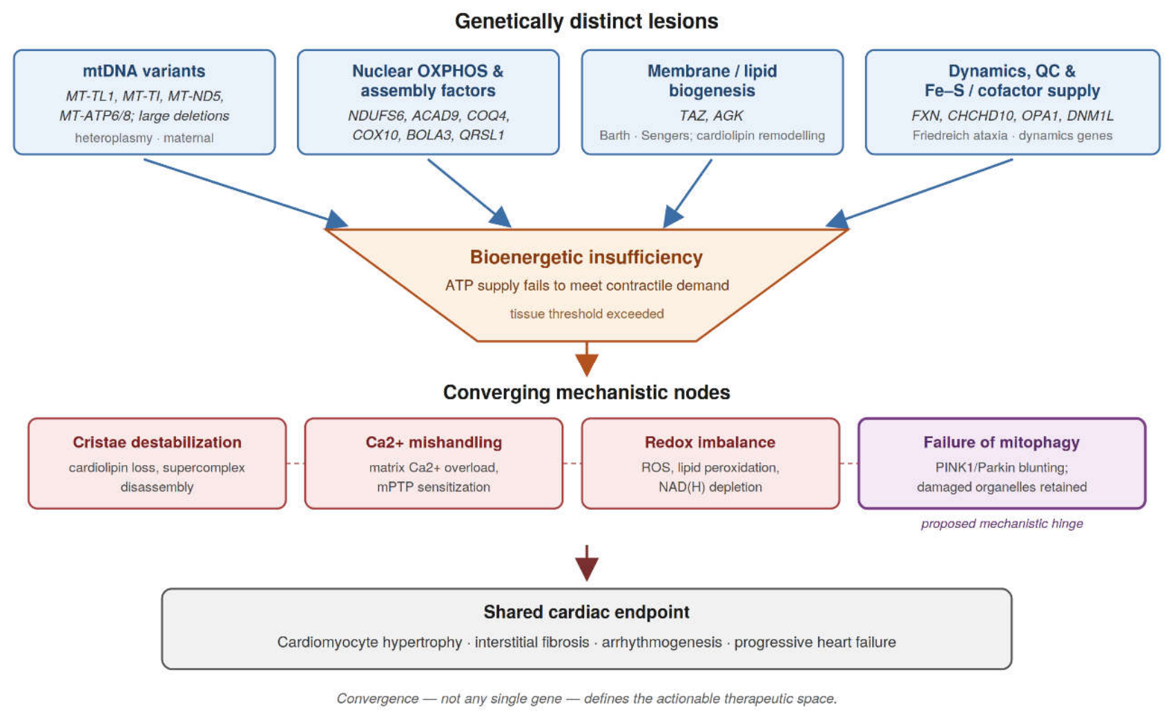
Figure 2.
The cardiomyocyte mitochondrion and its four interdependent functional domains. Bioenergetic supply (1), inner-membrane architecture (2) and calcium and redox handling (3) together generate and consume the proton-motive force, while dynamics and quality control (4) determine how long a damaged organelle persists. OMM, outer mitochondrial membrane; IMM, inner mitochondrial membrane; mPTP, mitochondrial permeability transition pore; CypD, cyclophilin D.
Figure 2.
The cardiomyocyte mitochondrion and its four interdependent functional domains. Bioenergetic supply (1), inner-membrane architecture (2) and calcium and redox handling (3) together generate and consume the proton-motive force, while dynamics and quality control (4) determine how long a damaged organelle persists. OMM, outer mitochondrial membrane; IMM, inner mitochondrial membrane; mPTP, mitochondrial permeability transition pore; CypD, cyclophilin D.
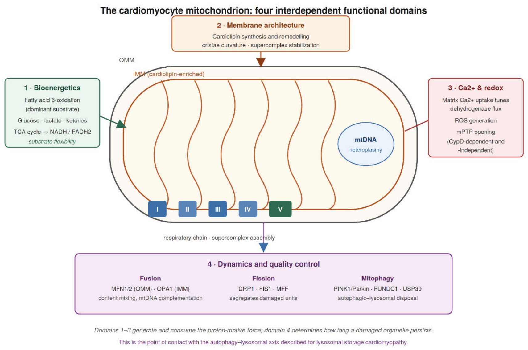
Table 1.
Core mitochondrial quality-control machinery in the cardiomyocyte and the cardiac consequences of its failure. FAO, fatty acid oxidation; mPTP, mitochondrial permeability transition pore; ROS, reactive oxygen species.
Table 1.
Core mitochondrial quality-control machinery in the cardiomyocyte and the cardiac consequences of its failure. FAO, fatty acid oxidation; mPTP, mitochondrial permeability transition pore; ROS, reactive oxygen species.
| Process | Key Effectors | Function in the Cardiomyocyte | Consequence of Failure | Ref. |
|---|---|---|---|---|
| Inner/outer membrane fusion | MFN1, MFN2, OPA1 | Content mixing and functional complementation between genomes; buffers heteroplasmic mtDNA load | Loss of complementation; unmasking of otherwise subthreshold variants; cristae disorganisation | [23] |
| Fission | DRP1 (DNM1L), FIS1, MFF | Segregates damaged subdomains for selective removal; enables organellar distribution | Excess fission fragments the network; insufficient fission prevents removal of damaged units | [23] |
| Mitophagy initiation | PINK1, Parkin, PARL | Senses depolarisation and tags damaged organelles by ubiquitination for autophagic clearance | Damaged organelles retained; sustained ROS output; contractile decline | [25,26] |
| Receptor-mediated mitophagy | FUNDC1, BNIP3/NIX | Ubiquitin-independent capture of mitochondria by autophagosomes, notably under hypoxia | Reduced clearance capacity when the canonical route is impaired | [27] |
| Negative regulation of mitophagy | USP30 | Deubiquitinates substrates and restrains PINK1/Parkin-dependent clearance | Unopposed activity blocks compensatory mitophagy; inhibition restores flux and survival in FAO-deficient hearts | [26] |
| Membrane lipid quality control | TAFAZZIN (TAZ), mitoregulin | Remodels and stabilises cardiolipin; maintains cristae curvature and supercomplex assembly | Damaged cristae, impaired respiration, arrested mitochondrial maturation | [19,20] |
| Permeability transition | mPTP, cyclophilin D (PPIF) | Calcium- and redox-gated pore governing the threshold for irreversible injury | Depolarisation, matrix swelling, necrotic cardiomyocyte loss | [21,22] |
Disclaimer/Publisher’s Note: The statements, opinions and data contained in all publications are solely those of the individual author(s) and contributor(s) and not of MDPI and/or the editor(s). MDPI and/or the editor(s) disclaim responsibility for any injury to people or property resulting from any ideas, methods, instructions or products referred to in the content. |
© 2026 by the authors. Licensee MDPI, Basel, Switzerland. This article is an open access article distributed under the terms and conditions of the Creative Commons Attribution (CC BY) license (http://creativecommons.org/licenses/by/4.0/).
Copyright: This open access article is published under a Creative Commons CC BY 4.0 license, which permit the free download, distribution, and reuse, provided that the author and preprint are cited in any reuse.