

Submitted:
06 September 2026
Posted:
08 September 2026
You are already at the latest version
Abstract
Cancer now ranks among the leading causes of death in Kenya, with approximately 35,867 new cases and 22,888 deaths annually, and outcomes remain constrained less by the availability of therapy than by the diagnostic pathway that must precede it. A national pathology workforce numbering in the low hundreds serves a population exceeding fifty million, and delayed or absent tissue diagnosis drives late presentation across the five malignancies that account for most of the national burden. Artificial intelligence offers a mechanism to extend interpretive capacity in a system where human expertise cannot be scaled at the rate the disease burden demands. This communication sets out a prioritized research agenda spanning smartphone-assisted visual inspection of the cervix, breast ultrasound classification and histopathological grading, morphological and immunophenotypic triage of hematolymphoid neoplasms, radiomic triage of thoracic and hepatic disease, and AI-assisted radiotherapy contouring. We present a six-stage methodological pipeline covering stakeholder engagement, local dataset curation, transfer learning, validation, usability assessment, and data governance, together with a tiered adoption framework matched to laboratory capability and the workforce competencies required for safe clinical supervision of these systems. The agenda is directed at early-career investigators and is transferable to comparable low- and middle-income settings.
Keywords:
artificial intelligence
; cancer control
; global health
; cervical cancer
; digital pathology
; pathology workforce
; radiomics
; Kenya
; health equity
; sub-Saharan Africa
1. Introduction
In 2024, the World Health Organization (WHO) estimated that there were 20.6 million new cancer cases and 9.8 million deaths globally [1]. The Global status report on cancer 2026 by WHO reports that of all the cancer deaths, over 4.8 million occurred among adults aged 30 to 69 years representing a substantial burden of premature cancer mortality [2]. These estimates showed that 1 in 5 people develop cancer in their lifetime, with 1 in 9 men and 1 in 13 women dying from cancer before the age of 75 years. By 2050, the WHO predicts a 77% increase to 35 million new cancer cases. Despite progress in early detection of cancers, there’s a significant disparity in treatment outcomes depending on where the patients reside, for breast cancer, for example, it's 87% in high-income countries but only about 42% in low-income countries, and fewer than one in three countries currently include cancer care in their universal health coverage packages [2]. The 2023 - 24 global cancer estimates suggest that these trends could be getting worse, with cases projected to rise sharply by 2025, underscoring the urgent need to explore alternative approaches to screening, treatment and support [4,5]
In Sub-Saharan Africa (SSA) estimates show that in 2022, there were 1,154,584 new cancer cases and 754,574 deaths with around 80% of the incidence cases occurring in 15 Countries [6]. With only 3% of the world’s healthcare workforce, SSA has 24% of cancer global disease burden seriously impacting care [7]. It has also been noted that over the past 3 decades, the burden of selected cancers has increased in SSA [8]. A need exists for SSA Countries to monitor progress in cancer control planning [9].
In Kenya, like many low- and -middle income countries (LMIC) in SSA, cancers are leading public health challenges contributing substantially to morbidity, mortality and the population healthcare expenditure [10,11]. A Kenya Vital Statistics [12] report shows that by July 2025 cancer is currently recognized as one of the leading causes of death in Kenya after cardiovascular and infectious diseases [13]. The situation is also impacted by limited screening services, delayed diagnosis, and inadequate treatment and support [14,15]. Socioeconomic inequalities, low public awareness, inadequate health financing and unequal access to healthcare between urban and rural settings make the cancer situation in Kenya worse [16].
Approximately, 35,867 new cancer cases are reported annually in Kenya, 22,888 cancer-related deaths, and 76,165 people living with cancer within five years of diagnosis. Breast cancer remains the most frequently diagnosed cancer, accounting for 5,822 new cases (16.2%), followed by cervical cancer with 4,294 cases (12.0%), prostate cancer with 3,601 cases (10.0%), oesophageal cancer with 2,532 cases (7.1%), and colorectal cancer with 2,525 cases (7.0%). Collectively, these five cancers account for more than half of all cancer diagnoses in Kenya and represent the highest priorities for prevention, early detection, screening, diagnosis, and treatment interventions [17].
No treatment decision in oncology is made without a tissue or cytological diagnosis that assigns the tumour to a category, a grade, and molecular class. Kenya's capacity to generate a pathological diagnosis is a binding constraint on its cancer control programme. The Lancet Commission on Diagnostics [18] estimated that nearly half of the world's population has little or no access to diagnostic services, and the deficit falls hardest on histopathology, where the pathologist-to-population ratio across much of sub-Saharan Africa approaches one per million against approximately one per twenty-five thousand in high-income health systems [19,20]. Published counts for Kenya vary by source and by whether forensic and clinical pathologists are included, but every available estimate describes a workforce of a few hundred at most, concentrated in Nairobi and in a small number of teaching hospitals [21]. When biopsy results take several weeks or more to return, patients may experience substantial delays in starting treatment, during which time their disease may progress significantly. The effective utilization of artificial intelligence can advance aspects of cancer care delivery that are currently deleteriously impacted by severe healthcare workforce shortages [22] (Hricak, Lancet, 2026, Cancer Workforce). AI should not replace the workforce, but rather enhance and extend their capabilities and capacity.
Against this background, AI applications should be prioritized according to their potential to address specific bottlenecks across the cancer care pathway, particularly where shortages in diagnostic expertise, infrastructure, and specialist workforce contribute to delayed care. The following sections identify priority opportunities spanning screening, diagnosis, imaging pathology and treatment planning, with emphasis on interventions that are clinically relevant, technically feasible, and adaptable to resource-constrained settings.
2. Priority AI Interventions in Kenya
2.1. Cervical Cancer Screening
Cervical cancer remains the leading cause of cancer-related mortality among Kenyan women despite being one of the most preventable malignancies through early detection and treatment of precancerous lesions [27]. Kenya's national cervical cancer screening programme primarily relies on Visual Inspection with Acetic Acid (VIA), Pap smear cytology, and Human Papillomavirus (HPV) DNA testing [28]. However, each of these diagnostic tests faces substantial implementation challenges within resource-constrained health systems [29,30].
The VIA is inexpensive and suitable for low-resource settings but is highly operator-dependent, resulting in considerable inter-observer variability and inconsistent diagnostic performance [31]. Pap smear cytology requires trained cytotechnologists and pathologists who remain scarce in many Kenyan counties, leading to prolonged turnaround times and reduced screening coverage [32]. Although HPV DNA testing provides superior sensitivity, its widespread implementation is constrained by laboratory infrastructure, equipment costs, and supply-chain limitations [33].
Artificial intelligence offers an opportunity to standardise image interpretation and expand diagnostic capacity. Deep convolutional neural networks (CNNs) have demonstrated excellent performance in analysing cervical images captured after acetic acid application, enabling automated classification of cervical lesions using smartphone-acquired images [34]. AI-assisted smartphone systems can provide real-time decision support, reducing diagnostic subjectivity and facilitating deployment in primary healthcare facilities where specialist expertise is unavailable [35].
Within Kenya, an AI-enabled cervical screening platform could combine smartphone colposcopy with cloud-based or edge-computing (preference for bandwidth-constrained areas) deep-learning algorithms trained on annotated cervical images. Such systems could classify normal cervices, low-grade squamous intraepithelial lesions, and high-grade lesions while automatically identifying women requiring referral for colposcopy or treatment.
A feasible proof-of-concept study would involve community health workers performing VIA using smartphone imaging devices across county health facilities. AI-generated diagnoses would be compared against expert colposcopist assessments and histopathological confirmation (Human-in-the-loop). Primary outcome measures would include sensitivity, specificity, area under the receiver operating characteristic curve (AUC), inter-rater agreement, referral accuracy, and time-to-decision.
2.2. Breast Cancer Diagnosis
Breast cancer is now the most frequently diagnosed cancer among Kenyan women and remains a leading cause of cancer mortality because many patients present with advanced disease [36]. Breast ultrasound is widely available in Kenyan hospitals but remains highly operator-dependent, while histopathology reporting often experiences delays owing to shortages of consultant pathologists [37,38].
Artificial intelligence has demonstrated substantial potential in improving both radiological and pathological diagnosis. Convolutional neural networks trained using publicly available datasets such as the Breast Ultrasound Images Dataset (BUSI) have achieved high diagnostic accuracy for differentiating benign from malignant breast lesions [39]–[41]. Fine-tuning these models using locally acquired Kenyan ultrasound images could improve generalisability across diverse patient populations and imaging equipment. Explainable AI approaches, including Gradient-weighted Class Activation Mapping (Grad-CAM), further enhance clinical trust by highlighting diagnostically relevant image regions [42].
Digital pathology provides another transformative opportunity. Whole-slide imaging (WSI) enables histopathological slides to be digitised for AI analysis. Deep-learning algorithms have demonstrated high accuracy in automated tumour detection, mitotic counting, nuclear pleomorphism assessment, and Nottingham histological grading, thereby reducing reporting workload while improving consistency [43,44]. Furthermore, Digital pathology and AI can enhance pathology workflows by improving the objectivity and reproducibility of biomarker assessment. AI-assisted whole-slide image analysis may support more consistent evaluation of ER, HER2, and Ki67, including equivocal HER2 2+ results and emerging HER2-low categories. Overall, AI would be best positioned as a decision-support tool that complements pathologist expertise, reduces interobserver variability, and improves diagnostic efficiency [45].
A retrospective proof-of-concept study could evaluate diagnostic concordance between AI-generated pathology reports and consultant pathologist diagnoses using archived breast biopsy specimens from Kenyan referral hospitals. Agreement could be assessed using sensitivity, specificity, Cohen's κ coefficient, grading concordance, and reductions in reporting turnaround time.
2.3. Hematolymphoid Malignancies and the Morphological Gap
Solid tumours dominate national cancer control planning, yet haematological and lymphoid neoplasms carry a distinct diagnostic burden that no imaging algorithm will address. Burkitt lymphoma remains endemic across the malaria belt of East Africa, presents overwhelmingly in children, and is curable at rates that depend more on the interval between presentation and treatment than on any other single variable. Diffuse large B-cell lymphoma, Hodgkin lymphoma, and HIV-associated lymphoproliferative disease together account for a substantial share of the oncological workload at national referral hospitals, while acute leukaemias in children arrive with a clock already running. Accurate subclassification of these diseases depends on immunophenotyping by flow cytometry or immunohistochemistry and, increasingly, on cytogenetic and molecular studies, all of which are concentrated in a small number of Kenyan laboratories and are frequently unavailable at the point of first clinical contact [46,47].
Three applications are tractable with existing technology. Automated morphological screening of peripheral blood and bone marrow aspirate smears, whether through dedicated digital cell morphology systems or through microscope-mounted smartphone capture, can triage films containing blasts, dysplastic changes, or abnormal lymphoid populations to a pathologist, converting the scarce resource of expert interpretation into a review function rather than a primary screening function [48]. Deep-learning classification of haematoxylin and eosin sections from lymph node and extranodal biopsies can generate a prioritized differential and, in published work, predict immunophenotypic and molecular features that would otherwise consume an antibody panel the laboratory cannot afford to run on every case. Machine-learning interpretation of flow cytometry list-mode data can flag abnormal populations and standardize gating, a task in which inter-operator variability is well documented and in which local subspecialty expertise is thinnest [49].
A proof-of-concept study in this domain would be inexpensive relative to the imaging work described above, because the input material already exists in the archive. Stored Giemsa-stained films and archival lymph node blocks from national and academic referral hospitals could be digitized and used to train and validate a blast-detection and lymphoma-triage model against the consensus diagnosis of a haematopathology review panel [50]. Outcome measures of interest would include sensitivity for blasts, negative predictive value for the exclusion of acute leukaemia, concordance with WHO classification categories, and the reduction in immunohistochemical stains required per case. Negative predictive value deserves particular emphasis, because where the realistic alternative to an algorithm is no expert morphological review at all, the clinically decisive property of the tool is its capacity to safely release normal material rather than its accuracy on rare and difficult cases [51].
2.4. The Pre-Analytic Constraint on Computational Pathology
Every computational pathology proposal in this roadmap rests on an assumption that deserves to be stated rather than presumed, which is that a technically adequate slide exists. Whole-slide imaging digitizes only what the laboratory produced, and a model trained on well-fixed, evenly sectioned, consistently stained material from a high-resource reference laboratory will degrade against slides made under conditions of interrupted formalin supply, prolonged cold ischaemia, manual processing, variable microtome performance, and haematoxylin batches that drift across a working week. Stain variation alone has been shown to shift the output of deep-learning classifiers [52], and stain normalization, colour augmentation, and domain adaptation should therefore be written into the model development protocol rather than discovered during external validation.
Pathology in this context should be understood as a diagnostic system rather than histopathology alone. Cancer diagnosis depends on an interconnected pathology and laboratory medicine infrastructure spanning specimen acquisition and processing, histology and cytology, hematology and flow cytometry, immunohistochemistry, molecular diagnostics, and the laboratory information systems and quality processes that connect these activities. Artificial intelligence may improve individual steps within this system, but its clinical value ultimately depends on whether an adequate specimen can be obtained, processed, characterized, integrated with complementary laboratory findings, and reported in time to guide treatment. Investment in computational pathology should therefore strengthen the diagnostic pathway around the algorithm, including laboratory workforce, quality management, specimen logistics, interoperability, and access to ancillary testing.
This is not an argument against digital pathology in Kenya. There is an argument that the first investment in any national AI pathology programme belongs at the histology bench, and that algorithm development should be paired with standardization of fixation and staining together with participation in external quality assessment schemes. Scanner access deserves equal candour, since whole-slide scanners remain concentrated in a small number of institutions and the realistic near-term substitute is microscope-mounted digital capture of selected fields, which constrains model architecture and should be reflected in study design from the outset [53]. The diagnostic pathway begins with a clinician who decides to biopsy, continues through a specimen that reaches a laboratory intact and correctly labelled, and ends with a report that reaches the treating team in time to change management. Artificial intelligence can compress the interpretive segment of that pathway and cannot manufacture the segments preceding it.
2.5. Cancer Imaging and Radiomics
Medical imaging underpins cancer diagnosis, staging, treatment planning, and surveillance. However, Kenya continues to experience a severe shortage of subspecialised oncological radiologists, particularly outside tertiary referral centres [54]. Consequently, subtle lesions may be overlooked, reporting delays are common, and advanced quantitative imaging biomarkers remain underutilised.
Artificial intelligence offers opportunities to augment radiological interpretation while introducing radiomics into routine oncology practice. Radiomics extracts quantitative imaging features describing tumour morphology, texture, and heterogeneity that are often imperceptible to human observers [55]. When combined with machine-learning algorithms, these features can improve tumour characterisation, predict treatment response, and estimate prognosis.
Transfer learning using open-source chest X-ray models may facilitate automated detection of pulmonary metastases, while nnU-Net has emerged as one of the most successful architectures for automated medical image segmentation, including liver lesions on computed tomography (CT) scans [56,57]. Automated segmentation enables objective tumour volume measurement and facilitates longitudinal treatment monitoring.
A Kenyan proof-of-concept study could utilize de-identified abdominal CT scans from Kenyatta National Hospital to evaluate AI-based liver lesion segmentation. Performance could be assessed using Dice Similarity Coefficient, Hausdorff Distance, and volumetric agreement against expert manual annotations. Extracted radiomic signatures could then be correlated with treatment response, recurrence, and overall survival.
2.6. Radiotherapy Planning and Digital Pathology
Radiotherapy planning requires precise delineation of tumours and surrounding organs at risk (OARs). In Kenya, manual contouring contributes substantially to treatment delays because of limited numbers of radiation oncologists and medical physicists [58].
Deep-learning segmentation architectures such as U-Net and DeepLabV3+ have demonstrated excellent performance in automated OAR contouring across multiple anatomical sites, substantially reducing planning time while maintaining expert-level contour quality [59,60]. Such systems may significantly improve workflow efficiency for cervical brachytherapy and external beam radiotherapy.
Artificial intelligence also offers opportunities for computational pathology. Molecular biomarkers such as HER2, Ki-67, and microsatellite instability (MSI) increasingly guide precision oncology but remain expensive because they require immunohistochemistry (IHC), polymerase chain reaction (PCR), or next-generation sequencing [61,62]. Recent advances in weakly supervised deep learning have shown that clinically relevant molecular alterations can be predicted directly from routine H&E-stained whole-slide images, thereby reducing unnecessary molecular testing [63].
A proof-of-concept implementation in Kenya could compare conventional manual cervical brachytherapy planning with AI-assisted workflows to quantify reductions in contouring time while evaluating contour quality using Dice Similarity Coefficients and clinician acceptability scores. Simultaneously, AI models predicting MSI status from archived digital pathology slides could be validated against PCR or IHC results, with evaluation based on diagnostic accuracy, cost-effectiveness, and workflow efficiency.
3. Methodological Roadmap for Early-Career Researchers
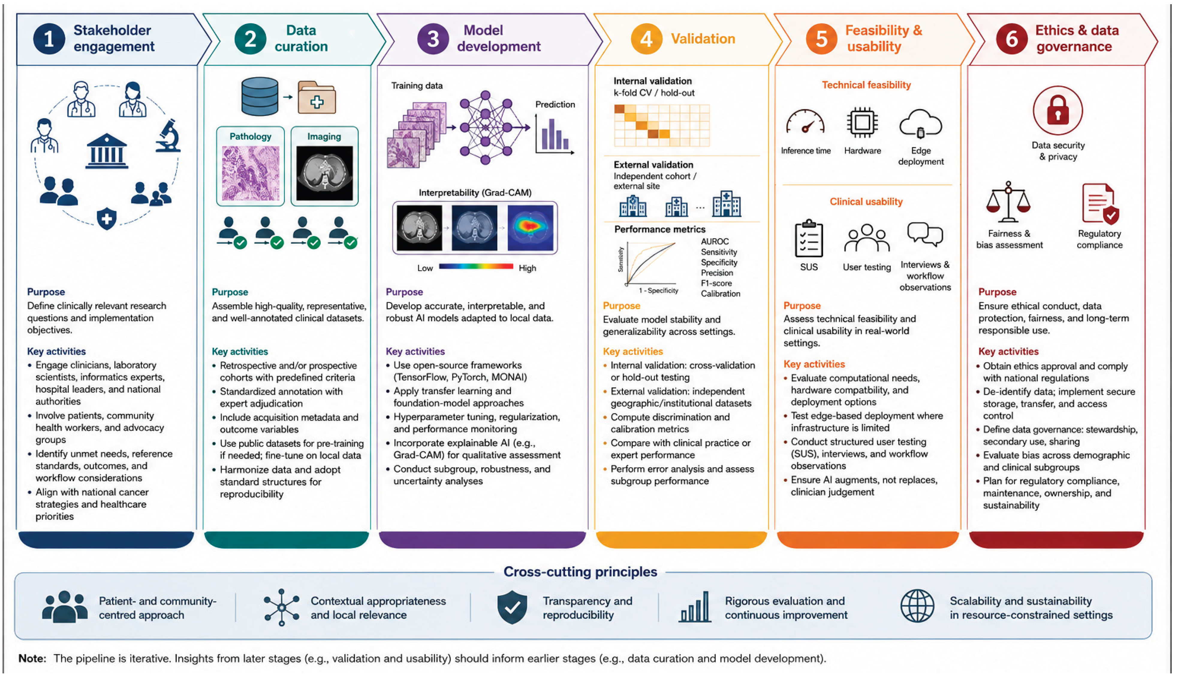
We present a six-stage methodological pipeline for conducting proof-of-concept artificial intelligence (AI) studies in cancer research within resource-constrained healthcare systems (Figure 1 ). The pipeline provides a structured framework that integrates clinical problem definition, data generation, model development, validation, implementation assessment, and governance considerations.
Unlike conventional AI development workflows that prioritize predictive performance, this framework emphasizes clinical relevance, representative datasets, implementation readiness, and responsible AI governance. Although developed using Kenya as an exemplar LMIC setting, the pipeline is designed to be transferable to comparable healthcare environments and to support early-career researchers undertaking clinically oriented AI research.
3.1. Stakeholder Engagement
The pipeline begins with stakeholder engagement to define clinically relevant research questions and implementation objectives before model development. Clinical stakeholders, including oncologists, pathologists, radiologists, surgeons, laboratory scientists, and clinical informatics specialists, identify priority clinical problems, establish reference standards, define outcome measures, and evaluate integration within existing diagnostic and treatment workflows. Engagement with hospital administrators and national health authorities aligns research activities with institutional priorities and national cancer control strategies.
Patient representatives, community health workers, and cancer advocacy organisations are incorporated throughout the planning phase to identify barriers to implementation, improve acceptability, and ensure that proposed AI solutions address locally relevant healthcare needs. Early stakeholder engagement therefore informs study design, data acquisition, annotation protocols, evaluation criteria, and implementation planning.
3.2. Data Curation
Clinical datasets are assembled from retrospective and/or prospective cohorts using predefined inclusion and exclusion criteria. Dataset development prioritizes clinical representativeness, annotation quality, and demographic diversity rather than dataset size alone. Proof-of-concept studies typically include several hundred participants, although sample size requirements depend on disease prevalence, imaging modality, and the intended clinical application.
Annotations are generated using standardized protocols developed collaboratively with domain experts. Pathology datasets include tumour classification, grading, and segmentation according to established diagnostic criteria, whereas imaging datasets include acquisition metadata together with clinically relevant outcome variables. Annotation discrepancies are documented and resolved through expert adjudication to preserve annotation consistency while acknowledging clinically meaningful uncertainty.
Where sufficiently large local datasets are unavailable, publicly available datasets may be used for pre-training before transfer learning and fine-tuning using locally acquired data. Because disease epidemiology, imaging protocols, healthcare delivery, and population characteristics differ across settings, externally derived datasets are not considered substitutes for representative local cohorts. Data harmonization procedures and standardized data structures are incorporated to improve reproducibility and facilitate future multicentre collaborations.
3.3. Model Development
Models are developed using open-source deep learning frameworks (TensorFlow, PyTorch, or MONAI) to maximize reproducibility, transparency, and accessibility. Transfer learning and foundation-model approaches are preferentially adopted to reduce annotation requirements while enabling adaptation to local clinical datasets.
Model optimization includes hyperparameter tuning, regularization, and iterative performance monitoring using predefined validation datasets. Explainable AI techniques, including Gradient-weighted Class Activation Mapping (Grad-CAM), are incorporated to enable qualitative assessment of prediction localization by clinical experts. These visualization methods support interpretation of model outputs but are not regarded as definitive evidence of model reasoning.
Model development further includes robustness testing, uncertainty estimation, and subgroup analyses to evaluate performance across demographic, clinical, and disease-specific subpopulations and to identify potential sources of algorithmic bias before external evaluation.
3.4. Validation
Model evaluation follows a sequential validation strategy comprising both internal and external assessment. Internal validation employs cross-validation or independent hold-out datasets to assess model stability during development. External validation uses geographically or institutionally independent datasets to evaluate generalizability across healthcare settings.
Performance is quantified using standard discrimination metrics, including the area under the receiver operating characteristic curve (AUROC), sensitivity, specificity, precision, recall, and F1-score, together with calibration analyses where appropriate. Whenever suitable reference standards exist, AI performance is compared with expert interpretation or current clinical practice. Comprehensive error analysis is performed to identify clinically significant failure modes, characterize model uncertainty, and evaluate performance variability across patient subgroups.
3.5. Feasibility and Usability Assessment
Implementation feasibility is assessed by evaluating computational requirements, inference time, hardware compatibility, and deployment within representative clinical environments. In settings with constrained computational infrastructure, edge-based deployment strategies are considered to minimize dependence on cloud computing and uninterrupted internet connectivity. Teams should incorporate implementation science principles to rigorously evaluate the implementation process to emphasize effectiveness and sustainability.
Clinical usability is evaluated through structured user testing involving intended end-users. Standardized usability instruments, including the System Usability Scale (SUS), are combined with qualitative interviews and workflow observations to assess usability, interpretability, workflow integration, and user acceptance. Throughout the evaluation process, AI systems are considered clinical decision-support tools intended to augment, rather than replace, clinician decision-making.
3.6. Ethics and Data Governance
All studies should be conducted following institutional ethics approval and applicable national regulations governing human participant research, data protection, and information security. Clinical datasets are de-identified before analysis, and secure procedures are implemented for data storage, transfer, and controlled access. Data governance frameworks define responsibilities for data stewardship, secondary data use, institutional ownership, and collaborative data sharing.
Bias assessment is integrated throughout model development by evaluating performance across demographic and clinical subgroups. Fairness analyses identify systematic performance disparities that could contribute to inequitable healthcare delivery and inform subsequent model refinement. Long-term considerations, including regulatory compliance, post-deployment monitoring, model maintenance, sustainability, and institutional capacity building, are incorporated during study planning to facilitate responsible clinical translation and sustainable implementation of AI technologies within LMIC healthcare systems.
Digital pathology raises a governance question that is different from radiological imaging. A whole-slide image is a permanent, high-resolution, and effectively inexhaustible representation of a patient's tissue, and once it leaves the institution that produced it, it can be used to train commercial models indefinitely without further reference to the patient, the reporting pathologist, or the hospital. African datasets have historically been exported under collaborative arrangements that returned publications rather than durable capacity, and a Kenyan artificial intelligence cancer programme should therefore set its terms in advance rather than negotiate them retrospectively.
Those terms should specify that primary image repositories remain under Kenyan institutional custody, that model weights derived from Kenyan data are jointly owned, that any commercial application requires a separately negotiated agreement (as proposed by the National Data Governance policy), and that collaborating institutions commit to reciprocal training and infrastructure investment rather than to authorship alone. The Data Protection Act, 2019 and the oversight framework of the National Commission for Science, Technology and Innovation provide the statutory basis, and study protocols should address secondary use explicitly rather than relying on broad consent language that patients cannot meaningfully evaluate.
4. Workforce, Training, and Resource-Stratified Adoption
A roadmap that ends at model validation stops short of the point at which patients benefit. The rate-limiting factor for artificial intelligence in Kenyan cancer care will not be model performance, which is already adequate for several of the tasks described above, but the readiness of the clinical workforce to interpret, supervise, and override these systems. Pathologists, radiologists, oncologists, clinical officers, and community health workers will be asked to act on probabilistic outputs generated by models whose training data they did not select and whose failure modes they have not been taught to recognize. Building that capacity is a curricular problem, and it should be planned alongside the technical work rather than appended to it once deployment has begun.
Four competencies define the minimum standard for safe use. Clinical users should be able to state what a given model was trained to do and on which population, recognize the case types that fall outside its training distribution, interpret a probability score or a saliency map as evidence to be weighed against the clinical picture rather than as a verdict to be transcribed, and articulate where diagnostic and medicolegal responsibility resides when the model and the clinician disagree.
These competencies map onto the existing architecture of medical education in Kenya, where they can be introduced during undergraduate pathology teaching, consolidated within Master of Medicine training in pathology and radiology, and maintained through continuing professional development delivered by the Kenya Association of Pathologists and the College of Pathologists of East, Central and Southern Africa. Embedding artificial intelligence literacy within postgraduate pathology training carries a second benefit, which is that the trainees who annotate the datasets become the consultants who later supervise the deployed systems, and that continuity is the most reliable safeguard against uncritical adoption.
Adoption should be stratified by demonstrated laboratory capability rather than presented as a single national ambition, since deploying a whole-slide imaging workflow into a facility without reliable tissue processing produces a costly failure that sets the wider programme back. Table 1 proposes a tiered framework in which each level specifies the enabling requirements that must be met before the corresponding tools are introduced, and in which the deliverable at lower tiers is accurate triage and referral rather than definitive diagnosis.
5. Expected Impact and Conclusions
In conclusion, successful translation of AI into routine clinical practice requires methodological approaches that extend beyond algorithm development to encompass clinical validation, implementation science, governance, and healthcare system integration. The six-stage pipeline presented here provides a practical and reproducible framework to guide early-career researchers in developing clinically relevant, transparent, and ethically responsible AI systems for cancer diagnosis within resource-constrained settings.
By aligning computational innovation with clinical priorities, implementation readiness, and responsible data governance, the framework supports the generation of high-quality translational evidence necessary to advance AI from proof-of-concept studies toward sustainable clinical deployment. Wider adoption of similar methodological frameworks may strengthen AI research capacity, promote equitable innovation, and contribute to reducing disparities in access to high-quality cancer diagnostics across sub-Saharan Africa and other LMICs.
Author Contributions
Conceptualization, PM. and CNN.; methodology, AKW; original draft preparation, PM and CNN.; review and editing, PJ, NN, LW, KMM; supervision, AKW. All authors have read and agreed to the published version of the manuscript.
Funding
This research received no external funding.
Institutional Review Board Statement
Not applicable.
Informed Consent Statement
Not applicable.
Data Availability Statement
No new empirical data were created or analyzed in this study. Data sharing is not applicable to this article.
Conflicts of Interest
The authors declare no conflict of interest.
References
- Sung, H.; Filho, A.M.; Laversanne, M.; et al. Global cancer statistics 2024: GLOBOCAN estimates of incidence and mortality worldwide for 34 cancers in 186 countries. CA Cancer J. Clin. 2026, 76(4), e70090. [Google Scholar] [CrossRef] [PubMed]
- Global status report on cancer 2026: the future we choose together. Available online: https://www.who.int/publications/i/item/9789240123977 (accessed on 30 July 2026).
- Global cancer burden growing, amidst mounting need for services. Available online: https://www.who.int/news/item/01-02-2024-global-cancer-burden-growing--amidst-mounting-need-for-services (accessed on 26 July 2026).
- Sung, H.; Filho, A.M.; Laversanne, M.; et al. Global cancer statistics 2024: GLOBOCAN estimates of incidence and mortality worldwide for 34 cancers in 186 countries. CA Cancer J. Clin. 2026, 76(4), e70090. [Google Scholar] [CrossRef] [PubMed]
- Luo, Q.; Smith, D.P. Global cancer burden: progress, projections, and challenges. The Lancet 2025, 406(10512), 1536–1537. [Google Scholar] [CrossRef] [PubMed]
- Ugwu, L.E.; Ajele, W.K.; Idemudia, E.S.; et al. Cancer in Africa, 2022: Incidence, Mortality, and Age-Patterned Fatality From GLOBOCAN Across 36 Cancer Types. JCO Glob. Oncol. 2026, 12(4), e2500663. [Google Scholar] [CrossRef] [PubMed]
- Cancer Atlas. Available online: https://canceratlas.cancer.org/burden-of-cancer/cancer-in-sub-saharan-africa/ (accessed on 26 July 2026).
- Bolarinwa, O.; Bashir, S.G.; Okyere, J.; et al. Burden, Regional Trends and Risk Factors of Breast, Cervical, Uterine, and Ovarian Cancers in Sub-Saharan Africa, 1990-2023: The Global Burden of Disease 2023. Int. J. Environ. Res. Public Health 2026, 23(4), 419. [Google Scholar] [CrossRef] [PubMed]
- Bray, F.; Parkin, D.M.; African Cancer Registry Network. Cancer in sub-Saharan Africa in 2020: a review of current estimates of the national burden, data gaps, and future needs. Lancet Oncol. 2022, 23(6), 719–728. [Google Scholar] [CrossRef] [PubMed]
- Omotoso, O.; Teibo, J.O.; Atiba, F.A.; et al. Addressing cancer care inequities in sub-Saharan Africa: current challenges and proposed solutions. Int. J. Equity Health 2023, 22(1), 189. [Google Scholar] [CrossRef] [PubMed]
- Cancer in sub-Saharan Africa: a Lancet Oncology Commission - The Lancet Oncology. Available online: https://www.thelancet.com/journals/lanonc/article/PIIS1470-2045(21)00720-8/abstract (accessed on 28 July 2026).
- Kenya Vital Statistics Report - 2024 - Kenya National Bureau of Statistics. Available online: https://www.knbs.or.ke/reports/kenya-vital-statistics-report-2024/ (accessed on 30 July 2026).
- Wambalaba, F.; Wambalaba, A. The Inequity of Cancer Treatment Services in Kenya. In The Landscape of Global Health Inequity; Son, B.W.K., Ed.; Springer Nature Switzerland, 2024; pp. 37–46. [Google Scholar] [CrossRef]
- Nyangasi, M.F.; McLigeyo, A.A.; Kariuki, D.; Mithe, S.; Orwa, A.; Mwenda, V. Decentralizing cancer care in sub-Saharan Africa through an integrated regional cancer centre model: The case of Kenya. PLoS Glob. Public Health 2023, 3(9), e0002402. [Google Scholar] [CrossRef] [PubMed]
- Kizub, D.A.; Naik, S.; Abogan, A.A.; et al. Access to and Affordability of World Health Organization Essential Medicines for Cancer in Sub-Saharan Africa: Examples from Kenya, Rwanda, and Uganda. Oncologist 2022, 27(11), 958–970. [Google Scholar] [CrossRef] [PubMed]
- How Social Determinants of Health and Health System Factors Shape the Breast Cancer Continuum in Kenya | Current Breast Cancer Reports | Springer Nature Link. Available online: https://link.springer.com/article/10.1007/s12609-025-00596-z (accessed on 28 July 2026).
- Kenya Records 35,867 New Cancer Cases in Latest GLOBOCAN 2024 Estimates – National Cancer Institute Kenya. 10 July 2026. Available online: https://wp.ncikenya.go.ke/2026/07/10/kenya-records-35867-new-cancer-cases-in-latest-globocan-2024-estimates/ (accessed on 28 July 2026).
- The Lancet Commission on diagnostics: transforming access to diagnostics - The Lancet. Available online: https://www.thelancet.com/journals/lancet/article/PIIS0140-6736(21)00673-5/fulltext (accessed on 11 August 2026).
- Mudenda, V.; Malyangu, E.; Sayed, S.; Fleming, K. Addressing the shortage of pathologists in Africa: Creation of a MMed Programme in Pathology in Zambia. Afr. J. Lab Med. 2020, 9(1), 974. [Google Scholar] [CrossRef] [PubMed]
- Wilson, M.L.; Fleming, K.A.; Kuti, M.A.; Looi, L.M.; Lago, N.; Ru, K. Access to pathology and laboratory medicine services: a crucial gap. The Lancet 2018, 391(10133), 1927–1938. [Google Scholar] [CrossRef] [PubMed]
- Improving pathology and laboratory medicine in low-income and middle-income countries: roadmap to solutions - The Lancet. Available online: https://www.thelancet.com/journals/lancet/article/PIIS0140-6736(18)30459-8/abstract (accessed on 11 August 2026).
- Hricak, H.; Ward, Z.J.; Moraes, F.Y.; et al. Cancer workforce-a global crisis: a Lancet Oncology Commission. Lancet Oncol. 2026, 27(6), e279–e316. [Google Scholar] [CrossRef] [PubMed]
- Kibe, L.W.; Kwanya, T.; Nyagowa, H. Cancer research production and dissemination in East African Community countries. Inf. Dev. Published online. 2025, 02666669251325463. [CrossRef]
- Hutchinson, B.; Watts, R.; Nyangasi, M.; et al. An economic evaluation of breast cancer interventions in Kenya. eClinicalMedicine 2024, 77. [Google Scholar] [CrossRef] [PubMed]
- Hughes, J.G.; Tamirisa, K.; Dharani, A.; et al. Leveraging Artificial Intelligence to Enhance Oncology Care Delivery in Sub-Saharan Africa. Mayo Clin. Proc. Digit. Health 2026, 4(1). [Google Scholar] [CrossRef] [PubMed]
- Dako, F.; Moraes, F.Y.; Doo, F.; et al. Digital health and artificial intelligence innovations for oncology in sub-Saharan Africa. Lancet Oncol. 2025, 26(10), e547–e557. [Google Scholar] [CrossRef] [PubMed]
- Mwenda, V.; Mburu, W.; Bor, J.P.; et al. Cervical cancer programme, Kenya, 2011–2020: lessons to guide elimination as a public health problem. Ecancermedicalscience 2022, 16, 1442. [Google Scholar] [CrossRef] [PubMed]
- Orang’o, E.O.; Were, E.; Rode, O.; et al. Novel concepts in cervical cancer screening: a comparison of VIA, HPV DNA test and p16INK4a/Ki-67 dual stain cytology in Western Kenya. Infect. Agent Cancer 2020, 15, 57. [Google Scholar] [CrossRef] [PubMed]
- Mwenda, V.; Murage, D.; Kilonzo, C.; et al. Baseline assessment of cervical cancer screening and treatment capacity in 25 counties in Kenya, 2022. Front Oncol. 2024, 14. [Google Scholar] [CrossRef] [PubMed]
- Adhiambo, H.F.; Owidi, E.; Okello, P.; et al. Barriers and facilitators to implementing a single-visit, screen-and-treat approach with thermal ablation for cervical cancer prevention in Kenya. PLoS Glob. Public Health 2025, 5(9), e0005166. [Google Scholar] [CrossRef] [PubMed]
- David, J.; Joshi, V.; Jebin Aaron, D.; Baghel, P. A Comparative Analysis of Visual Inspection With Acetic Acid, Cervical Cytology, and Histopathology in the Screening and Early Detection of Premalignant and Malignant Lesions of the Cervix. Cureus 14(9), e29762. [CrossRef] [PubMed]
- Anangwe, N.; Steimgrimson, J.; Cu-Uvin, S. Evaluation of pathology resources for cervical cancer detection between 2018 and 2022: a retrospective study at Moi Teaching and Referral Hospital, Western Kenya. BMC Cancer 2025, 25, 203. [Google Scholar] [CrossRef] [PubMed]
- Siferih, M.; Gedif, G.; Lamesgen, A.; et al. Point-of-care HPV testing for cervical cancer screening in Sub-Saharan Africa: platform diversity, diagnostic performance, implementation feasibility, and determinants-a scoping review with contextual considerations for Ethiopia. BMC Public Health 2026, 26(1), 990. [Google Scholar] [CrossRef] [PubMed]
- Wang, X.; Wang, Q.; Ding, G.; Wang, J.; Tang, Y.; Feng, Y. Artificial intelligence for colposcopic and cytological image analysis in early cervical cancer detection. iScience 2026, 29(2), 114627. [Google Scholar] [CrossRef] [PubMed]
- AI delivers cervical cancer screening to rural areas of Kenya. Available online: https://healthcare-in-europe.com/en/news/ai-delivers-cervical-cancer-screening-to-rural-areas-of-kenya.html (accessed on 28 July 2026).
- Daniels-Donkor, S.S.; Afaya, A. Internet use and uptake of clinical breast examination among Kenyan women of reproductive age. Sci. Rep. 2025, 15, 42574. [Google Scholar] [CrossRef] [PubMed]
- Moturi, A.K.; Robert, B.N.; Bahati, F.; Macharia, P.M.; Okiro, E.A. Investigating rapid diagnostic testing in Kenya’s health system, 2018–2020: validating non-reporting in routine data using a health facility service assessment survey. BMC Health Serv. Res. 2023, 23, 306. [Google Scholar] [CrossRef] [PubMed]
- Iacob, R.; Iacob, E.R.; Stoicescu, E.R.; et al. Evaluating the Role of Breast Ultrasound in Early Detection of Breast Cancer in Low- and Middle-Income Countries: A Comprehensive Narrative Review. Bioengineering 2024, 11(3), 262. [Google Scholar] [CrossRef] [PubMed]
- Liu, J.; Pian, L.; Chen, J.; et al. Artificial intelligence in breast ultrasound: a systematic review of research advances. Front Oncol. 15, 1619364. [CrossRef] [PubMed]
- Parvin, S.; Rozario, L.J.; Islam, MdE. Artificial intelligence in breast cancer diagnosis: A systematic review of imaging modalities, methods, and datasets. Inform. Med. Unlocked 2026, 64, 101786. [Google Scholar] [CrossRef]
- Ali, A.; Alghamdi, M.; Marzuki, S.S.; et al. Exploring AI Approaches for Breast Cancer Detection and Diagnosis: A Review Article. Breast Cancer (Dove Med Press) 2025, 17, 927–947. [Google Scholar] [CrossRef] [PubMed]
- Ghauth, K.I.; Kustiawan, Y.A. Explainable Artificial Intelligence (XAI) for Cancer Classification in Medical Imaging: A Systematic Review. Mach. Learn. Knowl. Extr. 2026, 8(5), 134. [Google Scholar] [CrossRef]
- Dang, C.; Qi, Z.; Xu, T.; et al. Deep Learning–Powered Whole Slide Image Analysis in Cancer Pathology. Lab. Investig. 2025, 105(7), 104186. [Google Scholar] [CrossRef] [PubMed]
- Rad, M.S.; Huang, J (Vincent); Hosseini, M.M.; et al. Deep learning for digital pathology: A critical overview of methodological framework. J. Pathol. Inform. 2025, 19, 100514. [Google Scholar] [CrossRef] [PubMed]
- Datwani, S.; Khan, H.; Niazi, M.K.K.; Parwani, A.V.; Li, Z. Artificial intelligence in breast pathology: Overview and recent updates. Hum. Pathol. 2025, 162, 105819. [Google Scholar] [CrossRef] [PubMed]
- Fedoriw, Y.; Silva, O.; Znaor, A.; Macintyre, E. Global view of haematolymphoid tumor classifications and their application in low- and middle-income countries. Histopathology 2025, 86(1), 6–16. [Google Scholar] [CrossRef] [PubMed]
- Alaggio, R.; Amador, C.; Anagnostopoulos, I.; et al. The 5th edition of the World Health Organization Classification of Haematolymphoid Tumours: Lymphoid Neoplasms. Leukemia 2022, 36(7), 1720–1748. [Google Scholar] [CrossRef] [PubMed]
- Manescu, P.; Narayanan, P.; Bendkowski, C.; et al. Detection of acute promyelocytic leukemia in peripheral blood and bone marrow with annotation-free deep learning. Sci. Rep. 2023, 13(1), 2562. [Google Scholar] [CrossRef] [PubMed]
- Zuromski, L.M.; Durtschi, J.; Aziz, A.; et al. Clinical validation of a real-time machine learning-based system for the detection of acute myeloid leukemia by flow cytometry. Cytom. B Clin. Cytom. 2025, 108(5), 392–403. [Google Scholar] [CrossRef] [PubMed]
- Chipidza, F.E.; Kayembe, M.K.A.; Nkele, I.; et al. Accuracy of Pathologic Diagnosis in Patients With Lymphoma and Survival: A Prospective Analysis From Botswana. JCO Glob. Oncol. 2021, 7, 1620–1632. [Google Scholar] [CrossRef] [PubMed]
- Naresh, K.N.; Raphael, M.; Ayers, L.; et al. Lymphomas in sub-Saharan Africa--what can we learn and how can we help in improving diagnosis, managing patients and fostering translational research? Br. J. Haematol. 2011, 154(6), 696–703. [Google Scholar] [CrossRef] [PubMed]
- Tellez, D.; Litjens, G.; Bándi, P.; et al. Quantifying the effects of data augmentation and stain color normalization in convolutional neural networks for computational pathology. Med. Image Anal. 2019, 58, 101544. [Google Scholar] [CrossRef] [PubMed]
- Huang, Y.N.; Peng, X.C.; Ma, S.; et al. Development of Whole Slide Imaging on Smartphones and Evaluation With ThinPrep Cytology Test Samples: Follow-Up Study. JMIR Mhealth Uhealth 2018, 6(4), e82. [Google Scholar] [CrossRef] [PubMed]
- The Hidden Physician: Understanding the Vital Role of Radiologists | Kenya Association of Radiologists (KAR). Available online: https://kar.co.ke/blogs/the-radiologist-you-may-never-meet-but-whose-expertise-your-life-depends-on (accessed on 30 July 2026).
- Pang, J.; Xiu, W.; Ma, X. Application of Artificial Intelligence in the Diagnosis, Treatment, and Prognostic Evaluation of Mediastinal Malignant Tumors. J. Clin. Med. 2023, 12(8), 2818. [Google Scholar] [CrossRef] [PubMed]
- Tangruangkiat, S.; Chaiwongkot, N.; Pamarapa, C.; et al. Diagnosis of focal liver lesions from ultrasound images using a pretrained residual neural network. J. Appl. Clin. Med. Phys. 2023, 25(1), e14210. [Google Scholar] [CrossRef] [PubMed]
- Li, Y.; Endo, H.; Hirata, K.; et al. Evaluation of 2D and 3D nnU-Net models with two-label and three-label strategies for automatic segmentation and total metabolic tumor volume estimation of metastatic differentiated thyroid carcinoma on FDG-PET/CT. Jpn. J. Radiol. Published online. 2026. [Google Scholar] [CrossRef] [PubMed]
- Abdihamid, O.; Affey, F.; Maina, J.; Ngaruiya, C. The current state of oncological emergency services in Kenya: challenges and opportunities. BMJ Glob. Health 2026, 11(1), e020895. [Google Scholar] [CrossRef] [PubMed]
- Smine, Z.; Poeta, S.; De Caluwé, A.; et al. Automated segmentation in planning-CT for breast cancer radiotherapy: A review of recent advances. Radiother. Oncol. 2025, 202, 110615. [Google Scholar] [CrossRef] [PubMed]
- Moran, K.; Poole, C.; Barrett, S. Evaluating deep learning auto-contouring for lung radiation therapy: A review of accuracy, variability, efficiency and dose, in target volumes and organs at risk. Phys. Imaging Radiat. Oncol. 2025, 33, 100736. [Google Scholar] [CrossRef] [PubMed]
- Bani, M.A. Smart Lies and Sharp Eyes: Pragmatic Artificial Intelligence for Cancer Pathology: Promise, Pitfalls, and Access Pathways. Cancers 2026, 18(3), 421. [Google Scholar] [CrossRef] [PubMed]
- Chaurasia, A.K.; Toohey, P.W.; Bennett, M.T.; Harris, H.C.; Hewitt, A.W. Automated quantification of Ki-67 expression in breast cancer from H&E-stained slides using a transformer-based regression model. Breast Cancer Res. 2025, 27, 206. [Google Scholar] [CrossRef] [PubMed]
- Ghaffari Laleh, N.; Muti, H.S.; Loeffler, C.M.L.; et al. Benchmarking weakly-supervised deep learning pipelines for whole slide classification in computational pathology. Med. Image Anal. 2022, 79, 102474. [Google Scholar] [CrossRef] [PubMed]
Disclaimer/Publisher’s Note: The statements, opinions and data contained in all publications are solely those of the individual author(s) and contributor(s) and not of MDPI and/or the editor(s). MDPI and/or the editor(s) disclaim responsibility for any injury to people or property resulting from any ideas, methods, instructions or products referred to in the content. |
© 2026 by the authors. Licensee MDPI, Basel, Switzerland. This article is an open access article distributed under the terms and conditions of the Creative Commons Attribution (CC BY) license (http://creativecommons.org/licenses/by/4.0/).
Copyright: This open access article is published under a Creative Commons CC BY 4.0 license, which permit the free download, distribution, and reuse, provided that the author and preprint are cited in any reuse.