

Submitted:
04 September 2026
Posted:
07 September 2026
You are already at the latest version
Abstract
Cardiovascular diseases (CVD) and osteoporosis are among the leading causes of illness and mortality among older people worldwide. Evidence suggests these diseases are interconnected through shared biological mechanisms and risk factors. However, the relationship between specific CVD subtypes and osteoporosis has not been fully established. This review examines current evidence on the association between CVD subtypes and skeletal health with focus on shared biological mechanisms, epidemiological findings, medication effects, and future research that may improve understanding of the heart-bone axis. A literature search was conducted on PubMed and through backward citation searching to identify relevant articles investigating CVD, bone mineral density, and osteoporosis.Current evidence indicates that several CVD subtypes, including heart failure, coronary artery disease, hypertension, atrial fibrillation, and stroke, are associated with an increased risk of reduced bone mineral density, osteoporosis, and hip fractures. Shared mechanisms such as chronic inflammation, oxidative stress, vascular calcification, endocrine dysregulation, and altered RANK/RANKL/OPG and Wnt/β-catenin signaling pathways may contribute to the development of both CVD and osteoporosis. Advances in genomics, large population-based cohorts, and causal inference methods have further strengthened understanding of the complex heart-bone relationship. However, there is still a need to understand the subtype-specific associations and mechanisms. Future research is needed to fully understand the causal pathways and improve the prevention, early detection, and management of CVD, osteoporosis, and osteoporotic hip fractures.
Keywords:
cardiovascular disease
; osteoporosis
; bone mineral density
; hip fracture
; heart-bone axis
; vascular calcification
1. Introduction
Cardiovascular diseases (CVD) are a significant cause of global morbidity and mortality and are a public health concern [1,2]. They are chronic disorders associated with the heart and circulatory system [3]. CVDs are caused by a number of risk factors including age, gender, genetic susceptibility, smoking, obesity, dyslipidemia, hypertension, diabetes, and behavioral factors [4,5]. Studies have reported associations between low bone mineral density (BMD) and higher incidence of stroke, heart failure (HF), acute myocardial infarction (AMI), and mortality [6,7]. Stroke and HF have been found to make patients prone to lower BMD [6]. Reduced BMD measured by dual energy X-ray absorptiometry (DXA) is associated with increased CVD risk in postmenopausal women [8]. Previous studies have highlighted the role of CVDs in the increased risk of osteoporotic fractures [9]. This review will focus on coronary artery disease (CAD)/ myocardial infarction (MI), stroke, atrial fibrillation (AF), peripheral arterial disease (PAD) and HF as the CVD subtypes.
CAD is among the common types of heart disease [5] which is characterized by blockage or narrowing of the coronary arteries due to plaque formation within the vessel wall [3] due to atherosclerosis, which is characterized by thickening and hardening of the arterial wall [5]. It is associated with the adverse effects on the cardiovascular system [3]. Congenital heart disease is associated with the structural defects of the heart while PAD is caused by reduced blood supply to the limbs [5].
Stroke is a neurological deficit caused by an acute focal injury to the central nervous system due to vascular causes [10,11], which results in acquired physical disability in adults [12]. It is caused by a wide range of risk factors with hypertension being the most important. The most common etiologic subtypes of ischemic stroke include cardioembolic, small-vessel, and large-artery atherosclerotic stroke [12].
AF is the most common type of arrhythmia [13] resulting from electrophysiological abnormalities in impulse generation or structural abnormalities of cellular connections that facilitate uniform impulse conduction [14]. Instead of fully contracting, the atrium fibrillates, resulting in an irregular heartbeat. Blood pools due to fibrillation, resulting in clots which block blood supply through arteries [5]. It is associated with an increased risk of HF, stroke, and death [9,15].
PAD is the narrowing of arteries caused by accumulation of fatty plaques [16]. It is characterized by stenosis and/or occlusion of the medium and large arteries resulting in reduced functional capacity and increased risk of cardiovascular morbidity [17]. It causes limited ambulation, decreased muscle strength, and increased risk of falling in older adults [18].
Osteoporosis is a common systemic skeletal disease that is characterized by microarchitectural deterioration of bone tissue, low bone mass, and an increase in bone fragility [19,20]. It is defined by hip or lumbar spine BMD that is less than or equal to 2.5 standard deviations below the mean BMD of a young adult reference population (T-score) [21]. DXA is widely used to diagnose osteoporosis [22]. According to World Health Organization criteria, BMD is classified as normal at a T-score of >= -1.0, as osteopenia between -1.0 and -2.5, and as osteoporosis at <= -2.5 [23]. CVD and osteoporosis usually coexist under clinical settings [1]. Diagnosis of CVD like MI, stroke, or HF is often followed by increased risk of osteoporotic fracture [24]. Furthermore, osteoporotic fracture related deaths are associated with decompensation of a CVD with the rise in the incidence of both osteoporosis and CVD being attributed to aging [25].
Due to the increasingly aging population, it is expected to see more co-existence between CVD and osteoporosis. Despite increasing evidence demonstrating coexistence between CVD and osteoporosis, current literature has largely focused on individual cardiovascular conditions and lacks subtype-specific analysis of the CVD-osteoporosis interaction. Therefore, it is important to understand how CVD subtypes and osteoporosis mutually influence the patient’s clinical condition. There is a need for clinicians to use an integrated approach in order to improve early diagnosis and treatment initiation and prognosis. The aim of this paper is to synthesize current evidence regarding the relationship between CVD subtypes and osteoporosis, analyze epidemiological findings within the vascular-bone axis, identify CVD subtypes strongly associated with osteoporosis, evaluate shared pathophysiological, biological, and genetic mechanisms, and highlight opportunities for precision medicine, risk prediction, identifying potentially potential therapeutic targets, and future research. The findings may contribute to improved understanding of interactions within the heart-bone axis and provide foundational information for future predictive modeling and precision medicine research.
2. Literature Search Strategy
A literature search was conducted on PubMed as well as using backward citation searching during the months of June to August 2026 to identify relevant studies ranging from the year 2001 to 2026. The search included keywords such as osteoporosis, BMD, CVD, hip fracture, CAD, MI, AF, stroke, vascular calcification, RANK/RANKL/OPG, Wnt/β-catenin and genome-wide association study. Only original English research articles, systematic reviews, and meta-analyses were included. Studies focusing on the relationship between CVD, osteoporosis, BMD, hip fractures, shared biological mechanisms, genetics, and medication effects were prioritized. Reference lists of relevant articles were reverse tracked to identify additional studies. The review excluded studies focusing solely on osteoporosis without cardiovascular outcomes or vice-versa, non-peer-reviewed articles, conference abstracts, and opinion papers without comprehensive review content.
3. Epidemiological and Clinical evidence
Osteoporosis is a subclinical condition until it is complicated by fractures [21]. Osteoporotic patients have a higher risk of CVD and related death compared to those with normal BMD [26]. Osteoporotic fractures are associated with a 5-fold increased risk for additional vertebral fractures which often result in disability and premature death [21].
Tanko et al., reported that women with osteoporosis have a 3.9-fold increased risk for experiencing a cardiovascular event [27]. A study by Marcovitz et al., 2005 showed that the risk of coronary artery stenosis above 50% was higher in individuals with osteoporosis compared to those without, showing that BMD is strongly associated with CAD [23,25]. Osteoporosis was associated with a 5.6-fold higher odds of CAD (odds ratio 5.6, 95% CI 2.6 to 12.0) after adjusting for other factors. Furthermore, both osteoporosis and osteopenia were significantly associated with vessel narrowing of any degree (p=0.0001) [23]. A retrospective review found that about 51% of women with osteoporosis or osteopenia were found to have a history of MI, stroke, or PAD compared to 38% who had normal BMD (P=0.05) [28]. Studies showed an association between low BMD and cardiovascular events, while Tanko et al. [27] reported an increased incidence of cardiovascular events among patients who had low BMD over a 6-to-9-year follow-up [27]. Yang & Huang [22], found that osteoporosis was associated with a 2.05-fold increased risk of CVD (95% CI 1.68–5.52) [22]. A meta-analysis by Veronese et al. [29], found that low BMD and fractures were associated with a higher incidence of coronary heart disease (CHD), cerebrovascular diseases, and death due to CVD. Individuals with low BMD had an increased risk of developing CVD during follow-up (11 studies; HR=1.33; 95% CI: 1.27-1.38; I2=53%), after adjusting for confounders. Furthermore, they found that a reduction in physical activity after a hip fracture is a key risk factor for CVD [29]. In a study by Fiechter et al., [30] a fully adjusted proportional hazards regression model showed that reduced BMD was a significant predictor of major adverse cardiac events (P = .015). They also found that interaction between sex and BMD was statistically significant (P = .007). Patients with abnormal myocardial perfusion or impaired left ventricular ejection fraction had significantly lower BMD (P < .05). However, this variation was noticed in men, not in women, suggesting that the association between low BMD and CVD is sex-dependent [30].
A study by Xu et al. [31], showed a significant association between CVD and hip fracture due to osteoporosis among older hospitalized patients. There was a 53% increased risk of hip fracture among patients aged 55 to 79 years. CVD subtype-specific analysis showed that patients with CVD had more than a 2-fold increased hip fracture risk [31].
Kaplan-Meier survival curves in a study by Katano et al. [32] showed that the rate of death and HF hospitalization was higher in patients with osteoporotic BMD at 2 or 3 sites compared to patients without osteoporosis (hazard ratio 3.45, P<0.01). Multivariate Cox regression analyses showed that osteoporotic BMD was an independent predictor of adverse events [32]. Katano et al. [32] also found that osteoporosis at two or three measured skeletal sites was independently associated with low appendicular skeletal muscle mass index and a higher risk of adverse events in patients with HF [32].
Epidemiological studies have shown that reduced BMD is associated with higher morbidity and mortality in stroke and HF [22]. A study of chronic HF in Japan found that patients with osteoporosis had a significantly increased incidence of adverse events like hospitalization or death (HR = 2.40, 95% CI 1.36 - 4.22) [32]. Study results showed that a 3-fold increase in risk of cardiovascular events in the presence of at least one prior vertebral fracture was associated with increased hospitalization and mortality. Regardless of the total BMD, the presence of a vertebral fracture was associated with an increase in cardiovascular risk [27]. Stroke is a risk factor for hip fracture which is a consequence of osteoporosis. Stroke causes immobility, which induces sarcopenia and bone loss [33].
4. Pathophysiological Links Between Osteoporosis and Cardiovascular Disease
4.1. Shared Risk Factors
Osteoporosis and CVD have shared etiological, genetic, and pathophysiological risk factors that influence their coexistence. Shared pathophysiology is highlighted by shared risk factors such as aging, smoking, physical activity, alcohol intake, hypertension, elevated body mass index (BMI), menopause, etc., which may concurrently promote or inhibit atherosclerosis and bone demineralization [4,8,26]. Furthermore, oxidative stress, inflammation, free radicals, and lipid metabolism are involved in both bone and cardiovascular health [7]. These risk factors can help explain the association between the two diseases [4,8,26]. BMD alteration has been associated with congestive HF and is modified by gender [30].
Figure 1.
Shared risk factors linking heart and bone health.
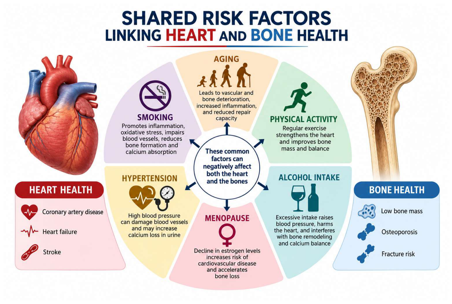
Trial analysis showed that the presence of osteoporosis was a risk factor for future acute cardiovascular events in postmenopausal women. Increased risk associated with osteoporosis was found to be independent of the traditional cardiovascular risk factors. Furthermore, the increased risk of cardiovascular disease was proportional to the severity of osteoporosis [27].
4.2. Biological Mechanisms
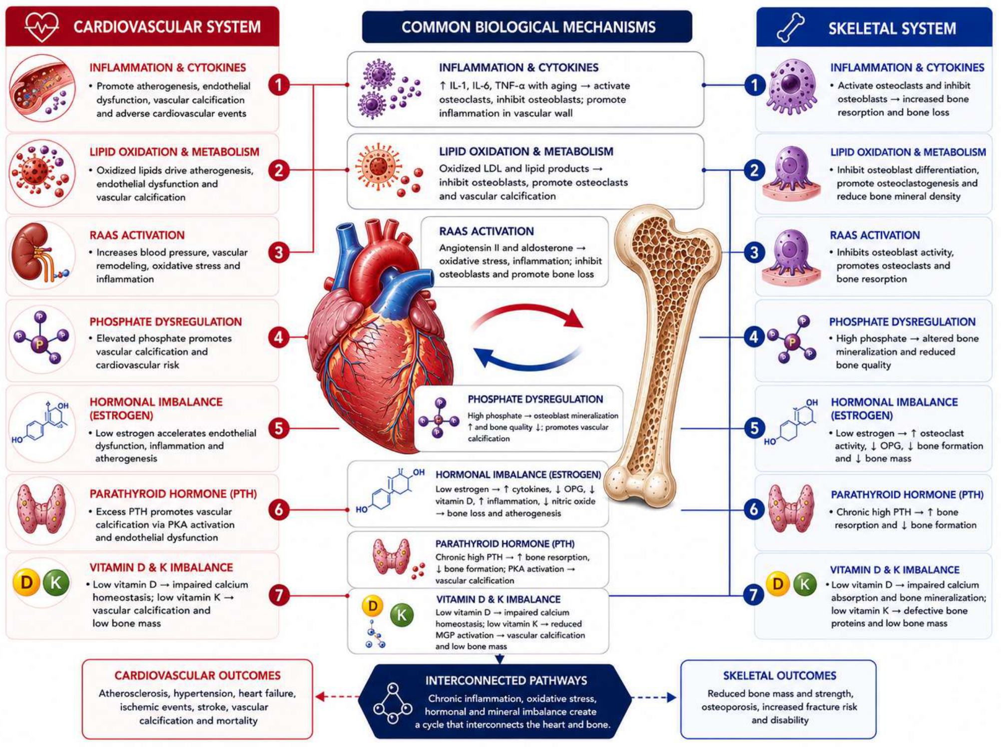
The common pathophysiological mechanisms include inflammatory cytokines, oxidized lipids, sex hormones, vitamin K deficiency, and vitamin D play a role in the progression of CVD and osteoporosis [4].
Cytokines and Inflammatory Markers
Inflammation plays a pivotal role in the stages of atherogenesis as well as in bone loss. Inflammatory diseases like lupus, rheumatoid arthritis, and Crohn’s disease are associated with an increased risk of osteoporosis and fractures. The pathogenesis of osteoporosis is influenced by systemic inflammatory processes, which are mediated by cytokines, including IL-6 and TNF-α. Increased levels of inflammatory cytokines such as IL-6 and TNF-α are associated with aging. IL-6 stimulates osteoclasts, therefore increasing the rate of bone remodeling and bone loss, it is also used as a marker of subclinical CVD in older adults [4]. TNF-α stimulates bone resorption and inhibits bone formation. Individuals with high inflammatory markers have been found to have increased risk of fracture [34] and cardiovascular events [4]
Lipid Oxidation and Metabolism
The 12/15 lipo-oxygenase system, which is involved in oxidation of lipids, is another biological link between CVD and osteoporosis [35]. Oxidized lipids play a role in atherogenesis and are a potential mechanism for the paradoxical occurrence of bone loss with vascular calcification. Lipid oxidation products have been found to have opposite effects on the differentiation of vascular calcifying cells (CVCs) and bone cells. Oxidized lipids stimulate osteoblast differentiation in CVCs by induction of alkaline phosphatase (ALP), which is a marker of osteoblastic differentiation, while reducing BMD [32,36]. They speed up atherogenesis and the activation of calcifying vascular cells while inhibiting differentiation of osteoblasts [27]. Expression of mediators of osteoclastic differentiation and suppression of terminal differentiation of stromal cells into osteoblasts are induced by oxidized low-density lipoprotein (LDL) cholesterol. Vascular calcification and inhibition of bone mineral formation are promoted by the accumulation of oxidized lipids in the arterial subendothelial area and skeletal bone arteries, respectively. Osteoporosis in postmenopausal women is associated with increased levels of LDL cholesterol and reduced HDL cholesterol [36].
Vitamin D & Calcium
Vitamin D is a crucial regulator of calcium homeostasis and bone mineralization [32]. Low Vit D levels decrease BMD and increase the risk of falls and fractures. Reduced Vit D also makes the musculoskeletal system more susceptible to arterial calcification, which increases cardiovascular risk factors. Furthermore, vitamin D deficiency stimulates the renin-angiotensin system, which leads to the development of hypertension, left ventricular hypertrophy, and coronary artery calcification, which is associated with congestive HF [37]. CHF patients have been found to have reduced circulating levels of vitamin D [32]. Calcium absorption is reduced in vitamin D deficiency, causing low serum calcium, hypersecretion of calcitriol, and the parathyroid hormone. This results in the mobilization of calcium and phosphorus from the bones to maintain optimal levels of calcium and phosphorus for bone turnover and metabolic processes [37]. On the other hand, calcium deposition leads to increased arterial stiffness and pulse wave velocity. Calcification reduces elasticity of vessels and negatively affects the hemodynamic parameters of the cardiovascular system, resulting in clinical and sub-clinical cardiovascular events [29].
Vitamin K
Vitamin K has a protective role in bone mass, as it is needed in vitamin K -dependent γ -carboxylation of bone proteins like matrix Gla protein (MGP) [37]. Low dietary and serum vitamin K is associated with low BMD and a higher risk of hip fractures. Deficiency results in incomplete γ -carboxylation and reduced function of MGP, which increases vascular calcification and CHD risk. High dietary vitamin K helps reduce vascular calcification progression, improve elasticity, and protect against CHD [36,37]. Low vitamin K intake (an important cofactor of uncarboxylated MGP activation) and increased levels of uncarboxylated MGP are highly associated with increased risk of cardiovascular calcification and mortality [36]. The relationship between osteoporosis and cardiovascular diseases is confounded by vitamin K [37].
Phosphate
Phosphate is an important component of bone structure as it plays an essential role in bone mineralization. Phosphate deficiency (hypophosphataemia) causes defective bone formation, while hyperphosphataemia stimulates mineralization in osteoblasts. Vascular calcification is stimulated by phosphate by increased calcium-phosphate. Hyperphosphataemia is a risk factor for CVD, while calcification is dependent on the concentrations of phosphate and pyrophosphate. Chronic kidney disease (CKD) patients have increased levels of serum phosphorus and parathyroid hormone (PTH), which are associated with an increased risk of cardiovascular mortality [36].
Hormones (Estrogen)
Estrogen regulates bone metabolism by inducing osteoclast apoptosis and expression of osteoprotegerin (OPG) by osteoblasts [38]. They play a role in the development of both CVD and osteoporosis through their effect on cytokines like IL-1, IL-6, TNF-alpha, and OPG. Low estrogen levels induce an increase in cytokines, a decrease in OPG, a reduction of serum vitamin D, increased inflammation, and reduced nitric oxide production, which are involved in the progression of bone loss and in atherogenesis [35]. The reduction in estrogen after menopause is the major cause of osteoporosis in women as they lose the protective effect of estrogen. This also contributes to the increase in postmenopausal cardiovascular risk [25].
Parathyroid Hormone (PTH)
The PTH has paradoxical effects on bone formation. Highly elevated levels inhibit osteoblast activity while increasing bone resorption. On the other hand, intermittent levels increase bone formation. PTH binds on its receptor on osteoblasts and involves intracellular signaling pathways and PTH-responsive transcription factors, such as cyclic AMP-responsive element-binding protein and RUNX2. PTH induces MGP expression in osteoblasts in bone formation, while in the vascular system, it activates protein Kinase A (PKA) which induces calcification. Osteoblastic differentiation and vascular cell mineralization are induced by the activation of PKA [36].
Renin-Angiotensin-Aldosterone System (RAAS)
The renin-angiotensin-aldosterone system plays a key role in hypertension as well as osteoporosis. RAAS is responsible for maintaining blood pressure and volume. Studies have shown the relationship between the alterations in the RAAS and the development of estrogen deficiency and osteoporosis. The interaction between angiotensin II (Ang II) (RAAS component) and the angiotensin II type 1 receptor (AT1R) inhibits osteoblast maturation [39]. Ang II elevates the Receptor Activator of Nuclear Factor Kappa-B Ligand (RANKL) and decreases osteoprotegerin, therefore promoting bone resorption through the activation of osteoclasts [40,41]. Upregulation of aldosterone (a RAAS hormone), which influences bone turnover by binding to mineralocorticoid receptors (MR) in osteoclasts, osteocytes and osteoblasts is promoted by the upregulation of Ang II. The upregulation of the Ang II, AngII/ AT1R interaction, OPG/RANKL ratio shift, and increase in aldosterone cause BMD decrease, which alters the microarchitecture of the bone, leading to osteoporosis development and progression. Activation of the Ang II/AT1R pathway contributes to cardiac remodeling and atherosclerosis [39]. Ang II also influences oxidative stress and Nuclear Factor kappa-light-chain-enhancer of activated B cells (NF-κB) activation, which decreases osteoblast differentiation and enhances osteoclastogenesis. This links RAAS activation to both vascular remodeling and bone loss. Hormonal pathways help highlight the complex relationship between skeletal and vascular health [42].
Vascular Calcification and Bone Mineralization
Vascular calcification is a major complication of CVD as well as a risk factor for myocardial infarction and cardiac death [38]. Bone mineralization and vascular calcification are highly organized processes that share common pathophysiological and anatomical features with arterial tissue calcification / absorption of calcium and phosphate, sharing similar mechanisms with bone mineralization [24,26]. Increased bone resorption and abnormal bone modeling cause excess release of calcium from the bone mass, resulting in hypercalcemia, with an increase in calcium ions being associated with increased risk of CVD [7].
Calcium deposits in coronary arteries affect the stability of atherosclerotic plaques and weaken vasomotor responses. Vascular calcification reduces arterial elasticity, leading to morbidity and mortality from hypertension, cardiac hypertrophy, myocardial infarction, and aortic stenosis among others [36].
Minerals in calcium deposits of atherosclerotic plaques have a similar chemical composition to hydroxyapatite crystals that form the bone matrix. Calcified plaques have been found to express bone matrix proteins such as type I collagen, gla (gamma carboxyglutamate)-containing proteins like matrix-gla protein and osteocalcin (bone-gla protein), bone morphogenetic protein (BMP)-2 and -4, osteonectin, osteopontin and bone sialoprotein [4]. Vascular calcification is regulated by BMP, osteopontin (OPN), ALP and MGP which are involved in osteogenesis Calcification is a result of bone formation which is initiated by osteoblast-like cells. The vascular smooth muscle cells (VSMC) differentiate into osteoblast-like cells, which form nodules and mineralize [26]. When VSMC are stimulated by BMP-2 and BMP-4, they begin to express osteoblast genes, including ALP, collagen I, and osteocalcin needed for bone formation. Plaques have also been seen to contain chondrocyte-like cells, osteoclast-like cells, and hematopoietic bone marrow cells [4]. BMP, RANKL, and oxidative stress promote the transformation of vascular smooth muscle cells into osteoblast-like cells [26].
OPG/RANK/RANKL Pathway
The OPG/RANK/RANKL pathway is a key regulator of bone resorption and also plays a role in vascular calcification [4]. RANKL, expressed by osteoblasts and stromal cells, is critical in bone metabolism as it regulates osteoclast formation by inducing the differentiation of monocyte–macrophage precursors into multinucleated osteoclasts and enhancing the activity of mature osteoclasts. It activates the protein kinase B through a process involving Src kinase and TNF receptor- associated factor 6 (TRAF6). Furthermore, binding to its receptor on osteoclasts activates Nuclear Factor kappa B (NF-κB) and Nuclear Factor of Activated T Cells 1 (NFATc1) to regulate the expression of specific genes required in osteoclast differentiation [43]. RANKL stimulates the production of reactive oxygen species (ROS), including oxygen-derived ions, free radicals, organic and inorganic peroxides, which are important in osteoclastogenesis [36].
Osteoprotegerin inhibits osteoclast differentiation and suppresses expression of cathepsin K by binding to RANKL and preventing its interaction with RANK. OPG also stimulates the expression of tissue inhibitor of metalloproteinases 1 (TIMP- 1), which suppress the bone-resorbing activity of osteoclasts [36]. Estrogens stimulate OPG, and a lack of them decreases OPG. OPG regulates both bone and vascular metabolism (West and O’Donnell 2018) and is associated with vascular calcification, arterial stiffness, severity of CHD, stroke, and atherosclerosis as well as cardiovascular morbidity and mortality [36]. Additionally, it is released from vascular smooth muscle cells in response to inflammatory stimuli, therefore having a protective role in decreasing vascular inflammation and atherosclerosis [8]. OPG regulates bone resorption through prevention of osteoclast (bone resorption cells) formation and survival via inhibition of RANKL [8]. In the CLARICOR trial, OPG levels were measured in patients with CAD, who were followed for up to 2.6 years for cardiovascular events and 6 years for all-cause mortality. OPG levels were significantly higher in non-survivors compared to survivors and were a strong predictor for all-cause mortality. It was found that OPG was an independent predictor of mortality after adjusting for conventional and clinical cardiovascular risk markers [44]. Serum OPG is a useful predictive marker to assess the risk of bone and CV disease in patients [8]. The study by Katano et al., [32] found that the serum level of RANKL was upregulated in mild HF patients [32]. The OPG/RANK/RANKL triad plays a role in both the bone and cardiovascular system, contributing to calcification of arteries and cardiovascular mortality. However, there is a need to understand their relationship with cardiovascular disease subtypes.
Figure 2.
Shared pathophysiological and biological mechanisms linking cardiovascular disease and osteoporosis. Chronic inflammation, oxidative stress, endocrine dysregulation, vascular calcification, and altered bone-remodeling pathways collectively contribute to cardiovascular dysfunction, bone loss, and an increased risk of osteoporosis and hip fracture.
Figure 2.
Shared pathophysiological and biological mechanisms linking cardiovascular disease and osteoporosis. Chronic inflammation, oxidative stress, endocrine dysregulation, vascular calcification, and altered bone-remodeling pathways collectively contribute to cardiovascular dysfunction, bone loss, and an increased risk of osteoporosis and hip fracture.
BMPs play an important role in osteoblast differentiation and increase collagen synthesis [26,36]. They inhibit osteoblast expression of collagenase 3, which reduces collagen degradation and maintenance of bone mass [36] and also play a mediatory role in vascular calcification. This is evidenced in atheromatous plaques, where the endothelial cells, foam cells, and smooth muscle cells show higher BMP-2 and BMP-4 expression [26]. Osteoblastic differentiation is induced by BMP -2 through induction of the transcription factor Msx2. BMPs have proinflammatory and pro-oxidant effects in arteries. Vascular endothelial cells and SMCs express BMP-2, which is regulated by proinflammatory stimuli like TNF. The stimulatory effects of glucocorticoids on osteoblastic cell differentiation are mediated by BMP-6 [36].
MGP is a mineral-binding protein that inhibits mineralization and indirectly interferes with the binding of BMP-2 to its receptor. OPN (a glycoprotein) accumulates in the extracellular matrix of the bone tissue and binds to hydroxyapatite and calcium. OPN is found in bone tissue as well as in atherosclerotic arteries, while elevated levels are associated with vascular calcification [26]. ALP is a marker of bone turnover and often found on the osteoblast surface and catalyzes the hydrolysis of esters. ALP levels are increased in smooth muscles by inflammation and oxidative stress, which in turn is associated with an increase in mineralization [26].
Wnt-β-Catenin Signaling Pathway
The Wnt-β-catenin pathway is essential for the maintenance of balance between bone formation and resorption. Reduction in Wnt activity prevents osteoblastogenesis while promoting bone resorption, leading to bone mass reduction and loss of microarchitecture [45,46,47]. Wnt signaling activates OPG, which is a decoy receptor to RANKL [45]. OPG inhibits osteoclast differentiation by binding to RANKL, thereby preventing its interaction with RANK and reducing osteoclast-mediated bone resorption [45,48]. Malfunctioning of the Wnt pathway is associated with cardiac disease, including myocardial infarction, HF, arrhythmias, and atherosclerosis. In atherosclerosis, it is involved in lipid deposition, cell proliferation, and cell migration [49].
Oxidative Stress
Studies have shown that oxidative stress is the common mechanism associated with vascular remodelling and endothelial dysfunction as well as affecting bone formation through stimulation of osteoclast activity while suppressing osteoblast differentiation. Oxidative stress promotes osteoclastogenesis and blocks osteoblast function in bone metabolism. Inflammatory cytokines and ROS activate the NF-κB transcription factor, which induces the expression of RANKL, which binds onto RANK on the osteoclast precursor, promoting osteoclast differentiation and activation. This results in enhanced bone resorption and osteoporosis pathogenesis [42]. ROS influences endothelial dysfunction, vascular remodeling, and inflammation in hypertension. Nitric oxide bioavailability is reduced and smooth muscle cell proliferation is promoted by ROS, therefore contributing to hypertension development [50]. Similar redox-sensitive pathways like NF-κB are upregulated in osteoporosis and increased inflammation and vascular injury. Oxidative stress is a shared mechanism between hypertension and osteoporosis, and this can have an effect on therapies used to target redox-pathways [42,51].

Table 1.
Shared pathophysiological factors between CVDs & Osteoporosis.
| Shared biological mechanism | Role in cardiovascular disease | Role in bone loss/osteoporosis |
|---|---|---|
| Inflammation and inflammatory cytokines | Chronic inflammation promotes endothelial dysfunction, atherosclerosis, and vascular remodeling. Elevated IL-1, IL-6, and TNF-α are associated with cardiovascular events and disease progression. | IL-1, IL-6, and TNF-α stimulate osteoclast differentiation and activity while suppressing osteoblast function, increasing bone resorption, and reducing bone formation. |
| Lipid oxidation and lipid metabolism | Oxidized LDL promotes endothelial dysfunction, atherosclerotic plaque formation, and vascular calcification. | Oxidized lipids inhibit osteoblast differentiation, enhance osteoclastogenesis, and are associated with reduced bone mineral density. |
| Vitamin D and Vitamin K | Vitamin D deficiency is associated with HF, while vitamin K deficiency contributes to vascular calcification through MGP activation interruption. | Vitamin D regulates calcium homeostasis and bone mineralization. Vitamin K is required for γ-carboxylation of bone proteins. |
| Phosphate homeostasis | Hyperphosphatemia promotes vascular calcification and increases cardiovascular morbidity and mortality. | Both phosphate deficiency and excess impair normal bone mineralization and alter bone quality. |
| Estrogen deficiency | Reduced estrogen increases inflammation, oxidative stress, and endothelial dysfunction, contributing to atherosclerosis. | Estrogen deficiency decreases OPG expression, increases osteoclast activity, and accelerates bone loss. |
| Parathyroid hormone (PTH) | Sustained elevation of PTH promotes vascular calcification. | Continuous PTH exposure stimulates bone resorption, whereas intermittent exposure promotes bone formation. |
| Renin–Angiotensin–Aldosterone System (RAAS) | RAAS activation contributes to hypertension, vascular remodeling, oxidative stress, and atherosclerosis. | Angiotensin II suppresses osteoblast maturation, increases RANKL expression and enhances osteoclast activity. |
| Vascular calcification and bone mineralization | Vascular smooth muscle cells undergo osteogenic differentiation, resulting in arterial calcification. | Increased bone resorption releases calcium and phosphate, contributing to deterioration of bone microarchitecture. |
| OPG/RANK/RANKL signaling pathway | Regulates vascular calcification, arterial stiffness, and cardiovascular mortality. | RANKL promotes osteoclastogenesis while OPG inhibits osteoclast differentiation. |
| Bone morphogenetic proteins (BMPs) | BMP-2 and BMP-4 promote osteogenic transformation of vascular smooth muscle cells. | BMPs regulate osteoblast differentiation, collagen synthesis, and bone formation. |
| Oxidative stress | Reactive oxygen species promote endothelial dysfunction, hypertension, and vascular remodeling. | Reactive oxygen species enhance osteoclastogenesis and suppress osteoblast differentiation. |
4.3. Genetic Association Between CVDs and Osteoporosis
Genetic factors that may explain the association between CVDs and hip fracture include telomere length as well as genes involved in cellular mechanisms shared by the vascular and bone systems [33]. Genetic factors play a role in the development of both CVD and osteoporosis, and polymorphisms in genes regulating bone, lipid metabolism, and inflammatory responses and pathways are common risk factors [52].
Genome-wide association studies (GWAS) have provided strong evidence of the genetic component of osteoporosis through the identification of genes involved in the RANK/RANKL/OPG axis and Wnt/Wnt-β-catenin signaling pathways [24,53]. The lipid plaques in atherosclerosis undergo calcification [25], while OPG is the possible molecular link between bone resorption and vascular calcification [54].
Research involving twins and families has shown that genetic inheritance explains nearly 50–85% of the observed variation in BMD [53]. A study of identical twin siblings showed that pseudoexposure of HF was associated with a 4-fold increased hip fracture rate as compared with those without CVD while, hip fracture rate doubled in identical twins pseudoexposed to stroke after a stroke event in the co-twin. Co-twin analysis showed that genes influence the development of CVD and fractures [33]
4.4. Cardiovascular Disease Subtypes and Osteoporosis
Heart Failure (HF)
HF and osteoporosis commonly occur together in patients over 50 years of age. Osteoporosis and HF share numerous pathological mechanisms and have a bi-directional relationship whereby each condition may influence the development of the other [24]. HF patients are prone to develop osteoporosis and fractures [55] because of low vitamin D levels, alteration of RAAS, high PTH, low testosterone levels, and metabolic effects of medications [24,56]. On the other hand, patients with osteoporosis are prone to develop HF because of vitamin D deficiency, elevated plasma PTH levels, and increased Fibroblast Growth Factor 23 (FGF-23) activity [24]. A study by Katano et al., showed that patients with HF have lower BMD compared to patients without CHF. It was found that changes in BMD are dependent on CHF severity, showing that CHF patients have bone fragility and are susceptible to fracture. Reduced BMD combined with increased incidence of fall events was found to likely be responsible for the high prevalence of fractures in CHF patients [32]. BMD predicts the incidence of HF, showing a bidirectional relationship between CHF and osteoporosis [32]. There is an increased risk of stroke and coronary events among women with osteoporosis [27,33].
Ischemic Heart Disease/Coronary Artery Disease and atherosclerosis
Coronary artery disease is characterized by atherosclerosis, which is the build-up of plaques within the arterial walls [57]. Atherosclerosis is due to a deposition of plaque, which is a combination of cholesterol, calcium, macrophage cells, and fibrous connective tissue inside coronary arteries [5]. It is associated with adverse effects on the cardiovascular system [3]. Abnormalities in the conventional bone-cell stimulation pathways are involved in the atheroma plaque formation in CAD. Osteoblast-like calcifying vascular cells found in atheroma plaques are phenotypically similar to osteoblasts in bone tissue. Bone tissue proteins like BMP-2, osteopontin, osteoprotegerin, and Gla protein have been found in atheroma plaques [25]. The osteoblast-like calcifying vascular cells are regulated by the same factors as osteoblasts. For example, elevation of osteoprotegerin is associated with CAD [25] and low volumetric BMD is associated with increased morbidity and mortality due to ischemic heart disease [24]. It inhibits the RANK-RANKL pathway, which is responsible for osteoclast stimulation; therefore, it acts as an autocrine and paracrine regulator of vascular calcification, which is the main marker of vascular disease [25].
Stroke
Stroke is a risk factor for osteoporosis and fracture [46]. There is a complex interplay between stroke and osteoporosis as they share risk factors, pathophysiological pathways, and genetic factors [58] . Stroke survivors are at high fall risk, which results in increased fracture risk depending on stroke type [59] which includes ischemic stroke, small vessel stroke, large artery atherosclerosis stroke and cardioembolic stroke [60]. This is attributed to decreased BMD [59]. The reduction in BMD observed in paralysed limbs compared to the non-paralysed limb following stroke may highlight the combined effects of reduced mobility and increased inflammation. Reduced mobility and muscle weakness enhance bone resorption and interfere with bone formation, resulting in rapid bone loss. The production of cytokines like interleukin-1 (IL-1) and tumor necrosis factor alpha (TNF-a) involved in stimulating osteoclastogenesis and suppression of osteoblast function is triggered by post-stroke inflammation [58].
Atrial Fibrillation (AF)
Decreased BMD is associated with the development of atherosclerosis in older adults. It is also associated with pathogenesis of AF [61]. AF is highly associated with HF and stroke, which in turn are highly associated with osteoporotic fractures [62]. Coronary artery calcification is a significant risk factor for AF which is linked to bone mass loss through the RANK/RANKL/OPG and Wnt-β-catenin signaling pathways. The use of warfarin for stroke prevention in AF may be a link between AF and osteoporotic fracture [13]. Wong et al. [9] found that patients with a history of AF had a 2-fold increased hazard of hip fracture compared to those without AF. Furthermore, they found that there was a higher absolute risk of hip fracture among women with AF, while the relative risk attributable to AF was similar in both men and women [9]. A study by Kim et al., 2018 [15] showed that the cumulative incidence of fractures was significantly higher among individuals with AF compared to those without (log-rank P<0.001) [15]
Peripheral Arterial Disease (PAD)
Studies have suggested an increased risk of hip fracture among individuals with PAD but the results have been inconsistent [18]. Mühlen et al.,[63] found that the prevalence of osteoporosis was higher among women with PAD compared to those without (P<0.05). After a 4-year follow-up period, PAD in women was associated with a higher rate of bone loss than women without PAD (p=0.05). However, this association was weakened after adjusting for age. PAD was also not associated with osteoporosis, among men [63]. A meta-analysis by Ungprasert et al. [18] showed a significant association between incident hip fracture and PAD (pooled RR 1.64 (95% CI, 1.17- 2.29)) amongst patients with PAD compared to those without PAD. Furthermore, subgroup analysis by study design showed a significant association between incident hip fracture and PAD for both prospective studies (pooled RR, 1.60; 95% CI, 1.12-2.28; p-value, 0.49) and retrospective studies (pooled RR, 1.72; 95% CI, 1.07-2.77; p-value <0.0001) [18]. Results from a study by Syu et al., [64] showed that individuals with osteoporosis were significantly at higher risk of PAD compared to those without osteoporosis (adjusted HR 1.18, 95% CI, 1.08-1.29). The results also showed a positive association between osteoporosis and the development of PAD. Patients with osteoporosis had an increased incidence of PAD over time [64].
Cardiovascular disease subtypes HF, stroke, ischemic heart disease, and peripheral atherosclerosis were associated with increased hip fracture. The multivariable-adjusted hazard ratio (HR) of hip fracture after a diagnosis of HF was 4.40 (95% confidence interval [CI], 3.43-5.63); the HR after a stroke was 5.09 (95% CI, 4.18-6.20); after an ischemic heart disease event, the HR was 2.32 (95% CI, 1.91-2.84) and HR after a diagnosis of peripheral atherosclerosis was 3.20 (95% CI, 2.28-4.50). This showed a diagnosis of CVD was highly associated with hip fracture [33].
5. Therapeutic Interactions and Clinical Implications
Pharmacologic treatments play an important role in the management of CVDs and osteoporosis, with various drugs influencing both the vascular and skeletal pathway. Some CVD medications influence bone metabolism through altering calcium processing, oxidative stress and inhibiting pathways, whereas osteoporosis medication can also affect cardiovascular physiology by affecting calcium balance, endothelial function, and inflammatory pathways [42].
Statins, which are lipid-lowering drugs, have an effect on bone health. They inhibit HMG-CoA reductase, which is the enzyme that catalyzes cholesterol biosynthesis [65] and the reduction of 3-hydroxy-3-methylglutaryl coenzyme A (HMG-CoA) to mevalonate. Statins reduce LDL-C and triglycerides to increase HDL-C, reducing the incidence of cardiovascular events. However, the use of statins is associated with higher BMD and reduced fracture risk. Statins stimulate bone formation and enhance osteoblast differentiation by increasing BMP-2 expression and production [4] and also have bone-protective effects [65]. The use of statins with a higher potency is associated with a lower risk of osteoporosis compared to those with a lower potency [66].
A survey in China found that bisphosphonate, which is a first-line drug for treatment of osteoporosis, significantly reduced the risk of all-cause mortality in patients with ischemic stroke or acute coronary syndrome [67].
Loop diuretics are associated with an increase in urinary calcium elimination, resulting in a decrease in serum calcium and an increase in PTH [65]. After adjusting for other risk factors, such as other medication use, gender, age, and weight, loop diuretics were associated with an increase in the risk of osteoporosis (OR 2.52, 95% CI 1.20–5.27) in a observational study of older patients who were admitted for HF evaluation [32].
Beta-blockers are used to treat HF by reducing stimulation of the sympathetic nervous system (SNS). SNS stimulation increase osteoclast activity through increased RANKL expression, whereas beta-blockade may reduce this effect and thereby benefit bone health [68]. In the Framingham study, beta-blockers were found to increase bone density at the spine and femur depending on dosage. Beta-blocker therapy in postmenopausal women was found to decrease the bone resorption marker (C-telopeptide) and improve bone microarchitecture [65,68]. The results from the Framingham study showed that treatment with beta-blockers was associated with higher bone density and reduced risk of fractures, which may have been attributed to RANKL production and a decrease in adrenergic signaling in osteoblasts [65].
Glucocorticoids are a risk factor for osteoporosis and atherosclerosis. Dexamethasone downregulates calcification inhibitors like OPN and MGP, therefore inducing calcification. Estrogens are involved in the vascular calcification process, and studies have shown that the BMP osteogenic pathway is blocked by the increase in MGP mRNA expression due to estrogen replacement therapy. Estrogens have a direct effect on vascular endothelial and bone cells, as estrogen receptors are expressed on osteoblasts, osteoclasts, SMCs and vascular endothelial cells [36]. Studies have shown how estrogen replacement in postmenopausal women decreases the risk of fractures by increasing bone mineral density. Estrogen deficiency is associated with increased bone resorption as well as vascular dysfunction, which leads to hypertension [42].
Use of anticoagulants and vitamin K antagonists (VKA) has been associated with an increase in osteoporotic fractures in older populations [69]. Warfarin (anticoagulant) affects the availability of vitamin K, which in turn affects MGP function by stopping the formation of Gla residues in MGP, resulting in rapid calcification in arteries. It is associated with calcification of coronary arteries and cardiac valves [36]. Inhibition of vitamin K-dependent carboxylation of proteins associated with bone metabolism compromises bone quality [69].
Table 2.
Effects of cardiovascular medication on osteoporosis.
| Medication class | Common examples | Primary mechanism of action | Impact on bone health | Mechanisms related to bone health |
|---|---|---|---|---|
| Thiazide and thiazide-like diuretics | Hydrochlorothiazide; chlorthalidone; indapamide | Inhibit the sodium–chloride cotransporter in the distal convoluted tubule, lowering blood pressure and reducing urinary calcium loss. | Increased bone mineral density and a lower risk of hip or other osteoporotic fractures. | Reduced urinary calcium excretion may improve calcium balance. Thiazides may promote osteoblast activity and bone formation. |
| Loop diuretics | Furosemide; bumetanide; torsemide | Block the sodium–potassium–chloride cotransporter in the thick ascending limb, producing strong natriuresis and diuresis. | Long-term exposure linked to increased bone loss and fracture risk | Increased urinary calcium excretion can stimulate secondary hyperparathyroidism and accelerate bone turnover. Dizziness and electrolyte disturbances may increase falls. |
| Beta-adrenergic blockers | Metoprolol; atenolol; carvedilol; propranolol | Block beta-adrenergic receptors, reducing heart rate, myocardial contractility and sympathetic activity. | Increases bone density, fewer fractures, | Reduced sympathetic signaling may decrease osteoclast activation and bone resorption. |
| Statins | Atorvastatin; rosuvastatin; simvastatin; pravastatin | Inhibit HMG-CoA reductase, reducing hepatic cholesterol synthesis and increasing LDL-receptor activity. | higher bone density and fewer fractures | Inhibition of the mevalonate pathway may reduce osteoclast prenylation and resorption. Statins promote bone morphogenetic protein-2 expression and osteoblast differentiation. |
| Angiotensin-converting enzyme inhibitors | Lisinopril; enalapril; ramipril | Reduce conversion of angiotensin I to angiotensin II and increase bradykinin, lowering vascular resistance and aldosterone activity. | Reduces osteoporosis, fracture risk and bone-density loss. | Lower angiotensin II activity reduces osteoclastogenesis, inflammation and oxidative stress |
| Angiotensin II receptor blockers | Losartan; valsartan; candesartan | Block angiotensin II type-1 receptors, reducing vasoconstriction, aldosterone release and vascular remodeling. | Reduces fracture risk | Reduced AT1-receptor signaling limits inflammatory cytokines, osteoclast differentiation and bone resorption. |
| Vitamin K antagonists | Warfarin | Inhibit vitamin K epoxide reductase, reducing activation of vitamin K-dependent coagulation proteins. | Decrease bone quality and increase fracture risk, | Vitamin K is required for gamma-carboxylation of osteocalcin and matrix Gla protein. Inhibition may impair mineral binding in bone and alter regulation of vascular calcification. |
| Unfractionated and low-molecular-weight heparins | Unfractionated heparin; enoxaparin; dalteparin | Potentiate antithrombin and inhibit thrombin and/or factor Xa. | prolonged treatment causes bone loss | Heparin may suppress osteoblast function, enhance osteoclast-mediated resorption and alter osteoprotegerin signaling. Risk increases with longer treatment duration . |
| Direct oral anticoagulants | Apixaban; rivaroxaban; edoxaban; dabigatran | Directly inhibit factor Xa or thrombin without blocking vitamin K recycling. | Reduce fracture risk | They do not interfere with vitamin K-dependent osteocalcin carboxylation. Reduced intracranial bleeding and simpler dosing may also indirectly reduce morbidity, although falls and bleeding remain relevant. |
Interpretation note: Effects may differ by dose, treatment duration, patient age, kidney function, comorbidities and baseline fracture or cardiovascular risk. This table is an evidence summary and is not a prescribing guide.
New treatment approaches with distinct mechanisms have been identified through the discovery of key pathways which regulate bone resorption and formation [19]. Treatments for osteoporosis have been found to have an effect on cardiovascular health. For example, bisphosphonates used as first-line therapeutic agents for treating osteoporosis have been found to increase AF risk [7]. Manipulating the OPG/RANKL pathway is a treatment approach for bone health and vascular calcification [8]. Trial results suggested that clinical management of postmenopausal women with osteoporosis should not focus on fracture prevention but also cardiovascular disease prevention [27].
6. Knowledge Gaps and Future Directions
The current evidence shows a complex relationship between CVD and osteoporosis, as they share several pathophysiological mechanisms. This relationship is influenced by factors including aging, physical inactivity, medication use, hormonal changes, renal dysfunction, and socioeconomic conditions. However, substantial knowledge gaps remain regarding CVD subtypes and osteoporosis, as current research focuses on CVD as a single broad category. Studies need to be conducted to understand how cardiovascular disease subtypes influence changes in BMD resulting in osteoporosis over time. Future studies should not group CVD into a single broad category but should be separated into CAD, MI, AF, HF, stroke, and PAD. This will help in understanding how these diseases may affect skeletal health through different pathways and mechanisms. For example, HF may cause BMD reduction through chronic inflammation, reduced mobility, and the use of loop diuretics, while stroke may increase the risk of hip fracture due to muscle weakness, reduced balance, and falls. Therefore, subtype analysis will identify the CVD that will need closer monitoring for osteoporosis and fracture risk.
Attention should be given to the biological and pathophysiological mechanisms within the heart-bone axis. Studies should investigate how chronic inflammation, oxidative stress, vascular calcification, endocrine alterations, and endothelial dysfunction among others jointly influence bone and cardiovascular health. Pathways like the RANK/RANKL/OPG and Wnt/β-catenin pathways, which play pivotal roles in bone formation, osteoclast activity, vascular smooth muscle cell transformation, and vascular calcification should be extensively studied. Combining biomarkers, imaging, and clinical outcomes may help understand the association of cardiovascular disease subtypes and osteoporosis.
Genetics-based research like GWAS can be used to identify loci associated with cardiovascular disease subtypes, osteoporosis, and BMD. Colocalization analysis can be used to establish whether cardiovascular and skeletal traits share genetic signals in the same genomic region. Mendelian randomisation may also help evaluate whether genetically proxied CVD traits influence osteoporosis-related outcomes or vice versa.
CVD and osteoporosis are frequently co-occurring conditions in older adults, and this adds additional challenges to their clinical management. Questions regarding the best therapeutic approach still remain: should managing cardiovascular disease symptoms be more critical or treatment of osteoporosis be prioritized in CVD patients? Cardiovascular medications like anticoagulants, loop diuretics and statins may affect bone metabolism and fracture risk. On the other hand, osteoporosis treatments like bisphosphonates and hormone therapy may have positive or negative cardiovascular effects. Current literature is inconsistent and dependent on treatment duration, disease severity at baseline and medication adherence. Treatment effects can be studied using trial emulations when observational datasets are not available or when randomized controlled trials are not feasible. Further research on the influence of medications needs to be carried out to fully understand these interactions and to develop patient-specific and comprehensive management strategies.
Large-scale databases like the UK Biobank and All of Us Research Program can be used to answer the questions regarding the association between cardiovascular disease subtypes and osteoporosis. These databases can be used to assess the relationship across different populations enabling cross-cohort comparisons which can highlight differences influenced by variations in ancestry, sex, healthcare services, etc. Stratified analysis can also highlight differences in the heart-bone relationship across groups based on age, sex, comorbidities, race and menopausal status among others. Artificial intelligence and machine learning techniques could be used to investigate the relationship between CVD subtypes and osteoporosis through the integration of clinical, laboratory, genomic, and imaging data. Multimodal prediction models can be used to further study this relationship to help identify individuals with different CVD subtypes.
Future research encompassing a combination of causal inference methods, longitudinal cohorts, imaging, genetic analysis, and biomarkers’ predictive models will help improve the understanding of the heart-bone axis, in this case cardiovascular disease subtypes and osteoporosis. This will help highlight intricate details regarding mechanisms, direction, and clinical implications of the relationship. The findings can help in the development of integrated screening and treatment of osteoporosis among patients based on the cardiovascular disease subtype they have while patients with osteoporosis also receive cardiovascular disease evaluation. This will help improve early detection, prevention, targeted treatment, and coordinated management of cardiovascular and skeletal diseases.
7. Conclusion
Current evidence supports the association between CVD and osteoporosis with the heart-bone axis being the main link between both conditions. Shared mechanisms like inflammation, oxidative stress, and vascular calcification contribute to both cardiovascular and skeletal health. The effects, however, vary across cardiovascular disease subtypes. Large cohorts, genomics, and advanced analysis methods are being used to understand the relationship. Further subtype-specific studies are needed to fully understand the causal pathways and develop better integrated strategies for prevention, early detection, and management of CVD and osteoporosis.
Funding
This research was in part supported by grants from the National Institutes of Health, USA (1R15HL172198, 1R15HL173852, and U19AG055373) and Blue Cross Blue Shield of Michigan Foundation (2025110031.SAP).
Conflicts of Interest
Dr. Weihua Zhou reports consulting fees from Pressiant Health Inc. and SynterMed Inc., outside the submitted work. All other authors declare no competing interests.
References
- Zhang, J.; Xu, P.; Liu, R.; Gyu, J. M.; Cao, P.; Kang, C. Osteoporosis and coronary heart disease: a bi-directional Mendelian randomization study. Front. Endocrinol. 2024, vol. 15, 1362428. [Google Scholar] [CrossRef]
- Chong, B. Global burden of cardiovascular diseases: projections from 2025 to 2050. Eur. J. Prev. Cardiol. 2025, vol. 32(no. 11), 1001–1015. [Google Scholar] [CrossRef]
- Frąk, W.; Wojtasińska, A.; Lisińska, W.; Młynarska, E.; Franczyk, B.; Rysz, J. Pathophysiology of Cardiovascular Diseases: New Insights into Molecular Mechanisms of Atherosclerosis, Arterial Hypertension, and Coronary Artery Disease. Biomedicines 2022, vol. 10(no. 8), 1938. [Google Scholar] [CrossRef]
- Farhat, G. N.; Cauley, J. A. The link between osteoporosis and cardiovascular disease. Clin. Cases Miner. Bone Metab. 2008, vol. 5(no. 1). [Google Scholar]
- Karunathilake, S. P.; Ganegoda, G. U. Secondary Prevention of Cardiovascular Diseases and Application of Technology for Early Diagnosis. BioMed Res. Int. 2018, vol. 2018, 1–9. [Google Scholar] [CrossRef]
- Xing, W. Bone mineral density in patients with chronic heart failure: a meta-analysis. Clin. Interv. Aging 2018, vol. Volume 13, 343–353. [Google Scholar] [CrossRef]
- Bhatta, L. Bone mineral density and risk of cardiovascular disease in men and women: the HUNT study. Eur. J. Epidemiol. 2021, vol. 36(no. 11), 1169–1177. [Google Scholar] [CrossRef]
- West, S. L.; O’Donnell, E. Cardiovascular disease and bone loss—new research in identifying common disease pathophysiologies and predictors. AME Med. J. 2018, vol. 3, 42–42. [Google Scholar] [CrossRef]
- Wong, C. X. Atrial fibrillation and risk of hip fracture: A population-based analysis of 113,600 individuals. Int. J. Cardiol. 2017, vol. 243, 229–232. [Google Scholar] [CrossRef]
- Sacco, R. L. An Updated Definition of Stroke for the 21st Century: A Statement for Healthcare Professionals From the American Heart Association/American Stroke Association. Stroke 2013, vol. 44(no. 7), 2064–2089. [Google Scholar] [CrossRef]
- Coupland, P.; Thapar, A.; Qureshi, M. I.; Jenkins, H.; Davies, A. H. The definition of stroke. J. R. Soc. Med. 2017, vol. 110(no. 1), 9–12. [Google Scholar] [CrossRef]
- Murphy, S. Jx.; Werring, D. J. Stroke: causes and clinical features. Medicine 2020, vol. 48(no. 9), 561–566. [Google Scholar] [CrossRef]
- Yu, Q. Bone mineral loss and risk of atrial fibrillation: a multicohort study. Eur. J. Prev. Cardiol. 2025, vol. 2025(no. 00), 1–10. [Google Scholar] [CrossRef]
- Joglar, J. A. 2023 ACC/AHA/ACCP/HRS Guideline for the Diagnosis and Management of Atrial Fibrillation: A Report of the American College of Cardiology/American Heart Association Joint Committee on Clinical Practice Guidelines. Circulation 2024, vol. 149(no. 1). [Google Scholar] [CrossRef]
- Kim, D. Effect of Atrial Fibrillation on the Incidence and Outcome of Osteoporotic Fracture ― A Nationwide Population-Based Study ―. Circ. J. 2018, vol. 82(no. 8), 1999–2006. [Google Scholar] [CrossRef]
- Shamaki, G. R.; Markson, F.; Soji-Ayoade, D.; Agwuegbo, C. C.; Bamgbose, M. O.; Tamunoinemi, B.-M. Peripheral Artery Disease: A Comprehensive Updated Review. Curr. Probl. Cardiol. 2022, vol. 47(no. 11), 101082. [Google Scholar] [CrossRef]
- Shu, J.; Santulli, G. Update on peripheral artery disease: Epidemiology and evidence-based facts. Atherosclerosis 2018, vol. 275, 379–381. [Google Scholar] [CrossRef]
- Ungprasert, P.; Wijarnpreecha, K.; Thongprayoon, C.; Cheungpasitporn, W. Peripheral arterial disease and risk of hip fracture: A systematic review and meta-analysis of cohort studies. J. Postgrad. Med. 2018, vol. 64(no. 4), 220–225. [Google Scholar] [CrossRef]
- Compston, J. E.; McClung, M. R.; Leslie, W. D. Osteoporosis. The Lancet 2019, vol. 393(no. 10169), 364–376. [Google Scholar] [CrossRef]
- Gregson, C. L. UK clinical guideline for the prevention and treatment of osteoporosis. Arch. Osteoporos. 2022, vol. 17(no. 1), 58. [Google Scholar] [CrossRef]
- LeBoff, M. S. The clinician’s guide to prevention and treatment of osteoporosis. Osteoporos. Int. 2022, vol. 33(no. 10), 2049–2102. [Google Scholar] [CrossRef]
- Yang, Y.; Huang, Y. Association between bone mineral density and cardiovascular disease in older adults. Front. Public Health 2023, vol. 11, 1103403. [Google Scholar] [CrossRef]
- Marcovitz, P. A. Usefulness of Bone Mineral Density to Predict Significant Coronary Artery Disease. Am. J. Cardiol. 2005, vol. 96(no. 8), 1059–1063. [Google Scholar] [CrossRef]
- Spoladore, R. Heart Failure and Osteoporosis: Shared Challenges in the Aging Population. J. Cardiovasc. Dev. Dis. 2025, vol. 12(no. 2), 69. [Google Scholar] [CrossRef]
- Laroche, M. Osteoporosis and ischemic cardiovascular disease. Jt. Bone Spine 2017, vol. 84(no. 4), 427–432. [Google Scholar] [CrossRef]
- Sprini, D. Correlation between osteoporosis and cardiovascular disease. Clin. Cases Miner. Bone Metab. 2014. [Google Scholar] [CrossRef]
- Tankó, L. B.; Christiansen, C.; Cox, D. A.; Geiger, M. J.; McNabb, M. A.; Cummings, S. R. Relationship Between Osteoporosis and Cardiovascular Disease in Postmenopausal Women. J. Bone Miner. Res. 2005, vol. 20(no. 11), 1912–1920. [Google Scholar] [CrossRef]
- Gupta, G.; Aronow, W. S. Atherosclerotic vascular disease may be associated with osteoporosis or osteopenia in postmenopausal women: A preliminary study. Arch. Gerontol. Geriatr. 2006, vol. 43(no. 2), 285–288. [Google Scholar] [CrossRef]
- Veronese, N. Relationship Between Low Bone Mineral Density and Fractures With Incident Cardiovascular Disease: A Systematic Review and Meta-Analysis. J. Bone Miner. Res. 2017, vol. 32(no. 5), 1126–1135. [Google Scholar] [CrossRef]
- Fiechter, M. Association between vertebral bone mineral density, myocardial perfusion, and long-term cardiovascular outcomes: A sex-specific analysis. J. Nucl. Cardiol. 2020, vol. 27(no. 3), 726–736. [Google Scholar] [CrossRef]
- Xu, B. Cardiovascular Disease and Hip Fracture among Older Inpatients in Beijing, China. BioMed Res. Int. 2013, vol. 2013, 1–6. [Google Scholar] [CrossRef]
- Katano, S. Clinical Risk Factors and Prognostic Impact of Osteoporosis in Patients With Chronic Heart Failure. Circ. J. 2020, vol. 84(no. 12), 2224–2234. [Google Scholar] [CrossRef]
- Sennerby, U. Cardiovascular Diseases and Risk of Hip Fracture. JAMA 2009, vol. 302(no. 15), 1666. [Google Scholar] [CrossRef]
- Cauley, J. A. Inflammatory Markers and Incident Fracture Risk in Older Men and Women: The Health Aging and Body Composition Study. J. Bone Miner. Res. 2007, vol. 22(no. 7), 1088–1095. [Google Scholar] [CrossRef]
- Sennerby, U.; Farahmand, B.; Ahlbom, A.; Ljunghall, S.; Michaëlsson, K. Cardiovascular diseases and future risk of hip fracture in women. Osteoporos. Int. 2007, vol. 18(no. 10), 1355–1362. [Google Scholar] [CrossRef]
- Lampropoulos, C. E.; Papaioannou, I.; D’Cruz, D. P. Osteoporosis—a risk factor for cardiovascular disease? Nat. Rev. Rheumatol. 2012, vol. 8(no. 10), 587–598. [Google Scholar] [CrossRef]
- Stojanovic, I.; Lazovic, M.; Lazovic, M.; Vuceljic, M. Association between atherosclerosis and osteoporosis, the role of vitamin D. Arch. Med. Sci. 2011, vol. 2, 179–188. [Google Scholar] [CrossRef]
- Osako, M. K. Estrogen Inhibits Vascular Calcification via Vascular RANKL System: Common Mechanism of Osteoporosis and Vascular Calcification. Circ. Res. 2010, vol. 107(no. 4), 466–475. [Google Scholar] [CrossRef]
- Mkhize, B. C.; Mosili, P.; Ngubane, P. S.; Sibiya, N. H.; Khathi, A. The Relationship between Renin–Angiotensin–Aldosterone System (RAAS) Activity, Osteoporosis and Estrogen Deficiency in Type 2 Diabetes. Int. J. Mol. Sci. 2023, vol. 24(no. 15), 11963. [Google Scholar] [CrossRef]
- Theoleyre, S.; Wittrant, Y.; Tat, S. K.; Fortun, Y.; Redini, F.; Heymann, D. The molecular triad OPG/RANK/RANKL: involvement in the orchestration of pathophysiological bone remodeling. Cytokine Growth Factor Rev. 2004, vol. 15(no. 6), 457–475. [Google Scholar] [CrossRef]
- Carbone, L. D. The renin-angiotensin aldosterone system and osteoporosis: findings from the Women’s Health Initiative. Osteoporos. Int. 2019, vol. 30(no. 10), 2039–2056. [Google Scholar] [CrossRef]
- Gutierrez, Y.; Shaju, R. A.; Sepulveda, A.; Coban, S. Exploring the Interplay Between Hypertension and Osteoporosis: A Narrative Review. Cureus 2025. [Google Scholar] [CrossRef]
- Nakashima, T.; Hayashi, M.; Takayanagi, H. New insights into osteoclastogenic signaling mechanisms. Trends Endocrinol. Metab. 2012, vol. 23(no. 11), 582–590. [Google Scholar] [CrossRef]
- Bjerre, M. Osteoprotegerin independently predicts mortality in patients with stable coronary artery disease: The CLARICOR trial. Scand. J. Clin. Lab. Invest. 2014, vol. 74(no. 8), 657–664. [Google Scholar] [CrossRef]
- Cao, L.; Wang, J. Roles of Wnt/β-Catenin Signaling in Osteoporosis, Disease Pathogenesis, and Natural Compound Intervention. Biomol. Ther. 2026, vol. 34(no. 1), 65–79. [Google Scholar] [CrossRef]
- Liu, B. Osteoporosis and fracture risk assessment in adults with ischaemic stroke. Osteoporos. Int. 2024, vol. 35(no. 7), 1243–1247. [Google Scholar] [CrossRef]
- Manolagas, S. C. Wnt signaling and osteoporosis. Maturitas 2014, vol. 78(no. 3), 233–237. [Google Scholar] [CrossRef]
- Gay; Towler, D. A. Wnt signaling in cardiovascular disease: opportunities and challenges. Curr. Opin. Lipidol. 2017, vol. 28(no. 5), 387–396. [Google Scholar] [CrossRef]
- Liu, Y.; Neogi, A.; Mani, A. The role of Wnt signalling in development of coronary artery disease and its risk factors. Open Biol. 2020, vol. 10(no. 10), 200128. [Google Scholar] [CrossRef]
- Rodrigo, R.; González, J.; Paoletto, F. The role of oxidative stress in the pathophysiology of hypertension. Hypertens. Res. 2011, vol. 34(no. 4), 431–440. [Google Scholar] [CrossRef]
- Qi, J. Exercise Training Attenuates Hypertension via Suppressing ROS/MAPK/NF-κB/AT-1R Pathway in the Hypothalamic Paraventricular Nucleus. Nutrients 2022, vol. 14(no. 19), 3968. [Google Scholar] [CrossRef]
- Kaur, J.; Rai, V.; Bhullar, G. The intersection of osteoporosis and cardiovascular disease in pathogenesis and treatment. Discov. Med. 2026, vol. 3(no. 1), 5. [Google Scholar] [CrossRef]
- Estrada, K. Genome-wide meta-analysis identifies 56 bone mineral density loci and reveals 14 loci associated with risk of fracture. Nat. Genet. 2012, vol. 44(no. 5), 491–501. [Google Scholar] [CrossRef]
- Shargorodsky, M.; Boaz, M.; Luckish, A.; Matas, Z.; Gavish, D.; Mashavi, M. Osteoprotegerin as an independent marker of subclinical atherosclerosis in osteoporotic postmenopausal women. Atherosclerosis 2009, vol. 204(no. 2), 608–611. [Google Scholar] [CrossRef]
- Verheyen, N. Prevalence and Prognostic Impact of Bone Disease in Chronic Heart Failure with Reduced Ejection Fraction. ESC Heart Fail. 2024, vol. 11(no. 3), 1730–1738. [Google Scholar] [CrossRef]
- Liu, X. The association between heart failure and risk of fractures: Pool analysis comprising 260,410 participants. Front. Cardiovasc. Med. 2022, vol. 9, 977082. [Google Scholar] [CrossRef]
- Azeez, T. A. Osteoporosis and cardiovascular disease: a review. Mol. Biol. Rep. 2023, vol. 50(no. 2), 1753–1763. [Google Scholar] [CrossRef]
- Wang, J.; Sun, Y.; Guo, X.; Zhang, Z.; Liang, H.; Zhang, T. The effect of stroke on the bone mineral density: A systematic review and meta-analysis. J. Nutr. Health Aging 2024, vol. 28(no. 4), 100189. [Google Scholar] [CrossRef]
- Lee, D. Fracture Risk Among Stroke Survivors According to Poststroke Disability Status and Stroke Type. Stroke 2024, vol. 55(no. 6), 1498–1506. [Google Scholar] [CrossRef]
- Fan, Z.; Zhao, J.; Chen, J.; Hu, W.; Ma, J.; Ma, X. Causal associations of osteoporosis with stroke: a bidirectional Mendelian randomization study. Osteoporos. Int. 2024, vol. 35(no. 12), 2127–2135. [Google Scholar] [CrossRef]
- Jørgensen, L.; Engstad, T.; Jacobsen, B. K. Bone Mineral Density in Acute Stroke Patients: Low Bone Mineral Density May Predict First Stroke in Women. Stroke 2001, vol. 32(no. 1), 47–51. [Google Scholar] [CrossRef]
- Wallace, E. R. Incident atrial fibrillation and the risk of fracture in the cardiovascular health study. Osteoporos. Int. 2017, vol. 28(no. 2), 719–725. [Google Scholar] [CrossRef]
- Von Mühlen, D.; Allison, M.; Jassal, S. K.; Barrett-Connor, E. Peripheral arterial disease and osteoporosis in older adults: the Rancho Bernardo Study. Osteoporos. Int. 2009, vol. 20(no. 12), 2071–2078. [Google Scholar] [CrossRef]
- Syu, D.-K. The Association between Osteoporosis and Peripheral Artery Disease: A Population-Based Longitudinal Follow-Up Study in Taiwan. Int. J. Environ. Res. Public. Health 2022, vol. 19(no. 18), 11327. [Google Scholar] [CrossRef]
- Trost, S.; Tesfaye, N.; Harindhanavudhi, T. The interplay between bone and heart health as reflected in medication effects: A narrative review. Womens Health 2023, vol. 19, 17455057231165549. [Google Scholar] [CrossRef]
- Jeong, J. S. Association of higher potency statin use with risk of osteoporosis and fractures in patients with stroke in a Korean nationwide cohort study. Sci. Rep. 2024, vol. 14(no. 1), 30825. [Google Scholar] [CrossRef]
- Wu, S.-T.; Chen, J.-F.; Tsai, C.-J. The impact of bisphosphonates on mortality and cardiovascular risk among osteoporosis patients after cardiovascular disease. J. Formos. Med. Assoc. 2021, vol. 120(no. 11), 1957–1966. [Google Scholar] [CrossRef]
- Khosla, S. Sympathetic β1-adrenergic signaling contributes to regulation of human bone metabolism. J. Clin. Invest. 2018, vol. 128(no. 11), 4832–4842. [Google Scholar] [CrossRef]
- Tsai, S. H. L. Comparative Risks of Fracture Among Direct Oral Anticoagulants and Warfarin: A Systematic Review and Network Meta-Analysis. Front. Cardiovasc. Med. vol. 9, 896952, May 2022. [CrossRef]
Disclaimer/Publisher’s Note: The statements, opinions and data contained in all publications are solely those of the individual author(s) and contributor(s) and not of MDPI and/or the editor(s). MDPI and/or the editor(s) disclaim responsibility for any injury to people or property resulting from any ideas, methods, instructions or products referred to in the content. |
© 2026 by the authors. Licensee MDPI, Basel, Switzerland. This article is an open access article distributed under the terms and conditions of the Creative Commons Attribution (CC BY) license (http://creativecommons.org/licenses/by/4.0/).
Copyright: This open access article is published under a Creative Commons CC BY 4.0 license, which permit the free download, distribution, and reuse, provided that the author and preprint are cited in any reuse.