
Submitted:
04 September 2026
Posted:
07 September 2026
You are already at the latest version
Abstract
Fibromyalgia (FM) is a chronic, widespread musculoskeletal pain syndrome of still unknown etiology, characterized by allodynia and hyperalgesia. It affects between 2% and 8% of the population and is significantly more prevalent in women than in men. It is attributed to physiological, endocrine, immunological, and neurobiological variations that influence both pain development and the response to pharmacological treatments. have Limited efficacy and a high incidence of adverse effects of current pharmacological treatments for FM have increased interest in the use of alternative therapies with medicinal plants. Non-polar extracts such as the essential oil of Salvia rosmarinus are included in herbal formulations suggesting its potential antinociceptive properties for nociplastic pain. However, the evidence regarding the analgesic efficacy and mechanisms of action according to sex remains unexplored. The objective in this study was to investigate the efficacy of a non-polar extract of S. rosmarinus, whose GC-MS profile is described, and to explore the involvement of gonadal hormone receptors in an experimental model of FM in male and female rats. The extract (10, 30 and 300 mg/kg, i.p.) or Gabapentin (30 mg/kg, i.p., as reference drug) were evaluated 30 minutes after their administration by using the Von Frey filaments and thermal acetone test for tactile and mechanical allodynia, respectively, and Randall Sellito test for mechanical hyperalgesia carried out for a period of 240 minutes. To determine the involvement of gonadal hormone receptors, reserpinized female rats were pretreated with the selective estrogen receptor β antagonist (ERβ, PHTPP, 25 µg/rat) 24 hours before the extract, while male rats received non-steroidal antiandrogen pretreatment with flutamide (10 mg/kg/day) for 14 days, followed by behavioral testing. The extract produced a dose-dependent antiallodynic and antihyperalgesic effect, with greater efficacy in females compared to males as this effect was significant starting at a dose of 30 mg/kg, while in males only the highest dose was significant. Blockade of ERβ receptors inhibited the antinociceptive effect in females, while androgen blockade facilitated the effect in males. The results of this study suggest that non-polar metabolites of S. rosmarinus are involved in its antinociceptive efficacy mediated by sex-dependent gonadal hormonal mechanisms for FM-type pain relief.
Keywords:
allodynia
; hyperalgesia
; fibromyalgia
; medicinal plants
; nociplastic pain
; Salvia rosmarinus
; sex
1. Introduction
Fibromyalgia (FM) is a chronic and generalized musculoskeletal pain syndrome of still unknown etiology, characterized by the presence of allodynia, hyperalgesia, and spontaneous pain, and accompanied by systemic symptoms such as fatigue, headache, gastrointestinal disorders, cognitive dysfunction, anxiety, and depression, which significantly impair the quality of life of patients [1]. It is considered one of the most prevalent musculoskeletal diseases in the world [2], affecting approximately 2 to 5% of population that can be strike at any age and ethnic groups, that might be worsen with determined lifestyle characteristics [3]. FM has a marked adult female predominance as 80 and 96% of diagnosed patients are women [4], but also adult men and children are among the patients who suffer from this debilitating disease [5]. This disorder is often seen in families, among siblings or mothers and their children [6].
A higher prevalence in women is generally observed in chronic pain. Although historically this difference was mainly attributed to psychosocial factors, there is currently growing evidence that sex-dependent biological mechanisms play an important role in pain susceptibility and expression [7]. Several studies have shown that males and females differ in physiological, endocrine, immunological, and neurobiological aspects that can influence both pain development and the response to pharmacological treatments [7,8]. Although most patients with chronic pain are women, it is a fact that much of the preclinical research continues to be conducted in male animals [7]. Consequently, international organizations such as the National Institutes of Health have promoted the inclusion of both sexes in preclinical research to improve the reproducibility and translation of findings [2,8].
The pathophysiology of FM is not yet fully understood, which complicates both its diagnosis and the development of effective therapeutic strategies, as well as understanding the differences between men and women [9]. Hormones can modify the response to analgesic treatments, contributing to differences observed between males and females in both experimental models and clinical conditions of chronic pain [10]. Thus, from an endocrine perspective, estrogens and androgens have been shown to modulate nociceptive sensitivity through their effects on sensory neurons, central circuits, and the immune system [11]. Estrogens participate in the regulation of multiple pathways involved in pain transmission, while testosterone appears to exert predominantly antinociceptive effects [12]. Currently, pharmacological treatment is used in both women and men, focusing on symptom relief, and primarily employing central nervous system (CNS) modulators, such as the antidepressants duloxetine and milnacipran, as well as the anticonvulsants pregabalin and gabapentin, among others [13,14]. However, these treatments have limited efficacy and are often associated with adverse effects [15].
Given the limitations of conventional treatments, interest in alternative therapies based on natural products has increased, as is the case with S. rosmarinus, a medicinal plant species with potential for pain management due to its wide spectrum of analgesic activity supported by antinociceptive and anti-inflammatory activities but also a potent antioxidant property in different kinds of pain [16]. Regarding its preparation, the essential oil of S. rosmarinus has been prescribed in humans topically for muscular and rheumatic pain, as well as trauma [17]. Its antinociceptive and anti-inflammatory properties have been reported in experimental models performed in male rats and mice, such as after oral administration of the oil (70–750 mg/kg), to significantly reduce carrageenan-induced edema, pleural effusion volume, and acetic acid-induced abdominal contortions, but did not alter the response in the hot plate test, suggesting that its analgesic effects are primarily related to peripheral mechanisms associated with inflammation [18]. Nevertheless, the essential oil also showed a dose-dependent antinociceptive effect (100–600 mg/kg, i.p.) in a model of arthritic pain in male rats, where the blockage of the 5-HT1A serotonin and endogenous opioid receptors significantly reduced this effect supporting the involvement of serotonergic and endogenous opioid mechanisms in the analgesic activity due not only peripheral but also possible CNS activity [19]. A similar non-polar preparation of S. rosmarinus is obtained by extracting with hexane, which compounds share similarities with bioactive molecules capable of interacting with hormonal signaling systems [20]. Some of its components such as phenolic diterpenes like carnosol and carnosic acid [21] have been shown to interact with estrogen and androgen receptors suggesting a possible modulatory effect on sex-dependent hormonal pathways [20]. However, whether S. rosmarinus extract can reduce chronic pain such as FM, and whether its effects differ between sexes, is unknown.
The reserpine-induced FM model was selected because it produces persistent nociceptive alterations, such as allodynia and hyperalgesia, relevant pain characteristics observed in human FM. The use of male and female Wistar rats allowed us to identify that the treatment’s efficacy on nociceptive responses may be sex dependent, partly due to hormonal influence, which is particularly relevant considering the marked prevalence of FM in women compared to men. The interest of this investigation is to look for the antihiperalgesic and antiallodinic-like responses of a non-polar extract of this medicinal plant to discriminate if its efficacy is dependent of the sex by evaluating in the female and male Wistar rats in the reserpine-induced FM and by identifying the involvement of estrogen and/or androgen receptors, respectively.
2. Results
2.1. Gas Chromatography Coupled to Mass Spectrometry (GC-MS) of S. rosmarinus Hexane Extract
GC-MS analysis identified 44 peaks representing the total chromatogram area of the hexane extract of S. rosmarinus (Figure 1). Sixteen of these peaks (83.18% of the total area) corresponded to natural products (Figure 2); the remainder (0.26%) corresponded to column bleed, to unknown compounds or compounds with no matches (16.56%) in the NIST library. (+)-2-bornanone (camphor, 24.47%) was the main component of this extract. The identified natural products are listed in Table 1.
2.2. Antinociceptive Effect of the Hexane Extract of S. rosmarinus on Reserpine-Induced Tactile Allodynia in Female and Male Rats
S. rosmarinus extract produced anti-allodynic effect in both male and female rats. But in female rats, the effect was significant from 10 mg/kg observed up to 210 and 240 minutes after administration (Figure 3A). This effect was enhanced after a dosage of 30 mg/kg, which significance was observed at 30 minutes with a maximum effect reached at 90 minutes that persisted until the end of the evaluation period. This response was comparable to that observed with GBP at the same dose, but the reference drug was significant until 120 minutes. The effect of the extract was also improved when dose was increased to 300 mg/kg; at this dosage but it started at 60 minutes and reached its maximum at 210 minutes (Treatment: F5,270 = 118.80, p < 0.0001; time: F8,270 = 17.57, p < 0.0001; interaction: F40,270 = 3.00, p < 0.0001) (Figure 3A). The AUC0-240 min from the time course curves showed that the extract at 10 mg/kg demonstrated the lowest antiallodynic response (14.94 ± 0.20%), that in a dose-dependent manner improved when it was increased at 30 mg/kg, that was the greatest anti-allodynic response (74.93 ± 3.58%) compared to that observed in the groups treated with GBP (46.40 ± 3.69%), or 300 mg/kg of the extract (46.34 ± 1.73%) (F5,30 = 48.36, p < 0.0001) (Figure 3B).
In male rats, the extract at 10 and 30 mg/kg did not produce significant effects. While antiallodynic response was significatively obtained when dose was increased at 300 mg/kg, starting with the maximum effect at 60 minutes and remaining until the end of the evaluation period. This effect was better to that obtained with GBP (30 mg/kg, i.p.), which produced a significant antiallodynic effect after 120 minutes of the administration, with a maximal effect observed at 240 minutes (Treatment: F5,270 = 97.94, p < 0.0001; time: F8,270 = 3.97, p = 0.0002; interaction: F40,270 = 1.584, p = 0.0185) (Figure 3C).
The AUC0-240 min from the time-course curves showed that the extract in male rats did not produce a significant response at testing doses of 10 mg/kg (18.19 ± 2.02%) and 30 mg/kg (18.34 ± 2.04%). A significant effect was obtained when the dose of the extract was increased to the maximal dose of 300 mg/kg (69.27 ± 10.29%), which effect was comparable and a bit better than that observed in the GBP-treated group (45.89 ± 5.48%) (F5,30 = 19.16, p < 0.0001) (Figure 3D).
2.3. Antinociceptive Effect of S. rosmarinus Extract on Reserpine-Induced Thermal Allodynia in Female and Male Rats
In contrast to tactil allodynia, S. rosmarinus extract produced a significant effect in thermal allodynia equivalent in both male and female rats. In addition, female rats showed a similar anti-allodynic response in all three doses evaluated (Figure 4A). This effect was significant from the first 30 minutes after administration; the effect reached a stable maximum intensity at approximately 120 minutes remaining until the end of the evaluation period. This effect was comparable to that observed with GBP (30 mg/kg) (Treatment: F5,270 = 83.98, p < 0.0001; time: F8,270 = 12.48, p < 0.0001; interaction: F40,270 = 2.27, p < 0.0001) (Figure 4A).
The analysis from the time-course curves as AUC0-240 min revealed the equivalent and significant antiallodynic effect produced in all doses of the extract tested such as 10 mg/kg (63.01 ± 4.74%), 30 mg/kg (64.05 ± 11.35%), and 300 mg/kg (53.83 ± 7.16%), and in a similar manner than GBP (59.94 ± 7.99%) (F₅,₃₀ = 48.36, p < 0.0001) (Figure 4B).
In male rats as in female rats, the antiallodynic response was equivalent in all three doses evaluated (Figure 4C). However, doses of 10 and 30 mg/kg reached their maximum effect at 60 minutes, and in particular, these doses exhibited fluctuations in the magnitude of the response throughout the experimental period, where at 10 and 30 mg/kg variations were observed between 120 and 240 minutes; at 300 mg/kg, the maximum effect observed at 60 minutes was maintained throughout the evaluation period. This effect was comparable to that observed with GBP, which produced a significant effect starting at 60 minutes and reached its maximum magnitude at 90 minutes, remaining in effect for the 240 minutes of evaluation in a similar manner than that observed in female rats (Treatment: F5,270 = 86.61, p < 0.0001; time: F8,270 = 16.01, p < 0.0001; interaction: F40,270 = 2.892, p < 0.0001) (Figure 4C).
The AUC₀₋₂₄₀ min obtained from the time-course curves revealed that all three doses of the extract produced a significant antiallodynic effect compared to the reserpine-treated group. The magnitude of this effect was similar among the evaluated doses 10 mg/kg (58.87 ± 5.85%), 30 mg/kg (68.00 ± 14.51%) and 300 mg/kg (67.17 ± 7.48%); and GBP (68.70 ± 7.87%) (F₅,₃₀ = 27.01, p < 0.0001) (Figure 4D).
2.4. Antihyperalgesic Effect of the Hexane Extract of S. rosmarinus on Reserpine-Induced Mechanical Hyperalgesia in Female and Male Rats
Regarding the antihyperalgesic effect, a dose-response was observed in female rats since the lowest dose of 10 mg/kg of S. rosmarinus showed a significant response from the first 60 min and remained at this level until the end of the evaluation in comparison to the effect observed in the reserpine group. This antihyperalgesic effect was improved when doses were increased to 30 mg/kg and 300 mg/kg. The effect was evident starting 30 minutes after administration reached as its maximum intensity and was maintained throughout the 240-minute evaluation period (Figure 5A). The effect of GBP (30 mg/kg) was similar to the lowest dose of the extract administration, which produced a significant antihyperalgesic effect from 30 minutes onwards but showing a delay in reaching the maximum effect observed in the presence of the extract (Treatment: F5,270 = 86.12, p < 0.0001; time: F8,270 = 9.79, p < 0.0001; interaction: F40,270 = 2.49, p < 0.0001) (Figure 5A).
The AUC0-240 min obtained from the time-course curves analysis revealed that the effects produced by the extract at 10 mg/kg (37.68 ± 3.22%) were significant and improved by increasing doses to 30 mg/kg (72.19 ± 2.33%), which was almost similar to that observed at 300 mg/kg (82.13 ± 2.92%) in comparison to the reserpine group. In an interesting manner, this effect was greater than that produced by GBP (46.13 ± 2.07%) (F₅,₃₀ = 18.57, p < 0.0001) (Figure 5B).
In the case of male rats, the extract at 10 and 30 mg/kg did not produce a significant antihyperalgesic effect; until the dose was increased to 300 mg/kg, a significant response was evident 30 minutes after administration, reaching its maximum magnitude at that same moment and remaining throughout the evaluation period. This effect was superior to that observed with GBP (30 mg/kg), which significant antihyperalgesic effect started at 150 minutes and was maintained throughout the rest of the experiment (Treatment: F5,270 = 89.64, p < 0.0001; time: F8,270 = 5.91, p < 0.0001; interaction: F40,270 = 1.59, p < 0.0001) (Figure 5C).
The AUC₀-240 min from the time-course curves analysis showed that administration of the extract at a dose of 300 mg/kg produced the greatest effect (82.13 ± 4.65%), which was better than that observed with GBP (30 mg/kg) administration (37.68 ± 1.57%) (F₅,₃₀ = 27.01, p < 0.0001) (Figure 5D).
2.5. Participation of the Estrogen Receptor β in the Antihyperalgesic and Antiallodinic Effects of a Non-Polar Extract of the of S. rosmarinus in the FM Model in Female Rats
Time course curve shows that administration of PHTPP (25 µg/rat) did not produce an anti-allodynic effect per se on tactile allodynia throughout the experimental analysis. However, the significant effect produced by the Hex-Ext (30 mg/kg, i.p.) from the first 30 min and maintained during the 240 minutes of evaluation period was totally prevented by PHTPP (25 µg/rat), as no anti-allodynic effect was observed (Treatment: F4,225 = 273.10, p < 0.0001; time: F8,225 = 4.10, p < 0.0001; interaction: F32,225 = 2.82, p < 0.0001) (Figure 6A). Time course of AUC0-240 min shows that the administration of PHTPP (25 µg/rat) produced no effect, but its presence inhibited the significant antiallodynic effect observed preliminarily with the extract alone at 30 mg/kg (74.93 ± 3.58%), which was reduced to 13.81 ± 2.58%) (F4.25 = 71.88, p < 0.0001) (Figure 6B).
In thermal allodynia, the administration of PHTPP (25 µg/rat) did not produce an antiallodynic effect per se. However, the significant antiallodynic response induced by the S. rosmarinus extract at a dose of 30 mg/kg in thermal allodynia, starting 60 minutes after administration and lasting for the 240 minutes of evaluation, was partially reduced (Treatment: F4,225 = 156.90, p < 0.0001; time: F8,225 = 4.85, p < 0.0001; interaction: F32,225 = 2.81, p < 0.0001) (Figure 6C). The temporal evolution of the AUC0-240 min reinforces that the administration of PHTPP (25 µg/rat) did not produce an antiallodinic effect. However, it reduced the significant response of the 30 mg/kg of the extract from 64.05 ± 11.35% to 49.45 ± 6.95% (F4,25 = 28.40, p < 0.0001) (Figure 6D).
As it was observed in tactile and thermal allodynia, the administration of PHTPP (25 µg/rat) did not produce an antihyperalgesic effect on its own. However, in a similar manner to tactile allodynia, it completely inhibited the significant antihyperalgesic response of S. rosmarinus extract (30 mg/kg, i.p.) in female rats. (Treatment: F4,225 = 251.60, p < 0.0001; time: F8,225 = 3.69, p < 0.0001; interaction: F32,225 = 3.17, p < 0.0001) (Figure 6E). The AUC0-240 min of the time course shows that the administration of PHTPP (25 µg/rat) did not produce an antihyperalgesic effect, but the antihyperalgesic effect produced by the 30 mg/kg of the S. rosmarinus extract (72.19 ± 2.33%) was completely inhibited by this antagonist (F4.25 = 67.93, p < 0.0001) (Figure 6F).
2.6. Participation of the Androgen Receptor in the Antihyperalgesic and Antiallodinic Effects of a Non-Polar Extract of S. rosmarinus in the Fm Model in Male Rats
In the time course of tactile allodynia evaluated in males, it was observed that the administration of flutamide (10 mg/kg) produced no effect on its own during the entire evaluation. However, its presence caused the significant effect of the S. rosmarinus extract to have a shorter latency, and the anti-allodynic response was significant from 1 h prior, further enhancing the effect, which reached its maximum at 180 min and persisted until the end of the evaluation (Treatment: F4,225 = 269.60, p < 0.0001; time: F8,225 = 3.67, p < 0.0001; interaction: F32,225 = 4.87, p < 0.0001) (Figure 7A). The AUC0-240 min from these time courses allows us to observe no response after flutamide administration (10 mg/kg, i.p.) on its own. Nevertheless, the significant antiallodynic response of 30 mg/kg (18.34 ± 2.04%) was almost duplicated in male rats that received pretreatment with flutamide (10 mg/kg) until 31.28 ± 2.39% (F4.25 = 118.40, p < 0.0001) (Figure 7B).
In the case of thermal allodynia, the administration of flutamide (10 mg/kg) also did not produce an anti-allodynic effect per se. Interestingly, when male rats received the S. rosmarinus extract at 30 mg/kg, i.p. and pretreatment with flutamide (10 mg/kg) resulted in a significant anti-allodyinic effect that was maintained until the end of the evaluation, rather than a progressive decrease in this response in the last hour of measurement in male rats receiving the extract only (Treatment: F4,225 = 198.10, p < 0.0001; time: F8,225 = 5.00, p < 0.0001; interaction: F32,225 = 3.44, p < 0.0001) (Figure 7C). The AUC0-240 min obtained from the time curves shows that administration of flutamide (10 mg/kg) alone did not cause any effect alone, and although changes in latency and time to maximum effect are observed over the time course curves, the expression of the AUC0-240 min indicates that the antiallodinic effect of the S. rosmarinus extract (30 mg/kg, i.p.) of 68.00 ± 14.51% was maintained at 68.42 ± 11.46% in the presence of the antagonist (F4,25 = 32.11, p < 0.0001) (Figure 7D).
Similar to that observed in tactile allodynia, administration of flutamide (10 mg/kg) did not produce changes in the mechanical hyperalgesia response, but its pretreatment in male rats administered with S. rosmarinus extract (30 mg/kg) enhanced the significant antihyperalgesic effect (Figure 7E) (Treatment: F4,225 = 148.20, p < 0.0001; time: F8,225 = 4.16, p < 0.0001; interaction: F32,225 = 1.61, p < 0.0001). The AUC0-240 min confirms that flutamide (10 mg/kg) does not produce an effect by itself, but improves the significance of the antihyperalgesic effect (54.05 ± 0.92%) preliminary produced by the extract at a dose of 30 mg/kg (31.44 ± 3.27%) (F4,25 = 27.44, p < 0.0001) (Figure 7F).
3. Discussion
Non-polar extracts such as the essential oil of S. rosmarinus are included in herbal formulations for human use suggesting its potential antinociceptive properties for nociplastic or FM pain. The hexane extract resembles bioactive essential oils prepared with S. rosmarinus batches from all over the world, as monoterpenes (namely 1,8-cineol, camphor, and α- and β-pinene) considered chemical markers in such bioactive extracts [22]. In this study, the antiallodynic and antihyperalgesic effects of the aerial parts of S. rosmarinus prepared as a non-polar hexane extract was evaluated in the experimental FM because of the differential efficacious detected by separating female and male rats; additionally, participation of androgens and estrogens receptors was determined as part of the mechanisms of action of this medicinal plant used to relief pain, reinforcing its wide spectrum, not only for the well-known functional antinociceptive activity but also for dysfunctional and nociplastic pain diseases.
FM is a chronic pain syndrome characterized by widespread musculoskeletal pain associated with extreme fatigue, sleep disturbances, and cognitive issues [1]. Scientists believe the brain amplifies pain signals; while there is no cure, the focus is on finding comprehensive treatments to improve quality of life, including natural products with antioxidant properties such as S. rosmarinus [21,23,24]. The widespread pain associated with FM highlights two key diagnostic symptoms: allodynia, where harmless stimuli (such as the touch of clothing) cause pain, and hyperalgesia, an exaggerated and severe response to a stimulus that would normally cause only mild discomfort [1,25]. It is also important to note that FM predominantly affects women (accounting for up to 80% of cases [26], though men also suffer from it; while the condition is similar, it may manifest with certain differences [27]. Thus, the aim of this project was to evaluate the efficacy of a non-polar S. rosmarinus extract —a plant noted in traditional medicine for pain relief [28]—in alleviating pain-related behaviors such as allodynia and hyperalgesia in male and female rats. This was achieved by inducing an experimental FM model through the subcutaneous administration of reserpine (1 mg/kg) over three days, which lowered the mechanical stimulation threshold—assessed via von Frey filaments and thermic tactile allodynia. Additionally, mechanical hyperalgesia was measured using the Randall-Selitto apparatus, while cold sensitivity was determined via the acetone test, a condition characterized as thermal allodynia. Collectively, these behaviors confirm the successful establishment of the experimental FM model [29,30]. Although the literature reports that repeated reserpine administration induces both FM-characteristic nociceptive behaviors to a similar degree in male and female rats—reproducing a state of generalized, persistent pain [31]—it remains unknown whether treatments might generate sex-related differences in pain relief.
The reason why reserpine administration induces generalized pain is that this treatment regimen causes the depletion of biogenic amines—primarily dopamine, noradrenaline, and serotonin (5-hydroxytryptamine, 5-HT)—through the inhibition of the vesicular monoamine transporter (VMAT) in CNS structures such as the spinal cord, thalamus, and prefrontal cortex [32]. Given that these neurotransmitters play a fundamental role in descending pain-inhibitory pathways, their reduction promotes altered nociceptive processing and the development of central sensitization phenomena, which are considered key mechanisms in the pathophysiology of FM [33]. In this regard, the reduction in thresholds observed in the present study supports the validity of the model used and confirms that the animals developed a state of hypersensitivity consistent with the sensory alterations described in FM patients, thereby establishing the model as a suitable tool for evaluating the antinociceptive potential of S. rosmarinus for this kind of pain.
Once the FM experimental model was established, intraperitoneal administration of the non-polar S. rosmarinus extract significantly reduced reserpine-induced tactile allodynia, thermal allodynia, and mechanical hyperalgesia, demonstrating an antinociceptive effect in both sexes. Overall, these results indicate that the non-polar extract can attenuate the state of hypersensitivity and allodynia characteristic of this model and of FM [29,30,31,33]. However, it should be noted that the magnitude of the antinociceptive effect varied significantly depending on the behavioral test, the administered dose, and the sex of the animals. The observed efficacy was comparable to, or even greater than, that of the reference drug GBP, depending on the experimental conditions. These findings highlight the pharmacological potential of the non-polar S. rosmarinus extract as a source of compounds with analgesic activity against nociplastic pain. These results align with studies attributing antinociceptive, anti-inflammatory, and antioxidant properties to this plant—properties associated with monoterpenes, diterpenes, and triterpenes found in the plant’s lipophilic fraction [23].
Sex differences observed in the pharmacological antinociception of S. rosmarinus were related to the type of pain behavioral response evaluated in the reserpine-induced FM. All doses of S rosmarinus were effective in males and females in thermal-allodynic response in the same range of dose and magnitude of effect; in contrast to the response observed in tactile allodynia and mechanical hyperalgesia, where female rats were more sensitive to the analgesic and antiallodynic effect of S. rosmarinus than males. In this FM model, Transient Receptor Potential A1 (TRPA1) activation has been shown to contribute to the onset of both mechanical and cold allodynia, and its pharmacological blockade significantly reduces both nociceptive responses [34]. Interestingly, TRPA1 could be activated by an ERβ agonist [35]; however, PHTPP administration did not cancel the antiallodynic effect of the extract, neither in females nor in males, suggesting participation of another mechanism of action. Several reports indicate a crosstalk between estrogen receptors; in fact, ERβ could be repressed by the activation of ERα [36,37]. Further, it was reported that the antiallodynic effect of AC 186, a pure agonist to ERβ, is canceled by the antagonism of ERα [38]. Therefore, it is possible that some of the compounds reported in the extract of S. rosmarinus that possess affinity for ERα could induce their anti-allodynic effect via TRPA1 through ERα receptor modulation. According to the GC-MS analysis of the S. officinalis hexane extract, at least 16 constituents were detected, such as (+)-2-Bornanone (camphor) as the most abundant; this monoterpene has been reported to activate and desensitize the TRPV1 channel through a vanilloid site-independent mechanism, as well as inhibit TRPA1 in a murine model of neuropathic pain [39], where camphor reduced hyperalgesia and hyperexcitability of dorsal root ganglion neurons, an effect attributed mainly to blocking TRPA1-mediated responses [40]. In a similar manner, borneol has been shown to inhibit TRPA1-mediated cationic currents in trigeminal ganglion neurons, and to reduce responses induced by nicotine, an agonist of this channel [41], thereby reducing mechanical hyperalgesia in murine models of chronic neuropathic and inflammatory pain [42]. Whereas in the case of 1,8-cineole, an inhibition of TRPA1 channels was related to nociception and thermal sensitivity [43]. Finally, camphene reduced thermal and mechanical hyperalgesia by inhibiting CaV3.2 T-type calcium channels; an effect observed in both females and males with no difference [44]. Meanwhile, β-caryophyllene, considered an agonist of the CB2 cannabinoid receptor, reduced nociceptive behaviors in rats, with the effect being more pronounced in males [45]. Then, it is possible that TRPA1 participates in the anti-allodynic and antihyperalgesic effects induced by this non-polar extract of S. rosmarinus in females and males.
It has been reported that anti-hyperalgesic and antiallodynic (thermal and tactile) responses in males could be mediated by androgen receptors. Following this idea, flutamide, an unspecific AR antagonist, was evaluated in the presence of the extract in male rats. It was observed that flutamide did not block the effect of the extract, on the contrary, the antagonist increased the analgesic response of S rosmarinus, suggesting that androgen induction of the analgesic response might be mediated by the conversion of androgen to estrogen [46]. A limitation is that participation of estrogen receptors was not explored in the effect of the S. rosmarinus extract in males. However, the literature reports that estrogens, as androgenic metabolites, mediate neuroprotective effects on N27 (for female) and PC12 (for male) neuronal cells [47] and the antidepressant-like action of compounds that act on the serotonergic system and noradrenergic system [48], both involved in FM modulation (reviewed in [49]). Future specific experiments using an aromatase inhibitor such as formestane (4-hydroxyandrostenedione) may help corroborate this proposal.
Regarding female rats, the antagonist to ERβ inhibited the effect of S. rosmarinus in tactile allodynia and mechanical hyperalgesia suggesting the participation of ERβ in the effect of its non-polar extract. For ERα, some active compounds present in this extract are consistently reported to have affinity for it and for androgen receptors [20,21]; however, for ERβ, evidence is sparse; for example, two estrogen receptor-β agonists, ERB-041 and AC-186, blocked hyperalgesia and allodynia in models of inflammatory and chemical-induced pain [50]. Then, it is possible that both receptors, ERα and ERβ, participate in the extract’s effects. One possibility to explain the apparent discrepancy derived of the effect of PHTPP canceled the action of S rosmarinus in two models arises from the fact that some of these compounds could be acting as phytoestrogens on ERα, which in turn may contribute to the action ERβ via crosstalk [37,51] and promote the cancellation of its effect, as has been proposed for AMC 186, a pure agonist to ERβ, that could be inactivated by both ERα and ERβ antagonism [38]. If this is true, the antagonism of ERα also could cancel the effect of the extract of S. rosmarinus in both sexes. Future experiments could confirm this proposal.
Another non-exclusive explanation of why the effect of S. rosmarinus could be canceled by ERβ lies in the relation of estrogen and the serotonergic system. An involvement of estrogens in the regulation of the reserpine-induced FM model in rats was preliminary reported [31,52] where an estrogen restitution in ovariectomized rats produced robust anti-hyperalgesic and anti-allodynic effects [31]; therefore, it is likely that some compounds in S. rosmarinus act as phytoestrogens [53] as their effects are canceled by the antagonism of ERβ. Estrogens and phytoestrogens modulate the activity of the serotonergic system at several levels [49], mainly via ERβ activation, for example in the of 5-HT1A [53], 5-HT2A receptors [52] and tryptophan hydroxylase regulation [54], involved in FM regulation [49,52]. Other mechanism likely associated to the effect of S. rosmarinus in the FM model mediated by ERβ might involve the P2X3 receptor, which has a relevant role in nociception and inflammation upon activation by adenosine triphosphate (ATP). ERβ agonist promotes downregulation of the P2X3 receptor, which in turn decreases c-Fos and ERK, exerting anti-hyperalgesic effects [55]. Therefore, the extract of S. rosmarinus via ERβ could be acting on P2X3 and TRPA1 to induce anti-hyperalgesic and anti-allodynic actions in FM in male and female reserpine-treated rats.
These results reinforce previous research demonstrating the antinociceptive activity of S. rosmarinus in various experimental pain models—acting at local, peripheral, and central levels—and expand knowledge regarding its spectrum of activity. Extracts from this plant species have significantly reduced nociceptive behaviors in different models of chemical and inflammatory pain [16], where major triterpenes found in this plant—specifically ursolic acid and oleanolic acid—contributed significantly to its antinociceptive effect in those types of pain [55]. Although those studies utilized different pain models from the one employed in the present work, the results support the hypothesis that lipophilic metabolites present in the S. rosmarinus non-polar extract may play a role in reducing mechanical sensitivity in FM. Both with affinity to estrogens and androgen receptors. Tactile allodynia is a key manifestation of the central sensitization characteristic of FM, as it reflects a painful response to mechanical stimuli that does not elicit pain under physiological conditions. Administration of this extract significantly reduced reserpine-induced tactile allodynia in rats of both sexes; this indicates that the extract’s bioactive components can modulate pain-processing mechanisms involving neurochemical alterations in serotonin, noradrenaline, and dopamine neurotransmitter systems—neurotransmitters essential for the function of descending pain-inhibitory pathways [33,56]. Consequently, the recovery of the mechanical threshold observed following extract administration suggests an attenuation of the reserpine-induced state of central sensitization. However, this suggests a different mechanism of action since females respond to lower doses than males.
A particularly significant finding of this project was the marked difference in the pharmacological response profile between males and females. While the 30 mg/kg dose elicited the greatest antiallodynic effect in female rats, maximum efficacy in males was observed only at a dose of 300 mg/kg; this demonstrates that females are more sensitive to the non-polar constituents in S. rosmarinus extract than males. Depending on the type of stimulus (tactile versus thermal versus mechanical), this finding is of particular interest because, as previously noted, FM is more prevalent in women, and the mechanisms governing pain processing and modulation differ between the sexes. Sex hormones—particularly estrogens—modulate the activity of serotonergic, noradrenergic, and dopaminergic systems and influence inflammatory responses and neuronal excitability; these factors are closely linked to the development and maintenance of central sensitization [57]. Recent studies indicate that sex-specific neuroimmune mechanisms play a role in the persistence of chronic pain: spinal microglia activation predominates in males, whereas T lymphocytes and other adaptive immune cells play a greater role in females [7]. Such differences could alter responses to compounds with anti-inflammatory, antioxidant, and neuromodulatory properties—such as those found in S. rosmarinus—thereby helping to explain the divergent efficacy profiles observed between females and males in the present study. However, since the mechanisms underlying these differences were not directly assessed, this interpretation should be viewed as a hypothesis requiring confirmation through further research aimed at elucidating the pharmacological and molecular bases of the extract’s antinociceptive effect.
The antinociceptive activity of S. rosmarinus extracts in experimental FM is therefore associated in part with its non-polar chemical composition, as it primarily contains lipophilic compounds such as carnosic acid, carnosol, ursolic acid, oleanolic acid, and various monoterpenes and sesquiterpenes. These metabolites have been shown to possess antioxidant, anti-inflammatory, neuroprotective, and antinociceptive properties by decreasing pro-inflammatory cytokines, reducing oxidative stress, and modulating signaling systems involved in nociceptive transmission [58,59]—mechanisms that could collectively contribute to reducing pain-like behaviors induced in experimental FM in male or female rats, while also acknowledging that a hormonal component might be relevant and of interest for elucidating the mechanism of action underlying its pharmacological activity. Administration of the 30 mg/kg dose of the extract resulted in an onset of effect in female rats characterized by a shorter latency and greater intensity than that observed with gabapentin during the mechanical allodynia assessment period; in contrast, in males, the 300 mg/kg dose scarcely achieved efficacy comparable to the reference drug. Although these results do not establish the extract’s therapeutic superiority over GBP—given that the latter was administered at 30 mg/kg (i.p.)—they do highlight the pharmacological potential of S. rosmarinus as a source of bioactive compounds potentially useful for treating FM-associated pain. These findings expand upon the scientific evidence previously generated by the research group by demonstrating that the non-polar extract of S. rosmarinus—a plant used in traditional medicine—exerts anti-allodynic activity in a model of nociplastic pain such as FM. Furthermore, this response is sex-dependent, favoring females—a demographic with higher FM prevalence and incidence rates that had not been explored in previous studies. In contrast to tactile allodynia, the non-polar extract of S. rosmarinus produced a consistent anti-allodynic effect against thermal stimuli in both sexes. Data from both the time-course analysis and the area under the curve showed an equivalent antinociceptive effect; all three tested doses significantly reduced reserpine-induced thermal allodynia, demonstrating efficacy comparable among themselves and similar to that observed with GBP. No dose-response relationship was observed regarding the antinociceptive effect, as a plateau was reached at the lowest dose and did not change significantly with increasing doses. This behavior suggests that the mechanisms involved in thermal allodynia are highly sensitive to the metabolites present in the extract.
Finally, this non-polar extract of S. rosmarinus reduced mechanical hyperalgesia in a dose-dependent manner, particularly in female rats. Analysis of the time course and the area under the curve revealed that doses of 30 and 300 mg/kg produced a significant effect; the 300 mg/kg dose demonstrated the greatest efficacy, showing a percentage of inhibition higher than that observed with the reference drug, GBP. In contrast, among male rats, only the 300 mg/kg dose produced a significant reduction, as the 30 mg/kg dose showed merely a trend toward decreasing the nociceptive response, and the 10 mg/kg dose showed no difference compared to the group treated with reserpine and vehicle. These results indicate the existence of sex-related differences in the antinociceptive response regarding treatment sensitivity. The mechanical hyperalgesia observed following reserpine administration stems from a central sensitization process driven by the depletion of monoamines involved in the descending modulation of pain. Reserpine treatment has been shown to reduce monoamine levels in CNS regions associated with nociceptive processing, thereby inducing a persistent state of neuronal hyperexcitability characterized by a lowered mechanical threshold and an enhanced response to noxious stimuli [33]. This phenomenon aligns with the mechanisms described in FM, which involves central amplification of pain signals associated with altered glutamatergic transmission, N-Methyl-D-Aspartate (NMDA) receptor activation, and plastic changes in nociceptive pathways [60]. In addition to neuronal changes, neuroinflammation plays a significant role in maintaining mechanical hypersensitivity. The activation of glial cells—particularly microglia and astrocytes—promotes the release of proinflammatory mediators such as IL-1β and TNF-α, as well as neurotrophic factors; these substances increase the excitability of nociceptive neurons and sustain the state of central sensitization [61]. In this context, the antihyperalgesic activity of this non-polar extract of S. rosmarinus may be linked to the ability of its bioactive metabolites to modulate several processes associated with inflammation and oxidative stress.
The evidence found in this study suggest the involvement of hormonal factors in the extract’s antihyperalgesic response. Female rats showed greater sensitivity to the treatment, as intermediate doses (30 mg/kg) were sufficient to produce a significant effect, whereas males required the highest dose (300 mg/kg) to achieve a comparable response. These differences align with evidence demonstrating that the mechanisms involved in pain perception and modulation vary between the sexes. Factors such as sex hormones, immune cell activity, glial signaling, and differences in neurotransmission have been shown to modify both pain sensitivity and the response to analgesic treatments [7,57]. Specifically, it has been proposed that spinal microglia play a predominant role in pain sensitization in males, whereas mechanisms mediated by adaptive immune cells—such as T lymphocytes—are more significant in females [7]. These differences could influence the response to compounds found in S. rosmarinus that possess anti-inflammatory and antioxidant properties, resulting in distinct pharmacological profiles between the sexes. However, further studies are needed to evaluate and characterize the metabolites responsible for this effect and to determine the molecular targets involved in modulating peripheral and central sensitization.
The evidence of the antinociceptive effects of S. rosmarinus differing between female and male rats is relevant for understanding sex-related differences in FM pain. However, the reproducibility of the effects observed in both sexes of Wistar rats in the reserpine-induced FM model needs to be strengthened in other strains, species, or experimental models of nociplastic pain, as well as in clinical studies to determine its translational significance.
4. Materials and Methods
4.1. Plant Material
The aerial parts of S. rosmarinus Spenn. were acquired at the Sonora Market, Mexico City, on October 18, 2024. The specimen was authenticated by taxonomist Dr. Martha Gordillo and conserved as a reference in the herbarium of the Facultad de Ciencias, Universidad Nacional Autónoma de México, under receipt number 185759.
4.2. Extract Preparation
The dried aerial parts of S. rosmarinus (885 kg) were cut into small pieces and placed in 3 L of n-hexane using a container for maceration during 72 h x 3 times. After filtration and complete evaporation of solvent using a rotatory evaporator (RE301; Yamato rotatory evaporator, Japan), it was obtained a final product yield of 1.86% (16.43 g).
4.3. GC-MS Chromatographic Analysis
The hexane extract of S. rosmarinus was analyzed using an Agilent 7890B gas chromatography system coupled to an Agilent 5977A mass spectrometer (Agilent Technologies, Santa Clara, CA, USA). Compound separation was performed using an HP-5MS column (30 m × 0.25 mm × 0.25 μm). The oven program was configured as follows: initial temperature at 40 °C, held for 1 min, increased at 8 °C/min up to 310 °C, held for 8.25 min, and brought to equilibrium for 0.5 min at 350 °C. The extract was dissolved in GC-MS grade hexane and injected in split mode (20:1) at 280 °C. Helium at a constant flow rate of 1 mL/min was used as carrier gas. The electroionization potential was set to 70 eV, and a quadrupole mass analyzer was configured in SCAN mode, acquiring spectra from m/z 50 to 500. Data processing was performed using MassHunter Workstation Software (Unknown Analysis and Qualitative Analysis modules, v. B.07.00; Agilent Technologies, Santa Clara, CA, USA) Compound identification was performed by comparison with the NIST 14 mass spectrum library.
4.4. Animals
Wistar rats (200–230 g body weight, 7-8 weeks old, both sexes) provided by vivarium of the Instituto Nacional de Psiquiatría Ramón de la Fuente Muñiz (INPRFM) were used in groups of at least 6 animals. The animals were kept in acrylic cages with ad libitum access to food and water, in a temperature-controlled room (22 ± 2 °C) and under a 12-h light/dark cycle (light on at 7:00 AM and off at 7:00 PM) until the start of the experiments. No additional environmental enrichment was provided. All procedures were performed by trained personnel, and the animals were habituated to the experimental conditions for three days before testing to minimize stress. The animals were monitored daily throughout the experimental period to assess their overall health, behavioral changes, hypoactivity, body weight loss, and access to food and water. Hypoactivity and body weight loss were expected effects associated with reserpine administration; however, these remained within predefined acceptable limits, and no unexpected adverse events were observed. Humane endpoint criteria were established a priori, including signs of severe or persistent distress, body weight loss exceeding 20%, or inability to access food or water.
The studies were carried out in accordance with the guidelines of the local institution’s bioethics committee, as well as national (NOM-062-ZOO-1999) and international regulations for the care and use of laboratory animals. This research was approved by the Research Committee of the INPRFM (CEI/C/035/2018, Jun 04, 2018) reapproved CEI/C/035/2018/2026 (January 12, 2026), protocols NC123280.0 (September 10, 2012) and NC17073.0 (December 12, 2017).
4.5. Drugs and Reagents
The 4-[2-phenyl-5,7-bis(trifluoromethyl)pyrazolo[1,5-a]pyrimidine-3-yl]phenol (PHTPP) (β-estrogen receptor antagonist), flutamide (non-steroidal antiandrogenic drug), Gabapentin (GBP, reference drug), reserpine, acetic acid, and Tween-80 were purchased from Sigma-Aldrich (St. Louis, Mo, USA).
Reserpine was dissolved in 0.5% acetic acid and administered subcutaneously (s.c.) at a dosage of 1 mg/kg. All treatments, including reference drug GBP (30 mg/kg, i.p.) [62], were prepared fresh on the day of the experiment by either dissolving or resuspending the extract in 0.2% tween 80 to intraperitoneally (i.p.) administrate 10, 30, and 300 mg/kg, respectively. Treatments were administered in a volume of 0.1 mL/100 g of body weight. Flutamide was administered using almond oil 10 mg/kg, s.c. [63]. PHTPP was dissolved in 5% dimethyl sulfoxide (DMSO) in saline solution and subcutaneously administered at 25 μg/rat [64].
4.6. Experimental Design
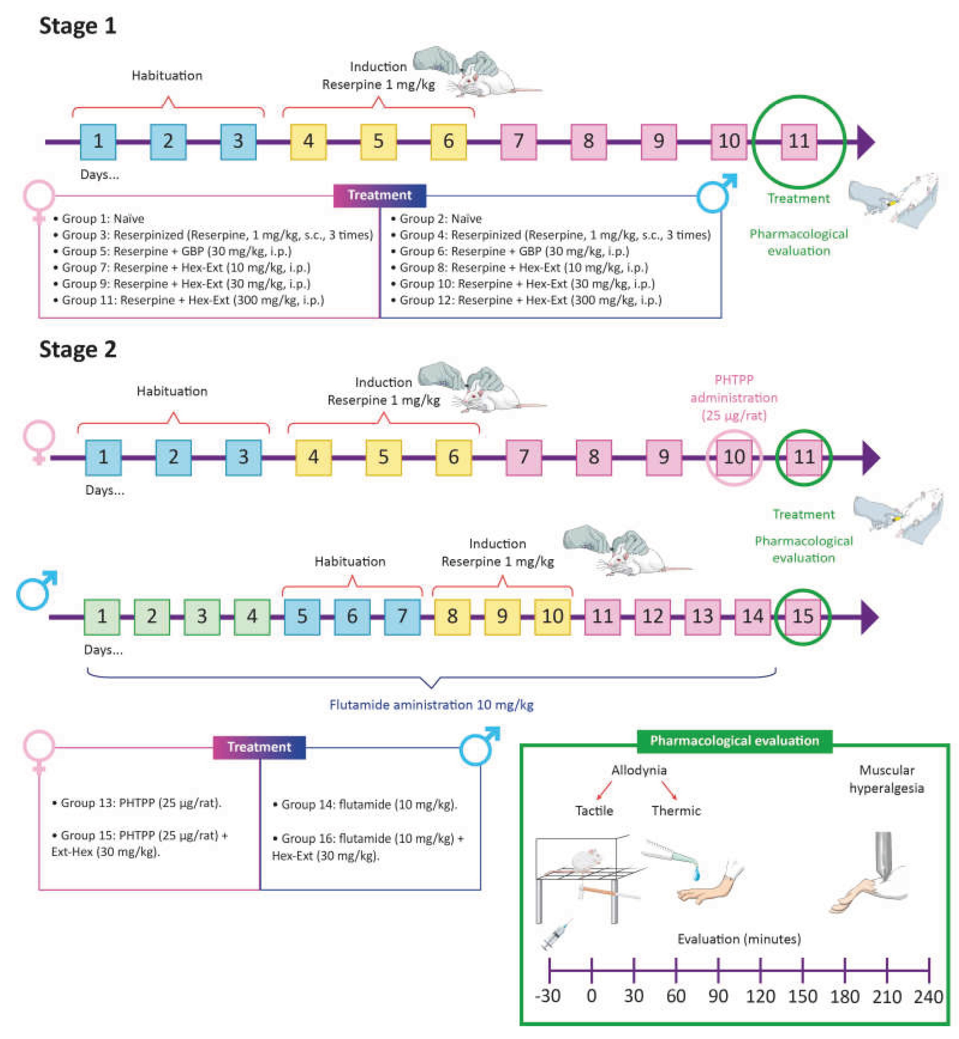
Experimental FM-type pain was induced according to a modified [31] previous method described [33]. Ninety-six rats (forty-eight females and forty-eight males) were habituated for three days prior to induction and to the measurements of hyperalgesia and allodynia. Reserpine was administered subcutaneously in the back of the neck once daily for three consecutive days, at a volume of 1 ml/kg (See timeline Figure 5).
The design included 16 experimental groups of at least 6 rats each randomly assigned (see time-line Figure 5). The acute treatments (single dose) were the following in each female or male subjects:
Stage 1
- Group 1 or 2: Naïve female or male rats.
- Group 3 or 4: Reserpinized female or male rats (Reserpine, 1 mg/kg, s.c., 3 times).
- Group 5 or 6: Reserpine + GBP (30 mg/kg, i.p.) female or male rats.
- Groups 7, 9, and 11 or 8, 10, and 12: Reserpine + Hex-Ext (10, 30, and 300 mg/kg, i.p.) female or male rats, respectively.
Stage 2
And additional groups were also explored to determine a possible mechanism of action involved, such as estrogens and androgens antagonism alone and on the antiallodynic and antihyperalgesic effect shown by the animals with the administration of Hex-Ext:
- Group 13: PHTPP (25 µg/rat 10 mg/kg) in female rats.
- Group 14: Flutamide (10 mg/kg) in male rats.
- Group 15: PHTPP (25 µg/rat) + Hex-Ext (30 mg/kg) in female rats.
- Group 16: Flutamide (10 mg/kg) + Hex-Ext (30 mg/kg) in male rats.
Flutamide was administered for 14 days prior to the test [61]. PHTPP was administered 24 hours before starting the test [64]. Treatments were administered on the 5th day after induction, when maximum nociception was detected based on hyperalgesia and allodynia thresholds [31,33]. All treatments were evaluated after 30 min of administration and over a 4-h time course, with measurements at 0, 30, 60, 120-, 150-, 180-, and 240-min post-administration. Data from the time curves were converted to AUC to determine efficacy.
Inclusion and exclusion criteria were established a priori based on the development of nociceptive responses following reserpine administration. Animals exhibiting predetermined thresholds for allodynia and hyperalgesia on the fifth day post-induction were included. Animals that did not reach these thresholds were excluded from the experimental groups. The model showed an approximate success rate of 80% in female rats and 70% in male rats, based on the development of predetermined thresholds for allodynia and hyperalgesia. No animals or data meeting the inclusion criteria were subsequently excluded from the statistical analyses.
To minimize potential confounding factors, all animals were kept in the same housing conditions and habituated to the experimental conditions for three days prior to testing. All behavioral assessments were conducted in the same laboratory, during the same period (8:00 a.m. to 2:00 p.m.), and by the same researchers throughout the study. Female and male rats were assessed separately to avoid potential interference associated with the simultaneous presence of animals of both sexes.
4.7. Assessment of Tactile Allodynia
The tactile response threshold was calibrated using Von Frey filaments (Stoelting Co., Wood Dale, IL, USA) with the up-and-down method. Rats were placed individually in transparent acrylic boxes without a bottom lid, on a wire mesh on an elevated platform, exposing the sole of the limb. Subsequently, the mid-portion of the right hind limb was stimulated with calibrated von Frey filaments (2, 4, 6, 8, 10, and 15 g).
A positive response was defined as limb withdrawal within a period of up to 6 s; a negative response was defined as no response at that time. Negative responses were marked with an “O” and positive responses with an “X”. After obtaining the first positive response, testing continued with the immediately smaller filament; if the new response was negative, the smaller filament was tested; if it was positive, the next larger filament was used, until a pattern of five measurements was completed, ranging from positive (X) to negative (O).
The paw withdrawal threshold was calculated as the tactile response threshold using an adaptation of Dixon’s top-down paradigm [65]. The 50% withdrawal threshold was calculated using the formula as follows [66]:
Where:
- Xf: value of the last von Frey filament used (in logarithmic units);
- κ: correction factor based on the response pattern (according to the calibration table and the number of positive and negative responses).
- δ: average difference between stimuli (in logarithmic units).
Figure 8.
Time-line of the experimental design. Stage 1: Dose-response evaluation and Stage 2: hormonal mechanism of action during allodynia and hyperalgesia in experimental fibromyalgia in female rats (impairs groups) and male rats (pair groups).
Figure 8.
Time-line of the experimental design. Stage 1: Dose-response evaluation and Stage 2: hormonal mechanism of action during allodynia and hyperalgesia in experimental fibromyalgia in female rats (impairs groups) and male rats (pair groups).
4.8. Cold Stimulus Threshold (Thermal Allodynia)
Thermal allodynia to a cold stimulus was assessed using the acetone test. For this test, each rat was individually placed into a transparent acrylic cylinder, which was located on a wire rig. A volume of 50 μL of acetone was then applied to the mid-dorsal surface of the right hind paw using a drip adapter on an insulin syringe. Immediately afterward, the licking or/and shaking time of their right hind paw was recorded for 60 seconds in each rat. This test was performed in triplicate, with a 5-minute interval between each measurement and the average value obtained was considered as the cold allodynia threshold.
4.9. Mechanical Hyperalgesia Assessment
Muscle pressure threshold was assessed using the Randall–Selitto apparatus (Ugo-Basile, Varese, Italy). For this assessment, rats were immobilized with a cloth, leaving the right hind paw exposed, and increasing linear pressure was applied to the medial gastrocnemius muscle (maximum force: 250 g). A positive response was considered when the rats withdrew their limb or vocalized. Each study was performed in triplicate, with a minimum inter-stimulus interval of 1 min [33,67].
4.10. Statistical Analysis
Data are expressed as the mean ± SEM of six repetitions. Data normality was assessed using the Shapiro–Wilk test, and all datasets met the normality assumption. Time-course curves were analyzed using repeated measures in a two-way analysis of variance (ANOVA) followed by Tukey’s post hoc test. The area under the curve (AUC) was calculated using the trapezoidal method, and data were analyzed using a one-way ANOVA followed by Dunnett’s post hoc test for comparison with the reserpinized group. Statistical analysis was performed using GraphPad Prism version 10.5.0 for Windows (GraphPad Software INC, La Jolla, CA, USA). A p < 0.05 was considered to declare a significant difference.
5. Conclusions
A non-polar extract of S. rosmarinus produced dose-dependent antiallodynic and antihyperalgesic effects, with greater efficacy in females than in males in a 1:10 ratio. Experimental evidence suggests that non-polar metabolites of S. rosmarinus are involved in its antinociceptive efficacy, mediated by sex-dependent gonadal hormonal mechanisms for FM-type pain relief, such as ERβ receptors in females and androgens in males. One limitation was the lack of exploration of estrogen receptor involvement in the effect of the non-polar S. rosmarinus extract in males. Blocking estrogen synthesis to eliminate endogenous estrogen production using an aromatase inhibitor such as 4-hydroxyandrostenedione could be useful in determining selective estrogen receptor modulation, which, based on the current results, will be interesting to explore in future targeted experiments.
Supplementary Materials
The following supporting information can be downloaded at the website of this paper posted on Preprints.org.
Author Contributions
Conceptualization, M.E.G.-T., J.S.-G., E.M.E.-C, M.D.-C. Methodology, M.E.G.-T., J.S.-G., B.O.-M. Software, M.E.G.-T., J.S.-G. Validation, M.E.G.-T., J.S.-G., E.M.E.-C, H.S.-C., L.A.M.-M., M.D.-C. Formal analysis, M.E.G.-T., J.S.-G, B.O.-M. Investigation, M.E.G.-T., J.S.-G., E.M.E.-C, H.S.-C., M.D.-C. Resources, M.E.G.-T., E.M.E.-C, L.A.M.-M., B.O.-M., M.D.-C. Data curation, M.E.G.-T., J.S.-G., M.D.-C. Writing—original draft preparation, M.E.G.-T., J.S.-G., E.M.E.C. Writing—review and editing, M.E.G.-T., J.S.-G., E.M.E.-C, H.S.-C., L.A.M.-M.; B.O.-M., M.D.-C. Visualization M.E.G.-T., J.S.-G., E.M.E.-C. Supervision, M.E.G.-T., E.M.E.-C, H.S.-C., L.A.M.-M., B.O.-M., M.D.-C. Project administration, M.E.G.-T.
Funding
This investigation was financed by INPRFM protocols NC123280.0 (September 10, 2012) and NC17073.0 (December 12, 2017) (M.E.G.-T). J.S.-G thanks fellowship by SECIHTI No. 923250 and BEIFI-IPN.
Institutional Review Board Statement
The experimental procedure followed the research was conducted in accordance with the internationally accepted principles for laboratory animal use and care as found in the European Community guidelines (2010/63/EU for the Protection of Laboratory Animals), the US guidelines (NIH publication #85-23, revised in 1985 and updated in 2011), the local Guides such as the Official Mexican Standard (NOM-062-ZOO-1999). This research was approved by the Research Committee of the INPRFM (CEI/C/035/2018, Jun 04, 2018) reapproved (January 12, 2026), NC123280.0 (September 10, 2012) and NC17073.0 (December 12, 2017).
Data Availability Statement
The presented data in this study are available upon request.
Acknowledgments
This investigation was financed by SECIHTI (before CONAHCYT) grant number 256448 (M.E.G.-T)/923250(J.S.-G.). We thank the students Gerardo Alcocer and Julio Trejo for their technical support and M. Sc. Everardo Tapia Mendoza from Laboratorio Nacional de Ciencias para la Investigación y la Conservación del Patrimonio Cultural (LANCIC), Instituto de Química, UNAM, CDMX 04510, México, for the technical support with GC-MS data acquisition.
Conflicts of Interest
The authors declare no conflicts of interest.
Abbreviations
The following abbreviations are used in this manuscript:
| ANOVA | Analysis of Variance |
| ATP | Adenosine Triphosphate |
| AUC | Area Under the Curve |
| CONAHCYT | Consejo Nacional de Humanidades, Ciencias y Tecnología |
| CNS | Central Nervous System |
| DMSO | Dimethyl Sulfoxide |
| ERβ | Estrogen Receptor β |
| FM | Fibromyalgia |
| GC-MS | Gas Chromatography coupled to Mass Spectrometry |
| GBP | Gabapentin |
| Hex-Ext | Hexane extract |
| i.p. | Intraperitoneal |
| INPRFM | Instituto Nacional de Psiquiatría Ramón de la Fuente Muñiz |
| NIST | National Institute of Standards and Technology |
| NMDA | N-Methyl-D-Aspartate |
| PHTPP | 4-[2-phenyl-5,7-bis(trifluoromethyl)pyrazolo[1,5-a]pyrimidine-3-yl]phenol |
| s.c. | subcutaneous |
| SECIHTI | Secretaria de Ciencia, Humanidades, Tecnología |
| 5-HT | Serotonin |
| 5-HT1A | Serotonin Receptor 1A |
| TRP | Transient Receptor Potential |
| VMAT | Vesicular Monoamine Transporter |
References
- Sarzi-Puttini, P.; Giorgi, V.; Marotto, D.; Atzeni, F. Fibromyalgia: An update on clinical characteristics, aetiopathogenesis and treatment. Nat. Rev. Rheumatol. 2020, 16, 645–660. [Google Scholar] [CrossRef] [PubMed]
- Ruschak, I.; Montesó-Curto, P.; Rosselló, L.; Aguilar Martín, C.; Sánchez-Montesó, L.; Toussaint, L. Fibromyalgia Syndrome Pain in Men and Women: A Scoping Review. Healthcare 2023, 11, 223. [Google Scholar] [CrossRef] [PubMed]
- Font Gayà, T.; Bordoy Ferrer, C.; Juan Mas, A.; Seoane-Mato, D.; Álvarez Reyes, F.; Delgado Sánchez, M.; Martínez Dubois, C.; Sánchez-Fernández, S.A.; Marena Rojas Vargas, L.; García Morales, P.V.; Olivé, A.; Rubio Muñoz, P.; Larrosa, M.; Navarro Ricós, N.; Sánchez-Piedra, C.; Díaz-González, F.; Bustabad-Reyes, S. Prevalence of Fibromyalgia and Associated Factors in Spain. Clin. Exp. Rheumatol. 2020, 38, S47–S52. [Google Scholar] [CrossRef]
- Cabo-Meseguer, A.; Cerdá-Olmedo, G.; Trillo-Mata, J.L. Fibromyalgia: Prevalence, Epidemiologic Profiles and Economic Costs. Med. Clin. (Barc.) 2017, 149, 441–448. [Google Scholar] [CrossRef] [PubMed]
- Marques, A.P.; Espírito Santo, A.S.; Berssaneti, A.A.; Matsutani, L.A.; Yuan, S.L.K. Prevalence of Fibromyalgia: Literature Review Update. Rev. Bras. Reumatol. 2017, 57, 356–363. [Google Scholar] [CrossRef] [PubMed]
- Park, D.J.; Lee, S.S. New Insights into the Genetics of Fibromyalgia. Korean J. Intern. Med. 2017, 32, 984–995. [Google Scholar] [CrossRef] [PubMed]
- Mogil, J.S. Qualitative Sex Differences in Pain Processing: Emerging Evidence of a Biased Literature. Nat. Rev. Neurosci. 2020, 21, 353–365. [Google Scholar] [CrossRef] [PubMed]
- Miller, L.R.; Marks, C.; Becker, J.B.; Hurn, P.D.; Chen, W.; Woodruff, T.; McCarthy, M.M.; Sohrabji, F.; Schiebinger, L.; Wetherington, C.L.; et al. Considering Sex as a Biological Variable in Preclinical Research. FASEB J. 2017, 31, 29–34. [Google Scholar] [CrossRef] [PubMed]
- Chinn, S.; Caldwell, W.; Gritsenko, K. Fibromyalgia Pathogenesis and Treatment Options Update. Curr. Pain Headache Rep. 2016, 20, 25. [Google Scholar] [CrossRef] [PubMed]
- Bartley, E.J.; Fillingim, R.B. Sex Differences in Pain: A Brief Review of Clinical and Experimental Findings. Br. J. Anaesth. 2013, 111, 52–58. [Google Scholar] [CrossRef] [PubMed]
- Craft, R.M.; Mogil, J.S.; Aloisi, A.M. Sex Differences in Pain and Analgesia: The Role of Gonadal Hormones. Eur. J. Pain 2004, 8, 397–411. [Google Scholar] [CrossRef] [PubMed]
- Chen, Q.; Zhang, W.; Sadana, N.; Chen, X. Estrogen receptors in pain modulation: cellular signaling. Biol. Sex. Differ. 2021, 12(1), 22. [Google Scholar] [CrossRef] [PubMed]
- Winslow, B.T.; Vandal, C.; Dang, L. Fibromyalgia: Diagnosis and Management. Am. Fam. Physician 2023, 107, 137–144. https://www.aafp.org/pubs/afp/issues/2023/0200/fibromyalgia.html. [CrossRef] [PubMed]
- Li, Y.; Li, Y.; Wu, S.; Long, M.; Chen, X.; Wang, Y.; Zhao, J.; He, B.; Chang, C.; Jiao, J. Pharmacologic Treatment of Fibromyalgia: An Update. Front. Pharmacol. 2025, 16, 1651181. [Google Scholar] [CrossRef] [PubMed]
- Moore, A.; Bidonde, J.; Fisher, E.; Häuser, W.; Bell, R.F.; Perrot, S.; Makri, S.; Straube, S. Effectiveness of Pharmacological Therapies for Fibromyalgia Syndrome in Adults: An Overview of Cochrane Reviews. Rheumatology 2025, 64, 2385–2394. [Google Scholar] [CrossRef] [PubMed]
- González-Trujano, M.E.; Peña, E.I.; Martínez, A.L.; Moreno, J.; Guevara-Fefer, P.; Déciga-Campos, M.; López-Muñoz, F.J. Evaluation of the Antinociceptive Effect of Rosmarinus officinalis L. Using Three Different Experimental Models in Rodents. J. Ethnopharmacol. 2007, 111, 476–482. [Google Scholar] [CrossRef] [PubMed]
- Borges, R.S.; Ortiz, B.L.S.; Pereira, A.C.M.; Keita, H.; Carvalho, J.C.T. Rosmarinus officinalis Essential Oil: A Review of Its Phytochemistry, Anti-Inflammatory Activity, and Mechanisms of Action Involved. J. Ethnopharmacol. 2019, 229, 29–45. [Google Scholar] [CrossRef] [PubMed]
- Takaki, I.; Bersani-Amado, L.E.; Vendruscolo, A.; Sartoretto, S.M.; Diniz, S.P.; Bersani-Amado, C.A.; Cuman, R.K.N. Anti-Inflammatory and Antinociceptive Effects of Rosmarinus officinalis L. Essential Oil in Experimental Animal Models. J. Med. Food 2008, 11, 741–746. [Google Scholar] [CrossRef] [PubMed]
- Martínez, A.L.; González-Trujano, M.E.; Pellicer, F.; López-Muñoz, F.J.; Navarrete, A. Antinociceptive Effect and GC/MS Analysis of Rosmarinus officinalis L. Essential Oil from Its Aerial Parts. Planta Med. 2009, 75, 508–511. [Google Scholar] [CrossRef] [PubMed]
- Johnson, J.J.; Syed, D.N.; Suh, Y.; Heren, C.R.; Saleem, M.; Siddiqui, I.A.; Mukhtar, H. Disruption of Androgen and Estrogen Receptor Activity in Prostate Cancer by a Novel Dietary Diterpene Carnosol: Implications for Chemoprevention. Cancer Prev. Res. 2010, 3, 1112–1123. [Google Scholar] [CrossRef] [PubMed]
- Ibrahim, N.; Abbas, H.; El-Sayed, N.S.; Gad, H.A.; et al. Rosmarinus officinalis L. Hexane Extract: Phytochemical Analysis, Nanoencapsulation, and In Silico, In Vitro, and In Vivo Anti-Photoaging Potential Evaluation. Sci. Rep. 2022, 12, 13102. [Google Scholar] [CrossRef] [PubMed]
- Chetia, M.P.; Ashraf, G.J.; Sahu, R.; Nandi, G.; Karunakaran, G.; Paul, P.; Dua, T.K. Rosemary (Rosmarinus officinalis L.) essential oil: A review of extraction technologies, and biological activities. Next Res. 2025, 2(3), 100545. [Google Scholar] [CrossRef]
- Ghasemzadeh Rahbardar, M.; Hosseinzadeh, H. Therapeutic Effects of Rosemary (Rosmarinus officinalis L.) and Its Active Constituents on Nervous System Disorders. Iran. J. Basic Med. Sci. 2020, 23, 1100–1112. [Google Scholar] [CrossRef] [PubMed]
- Siracusa, R.; Di Paola, R.; Cuzzocrea, S.; Impellizzeri, D. Fibromyalgia: Pathogenesis, Mechanisms, Diagnosis and Treatment Options Update. Int. J. Mol. Sci. 2021, 22, 3891. [Google Scholar] [CrossRef] [PubMed]
- Jurado-Priego, L.N.; Torres-Sánchez, L.; Castañeda-Corral, G.; Granados-Soto, V.; Rocha-González, H.I. Sex Differences and Estradiol Involvement in Fibromyalgia-Like Pain Induced by Reserpine in Rats. Eur. J. Pain 2021, 25, 1226–1239. [Google Scholar] [CrossRef] [PubMed]
- Cabo-Meseguer, A.; Cerdá-Olmedo, G.; Trillo-Mata, J.L. Fibromyalgia: Prevalence, Epidemiologic Profiles and Economic Costs. Med. Clin. (Barc.) 2017, 149, 441–448. [Google Scholar] [CrossRef] [PubMed]
- Macia, A.; Calvo, M.; et al. Sex-Related Differences in Symptoms and Psychosocial Outcomes in Patients with Fibromyalgia: A Prospective Questionnaire Study. Mayo Clin. Proc. Innov. Qual. Outcomes 2020, 4, 767–774. [Google Scholar] [CrossRef] [PubMed]
- Bendif, H.; Boudjeniba, M.; Miara, M.D.; Biqiku, L.; Bramucci, M.; Caprioli, G. Rosmarinus eriocalyx: An Alternative to Rosmarinus officinalis as a Source of Antioxidant Compounds. Food Chem. 2017, 218, 78–88. [Google Scholar] [CrossRef] [PubMed]
- Nagakura, Y. Giving Priority to Preclinical Pain Measures Resistant to Existing Drugs for Developing Innovative Analgesics. Drug Dev. Res. 2018, 79, 147–156. [Google Scholar] [CrossRef] [PubMed]
- Hernandez-Leon, A.; De la Luz-Cuellar, Y.E.; Granados-Soto, V.; González-Trujano, M.E.; Fernández-Guasti, A. Sex Differences and Estradiol Involvement in Hyperalgesia and Allodynia in an Experimental Model of Fibromyalgia. Horm. Behav. 2018, 97, 39–46. [Google Scholar] [CrossRef] [PubMed]
- Hernandez-Leon, A.; Fernández-Guasti, A.; Martínez, A.; Pellicer, F.; González-Trujano, M.E. Sleep Architecture Is Altered in the Reserpine-Induced Fibromyalgia Model in Ovariectomized Rats. Behav. Brain Res. 2019, 364, 383–392. [Google Scholar] [CrossRef] [PubMed]
- Mandela, P.; Chandley, M.; Xu, Y.Y.; Zhu, M.Y.; Ordway, G.A. Reserpine-Induced Reduction in Norepinephrine Transporter Function Requires Catecholamine Storage Vesicles. Neurochem. Int. 2010, 56, 760–767. [Google Scholar] [CrossRef] [PubMed]
- Nagakura, Y.; Oe, T.; Aoki, T.; Matsuoka, N. Biogenic Amine Depletion Causes Chronic Muscular Pain and Tactile Allodynia Accompanied by Depression: A Putative Animal Model of Fibromyalgia. Pain 2009, 146, 26–33. [Google Scholar] [CrossRef] [PubMed]
- Brum, E.S.; Fialho, M.F.P.; Souza Monteiro de Araújo, D.; Landini, L.; Marini, M.; Titiz, M.; Kuhn, B.L.; Frizzo, C.P.; Araújo, P.H.S.; Guimarães, R.M.; et al. Schwann Cell TRPA1 Elicits Reserpine-Induced Fibromyalgia Pain in Mice. Br. J. Pharmacol. 2024, 181, 3445–3461. [Google Scholar] [CrossRef] [PubMed]
- Greaves, E.; Grieve, K.; Horne, A.W.; Saunders, P.T.K. Elevated Peritoneal Expression and Estrogen Regulation of Nociceptive Ion Channels in Endometriosis. J. Clin. Endocrinol. Metab. 2014, 99, E1738–E1743. [Google Scholar] [CrossRef] [PubMed]
- Chen, Y.; Moutal, A.; Navratilova, E.; Hua, H.; Giddings, S.; Vakil, V.; et al. The Prolactin Receptor Long Isoform Regulates Nociceptor Sensitization and Opioid-Induced Hyperalgesia Selectively in Females. Sci. Transl. Med. 2020, 12, eaay7550. [Google Scholar] [CrossRef] [PubMed]
- Li, Y.; Li, Y.; Wu, S.; Long, M.; Chen, X.; Wang, Y.; Zhao, J.; He, B.; Chang, C.; Jiao, J. Pharmacologic Treatment of Fibromyalgia: An Update. Front. Pharmacol. 2025, 16, 1651181. [Google Scholar] [CrossRef] [PubMed]
- Ma, J.N.; McFarland, K.; Olsson, R.; Burstein, E.S. Estrogen Receptor Beta Selective Agonists as Agents to Treat Chemotherapeutic-Induced Neuropathic Pain. ACS Chem. Neurosci. 2016, 7, 1180–1187. [Google Scholar] [CrossRef] [PubMed]
- Xu, H.; Blair, N.T.; Clapham, D.E. Camphor Activates and Strongly Desensitizes the Transient Receptor Potential Vanilloid Subtype 1 Channel in a Vanilloid-Independent Mechanism. J. Neurosci. 2005, 25, 8924–8937. [Google Scholar] [CrossRef] [PubMed]
- Li, Z.; Gan, Y.; Kang, T.; Zhao, Y.; Huang, T.; Chen, Y.; Liu, J.; Ke, B. Camphor Attenuates Hyperalgesia in Neuropathic Pain Models in Mice. J. Pain Res. 2023, 16, 785–795. [Google Scholar] [CrossRef] [PubMed]
- Sherkheli, M.A.; Schreiner, B.; Haq, R.; Werner, M.; Hatt, H. Borneol Inhibits TRPA1, a Proinflammatory and Noxious Pain-Sensing Cation Channel. Pak. J. Pharm. Sci. 2015, 28, 1357–1363. https://pubmed.ncbi.nlm.nih.gov/26142526/. [PubMed]
- Jiang, J.; Shen, Y.Y.; Li, J.; Lin, Y.H.; Luo, C.X.; Zhu, D.Y. (+)-Borneol Alleviates Mechanical Hyperalgesia in Models of Chronic Inflammatory and Neuropathic Pain in Mice. Eur. J. Pharmacol. 2015, 757, 53–58. [Google Scholar] [CrossRef] [PubMed]
- Takaishi, M.; Fujita, F.; Uchida, K.; Yamamoto, S.; Sawada, M.; Hatai, C.; Shimizu, M.; Tominaga, M. 1,8-Cineole, a TRPM8 Agonist, Is a Novel Natural Antagonist of Human TRPA1. Mol. Pain 2012, 8, 86. [Google Scholar] [CrossRef] [PubMed]
- Gadotti, V.M.; Huang, S.; Zamponi, G.W. The Terpenes Camphene and Alpha-Bisabolol Inhibit Inflammatory and Neuropathic Pain via CaV3.2 T-Type Calcium Channels. Mol. Brain 2021, 14, 166. [Google Scholar] [CrossRef] [PubMed]
- Ceccarelli, I.; Fiorenzani, P.; Pessina, F.; Pinassi, J.; Aglianò, M.; Miragliotta, V.; Aloisi, A.M. The CB2 Agonist β-Caryophyllene in Male and Female Rats Exposed to a Model of Persistent Inflammatory Pain. Front. Neurosci. 2020, 14, 850. [Google Scholar] [CrossRef] [PubMed]
- Plumb, A.N.; Lesnak, J.B.; Kolling, L.J.; Marcinkiewicz, C.A.; Sluka, K.A. Local Synthesis of Estradiol in the Rostral Ventromedial Medulla Protects against Widespread Muscle Pain in Male Mice. eNeuro 2024, 11, ENEURO.0332-24.2024. [Google Scholar] [CrossRef] [PubMed]
- Duong, P.; Tenkorang, M.A.A.; Trieu, J.; McCuiston, C.; Rybalchenko, N.; Cunningham, R.L. Neuroprotective and Neurotoxic Outcomes of Androgens and Estrogens in an Oxidative Stress Environment. Biol. Sex. Differ. 2020, 11, 12. [Google Scholar] [CrossRef] [PubMed]
- Martínez-Mota, L.; Cruz-Martínez, J.J.; Márquez-Baltazar, S.; Fernández-Guasti, A. Estrogens Participate in the Antidepressant-Like Effect of Desipramine and Fluoxetine in Male Rats. Pharmacol. Biochem. Behav. 2008, 88, 332–340. [Google Scholar] [CrossRef] [PubMed]
- Paredes, S.; Cantillo, S.; Candido, K.D.; Knezevic, N.N. An Association of Serotonin with Pain Disorders and Its Modulation by Estrogens. Int. J. Mol. Sci. 2019, 20, 5729. [Google Scholar] [CrossRef] [PubMed]
- Leventhal, L.; Brandt, M.R.; Cummons, T.A.; Piesla, M.J.; Rogers, K.E.; Harris, H.A. An Estrogen Receptor-Beta Agonist Is Active in Models of Inflammatory and Chemical-Induced Pain. Eur. J. Pharmacol. 2006, 553, 146–148. [Google Scholar] [CrossRef] [PubMed]
- Chen, Q.; Zhang, W.; Sadana, N.; Chen, X. Estrogen Receptors in Pain Modulation: Cellular Signaling. Biol. Sex Differ. 2021, 12, 22. [Google Scholar] [CrossRef] [PubMed]
- Athnaiel, O.; Cantillo, S.; Paredes, S.; Knezevic, N.N. The Role of Sex Hormones in Pain-Related Conditions. Int. J. Mol. Sci. 2023, 24, 1866. [Google Scholar] [CrossRef] [PubMed]
- Ito, A.; Takeda, M.; Furue, H.; Shibata, K.; Hori, M.; Sagai, H.; Sakurada, T.; Yoshimura, M. Administration of Estrogen Shortly after Ovariectomy Mimics the Antinociceptive Action and Change in 5-HT1A-Like Receptor Expression Induced by Calcitonin in Ovariectomized Rats. Bone 2004, 35, 697–703. [Google Scholar] [CrossRef] [PubMed]
- Nomura, M.; Akama, K.T.; Alves, S.E.; Korach, K.S.; Gustafsson, J.A.; Pfaff, D.W.; Ogawa, S. Differential Distribution of Estrogen Receptor (ER)-Alpha and ER-Beta in the Midbrain Raphe Nuclei and Periaqueductal Gray in Male Mouse: Predominant Role of ER-Beta in Midbrain Serotonergic Systems. Neuroscience 2005, 130, 445–456. [Google Scholar] [CrossRef] [PubMed]
- Martínez, A.L.; González-Trujano, M.E.; Chávez, M.; Pellicer, F. Antinociceptive Effectiveness of Triterpenes from Rosemary in Visceral Nociception. J. Ethnopharmacol. 2012, 142, 28–34. [Google Scholar] [CrossRef] [PubMed]
- Nagakura, Y. Therapeutic Approaches to Nociplastic Pain Based on Findings in the Reserpine-Induced Fibromyalgia-Like Animal Model. J. Pharmacol. Exp. Ther. 2022, 381, 106–119. [Google Scholar] [CrossRef] [PubMed]
- Fillingim, R.B.; King, C.D.; Ribeiro-Dasilva, M.C.; Rahim-Williams, B.; Riley, J.L. Sex and Gender Differences in Pain and Analgesia. Prog. Brain Res. 2010, 186, 141–157. [Google Scholar] [CrossRef] [PubMed]
- Nieto, G.; Ros, G.; Castillo, J. Antioxidant and Antimicrobial Properties of Rosemary (Rosmarinus officinalis, L.): A Review. Medicines 2018, 5, 98. [Google Scholar] [CrossRef] [PubMed]
- Rahbardar, M.G.; Hosseinzadeh, H. Therapeutic Effects of Rosemary (Rosmarinus officinalis L.) and Its Active Constituents on Nervous System Disorders. Iran. J. Basic Med. Sci. 2020, 23, 1100–1112. [Google Scholar] [CrossRef] [PubMed]
- Woolf, C.J. Central Sensitization: Implications for the Diagnosis and Treatment of Pain. Pain 2011, 152, S2–S15. [Google Scholar] [CrossRef] [PubMed]
- Ji, R.R.; Berta, T.; Nedergaard, M. Glia and Pain: Is Chronic Pain a Gliopathy? Pain 2013, 154 (Suppl. 1), S10–S28. [Google Scholar] [CrossRef] [PubMed]
- Zamora-Díaz, I.-Y.; González-Trujano, M.E.; Martínez-Vargas, D.; Moreno-Pérez, G.F.; Hernandez-Leon, A.; Narváez-González, H.F.; Ventura-Martínez, R.; Pellicer, F.; López-Muñoz, F.J. Pharmacological Interactions of Sulforaphane and Gabapentin in a Murine Fibromyalgia-Like Pain Model. Biomed. Pharmacother. 2025, 184, 117929. [Google Scholar] [CrossRef] [PubMed]
- Jiménez-Rubio, G.; Herrera-Pérez, J.J.; Martínez-Mota, L. Effect of Androgen Receptor Blockade on Spatial Memory in Young and Aged Male Rats in the Barnes Maze. Horm. Behav. 2025, 170, 105711. [Google Scholar] [CrossRef] [PubMed]
- Valdés-Sustaita, B.; Estrada-Camarena, E.; González-Trujano, M.E.; López-Rubalcava, C. Estrogen Receptors-β and Serotonin Mediate the Antidepressant-Like Effect of an Aqueous Extract of Pomegranate in Ovariectomized Rats. Neurochem. Int. 2021, 142, 104904. [Google Scholar] [CrossRef] [PubMed]
- Dixon, W.J. Staircase Bioassay: The Up-and-Down Method in Small Samples. J. Am. Stat. Assoc. 1991, 46, 46–53. [Google Scholar] [CrossRef]
- Chaplan, S.R.; Bach, F.W.; Pogrel, J.W.; Chung, J.M.; Yaksh, T.L. Quantitative Assessment of Tactile Allodynia in the Rat Paw. J. Neurosci. Methods 1994, 53, 55–63. [Google Scholar] [CrossRef] [PubMed]
- Deuis, J.R.; Dvorakova, L.S.; Vetter, I. Methods Used to Evaluate Pain Behaviors in Rodents. Front. Mol. Neurosci. 2017, 10, 284. [Google Scholar] [CrossRef] [PubMed]
Figure 1.
Total ion current chromatogram of the S. rosmarinus hexane extract.
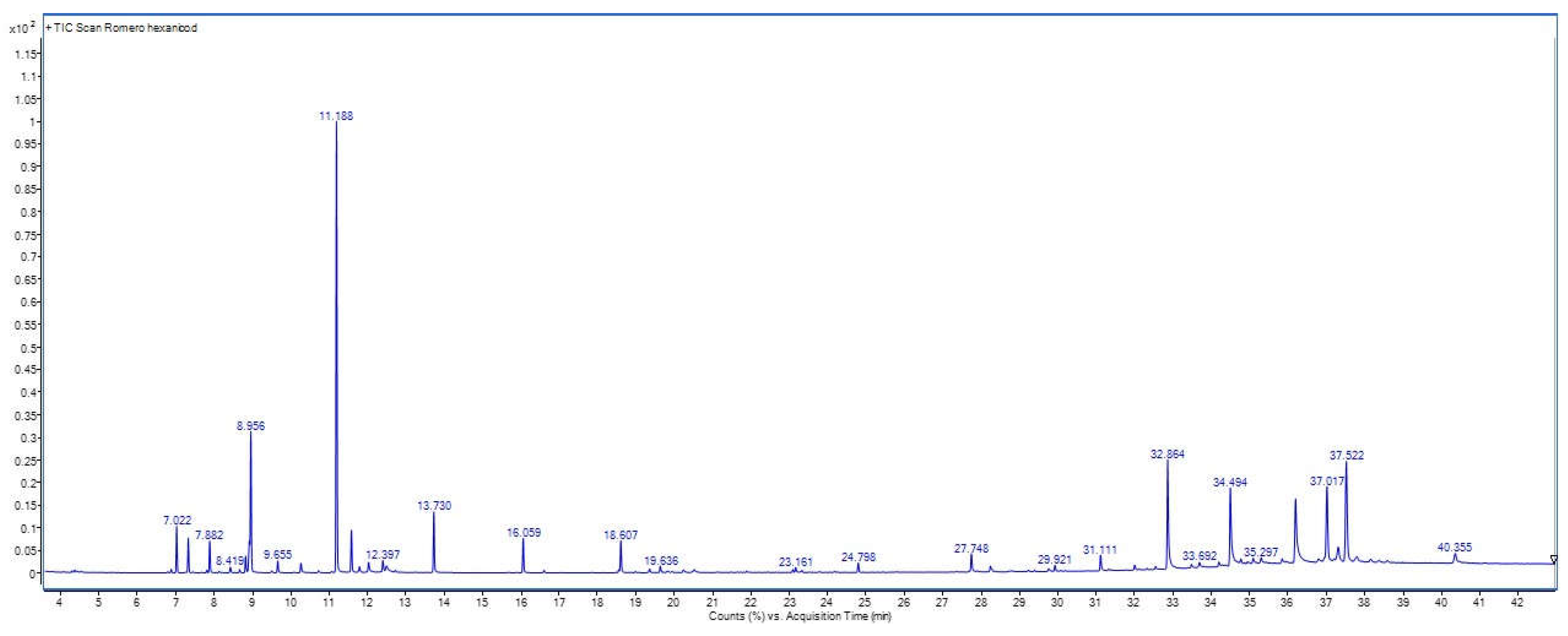
Figure 2.
Chemical structure of the 16 compounds identified in the hexane extract of S. rosmarinus.
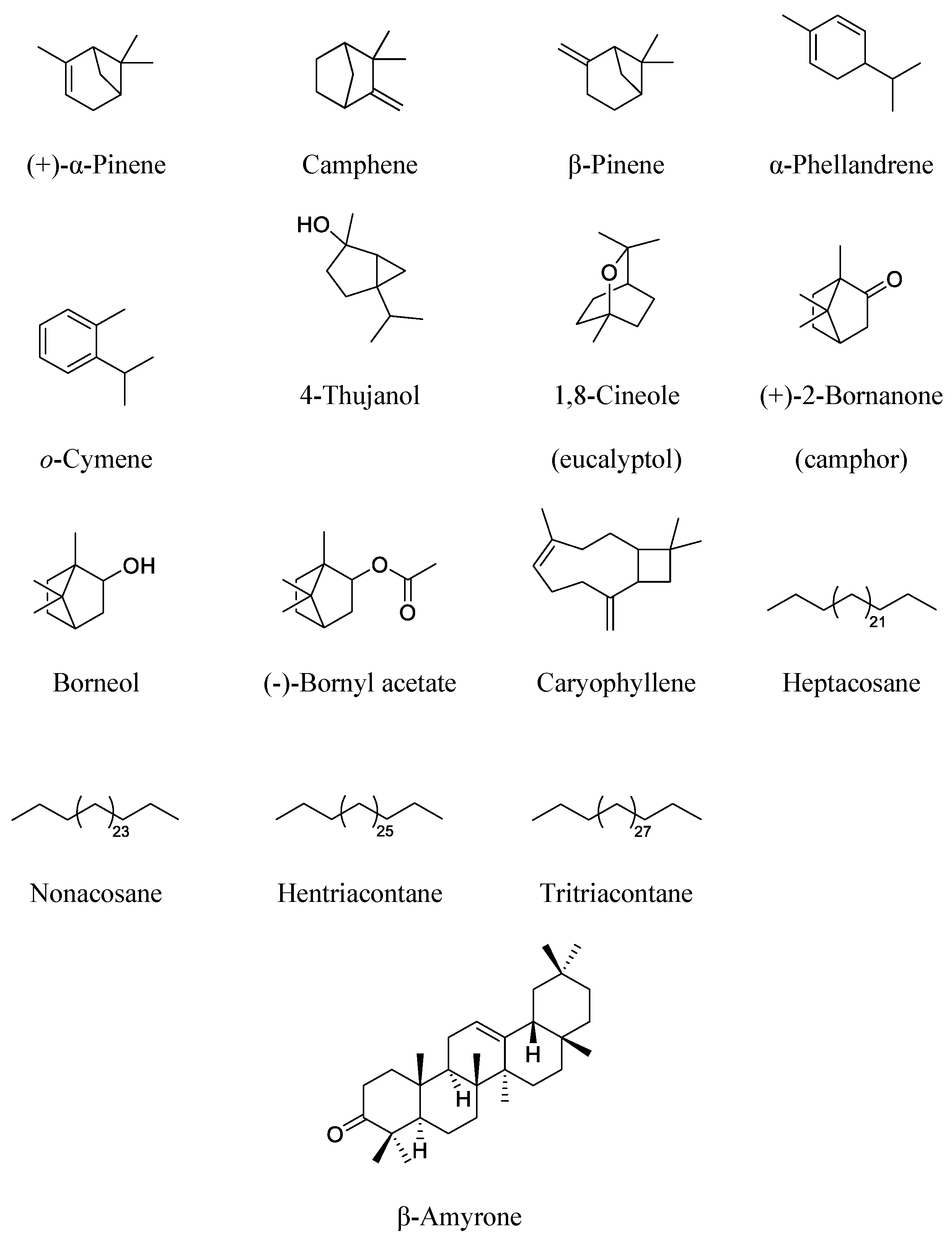
Figure 3.
Time-course curves of the antiallodyne effect of Salvia rosmarinus extract (Hex-Ext) and gabapentin (GBP) of reserpinized (A) female and (C) male rats in tactil allodynia. Each point represents the mean ± SEM of 6 rats. Two-way ANOVA followed by Tukey’s post-hoc test, *p < 0.05, **p < 0.01, ***p < 0.001, and ****p < 0.0001 vs. reserpine group (1 mg/kg, s.c.). Area under the curve (AUC) obtained from the time courses of the antiallodynic effect observed in (B) female or (D) male rats. Each bar represents the mean ± SEM of 6 rats. One-way ANOVA followed by a Dunnett’s post-hoc test. *p < 0.05, **p < 0.01, ***p < 0.001 and ****p < 0.0001. #### p < 0.0001 reserpine vs. naïve groups.
Figure 3.
Time-course curves of the antiallodyne effect of Salvia rosmarinus extract (Hex-Ext) and gabapentin (GBP) of reserpinized (A) female and (C) male rats in tactil allodynia. Each point represents the mean ± SEM of 6 rats. Two-way ANOVA followed by Tukey’s post-hoc test, *p < 0.05, **p < 0.01, ***p < 0.001, and ****p < 0.0001 vs. reserpine group (1 mg/kg, s.c.). Area under the curve (AUC) obtained from the time courses of the antiallodynic effect observed in (B) female or (D) male rats. Each bar represents the mean ± SEM of 6 rats. One-way ANOVA followed by a Dunnett’s post-hoc test. *p < 0.05, **p < 0.01, ***p < 0.001 and ****p < 0.0001. #### p < 0.0001 reserpine vs. naïve groups.
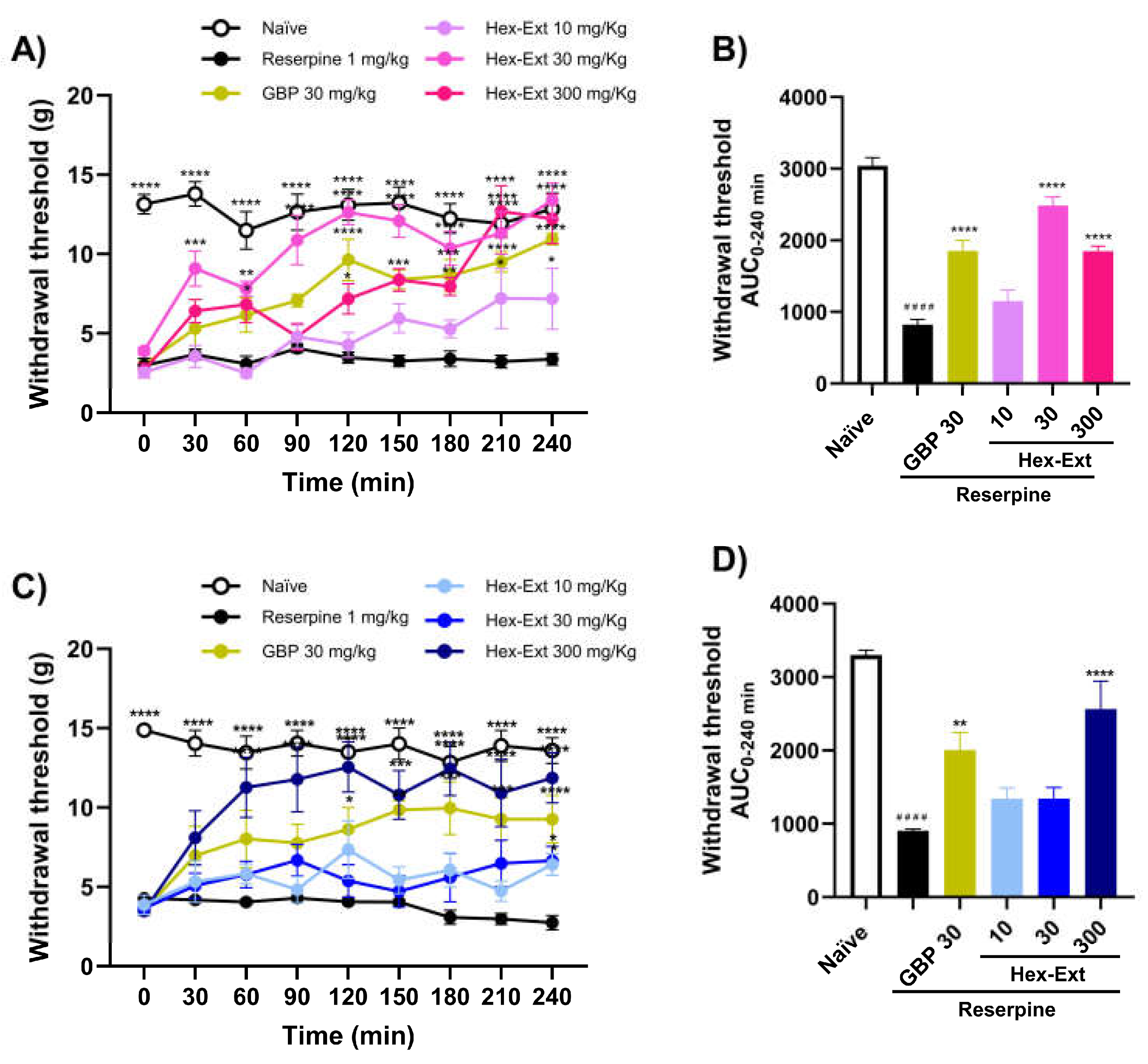
Figure 4.
Time-course curves of the antiallodynic effect of Salvia rosmarinus extract (Hex-Ext) and gabapentin (GBP) in reserpinized (A) female and (C) male rats in thermal allodynia. Each point represents the mean ± SEM of 6 rats. Two-way ANOVA followed by Tukey’s post-hoc test, *p<0.05, **p<0.01, ***p < 0.001, and ****p < 0.0001 vs. reserpine group (1 mg/kg, s.c.). Area under the curve (AUC) obtained from the time courses of the antiallodynic effect obtained in (B) female or (D) male rats. Each bar represents the mean ± SEM of 6 rats. One-way ANOVA followed by a Dunnett’s post-hoc test. *p < 0.05, **p < 0.01, ***p < 0.001 and ****p < 0.0001. #### p < 0.0001 reserpine vs. naïve groups.
Figure 4.
Time-course curves of the antiallodynic effect of Salvia rosmarinus extract (Hex-Ext) and gabapentin (GBP) in reserpinized (A) female and (C) male rats in thermal allodynia. Each point represents the mean ± SEM of 6 rats. Two-way ANOVA followed by Tukey’s post-hoc test, *p<0.05, **p<0.01, ***p < 0.001, and ****p < 0.0001 vs. reserpine group (1 mg/kg, s.c.). Area under the curve (AUC) obtained from the time courses of the antiallodynic effect obtained in (B) female or (D) male rats. Each bar represents the mean ± SEM of 6 rats. One-way ANOVA followed by a Dunnett’s post-hoc test. *p < 0.05, **p < 0.01, ***p < 0.001 and ****p < 0.0001. #### p < 0.0001 reserpine vs. naïve groups.
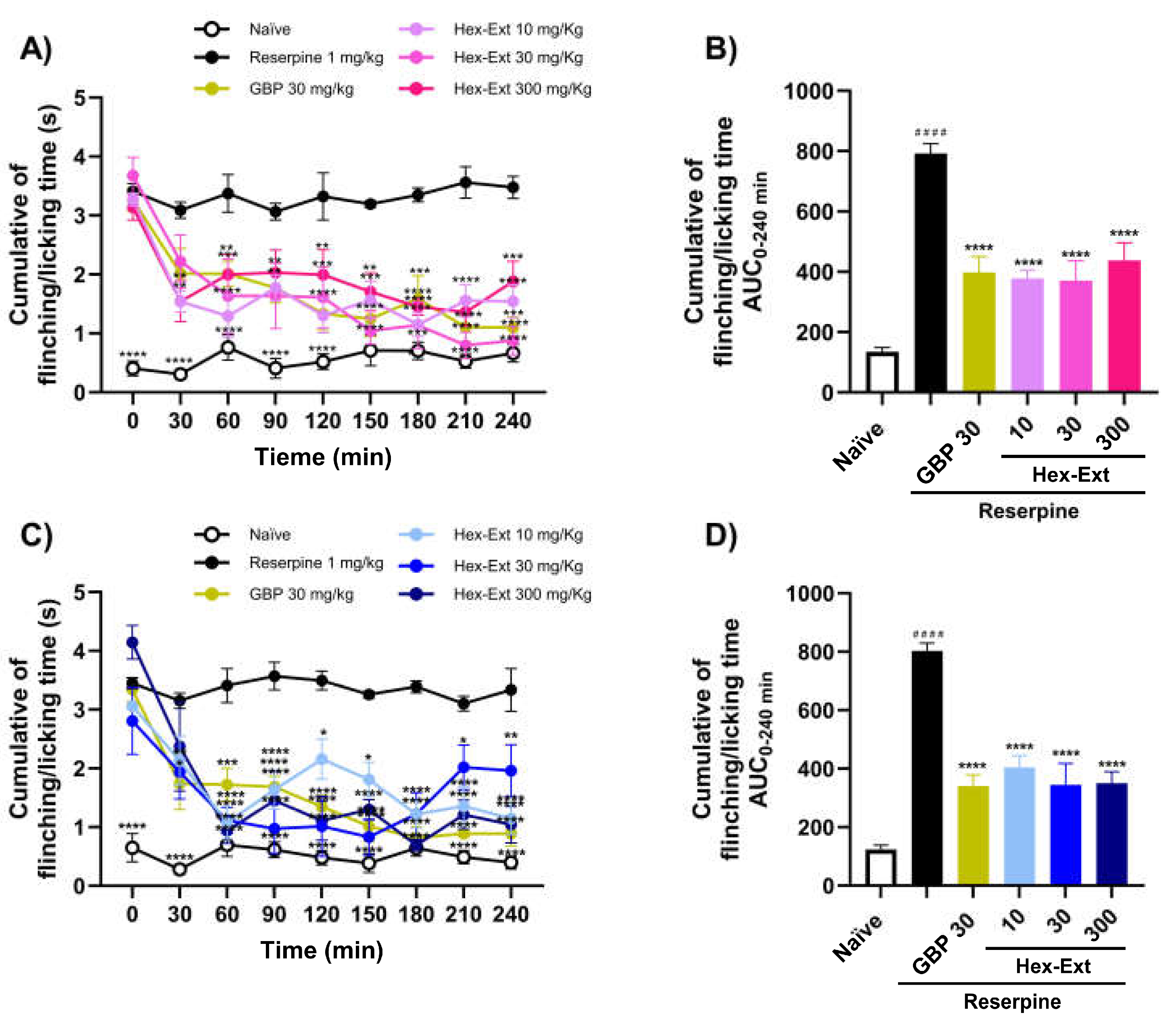
Figure 5.
Time-course curves of the antihyperalgesic effect of the Salvia rosmarinus extract (Hex-Ext) and gabapentin (GBP) in reserpinized (A) female or (C) male rats in mechanical hyperalgesia. Each point represents the mean ± SEM of 6 rats. Two-way ANOVA followed by Tukey’s post hoc test, *p < 0.05, **p < 0.01, ***p < 0.001, and ****p < 0.0001 vs reserpine group (1 mg/kg, s.c.). Area under the curve (AUC) obtained from the time courses of the antihyperalgesic effect observed in (B) female or (D) male rats. Each bar represents the mean ± SEM of 6 rats. One-way ANOVA followed by Dunnett’s post hoc test. *p < 0.05, **p < 0.01, ***p < 0.001, and ****p < 0.0001. #### p < 0.0001 reserpine vs. naïve groups.
Figure 5.
Time-course curves of the antihyperalgesic effect of the Salvia rosmarinus extract (Hex-Ext) and gabapentin (GBP) in reserpinized (A) female or (C) male rats in mechanical hyperalgesia. Each point represents the mean ± SEM of 6 rats. Two-way ANOVA followed by Tukey’s post hoc test, *p < 0.05, **p < 0.01, ***p < 0.001, and ****p < 0.0001 vs reserpine group (1 mg/kg, s.c.). Area under the curve (AUC) obtained from the time courses of the antihyperalgesic effect observed in (B) female or (D) male rats. Each bar represents the mean ± SEM of 6 rats. One-way ANOVA followed by Dunnett’s post hoc test. *p < 0.05, **p < 0.01, ***p < 0.001, and ****p < 0.0001. #### p < 0.0001 reserpine vs. naïve groups.
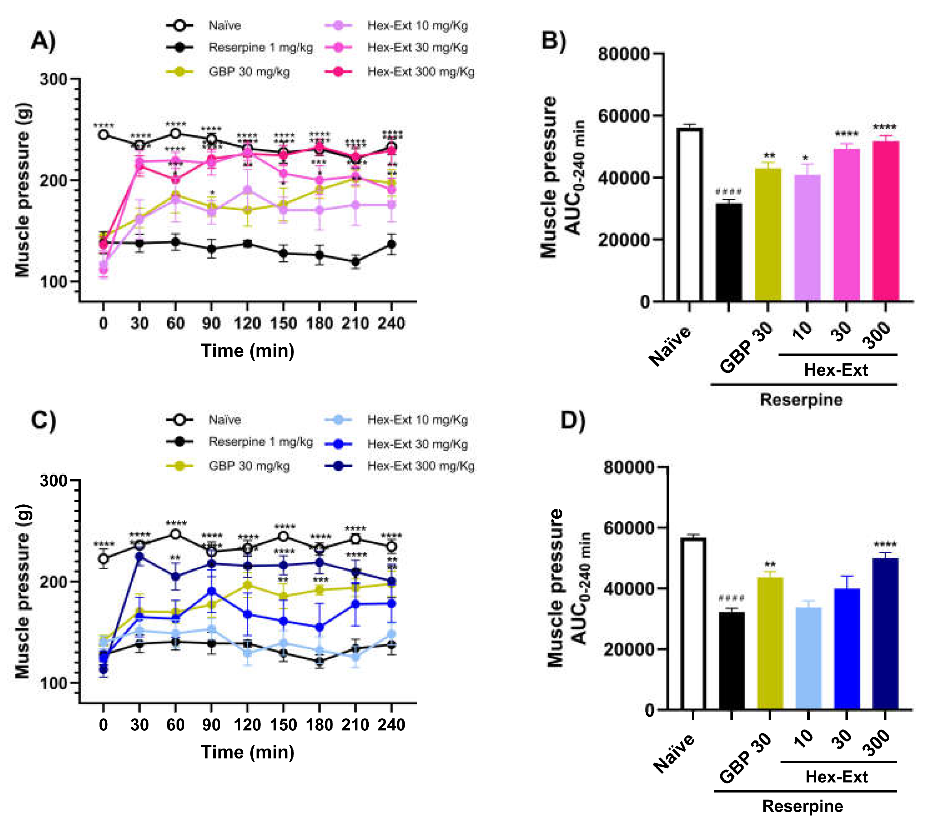
Figure 6.
Time-course curves of the antinociceptive effects of Salvia rosmarinus extract (Hex-Ext) (30 mg/kg, i.p.) in female rats alone and in the presence of PHTPP (25 µg/rat) in (A) tactile and (C) thermal allodynia, as well as in (E) mechanical hyperalgesia. Each point represents the mean ± SEM of 6 rats. Two-way ANOVA followed by Tukey’s post-hoc test, *p < 0.05, **p < 0.01, ***p < 0.001 and ****p < 0.0001 vs. reserpine group (1 mg/kg, s.c.). Area under the curve (AUC) obtained from the time courses of the S. rosmarinus hexane extract (Hex-Ext) (30 mg/kg, i.p.), alone and combined with PHTPP (25 µg/rat) in (B) tactile and (D) thermal allodynia, as well as in (F) mechanical hyperalgesia. Each bar represents the mean ± SEM of 6 rats. One-way ANOVA followed by Dunnett ‘s post-hoc test. **p<0.01, ***p < 0.001, and ****p < 0.0001. #### p < 0.0001 reserpine vs. naïve groups.
Figure 6.
Time-course curves of the antinociceptive effects of Salvia rosmarinus extract (Hex-Ext) (30 mg/kg, i.p.) in female rats alone and in the presence of PHTPP (25 µg/rat) in (A) tactile and (C) thermal allodynia, as well as in (E) mechanical hyperalgesia. Each point represents the mean ± SEM of 6 rats. Two-way ANOVA followed by Tukey’s post-hoc test, *p < 0.05, **p < 0.01, ***p < 0.001 and ****p < 0.0001 vs. reserpine group (1 mg/kg, s.c.). Area under the curve (AUC) obtained from the time courses of the S. rosmarinus hexane extract (Hex-Ext) (30 mg/kg, i.p.), alone and combined with PHTPP (25 µg/rat) in (B) tactile and (D) thermal allodynia, as well as in (F) mechanical hyperalgesia. Each bar represents the mean ± SEM of 6 rats. One-way ANOVA followed by Dunnett ‘s post-hoc test. **p<0.01, ***p < 0.001, and ****p < 0.0001. #### p < 0.0001 reserpine vs. naïve groups.
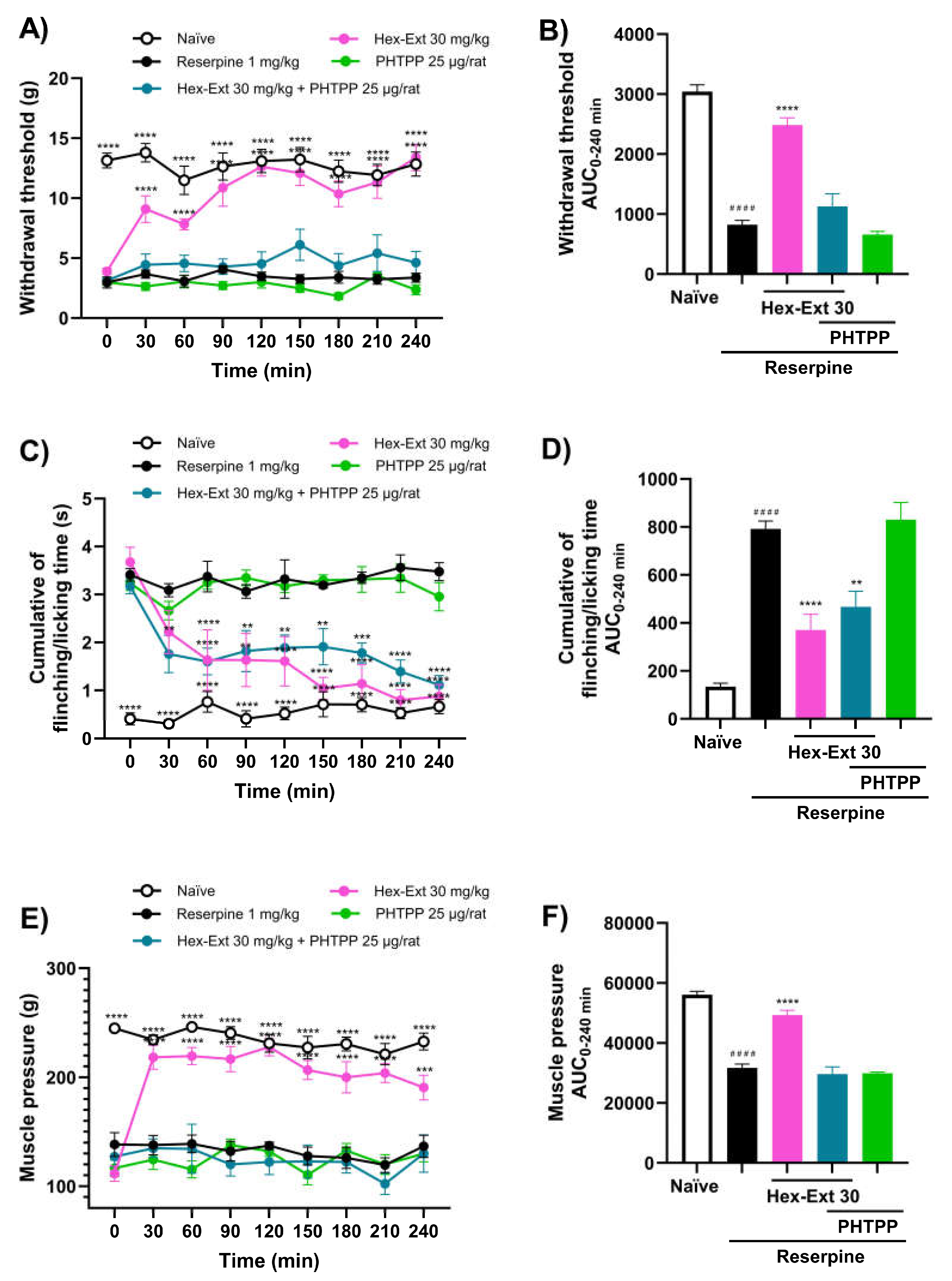
Figure 7.
Time-course curves of the Salvia rosmarinus extract (Hex-Ext, 30 mg/kg, i.p.) effects on (A) tactile and (C) thermal allodynia, and (E) hyperalgesia alone and in the presence of flutamide (10 mg/kg). Each point represents the mean ± SEM of 6 male rats. Two-way ANOVA followed by Tukey’s post-hoc test, *p<0.05, **p<0.01, ***p<0.001 and ****p<0.0001 vs. reserpine group (1 mg/kg, s.c.). Area under the curve (AUC) obtained from the time courses of the effects of S. rosmarinus extract (Hex-Ext, 30 mg/kg, i.p.), flutamide (10 m g/kg, i.p.), and their combination on (B) tactile and (D) thermal antiallodynia, and (F) mechanical hyperalgesia after their administration. Each bar represents the mean ± SEM of 6 rats. One-way ANOVA followed by Dunnett’s post-hoc test. *p<0.05, **p<0.01, ***p < 0.001, and ****p < 0.0001. #### p < 0.0001 reserpine vs. Naïve groups.
Figure 7.
Time-course curves of the Salvia rosmarinus extract (Hex-Ext, 30 mg/kg, i.p.) effects on (A) tactile and (C) thermal allodynia, and (E) hyperalgesia alone and in the presence of flutamide (10 mg/kg). Each point represents the mean ± SEM of 6 male rats. Two-way ANOVA followed by Tukey’s post-hoc test, *p<0.05, **p<0.01, ***p<0.001 and ****p<0.0001 vs. reserpine group (1 mg/kg, s.c.). Area under the curve (AUC) obtained from the time courses of the effects of S. rosmarinus extract (Hex-Ext, 30 mg/kg, i.p.), flutamide (10 m g/kg, i.p.), and their combination on (B) tactile and (D) thermal antiallodynia, and (F) mechanical hyperalgesia after their administration. Each bar represents the mean ± SEM of 6 rats. One-way ANOVA followed by Dunnett’s post-hoc test. *p<0.05, **p<0.01, ***p < 0.001, and ****p < 0.0001. #### p < 0.0001 reserpine vs. Naïve groups.
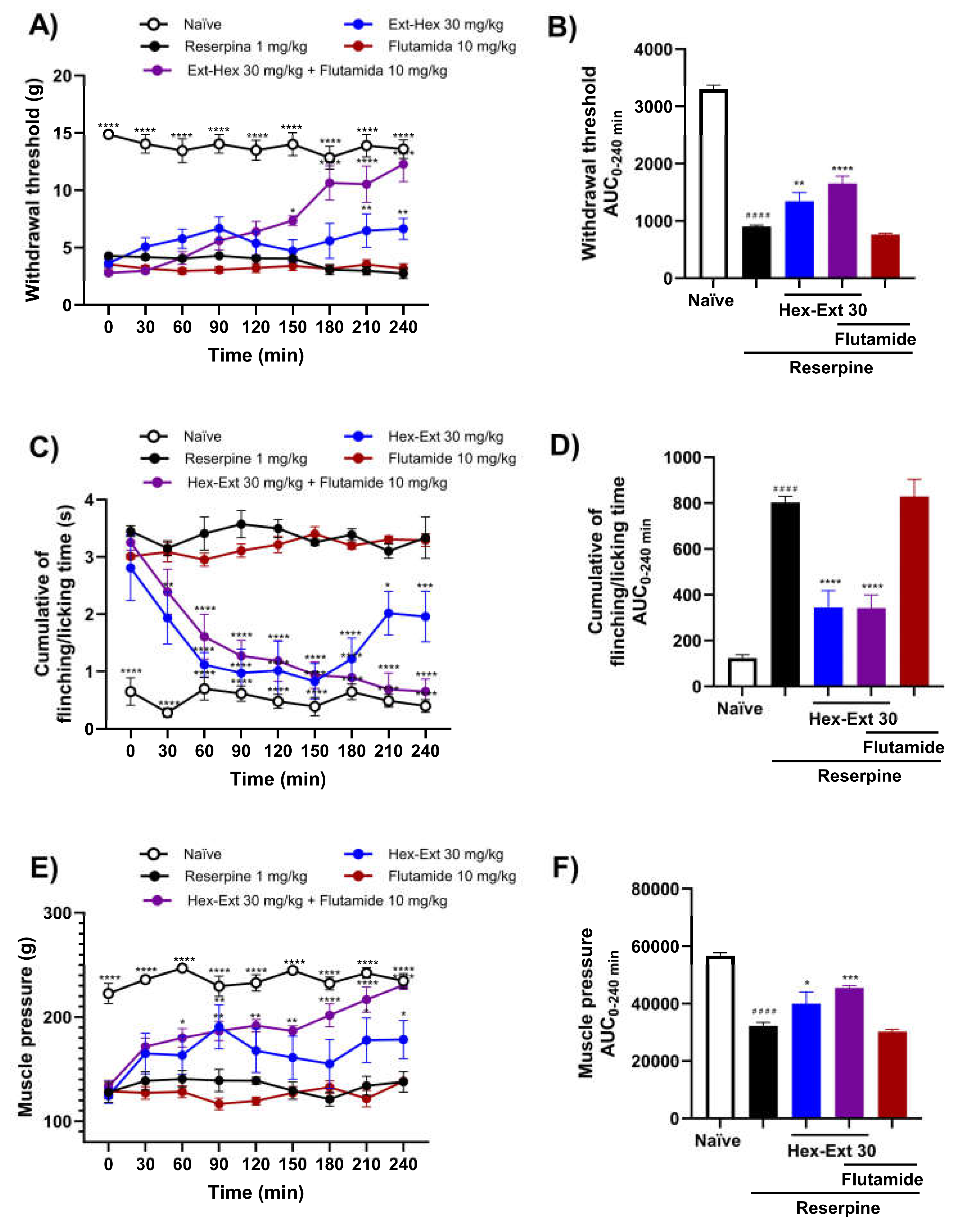

Table 1.
Identified compounds in the hexane extract of S. rosmarinus.
| Peak | Rt (min) | %Relative area | Compound | Molecular Formula | MW (g/mol) |
|---|---|---|---|---|---|
| 1 | 7.022 | 2.23 | (+)-α-Pinene | C10H16 | 136.24 |
| 2 | 7.326 | 1.71 | Camphene | C10H16 | 136.24 |
| 3 | 7.882 | 1.66 | β-Pinene | C10H16 | 136.24 |
| 4 | 8.419 | 0.33 | α-Phellandrene | C10H16 | 136.24 |
| 5 | 8.82 | 0.92 | o-Cymene | C10H14 | 134.22 |
| 6 | 8.917 | 0.48 | 4-Thujanol | C10H18O | 154.25 |
| 7 | 8.956 | 5.34 | 1,8-Cineole (eucalyptol) | C10H18O | 154.25 |
| 8 | 11.188 | 24.47 | (+)-2-Bornanone (camphor) | C10H16O | 152.24 |
| 9 | 11.576 | 2.37 | Borneol | C10H18O | 154.25 |
| 10 | 13.73 | 3.38 | (-)-Bornyl acetate | C12H20O2 | 196.29 |
| 11 | 16.059 | 1.96 | Caryophyllene | C15H24 | 204.36 |
| 12 | 31.111 | 1.07 | Heptacosane | C27H56 | 380.75 |
| 13 | 32.864 | 8.15 | Nonacosane | C29H60 | 408.80 |
| 14 | 34.494 | 5.65 | Hentriacontane | C31H64 | 436.85 |
| 15 | 36.196 | 7.58 | Tritriacontane | C33H68 | 464.91 |
| 16 | 37.017 | 6.47 | β-Amyrone | C30H48O | 424.71 |
Disclaimer/Publisher’s Note: The statements, opinions and data contained in all publications are solely those of the individual author(s) and contributor(s) and not of MDPI and/or the editor(s). MDPI and/or the editor(s) disclaim responsibility for any injury to people or property resulting from any ideas, methods, instructions or products referred to in the content. |
© 2026 by the authors. Licensee MDPI, Basel, Switzerland. This article is an open access article distributed under the terms and conditions of the Creative Commons Attribution (CC BY) license.
Copyright: This open access article is published under a Creative Commons CC BY 4.0 license, which permit the free download, distribution, and reuse, provided that the author and preprint are cited in any reuse.