

Submitted:
04 September 2026
Posted:
07 September 2026
You are already at the latest version
Abstract
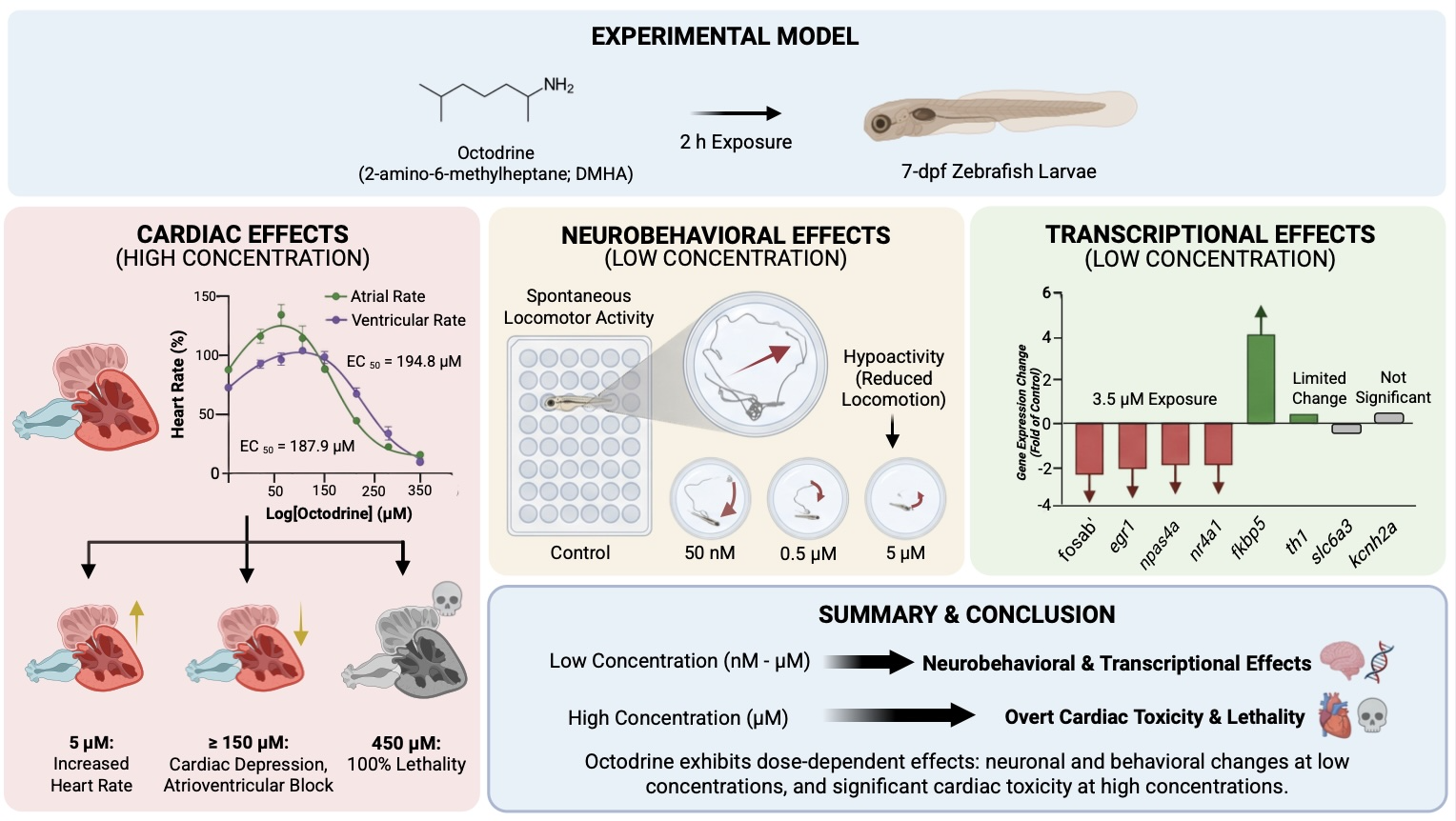
Octodrine (2-amino-6-methylheptane; DMHA) is a sympathomimetic stimulant detected in sports-performance and weight-loss supplements despite limited toxicological characterization and regulatory concerns. Here, we used 7-day-post-fertilization zebrafish larvae to characterize the acute cardiac, neurobehavioral, and transcriptional effects of octodrine after 2 h of exposure. Cardiac responses were biphasic: 5 µM octodrine significantly increased atrial and ventricular rates, whereas concentrations ≥150 µM produced progressive cardiac depression and higher concentrations disrupted atrioventricular conduction. Exposure to 450 µM resulted in 100% lethality. Nonlinear concentration–response analysis yielded EC50 values of 194.8 µM for atrial rate and 187.9 µM for ventricular rate. At substantially lower concentrations, octodrine significantly reduced spontaneous locomotor activity, with effects detectable at 50 nM and more sustained hypoactivity at 0.5 and 5 µM across most of the 2 h exposure period. At the transcriptional level, exposure to 3.5 µM suppressed several neuronal activity–responsive genes, including fosab, egr1, and npas4a, while nr4a1 was downregulated at higher concentrations. The stress-responsive glucocorticoid target fkbp5 was induced at selected concentrations, whereas th1 and kcnh2a expression was not significantly altered. Together, these findings reveal a marked separation between low-concentration neurobehavioral and transcriptional effects and overt cardiac toxicity at substantially higher concentrations. The results provide an initial in vivo toxicological profile of octodrine and support further investigation of its neuropharmacological mechanisms, internal exposure, and cardiovascular safety.
Keywords:
octodrine
; DMHA
; zebrafish
; neurobehavior
; cardiotoxicity
; atrioventricular block
; immediate early genes
; dietary supplements
1. Introduction
The expanding market for pre-workout, thermogenic, weight-loss, and performance-enhancing supplements has increased human exposure to pharmacologically active stimulants for which efficacy and safety are often incompletely characterized. In addition to conventional ingredients such as caffeine, analytical surveys have repeatedly identified synthetic or experimental sympathomimetic compounds in commercially available products, sometimes under ambiguous ingredient names or with substantial discrepancies between labelled and measured contents [1,2]. This is of particular concern in sports supplements, where stimulant combinations may result in poorly predictable pharmacological interactions and may also expose athletes to substances prohibited in competition. Among the compounds that have emerged in this context is octodrine, also known as 2-amino-6-methylheptane or 1,5-dimethylhexylamine (DMHA).
Octodrine is not a newly synthesized compound. It was pharmacologically investigated during the mid-twentieth century and used in pharmaceutical preparations, including nasal decongestant formulations, before largely disappearing from medical use. Decades later, it re-emerged as an ingredient in pre-workout and weight-loss supplements, frequently promoted as an alternative to other sympathomimetic stimulants such as 1,3-dimethylamylamine (DMAA) [3]. Chemical analyses have confirmed the presence of octodrine in commercial sports and weight-loss supplements. Octodrine, together with other experimental or prohibited stimulants, has been identified in products marketed in the United States [1], while this compound has been found in several commercial supplements, with reported amounts reaching 112 mg per serving [4]. The latter study also found a racemic enantiomeric distribution and synthetic by-products, supporting a synthetic rather than botanical origin of the octodrine detected in these products. Regulatory concerns have consequently increased: the U.S. Food and Drug Administration considers DMHA an unsafe food additive that does not meet the statutory definition of a dietary ingredient and considers dietary supplements containing it to be adulterated [5]. Octodrine is also classified by the World Anti-Doping Agency as a specified stimulant prohibited in competition [6].
Despite this pattern of human exposure, the pharmacology and toxicology of octodrine remain remarkably poorly characterized. Much of the experimental evidence originates from studies conducted more than seven decades ago. In the classical study by Fellows (1947) [7], 2-amino-6-methylheptane displayed pressor activity and increased cardiac rate and contractile amplitude in anesthetized dogs, together with local anesthetic effects, whereas central nervous system stimulation was minimal in rats at non-toxic doses. Other early studies similarly described cardiovascular and respiratory actions, but these investigations predated modern molecular and neurobehavioral approaches [3]. More recent literature has focused predominantly on the occurrence of octodrine in supplements, analytical detection, regulatory issues, and doping rather than on experimental characterization of its biological effects. Although octodrine is generally marketed and described as a stimulant and catecholaminergic actions have been proposed, its molecular targets and the contribution of dopaminergic, adrenergic, or other neuronal pathways remain poorly resolved. Likewise, contemporary experimental information on its potential cardiotoxicity is extremely limited. This lack of mechanistic toxicological information contrasts with both its availability in commercial products and the cardiovascular adverse effects associated with sympathomimetic stimulant exposure.
Zebrafish provide a particularly suitable vertebrate model for addressing this knowledge gap because neurobehavioral and cardiovascular effects can be evaluated within the same intact organism over short exposure periods. Early-life zebrafish possess functional monoaminergic systems and exhibit quantifiable locomotor responses to psychoactive compounds, while their optical accessibility permits direct assessment of atrial and ventricular activity and atrioventricular conduction [8,9]. This combination has proven useful for the integrated characterization of stimulant compounds. For example, synthetic cathinones and classical psychostimulants produce compound-specific alterations in locomotor activity, cardiac rate, rhythmicity, and atrioventricular conduction in zebrafish, demonstrating that neurobehavioral and cardiovascular liabilities can be resolved within a common experimental framework [9,10]. The model further enables functional phenotypes to be linked with rapid molecular responses, including changes in genes associated with monoaminergic signalling, neuronal activity, cellular stress, and cardiac electrophysiology.
In the present study, we therefore used 7-day-post-fertilization (dpf) zebrafish larvae to provide an integrated characterization of the acute effects of octodrine across neurobehavioral, cardiovascular, and molecular levels. Spontaneous locomotor activity was assessed as a sensitive functional readout of neuroactive effects, whereas high-speed cardiac imaging was used to quantify atrial and ventricular rate and atrioventricular conduction. To explore molecular responses associated with these phenotypes, we additionally examined the expression of genes involved in dopamine synthesis and transport (th1 and slc6a3), neuronal activity-dependent transcription (fosab, egr1, npas4a, and nr4a1), stress signalling (fkbp5), and cardiac repolarization (kcnh2a). By integrating these complementary endpoints across concentration ranges, the study aimed to define the acute neurofunctional and cardiovascular hazard profile of octodrine and to identify molecular responses that may help guide subsequent mechanistic investigation.
2. Results
2.1. Acute Octodrine Exposure Impairs Cardiac Function and Atrioventricular Conduction
Exposure to 450 µM octodrine resulted in 100% lethality and was therefore excluded from all subsequent quantitative cardiac analyses. Among surviving larvae, octodrine produced a biphasic effect on cardiac activity (Figure 1A; individual data are available at Supplementary Dataset 1). Generalized estimating equations (GEE) analysis revealed a strong overall effect of octodrine concentration on heart rate (Wald χ2(5) = 2286.91, p < 0.001), as well as a significant concentration × cardiac chamber interaction (Wald χ2(3) = 12.39, p = 0.006), whereas no significant effect of experimental replicate was detected (Wald χ2(2) = 3.61, p = 0.165). At 5 µM, atrial and ventricular rates were significantly increased relative to the corresponding controls (both Holm-adjusted p < 0.001). The numerical reduction observed at 50 µM did not reach statistical significance (both Holm-adjusted p = 0.076). From 150 µM onward, octodrine produced a marked and progressive reduction in both atrial and ventricular rates (all Holm-adjusted p < 0.001). Full GEE model statistics, estimated marginal means, and Holm-adjusted planned contrasts for cardiac rate are provided in Supplementary Table S1.
Concentration–response analysis confirmed a similar sensitivity of atrial and ventricular function to octodrine. Nonlinear regression analysis yielded an EC50 of 194.8 μM for atrial rate (95% CI: 179.2–210.0 μM; R2 = 0.805) and 187.9 μM for ventricular rate (95% CI: 173.5–201.8 μM; R2 = 0.826).
Octodrine also altered atrioventricular conduction at the higher concentrations tested (Figure 1B,C). A 1:1 atrial-to-ventricular conduction ratio was observed in all larvae exposed to concentrations up to 150 µM (Figure 1B). At 250 µM, a small proportion of larvae exhibited 2:1 AV conduction, whereas at 350 µM both 2:1 and >2:1 conduction ratios were observed. Accordingly, the mean A/V ratio remained close to 1 up to 150 µM, increased at 250 µM, and reached its highest value at 350 µM (Figure 1C). At 350 µM, the A/V ratio was significantly higher than in the control, 5, 50, and 150 µM groups.
2.2. Octodrine Reduces Spontaneous Locomotor Activity
Octodrine exposure significantly altered spontaneous locomotor activity during the 2 h exposure period (Figure 2; individual data are available at Supplementary Dataset 1). GEE analysis revealed significant effects of treatment (Wald χ2(3) = 41.37, p < 0.001) and time interval (Wald χ2(7) = 593.65, p < 0.001), together with a significant treatment × time interaction (Wald χ2(21) = 44.87, p = 0.002), indicating that the effect of octodrine varied over the course of the exposure. Full GEE model statistics, estimated marginal means, and time-specific Holm-adjusted contrasts for locomotor activity are provided in Supplementary Table S2.
A reduction in distance travelled was already evident during the first 15 min of exposure. At 50 nM, locomotor activity was significantly lower than in vehicle controls during the 0–15, 15–30, 30–45, 45–60, and 75–90 min intervals (Holm-adjusted p < 0.05), whereas differences were not significant during the 60–75, 90–105, or 105–120 min intervals. Exposure to 500 nM produced a more sustained reduction, with significantly decreased locomotor activity throughout the first seven recording intervals (0–105 min; all Holm-adjusted p ≤ 0.0168), but not during the final 105–120 min interval (p = 0.132). The strongest and most consistent reduction was observed at 5 µM, which showed the lowest estimated locomotor activity throughout the recording period and differed significantly from controls during each of the first seven intervals (all Holm-adjusted p ≤ 0.0013). The difference at 105–120 min did not reach statistical significance (p = 0.090).
2.3. Octodrine Modulates Neuronal Activity- and Stress-Responsive Gene Expression
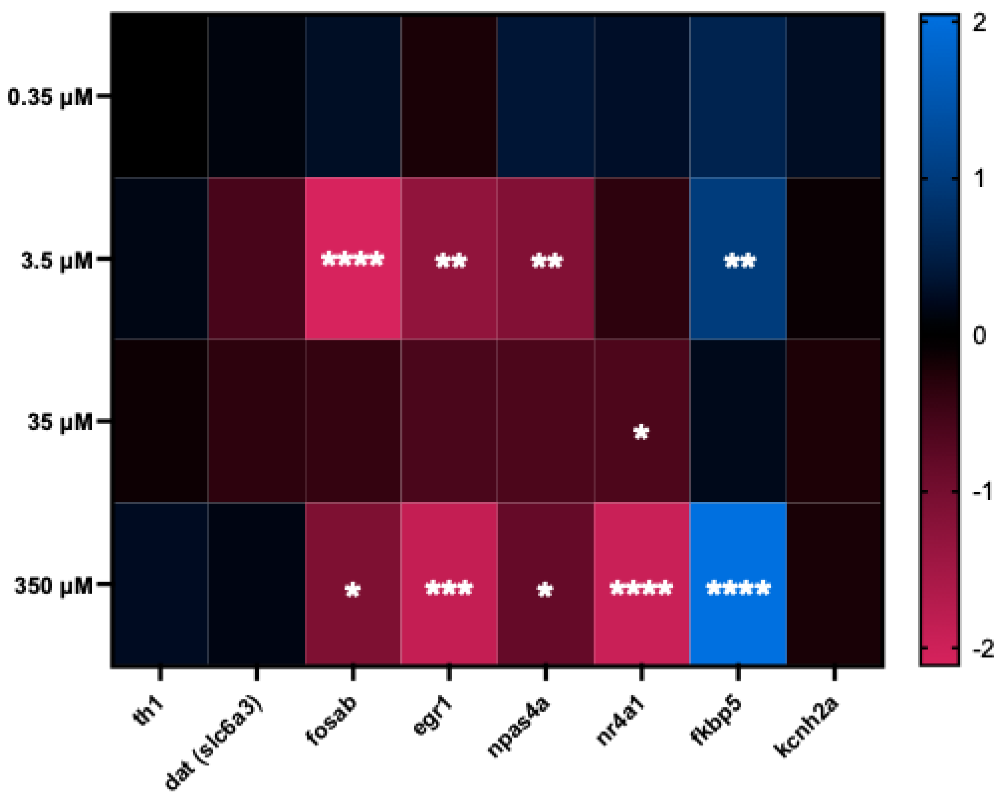
Exposure to octodrine for 2 h produced concentration-related changes in the transcriptional profile of genes associated with dopaminergic function, neuronal activity, stress signalling, and cardiac electrophysiology (Figure 3; individual data are available in Supplementary Dataset 1). Expression of the reference gene was stable across concentrations (ppiaa: F(4, 35) = 0.351, p = 0.841). Global analyses revealed significant effects of octodrine concentration on slc6a3 (F(4, 35) = 3.22, p = 0.024), fosab (F(4, 35) = 7.99, p < 0.001), egr1 (F(4, 35) = 13.03, p < 0.0001), npas4a (H(4) = 19.35, p < 0.001), nr4a1 (F(4, 35) = 26.11, p < 0.0001), and fkbp5 (F(4, 35) = 29.52, p < 0.0001), whereas no significant overall effects were detected for th1 or kcnh2a. Full global and post hoc statistics are provided in Supplementary Table S3.
At 0.35 µM, transcriptional effects were limited, with a modest but significant induction of fkbp5 (p = 0.031) and no significant changes in the other genes examined. At 3.5 µM, a broader transcriptional response became evident, characterized by significant downregulation of the neuronal activity-responsive genes fosab (p < 0.001), egr1 (p < 0.001), and npas4a (p = 0.044), together with induction of fkbp5 (p < 0.001). nr4a1 was not significantly altered at this concentration.
At 35 µM, nr4a1 was significantly downregulated (p= 0.048), whereas the other genes did not differ significantly from the vehicle control. At 350 µM, fosab, egr1, npas4a, and nr4a1 were significantly downregulated, while fkbp5 was strongly induced. Although the overall concentration effect for slc6a3 was significant, none of the individual octodrine concentrations differed significantly from the vehicle control after Dunnett adjustment. Similarly, th1 and kcnh2a showed no significant pairwise changes across the concentration range.
3. Discussion
The present study provides an integrated in vivo characterization of the acute effects of octodrine across behavioural, cardiac, and molecular endpoints. Three main findings emerge. First, spontaneous locomotor activity was reduced at submicromolar-to-low-micromolar concentrations, well below those required to produce overt cardiac dysfunction. Second, a similar low-micromolar exposure range was associated with reduced expression of several neuronal activity-responsive transcripts, while the dopaminergic synthesis and transport markers th1 and slc6a3 were comparatively preserved. Third, at higher concentrations, octodrine caused pronounced bradycardia and atrioventricular conduction abnormalities, revealing a distinct cardiovascular toxicity domain. This separation of neurofunctional and cardiac effect concentrations is particularly relevant because octodrine remains pharmacologically under-characterized despite its documented presence at substantial doses in sports and weight-loss supplements and continuing regulatory concern [1,3,4,5].
The behavioural data indicate that the nervous system is a highly sensitive target of acute octodrine exposure. Significant reductions in spontaneous locomotion were detected even at 50 nM, although this effect was less consistently maintained over time, whereas 0.5 and 5 µM produced more sustained hypoactivity. Locomotor activity also progressively decreased over the recording period in vehicle-control larvae. Such time-dependent reductions in spontaneous locomotion following transfer to a behavioural recording environment have previously been described in zebrafish larvae and interpreted as habituation to the testing apparatus [11,12]. In the present study, recording was initiated immediately after addition of the exposure solutions and placement of the plate in the DanioVision chamber, without a preceding acclimation period, which likely contributed to the pronounced temporal decline in control activity. Importantly, this common time-dependent change was explicitly incorporated into the GEE model, allowing treatment-related effects and their temporal evolution to be distinguished from the overall decline in locomotor activity. Notably, the cardiac response observed at 5 µM consisted of increased rather than depressed atrial and ventricular rates, while pronounced bradycardia and AV conduction abnormalities required substantially higher concentrations. This separation argues against the hypoactivity being a secondary consequence of impaired cardiac function and instead supports a direct or indirect neuropharmacological action of octodrine. Interestingly, a comparable separation between neurobehavioral and cardiovascular effect concentrations has recently been observed for synthetic cathinones in zebrafish, with locomotor suppression occurring in the low-micromolar range and marked bradycardia and AV conduction impairment requiring substantially higher concentrations [10,13]. Although octodrine and synthetic cathinones differ substantially in chemical structure and pharmacology, these findings illustrate the ability of an integrated zebrafish approach to resolve distinct neurobehavioral and cardiovascular response domains across stimulant classes. Nevertheless, reduced locomotion per se does not identify a specific neurotransmitter mechanism. Octodrine has historically been classified as a stimulant or sympathomimetic compound, but its molecular targets and actions at monoamine transporters or receptors remain poorly defined, and the available pharmacological evidence is remarkably sparse [3,7].
The transcriptional profile provides additional evidence that neural function is affected within the same low-micromolar concentration range. The genes examined here represent complementary activity-responsive transcriptional markers in zebrafish. fosab, the zebrafish orthologue of mammalian c-FOS, is widely used as a molecular readout integrating recent neuronal activity in the larval brain [14], whereas egr1 is an immediate-early gene rapidly induced by sensory stimulation in zebrafish larvae [15]. Similarly, npas4a is predominantly expressed in the zebrafish brain and is strongly induced by neuronal activation [16], while nr4a1 has been identified among neuronal activity-dependent immediate-early genes associated with neuroplastic responses in the zebrafish brain [17]. The coordinated reduction of fosab, egr1, and npas4a at 3.5 µM is therefore consistent with the hypoactive behavioural phenotype observed within a closely overlapping concentration range. Interestingly, a similar transcriptional pattern has been reported in zebrafish following developmental exposure to amphetamine, nicotine, and oxycodone, all of which produced downregulation of fosab, egr1, and npas4a together with alterations in locomotor behaviour [18]. Although the exposure paradigms differ substantially, these findings support the interpretation that coordinated suppression of neuronal activity-responsive genes can accompany drug-induced neurobehavioral alterations in zebrafish. Acute modulation of the same transcriptional network has also been observed following 2 h exposure to the synthetic cathinone MDPV in 3 dpf zebrafish embryos, although with a markedly different concentration-dependent profile [13]. In that study, fosab and egr1 were upregulated at higher MDPV concentrations, whereas npas4a and nr4a1 showed biphasic regulation, including downregulation at lower concentrations and induction at the highest concentration tested [13]. Thus, rapid modulation of neuronal activity-responsive genes appears to be a recurrent feature of acute stimulant exposure in zebrafish, while the direction and magnitude of the response may depend on compound identity, concentration, and developmental stage.
In contrast, th1 expression was not significantly altered, while slc6a3 showed only a modest overall concentration effect, with no individual octodrine concentration differing significantly from the vehicle control. These findings suggest that the acute response does not involve major transcriptional remodelling of dopamine synthesis or dopamine transporter expression within the 2 h exposure window. Importantly, this does not exclude acute effects of octodrine on dopaminergic neurotransmission through changes in transporter function, dopamine release, receptor activation, or other non-transcriptional mechanisms, none of which can be inferred from whole-larva transcript abundance.
The induction of fkbp5 provides an additional indication that octodrine engages stress-responsive signalling. In zebrafish, fkbp5 is a well-characterized glucocorticoid receptor (GR)-responsive gene that participates in feedback regulation of glucocorticoid signalling. Its expression exhibits GR-dependent regulation, and genetic disruption of GR signalling markedly alters fkbp5 transcription in zebrafish [19,20]. Interestingly, fkbp5 was also significantly induced following a 2 h exposure to MDPV in zebrafish embryos, providing an additional precedent for rapid engagement of stress-responsive transcription by structurally distinct stimulant compounds [13]. The induction observed following low-micromolar octodrine exposure is therefore compatible with engagement of glucocorticoid-responsive signalling and may reflect activation of the hypothalamic–pituitary–interrenal stress axis. However, fkbp5 expression alone cannot establish either increased cortisol production or activation of the HPI axis, and these possibilities should be tested directly. At 350 µM, the interpretation must be even more conservative since cardiac function is already severely compromised. Transcriptional alterations at this concentration may consequently reflect generalized physiological stress, altered tissue oxygenation, or systemic toxicity rather than a primary neuronal response. Measurements of whole-body cortisol, monoamine concentrations, and spatially resolved neuronal activity would help distinguish these mechanisms.
Cardiac responses to octodrine were biphasic. At 5 µM, atrial and ventricular rates were modestly but significantly increased, whereas concentrations ≥150 µM produced a progressive reduction in cardiac rate. Thus, overt cardiac toxicity was considerably less sensitive than the locomotor phenotype. Atrial and ventricular rates declined in parallel, with EC50 values close to 190 µM, while AV conduction abnormalities emerged at the upper end of the concentration range. Thus, overt cardiac toxicity was considerably less sensitive than the locomotor phenotype. Atrial and ventricular rates declined in parallel, with EC50 values close to 190 µM, while AV conduction abnormalities emerged at the upper end of the concentration range. Zebrafish are particularly sensitive to pharmacological perturbation of cardiac repolarization, and compounds that interfere with repolarizing currents can produce pronounced bradycardia and AV block [21]. More recent functional studies have further demonstrated that both ERG potassium-channel blockade and inhibition of T-type calcium channels can produce bradycardia and AV conduction block in zebrafish larvae through different electrophysiological mechanisms [22]. Thus, the cardiac phenotype observed here is compatible with disruption of cardiac excitability or conduction but does not, by itself, identify the underlying molecular target.
In this context, the absence of significant changes in kcnh2a expression should be interpreted cautiously. Although human cardiac IKr is mediated by KCNH2/hERG, the corresponding repolarizing current in the zebrafish heart is predominantly mediated by the paralogue Kcnh6a rather than Kcnh2a [22]. Therefore, unchanged kcnh2a transcript abundance cannot be used to infer preservation of ERG-mediated repolarization. Moreover, direct pharmacological inhibition of ion-channel function would not necessarily require transcriptional modulation. Interestingly, acute MDPV exposure in zebrafish also produces marked bradycardia and AV block without significant transcriptional modulation of kcnh6a or kcnq1 [13], further illustrating the limitations of transcript abundance as a surrogate for acute cardiac ion-channel function. Direct electrophysiological or pharmacological studies, together with assessment of kcnh6a and other relevant cardiac ion-channel targets, would therefore be required to determine the mechanism underlying octodrine-induced conduction abnormalities.
The toxicological relevance of these findings lies primarily in hazard identification rather than direct prediction of human risk. Analyses of commercially available supplements have detected approximately 72 mg of octodrine per serving, more than twice the highest historical pharmaceutical dose, while another survey reported concentrations reaching 112 mg per serving [1,4]. These findings demonstrate that consumers may be exposed to pharmacologically substantial quantities of octodrine despite the limited information available regarding its safety. Nevertheless, nominal waterborne concentrations in zebrafish cannot be directly translated into human doses or circulating concentrations. Zebrafish larvae can absorb, metabolize, accumulate, and excrete xenobiotics to markedly different extents depending on compound-specific physicochemical and metabolic properties, such that nominal bath concentration may differ substantially from internal exposure [23]. Determination of octodrine uptake, internal concentrations, and metabolite profiles would therefore be necessary before quantitative comparisons with human exposure can be attempted. The same consideration applies to establishing whether the low-micromolar concentrations associated with behavioural effects in the present study are achievable in human plasma or target tissues.
Several limitations should therefore be considered. The study evaluated only acute exposure and a relatively small molecular panel, and transcript measurements were performed in whole larvae, limiting both anatomical and cell-type resolution. The experiments did not directly quantify internal octodrine concentrations, monoamines, cortisol, neuronal activity, or cardiac electrophysiology. Moreover, gene-expression measurements provide information on transcriptional responses but cannot establish rapid pharmacological interactions with neurotransmitter transporters, receptors, or ion channels. Despite these limitations, the convergence between locomotor suppression and reduced expression of multiple neuronal activity-responsive genes within the low-micromolar range, together with a distinct high-concentration cardiac phenotype, provides a coherent initial characterization of the acute effects of this poorly studied stimulant used in dietary supplements and identifies specific neuroendocrine and electrophysiological mechanisms warranting further investigation.
3.1. Conclusions
Acute octodrine exposure produced two clearly separable toxicological response domains in zebrafish larvae. Neurobehavioral effects and reduced expression of neuronal activity-responsive transcripts occurred at nanomolar-to-low-micromolar concentrations, whereas marked bradycardia and atrioventricular conduction impairment required substantially higher concentrations. The absence of significant changes in th1, together with only a modest overall effect on slc6a3 that did not translate into significant treatment-versus-control differences, argues against major acute transcriptional remodelling of dopaminergic machinery, although non-transcriptional effects on monoaminergic signalling cannot be excluded. In parallel, fkbp5 induction is consistent with engagement of glucocorticoid-responsive stress signalling. At higher concentrations, the cardiac phenotype indicates disruption of excitability and/or conduction, although its molecular basis remains unresolved. Collectively, these findings provide an initial integrated toxicological profile of octodrine and support targeted studies on monoaminergic signalling, glucocorticoid responses, internal dosimetry, and cardiac electrophysiology.
4. Material and Methods
4.1. Zebrafish Maintenance and Embryo Production
Adult wild-type short-fin sexually mature zebrafish, within the typical standard length range for breeding stock (3.0–3.5 cm), were obtained from a commercial supplier (Exopet, Madrid, Spain) and were maintained under a 12 h: 12 h light/dark cycle and a temperature of 28 ± 1ºC at the Research and Development Center (CID-CSIC) in a recirculating water system (Aquaneering Inc., San Diego, CA, USA).
Embryos were obtained through natural spawning in breeding tanks from groups composed by three females and two males. Fertilized eggs were collected within 30 min post-spawning (lights-on). Only high-quality embryos at the 50%-epiboly stage were selected under a stereomicroscope (Nikon SMZ1500, Champigny-sur-Marne, France). Embryos were transferred to a crystallizing dish containing embryo water [Milli-Q water with 90 mg/L Instant Ocean (Aquarium Systems, Sarrebourg, France) and 0.58 mM CaSO4·2H2O, pH 6.5–7.0, 750–900 μS/cm conductivity] and incubated at 28 ± 1 ◦C in a climatic chamber (POL-EKO APARATURA KK350, Wodzisław Śląski, Poland) under a 12 h:12 h light/dark cycle until the start of the experiments at 7 dpf.
All procedures were approved by the Institutional Animal Care and Use Committees at the CID-CSIC and conducted in accordance with the institutional guidelines under a license from the local government (agreement number 11336).
4.2. Chemicals
Octodrine (CAS 543-82-8) was purchased as an analytical standard from Sigma-Aldrich. Stock solutions (50 mM) were freshly prepared in DMSO on the day of each experiment. Working solutions were prepared by dilution in embryo water and contained a final DMSO concentration of 1% (v/v) in all exposure groups. The corresponding vehicle control also contained 1% DMSO. A constant DMSO concentration was maintained across treatments to avoid concentration-dependent differences in solvent exposure and to accommodate the highest octodrine concentration tested (450 µM), which required 0.9% (v/v) of the 50 mM DMSO stock solution. Although 1% DMSO is at the upper end of concentrations commonly used as a vehicle in zebrafish assays, acute exposures at this concentration have been reported to produce no detectable alterations in larval locomotor behaviour [24]. The pH of all exposure solutions was adjusted to approximately 8.3 with 1 M NaHCO3. This value was selected as a compromise between approaching the pKa of octodrine (pKa = 10.42) and maintaining exposure conditions within a pH range physiologically tolerated by zebrafish larvae [25].
4.3. Cardiotoxicity Assessment
The cardiotoxicity assessment followed the protocol described elsewhere [13]. 7 dpf larvae were exposed for 2 h to 5–450 μM octodrine in 48-well plates (one larva per well, 1 mL solution). To minimize movement during imaging, larvae were briefly anesthetized (0.08 mg/mL tricaine methane-sulfonate [MS-222], 10 s) and embedded in 3% methylcellulose on a depression microscope slide. Cardiac activity was recorded laterally with a high-speed camera (ace U acA1440-220 um, Basler AG, Ahrensburg, Germany) attached to a stereomicroscope (SMZ1500, Nikon, Champigny sur Marne, France) at 100 fps for 20 s using Pylon Viewer 64-Bit software (9.1.0.1614) at 28 ± 1 ◦C.
Atrial and ventricular beat frequencies were quantified using DanioScope v1.0 (Noldus Information Technology, Wageningen, The Netherlands), which derives heart rate from pixel-intensity fluctuations over time [26]. Atrial and ventricular activity were analyzed separately to assess chamber-specific effects of octodrine. The atrioventricular (AV) ratio, defined as the ratio between atrial and ventricular beat frequencies, was calculated for each embryo as an indicator of conduction efficiency (1.0 = full AV conduction).
To quantify the degree of conduction impairment, the AV ratio was transformed into percentage AV block according to the expression:

Concentration–response relationships were analyzed by nonlinear regression using a four-parameter logistic model (variable slope) in GraphPad Prism 10. Concentrations were log10-transformed prior to curve fitting. Top and bottom values were constrained to 100 and 0, respectively. The half-maximal effective concentration (EC50) and corresponding 95% confidence intervals were estimated separately for atrial and ventricular responses. Goodness-of-fit was assessed using the coefficient of determination (R2). The 450 µM group was excluded from concentration–response modelling because this concentration resulted in 100% lethality.
4.4. Behavioural Assessment
Basal locomotor activity (BLA) was assessed in 7 dpf zebrafish larvae exposed for 2 h to 50 nM, 500 nM and 5 μM octodrine. Vehicle control larvae exposed to embryo water containing 1% DMSO were used. Recordings were made on the DanioVision platform (Noldus Information Technology, Wageningen, The Netherlands) under near-infrared illumination with temperature maintained at 28 ◦C. Video tracking was performed using EthoVision XT software (v13 for recording, v16 for analysis).
Immediately after addition of the exposure solutions, the plate was transferred to the DanioVision chamber and recording was initiated without a prior acclimation period. Locomotor behavior was quantified during eight consecutive 15-min observation windows over a 120-min period (0–15, 15–30, 30–45, 45–60, 60–75, 75–90, 90–105, and 105–120 min). For each larva, BLA was defined as the total distance (cm) travelled within each window [13].
4.5. RNA Preparation and qRT-PCR Analysis
Total RNA was extracted from pools of six 7 dpf zebrafish larvae (control or exposed to 0.35–350 μM octodrine) using TRIzol Reagent (Invitrogen, Carlsbad, CA, USA). RNA concentration and purity were determined spectrophotometrically (NanoDrop ND-8000, NanoDrop Technologies, Wilmington, DE, USA). Following DNase I treatment (Ambion, Austin, TX, USA), RNA was reverse-transcribed using the First Strand cDNA Synthesis Kit (Roche Diagnostics, Mannheim, Germany).
Quantitative real-time PCR (qRT-PCR) was performed on a LightCycler 480 System (Roche Diagnostics) using SYBR Green Master Mix (Roche) under the following cycling conditions: 95 °C for 15 min, followed by 45 cycles of 95 °C for 10 s and 60 °C for 30 s. Each experimental condition included eight biological replicates and three technical replicates. Primers targeting th1, slc6a3, fosab, egr1, npas4a, nr4a1, fkbp5, and kcnh2a were designed using Primer-BLAST (National Center for Biotechnology Information, Bethesda, MD, USA). Primer sequences for these genes, together with those for the reference gene ppiaa (peptidyl-prolyl isomerase A), are listed in Supplementary Table S4.
Gene expression levels were normalized against the reference gene ppiaa. Relative expression was calculated using the comparative Ct (ΔΔCt) method. For graphical representation, results are expressed as log2 fold change (log2FC) relative to the control group. Statistical analyses were performed using individual ΔCt values, which satisfy the assumptions of parametric testing more appropriately than transformed fold-change values. Normality and homogeneity of variance were assessed prior to statistical analysis [13].
4.6. Data Analysis
Statistical analyses were performed using IBM SPSS Statistics and GraphPad Prism version 10.0 (GraphPad Software, San Diego, CA, USA), as specified below. Statistical significance was set at p < 0.05.
Atrial and ventricular heart rates were analysed jointly using generalized estimating equations (GEE), with octodrine concentration and cardiac chamber as fixed factors, their interaction included in the model, and experimental replicate entered as a blocking factor. Larva identity was specified as the subject variable, with atrial and ventricular measurements treated as repeated observations within each subject. GEE was selected to account for the within-larva correlation between cardiac chambers while providing robust inference in the presence of heteroscedasticity. A normal distribution with identity link, robust covariance estimator, and unstructured working correlation matrix were used. Planned comparisons between each octodrine concentration and the corresponding vehicle control were adjusted for multiple testing using the sequential Bonferroni (Holm) procedure.
Concentration–response relationships were evaluated by nonlinear regression using a four-parameter logistic model (variable slope), from which EC50 values and their corresponding 95% confidence intervals were estimated.
The differences between the concentrations in A/V ratio were evaluated by one-way ANOVA, followed by Tukey’s post hoc test for multiple comparisons.
Locomotor activity was measured repeatedly in the same larvae over time, resulting in correlated within-subject observations that violate the independence assumption of conventional between-group tests. Therefore, locomotor activity data were analysed using GEE to account for the repeated measurements obtained from the same larva across eight consecutive 15-min intervals. Distance travelled was modelled using a gamma distribution with a log link function because distance-travelled data were strictly positive and right-skewed. Larva identity was specified as the subject variable and time interval as the within-subject repeated factor. Octodrine treatment, time interval, and their interaction were included as fixed effects, while experimental replicate was included as a blocking factor. An autoregressive first-order [AR(1)] working correlation structure and a robust sandwich covariance estimator were used. Estimated marginal means were calculated on the original response scale, and planned comparisons between each octodrine concentration and the corresponding vehicle control within each time interval were adjusted for multiple testing using the sequential Bonferroni (Holm) procedure.
Gene-expression analyses were performed on individual ΔCt values. For each gene, one-way ANOVA followed by Dunnett’s multiple-comparisons test against the vehicle control was used when residuals satisfied normality and homogeneity-of-variance assumptions. When these assumptions were not met, Kruskal–Wallis analysis followed by Dunn’s multiple-comparisons test with Holm adjustment was used. Relative gene expression was calculated using the 2^−ΔΔCt method and expressed graphically as log2 fold change relative to the vehicle control.
Author Contributions
Conceptualization: D.R., I.F., C.F.A.-L., E.K.C.-D.; Methodology: O.A.; Formal Analysis: D.R., E.K.C.-D; Investigation: E.K.C.-D., O.A., E.P.; Resources: D.R.; Writing—Original Draft Preparation: D.R., E.K.C.-D., O.A., E.P.; Writing—Review and Editing: D.R., E.K.C.-D., O.A., E.P., I.F., C.F.A.-L.; Visualization: D.R., E.K.C-D.; Supervision: D.R, I.F., C.F.A.-L.; Project Administration: D.R.; Funding Acquisition: D.R. All authors have read and agreed to the published version of the manuscript.
Funding
This work was funded by MICIU/AEI/10.13039/501100011033 (grant number PID2023-148502OB-C21). O.A. was supported by MICIU/AEI/10.13039/501100011033, co-financed by the Spanish Government and the European Social Fund (grant number PREP2022-104748). This work was also partially supported by the Coordination for Superior Level Staff Improvement (CAPES, Brazil) through an Institutional Sandwich Doctorate Program Abroad (PDSE) fellowship (Process No. 88881.220305/2025-01) awarded to E.K.C.-D.
Institutional Review Board Statement
All procedures were approved by the Institutional Animal Care and Use Committees at the CID-CSIC and conducted in accordance with the institutional guidelines under a license from the local government (agreement number 11336, 18 May 2022).
Informed Consent Statement
Not applicable.
Data Availability Statement
The original contributions presented in this study are included in the article/Supplementary Material. Further inquiries can be directed to the corresponding author.
Acknowledgments
During the preparation of this manuscript, the authors used ChatGPT v5 for the purpose of proofreading of English writing. The authors have reviewed and edited the output and take full responsibility for the content of this publication.
Conflicts of Interest
The authors declare no conflicts of interest.
References
- Cohen, P.A.; Travis, J.C.; Keizers, P.H.J.; Deuster, P.; Venhuis, B.J. Four experimental stimulants found in sports and weight loss supplements: 2-amino-6-methylheptane (octodrine), 1,4-dimethylamylamine (1,4-DMAA), 1,3-dimethylamylamine (1,3-DMAA) and 1,3-dimethylbutylamine (1,3-DMBA). Clin. Toxicol. 2018, 56, 421–426. [Google Scholar] [CrossRef] [PubMed]
- Walpurgis, K.; Thomas, A.; Geyer, H.; Mareck, U.; Thevis, M. Dietary Supplement and Food Contaminations and Their Implications for Doping Controls. Foods 2020, 9, 1012. [Google Scholar] [CrossRef] [PubMed]
- Catalani, V.; Prilutskaya, M.; Al-Imam, A.; Marrinan, S.; Elgharably, Y.; Zloh, M.; Martinotti, G.; Chilcott, R.; Corazza, O. Octodrine: New Questions and Challenges in Sport Supplements. Brain Sci. 2018, 8, 34. [Google Scholar] [CrossRef] [PubMed]
- Wang, M.; Haider, S.; Chittiboyina, A.G.; Parcher, J.F.; Khan, I.A. 1,5-Dimethylhexylamine (octodrine) in sports and weight loss supplements: Natural constituent or synthetic chemical? J. Pharm. Biomed. Anal. 2018, 152, 298–305. [Google Scholar] [CrossRef] [PubMed]
- FDA. DMHA in Dietary Supplements. U.S. Food and Drug Administration. 2020. Available online: https://www.fda.gov/food/information-select-dietary-supplement-ingredients-and-other-substances/dmha-dietary-supplements (accessed on 25 August 2026).
- World Anti-Doping Agency (WADA). The 2026 Prohibited List: World Anti-Doping Code International Standard; WADA: Montreal, QC, Canada, 2025; Available online: https://www.wada-ama.org/sites/default/files/2025-09/2026list_en_final_clean_september_2025.pdf (accessed on 28 August 2026).
- Fellows, E.J. The Pharmacology of 2-amino-6-methylpheptane. J. Pharmacol. Exp. Ther. 1947, 90, 351–358. [Google Scholar] [CrossRef]
- Parng, C.; Roy, N.M.; Ton, C.; Lin, Y.; McGrath, P. Neurotoxicity assessment using zebrafish. J. Pharmacol. Toxicol. Methods 2007, 55, 103–112. [Google Scholar] [CrossRef] [PubMed]
- Teixidó, E.; Riera-Colomer, C.; Raldúa, D.; Pubill, D.; Escubedo, E.; Barenys, M.; López-Arnau, R. First-Generation Synthetic Cathinones Produce Arrhythmia in Zebrafish Eleutheroembryos: A New Approach Methodology for New Psychoactive Substances Cardiotoxicity Evaluation. Int. J. Mol. Sci. 2023, 24. [Google Scholar] [CrossRef] [PubMed]
- Aljabasini, O.; Tagkalidou, N.; Pazos, M.D.; García-Díez, G.; Prats, E.; Seco, R.; Berzosa, X.; López-Arnau, R.; Raldua, D. Integrated Assessment of Neurobehavioral and Cardiotoxic Effects of Pyrrolidine-Containing Cathinones in Zebrafish: Structural Determinants of Functional Safety Profiles. Int. J. Mol. Sci. 2026, 27, 3141. [Google Scholar] [CrossRef]
- Lange, M.; Froc, C.; Grunwald, H.; Norton, W.H.J.; Bally-Cuif, L. Pharmacological analysis of zebrafish lphn3.1 morphant larvae suggests that saturated dopaminergic signaling could underlie the ADHD-like locomotor hyperactivity. Prog. Neuro-Psychopharmacol. Biol. Psychiatry 2018, 84, 181–189. [Google Scholar] [CrossRef] [PubMed]
- MacPhail, R.C.; Brooks, J.; Hunter, D.L.; Padnos, B.; Irons, T.D.; Padilla, S. Locomotion in larval zebrafish: Influence of time of day, lighting and ethanol. Neurotoxicology 2009, 30, 52–58. [Google Scholar] [CrossRef] [PubMed]
- Aljabasini, O.; Tagkalidou, N.; Bedrossiantz, J.; Prats, E.; Arnau, R.L.; Raldua, D. Integrated Assessment of the Cardiotoxic and Neurobehavioral Effects of 3,4-Methylenedioxypyrovalerone (MDPV) in Zebrafish Embryos. Int. J. Mol. Sci. 2026, 27, 59. [Google Scholar] [CrossRef] [PubMed]
- Shainer, I.; Kuehn, E.; Laurell, E.; Al Kassar, M.; Mokayes, N.; Sherman, S.; Larsch, J.; Kunst, M.; Baier, H. A single-cell resolution gene expression atlas of the larval zebrafish brain. Sci. Adv. 2023, 9. [Google Scholar] [CrossRef] [PubMed]
- Boyer, B.; Ernest, S.; Rosa, F. Egr-1 induction provides a genetic response to food aversion in zebrafish. Front. Behav. Neurosci. 2013, 7. [Google Scholar] [CrossRef] [PubMed]
- Klarić, T.; Lardelli, M.; Key, B.; Koblar, S.; Lewis, M. Activity-dependent expression of neuronal PAS domain-containing protein 4 (npas4a) in the developing zebrafish brain. Front. Neuroanat. 2014, 8, 148. [Google Scholar] [CrossRef] [PubMed]
- Oliveira, R.F.; Simões, J.M.; Teles, M.C.; Oliveira, C.R.; Becker, J.D.; Lopes, J.S. Assessment of fight outcome is needed to activate socially driven transcriptional changes in the zebrafish brain. Proc. Natl. Acad. Sci. 2016, 113. [Google Scholar] [CrossRef] [PubMed]
- Mech, A.M.; Merteroglu, M.; Sealy, I.M.; Teh, M.-T.; White, R.J.; Havelange, W.; Brennan, C.H.; Busch-Nentwich, E.M. Behavioral and Gene Regulatory Responses to Developmental Drug Exposures in Zebrafish. Front. Psychiatry 2022, 12, 795175. [Google Scholar] [CrossRef] [PubMed]
- Gans, I.M.; Grendler, J.; Babich, R.; Jayasundara, N.; Coffman, J.A. Glucocorticoid-Responsive Transcription Factor Krüppel-Like Factor 9 Regulates fkbp5 and Metabolism. Front. Cell Dev. Biol. 2021, 9. [Google Scholar] [CrossRef] [PubMed]
- Eachus, H.; Oberski, L.; Paveley, J.; Bacila, I.; Ashton, J.-P.; Esposito, U.; Seifuddin, F.; Pirooznia, M.; Elhaik, E.; Placzek, M.; et al. Glucocorticoid receptor regulates protein chaperone, circadian clock and affective disorder genes in the zebrafish brain. Dis. Model. Mech. 2023, 16. [Google Scholar] [CrossRef] [PubMed]
- Milan, D.J.; Peterson, T.A.; Ruskin, J.N.; Peterson, R.T.; MacRae, C.A. Drugs that induce repolarization abnormalities cause bradycardia in zebrafish. Circulation 2003, 107, 1355–1358. [Google Scholar] [CrossRef] [PubMed]
- Salgado-Almario, J.; Molina, Y.; Vicente, M.; Martínez-Sielva, A.; Rodríguez-García, R.; Vincent, P.; Domingo, B.; Llopis, J. ERG potassium channels and T-type calcium channels contribute to the pacemaker and atrioventricular conduction in zebrafish larvae. Acta Physiol. 2024, 240. [Google Scholar] [CrossRef] [PubMed]
- Achenbach, J.C.; Hui, J.P.M.; Berrue, F.; Woodland, C.; Ellis, L.D. Evaluation of the Uptake, Metabolism, and Secretion of Toxicants by Zebrafish Larvae. Toxicol. Sci. 2022, 190, 133–145. [Google Scholar] [CrossRef] [PubMed]
- Hedge, J.M.; Hunter, D.L.; Sanders, E.; Jarema, K.A.; Olin, J.K.; Britton, K.N.; Lowery, M.; Knapp, B.R.; Padilla, S.; Hill, B.N. Influence of Methylene Blue or Dimethyl Sulfoxide on Larval Zebrafish Development and Behavior. Zebrafish 2023, 20, 132–145. [Google Scholar] [CrossRef] [PubMed]
- Andrade, T.S.; Henriques, J.F.; Almeida, A.R.; Soares, A.M.V.M.; Scholz, S.; Domingues, I. Zebrafish embryo tolerance to environmental stress factors—Concentration–dose response analysis of oxygen limitation, pH, and UV-light irradiation. Environ. Toxicol. Chem. 2016, 36, 682–690. [Google Scholar] [CrossRef] [PubMed]
- Gauvrit, S.; Bossaer, J.; Lee, J.; Collins, M.M. Modeling Human Cardiac Arrhythmias: Insights from Zebrafish. J. Cardiovasc. Dev. Dis. 2022, 9, 13. [Google Scholar] [CrossRef] [PubMed]
Figure 1.
Cardiac effects of acute octodrine exposure in 7 dpf zebrafish larvae. (A) Atrial and ventricular heart rates, expressed as beats per minute (bpm), following 2 h exposure to octodrine. Data are presented as estimated marginal means with 95% confidence intervals obtained from the generalized estimating equation (GEE) model. Asterisks indicate Holm-adjusted planned comparisons between each octodrine concentration and the corresponding vehicle control within each cardiac chamber (**** p < 0.0001; ns, not significant). Because atrial and ventricular comparisons yielded the same significance category at each concentration, a single significance annotation is shown per concentration. (B) Distribution of atrioventricular conduction patterns (1:1, 2:1, and >2:1) following octodrine exposure. (C) Atrial-to-ventricular (A/V) ratio following octodrine exposure. Data are presented as mean ± SD. Brackets indicate pairwise comparisons evaluated by one-way ANOVA followed by Tukey’s post hoc test; asterisks denote statistically significant differences (p < 0.05). The 450 µM group is not shown because this concentration resulted in 100% lethality and was excluded from quantitative heart-rate analysis. For all panels, n = 24 larvae per group (8 larvae per group in each of three independent experiments).
Figure 1.
Cardiac effects of acute octodrine exposure in 7 dpf zebrafish larvae. (A) Atrial and ventricular heart rates, expressed as beats per minute (bpm), following 2 h exposure to octodrine. Data are presented as estimated marginal means with 95% confidence intervals obtained from the generalized estimating equation (GEE) model. Asterisks indicate Holm-adjusted planned comparisons between each octodrine concentration and the corresponding vehicle control within each cardiac chamber (**** p < 0.0001; ns, not significant). Because atrial and ventricular comparisons yielded the same significance category at each concentration, a single significance annotation is shown per concentration. (B) Distribution of atrioventricular conduction patterns (1:1, 2:1, and >2:1) following octodrine exposure. (C) Atrial-to-ventricular (A/V) ratio following octodrine exposure. Data are presented as mean ± SD. Brackets indicate pairwise comparisons evaluated by one-way ANOVA followed by Tukey’s post hoc test; asterisks denote statistically significant differences (p < 0.05). The 450 µM group is not shown because this concentration resulted in 100% lethality and was excluded from quantitative heart-rate analysis. For all panels, n = 24 larvae per group (8 larvae per group in each of three independent experiments).
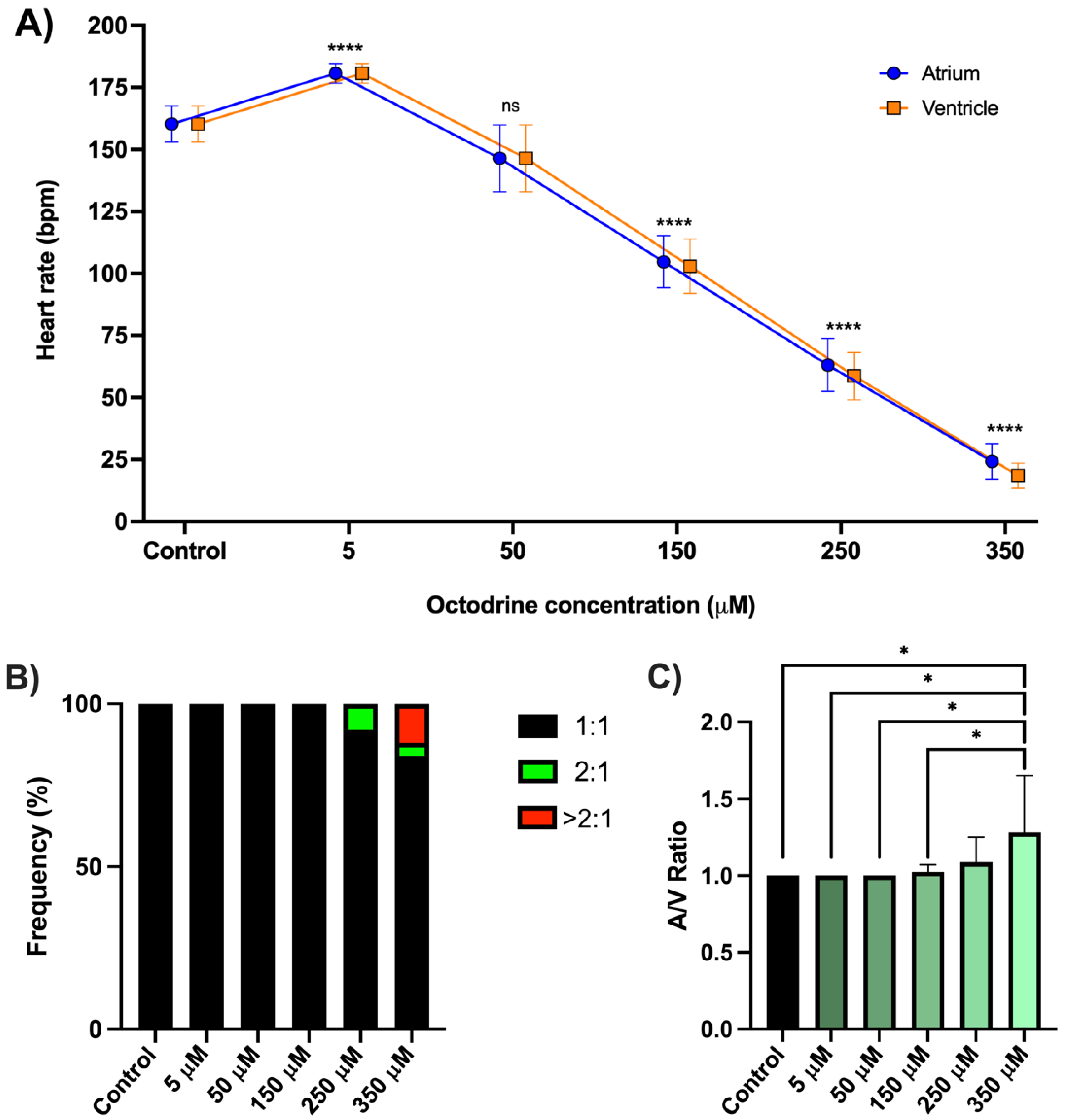
Figure 2.
Effects of octodrine on spontaneous locomotor activity in zebrafish larvae. Locomotor activity was quantified as distance travelled during eight consecutive 15-min intervals over a 2 h exposure period (n = 36 larvae per group; 12 larvae per group in each of three independent experiments). Data are presented as estimated marginal means with 95% confidence intervals obtained from a generalized estimating equation (GEE) model using a gamma distribution with log link, robust covariance estimation, and an AR(1) working correlation structure. Coloured significance annotations correspond to the respective octodrine treatment and indicate planned comparisons with the vehicle control within each time interval after sequential Bonferroni (Holm) correction (* p < 0.05, ** p < 0.01, *** p < 0.001, **** p < 0.0001; ns, not significant).
Figure 2.
Effects of octodrine on spontaneous locomotor activity in zebrafish larvae. Locomotor activity was quantified as distance travelled during eight consecutive 15-min intervals over a 2 h exposure period (n = 36 larvae per group; 12 larvae per group in each of three independent experiments). Data are presented as estimated marginal means with 95% confidence intervals obtained from a generalized estimating equation (GEE) model using a gamma distribution with log link, robust covariance estimation, and an AR(1) working correlation structure. Coloured significance annotations correspond to the respective octodrine treatment and indicate planned comparisons with the vehicle control within each time interval after sequential Bonferroni (Holm) correction (* p < 0.05, ** p < 0.01, *** p < 0.001, **** p < 0.0001; ns, not significant).
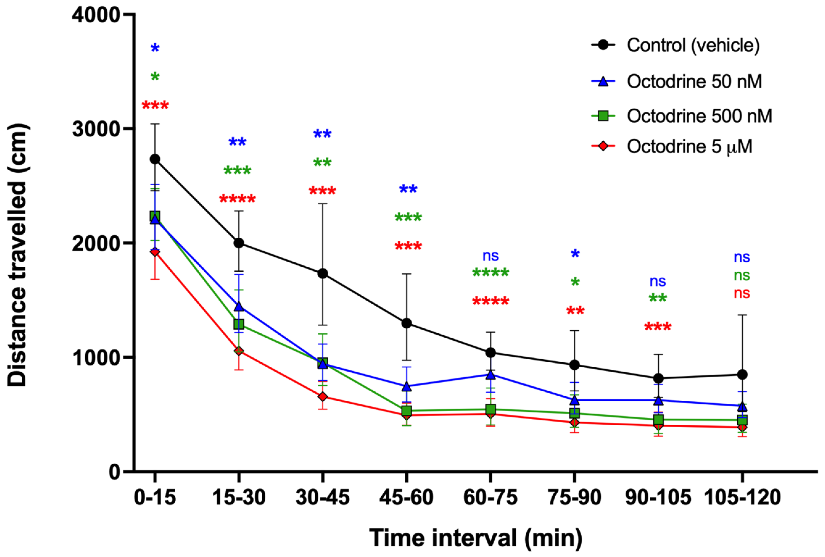
Figure 3.
Relative expression of genes associated with dopaminergic function, neuronal activity, stress signalling, and cardiac electrophysiology following acute octodrine exposure. Heat map showing mean log2 fold changes (log2FC) in th1, slc6a3 (DAT), fosab, egr1, npas4a, nr4a1, fkbp5, and kcnh2a after 2 h exposure to 0.35–350 µM octodrine (n = 8 samples per group). Values are expressed relative to the vehicle control, which was set to log2FC = 0 and is therefore not displayed. Statistical analyses were performed on individual ΔCt values using one-way ANOVA followed by Dunnett’s multiple-comparisons test against the vehicle control, except for npas4a, which was analysed using the Kruskal–Wallis test followed by Dunn’s multiple-comparisons test with Holm adjustment. Asterisks indicate statistically significant differences (*p < 0.05, ** p < 0.01, *** p < 0.001, **** p < 0.0001).
Figure 3.
Relative expression of genes associated with dopaminergic function, neuronal activity, stress signalling, and cardiac electrophysiology following acute octodrine exposure. Heat map showing mean log2 fold changes (log2FC) in th1, slc6a3 (DAT), fosab, egr1, npas4a, nr4a1, fkbp5, and kcnh2a after 2 h exposure to 0.35–350 µM octodrine (n = 8 samples per group). Values are expressed relative to the vehicle control, which was set to log2FC = 0 and is therefore not displayed. Statistical analyses were performed on individual ΔCt values using one-way ANOVA followed by Dunnett’s multiple-comparisons test against the vehicle control, except for npas4a, which was analysed using the Kruskal–Wallis test followed by Dunn’s multiple-comparisons test with Holm adjustment. Asterisks indicate statistically significant differences (*p < 0.05, ** p < 0.01, *** p < 0.001, **** p < 0.0001).
Disclaimer/Publisher’s Note: The statements, opinions and data contained in all publications are solely those of the individual author(s) and contributor(s) and not of MDPI and/or the editor(s). MDPI and/or the editor(s) disclaim responsibility for any injury to people or property resulting from any ideas, methods, instructions or products referred to in the content. |
© 2026 by the authors. Licensee MDPI, Basel, Switzerland. This article is an open access article distributed under the terms and conditions of the Creative Commons Attribution (CC BY) license (http://creativecommons.org/licenses/by/4.0/).
Copyright: This open access article is published under a Creative Commons CC BY 4.0 license, which permit the free download, distribution, and reuse, provided that the author and preprint are cited in any reuse.