

Submitted:
03 September 2026
Posted:
07 September 2026
You are already at the latest version
Abstract
Sepsis-associated renal injury involves interacting oxidative, inflammatory, and microvascular mechanisms. Molsidomine, a nitric oxide donor, may modulate these pathways. We investigated whether molsidomine pretreatment attenuates renal injury in cecal ligation and puncture (CLP)-induced polymicrobial sepsis. Forty male Wistar albino rats were randomized to Sham, molsidomine (MOL), CLP, or MOL+CLP groups. Molsidomine (10 mg/kg/day) was administered orally for 14 days before surgery. At 24 h, renal biochemical, histopathological, and immunohistochemical outcomes were assessed. Seven septic animals died before the endpoint, resulting in outcome-specific numbers of evaluable animals. The primary outcome was total renal histopathological damage score. The total renal histopathological damage score was lower in MOL+CLP than CLP animals [median (IQR), 2 (1–3) vs. 5.5 (5–6); overall p < 0.001], with reductions across all four histopathological components. Renal MDA was lower in MOL+CLP than CLP animals (106.41 ± 9.03 vs. 136.45 ± 20.27 nmol/g), whereas SOD (36.23 ± 4.19 vs. 24.12 ± 2.23 U/g) and GPx (99.43 ± 26.38 vs. 61.54 ± 9.10 U/mg) activities were higher. VEGF and P-selectin H-scores were also markedly lower in MOL+CLP than CLP animals. Conversely, renal NO, GSH, TOS, OSI, and TNF-α and serum creatinine, BUN, and urea did not differ significantly between the two septic groups. Molsidomine pretreatment attenuated renal structural injury, selected redox abnormalities, and VEGF/P-selectin immunoreactivity in experimental polymicrobial sepsis. This renoprotective phenotype was not accompanied by uniform biochemical or renal functional improvement. These proof-of-concept findings warrant evaluation using post-sepsis treatment protocols and longer-term renal outcomes.
Keywords:
acute kidney injury
; cecal ligation and puncture
; molsidomine
; nitric oxide
; oxidative stress
; sepsis
1. Introduction
Sepsis is a life-threatening syndrome caused by a dysregulated host response to infection that results in acute organ dysfunction (1). Despite advances in antimicrobial and supportive care, sepsis remains a major cause of morbidity and mortality worldwide (2). Sepsis-induced organ injury arises from interconnected inflammatory, metabolic, and vascular disturbances. In particular, oxidative and nitrosative stress, endothelial activation, and microcirculatory dysfunction disrupt cellular homeostasis and tissue oxygen utilization (3). The kidney is particularly vulnerable to these insults, making renal injury a clinically important manifestation of sepsis-associated organ dysfunction (4).
Sepsis-associated acute kidney injury (SA-AKI) is a common and heterogeneous complication of sepsis associated with adverse short- and long-term outcomes (4). Current concepts extend beyond global renal hypoperfusion, emphasizing dysregulated inflammation, microcirculatory alterations, metabolic reprogramming, and tubular stress (5). Endothelial activation can increase vascular permeability, leukocyte–endothelial interactions, and microvascular flow heterogeneity, impairing tissue oxygen and nutrient delivery (5). Concurrently, excessive reactive oxygen species generation and impaired antioxidant defenses can disrupt mitochondrial function and tubular epithelial homeostasis (6). Together, these vascular, inflammatory, and redox disturbances contribute to septic renal injury and provide a rationale for investigating renal oxidative and endothelial responses (6).
Experimental models are essential for investigating sepsis pathobiology and pharmacological responses under controlled conditions (7). Cecal ligation and puncture (CLP) is a reference model of polymicrobial sepsis that generates a sustained intra-abdominal infectious focus and reproduces key systemic features of human sepsis (7). CLP also produces renal functional and structural alterations and is widely used to investigate sepsis-associated renal injury (8). In this context, nitric oxide signaling is particularly relevant because of its involvement in vascular homeostasis, redox regulation, and the renal response to sepsis (9).
Nitric oxide (NO) has a context-dependent role in septic renal pathophysiology, determined by its cellular source, concentration, and redox environment (9). Under physiological conditions, constitutive NO production through endothelial and neuronal nitric oxide synthases regulates renal vascular tone, intrarenal blood flow, and glomerular hemodynamics (9). During sepsis, upregulation of inducible nitric oxide synthase (iNOS) increases NO generation and alters its intrarenal distribution (10). In an oxidant-rich environment, NO reacts with superoxide to generate peroxynitrite, promoting protein modification, mitochondrial dysfunction, and tubular injury (11). Thus, the renal effects of NO in sepsis reflect the balance between constitutive NO signaling and excessive nitrosative stress rather than NO abundance alone (11).
Molsidomine is a sydnonimine-class vasodilator and nitric oxide donor used clinically for the prophylactic treatment of angina pectoris (12). It is converted to the active metabolite linsidomine (SIN-1), which spontaneously decomposes to release NO and activate the soluble guanylate cyclase–cyclic guanosine monophosphate pathway (12). Unlike organic nitrates, this mechanism does not require thiol-dependent enzymatic bioactivation and is less susceptible to pharmacological tolerance (13). Experimental studies also indicate that molsidomine can modulate renal oxidative stress, antioxidant defenses, and structural injury in models of kidney damage (14). These properties provide a rationale for investigating whether molsidomine pretreatment modifies the renal response to polymicrobial sepsis, which remains insufficiently characterized.
Accordingly, the present study aimed to determine whether molsidomine pretreatment modifies the renal response to experimental polymicrobial sepsis induced by cecal ligation and puncture. We hypothesized that molsidomine pretreatment would attenuate CLP-induced renal injury, primarily reflected by a reduction in the total histopathological damage score, while concurrently ameliorating renal oxidative stress and endothelial activation. To test this hypothesis, renal biochemical indices of redox homeostasis, histopathological alterations, and VEGF and P-selectin immunoreactivity were evaluated across the experimental groups.
2. Results
2.1. Renal Histopathological Injury: Primary Outcome
The primary outcome, total renal histopathological damage score, differed significantly across the four experimental groups (overall p < 0.001). The score was highest in the CLP group and was lower in the MOL+CLP group [median (IQR), 5.5 (5–6) vs. 2 (1–3), respectively] (Table 1). Sham kidneys showed preserved renal architecture, whereas CLP was associated with tubular injury, interstitial inflammatory cell infiltration, and vascular congestion. These histopathological alterations were less pronounced in the MOL+CLP group.
Tubular epithelial degeneration, tubular necrosis, interstitial inflammatory cell infiltration, and vascular congestion each differed significantly across the experimental groups (overall p < 0.001 for all four components), with higher scores after CLP and lower scores in the MOL+CLP group (Table 1). Representative renal histopathological findings across the four groups are shown in Figure 1.
2.2. Renal Oxidative Stress, Antioxidant Status, and Inflammatory Markers
Renal MDA concentrations differed significantly among the experimental groups (overall p = 0.003). MDA was higher in the CLP group than in the Sham and MOL groups and lower in the MOL+CLP group than in the CLP group (136.45 ± 20.27 vs. 106.41 ± 9.03 nmol/g) (Table 2). NO and GSH also differed significantly among groups (overall p = 0.004 and p = 0.030, respectively), but neither differed significantly between the CLP and MOL+CLP groups after multiplicity-adjusted pairwise comparisons.
SOD and GPx activities differed significantly among groups (overall p < 0.001 for both) and were higher in the MOL+CLP group than in the CLP group (SOD: 36.23 ± 4.19 vs. 24.12 ± 2.23 U/g; GPx: 99.43 ± 26.38 vs. 61.54 ± 9.10 U/mg). CAT and TAS did not differ significantly among groups (p = 0.157 and p = 0.364, respectively). TOS, OSI, and TNF-α showed significant overall group differences (p = 0.009, p = 0.004, and p = 0.045, respectively), but none differed significantly between the CLP and MOL+CLP groups after multiplicity-adjusted pairwise comparisons (Table 2).
2.3. Serum Renal Biochemical Findings
Serum BUN and urea concentrations differed significantly among the experimental groups (overall p = 0.025 for both) and were higher in the CLP and MOL+CLP groups than in the Sham and MOL groups. Serum creatinine also differed significantly among groups (overall p = 0.004), with a higher concentration in the CLP group than in the Sham group [median (IQR), 0.50 (0.10) vs. 0.40 (0.10) mg/dL]. However, no significant differences were observed between the CLP and MOL+CLP groups for serum creatinine, BUN, or urea after multiplicity-adjusted pairwise comparisons (Supplementary Table S1).
2.4. Renal Immunohistochemical Findings
Renal VEGF and P-selectin immunoreactivity differed significantly among the experimental groups (overall p < 0.001 for both). VEGF H-scores were lower in the MOL+CLP group than in the CLP group (47.5 ± 10.35 vs. 135.0 ± 20.70), with no significant difference between the MOL+CLP and Sham groups. Similarly, P-selectin H-scores were lower in the MOL+CLP group than in the CLP group (37.5 ± 10.35 vs. 135.0 ± 14.14), whereas the Sham, MOL, and MOL+CLP groups did not differ significantly (Table 3). Representative renal VEGF and P-selectin immunostaining across the experimental groups is shown in Figure 1.
2.5. Physiological Responses to CLP and Molsidomine Pretreatment
Heart rate differed significantly among the experimental groups (overall p = 0.019) and was higher in both septic groups than in the Sham and MOL groups, whereas no significant difference was observed between the CLP and MOL+CLP groups. Systolic blood pressure and mean arterial pressure also differed significantly among groups (overall p = 0.003 and p = 0.017, respectively), while diastolic blood pressure did not differ significantly (overall p = 0.100). Despite the overall differences in systolic blood pressure and mean arterial pressure, neither parameter differed significantly between the CLP and MOL+CLP groups after multiplicity-adjusted pairwise comparisons. Preoperative body temperature was comparable among groups (overall p = 0.171). At the end of the experiment, body temperature differed significantly among groups (overall p = 0.023), but no significant difference was observed between the CLP and MOL+CLP groups after multiplicity-adjusted pairwise comparisons (Supplementary Table S2).
3. Discussion
The principal finding of this study was that molsidomine pretreatment attenuated renal structural injury in CLP-induced polymicrobial sepsis. The prespecified total renal histopathological damage score was lower in the MOL+CLP group, with reductions across all four histopathological components. Molsidomine also reduced renal MDA and increased SOD and GPx activities, although several other redox and inflammatory indices remained unchanged. VEGF and P-selectin immunoreactivity were likewise markedly attenuated. Overall, molsidomine pretreatment was associated with structural, selected redox, and immunohistochemical protection rather than uniform normalization of the renal response to sepsis.
The marked reduction in total renal histopathological damage score provides the strongest evidence of molsidomine-associated renoprotection in our model. Contemporary concepts describe SA-AKI as a heterogeneous process in which tubular, inflammatory, metabolic, and microvascular disturbances interact rather than reflecting renal hypoperfusion alone (22). Oxidative stress can amplify tubular and endothelial injury through mitochondrial dysfunction, impaired oxygen utilization, and disruption of cellular homeostasis (23). Endothelial activation further contributes to renal injury by promoting vascular permeability, leukocyte–endothelial interactions, and microcirculatory dysfunction (24). Against this mechanistic background, the concurrent attenuation of tubular epithelial degeneration, tubular necrosis, interstitial inflammatory cell infiltration, and vascular congestion supports a broad structural protective effect of molsidomine.
CLP increased renal MDA and reduced SOD and GPx activities, whereas molsidomine pretreatment lowered MDA and increased both antioxidant enzyme activities, indicating partial restoration of renal redox balance. Similarly, Xia et al. reported that TAK-242 reduced oxidative stress, restored SOD activity, and ameliorated renal injury in experimental SA-AKI (25). Zhu et al. demonstrated that sivelestat reduced MDA while increasing SOD and GPx activities and attenuating renal histopathological injury after CLP (26). Karaboğa et al. likewise showed that ebselen ameliorated sepsis-induced renal injury through modulation of oxidative stress together with endoplasmic reticulum stress and apoptosis (27). Collectively, these studies support oxidative imbalance as an important component of septic renal injury and provide a consistent experimental context for the selective redox protection observed with molsidomine.
Molsidomine did not significantly alter renal NO, GSH, TOS, OSI, or TNF-α, nor did it improve serum creatinine, BUN, or urea compared with untreated septic animals, indicating a non-uniform renal response. Wiersema et al. identified septic AKI subphenotypes with distinct inflammatory profiles, renal recovery, and 90-day mortality (28). Molinari et al. demonstrated that functional AKI staging and tubular stress biomarkers provide complementary information, supporting dissociation between functional impairment and cellular injury (29). Similarly, the PHENAKI study identified distinct SA-AKI subphenotypes by integrating kidney function variables with urinary TIMP-2×IGFBP7 (30). Thus, structural and selective redox protection without parallel improvement in conventional functional indices may reflect differential renal responses rather than uniform recovery.
The renoprotective profile of molsidomine in our CLP model is consistent with findings from other experimental kidney injury models. Chander and Chopra showed that molsidomine attenuated renal dysfunction, morphological injury, and oxidative stress in rhabdomyolysis-induced renal injury (31). Rodriguez-Peña et al. reported reduced renal dysfunction, inflammatory cell infiltration, and adhesion-molecule expression, including P-selectin, following intrarenal molsidomine administration in renal ischemia–reperfusion injury (32). Karakoc et al. similarly demonstrated reduced tubular degeneration and inflammatory histological changes in cisplatin nephrotoxicity, despite nonsignificant improvements in several oxidative stress markers (33). Together, these studies parallel the structural protection and selective biochemical improvement observed here and extend experimental evidence of molsidomine-associated renoprotection to CLP-induced polymicrobial sepsis.
Our histopathological and immunohistochemical findings further support the involvement of endothelial and microvascular alterations in septic renal injury. Molema et al. characterized renal microvascular endothelial dysfunction in SA-AKI by altered perfusion, permeability, endothelial signaling, and inflammatory interactions (34). Herter et al. demonstrated renal P-selectin upregulation after experimental sepsis and showed that its blockade reduced leukocyte adhesion and CLP-induced kidney injury (35). Renal VEGF responses appear to be model- and compartment-dependent; Xu et al. demonstrated glomerular endothelial injury accompanied by reduced renal VEGF expression in LPS-induced septic AKI (36). Consistent with these observations, molsidomine markedly attenuated CLP-induced renal VEGF and P-selectin immunoreactivity alongside reduced vascular congestion and overall histopathological injury.
The effects of molsidomine should also be interpreted within the context-dependent biology of nitric oxide. Lundberg and Weitzberg emphasized that NO signaling depends on its cellular source, concentration, redox environment, and temporal pattern, permitting physiological signaling or nitrosative injury under different conditions (37). In the kidney, NO bioavailability regulates vascular tone, regional blood flow, oxygen delivery, and tubular homeostasis, whereas oxidative stress can disrupt this balance (38). Betrie et al. showed that renal arterial tempol administration prevented medullary hypoperfusion, hypoxia, and AKI during ovine Gram-negative sepsis (39). In our study, renal NO did not differ between CLP and MOL+CLP, despite improvements in selected redox and endothelial indices, suggesting modulation of NO-related signaling rather than increased total renal NO.
The translational interpretation of our findings requires caution because molsidomine was administered for 14 days before CLP rather than after sepsis onset. Osuchowski et al. emphasized that clinically relevant treatment timing, supportive care, and organ dysfunction endpoints are essential for improving the translational value of preclinical sepsis models (40). Guillon et al. highlighted important differences between rodent models and ICU patients, including comorbidities and supportive care (41). Marshall underscored the recurrent failure of experimental sepsis therapies to translate successfully into clinical practice (42). Accordingly, our findings establish proof-of-concept for prophylactic renoprotection but require confirmation using post-sepsis treatment protocols and clinically relevant supportive care.
Limitations
This study has several limitations. First, the CLP model cannot fully reproduce the complexity and heterogeneity of human sepsis. Molsidomine was administered for 14 days before CLP; therefore, the findings reflect a prophylactic rather than therapeutic intervention and cannot establish efficacy when treatment is initiated after sepsis onset. Renal assessment relied on biochemical, histopathological, and immunohistochemical measures without direct evaluation of glomerular filtration or renal hemodynamics. Furthermore, the 24-h endpoint precluded assessment of longer-term renal recovery and survival. Only male animals and a single molsidomine dose were studied, and the molecular mechanisms underlying the observed protection, including NOS activity and downstream nitrosative pathways, were not directly investigated.
4. Materials and Methods
4.1. Study Design, Ethics, and Reporting
This randomized, controlled experimental study evaluated the effects of molsidomine pretreatment on renal injury in a rat model of cecal ligation and puncture (CLP)-induced polymicrobial sepsis. The protocol was approved by the Inonu University Local Ethics Committee for Animal Experiments (Approval No. 2024/17-2; approval date: 18 October 2024, Malatya, Türkiye). All procedures complied with institutional animal-care guidelines, and the study was reported in accordance with the ARRIVE 2.0 guidelines (15).
4.2. Animal Attrition and Analysis Populations
Of the 40 randomized animals, three in the CLP group and four in the MOL+CLP group died before the scheduled 24-h endpoint. Renal tissue biochemical data were therefore available for 8, 8, 9, and 8 animals in the Sham, MOL, CLP, and MOL+CLP groups, respectively. One additional CLP kidney specimen was unsuitable for histopathological and immunohistochemical evaluation, resulting in eight evaluable animals per group for these analyses.
4.3. Animals, Randomization, and Experimental Groups
Forty male Wistar albino rats (10–12 weeks; 250–350 g) were obtained from the Inonu University Experimental Animal Research and Production Center and housed under controlled conditions (25 ± 2 °C, 60 ± 5% relative humidity, 12-h light/dark cycle) with ad libitum access to standard chow and water. Using a computer-generated randomization table, rats were allocated to four groups: Sham (n = 8), MOL (n = 8), CLP (n = 12), and MOL+CLP (n = 12). Because attrition was anticipated following CLP, additional animals were initially allocated to the septic groups. Three animals in the CLP group and four animals in the MOL+CLP group died before the scheduled 24-h endpoint. Renal biochemical analyses were therefore available for nine CLP and eight MOL+CLP animals. One additional CLP kidney specimen was unsuitable for histopathological and immunohistochemical evaluation, resulting in eight evaluable animals per group for these analyses.
4.4. Molsidomine Pretreatment and Cecal Ligation and Puncture Procedure
Molsidomine (10 mg/kg/day; Sigma-Aldrich, St. Louis, MO, USA) was administered by oral gavage once daily for 14 days before surgery; this dose was selected based on previous experimental evidence in rat models of renal injury (16). MOL and MOL+CLP rats received molsidomine, whereas Sham and CLP rats received saline during the corresponding pretreatment period. For CLP, rats were anesthetized with intraperitoneal ketamine (100 mg/kg) and xylazine (10 mg/kg), followed by a 1-cm midline laparotomy. The cecum was exteriorized, ligated distal to the ileocecal junction without intestinal obstruction, punctured twice with a sterile 22-gauge needle, and gently compressed to extrude a small amount of fecal material before abdominal closure. Sham and MOL rats underwent laparotomy and cecal manipulation without ligation or puncture. Buprenorphine (0.05 mg/kg) was administered for postoperative analgesia, and CLP and MOL+CLP rats received 1 mL sterile saline subcutaneously for fluid resuscitation. No antibiotics were administered.
4.5. Physiological Assessment, Blood Sampling, and Serum Biochemistry
Heart rate (HR), systolic and diastolic blood pressure (SBP and DBP), mean arterial pressure (MAP), and body temperature were assessed during the experimental protocol. At the scheduled 24-h endpoint after CLP or sham surgery, surviving animals were anesthetized with intraperitoneal ethyl carbamate (1.2 g/kg). Blood was collected from the inferior vena cava and centrifuged at 2500 rpm for 7 min. Serum creatinine, blood urea nitrogen (BUN), and urea were measured as indices of renal function. Animals were then euthanized by exsanguination, and the kidneys were collected for biochemical, histopathological, and immunohistochemical analyses. Kidney samples intended for biochemical analyses were stored at −80 °C, whereas specimens intended for histopathological and immunohistochemical evaluation were fixed in 10% neutral buffered formalin.
4.6. Renal Tissue Biochemical Analyses
Kidney tissues were homogenized and centrifuged to obtain supernatants for biochemical analyses. Malondialdehyde (MDA) was measured spectrophotometrically using the Uchiyama–Mihara method (17). Nitric oxide (NO) was assessed from stable nitrite/nitrate metabolites using the Griess reaction (18). Reduced glutathione (GSH) was determined using Ellman’s method (19). Superoxide dismutase (SOD), catalase (CAT), and glutathione peroxidase (GPx) activities were measured spectrophotometrically. Total oxidant status (TOS) was determined using Erel’s automated colorimetric method (20), and total antioxidant status (TAS) was measured using Erel’s automated colorimetric method (21). The oxidative stress index (OSI) was calculated as the TOS/TAS ratio and expressed in arbitrary units. Renal tissue tumor necrosis factor-alpha (TNF-α) was measured using a rat-specific ELISA kit (Biotek, China; catalog no. YLA0118RA) according to the manufacturer’s instructions.
4.7. Histopathological Examination
Formalin-fixed kidney specimens were paraffin-embedded, sectioned at 5–6 µm, and stained with hematoxylin and eosin (H&E). An experienced histologist blinded to group allocation semi-quantitatively assessed four prespecified renal injury components: tubular epithelial degeneration, tubular necrosis, interstitial inflammatory cell infiltration, and vascular congestion. Each component was scored as 0 (absent/normal), 1 (mild), 2 (moderate), or 3 (severe). The four component scores were summed to obtain the total renal histopathological damage score, ranging from 0 to 12, with higher scores indicating greater renal injury.
4.8. Immunohistochemical Analysis
Immunohistochemistry was performed on 4-µm kidney sections to evaluate vascular endothelial growth factor (VEGF) and P-selectin expression. Following antigen retrieval in citrate buffer (pH 6.0), sections were incubated overnight at 4 °C with primary antibodies against VEGF (Santa Cruz Biotechnology, USA; C-1, sc-7269; 1:100) and P-selectin (Santa Cruz Biotechnology, USA; sc-8419; 1:200). Immunoreactivity was visualized using biotinylated secondary antibodies and 3,3′-diaminobenzidine (DAB). Immunostaining was evaluated by an observer blinded to group allocation using the semi-quantitative H-score: Σ[Pi × (i + 1)], where Pi represents the percentage of cells at each staining intensity and i represents the corresponding intensity score (0–3).
4.10. Study Outcomes and Sample-Size Considerations
The individual animal was considered the experimental unit. The total renal histopathological damage score was designated as the primary outcome, with the CLP versus MOL+CLP comparison representing the principal comparison of interest. Secondary outcomes included renal redox and inflammatory indices, individual histopathological components, VEGF and P-selectin immunoreactivity, serum renal biochemical indices, and physiological variables. Pre-experimental sample-size planning (α = 0.05; 80% power) indicated a minimum of eight evaluable animals per group. To account for anticipated attrition associated with CLP, 12 animals were initially allocated to each septic group, whereas eight animals were allocated to each non-septic group. The original effect-size assumption used for sample-size planning was unavailable for retrospective verification; therefore, no post hoc power calculation was performed.
4.11. Statistical Analysis
Statistical analyses were performed using IBM SPSS Statistics for Windows, version 26.0 (IBM Corp., Armonk, NY, USA). Analyses were conducted using all evaluable animals with available data for the respective outcome. Data distribution was assessed using the Shapiro–Wilk test together with visual inspection, and continuous data are presented as mean ± standard deviation (SD) or median (interquartile range [IQR]), as appropriate. Normally distributed continuous outcomes were compared across the four groups using one-way analysis of variance (ANOVA). Homogeneity of variances was assessed using Levene’s test, followed by Tukey’s honestly significant difference or Tamhane’s T2 test for post hoc pairwise comparisons, as appropriate. Ordinal and non-normally distributed outcomes were analyzed using the Kruskal–Wallis test followed by Dunn’s pairwise comparisons. For the primary outcome, the prespecified principal comparison was CLP versus MOL+CLP. For secondary outcomes, multiplicity was controlled within predefined outcome families using the Holm procedure for pairwise comparisons. All tests were two-sided, and exact p values were reported whenever possible; values below 0.001 were reported as p < 0.001. An adjusted p < 0.05 was considered statistically significant for multiplicity-adjusted comparisons.
5. Conclusion
Molsidomine pretreatment attenuated renal structural injury, selected oxidative stress abnormalities, and endothelial-associated immunohistochemical changes in CLP-induced polymicrobial sepsis. The concordant histopathological, redox, and VEGF/P-selectin findings support a renoprotective phenotype, although the absence of improvement across several biochemical and conventional renal functional indices indicates that this protection was not uniform. Given the prophylactic design, these findings should be considered proof-of-concept rather than evidence of therapeutic efficacy after sepsis onset. Further studies using post-sepsis treatment protocols, clinically relevant supportive care, and longer-term renal outcomes are needed to determine the translational potential of molsidomine.
Supplementary Materials
The following supporting information can be downloaded at the website of this paper posted on Preprints.org: Table S1: Serum renal biochemical indices across experimental groups; Table S2: Physiological parameters across experimental groups.
Author Contributions
Conceptualization, A.K.; methodology, A.K., Z.B.K., M.E.B. and O.O.; formal analysis, A.K.; validation, S.Y.; investigation, Z.B.K., M.E.B., O.O., Z.U. and F.D.; histopathological and immunohistochemical evaluation, E.K., A.B.L. and M.G.; data curation, A.K., O.O., Z.U. and F.D.; writing—original draft preparation, A.K.; writing—review and editing, H.P.; supervision, A.K.; project administration, A.K. All authors have read and agreed to the published version of the manuscript.
Funding
This research was funded by the Inonu University Scientific Research Projects Coordination Unit (BAP), Project No. TCD-2025-3808.
Institutional Review Board Statement
The animal study protocol was approved by the Inonu University Local Ethics Committee for Animal Experiments (Approval No. 2024/17-2; approval date: 18 October 2024, Malatya, Türkiye).
Informed Consent Statement
Not applicable.
Data Availability Statement
The original contributions presented in this study are included in the article/supplementary material. Further inquiries can be directed to the corresponding author.
Acknowledgments
The authors thank the staff of the Inonu University Experimental Animal Research and Production Center for their assistance with animal care and laboratory procedures.
Conflicts of Interest
The authors declare no conflicts of interest.
References
- Singer, M.; Deutschman, C.S.; Seymour, C.W.; Shankar-Hari, M.; Annane, D.; Bauer, M.; et al. The Third International Consensus Definitions for Sepsis and Septic Shock (Sepsis-3). JAMA 2016, 315(8), 801–810. [Google Scholar] [CrossRef]
- Rudd, K.E.; Johnson, S.C.; Agesa, K.M.; Shackelford, K.A.; Tsoi, D.; Kievlan, D.R.; et al. Global, regional, and national sepsis incidence and mortality, 1990-2017: analysis for the Global Burden of Disease Study. Lancet 2020, 395(10219), 200–211. [Google Scholar] [CrossRef]
- Joffre, J.; Hellman, J.; Ince, C.; Ait-Oufella, H. Endothelial responses in sepsis. Am. J. Respir. Crit. Care Med. 2020, 202(3), 361–370. [Google Scholar] [CrossRef]
- Zarbock, A.; Nadim, M.K.; Pickkers, P.; Gomez, H.; Bell, S.; Joannidis, M.; et al. Sepsis-associated acute kidney injury: consensus report of the 28th Acute Disease Quality Initiative workgroup. Nat. Rev. Nephrol. 2023, 19(6), 401–417. [Google Scholar] [CrossRef]
- Gómez, H.; Kellum, J.A. Sepsis-induced acute kidney injury. Curr. Opin. Crit. Care 2016, 22(6), 546–553. [Google Scholar] [CrossRef]
- Wang, C.; Liu, Q.; Wang, H.; Zhang, Q.; Yang, B.; Liu, H.; et al. Mitochondrial dysfunction in sepsis-associated acute kidney injury: mechanisms and therapeutic potential. Front Immunol. 2026, 17, 1829288. [Google Scholar] [CrossRef]
- Sjaastad, F.V.; Jensen, I.J.; Berton, R.R.; Badovinac, V.P.; Griffith, T.S. Inducing experimental polymicrobial sepsis by cecal ligation and puncture. Curr. Protoc. Immunol. 2020, 131(1), e110. [Google Scholar] [CrossRef]
- González-Nicolás, M.Á.; Lázaro, A. Induction of sepsis in a rat model by the cecal ligation and puncture technique. Application for the study of experimental acute renal failure. Methods Cell Biol. 2025, 192, 69–82. [Google Scholar] [CrossRef]
- Oliveira, F.R.M.B.; Assreuy, J.; Sordi, R. The role of nitric oxide in sepsis-associated kidney injury. Biosci. Rep. 2022, 42(7), BSR20220093. [Google Scholar] [CrossRef]
- Heemskerk, S.; Masereeuw, R.; Russel, F.G.M.; Pickkers, P. Selective iNOS inhibition for the treatment of sepsis-induced acute kidney injury. Nat. Rev. Nephrol. 2009, 5(11), 629–640. [Google Scholar] [CrossRef]
- Lankadeva, Y.R.; Okazaki, N.; Evans, R.G.; Bellomo, R.; May, C.N. Targeting the nitric oxide pathway in the kidney: a novel therapeutic approach to sepsis-associated acute kidney injury. Clin. Sci. (Lond) 2019, 133(5), 577–588. [Google Scholar]
- Rosenkranz, B.; Winkelmann, B.R.; Parnham, M.J. Clinical pharmacokinetics of molsidomine. Clin. Pharmacokinet. 1996, 30(5), 372–384. [Google Scholar] [CrossRef]
- Feelisch, M.; Ostrowski, J.; Noack, E. On the mechanism of NO release from sydnonimines. J. Cardiovasc Pharmacol. 1989, 14 Suppl 11, S13–S22. [Google Scholar] [CrossRef]
- Singh, P.; Singh, N.; Jaggi, A.S. Therapeutic insight into molsidomine, a nitric oxide donor in streptozotocin-induced diabetic nephropathy in rats. Indian J. Pharmacol. 2016, 48(5), 544–549. [Google Scholar] [CrossRef]
- Percie du Sert, N.; Hurst, V.; Ahluwalia, A.; Alam, S.; Avey, M.T.; Baker, M.; et al. The ARRIVE guidelines 2.0: Updated guidelines for reporting animal research. PLoS Biol. 2020, 18(7), e3000410. [Google Scholar] [CrossRef]
- Chander, V.; Chopra, K. Renal protective effect of molsidomine and L-arginine in ischemia-reperfusion induced injury in rats. J. Surg. Res. 2005, 128(1), 132–139. [Google Scholar] [CrossRef]
- Uchiyama, M.; Mihara, M. Determination of malonaldehyde precursor in tissues by thiobarbituric acid test. Anal. Biochem. 1978, 86(1), 271–278. [Google Scholar] [CrossRef]
- Green, L.C.; Wagner, D.A.; Glogowski, J.; Skipper, P.L.; Wishnok, J.S.; Tannenbaum, S.R. Analysis of nitrate, nitrite, and [15N]nitrate in biological fluids. Anal. Biochem. 1982, 126(1), 131–138. [Google Scholar] [CrossRef]
- Ellman, G.L. Tissue sulfhydryl groups. Arch. Biochem Biophys. 1959, 82(1), 70–77. [Google Scholar] [CrossRef]
- Erel, O. A new automated colorimetric method for measuring total oxidant status. Clin. Biochem. 2005, 38(12), 1103–1111. [Google Scholar] [CrossRef]
- Erel, O. A novel automated method to measure total antioxidant response against potent free radical reactions. Clin. Biochem. 2004, 37(2), 112–119. [Google Scholar] [CrossRef]
- Legrand, M.; Bagshaw, S.M.; Bhatraju, P.K.; Bihorac, A.; Caniglia, E.C.; Khanna, A.K.; et al. Sepsis-associated acute kidney injury: recent advances in enrichment strategies, sub-phenotyping and clinical trials. Crit. Care 2024, 28(1), 92. [Google Scholar] [CrossRef]
- Ow, C.P.C.; Trask-Marino, A.; Betrie, A.H.; Evans, R.G.; May, C.N.; Lankadeva, Y.R. Targeting oxidative stress in septic acute kidney injury: from theory to practice. J. Clin. Med. 2021, 10(17), 3798. [Google Scholar] [CrossRef]
- Raia, L.; Zafrani, L. Endothelial activation and microcirculatory disorders in sepsis. Front Med. 2022, 9, 907992. [Google Scholar] [CrossRef]
- Xia, Y.M.; Guan, Y.Q.; Liang, J.F.; Wu, W.D. TAK-242 improves sepsis-associated acute kidney injury in rats by inhibiting the TLR4/NF-κB signaling pathway. Ren. Fail. 2024, 46(1), 2313176. [Google Scholar] [CrossRef]
- Zhu, W.; Ou, Y.; Wang, C.; An, R.; Lai, J.; Shen, Y.; et al. A neutrophil elastase inhibitor, sivelestat, attenuates sepsis-induced acute kidney injury by inhibiting oxidative stress. Heliyon 2024, 10(8), e29366. [Google Scholar] [CrossRef]
- Karaboğa, İ.; Okuyan, H.M.; Doğan, S.; Ayçiçek, Ş.Ö.; Çakıroğlu, H. Ebselen alleviates sepsis-induced acute kidney injury by regulating endoplasmic reticulum stress, apoptosis, and oxidative stress. Vet. Med. Sci. 2025, 11(2), e70318. [Google Scholar] [CrossRef]
- Wiersema, R.; Jukarainen, S.; Vaara, S.T.; Poukkanen, M.; Lakkisto, P.; Wong, H.; et al. Two subphenotypes of septic acute kidney injury are associated with different 90-day mortality and renal recovery. Crit. Care 2020, 24(1), 150. [Google Scholar] [CrossRef]
- Molinari, L.; Del Rio-Pertuz, G.; Smith, A.; Landsittel, D.P.; Singbartl, K.; Palevsky, P.M.; et al. Utility of biomarkers for sepsis-associated acute kidney injury staging. JAMA Netw. Open 2022, 5(5), e2212709. [Google Scholar] [CrossRef]
- Dépret, F.; Hollinger, A.; Cariou, A.; Deye, N.; Vieillard-Baron, A.; Fournier, M.C.; et al. The combination of kidney function variables with cell cycle arrest biomarkers identifies distinct subphenotypes of sepsis-associated acute kidney injury: a post-hoc analysis (the PHENAKI study). Ann. Intensive Care 2024, 14(1), 41. [Google Scholar]
- Chander, V.; Chopra, K. Molsidomine, a nitric oxide donor and L-arginine protects against rhabdomyolysis-induced myoglobinuric acute renal failure. Biochim Biophys. Acta 2005, 1723(1-3), 208–214. [Google Scholar] [CrossRef]
- Rodriguez-Peña, A.; Garcia-Criado, F.J.; Eleno, N.; Arevalo, M.; Lopez-Novoa, J.M. Intrarenal administration of molsidomine, a molecule releasing nitric oxide, reduces renal ischemia-reperfusion injury in rats. Am. J. Transplant. 2004, 4(10), 1605–1613. [Google Scholar] [CrossRef]
- Karakoc, H.T.E.; Altintas, R.; Parlakpinar, H.; Polat, A.; Samdanci, E.; Sagir, M.; et al. Protective effects of molsidomine against cisplatin-induced nephrotoxicity. Adv. Clin. Exp. Med. 2015, 24(4), 585–593. [Google Scholar] [CrossRef]
- Molema, G.; Zijlstra, J.G.; van Meurs, M.; Kamps, J.A.A.M. Renal microvascular endothelial cell responses in sepsis-induced acute kidney injury. Nat. Rev. Nephrol. 2022, 18(2), 95–112. [Google Scholar] [CrossRef]
- Herter, J.M.; Rossaint, J.; Spieker, T.; Zarbock, A. Adhesion molecules involved in neutrophil recruitment during sepsis-induced acute kidney injury. J. Innate Immun. 2014, 6(5), 597–606. [Google Scholar] [CrossRef]
- Xu, C.; Chang, A.; Hack, B.K.; Eadon, M.T.; Alper, S.L.; Cunningham, P.N. TNF-mediated damage to glomerular endothelium is an important determinant of acute kidney injury in sepsis. Kidney Int. 2014, 85(1), 72–81. [Google Scholar] [CrossRef]
- Lundberg, J.O.; Weitzberg, E. Nitric oxide signaling in health and disease. Cell 2022, 185(16), 2853–2878. [Google Scholar] [CrossRef]
- Carlström, M. Nitric oxide signalling in kidney regulation and cardiometabolic health. Nat. Rev. Nephrol. 2021, 17(9), 575–590. [Google Scholar] [CrossRef]
- Betrie, A.H.; Ma, S.; Ow, C.P.C.; Peiris, R.M.; Evans, R.G.; Ayton, S.; et al. Renal arterial infusion of tempol prevents medullary hypoperfusion, hypoxia, and acute kidney injury in ovine Gram-negative sepsis. Acta Physiol. (Oxf) 2023, 239(1), e14025. [Google Scholar] [CrossRef]
- Osuchowski, M.F.; Ayala, A.; Bahrami, S.; Bauer, M.; Boros, M.; Cavaillon, J.M.; et al. Minimum Quality Threshold in Pre-Clinical Sepsis Studies (MQTiPSS): an international expert consensus initiative for improvement of animal modeling in sepsis. Infection 2018, 46(5), 687–691. [Google Scholar] [CrossRef]
- Guillon, A.; Preau, S.; Aboab, J.; Azabou, E.; Jung, B.; Silva, S.; et al. Preclinical septic shock research: why we need an animal ICU. Ann. Intensive Care 2019, 9(1), 66. [Google Scholar] [CrossRef]
- Marshall, J.C. Why have clinical trials in sepsis failed? Trends Mol. Med. 2014, 20(4), 195–203. [Google Scholar] [CrossRef]
Figure 1.
Representative renal histopathological and immunohistochemical findings across the experimental groups. (A–D) Sham group; (E–H) MOL group; (I–L) CLP group; and (M–P) MOL+CLP group. The first and second columns show representative H&E-stained renal sections at lower and higher magnification, respectively; the third and fourth columns show VEGF and P-selectin immunohistochemical staining, respectively. Sham and MOL kidneys show preserved glomerular and tubular architecture (A, B, E, F). CLP kidneys show prominent tubular injury and necrosis, interstitial inflammatory cell infiltration, and vascular congestion (I, J), whereas these changes are less pronounced in MOL+CLP kidneys (M, N). VEGF and P-selectin immunoreactivity is increased in the CLP group (K, L) and lower in the MOL+CLP group (O, P). In H&E sections, g, p, and d denote glomerulus, proximal tubule, and distal tubule, respectively. Arrowheads identify glomeruli/glomerular alterations, single arrows indicate vascular congestion, and double-headed arrows indicate areas of tubular epithelial injury or inflammatory cell infiltration where shown. Asterisks identify representative renal tubular or immunoreactive areas according to the corresponding panel. Scale bars = 100 μm in A, E, I, and M and 50 μm in B–D, F–H, J–L, and N–P. CLP, cecal ligation and puncture; H&E, hematoxylin and eosin; MOL, molsidomine; VEGF, vascular endothelial growth factor.
Figure 1.
Representative renal histopathological and immunohistochemical findings across the experimental groups. (A–D) Sham group; (E–H) MOL group; (I–L) CLP group; and (M–P) MOL+CLP group. The first and second columns show representative H&E-stained renal sections at lower and higher magnification, respectively; the third and fourth columns show VEGF and P-selectin immunohistochemical staining, respectively. Sham and MOL kidneys show preserved glomerular and tubular architecture (A, B, E, F). CLP kidneys show prominent tubular injury and necrosis, interstitial inflammatory cell infiltration, and vascular congestion (I, J), whereas these changes are less pronounced in MOL+CLP kidneys (M, N). VEGF and P-selectin immunoreactivity is increased in the CLP group (K, L) and lower in the MOL+CLP group (O, P). In H&E sections, g, p, and d denote glomerulus, proximal tubule, and distal tubule, respectively. Arrowheads identify glomeruli/glomerular alterations, single arrows indicate vascular congestion, and double-headed arrows indicate areas of tubular epithelial injury or inflammatory cell infiltration where shown. Asterisks identify representative renal tubular or immunoreactive areas according to the corresponding panel. Scale bars = 100 μm in A, E, I, and M and 50 μm in B–D, F–H, J–L, and N–P. CLP, cecal ligation and puncture; H&E, hematoxylin and eosin; MOL, molsidomine; VEGF, vascular endothelial growth factor.
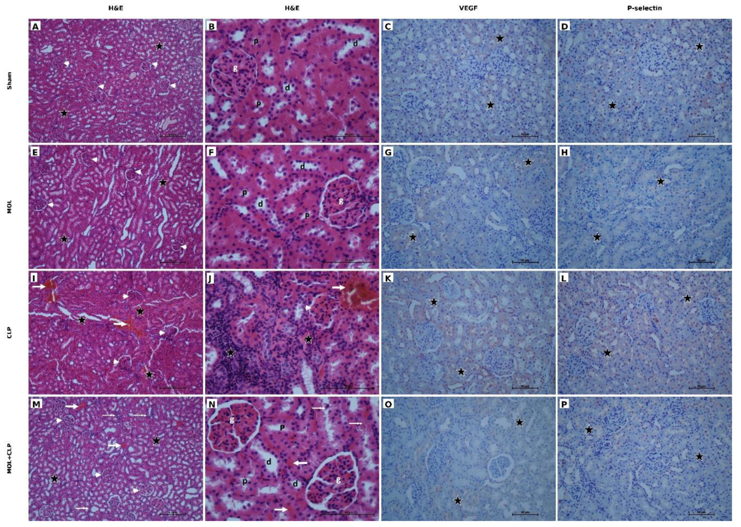

Table 1.
Renal histopathological injury scores across experimental groups.
| Histopathological parameter | Sham (n = 8) | MOL (n = 8) | CLP (n = 8) | MOL+CLP (n = 8) | Overall p value |
| Tubular epithelial degeneration | 0 (0–0)ᵃ | 0 (0–0)ᵃ | 1.5 (1–2)ᵇ | 1 (0–1)ᶜ | <0.001 |
| Tubular necrosis | 0 (0–0)ᵃ | 0 (0–0)ᵃ | 1 (1–1)ᵇ | 0 (0–0)ᶜ | <0.001 |
| Interstitial inflammatory cell infiltration | 0 (0–0)ᵃ | 0 (0–0)ᵃ | 2 (1–2)ᵇ | 1 (0–1)ᶜ | <0.001 |
| Vascular congestion | 0 (0–0)ᵃ | 0 (0–0)ᵃ | 1.5 (1–2)ᵇ | 1 (0–1)ᶜ | <0.001 |
| Total renal histopathological damage score | 0 (0–0)ᵃ | 0 (0–0)ᵃ | 5.5 (5–6)ᵇ | 2 (1–3)ᶜ | <0.001 |
Data are presented as median (interquartile range [IQR]). Histopathological renal injury was assessed by scoring tubular epithelial degeneration, tubular necrosis, interstitial inflammatory cell infiltration, and vascular congestion from 0 (absent/normal) to 3 (severe). The total renal histopathological damage score was designated as the primary outcome, with CLP versus MOL+CLP as the prespecified principal comparison. Overall group differences were assessed using the Kruskal–Wallis test followed by Dunn’s pairwise comparisons. For the four secondary histopathological component outcomes, multiplicity across pairwise comparisons was controlled using the Holm procedure. Within each secondary histopathological component row, values that do not share a superscript letter indicate a statistically significant pairwise difference after multiplicity adjustment, whereas values sharing at least one letter are not significantly different. For the primary outcome row, superscript letters indicate the results of Dunn’s pairwise comparisons. Overall p values represent the omnibus Kruskal–Wallis results and are not multiplicity-adjusted. An adjusted p < 0.05 was considered statistically significant for multiplicity-adjusted comparisons. Sample sizes represent animals with evaluable histopathological specimens.; Abbreviations: CLP, cecal ligation and puncture; IQR, interquartile range; MOL, molsidomine.
Table 2.
Renal tissue oxidative stress, antioxidant defense, and inflammatory markers across experimental groups.
Table 2.
Renal tissue oxidative stress, antioxidant defense, and inflammatory markers across experimental groups.
| Parameter | Sham (n = 8) | MOL (n = 8) | CLP (n = 9) | MOL+CLP (n = 8) | Overall p value |
| MDA (nmol/g) | 99.13 ± 9.96ᵃ | 107.34 ± 11.20ᵃ | 136.45 ± 20.27ᵇ | 106.41 ± 9.03ᵃ | 0.003 |
| NO (µmol/g) | 18.40 ± 3.65ᵃ | 19.60 ± 2.42ᵃ | 26.42 ± 5.88ᵇ | 20.84 ± 4.67ᵃᵇ | 0.004 |
| GSH (µmol/g) | 41.94 ± 2.05ᵃ | 41.20 ± 2.35ᵃ | 36.72 ± 4.76ᵇ | 40.14 ± 1.68ᵃᵇ | 0.030 |
| SOD (U/g) | 38.92 ± 7.53ᵃ | 37.49 ± 2.84ᵃ | 24.12 ± 2.23ᵇ | 36.23 ± 4.19ᵃ | <0.001 |
| CAT (K/g) | 215.10 ± 45.39 | 221.08 ± 68.94 | 159.42 ± 58.77 | 187.52 ± 66.48 | 0.157 |
| GPx (U/mg) | 96.80 ± 11.13ᵃ | 86.70 ± 7.24ᵃ | 61.54 ± 9.10ᵇ | 99.43 ± 26.38ᵃ | <0.001 |
| TAS (mmol Trolox eq/L) | 3.35 ± 0.34 | 3.20 ± 0.35 | 3.07 ± 0.54 | 3.03 ± 0.24 | 0.364 |
| TOS (µmol H₂O₂ eq/L) | 7.29 ± 0.73ᵃ | 8.29 ± 0.98ᵃᵇ | 10.56 ± 2.31ᵇ | 9.46 ± 2.59ᵃᵇ | 0.009 |
| OSI (AU) | 2.20 ± 0.37ᵃ | 2.63 ± 0.46ᵃᵇ | 3.51 ± 0.88ᵇ | 3.15 ± 0.93ᵃᵇ | 0.004 |
| TNF-α (pg/mL) | 288.42 ± 10.99ᵃ | 307.88 ± 45.35ᵃᵇ | 337.25 ± 51.83ᵇ | 321.75 ± 54.65ᵃᵇ | 0.045 |
Data are presented as mean ± SD. Overall group differences were assessed using one-way ANOVA. Homogeneity of variances was evaluated using Levene’s test, followed by Tukey’s HSD or Tamhane’s T2 test for pairwise comparisons, as appropriate. Multiplicity across pairwise comparisons within the renal tissue biochemical outcome family was controlled using the Holm procedure. Within each row, values that do not share a superscript letter indicate a statistically significant pairwise difference after multiplicity adjustment; values sharing at least one letter are not significantly different. Overall p values represent the omnibus ANOVA results and are not multiplicity-adjusted. An adjusted p < 0.05 was considered statistically significant for pairwise comparisons. Sample sizes represent animals with evaluable renal tissue biochemical measurements.; Abbreviations: AU, arbitrary units; CAT, catalase; CLP, cecal ligation and puncture; GPx, glutathione peroxidase; GSH, reduced glutathione; MDA, malondialdehyde; MOL, molsidomine; NO, nitric oxide; OSI, oxidative stress index; SD, standard deviation; SOD, superoxide dismutase; TAS, total antioxidant status; TNF-α, tumor necrosis factor-alpha; TOS, total oxidant status.
Table 3.
Renal VEGF and P-selectin immunoreactivity across experimental groups.
| Parameter | Sham (n = 8) | MOL (n = 8) | CLP (n = 8) | MOL+CLP (n = 8) | Overall p value |
| VEGF immunoreactivity, H-score | 32.5 ± 7.07ᵃ | 27.5 ± 7.07ᵃ | 135.0 ± 20.70ᵇ | 47.5 ± 10.35ᵃ | <0.001 |
| P-selectin immunoreactivity, H-score | 25.0 ± 9.26ᵃ | 23.75 ± 9.16ᵃ | 135.0 ± 14.14ᵇ | 37.5 ± 10.35ᵃ | <0.001 |
Data are presented as mean ± SD. Overall group differences were assessed using one-way analysis of variance (ANOVA). Variance homogeneity was assessed using Levene’s test, followed by Tukey’s HSD or Tamhane’s T2 test for post hoc pairwise comparisons, as appropriate. Multiplicity across pairwise comparisons was controlled within the predefined immunohistochemical outcome family using the Holm procedure. Within each row, values that do not share a superscript letter indicate a statistically significant pairwise difference after multiplicity adjustment, whereas values sharing at least one letter are not significantly different. Overall p values represent the omnibus ANOVA results and are not multiplicity-adjusted. An adjusted p < 0.05 was considered statistically significant for multiplicity-adjusted comparisons. H-scores were calculated as Σ[Pi × (i + 1)], where Pi represents the percentage of cells at each staining intensity and i represents the corresponding intensity score (0–3). Sample sizes represent animals with evaluable immunohistochemical specimens.; Abbreviations: ANOVA, analysis of variance; CLP, cecal ligation and puncture; HSD, honestly significant difference; MOL, molsidomine; SD, standard deviation; VEGF, vascular endothelial growth factor.
Disclaimer/Publisher’s Note: The statements, opinions and data contained in all publications are solely those of the individual author(s) and contributor(s) and not of MDPI and/or the editor(s). MDPI and/or the editor(s) disclaim responsibility for any injury to people or property resulting from any ideas, methods, instructions or products referred to in the content. |
© 2026 by the authors. Licensee MDPI, Basel, Switzerland. This article is an open access article distributed under the terms and conditions of the Creative Commons Attribution (CC BY) license (http://creativecommons.org/licenses/by/4.0/).
Copyright: This open access article is published under a Creative Commons CC BY 4.0 license, which permit the free download, distribution, and reuse, provided that the author and preprint are cited in any reuse.